Topical non-steroidal anti-inflammatory agents for diabetic cystoid macular oedema
Upload
independentCategory
view
4download
0
A Novel Steroidal Inhibitor of Estrogen-Related Receptor α(ERRα)
Sarah J. Duellman1, Joy M. Calaoagan1, Barbara G. Sato1, Richard Fine2, BorisKlebansky2, Wan-Ru Chao1, Peter Hobbs1, Nathan Collins1, Lidia Sambucetti1, and Keith R.Laderoute1
1Biosciences Division, SRI International, Menlo Park, California, 940252BioComputing Group, Inc., Oradell, NJ, 07649
AbstractThe orphan nuclear receptor estrogen-related receptor α (ERRα) has been implicated in thedevelopment of various human malignancies, including breast, prostate, ovary, and colon cancer.ERRα, bound to a co-activator protein (e.g., peroxisome proliferator receptor γ co-activator-1α,PGC-1α), regulates cellular energy metabolism by activating transcription of genes involved invarious metabolic processes, such as mitochondrial genesis, oxidative phosphorylation, and fattyacid oxidation. Accumulating evidence suggests that ERRα is a novel target for solid tumortherapy, conceivably through effects on the regulation of tumor cell energy metabolism associatedwith energy stress within solid tumor microenvironments. This report describes a novel steroidalantiestrogen (SR16388) that binds selectively to ERRα, but not to ERRβ or ERRγ, as determinedusing a time-resolved fluorescence resonance energy transfer assay. SR16388 potently inhibitsERRα's transcriptional activity in reporter gene assays, and prevents endogenous PGC-1α andERRα from being recruited to the promoters or enhancers of target genes. Representative in vivoresults show that SR16388 inhibited the growth of human prostate tumor xenografts in nude miceas a single agent at 30 mg/kg given once daily and 100 mg/kg given once weekly. In acombination study, SR16388 (10 mg/kg, once daily) and paclitaxel (7.5 mg/kg, twice weekly)inhibited the growth of prostate tumor xenografts in nude mice by 61% compared to untreatedxenograft tumors. SR16388 also inhibited the proliferation of diverse human tumor cell lines aftera 24-h exposure to the compound. SR16388 thus has utility both as an experimental antitumoragent and as a chemical probe of ERRα biology.
KeywordsEstrogen-related receptor α; steroid; antitumor agent; metabolism
1. IntroductionEstrogen-related receptor α (ERRα) belongs to the nuclear receptor (NR) superfamily, whichis a group of 48 structurally related, ligand-activated transcription factors [1-3]. The ERRfamily (the NR3B subgroup) consists of ERRα, -β, and -γ [3]. The ERRs are classified as
Address correspondence to: Sarah J. Duellman, 333 Ravenswood Ave., Menlo Park, CA, 94025. Fax: 01-650-859-5816, Tel:01-650-859-3505, [email protected]'s Disclaimer: This is a PDF file of an unedited manuscript that has been accepted for publication. As a service to ourcustomers we are providing this early version of the manuscript. The manuscript will undergo copyediting, typesetting, and review ofthe resulting proof before it is published in its final citable form. Please note that during the production process errors may bediscovered which could affect the content, and all legal disclaimers that apply to the journal pertain.
NIH Public AccessAuthor ManuscriptBiochem Pharmacol. Author manuscript; available in PMC 2011 September 15.
Published in final edited form as:Biochem Pharmacol. 2010 September 15; 80(6): 819–826. doi:10.1016/j.bcp.2010.05.024.
NIH
-PA Author Manuscript
NIH
-PA Author Manuscript
NIH
-PA Author Manuscript
orphan receptors because they do not bind any known natural or endogenous small-moleculeligands [2,3]. For example, although the ERRs are highly similar at both the primarysequence and structural levels to the classical estrogen receptors (ERα and -β), the ERRs donot bind estrogen (e.g., 17β-estradiol; E2). Substantial evidence supports a physiologicalmodel of ERR function in which the receptors regulate energy metabolism by directlyinteracting with certain transcriptional co-regulators, including peroxisome-proliferatoractivated receptor γ coactivator-1α (PGC-1α) and PGC-1β, steroid receptor co-activators,and the co-repressor nuclear receptor interacting protein 140 (RIP140) [2,3]. Co-activatorsof ERRs (e.g., PGC-1α) positively regulate fundamental metabolic processes, includingmitochondrial genesis, oxidative phosphorylation, fatty acid oxidation, and generation ofreactive oxygen species. Co-repressors, such as RIP140, that bind to ERRs compete withERR co-activators to negatively regulate ERR-dependent gene expression. Organism-wideexpression profiling of the ERR isoforms determined that ERRα is widely distributed, withsignificant protein expression in most adult tissues [4]. In general, ERRβ and -γ showrestricted expression patterns and are found at lower levels compared to ERRα. Knockoutstudies of the ERR family members have revealed that each receptor has tissue- andfunction-specific metabolic phenotypes that are important for adaptation to energy stress atthe whole body level. The knockout studies also indicate limited in vivo compensationamong the ERR family members [1-3,5,6].
The pleiotropic effect of ERR activity on energy metabolism has generated interest in thepossibility that specific ERRs could be targets for the discovery of new therapies fordiseases such as type 2 diabetes, progressive heart failure, osteoporosis, and cancer [2,3].Synthetic small-molecule ligands have been identified for the ERR family, such asdiethylstilbestrol, and for ERRβ and -γ, such as 4-hydroxytamoxifen (4-OHT); the 4-OHTderivative GSK5182; and the phenolic acyl hydrazones DY131 and GSK5182 [2]. Selectiveligands (inverse agonists) of ERRα have also been reported, including thethiadiazoleacrylamide XCT790 [7], N-[(2Z)-3-(4,5-dihydro-1,3-thiazol-2-yl)-1,3-thiazolidin-2-yl idene]-5H dibenzo[a,-d] [7]annulen-5-amine [8], and cyclohexylmethyl-(1-p-tolyl-1H-indol-3-ylmethyl)-amine [9].
Here we report a novel, purely steroidal antiestrogen, designated SR16388 [21-[2-(N,N-dimethylamino)ethyl]oxy-7a-methyl-19-norpregna-1,3,5(10),17(20)-tetraen-3-ol; Fig. 1]with strong selectivity for binding to ERRα over both ERRβ and ERRγ. Since SR16388 isessentially an estrogen molecule with a basic side chain attached at position C17, the affinityof SR16388 for ERRα is striking considering the lack of significant E2 binding to thisreceptor [5].
Representative in vitro and in vivo results provided below show that SR16388 inhibits theproliferation of diverse human tumor cell lines and substantially delays the growth of humantumor xenografts—independently of estrogen receptor status. Mechanistically, we show thatSR16388 inhibits the binding of ERRα to a critical peptide from its co-activator PGC-1α in acell-free assay, and strongly inhibits ERRα's ability to occupy promoter or enhancerelements and activate transcription from selected target genes in human cancer cells. Wealso provide a computational model of the ligand-binding domain (LBD) of human ERRαcontaining SR16388, which suggests how this antiestrogen could act as an inhibitor ofcellular ERRα activity.
2. Materials and Methods2.1 Cell culture and reagents
All chemicals were purchased from Sigma Chemical Company (St. Louis, MO) unlessotherwise noted. PC3 human prostate cancer cells were grown in RPMI 1640 medium
Duellman et al. Page 2
Biochem Pharmacol. Author manuscript; available in PMC 2011 September 15.
NIH
-PA Author Manuscript
NIH
-PA Author Manuscript
NIH
-PA Author Manuscript

(Invitrogen, Carlsbad, CA) supplemented with 10% fetal bovine serum (FBS). MCF7 breastcancer cells were grown in Dulbecco modified Eagle's medium (DMEM, Invitrogen)supplemented with 5% FBS and 20% F12 nutrient mixture (Invitrogen). All cells weregrown at 37°C in a humidified 5% CO2 atmosphere. Both cell lines were obtained fromAmerican Type Culture Collection (Manassas, VA).
2.2 Cell viability assayThe alamarBlue reagent (Invitrogen) was used to determine the effect of SR16388 on theproliferation and viability of cancer cell lines. Cells were plated in 96-well plates at 1000cells per well, and 6-point serial dilution dose-response curves were obtained. Briefly, cellswere incubated with SR16388 at 37°C for 24 h, and then the medium was replaced withfresh medium. On day 4 after treatment, alamarBlue was added to the cells, and theincubation was continued for 3 h. Fluorescence from the reduced reagent was measuredusing a BioTek Synergy 2 fluorescence plate reader (Winooski, VT) with excitation at 530nm and emission at 590 nm (30 nm bandwidth).
2.3 TR-FRET assayThe LanthaScreen time-resolved fluorescence resonance energy transfer (TR-FRET) co-activator assay series (Invitrogen) was used to analyze the interaction of the LBD of ERRα, -β, or -γ, and ERα, or -β with a PGC-1α peptide according to the manufacturer's instructions.A 12-point 1:3 serial dilution dose-response curve was obtained by incubating the bindingreaction in the presence of compounds for 1 h at room temperature. TR-FRET was measuredwith an Analyst HT fluorescence plate reader (Molecular Devices, Sunnyvale, CA) withexcitation at 360 nm and emission at 495 nm (10 nm bandwidth) and 520 nm (25 nmbandwidth), 100 μs lag time, and 200 μs integration time. To determine the one-halfmaximal effective concentrations (EC50), nonlinear regression, sigmoidal dose-response(variable slope) curves were calculated using GraphPad Prism version 4.03 (GraphPadSoftware, San Diego, CA).
2.4 Luciferase reporter assayThe dual-luciferase reporter assay system (Promega, Madison, WI) was used to determineERRα's transcriptional activity in the presence of various compounds. Cells were plated in6-well plates and transiently transfected with 1 μg pERRE(5×)TAffLuc [10] (which containsan estrogen-related receptor response element, ERRE), 20 ng pTA-srLuc [10], 450 ngpcDNA3.1-hERRα1 [11], and 6 μg pcDNA3/HA-hPGC-1α [12] using the TurboFectin 8.0transfection reagent (OriGene, Rockville, MD) according to the manufacturer's instructions.After 24 h, transfected cells were treated with DMSO, 5 μM ICI 182,780 (Fulvestrant), or 5μM SR16388. At 48 h posttransfection, cells were washed with PBS and lysed according tothe dual-luciferase reporter assay instructions. A total of 20 μl of each lysate was analyzedfor luciferase activity; relative light units (RLU) were normalized for transfection efficiencyby comparison to the Renilla luciferase signal. Statistical analysis was done using theunpaired Student's t-test.
2.5 Chromatin immunoprecipitation assayMCF7 cells were treated with DMSO or 5 μM SR16388 for 6 or 24 h. The cells were fixed,and a chromatin immunopreciptation (ChIP) assay was performed using a ChIP-IT Expresskit (Active Motif, Carlsbad, CA) according to the manufacturer's instructions. The followingprimary antibodies were used for the ChIP assays: anti-PGC-1α (H-300, Santa CruzBiotechnology, Santa Cruz, CA); anti-ERRα (07-662, Millipore, Temecula, CA); and anegative control IgG antibody (Active Motif). Primers used for quantitative PCR (qPCR)were the following: VEGF, forward 5′ CACCAGCTCACCCTGGTATT and reverse 5′
Duellman et al. Page 3
Biochem Pharmacol. Author manuscript; available in PMC 2011 September 15.
NIH
-PA Author Manuscript
NIH
-PA Author Manuscript
NIH
-PA Author Manuscript

ACTTCCCTCTCCCTGCTCTC; ERRα, forward 5′ CTTCCCCGTGACCTTCATT andreverse 5′ AGCCGACTTAAAACATGCAATA; Acadm, forward 5′AACGCAGAAAACCAAACCAG and reverse 5′ CATGCTCCGTGACCCTTG. Threeindependent replicate measurements were acquired for each experiment, and each qPCRmeasurement was done in duplicate. Results were reported as fold enrichment, whichrepresents the difference in signal relative to that for the IgG negative control antibody.Statistical analysis was performed with the Wilcoxon signed rank test.
2.6 PC3 prostate cancer xenograftsPC3 human prostate cancer cells (3 × 10⁶) were suspended in 100 μl of a 1:1 mixture of cellculture medium and Matrigel and implanted subcutaneously in the right flank region of nudemice (BALB/c [nu/nu], Taconic, Germantown, NY). Mice were monitored for tumor growthdaily after cell implantation, and when tumor volumes reached 80–100 mm3, mice wererandomized into appropriate groups of 10 mice each. Treatment with SR16388 alone or incombination with paclitaxel was initiated on the day after randomization. SR16388 wasorally administered once daily at 10 mg/kg or 30 mg/kg, and once weekly at 100 mg/kg. Forthe combination study, SR16388 was orally administered once daily at 10 mg/kg, andpaclitaxel was administered twice weekly by intraperitoneal injection at 7.5 mg/kg.Treatments were continued for 4 weeks. During the study, tumor volumes were measuredtwice weekly and body weights once weekly. Tumor volumes were measured using theformula V = L × W × H × π/6, where L and W represent the longer and shorter tumordiameters, respectively, and H represents tumor depth. Tumor growth rates were evaluatedstatistically between the control and each treatment group using the Student-Newman-Keulsmethod. No significant compound-related effects on body weight or any other signs of overttoxicity were observed in any of the groups. All procedures were approved by the SRIInstitutional Animal Care and Use Committee.
2.7 Molecular modelingThe primary structure of human ERRα that was used for the current study was Protein DataBank (PDB) entry 2PJL, which reveals an inverse agonist bound to the ERRα LBD [9]. Inplacing SR16388 in this structure, we were guided by two other crystal structures of relatedcompounds bound to the related receptor ERα. The fused-ring portion of SR16388 is highlysimilar to that of E2, for which a crystal structure bound to ERα (PDB entry 1ERE) isknown [13]. The tail portion of SR16388 resembles that of 4-OHT, for which a crystalstructure with the compound bound to ERα (PDB entry 1ERT) is also known [14].Structural superposition of 1ERE and 1ERT onto 2PJL yielded a compelling template for theconstruction of the model of bound SR16388, with the 4-OHT tail spatially occupying theregion where the SR16388 tail would be placed if it were attached to the homologous fused-ring portion of E2. The model was built by superimposing the fused-ring portion ofSR16388 onto that of E2, and subsequently orienting its tail portion to follow the tail of 4-OHT in the superimposed active sites of 1ERE, 1ERT, and 2PLJ. The resulting model wasthen subjected to energy minimization using the program Gromacs [15].
3. Results3.1 SR16388 inhibits human tumor cell proliferation
We performed cell proliferation/viability assays using alamarBlue to determine the effect ofSR16388 (Fig. 1) on the proliferation of a panel of human tumor cell lines (Table 1).SR16388 showed a strong antiproliferative effect toward these cell lines, which includedboth ER-positive and ER-negative cells. Although SR16388 is an antiestrogen [manuscriptsubmitted], these results demonstrate that SR16388 has cytotoxicity toward diverse humancancer cells that does not depend on the presence of the ERs. Indeed, it is unlikely that all
Duellman et al. Page 4
Biochem Pharmacol. Author manuscript; available in PMC 2011 September 15.
NIH
-PA Author Manuscript
NIH
-PA Author Manuscript
NIH
-PA Author Manuscript

the tumor cell types in this diverse panel require the ERs for survival (e.g. ER-negativeMDA-MB-231 cells [16] are sensitive to SR16388). Furthermore, an NCI-60Developmental Therapeutics Program Human Tumor Cell Line screen showed thatSR16388 was cytotoxic to most cell lines in the screen (data not shown). In this screen, allsensitive tumor cell lines showed a percentage of growth between -50 and -100 in responseto treatment with SR16388 (e.g. 10 μM for 48 h) relative to untreated cells, which isconsistent with a cytotoxic endpoint in this screen [17]. In part because ERs were notrequired for the cytotoxicity of SR16388 in the alamarBlue assay, we investigated whetherthe compound could target the closely related ERR orphan nuclear receptors. As mentionedabove, the ERRs (ERRα, -β, and -γ) do not bind any known endogenous ligand, includingestrogen.
3.2 SR16388 selectively inhibits the interaction of ERRα with PGC-1αTo investigate the effect of SR16388 on the ability of ERRα, -β, and -γ to regulatetranscription, we first used a TR-FRET assay that measures the direct interaction betweenthe LBD of each ERR and a target peptide from PGC-1α, which is an established co-activator of both the classic ERs and the ERRs (reviewed in [18]). SR16388 inhibited theinteraction of ERRα with the PGC-1α peptide (EC50 = 0.7 ± 0.1 μM), but did notcomparably inhibit the interaction of ERRβ (EC50 >10 μM) or ERRγ (EC50 >10 μM) withthe peptide (Fig. 2). The ERRα inverse agonist XCT790 had a lower EC50 than SR16388 inthe ERRα TR-FRET cell-free assay; however, as discussed below, XCT790 was not aseffective as SR16388 in cell-based assays of ERRα activity. Because SR16388 belongs to agroup of related molecules that includes an established antiestrogen (TAS-108 [19]), wetested the ability of SR16388 to inhibit ERα and ERβ in this TR-FRET assay as well.SR16388 inhibited both ERα (EC50 = 0.2 ± 0.2 μM) and ERβ (EC50 = 0.5 ± 0.2 μM). Thesefindings indicate that SR16388 could have antiestrogenic activity in tumor cells containingsignificant expression of ERα or -β. As mentioned above, however, the generalantiproliferative effect of SR16388 toward diverse human tumor cell lines (Table 1)suggests that this molecule has targets in addition to the classic ERs, such as ERRα.
3.3 SR16388 inhibits ERRα's transcriptional activation functionERRα constitutively activates transcription through site-specific DNA binding to ERREs inthe promoter or enhancer regions of its target genes [2,3]. Therefore, we next determinedwhether SR16388 could inhibit the ability of ERRα to stimulate expression of an ERRE-dependent luciferase reporter gene in MCF7 human breast cancer cells and PC3 humanprostate cancer cells. The cells were transfected with separate expression vectors for ERRαand PGC-1α, along with an ERRE-firefly luciferase reporter construct and a TATA-Renillaluciferase transfection control construct. In cells treated with the vehicle control (DMSO),ERRα and PGC-1α were able to co-activate transcription of the luciferase construct. In cellstreated with SR16388, however, transcriptional activation of this co-activator complex wassignificantly inhibited (Fig. 3). ICI 182,780 was also able to inhibit ERRα-mediatedtranscription to an intermediate level, but only in MCF7 cells (Fig. 3). Because ICI 182,780is an ER inhibitor [18], we hypothesize that the negative effect on ERRα-dependent geneexpression detected in ICI 182,780-treated MCF7 cells was a result of their relatively highlevel of ERα expression, which is not observed in PC3 cells. Equimolar XCT790 did notinhibit luciferase expression in this assay after a 24-h treatment (data not shown). In general,to inhibit ERRα activity XCT790 seems to require longer exposure times at higherconcentrations (e.g., 10 μM for 48 h, [19-21]) than that used here (5 μM for 24 h), whichexplains why SR16388 was much more effective than XCT790 as an inhibitor of ERRα-dependent gene expression in the present assay. It is important to note that the signal for therenilla luciferase transfection control construct was not significantly different for the DMSOand SR16388 treated samples (data not shown), indicating that the inhibitory effect of
Duellman et al. Page 5
Biochem Pharmacol. Author manuscript; available in PMC 2011 September 15.
NIH
-PA Author Manuscript
NIH
-PA Author Manuscript
NIH
-PA Author Manuscript
SR16388 on ERRE-dependent firefly luciferase activity was due to disruption of ERRα-dependent transcription of the ERRE-firefly luciferase construct rather than a cytotoxiceffect.
The binding of ERRα to ERREs in the promoter or enhancer regions of selected endogenoustarget genes, and thus the recruitment of PGC-1α to these specific DNA-binding elements,was also disrupted by SR16388. Here, we used ChIP assays and qPCR to monitor the effectof SR16388 on endogenous ERRα and PGC-1α occupancy at established ERREs thatregulate the genes for ERRα (ESRRA) itself [22-25], vascular endothelial growth factor(VEGFA) [26], and acyl-coenzyme A dehydrogenase (Acadm) [27,28]. MCF7 cells weretreated with either DMSO or 5 μM SR16388 for 6 or 24 h. Chromatin wasimmunoprecipitated using anti-PGC-1α, anti-ERRα, or negative control IgG antibodies, andqPCR amplification was performed for each of the indicated ERRα-regulated genes. Foldenrichment values were calculated relative to that of the IgG control to evaluate the relativeoccupancy of each endogenous gene by ERRα or PGC-1α. After 24 h of SR16388 treatment,the relative occupancy of all ERREs for both proteins was significantly decreased (p ≤0.05,Fig. 4). After 6 h of treatment, both ERRα and PGC-1α relative occupancies were decreasedcompared to those in DMSO treated cells for the ERRα and Acadm ERREs (p ≤0.05). Thefold enrichment of ERRα occupancy at these ERREs, however, was higher at 6 h than at 24h (p ≤0.05) for all the ERREs tested. On the other hand, the fold enrichment of PGC-1αoccupancy was equally diminished at both time points, indicating that PGC-1α was releasedfrom the ERREs shortly after the addition of SR16388 to the cells. It is possible that theobserved decreases in ERRα occupancy were not the result of direct inhibition of thebinding of ERRα to target genes, but rather of an overall decrease in the total cellular ERRαprotein level due to the disruption of transcription of the ERRα gene, which has a positiveautoregulatory feedback loop [2,24,25]. In summary, the luciferase reporter gene and ChIPassay experiments described here demonstrate that SR16388 significantly inhibited bothtransfected and endogenous ERRα activity in cultured human cancer cells.
3.4 SR16388 inhibits tumor growth in PC3 human prostate cancer xenograftsWe performed PC3 human prostate cancer xenograft studies in nude mice to investigate theantitumor efficacy of SR16388. Figure 5A shows that SR16388 alone had a stronginhibitory effect on the growth of PC3 tumor xenografts. At the final measurement, thevolumes of tumors from the mice treated with SR1633 doses of 100 mg/kg weekly and with30 or 10 mg/kg daily were 34%, 44%, and 75% of the size of the tumors in control mice,respectively. SR16388 also inhibited tumor growth when used in combination withpaclitaxel, a chemotherapeutic used for hormone-refractory prostate cancer (reviewed in[29]; Fig. 5B). In the combination study, mice were treated with 10 mg/kg of SR16388 (oraldaily dose) and 7.5 mg/kg paclitaxel (intraperitoneal injection, twice/week). At the finalmeasurement, the tumor growth was inhibited by 22% after treatment with paclitaxel alone,24% with SR16388 alone, and 61% with SR16388 and paclitaxel in combination. Nosignificant compound-related effects on body weight or any other signs of overt toxicitywere observed in any of the groups.
3.5 Computational modeling of the binding pose of SR16388 in the ERRα LBDThe computational model of SR16388 bound in the ERRα LBD suggests a strong ionicinteraction between the compound and charged amino acids E331 and D329 of ERRα (Fig.6). The x-ray crystal structure used to generate this model [9] shows that the ERRα LBDcontacts a leucine-rich nuclear receptor interacting motif (LxxLL) helix of PGC-1α throughhelices 11 and 12 of ERRα (Fig. 6). The crystal structure 2PJL [9] revealed displacement ofhelix 12 away from its active conformation (PDB structures 1XB7 [30] and 3D24 [31]) toaccommodate introduction of an inverse agonist. In addition to this displacement, our model
Duellman et al. Page 6
Biochem Pharmacol. Author manuscript; available in PMC 2011 September 15.
NIH
-PA Author Manuscript
NIH
-PA Author Manuscript
NIH
-PA Author Manuscript

of SR16388 suggests additional displacement of the last turn of helix 11 to accommodateSR16388. Based on our model of SR16388 binding to the LBD, we hypothesize that the C17side chain of SR16388 extends toward these helices and causes a conformational change thatprevents or disrupts binding of PGC-1α. In future work it will be important to validate thismodel of SR16388 binding to ERRα experimentally through co-crystallization andmutational studies.
4. DiscussionSR16388—originally developed as a selective estrogen receptor modulator (SERM) [18]—is a member of a compound series that includes TAS-108, which is under development as ahormonal therapy for breast cancer [32,33]. SR16388 is the first member of this series foundto have significant inhibitory (inverse agonist) activity toward ERRα-dependent geneexpression in human cancer cells. This conclusion is based on the following experimentalfindings: (1) SR16388 disrupted the interaction between the human ERRα LBD and aPGC-1α co-activator peptide in a cell-free TR-FRET assay (Fig. 2); (2) SR16388 inhibitedconstitutive human ERRα-dependent reporter gene expression in transient transfectionassays of human cancer cells (Fig. 3); and (3) SR16388 disrupted the occupancy of ERREsby ERRα and PGC-1α in three endogenous genes in human cancer cells regulated by ERRα(ESRRA, VEGFA, Acadm) (Fig. 4). Although the established ERRα inverse agonist XCT790was more effective as an inhibitor of the interaction between ERRα and its PGC-1α targetpeptide in the TR-FRET assay (Fig. 2), XCT790 failed to show significant inhibition ofERRα activity in the ERRE-dependent reporter gene assay described above (data notshown).
The therapeutic focus of SR16388 and related molecules such as TAS-108 is the treatmentof solid tumors such as breast and prostate carcinomas. SR16388, which inhibits theproliferation or survival of diverse human cancer cell lines in vitro (Table 1), also inhibitedthe growth of PC3 human prostate cancer xenografts in nude mice (Figure 5). Taken withthe findings presented here showing that SR16388 is an ERRα inhibitor, it is possible thatthe cytotoxicity of SR16388 could involve disrupting ERRα dependent gene expression,depending on the tumor cell line. ERR expression has been investigated in diverse humantumor cell lines and primary tumor types, including cancers derived from breast, prostate,ovary, endometrium, and colon [3,8,34-41]. Evidence supporting a role for deregulated ERRexpression or activity in human cancer is still emerging, but recent research indicates thatERRα in particular is involved in the development or progression of certain tumor types. Forexample, ERRα expression was reported to be significantly higher in clinical specimens ofhuman breast, ovarian, endometrial, and prostate cancer than it was in the correspondingnormal tissues [3,34-38,40]. ERRα can bind to the same DNA elements as ERs (estrogen-response elements, EREs) in vitro [35]. In breast cancer cells, ERα and ERRα can regulatethe same genes through binding a hybrid DNA element containing an ERRE within an ERE[38,42]. The genes that are co-regulated by these two nuclear receptors are known tocontribute to the development or progression of human breast cancer [42]. ERRα may thuscompensate for the loss of ER activity in breast cancers that become resistant to hormonal(antiestrogen) therapy. ERRα is also able to occupy androgen receptor response elements inthe regulatory regions of genes sensitive to androgens [43], which suggests that ERRαcontributes to phenotypes associated with tumors such as prostate cancer. Finally, given theimportance of ERRα as a positive regulator of cellular energy metabolism, it is possible thatERRα more generally contributes to tumor development as a key component of the adaptiveresponse of tumor cells to energy stress within solid tumor microenvironments (e.g.,reviewed in [44,45]). For example, using xenografts prepared from ER-negative MDA-MB-231 human breast cancer cells expressing siRNA targeting ERRα, it was found thatgrowth of the ERRα knockdown tumors was significantly attenuated compared to that of
Duellman et al. Page 7
Biochem Pharmacol. Author manuscript; available in PMC 2011 September 15.
NIH
-PA Author Manuscript
NIH
-PA Author Manuscript
NIH
-PA Author Manuscript
tumors expressing control siRNA [38]. Moreover, the proliferation of the ERRα-knockdowncells was not inhibited in vitro, suggesting that ERRα is required for MDA-MB-231 cells toadapt to microenvironmental energy stress within the rapidly growing tumor xenografts. Itwill be important in future work to test the efficacy of SR16388 in ERRα-knockdown tumorxenograft models to investigate the specificity of the compound for ERRα.
In summary, SR16388 is a purely steroidal antiestrogen that selectively targets ERRα in theERR subgroup of human nuclear hormone receptors. This selectivity is potentially importantfor breast cancer therapy because ERRα expression is correlated with an unfavorable clinicaloutcome in primary breast tumors, whereas ERRγ expression is correlated with a favorableoutcome [46]. ERRα expression was also reported to be associated with an increased risk ofrecurrence and a poor prognosis in breast cancer [47]. While the mechanistic contribution ofERRα to tumor development or progression is not yet clear, we hypothesize that ERRαactivity is required for the adaptation of tumor cell metabolism to energy stress withinpathophysiological tumor microenvironments. Thus, SR16388 may be cytotoxic towardenergetically stressed tumor cells in part by disrupting ERRα-dependent gene expression.
AcknowledgmentsWe would like to thank Dr. Janet Mertz and Dr. Anastasia Kralli for generously providing reagents. This work wasfunded in part by NIH/NCI CA73807 (K.R.L.).
References1. Alaynick WA. Nuclear receptors, mitochondria and lipid metabolism. Mitochondrion. 2008; 8:329–
37. [PubMed: 18375192]2. Giguere V. Transcriptional control of energy homeostasis by the estrogen-related receptors.
Endocrine reviews. 2008; 29:677–96. [PubMed: 18664618]3. Tremblay AM, Giguere V. The NR3B subgroup: an ovERRview. Nuclear receptor signaling. 2007;
5:e009. [PubMed: 18174917]4. Bookout AL, Jeong Y, Downes M, Yu RT, Evans RM, Mangelsdorf DJ. Anatomical profiling of
nuclear receptor expression reveals a hierarchical transcriptional network. Cell. 2006; 126:789–99.[PubMed: 16923397]
5. Giguere V, Yang N, Segui P, Evans RM. Identification of a new class of steroid hormone receptors.Nature. 1988; 331:91–4. [PubMed: 3267207]
6. Luo J, Sladek R, Carrier J, Bader JA, Richard D, Giguere V. Reduced fat mass in mice lackingorphan nuclear receptor estrogen-related receptor alpha. Molecular and cellular biology. 2003;23:7947–56. [PubMed: 14585956]
7. Busch BB, Stevens WC Jr, Martin R, Ordentlich P, Zhou S, Sapp DW, et al. Identification of aselective inverse agonist for the orphan nuclear receptor estrogen-related receptor alpha. Journal ofmedicinal chemistry. 2004; 47:5593–6. [PubMed: 15509154]
8. Chisamore MJ, Cunningham ME, Flores O, Wilkinson HA, Chen JD. Characterization of a novelsmall molecule subtype specific estrogen-related receptor alpha antagonist in MCF-7 breast cancercells. PLoS One. 2009; 4:e5624. [PubMed: 19462000]
9. Kallen J, Lattmann R, Beerli R, Blechschmidt A, Blommers MJ, Geiser M, et al. Crystal structure ofhuman estrogen-related receptor alpha in complex with a synthetic inverse agonist reveals its novelmolecular mechanism. The Journal of biological chemistry. 2007; 282:23231–9. [PubMed:17556356]
10. Ariazi EA, Kraus RJ, Farrell ML, Jordan VC, Mertz JE. Estrogen-related receptor alpha1transcriptional activities are regulated in part via the ErbB2/HER2 signaling pathway. Mol CancerRes. 2007; 5:71–85. [PubMed: 17259347]
11. Kraus RJ, Ariazi EA, Farrell ML, Mertz JE. Estrogen-related receptor alpha 1 actively antagonizesestrogen receptor-regulated transcription in MCF-7 mammary cells. The Journal of biologicalchemistry. 2002; 277:24826–34. [PubMed: 11986328]
Duellman et al. Page 8
Biochem Pharmacol. Author manuscript; available in PMC 2011 September 15.
NIH
-PA Author Manuscript
NIH
-PA Author Manuscript
NIH
-PA Author Manuscript

12. Knutti D, Kaul A, Kralli A. A tissue-specific coactivator of steroid receptors, identified in afunctional genetic screen. Molecular and cellular biology. 2000; 20:2411–22. [PubMed:10713165]
13. Brzozowski AM, Pike AC, Dauter Z, Hubbard RE, Bonn T, Engstrom O, et al. Molecular basis ofagonism and antagonism in the oestrogen receptor. Nature. 1997; 389:753–8. [PubMed: 9338790]
14. Weichsel A, Gasdaska JR, Powis G, Montfort WR. Crystal structures of reduced, oxidized, andmutated human thioredoxins: evidence for a regulatory homodimer. Structure. 1996; 4:735–51.[PubMed: 8805557]
15. Van Der Spoel D, Lindahl E, Hess B, Groenhof G, Mark AE, Berendsen HJ. GROMACS: fast,flexible, and free. Journal of computational chemistry. 2005; 26:1701–18. [PubMed: 16211538]
16. Mukhopadhyay R, Theriault RL, Price JE. Increased levels of alpha6 integrins are associated withthe metastatic phenotype of human breast cancer cells. Clinical & experimental metastasis. 1999;17:325–32. [PubMed: 10545019]
17. Shoemaker RH. The NCI60 human tumour cell line anticancer drug screen. Nature reviews. 2006;6:813–23.
18. Lonard DM, Smith CL. Molecular perspectives on selective estrogen receptor modulators(SERMs): progress in understanding their tissue-specific agonist and antagonist actions. Steroids.2002; 67:15–24. [PubMed: 11728517]
19. Debevec D, Christian M, Morganstein D, Seth A, Herzog B, Parker M, et al. Receptor interactingprotein 140 regulates expression of uncoupling protein 1 in adipocytes through specificperoxisome proliferator activated receptor isoforms and estrogen-related receptor alpha. Molecularendocrinology (Baltimore, Md. 2007; 21:1581–92.
20. Lanvin O, Bianco S, Kersual N, Chalbos D, Vanacker JM. Potentiation of ICI182,780(Fulvestrant)-induced estrogen receptor-alpha degradation by the estrogen receptor-relatedreceptor-alpha inverse agonist XCT790. The Journal of biological chemistry. 2007; 282:28328–34.[PubMed: 17631492]
21. Willy PJ, Murray IR, Qian J, Busch BB, Stevens WC Jr, Martin R, et al. Regulation ofPPARgamma coactivator 1alpha (PGC-1alpha) signaling by an estrogen-related receptor alpha(ERRalpha) ligand. Proceedings of the National Academy of Sciences of the United States ofAmerica. 2004; 101:8912–7. [PubMed: 15184675]
22. Barry JB, Giguere V. Epidermal growth factor-induced signaling in breast cancer cells results inselective target gene activation by orphan nuclear receptor estrogen-related receptor alpha. Cancerresearch. 2005; 65:6120–9. [PubMed: 16024613]
23. Liu D, Zhang Z, Gladwell W, Teng CT. Estrogen stimulates estrogen-related receptor alpha geneexpression through conserved hormone response elements. Endocrinology. 2003; 144:4894–904.[PubMed: 12960079]
24. Laganiere J, Tremblay GB, Dufour CR, Giroux S, Rousseau F, Giguere V. A polymorphicautoregulatory hormone response element in the human estrogen-related receptor alpha(ERRalpha) promoter dictates peroxisome proliferator-activated receptor gammacoactivator-1alpha control of ERRalpha expression. The Journal of biological chemistry. 2004;279:18504–10. [PubMed: 14978033]
25. Mootha VK, Handschin C, Arlow D, Xie X, St Pierre J, Sihag S, et al. Erralpha and Gabpa/bspecify PGC-1alpha-dependent oxidative phosphorylation gene expression that is altered indiabetic muscle. Proceedings of the National Academy of Sciences of the United States ofAmerica. 2004; 101:6570–5. [PubMed: 15100410]
26. Arany Z, Foo SY, Ma Y, Ruas JL, Bommi-Reddy A, Girnun G, et al. HIF-independent regulationof VEGF and angiogenesis by the transcriptional coactivator PGC-1alpha. Nature. 2008;451:1008–12. [PubMed: 18288196]
27. Sladek R, Bader JA, Giguere V. The orphan nuclear receptor estrogen-related receptor alpha is atranscriptional regulator of the human medium-chain acyl coenzyme A dehydrogenase gene.Molecular and cellular biology. 1997; 17:5400–9. [PubMed: 9271417]
28. Vega RB, Kelly DP. A role for estrogen-related receptor alpha in the control of mitochondrial fattyacid beta-oxidation during brown adipocyte differentiation. The Journal of biological chemistry.1997; 272:31693–9. [PubMed: 9395511]
Duellman et al. Page 9
Biochem Pharmacol. Author manuscript; available in PMC 2011 September 15.
NIH
-PA Author Manuscript
NIH
-PA Author Manuscript
NIH
-PA Author Manuscript

29. Mancuso A, Oudard S, Sternberg CN. Effective chemotherapy for hormone-refractory prostatecancer (HRPC): present status and perspectives with taxane-based treatments. Critical reviews inoncology/hematology. 2007; 61:176–85. [PubMed: 17074501]
30. Kallen J, Schlaeppi JM, Bitsch F, Filipuzzi I, Schilb A, Riou V, et al. Evidence for ligand-independent transcriptional activation of the human estrogen-related receptor alpha (ERRalpha):crystal structure of ERRalpha ligand binding domain in complex with peroxisome proliferator-activated receptor coactivator-1alpha. The Journal of biological chemistry. 2004; 279:49330–7.[PubMed: 15337744]
31. Greschik H, Althage M, Flaig R, Sato Y, Chavant V, Peluso-Iltis C, et al. Communication betweenthe ERRalpha homodimer interface and the PGC-1alpha binding surface via the helix 8-9 loop.The Journal of biological chemistry. 2008; 283:20220–30. [PubMed: 18441008]
32. Buzdar AU. TAS-108: a novel steroidal antiestrogen. Clin Cancer Res. 2005; 11:906s–8s.[PubMed: 15701885]
33. Kumagai Y, Fujita T, Ozaki M, Yokota S, Maeda M, Shida M, et al. Safety, tolerability andpharmacokinetics of TAS-108, a novel anti-oestrogen, in healthy post-menopausal Japanesewomen: a phase I single oral dose study. Basic & clinical pharmacology & toxicology. 2009;104:352–9. [PubMed: 19175362]
34. Cheung CP, Yu S, Wong KB, Chan LW, Lai FM, Wang X, et al. Expression and functional studyof estrogen receptor-related receptors in human prostatic cells and tissues. J Clin EndocrinolMetab. 2005; 90:1830–44. [PubMed: 15598686]
35. Stein RA, McDonnell DP. Estrogen-related receptor alpha as a therapeutic target in cancer. EndocrRelat Cancer. 2006; 13 1:S25–32. [PubMed: 17259555]
36. Fujimoto J, Alam SM, Jahan I, Sato E, Sakaguchi H, Tamaya T. Clinical implication of estrogen-related receptor (ERR) expression in ovarian cancers. J Steroid Biochem Mol Biol. 2007;104:301–4. [PubMed: 17509876]
37. Fujimura T, Takahashi S, Urano T, Kumagai J, Ogushi T, Horie-Inoue K, et al. Increasedexpression of estrogen-related receptor alpha (ERRalpha) is a negative prognostic predictor inhuman prostate cancer. Int J Cancer. 2007; 120:2325–30. [PubMed: 17294452]
38. Stein RA, Chang CY, Kazmin DA, Way J, Schroeder T, Wergin M, et al. Estrogen-related receptoralpha is critical for the growth of estrogen receptor-negative breast cancer. Cancer Res. 2008;68:8805–12. [PubMed: 18974123]
39. Chisamore MJ, Wilkinson HA, Flores O, Chen JD. Estrogen-related receptor-alpha antagonistinhibits both estrogen receptor-positive and estrogen receptor-negative breast tumor growth inmouse xenografts. Mol Cancer Ther. 2009; 8:672–81. [PubMed: 19276159]
40. Fujimoto J, Sato E. Clinical implication of estrogen-related receptor (ERR) expression in uterineendometrial cancers. J Steroid Biochem Mol Biol. 2009; 116:71–5. [PubMed: 19416752]
41. Stein RA, Gaillard S, McDonnell DP. Estrogen-related receptor alpha induces the expression ofvascular endothelial growth factor in breast cancer cells. J Steroid Biochem Mol Biol. 2009;114:106–12. [PubMed: 19429439]
42. Deblois G, Hall JA, Perry MC, Laganiere J, Ghahremani M, Park M, et al. Genome-wideidentification of direct target genes implicates estrogen-related receptor alpha as a determinant ofbreast cancer heterogeneity. Cancer research. 2009; 69:6149–57. [PubMed: 19622763]
43. Teyssier C, Bianco S, Lanvin O, Vanacker JM. The orphan receptor ERRalpha interferes withsteroid signaling. Nucleic Acids Res. 2008; 36:5350–61. [PubMed: 18697814]
44. Cairns R, Papandreou I, Denko N. Overcoming physiologic barriers to cancer treatment bymolecularly targeting the tumor microenvironment. Mol Cancer Res. 2006; 4:61–70. [PubMed:16513837]
45. Bristow RG, Hill RP. Hypoxia and metabolism. Hypoxia, DNA repair and genetic instability. NatRev Cancer. 2008; 8:180–92. [PubMed: 18273037]
46. Ariazi EA, Clark GM, Mertz JE. Estrogen-related receptor alpha and estrogen-related receptorgamma associate with unfavorable and favorable biomarkers, respectively, in human breast cancer.Cancer research. 2002; 62:6510–8. [PubMed: 12438245]
Duellman et al. Page 10
Biochem Pharmacol. Author manuscript; available in PMC 2011 September 15.
NIH
-PA Author Manuscript
NIH
-PA Author Manuscript
NIH
-PA Author Manuscript
47. Suzuki T, Miki Y, Moriya T, Shimada N, Ishida T, Hirakawa H, et al. Estrogen-related receptoralpha in human breast carcinoma as a potent prognostic factor. Cancer research. 2004; 64:4670–6.[PubMed: 15231680]
Abbreviations
4-OHT 4-hydroxytamoxifen
ChIP chromatin immunoprecipitation
ERR estrogen-related receptor
ER estrogen receptor
ERRE estrogen-related receptor response element
ERE estrogen response element
EC50 one-half maximal effective concentration
E2 17β-estradiol
FBS fetal bovine serum
LBD ligand binding domain
NR nuclear receptor
PGC peroxisome-proliferator activated receptor coactivator
PDB protein data bank
RLU relative light units
TR-FRET time-resolved fluorescence resonance energy transfer
Duellman et al. Page 11
Biochem Pharmacol. Author manuscript; available in PMC 2011 September 15.
NIH
-PA Author Manuscript
NIH
-PA Author Manuscript
NIH
-PA Author Manuscript

Figure 1.Structure of SR16388. SR16388 (21-[2-(N,N-Dimethylamino)ethyl]oxy-7a-methyl-19-norpregna-1,3,5(10),17(20)-tetraen-3-ol citrate salt) is an orally active compound thatbelongs to the antiestrogen class of therapeutic agents. SR16388 is a potent and selectiveinhibitor of human ERRα, which does not bind estrogen (E2).
Duellman et al. Page 12
Biochem Pharmacol. Author manuscript; available in PMC 2011 September 15.
NIH
-PA Author Manuscript
NIH
-PA Author Manuscript
NIH
-PA Author Manuscript

Figure 2.SR16388 disrupts the binding of the ERRα-LBD, but not the ERRβ or -γ LBDs, to aPGC-1α peptide. A TR-FRET assay was used to determine whether various compoundscould inhibit the interaction of an ERRα, -β, or -γ LBD-glutathione S-transferase fusionprotein with a co-activator peptide from PGC-1α (aa 135-153,EAEEPSLLKKLLLAPANTQ). Known inhibitors of the ERRs were used as positivecontrols: XCT790 for ERRα, and 4-hydroxytamoxifen (4-OHT) for ERRβ and -γ. E2 wasincluded in the ERRα TR-FRET assay to confirm that E2 does not bind ERRα. ND = notdetermined.
Duellman et al. Page 13
Biochem Pharmacol. Author manuscript; available in PMC 2011 September 15.
NIH
-PA Author Manuscript
NIH
-PA Author Manuscript
NIH
-PA Author Manuscript
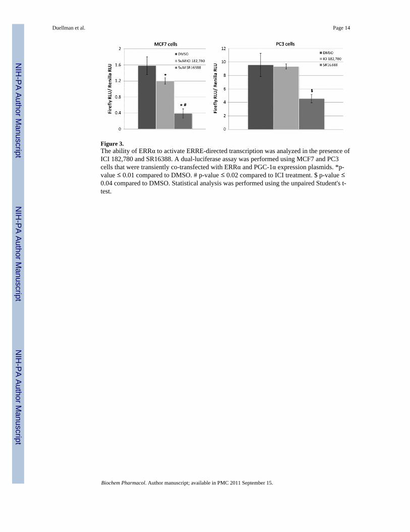
Figure 3.The ability of ERRα to activate ERRE-directed transcription was analyzed in the presence ofICI 182,780 and SR16388. A dual-luciferase assay was performed using MCF7 and PC3cells that were transiently co-transfected with ERRα and PGC-1α expression plasmids. *p-value ≤ 0.01 compared to DMSO. # p-value ≤ 0.02 compared to ICI treatment. $ p-value ≤0.04 compared to DMSO. Statistical analysis was performed using the unpaired Student's t-test.
Duellman et al. Page 14
Biochem Pharmacol. Author manuscript; available in PMC 2011 September 15.
NIH
-PA Author Manuscript
NIH
-PA Author Manuscript
NIH
-PA Author Manuscript

Figure 4.Treatment with SR16388 decreases the association of ERRα and PGC-1α with target genepromoters. MCF-7 cells were treated with DMSO or 5 μM SR16388 for 6 or 24 h followedby ChIP using anti-ERRα, anti-PGC-1α, or control IgG antibodies. qPCR was performedusing primer sets designed around established ERREs in the promoter or enhancer regions ofthe target genes [ERRα (ESRRA), VEGFA, Acadm]. Three independent replicatemeasurements were acquired for each experiment. The fold enrichment represents thedifference in signal relative to the IgG negative antibody control. Statistical analysis wasperformed using the Wilcoxon signed rank test.
Duellman et al. Page 15
Biochem Pharmacol. Author manuscript; available in PMC 2011 September 15.
NIH
-PA Author Manuscript
NIH
-PA Author Manuscript
NIH
-PA Author Manuscript

Figure 5.SR16388 inhibits tumor growth of PC3 human prostate cancer xenografts in nude mice. PC3cells were implanted subcutaneously in the right flank region of nude mice. When tumorvolumes reached 80–100 mm3, mice were treated with SR16388 with or without paclitaxel.Treatments were continued for 4 weeks, and differences in tumor growth rates between thecontrol group and each treatment group were evaluated statistically. No significantcompound-related effects on body weight or any other signs of overt toxicity were observedin any of the groups. *Tumor volumes were significantly reduced when compared withcontrol, p ≤ 0.02.
Duellman et al. Page 16
Biochem Pharmacol. Author manuscript; available in PMC 2011 September 15.
NIH
-PA Author Manuscript
NIH
-PA Author Manuscript
NIH
-PA Author Manuscript

Figure 6.Computational model of SR16388 bound to the LBD of ERRα. We hypothesize that thesteroidal scaffold of SR16388 fits within the LBD of ERRα (amino acids 290–519) whilethe C-17 side chain extends toward helices 11 and 12 (H11 and H12), resulting inconformational changes that prevent or disrupt co-activator binding. Residues E331 andD329 are thought to be important for SR16388 binding and activity.
Duellman et al. Page 17
Biochem Pharmacol. Author manuscript; available in PMC 2011 September 15.
NIH
-PA Author Manuscript
NIH
-PA Author Manuscript
NIH
-PA Author Manuscript

NIH
-PA Author Manuscript
NIH
-PA Author Manuscript
NIH
-PA Author Manuscript
Duellman et al. Page 18
Table 1
Alamar Blue proliferation/viability toxicity assays were used to determine the IC50 of SR16388 in variouscancer cell lines after a 24-h exposure to the compound.
Cell line IC50 (μM)
PC3 androgen-independent prostate cancer 3.0 ± 0.1
LNCaP C4-2B androgen receptor-positive, androgen-independent prostate cancer 0.9 ± 0.1
MDA-MB-231 ER-negative breast cancer 6.2 ± 0.1
MCF7 ER-positive breast cancer 2.2 ± 0.1
Panc-1 pancreatic cancer 2.8 ± 0.1
MiaPaCa2 pancreatic cancer 1.3 ± 0.1
RCC4 renal cell carcinoma 2.9 ± 0.1
RCC4/VHL renal cell carcinoma 0.5 ± 0.1
786-0 renal cell carcinoma 1.1 ± 0.1
786-0/VHL renal cell carcinoma 0.7 ± 0.1
A498 renal call carcinoma 1.0 ± 0.1
A498/VHL renal cell carcinoma 0.6 ± 0.1
Biochem Pharmacol. Author manuscript; available in PMC 2011 September 15.
Copyright © 2022 FDOKUMEN