Localization and distribution of estrogen receptors and ...
187
Localization and distribution of estrogen receptors and progesterone receptors in the bovine ovary in relation to the cell dynamics Localization and distribution of estrogen receptors and progesterone receptors in the bovine ovary in relation to the cell dynamics Department of Morphology Faculty of Veterinary Medicine, Ghent University Salisburylaan 133, B-9820 Merelbeke 2006 Mylène D’Haeseleer Mylène D’Haeseleer FACULTEIT DIERGENEESKUNDE approved by EAEVE FACULTEIT DIERGENEESKUNDE approved by EAEVE 2006
-
Upload
khangminh22 -
Category
Documents
-
view
0 -
download
0
Transcript of Localization and distribution of estrogen receptors and ...

Localization and distribution
of estrogen receptors and
progesterone receptors
in the bovine ovary in
relation to the cell dynamics
Localization and distribution of estrogen receptors and progesterone receptors in the bovine ovary in relation to the cell dynam
ics
Department of Morphology
Faculty of Veterinary Medicine, Ghent University
Salisburylaan 133, B-9820 Merelbeke
2006
Mylène D’Haeseleer
Mylène D
’Haeseleer
FACULTEIT DIERGENEESKUNDEapproved by EAEVE
FACULTEIT DIERGENEESKUNDEapproved by EAEVE
2006

Localization and distribution of estrogen
receptors and progesterone receptors in the bovine ovary in relation to the cell dynamics
Mylène D’HAESELEER
Thesis to obtain the academic degree of Doctor of Veterinary Science (PhD) Faculty of Veterinary Medicine, Ghent University
2006
Promoter: Prof. Dr. W. Van den Broeck Copromoter: Prof. Dr. P. Simoens
Department of Morphology Faculty of Veterinary Medicine, Ghent University
Salisburylaan 133, B-9820 Merelbeke

The lay-out of the cover “Picasso Cow” was designed by Bart De Pauw
ISBN 90-5864-097-3
TABLE OF CONTENTS
List of abbreviations
GENERAL INTRODUCTION 1
AIMS OF THE STUDY 31
CHAPTER 1 SEX STEROID HORMONE RECEPTORS IN THE BOVINE OVARY 35
1.1 Localization of estrogen receptors within various bovine ovarian cell types 37 at different stages of the estrous cycle 1.1.1 Cell-specific localization of estrogen receptor beta (ESR2) mRNA 39
within various bovine ovarian cell types using in situ hybridization
1.1.2 Localization of estrogen receptors within various bovine ovarian cell 61 types at different stages of the estrous cycle
1.2 Cell-specific localization of progesterone receptors in the bovine ovary at 87 different stages of the estrous cycle
CHAPTER 2 CELL DEATH IN THE CYCLIC BOVINE OVARY 107
Cell-specific localization of apoptosis in the bovine ovary at different stages of the estrous cycle
CHAPTER 3 PROLIFERATION PATTERNS IN THE CYCLIC BOVINE OVARY 131
Proliferation patterns in the bovine ovary at different stages of the estrous cycle
GENERAL DISCUSSION 147
SUMMARY 159
SAMENVATTING 165
CURRICULUM VITAE 171
BIBLIOGRAPHY 173
DANKWOORD 177


LIST OF ABBREVIATIONS
A adenine LH luteinising hormone
AEC amino ethyl carbazole ll large lutein cells
AS apoptotic score mRNA messenger RNA
C cytosine OAF obliterative atretic follicle
CA corpus albicans PBS phosphate-buffered saline
CAF cystic atretic follicle PCR polymerase chain reaction
CH corpus hemorrhagicum PdF primordial follicle
CL corpus luteum PF primary follicle
cs capsular stroma PR progesterone receptor
DAB diaminobenzidine PRID progesterone releasing intravaginal device
DAPI 4', 6-diamidino-2- phenylindole dihydrochloride ps perivascular stroma
DIG digoxigenin RNA ribonucleic acid
DNA deoxyribonucleic acid RNase ribonuclease
DNase desoxy nuclease SE surface epithelium
DS deep stroma SEM standard error of the mean
EDTA ethylene diamino tetra-acetic acid SER score of estrogen receptor
ER estrogen receptor SF secondary follicle
ERα, ESR1 estrogen receptor-alpha sl small lutein cells
ERβ, ESR2 estrogen receptor-beta SPR score of progesterone receptor
ERγ, ESR3 estrogen receptor-gamma SS superficial stroma
fc follicle cells SSPE sodium chloride sodium phosphate / EDTA
fd follicle diameter T thymine
FSH follicle stimulating hormone TA tunica albuginea
G guanine te theca externa
gc granulosa cells tf theca follicularis
HRP horseradish peroxidase ti theca interna
HSS high sensitive streptavidine Tris tris(hydroxymethyl)methylamine
is internal stroma TUNEL terminal deoxynucleotidyl transferase mediated dUTP nick end labeling
kb kilobase v/v volume/volume
kDa kilodalton VTF vital tertiary follicle
KO knockout


GENERAL INTRODUCTION


General Introduction
3
The bovine ovary
Embryology
Harbouring genetic information to be passed down from generation to generation, the
ovaries are crucial structures for the survival of the species. The germinal component
originates from primordial germ cells that, after migration from the yolk sac, colonize the
gonadal primordium. The germ cells together with the surrounding stroma cells form the pool
of resting primordial follicles in the ovary (Motta et al., 1997).
The ovary of a bovine foetus of 110 days contains more than 2 million germ cells.
However, at birth this number dramatically decreases to ca 100 000 germ cells enclosed in
primordial follicles and is progressively further reduced to 2500 in ten years old cattle
(Erickson, 1966a, 1966b).
Morphology of the bovine ovary (Fig. 1) The ovarian cortex and medulla
The bovine ovary consists of a central medulla or zona vasculosa and a peripheral cortex or
zona parenchymatosa. The medulla is highly vascularized and consists of connective tissue
and a dense network of nerves. The cortex contains both stromal and parenchymatous
structures (Schaller, 1992). The ovary is covered by a simple squamous or cuboidal surface
epithelium. Beneath the epithelium lies a layer of dense connective tissue named the tunica
albuginea and an amount of ovarian stroma, consisting of spindle-shaped cells arranged in
whorls (Dellmann and Eurell, 1998). This ovarian stroma can be further subdivided into a
superficial part (superficial stroma) and a deep part (deep stroma), both containing ovarian
parenchymatous structures.
The ovarian parenchymatous structures
The ovarian parenchymatous structures consist of the follicles in various stages of
development, the corpora hemorrhagica, the corpora lutea and the corpora albicantia. The
ovarian follicles and corpora hemorrhagica can be found in the superficial stroma, while the
corpora lutea and corpora albicantia are mainly embedded in the deep stroma.

4 General Introduction
Four main types of vital follicles can be distinguished, namely primordial, primary,
secondary and tertiary follicles (Nomina Histologica, 1994). A primordial follicle is
composed of a primary oocyte enveloped by a single layer of flattened follicular cells (follicle
diameter (fd) < 0.04 mm). A primary follicle is composed of a primary oocyte enveloped by a
single layer of cuboidal or columnar follicular cells (fd: 0.04-0.08 mm). A secondary follicle
consists of a primary oocyte surrounded by a stratified follicular epithelium, a basal
membrane, and a developing follicular theca (fd: 0.08-0.13 mm). A tertiary follicle, also
called an antral or vesicular follicle, is characterized by the development of a central cavity,
the antrum. In the early stages the tertiary follicle has a diameter of 0.13-0.25 mm while in
later stages the follicle matures and reaches its maximum size just prior to ovulation (fd: 0.25-
20 mm). The latter stage of follicle is termed a preovulatory or Graafian follicle (Gougeon,
1996; Braw-Tal and Yossefi 1997). As the antrum enlarges through the accumulation of
liquor folliculi, the primary oocyte is displaced eccentrically, usually in a part of the follicle
nearest to the centre of the ovary. The oocyte then lies in a protruding accumulation of
granulosa cells, called the cumulus oophorus (Dellmann and Eurell, 1998). The granulosa
cells of tertiary follicles are surrounded by a layer of theca cells which differentiates into two
layers, viz. an inner vascular theca interna and an outer supportive theca externa. The theca
interna cells are spindle-shaped and located in a delicate reticular fiber network. The theca
externa layer consists of a thin sheet of loose connective cells arranged concentrically around
the theca interna. Blood vessels of the theca externa supply capillaries to the theca interna
(Priedkalns et al., 1968; Ham and Cormack, 1979; Braw-Tal and Yossefi, 1997; Dellmann
and Eurell, 1998). Graafian follicles are the major source of ovarian estrogens that are
produced cyclically during reproductive life. Shortly before ovulation the first meiotic
division is finalized, whereby the primary oocyte is transformed into the secondary oocyte
and a first polar body. In response to preovulatory gonadotrophin surges the dominant follicle
ovulates to release the secondary oocyte for fertilization. After ovulation, the follicular wall
collapses and the granulosa layers form large folds protruding into the residual lumen. As a
result of the slight hemorrhage, which occurs at the time of ovulation, a blood clot is formed
in the centre. The resulting structure is called the corpus hemorrhagicum (Dellmann and
Eurell, 1998). This corpus hemorrhagicum further develops into a corpus luteum (or yellow
body), which is a transient endocrine gland. The granulosa cells are localized in the centre of
the corpus luteum and are referred to as small lutein cells, while theca interna cells are located
in the periphery and are called large lutein cells. In the cyclic cow, the corpus luteum is only
functional for 17-18 days. In the early luteal stage (days 2-4 of the ovarian cycle) the lutein
General Introduction
5
cells of the corpus luteum exhibit their greatest ability to proliferate and start secreting
progesterone. In the secretory stage (days 5-17) progesterone secretion reaches its maximum
levels. A dense network of vessels develops around the corpus luteum and may function in the
transport of hormones. Luteal regression occurs from day 18 onwards. Shrinkage of the
corpus luteum takes place rapidly after day 19 and is completed 1 to 2 days after oestrus
(Dellmann and Eurell, 1998). The corpus albicans is the "white body" and consists of scar
tissue formed by regression of the corpus luteum. The colour varies with species and is red or
white in the bovine. The corpus albicans is found deeper in the ovary than the corpus luteum
and appears very slowly. It is characterized by small lutein cells which enclose a few lipid
droplets, and a large amount of connective tissue (Dellmann and Eurell, 1998).
Follicular atresia can be observed at any stage of follicular development. In atretic
primordial, primary and secondary follicles the oocyte shrinks and subsequently degenerates,
followed by degeneration of the follicle cells (Van Wezel and Rodgers, 1996; Depalo et al.,
2003). At the end no scar tissue is left. Atretic changes in tertiary follicles may result in the
formation of two different morphological types of atretic follicles, namely cystic and
obliterative atretic tertiary follicles. In cystic atretic tertiary follicles the granulosa layer is
invaded by vascularized strands of connective tissue, after which the granulosa cells
desquamate into the follicular cavity. In more advanced stages of atresia the follicle collapses,
the walls are thrown into folds, more connective tissue and blood vessels are found, and rapid
resorption of the degenerated granulosa cells takes place. In obliterative atretic follicles, the
basement membrane between theca interna cells and granulosa cells thickens and is now
referred to as the glassy membrane or membrane of Slavjanski. Subsequently the theca interna
cells further increase in size and are arranged in radial layers or cords around the oocyte.
Eventually there is a complete breakdown of the obliterative atretic follicles by invading
connective tissue cells (Dellmann and Eurell, 1998).
Pathological cystic follicles are different from atretic follicles (Brambell, 1956). A cystic
follicle is a large (more than 2.5 cm in diameter), persistent, spherical structure on one of the
ovaries. The more common type is a follicular cyst, found when the follicle fails to rupture
but continues to grow (Peter, 2004). Less common are luteal cysts where the corpus luteum
fails to regress and continues to produce progesterone, blocking further follicle development
(Garverick, 1997).

6 General Introduction
l
b a k d g c
o
e
f
i
j
p h
m
n
Fig. 1: Diagram of the mammalian ovary illustrating the development and regression of follicles and corpora
lutea. (a) Primordial follicle, (b) primary follicle, (c) secondary follicle, (d) tertiary follicle, (e) Graafian follicle,
(f) cystic and (g) obliterative atretic follicles, (h) corpus hemorrhagicum, (i) corpus luteum, (j) corpus albicans,
(k) rete ovarii, (l) ovarian blood vessels, (m) deep stroma, (n) superficial stroma, (o) tunica albuginea, (p) surface
epithelium.
The bovine estrous cycle
Physiological and endocrinological characteristics
Domestic ruminants are polyoestrous animals. In normal heifers the first oestrus starts at
the age of 8 to 12 months. Each estrous cycle takes 20 till 21 days, but cycle length may vary
between 15 and 25 days (Sirois and Fortune, 1988; Ruckebusch et al., 1991; De Rensis and
Peters, 1999). The reproductive cycle of the normal cycling cow is coordinated by hormones
produced by the hypothalamus, pituitary gland and ovary. Gonadotrophic releasing hormone,
secreted by the hypothalamus, stimulates the adenohypophysis to secrete two gonadotrophic
hormones, namely follicle stimulating hormone (FSH) and luteinising hormone (LH). Both
gonadotrophins circulate into the blood and regulate the reproductive cycle through a series of
feedback mechanisms acting on the hypothalamus and pituitary gland (Peters and Lamming,

General Introduction
7
1983). In the ovary the regulation of follicular development is a complex process and involves
interactions between the gonadotrophins FSH and LH and intraovarian regulators such as
steroid hormones, growth factors and cytokines (Asselin et al., 2000).
In cattle, the initial stages of folliculogenesis (from primordial to secondary follicles) occur
independent of gonadotrophin stimulation (Crowe et al., 2001; Gong, 2002). From the
secondary follicular stage onwards, follicular development is a FSH dependent process as
FSH is responsible for survival of the follicles (Gougeon, 1996). A group of secondary
follicles are selected to continue growth to tertiary follicles by increasing concentrations of
FSH. FSH stimulation causes expression of aromatase in the granulosa cells of the follicles,
thus enabling the granulosa cells to convert androgens produced by the theca cells into
estrogens (Richards, 1994). The circulating estrogens cause an inhibition of gonadotrophin
release. One follicle of a selected group of tertiary follicles, called the dominant follicle, has
an aromatase activity much more sensitive to FSH than the other smaller follicles. As a
consequence, this dominant follicle is still capable of producing large amounts of estrogens
even when the concentration of circulating gonadotrophins decreases (Gougeon, 1996).
Concomitantly, increased concentrations of local growth factors and an elaborate
perifollicular vasculature contribute to a positive selection of the dominant follicle, thus
ensuring its final growth to a Graafian follicle which can ovulate (Coppola et al., 2005).
Progesterone plays also a major role in the selection of the dominant follicle. If circulating
progesterone levels remain high after the dominant follicle has attained its maximum size,
then all follicles of that particular wave including the dominant follicle undergo atresia
(Taylor et al., 1993; Yang and Rajamahendran, 2000a). However, if circulating progesterone
levels decrease during the growth phase of the dominant follicle, then this dominant follicle
will ovulate while the other follicles of the cohort become atretic. Although the role of steroid
hormones is of great importance, the exact mechanism that initiates the selection of the
Graafian follicle and the regression of the non-ovulatory dominant follicle is not yet fully
elucidated.
When a certain threshold of estrogens in the blood is reached, the negative feedback on
gonadotrophin production and secretion is changed into a positive feedback. This results in
peak levels of LH in the blood, triggering the ovulation. LH binds to LH receptors, which in
secondary follicles are only localized on the theca cells, while in tertiary follicles they are also

8 General Introduction present on the granulosa cells (Shima et al., 1987; Yamoto et al., 1992). At ovulation the
follicle ruptures and develops into a corpus hemorrhagicum. In response to the binding of
pituitary LH to receptors on cells of the ruptured follicular wall, the granulosa cells are
converted in lutein cells producing high levels of progesterone (Ruckebusch et al., 1991;
Dellmann and Eurell, 1998).
Follicle development in the bovine ovary (Fig. 2)
Primordial follicles undergo initial recruitment to enter the growing pool of primary
follicles. Thereafter, the primary follicles continue their growth to the secondary stage and
develop further into tertiary follicles (antral stage). The time required between the initial
recruitment of a primordial follicle and its growth to the antral stage is about 180 days in
cattle (Lussier et al., 1987). In the bovine ovary the growth of antral follicles occurs in a
wave-like pattern. Each wave is characterized by the recruitment of a group of three to six
follicles and the selection and dominance of a single dominant follicle (D) while the
remaining recruited follicles regress (Savio et al., 1988). The number of growth waves during
the estrous cycle is determined by the length of the luteal phase. Usually, there are two or
three waves of follicular development during each bovine estrous cycle (Celik et al., 2005),
although cycles with four waves or even only one wave exist (De Rensis and Peters, 1999).
Fig. 2 illustrates the selection of a dominant follicle in a three waves growth pattern. The first
wave begins on the day of ovulation. During the next few days, one of the follicles develops
to a dominant stage, while the others become subordinate. The dominant follicle of the first
wave reaches its largest size on day 5 or 6 of the estrous cycle. This dominant follicle
maintains its morphological and functional dominance until around day 7, subsequently
becomes atretic and begins to regress between days 8 and 9, to be replaced by a second wave
of follicular growth. The second wave emerges at about 9 days and is followed by another
wave at 16 days postovulation. The dominant follicle of the third wave becomes the ovulatory
follicle (OV, Graafian follicle) (Ginther et al., 1996; De Rensis and Peters, 1999; Mapletoft et
al., 2002).

General Introduction
9
CYCLIC RECRUITMENT
INITIAL RECRUITMENT
primordial follicles
primary follicles
secondary follicles
tertiary follicles
CONTINUING INITIAL RECRUITMENT
Day of cycle
Graafian follicle
Fig. 2: Schematic presentation of follicle recruitment, selection and dominance in the bovine ovary during the
estrous cycle (after McGee and Hsueh, 2000; Aerts and Bols, 2004).
Morphological characteristics
In this study 23 cows were used. The stage of the estrous cycle of each cow was
determined at the moment of slaughter on the basis of the morphological appearance of the
ovarian follicles and corpora lutea and the presence of uterine cervical mucus (Ireland et al.,
1980; Arthur et al., 1983). Additionally, the plasma progesterone concentration was
determined for each individual animal and compared with the findings described by Sartori et
al. (2004). The ovarian morphological characteristics and the corresponding plasma
progesterone levels were required to classify the animals into 5 groups, viz., oestrus,
metoestrus, early dioestrus, late dioestrus and prooestrus (Fig. 3, Table 1).

10 General Introduction
Cycle stage Day Plasma Morphologic characteristics progesterone concentration ng/ml
Oestrus D0 <0.30 Clear cervical mucus and presence of a mature
follicle.
Regressing corpora lutea.
Metoestrus D1-3 0.20 - 0.40 Collapsed remnants of the ovulated follicle,
corpus hemorrhagicum.
Early dioestrus D4-8 0.70 - 2.65 Ovarian epithelium covering the rupture point
of the ovulated follicle to form the apex of a
new corpus luteum.
Late dioestrus D9-16 2.90 - 4.70 Fully developed red or brown corpus luteum
with vasculature visible around its periphery.
Prooestrus D17-21 1.50 - 2.20 Brightly orange or yellow corpus luteum.
Appearance of a mature follicle.
Table 1: Classification of cows in five different cycle stages with progesterone levels and major ovarian
features (after Ireland et al., 1980; Sartori et al., 2004).
Oestrus is the period (16 to 20 hours) of sexual receptiveness. The day of oestrus is
considered as 'day 0' of the estrous cycle. About 30 hours after the onset of oestrus, ovulation
occurs, preceded by a surge of LH. The estrogen levels decline at the end of oestrus.
Metoestrus is the period (2 to 3 days) following oestrus, characterised by initial progesterone
secretion. Early dioestrus is the period (about 5 days) of corpus luteum development and
increasing progesterone levels. Late dioestrus is the phase (about 8 days) of the active corpus
luteum when the progesterone level reaches its maximum. Prooestrus is the period (about 4
days) of regression of the corpus luteum and maturation of a new follicle (Arthur et al., 1983;
Dellmann and Eurell, 1998) (Fig. 3).

General Introduction
11
P O M E D L D P
16 17 18 19 20 21 1 2 3 4 5 6 7 8 9 10 11 12 13 14 15 16 17 18 19 20 21
Day of cycle
Hor
mon
e C
once
ntra
tion
16 17 18 19 20 21 1 2 3 4 5 6 7 8 9 10 11 12 13 14 15 16 17 18 19 20 2116 17 18 19 20 21 1 2 3 4 5 6 7 8 9 10 11 12 13 14 15 16 17 18 19 20 21
a
b
c
[55 ng/ml]
[6,5 ng/ml]
[180 pg/ml]
Fig. 3: Schematic presentation of the hormonal changes that characterize the estrous cycle of the normal cycling
cow. (P) Prooestrus, (O) oestrus, (M) metoestrus, (ED) early dioestrus, (LD) late dioestrus, (a) serum
progesterone level, (b) serum estrogen level, (c) serum luteinizing hormone level (values from McDonald and
Pineda; 1989).
The estrogen receptor (ER) and the progesterone receptor (PR)
The steroid hormone receptor superfamily
The steroid hormone receptor superfamily consists of a large number of proteins,
originating from a common ancestral gene and therefore considered a gene superfamily (Tsai
and O'Malley, 1994). This family includes receptors for the steroid hormones (estrogen,
progesterone, glucocorticoid, mineralocorticoid and androgen), receptors for non-steroids, and
several receptors for unknown ligands (orphan receptors). The estrogen receptor and the
progesterone receptor are described in more detail hereafter.

12 General Introduction The steroid hormone action (Fig. 4)
The concentration of the sex steroid hormone receptors in the various body tissues
including the ovary is modulated by circulating and local amounts of sex steroid hormones
(Seidman et al., 1997). Therefore sex steroid hormone receptors in the ovary are valuable
mirrors of the action and concentration of sex steroid hormones.
+
+
HRE
protein cellular response
mRNA
DNA
1 2
3
4
Fig. 4: Simplified model of steroid hormone action (after Tsai and O’Malley, 1994).
(1) Cell membrane, (2) cytoplasm, (3) nuclear envelope, (4) nucleus.
sex steroid hormone, inactive receptor, activated receptor.
Sex steroid hormones are hydrophobic molecules that pass through the cell membranes and
interact with their specific receptors that are localized in the cell nucleus or cytoplasm in an
inactive form (King and Greene, 1984; Perrot-Applanat et al., 1985). After binding of the
hormone on its receptor, the latter undergoes conformational changes. The activated receptor
can bind effectively to a hormone receptor element (HRE), i.e., a particular DNA sequence
which is located in the region of hormone responsive genes. The DNA-bound receptor ligand
complex is then capable of either activating or repressing target gene transcription (Beekman
et al., 1993). The resulting mRNA molecules will be transferred to the cell cytoplasm where
they are translated into specific proteins that will induce the cellular response to hormone
action.

General Introduction
13
Structure of the estrogen receptor (ER) and the progesterone receptor (PR) (Fig. 5)
The steroid receptors ER and PR are transcription factors composed of six domains (A-F)
responsible for specific functions. The N-terminal A/B domain contains an activation function
(AF-1) that contributes to transcriptional activity. The C domain is the DNA-binding domain
and contains two zinc finger motifs. This domain contributes to receptor dimerization, binding
of the liganded receptor to its target genes (= response elements), and finally to the
transactivation function of the receptor. Domain D is the hinge region that allows
conformational changes during activation of the receptor. This domain contains conserved
amino acids which play a role in binding to heat shock proteins, nuclear localization and
stabilization of the DNA binding. The E domain is the ligand-binding domain and contains
the ligand binding site and the transactivation functions AF-2 and AF-2a (Peters and Khan,
1999). The function of domain F is still unclear, but it is probably involved in modulating
transcriptional activation (Pavao and Traish, 2001). Ligand binding may influence the
stability of the receptor. In particular, it has been shown that in the absence of ligand, the half-
life of ERα is about 4-5 h, whereas estradiol binding accelerates receptor degradation,
reducing its half-life to 3-4 h. (Eckert et al., 1984; Nardulli and Katzenellenbogen, 1986).
Fig. 5: Model of the receptor protein with several regions responsible for specific functions. A/B:
transcriptional activation domain; C: DNA binding and dimerization domain; D: nuclear localization domain;
E: ligand binding, dimerization and transcriptional domain; F: unknown function (Eckert et al., 1984).

14 General Introduction
Estrogen receptor subtypes
Two estrogen receptor subtypes are known, namely the ERα which was first described by
Walter et al. in 1986, and the ERβ which was described by Kuiper in 1996. Both receptors are
encoded by a different gene, namely the ERα and the ERβ gene. In addition to the presence of
the two receptor subtypes, the expression of several isoforms from the ERα and ERβ genes
greatly enlarges the diversity of ER. These isoforms are formed by alternative splicing leading
to deletions of certain exons (Walther et al., 1999; Fang et al., 2003). Recently, a third
subtype of estrogen receptor, the ERγ, has been detected in the teleost Micropogonias
undulatus (Hawkins et al., 2000), in mouse (Lorke et al., 2000) and in humans (Heard et al.,
2000).
Estrogen receptor α isoforms
Several ERα isoforms have been identified which can be expressed in various species and
tissues. In the MCF6 human breast cancer cell line two isoforms of the ERα gene are
expressed, namely the ERα66 isoform, 66 kDa in size, and the ERα46 isoform, 46 kDa in size
(Cullen et al., 2000; Flouriot et al., 2000; Métivier et al., 2004). Additionally, the expression
of ERα46 was reported in osteoblasts (Denger et al, 2001) and in endothelial cells (Li et al.,
1998). In chicken two ERα isoforms were identified, the ERα66 isoform and the ERα61
isoform, 61 kDa in size. In cockerels and hens, both variants are expressed in the
adenohypophysis and basal hypothalamus (Griffin et al., 2001). In rainbow trout two ERα
isoforms are found, namely the ERα1 and ERα2. Comparison of ERα1 with ERα2 reveals a
high similarity in the conserved DNA-binding domain (91%) and ligand-binding domain
(89%) (Bouma and Nagler, 2001).
Estrogen receptor β isoforms
A large number of ERβ isoforms with variable tissue expression has been reported. Several
isoforms are found in the human testis. These include the ERβ2 isoform which is also known
as ERβcx (Ogawa et al., 1998), the ERβ3, ERβ4 and ERβ5 isoforms (Moore et al., 1998), the
ERβ1δ5 isoform which lacks exon 5 (Inoue et al., 2000), and the ERβM isoform containing
exons 5-8 and exon M (Shoda et al., 2002). Some of these isoforms namely ERβ2 and
ERβ2δ5 have been associated with human breast cancer (Skrzyczak et al., 2003; Davies et al.,
2004). Additionally, 3 ERβ isoforms (δ11, δ21 and δ31) were demonstrated in the bovine
reproductive organs (Walther et al., 1999).

General Introduction
15
Progesterone receptor isoforms
All isoforms of PR identified so far are derived from a single gene as a consequence of
alternate initiation of transcription by distinct promoters (Kastner et al., 1990b; Taylor et al.,
2006).
In humans, macaques, rats, mice and chickens two isoforms of PR have been described,
namely PRA, 94 kDa in size, and PRB, 118 kDa in size (Christensen et al., 1991; Ilenchuck
and Walters, 1987; Hovland et al., 1998; Wei et al., 1997; Bethea and Widmann, 1998; Pieber
et al., 2001; Lessey et al., 1983; Aupperlee et al., 2005; Kastner et al., 1990a; Gonzalez-
Moran and Camacho-Arroyo, 2001). PRA is the predominant isoform in humans and rodents
(Vegeto et al., 1993; Graham and Clarke, 1997) and is widely expressed in the macaque
reproductive system (Bethea and Widmann, 1998), in the human uterus (Mote et al., 1999;
Mangal et al., 1997) and in human breast cancers (Graham et al., 1995). A third isoform,
called PRC and 60 kDa in size, was detected in the progestin responsive human breast cancer
cell line T47D (Wei et al., 1996). This isoform is inactive by itself, but it may inhibit the
activity of both PRA and PRB in the presence of progesterone (Wei and Miner, 1994).
Recently, two new isoforms PRS and PRM have been identified and partially characterised in
human reproductive tissues (Taylor et al., 2006).
Cell dynamics in the bovine ovary
Apoptosis in follicles and corpora lutea of the bovine ovary
Atresia of ovarian follicles is an important process, as it accounts for the loss of 99% of the
follicles in the mammalian ovary (Byskov, 1978; Greenwald and Terranova, 1988; Mariana et
al., 1991). This follicular atresia is mediated by a highly organized type of cell death called
apoptosis or programmed cell death (Hsueh et al., 1994; Billig et al., 1996; Kaipia and Hsueh,
1997; Driancourt, 2001).
During follicular development, endocrine factors such as FSH and LH, and paracrine
factors including growth factors, activin and interleukin 1β as well as estrogens activate
different intracellular pathways to rescue the follicles from apoptotic demise. In contrast,
16 General Introduction tumor necrosis factor α, androgens, and free radicals function as atretogenic factors (Kaipia
and Hsueh, 1997). Additionally, specific genes have been identified that encode proteins
responsible for the initiation, progression and completion of cell death, particularly a large
family of B-cell lymphomal leukemia-2 (Bcl-2)-related proteins (Tilly, 2003). All these
diverse signals probably converge on specific intracellular pathways to regulate apoptosis in
the ovarian follicles. Because of these multiple signals, it is still not completely understood
which are the critical factors that discriminate between follicles destined for elimination by
apoptosis and follicles that will continue to develop to reach the final stage of a preovulatory
follicle.
During the growth and development of follicles, atresia occurs at every stage of follicular
development (Gougeon, 1996; Yang and Rajamahendran, 2000b). In humans, quiescent
follicles are the most susceptible to apoptosis (Depalo et al., 2003). In this context, various
authors have investigated gonadotrophin control of primordial and primary follicles
(Gougeon, 1996; McGee and Hsueh, 2000) and some reports show that in quiescent follicles
FSH does not act as a survival factor but that various locally produced growth factors are
involved in the cellular control mechanisms (Markstrom et al., 2002). The most common
signs of atresia in primordial, primary and secondary bovine follicles are firstly shrinking of
the oocyte and degeneration of the follicle cells (Yang and Rajamahendran, 2000b). The
follicle will subsequently be resorbed without leaving a trace (Bloom and Fawcett, 1975). In
contrast, atresia of tertiary follicles occurs differently and is first reflected by apoptosis in the
inner granulosa cell layer and reduction in granulosa cell function, especially the loss of
aromatase activity (Amsterdam et al., 2003; Tajima et al., 2002). In a further stage apoptosis
leads to almost total destruction of the granulosa cell layer and theca cells (Isobe and
Yoshimura, 2000). Studies in bovine Graafian follicles suggest that the local and
chronological progress of apoptosis from the granulosa layer towards the embedded oocyte is
triggered by a threshold quantity of cells in the theca and granulosa layers and in the cumulus
oophorus (Bendell et al., 1988; Zeuner et al., 2003).
Apoptosis has also been implicated as the mechanism underlying structural regression of
the corpora lutea. The onset of apoptosis in the bovine corpus luteum is not observed until
progesterone production has declined (Juengel et al., 1993). This suggests that progesterone
may prevent apoptosis (Okuda et al., 2004). However, it is not known whether the decline in
progesterone production and its auto- or paracrine actions within the corpus luteum influence

General Introduction
17
receptor expression. Therefore, it is of interest to elucidate the role of progesterone and its
receptors in the corpus luteum.
Cell proliferation in follicles and corpora lutea of the bovine ovary
During the bovine estrous cycle, the ovary is subjected to extensive tissue remodelling that
consists of sequential phases of cell proliferation and cell death in the different ovarian
structures (Fortune et al., 1994; Gaytan et al., 1996). The growth of ovarian follicles,
ovulation and the formation of the corpus luteum are basic events in the proliferation process.
This process is regulated by specific responses to gonadotrophins, steroid hormones and
growth factors (Pedersen, 1970; Richards, 1980; Hirshfield, 1991).
In general, follicle growth consists of two distinct consecutive phases. The first phase is
characterized by the transformation of follicle cells from a flattened shape to cuboidal cells,
and the second phase is characterized by proliferation of the follicle cells. Both phases are
regulated by locally produced inhibitory and stimulatory factors (Braw-Tal, 2002). In
primordial follicles, the oocyte is surrounded by a single layer of non-dividing follicle cells.
Primordial follicles leave this quiescent state and initiate a phase of slow growth. Studies in
rat showed that the transition from a primordial follicle to a follicle with a single layer of
columnar follicle cells might take several weeks (Hirshfield, 1991). In the secondary and
tertiary follicles, the follicle/granulosa cells acquire enhanced responsiveness to FSH and start
producing estradiol (Richards, 1975, 1980). Exposure to FSH and estradiol triggers a rapid
burst of proliferation that results in the formation of large tertiary follicles (Rao et al., 1978)
characterized by the presence of both granulosa cells and a surrounding layer of theca cells
(Uilenbroek and Richards, 1979). The LH surge triggers dramatic changes in both follicular
structure and function and terminates follicular growth (Rao et al., 1978; Hirshfield, 1991).
However, the mechanism by which the growth and differentiation of theca cells in bovine
ovarian follicles are controlled is not yet elucidated. Some reports suggest the importance of
granulosa-theca interactions (Kotsuji et al., 1990) while others indicate that specific growth
factors could promote cell proliferation (Asselin et al., 2000). The follicle ruptures during
ovulation and the granulosa cells luteinize to form the corpus luteum (Lobel and Levy, 1968;
Meidan and Girsh, 1997; Meidan et al., 1999). The lutein cells undergo intense proliferation
during the early stages of luteal development, but during functional luteal regression the

18 General Introduction cellular proliferation activity is much lower (Lei et al., 1991). Finally, the lutein cells undergo
terminal differentiation and cease to divide (Richards, 1980).

General Introduction
19
References
Aerts JMJ, Bols PEJ. De boviene ovariële folliculaire dynamiek. Deel II: Antrale ontwikkeling, exogene
beïnvloeding en toekomstperspectieven. Vlaams Diergeneeskundig Tijdschrift 2004; 73: 250-259.
Amsterdam A, Sasson R, Keren-Tal I, Aharoni D, Dantes A, Rimon E, Land A, Cohen T, Dor Y, Hirsh L.
Alternative pathways of ovarian apoptosis: Death for life. Biochemical Pharmacology 2003; 66: 1355-
1362.
Arthur GH, Noakes DE, Pearson H. The estrous cycle and its control. In: Veterinary Reproduction and
Obstetrics (Theriogenology), Baillière Tindall, London 1983; pp. 3-45.
Asselin E, Xiao CW, Wang YF, Tsang BK. Mammalian follicular development and atresia: Role of apoptosis. Biological Signals and Receptors 2000; 9: 87-95.
Aupperlee MD, Kyle TS, Kariagina A, Haslam SZ. Progesterone receptor isoforms A and B: Temporal and
spatial differences in expression during murine mammary gland development. Endocrinology 2005; 146:
3577-3588.
Beekman JM, Allan GF, Tsai MJ, O'Malley BW. Transcription activation by the estrogen receptor requires
a conformational change in the ligand binding domain. Molecular Endocrinology 1993; 7: 1266-1274.
Bendell JJ, Lobb DK, Chuma A, Gysler M, Dorrington JH. Bovine theca cells secrete factor(s) that
promote granulosa cell proliferation. Biology of Reproduction 1988; 38: 790-797.
Bethea CL, Widmann AA. Differential expression of progestin receptor isoforms in the hypothalamus,
pituitary and endometrium of rhesus macaques. Endocrinology 1998; 139: 677-687.
Billig H, Chun SY, Eisenhauer K, Hsueh AJ. Gonadal cell apoptosis: Hormone-regulated cell demise.
Human Reproduction Update 1996; 2: 103-117.
Bloom WMD, Fawcett WMD. Female Reproductive System In: A textbook of histology. Tenth Edition.
Saunders Company, Philadelpia, London, Toronto 1975; pp. 873-876.
Bouma J, Nagler JJ. Estrogen receptor-α protein localization in the testis of the rainbow trout
(Oncorhynchus mykiss) during different stages of the reproduction cycle. Biology of Reproduction 2001;
65: 60-65.
Brambell FWR. Ovarian changes. In: Marshall's Physiology of Reproduction, Longman, Green and Co,
London 1956; pp. 397-542.

20 General Introduction Braw-Tal R. The initiation of follicle growth: The oocyte or the somatic cells? Molecular and Cellular
Endocrinology 2002; 187: 11-18.
Braw-Tal R, Yossefi S. Studies in vivo and in vitro on the initiation of follicle growth in the bovine ovary.
Journal of Reproduction and Fertility 1997; 109: 165-171.
Byskov AG. Follicular atresia. In: The Vertebrate Ovary. Plenum Press, New York 1978; pp. 533-562.
Celik HA, Aydin I, Senda S, Dinc DA. Number of follicular waves and their effect on pregnancy rate in the
cow. Reproduction in Domestic Animals 2005; 40: 87-92.
Christensen K, Estes PA, Onate SA, Beck CA, De Marzo A, Altmann M, Lieberman BA, St John J,
Nordeen SK, Edwards DP. Characterization and functional properties of the A and B forms of human
progesterone receptors synthesized in baculovirus system. Molecular Endocrinology 1991; 5: 1755-1770.
Coppola F, Ferrari B, Barusi L, Caccavari V, Salvarani MC, Piantelli G. Follicular fluid levels of vascular
endothelial growth factor and early corpus luteum function during assisted reproductive technology cycles.
Journal of Experimental and Clinical Assisted Reproduction 2005; 2: 13-19.
Crowe MA, Kelly P, Driancourt MA, Boland MP, Roche JF. Effects of follicle-stimulating hormone with
and without luteinizing hormone on serum hormone concentrations, follicle growth, and intrafollicular
estradiol and aromatase activity in gonadotrophin-releasing hormone-immunized heifers. Biology of
Reproduction 2001; 64: 368-374.
Cullen R, Ma Guire T, Diggin P, Hill A, McDermott E, O'Higgins N, Duffy MJ. Detection of estrogen
receptor-beta mRNA in breast cancer using RT-PCR. International Journal of Biological Markers 2000;
15: 114-115.
Davies MPA, O'Neill PA, Innes H, Sibson DR, Prime W, Holcombe C, Foster CS. Correlation of mRNA
for oestrogen receptor beta splice variants ERβ1, ERβ2/ ERβcx and ERβ5 with outcome in endocrine-
treated breast cancer. Journal of Molecular Endocrinology 2004; 33: 773-782.
Dellmann HD, Eurell JA. Female Reproductive System. In: Textbook of Veterinary Histology. Fifth
Edition. Williams and Wilkins, London 1998; pp. 247-269.
Denger S, Reid G, Kos M, Flouriot G, Parsch D, Brand H, Korach KS, Sonntag-Buck V, Gannon F. ERα
gene expression in human primary osteoblasts evidence for the expression of two receptor proteins.
Molecular Endocrinology 2001; 15: 2064-2077.

General Introduction
21
Depalo R, Nappi L, Loverro G, Bettocchi S, Caruso AL, Valentini AM, Selvaggi L. Evidence of apoptosis
in human primordial and primary follicles. Human Reproduction 2003; 18: 2678-2682.
De Rensis F, Peters AR. The control of follicular dynamics by PGF2α, GnRH, hCG and oestrus
synchronization in cattle. Reproduction in Domestic Animals 1999; 34: 49-59.
Driancourt MA. Regulation of ovarian follicular dynamics in farm animals. Implications for manipulation
of reproduction. Theriogenology 2001; 55: 1211-1239.
Eckert RL, Mullick A, Rorke EA, Katzenellenbogen BS. Estrogen receptor synthesis and turnover in MCF-
7 breast cancer cells measured by a density shift technique. Endocrinology 1984; 114: 629-637.
Erickson BH. Development and radio response of the prenatal bovine ovary. Journal of Reproduction and
Fertility 1966a; 11: 97-105.
Erickson BH. Development and senescence of the postnatal bovine ovary. Journal of Animal Science
1966b ; 25: 800-805.
Fang YO, Weng YZ, Huang WO, Sun L. Localization of the estrogen receptor alpha and beta-subtype in
the nervous system, Hatschek's pit and gonads of amphioxus, Branchiostoma belcheri. Shi Yan Sheng Wu
Xue Bao 2003; 36: 368-374.
Flouriot G, Brand H, Denger S, Métivier R, Kos M, Reid G, Sonntag-Buck V, Gannon F. Identification of a
new isoform of the human estrogen receptor-α (hER-α) that is encoded by distinct transcripts and that is
able to repress hER-α activation function. The EMBO Journal 2000; 19: 4688-4700.
Fortune, JE. Ovarian follicular-growth and development in mammals. Biology of Reproduction 1994; 50: 225-232.
Garverick HA. Ovarian follicular cysts in dairy cows. Journal of Dairy Science 1997; 80: 995-1004.
Gaytan F, Morales C, Bellido C, Aguilar E, Sanchez-Criado JE. Proliferative activity in the different
ovarian compartments in cycling rats estimated by the 5-bromodeoxyuridine technique. Biology of
Reproduction 1996; 54: 1356-1365.
Ginther OJ, Wiltbank MC, Fricke PM, Gibbons JR, Kot K. Selection of the dominant follicle in cattle.
Biology of Reproduction 1996; 55: 1187-1194.
Gong JG. Influence of metabolic hormones and nutrition on ovarian follicle development in cattle: practical
implications. Domestic Animal Endocrinology 2002; 23: 229-241.

22 General Introduction Gonzalez-Moran G, Camacho-Arroyo I. Immunohistochemical localization of progesterone receptor
isoforms in the chick pre-follicular ovary. Anatomia Histologia Embryologia 2001; 30: 153-158.
Gougeon A. Regulation of ovarian follicular development in primates: facts and hypotheses. Endocrine
Reviews 1996; 17: 121-155.
Graham JD, Clarke CL. Physiological action of progesterone in target tissues. Endocrine Reviews 1997; 18:
502-519.
Graham JD, Yeates C, Balleine RL, Harvey SS, Milliken JS, Bilous AM, Clarke CL. Characterization of
progesterone receptor A and B expression in human breast cancer. Cancer Research 1995; 55: 5063-5068.
Greenwald GS, Terranova PF. Follicular selection and its control. In: The Physiology of Reproduction.
Raven Press, New York 1988; pp. 387-445.
Griffin C, Flouriot G, Sharp P, Greene G, Gannon F. Distribution analysis of the two chicken estrogen receptor-alpha isoforms and their transcripts in the hypothalamus and anterior pituitary gland. Biology of
Reproduction 2001; 65: 1156-1163.
Ham AW, Cormack DH. Histology. Eighth Edition. Lippincott Company, Germany 1979; pp. 838-848.
Hawkins MB, Thornton JW, Crews D, Skipper JK, Dotte A, Thomas P. Identification of a third distinct
estrogen receptor and reclassification of estrogen receptors in teleosts. Proceedings of the National
Academy of Sciences in the United States of America 2000; 97: 10751-10756.
Heard DJ, Norby PL, Holloway J, Vissing H. Human ERR-gamma, a third member of the estrogen
receptor-related receptor (ERR) subfamily of orphan nuclear receptors: tissue-specific isoforms are
expressed during development in the adult. Molecular Endocrinology 2000; 14: 382-392.
Hirshfield AN. Development of follicles in the mammalian ovary. International Review of Cytology 1991;
124: 43-101.
Hovland AR, Powell RL, Takimoto GS, Tung L, Horwitz KB. An N-terminal inhibitory function, IF,
suppresses transcription by the A-isoform but not the B-isoform of human progesterone receptors. Journal
of Biological Chemistry 1998; 273: 5455-5460.
Hsueh AJW, Billig H, Tsafriri A. Ovarian follicle atresia: A hormonally controlled apoptotic process. Endocrine Reviews 1994; 15: 707-724.
General Introduction
23
Ilenchuck TT, Walters MR. Rat uterine progesterone receptor analysed by [3 H]R5020 photoaffinity
labelling: Evidence that the A and B subunits are not equimolar. Endocrinology 1987; 120: 1449-1456.
Inoue S, Ogawa K, Horie K, Hoshino S, Goto A, Hosoi T, Tsutsumi O, Muramatsu M, Ouchi Y. An
estrogen receptor beta isoform that lacks exon 5 has dominant negative activity on both ERalpha and
ERbeta. Biochemical and Biophysical Research Communications 2000; 279: 814-819.
Ireland JJ, Murphee RL, Coulson PB. Accuracy of predicting stages of bovine oestrous cycle by gross
appearance of the corpus luteum. Journal of Dairy Science 1980; 63: 155-160.
Isobe N, Yoshimura Y. Localization of apoptotic cells in the cystic ovarian follicles of cows: A DNA-end
labelling histochemical study. Theriogenology 2000; 53: 897-904.
Juengel JL, Garverick HA, Johnson AL, Youngquist RS, Smith MF. Apoptosis during luteal regression in
cattle. Endocrinology 1993; 132: 249-254.
Kaipia A, Hsueh AJW. Regulation of ovarian follicle atresia. Annual Review of Physiology 1997; 59: 349-
363.
Kastner P, Bocquel MT, Turcotte B, Garnier JM, Horwitz KB, Chambon P, Gronemeyer H. Transient
expression of human and chicken progesterone receptors does not support alternative translational
initiation from a single mRNA as the mechanism generating two receptor isoforms. Journal of Biological
Chemistry 1990a; 265: 12163-12167.
Kastner P, Krust A, Turcotte B, Stropp U, Tora L, Gronemeyer H, Chambon P. Two distinct estrogen-
regulated promoters generate transcripts encoding the two functionally different human progesterone
receptor forms A and B. The EMBO Journal 1990b; 9: 1603-1614.
King WJ, Greene GL. Monoclonal antibodies localize oestrogen receptor in the nuclei of target cells.
Nature 1984; 307: 745-747.
Kotsuji F, Kamitani N, Goto K, Tominaga T. Bovine theca and granulosa cell interactions modulate their
growth, morphology, and function. Biology of Reproduction 1990; 43: 726-732.
Kuiper GGJM, Enmark E, Pelto-Juikko M, Nilsson S and Gustafsson J-Å. Cloning of a novel estrogen
receptor expressed in rat prostate and ovary. Proceedings of the National Academy of Sciences of the
United States of America 1996; 93: 5925-5930.
Lei ZM, Chegini N, Rao CV. Quantitative cell composition of human and bovine corpora lutea from
various reproductive states. Biology of Reproduction 1991; 44: 1148-1156.

24 General Introduction Lessey BA, Alexander PS, Horwitz KB. The subunit structure of human breast cancer progesterone
receptors: characterization by chromatography and photoaffinity labelling. Endocrinology 1983; 112: 1267-
1274.
Li J, Kim JM, Liston P, Li M, Miyazaki T, Mackenzie AE, Korneluk RG, Tsang BK. Expression of
inhibitor of apoptosis proteins (IAPs) in rat granulosa cells during ovarian follicular development and
atresia. Endocrinology 1998; 139: 1321-1328.
Lobel BL, Levy E. Enzymatic correlations of development, secretory function and regression of follicles
and corpora lutea in the bovine ovary. Formation, development and involution of corpora lutea. Acta
Endocrinology 1968; 59: 35-63.
Lorke DE, Susens U, Borgmeyer U, Hermans-Borgmeyer I. Differential expression of the estrogen
receptor-related receptor gamma in the mouse brain. Brain Research 2000; 77: 277-280.
Lussier JG, Matton P, Dufour JJ. Growth rates of follicles in the ovary of the cow. Journal of Reproduction
and Fertility 1987; 81: 301-307.
Mangal RK, Wiehle RD, Poindexter AN 3rd , Weigel NL. Differential expression of uterine progesterone
receptor forms A and B during the menstrual cycle. Journal of Steroid Biochemistry and Molecular
Biology 1997; 63: 195-202.
Mapletoft RJ, Bennett Steward K, Adams GP. Recent advances in the superovulation in cattle.
Reproduction Nutrition Development 2002; 42: 601-611.
Markstrom E, Svensson EC, Shao R. Survival factors regulating ovarian apoptosis-dependence on follicle
differentiation. Reproduction 2002; 123: 23-30.
Mariana JC, Monniaux D, Driancourt MA, Mauleon P. Folliculogenesis In: Reproduction in Domestic
Animals. Academic Press, San Diego 1991; pp. 119-171.
McCracken JA, Custer EE, Lamsa JC. Luteolysis: A neuroendocrine-mediated event. Physiological
Reviews 1999; 79: 263-323.
McDonald LE, Pineda MH, 4rd Edn. Hormone levels during the oestrous cycle of the cow in: Veterinary
Endocrinology and Reproduction. Lea & Febiger, Philadelphia London 1989; pp. 396-412.
McGee EA, Hsueh AJW. Initial and cyclic recruitment of ovarian follicles. Endocrine Reviews 2000; pp.
200-214.

General Introduction
25
Meidan R, Girsh E. Role of endothelial cells in the steroidogenic activity of the bovine corpus luteum.
Seminars in Reproductive Endocrinology 1997; 15: 371-382.
Meidan R, Milvae RA, Weiss S, Levy N, Friedam A. Intraovarian regulation of luteolysis. Journal of
Reproduction and Fertility 1999; 54: 217-228.
Métivier R, Penot G, Carmouche RP, Hübner MR, Reid G, Denger S, Manu D, Brand H, Kos M, Benes V,
Gannon F. Transcriptional complexes engaged by apo-estrogen receptor-α isoforms have divergent
outcomes. The EMBO journal 2004; 23: 3653-3666.
Moore JT, McKee DD, Slentz-Kesler K, Moore LB, Jones SA, Horne EL, Su JL, Kliewer SA, Lehmann
JM, Willson TM. Cloning and characterization of human estrogen receptor beta isoforms. Biochemical and
Biophysical Research and Communications 1998; 247: 75-78.
Mote PA, Balleine RL, McGowan EM, Clarke CL. Co-localization of progesterone receptors A and B by
dual immunofluorescent histochemistry in human endometrium during the menstrual cycle. Journal of
Clinical Endocrinology and Metabolism 1999; 84: 2963-2971.
Motta PM, Makabe S, Nottola SA. The ultrastructure of human reproduction. The natural history of the
female germ cell: origin, migration and differentiation inside the developing ovary. Human Reproduction
Update 1997; 3: 281-295.
Nardulli AM, Katzenellenbogen BS. Dynamics of estrogen receptor turnover in uterine cells in vitro and in
uteri in vivo. Endocrinology 1986; 119: 2038-20.
Nomina Histologica. International Committee on Veterinary Histological Nomenclature, Second Edition.
Cornell, New York 1994; pp. 22.
Ogawa S, Inoue S, Watanabe T, Orimo A, Hosoi T, Ouchi Y, Muramatsu M. Molecular cloning and
characterization of human ERβcx: A potential inhibitor of estrogen action in human. Nucleic Acids
Research 1998; 26: 3505-3512.
Okuda K, Korzekwa A, Shibaya M, Murakami S, Nishimura R, Tsubouchi M, Woclawek-Potocka I,
Skarzynski D. Progesterone is a suppressor of apoptosis in bovine luteal cells. Biology of Reproduction
2004; 71: 2065-2071.
Pavao M, Traish AM. Estrogen receptor antibodies: Specificity and utility in detection, localization and
analyses of estrogen receptor α and β. Steroids 2001; 66: 1-16.
Pedersen T. Follicular kinetics in the ovary of the cyclic mouse. Acta Endocrinology 1970; 64: 304-323.
26 General Introduction Perrot-Applanat M, Logeat F, Groyer-Picard MT, Milgrom E. Immunocytochemical study of mammalian
progesterone receptor using monoclonal antibodies. Endocrinology 1985; 116: 1473-1484.
Peter AT. An update on cystic ovarian degeneration in cattle. Reproduction in Domestic Animals 2004; 39:
1-7.
Peters GA, Khan SA. Estrogen receptor domains E and F: Role in dimerization and interaction with
coactivator RIP-140. Molecular Endocrinology 1999; 13: 286-296.
Peters AR, Lamming GE. Hormone patterns and reproduction in cattle. In Practice 1983; 5: 153-157.
Pieber D, Allport VC, Hills F, Johnson M, Bennett PR. Interactions between progesterone receptor
isoforms in myometrial cells in human labour. Molecular Human Reproduction 2001; 7: 875-879.
Priedkalns J, Weber AF, Zemjanis R. Qualitative and quantitative morphological studies of the cells of the
membrana granulosa, theca interna and corpus luteum of the bovine ovary. Zeitschrift fuer Zellforschung
und Microscopische Anatomie 1968; 85: 501-520.
Quirk SM, Cowan RG, Harman RM, Hu C-L, Porter DA. Ovarian growth and atresia: The follicular
relationship between cell proliferation and survival. Journal of Animal Science 2004; 82: 40-52.
Rao MC, Midgley AR Jr, Richards JS. Hormonal regulation of ovarian cellular proliferation. Cell 1978; 14:
71-78.
Richards JS. Estradiol receptor content in rat granulosa cells during follicular development. Endocrinology
1975; 97: 1174-1184.
Richards JS. Maturation of ovarian follicles: Actions and interactions of pituitary and ovarian hormones on
follicular cell differentiation. Physiological Reviews 1980; 60: 51-89.
Richards JS. Hormonal control of gene expression in the ovary. Endocrine Reviews 1994; 15: 725-751.
Ruckebusch Y, Phaneuf L-P, Dunlop R. Physiology of small and large animals. Eighth Edition. Decker,
Philadelphia, Hamilton 1991; pp. 563-572.
Sartori R, Haughian JM, Shaver RD, Rosa GJM, Wiltbank MC. Comparison of ovarian function and
circulating steroids in estrous cycles of Holstein heifers and lactating cows. Journal of Dairy Science 2004;
87: 905-920.

General Introduction
27
Savio JD, Keenan L, Boland MP, Roche JF. Pattern of growth of dominant follicles during the oestrous
cycle in heifers. Journal of Reproduction and Fertility 1988; 83: 663-671.
Schaller O. Splanchnologia. Illustrated Veterinary Anatomical Nomenclature. Ferdinand Enke Verlag
Stuttgart 1992; pp.212-213.
Seidman JD, Ashton MA, Norris HJ. Endocrine aspects of the female reproductive tract. In: Bloodworth's
Endocrine pathology, third Ed, Williame and Wilkins, Baltimore 1997; pp. 273-281.
Shima K, Kitayama S, Nakano R. Gonadotrophin binding sites in human ovarian follicles and corpora lutea
during the menstrual cycle. Obstetrics and Gynecology 1987; 69: 800-806.
Shoda T, Hirata S, Kato J, Hoshi K. Cloning of the novel isoform of the estrogen receptor beta cDNA
(ERbeta isoform M cDNA) from the human testicular cDNA library. Journal of Steroid Biochemistry and
Molecular Biology 2002; 82: 201-208.
Sirois J, Fortune JE. Ovarian follicular dynamics during the estrous cycle in heifers monitored by real-time
ultrasonography. Biology of Reproduction 1988; 39: 308-317.
Skinner MK, Coffey RJ Jr. Regulation of ovarian cell growth through the local production of transforming
growth factor-α by theca cells. Endocrinology 1988; 123: 2632-2638.
Skrzyczak MS, Szymczak SS, Lewandowski SL, Kalita KK, Szczylik CS, Jakowicki JJ, Kaczmarek LK.
Alteration of estrogen receptor beta variants expression during endometrial carcinogenesis. European
Journal of Biochemistry 2003; 2: 1-26.
Tajima K, Orisaka M, Hosokawa K, Amsterdam A, Kotsuji F. Effects of ovarian theca cells on apoptosis
and proliferation of granulosa cells: changes during bovine follicular maturation. Biology of Reproduction
2002; 66: 1635-1639.
Taylor AH, McParland PC, Taylor DJ, Bell SC. The progesterone receptor in human term amniochorion
and placenta is isoform C. Endocrinology 2006; 147: 687-693.
Taylor C, Rajamahendran R, Walton JS. Ovarian follicular dynamics and plasma luteinizing hormone
concentrations in norgestomet treated heifers. Animal Reproduction Science 1993; 32: 173-184.
Tilly JL. Apoptosis. In: Reproductive Medicine. Molecular, cellular and genetic fundamentals. Parthenon,
New York 2003; pp. 34-54.

28 General Introduction Tsai MJ, O'Malley BW. Molecular mechanisms of action of steroid/thyroid receptor superfamily members.
Annual Review of Biochemistry 1994; 63: 451-486.
Uilenbroek JThJ, Richards JS. Ovarian follicular development during the rat estrous cycle: gonadotrophin
receptors and follicular responsiveness. Biology of Reproduction 1979; 20: 1159-1165.
Van Wezel IL, Rodgers RJ. Morphological characterization of bovine primordial follicles and their
environment in vivo. Biology of Reproduction 1996; 55: 1003-1011.
Vegeto E, Shahbaz MM, Wen DX, Goldman ME, O'Malley BW, McDonnell DP. Human progesterone
receptor A form is a cell- and promotor-specific repressor of human progesterone receptor B function.
Molecular Endocrinology 1993; 7: 1244-1255.
Walter P, Green S, Greene G, Krust A, Bornert JM, Jeltsch JM, Staub A, Jensen E, Scrace G, Waterfield
M. Cloning of the human estrogen receptor cDNA. Proceedings of the National Academy of Sciences of the
United States of America 1986; 82: 7889-7893.
Walther N, Lioutas G, Tillmann G, Ivell R. Cloning of bovine estrogen receptor beta (ERbeta): Expression
of novel deleted isoforms in reproductive tissues. Molecular and Cellular Endocrinology 1999; 152: 37-45.
Wei LL, Miner R. Evidence for the resistance of a third progesterone receptor protein in human breast
cancer line T47D. Cancer Research 1994; 54: 340-343.
Wei LL, Hawkins P, Baker C, Norris B, Sheridan PL, Quinn PG. An amino-terminal truncated
progesterone receptor isoform, PRC enhances progestin-induced transcriptional activity. Molecular
Endocrinology 1996; 10: 1379-1387.
Wei LL, Norris BM, Baker CJ. An N-terminally truncated third progesterone receptor protein, PR(C),
forms heterodimers with PR(B) but interferes in PR(B)-DNA binding. Journal of Steroid Biochemistry and
Molecular Biology 1997; 62: 287-297.
Yamoto M, Shima K, Nakano R. Gonadotrophin receptors in human ovarian follicles and corpora lutea
throughout the menstrual cycle. Hormone Research 1992; 37: 5-11.
Yang MY, Rajamahendran R. Involvement of apoptosis in the atresia of nonovulatory dominant follicle
during the bovine estrous cycle. Biology of Reproduction 2000a; 63: 1313-1321.
Yang MY, Rajamahendran R. Morphological and biochemical identification of apoptosis in small, medium,
and large bovine follicles and the effects of follicle-stimulating hormone and insulin-like growth factor-I on
spontaneous apoptosis in cultured bovine granulosa cells. Biology of Reproduction 2000b; 62: 1209-1217.

General Introduction
29
Zeuner A, Müller K, Reguszynski K, Jewgenow K. Apoptosis within bovine follicular cells and its effect
on oocyte development during in vitro maturation. Theriogenology 2003; 59: 1421-1433.


AIMS OF THE STUDY


Aims of the Study 33
During the bovine estrous cycle, the different ovarian structures are subjected to extensive
tissue remodelling that consists of sequential phases of cell proliferation and cell death. These
processes can be influenced by locally produced estrogens and progesterone. Both hormones
exert their functions by binding to specific receptors, the estrogen receptors (ERα and ERβ)
and the progesterone receptors (PRA and PRB).
Data concerning related changes in the ER and PR expression in the bovine ovary are
scarce. This study was undertaken to obtain a better insight into the role of estrogen receptors
and progesterone receptors in the different cell types of the bovine ovary and to verify their
relation to cell proliferation and apoptotis throughout the estrous cycle.
The specific aims of the present thesis are:
to describe the morphological localization of estrogen receptors and progesterone receptors
in the different cell types of both ovaries in normal cycling cows
to evaluate whether quantitative differences in the presence of these receptors exist during
the different stages of the estrous cycle
to examine the presence of apoptosis in the bovine ovarian cell types during the different
cycle stages using three techniques, i.e., the detection of activated caspase-3, in situ DNA
end labelling (TUNEL) and a DNA fluorescent staining (DAPI)
to verify cell proliferation in the bovine ovarian cell types during the various estrous cycle
stages by means of immunohistochemical detection of Ki-67 expression
to study putative differences between the scores in active and inactive ovaries for each
parameter, i.e., estrogen and progesterone receptor expression, apoptosis and cell
proliferation
to determine the correlation of each parameter with the plasma progesterone concentration
in the various ovarian cell types


CHAPTER 1
SEX STEROID HORMONE RECEPTORS
IN THE BOVINE OVARY


1.1 Localization of estrogen receptors within various bovine
ovarian cell types at different stages of the estrous cycle


1.1.1 Cell-specific localization of estrogen receptor beta (ESR2) mRNA
within various bovine ovarian cell types using in situ hybridisation
Modified from:
CELL-SPECIFIC LOCALIZATION OF ESTROGEN RECEPTOR BETA (ESR2) mRNA WITHIN VARIOUS BOVINE OVARIAN CELL TYPES USING IN SITU HYBRIDISATION D’Haeseleer M, Van Poucke M, Van den Broeck W Anatomia Histologia Embryologia 2005; 34: 265-272 BOS TAURUS ESTROGEN RECEPTOR BETA, PARTIAL SEQUENCE D’Haeseleer M, Van Poucke M, Van den Broeck W NCBI 2005: GenBank Database Accession AY690422

Chapter 1.1.1 41
Summary
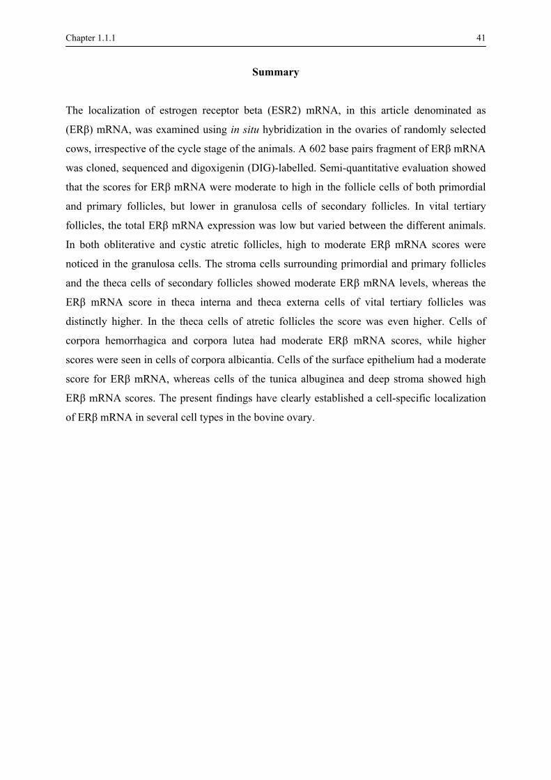
The localization of estrogen receptor beta (ESR2) mRNA, in this article denominated as
(ERβ) mRNA, was examined using in situ hybridization in the ovaries of randomly selected
cows, irrespective of the cycle stage of the animals. A 602 base pairs fragment of ERβ mRNA
was cloned, sequenced and digoxigenin (DIG)-labelled. Semi-quantitative evaluation showed
that the scores for ERβ mRNA were moderate to high in the follicle cells of both primordial
and primary follicles, but lower in granulosa cells of secondary follicles. In vital tertiary
follicles, the total ERβ mRNA expression was low but varied between the different animals.
In both obliterative and cystic atretic follicles, high to moderate ERβ mRNA scores were
noticed in the granulosa cells. The stroma cells surrounding primordial and primary follicles
and the theca cells of secondary follicles showed moderate ERβ mRNA levels, whereas the
ERβ mRNA score in theca interna and theca externa cells of vital tertiary follicles was
distinctly higher. In the theca cells of atretic follicles the score was even higher. Cells of
corpora hemorrhagica and corpora lutea had moderate ERβ mRNA scores, while higher
scores were seen in cells of corpora albicantia. Cells of the surface epithelium had a moderate
score for ERβ mRNA, whereas cells of the tunica albuginea and deep stroma showed high
ERβ mRNA scores. The present findings have clearly established a cell-specific localization
of ERβ mRNA in several cell types in the bovine ovary.
42 Chapter 1.1.1
Introduction
It has been well established that the gonadal steroid estrogen hormones exert a wide variety
of effects on growth, differentiation and functioning of many female and male reproductive
organs including the ovary, uterine tube/oviduct, uterus, vagina and mammary gland, and the
testis, epididymis and prostate (Clark et al., 1992). To exert their functions, estrogens are
retained with high affinity and specificity in target cells by binding to specific intranuclear
binding proteins, the estrogen receptors (ER) (Gronemeyer and Laudet, 1995). These
receptors belong to a large group of transcription factors, the so-called nuclear receptor
superfamily (Tsai and O'Malley, 1994), and are encoded by two different genes resulting in
two receptor subtypes, namely estrogen receptor alpha (ERα) and ERβ. After ligand binding
and dimerization, both receptors function as signal transducers and transcription factors to
modulate the expression of specific target genes by binding to target sequences called
"response elements" (Enmark and Gustafsson, 1999). In addition to acting as homodimers
(ERα/ERα or ERβ/ERβ), ERα and ERβ are also able to form heterodimers (ERα/ERβ)
(Tremblay et al., 1997).
The cloning and initial characterization of ERβ was first described in the rat prostate
(Kuiper et al., 1996) and subsequently orthologues have been found in humans (Mosselman et
al., 1996), mouse (Tremblay et al., 1997), pig (Kowalski et al., 2002; LaVoie et al., 2002),
sheep (Cardenas et al., 2001), monkey (Wu et al., 2000), goldfish (Tchoudakova et al., 1999)
and cattle (Rosenfeld et al., 1999; Walther et al., 1999). Within the different species, ERβ
shows the highest expression in prostate, ovary, lung, bladder, brain and epididymis (Kuiper
et al., 1996; Li et al., 1997). In addition, ERβ plays an important role in bone maintenance
(Turner et al., 1994), in the cardiovascular system (Farhat et al., 1996) and in some breast
cancers (Dotzlaw et al., 1997). ERβ also mediates activities in the central nervous system
(McCarthy and Pfaus, 1996). In the female reproductive tissues, ERβ has been found in the
ovary, uterus and mammary gland. In the human ovary, the receptor is located in stroma cells
of the cortex as well as in the granulosa cells (Byers et al., 1997). Therefore, ERβ might play
an important role in the regulation of follicular growth and oocyte development (Enmark and
Gustafsson, 1999).
The presence of ERβ in the bovine ovary has already been demonstrated by in situ
hybridization (Rosenfeld et al., 1999), but the cell specific localization of ERβ remains

Chapter 1.1.1 43
unclear. Therefore, the purpose of this study was to localize ERβ in the different cell types of
the bovine ovary.
Materials and methods
Animals and tissue sampling
Three adult cows, without symptoms of any reproductive pathology, were sampled at the
moment of slaughter at a local slaughterhouse. Immediately after exsanguination both ovaries
were collected. For in situ hybridization one part of the ovaries was fixed in a RNase free
phosphate buffered saline (PBS) solution containing 4% paraformaldehyde at 4°C for 8 hours,
dehydrated in graded methanol concentrations, cleared in xylene and embedded in paraffin
wax. Five µm thin sections were cut and mounted on poly-L-lysine-coated (Sigma
Diagnostics Inc., St. Louis, MO, USA) SuperFrost®Plus-slides (Menzel-Gläser,
Braunschweig, Germany) and dried overnight at 37°C. Another part of the ovaries was frozen
in liquid nitrogen and used for the generation of a probe for in situ hybridization.
Probe design Total RNA isolation and reverse transcription
Total RNA extraction from bovine ovarian tissue was performed using Trizol reagent
(Invitrogen Life Technologies, Merelbeke, Belgium) followed by DNA digestion using
DNase 1. Before isolation, 10 pg of rabbit globin mRNA (Invitrogen Life Technologies) was
added to each sample as an external standard. RNA isolation efficiency was checked by
polymerase chain reaction (PCR) with primers for rabbit globin sense and anti-sense. Reverse
transcription was done as described in the protocol from the Reverse Transcription kit
(Invitrogen Life Technologies). The bovine glyceraldehyde-3-phosphate dehydrogenase
(GAPDH) primers were used as an internal control for the complementary DNA (Yuan et al.,
2003).
PCR and verification
In order to amplify a specific fragment of 602 base pairs, two primers (left primer: 5'-
CTGCGGTCAATCCATCCTAC-3' and right primer: 5'-TCCGAGAGCCACACTTCAC-3')

44 Chapter 1.1.1 were designed based on the bovine ERβ sequence (Acc No.Y18017) by using the Primer 3
program (Rozen and Skaletsky, 1998). PCR was carried out on a T3 thermocycler (Biometra,
Göttingen, Germany) in 10 µl containing 25 ng complementary DNA, reaction buffer, 3 mM
MgCl2, 200 mM of each desoxynucleoside 5’-triphosphate, 200 mM of each primer and 0,5
units Taq Platinum polymerase (Invitrogen Life Technologies). The thermal cycling profile
was 10 min at 94°C for denaturation, followed by 40 cycles of 30 sec at 94°C, 30 sec at 58°C,
1 min at 72°C and a final elongation step of 10 min at 72°C. The 602 base pairs PCR product
was visualised on a 0.8% agarose gel, cloned in the pCR®2.1 Vector (Invitrogen Life
Technologies) and sequenced for verification using an ALFexpressTM Sequencing System
(Pharmacia, Uppsala, Sweden). In order to generate ERβ-specific RNA probes, the same
fragment was subcloned into the pGEM®-T easy vector (Promega, USA) according to
standard procedures (Sambrook et al., 1989). Subsequently the ligation product was
transformed into bacteria, viz., Subcloning Efficiency DH5αTM Competent Cells (Invitrogen
Life Technologies) and recombinant clones were identified by white: blue colour screening
using the chromogenic substrate X-gal (5-bromo-4-chloro-3-indol-β-D-galactoside)
(Sambrook et al., 1989). After culturing and harvesting the bacteria, plasmid DNA was
isolated using the HiSpeed TM Plasmid Purification kit (Qiagen, Westburg, The Netherlands)
and the orientation of the insert was determined by restriction digest analysis using the
enzymes PstI, SpeI and SalI (Fig. 1).
Fig. 1: Analysis of DNA by 0.8% agarose gel electrophoresis
with ethidium bromide staining; lane 1: 602-bp DNA fragment
of bovine ERβ following RT-PCR using ERβ-specific primers;
lane 2: 1 kb λ-marker; lanes 3 and 4: restriction enzyme digest
of the cloned DNA fragment using SpeI (lane 3) and PstI (lane
4) to ascertain the identity; lane 5: ERβ DNA fragment cut out
of the plasmid using EcoRI restriction enzyme digestion.
Chapter 1.1.1 45
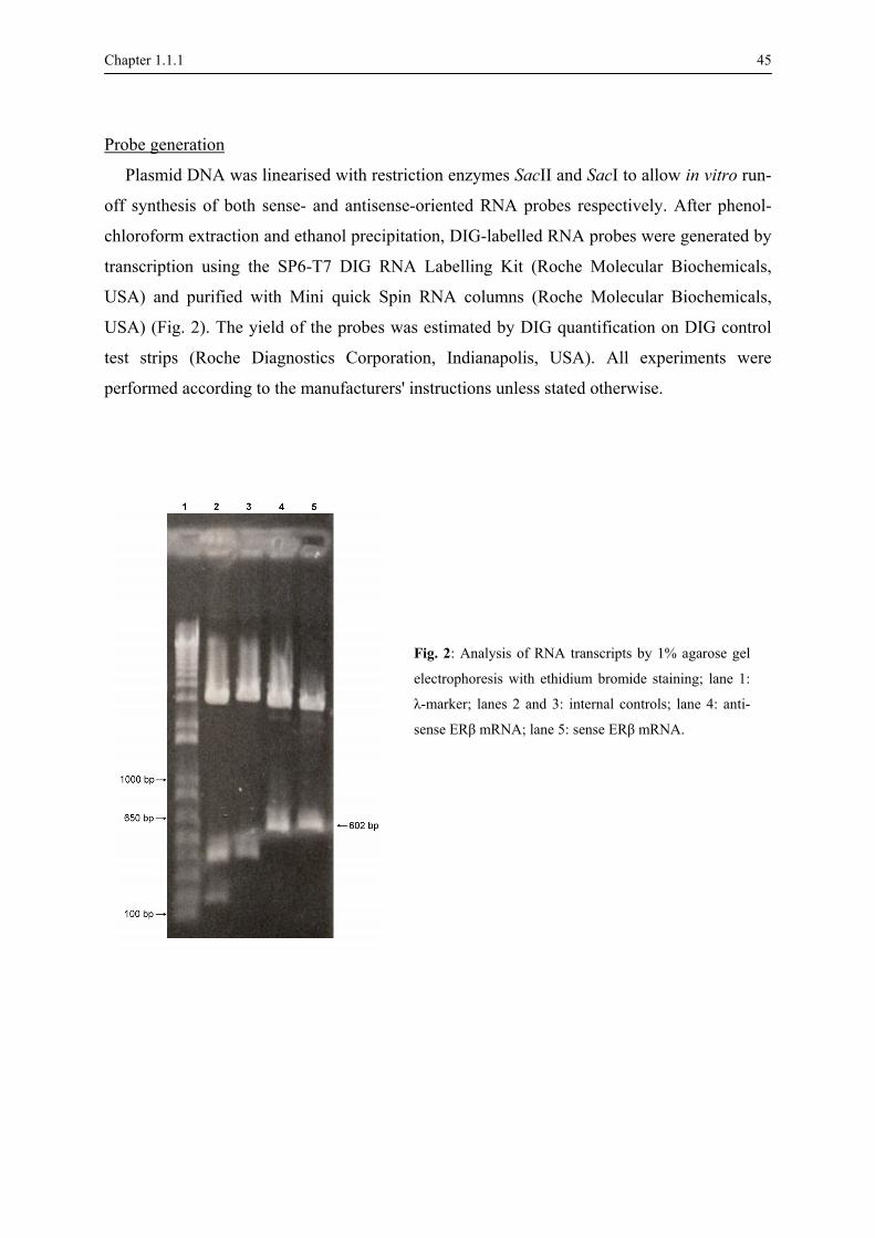
Probe generation
Plasmid DNA was linearised with restriction enzymes SacII and SacI to allow in vitro run-
off synthesis of both sense- and antisense-oriented RNA probes respectively. After phenol-
chloroform extraction and ethanol precipitation, DIG-labelled RNA probes were generated by
transcription using the SP6-T7 DIG RNA Labelling Kit (Roche Molecular Biochemicals,
USA) and purified with Mini quick Spin RNA columns (Roche Molecular Biochemicals,
USA) (Fig. 2). The yield of the probes was estimated by DIG quantification on DIG control
test strips (Roche Diagnostics Corporation, Indianapolis, USA). All experiments were
performed according to the manufacturers' instructions unless stated otherwise.
Fig. 2: Analysis of RNA transcripts by 1% agarose gel
electrophoresis with ethidium bromide staining; lane 1:
λ-marker; lanes 2 and 3: internal controls; lane 4: anti-
sense ERβ mRNA; lane 5: sense ERβ mRNA.

46 Chapter 1.1.1 Histological analysis In situ hybridization
ERβ mRNA was localized in ovarian tissue sections by in situ hybridization analysis.
Deparaffinized and rehydrated sections were rinsed in double concentrated SSPE (0.1 M
NaCl, 10 mM NaH2PO4 pH 7.4, 1 mM EDTA) and transferred into Tris-HCl (pH 7.6) for 5
min. Afterwards, slides were treated with 20 µg/ml Proteinase K (Roche Diagnostics,
Vilvoorde, Belgium) in 0.05 M Tris-HCl (pH 7.4) at 37°C for 30 min to make the RNA more
accessible to hybridization. Sections were fixed in 4% paraformaldehyde in PBS at room
temperature for 10 min. The slides were rinsed in PBS and acetylated in 0.2 M HCl for 15
min at room temperature. Prehybridization was performed by incubating the slides with a
hybridization buffer (Schwarzacher and Heslop-Harrison, 2000) in a humidified chamber at
42°C for 2 hours.
Hybridization was executed by incubating slides with the hybridization buffer containing
the anti-sense probe (200 ng/ml). As negative controls, two sections were incubated either
with hybridization buffer alone or with hybridization buffer containing the sense probe. The
slides were incubated in a humidified chamber (50 % deionized formamide + 50 % diethyl
pyrocarbonate water) at 42°C overnight. Posthybridization was carried out by incubating the
slides for 30 min at 37°C in RNase buffer (1 M Tris, pH 8.0, 5 M NaCl, 0.5 M EDTA)
containing RNase-A (50 µg/ml, Sigma, Belgium). Afterwards the slides were rinsed in double
concentrated SSPE for 15 min at 42°C, in SSPE for 30 min at 42°C and in SSPE for 15 min at
room temperature.
Immunohistochemical detection
Sections were incubated with 1:70 v/v sheep anti-DIG-peroxidase anti-binding fragments
(Roche Diagnostics, Vilvoorde, Belgium) for 1 hour at 37°C in a humidified chamber. The
sections were subsequently stained by incubation with DAB substrate (DAKO, Prosan,
Merelbeke, Belgium) for 10 minutes and counterstained with Mayer's hematoxylin for 5 sec
for better visualization of the cellular architecture of ovarian cell types.
Microscopic analysis
In all ovarian sections the different ovarian cell types were analysed by light microscopic
inspection. Staining scores were awarded in different follicles, corpora lutea, corpora

Chapter 1.1.1 47
albicantia, vascular endothelial cells, rete ovarii, deep and superficial stroma, tunica albuginea
and surface epithelium. All follicles were examined and classified into primordial, primary,
secondary and tertiary follicles according to the criteria listed in Nomina Histologica (1994).
In tertiary follicles the inner layers of granulosa cells facing the antral side were quantified
separately from granulosa layers of the basal side because they showed different staining
patterns. Atretic tertiary follicles were classified as obliterative or cystic (Dellmann and
Eurell, 1998).
For the semi-quantitative evaluation of ERβ mRNA the number of ERβ mRNA containing
cells, identified by a brown staining in the cytoplasm, was counted. For all cell types, at least
100 cells were counted in digital micrographs taken randomly at magnification 600 x
(Olympus BX61, Olympus Belgium, Aartselaar, Belgium). If too many cells were present in
one micrograph, 4 grids of 2500 µm² were placed randomly onto the micrograph and cells
were counted in these areas.
Staining intensity scores ranged from 0 (no staining), 1 (weak staining), 2 (moderate
staining) to 3 (strong staining). ERβ mRNA presence was evaluated using a score (SERβ
mRNA) which was calculated according to the formula:
SERβ mRNA = 0 n0 + 1 n1+ 2 n2 + 3 n3
with n0, n1, n2 and n3 are the percentage of cells exhibiting a staining intensity 0, 1, 2 and 3,
respectively (Boos et al., 1996).
Results
Production of ERβ mRNA probes for in situ hybridization
After sequence comparison of bovine ERα (Acc. No. 538775) with ERβ (Acc. No.
Y18017), an ERβ-specific region of 602 base pairs was selected from exon 1 (5'-UTR) to
exon 4. The amplified fragment (Acc. No. AY 690422) showed 99% sequence identity with
the sequence on which the primers were based because of 4 different nucleotides at position
19, 26, 57 and 202 (Table 1). With this fragment as a template, both sense- and antisense-
oriented RNA probes were synthesized with a yield of 200 ng/ml.

48 Chapter 1.1.1
ERβ sequence
Acc. No. Y18017 Acc. No. AY690422
Nucleotides Amino acid Nucleotides Amino acid
Codon 7 (nucleotides 19-21) GAT Asp AAT Asn
Codon 9 (nucleotides 25-27) TCA Ser TGA STOP
Codon 19 (nucleotides 55-57) AGT Ser AGC Ser
Codon 68 (nucleotides 202-204) CTG Leu TTG Leu
Table 1: Sequence comparison of bovine ERβ Acc. No. Y18017 with Acc. No. AY 690422.
Localization of ERβ mRNA in the bovine ovary
The in situ hybridization technique confirmed the presence of a specific and intense signal
in the different ovarian tissues when using the DIG-labelled RNA anti-sense probe (Fig. 3a)
while no signal was visible using the DIG- labelled RNA sense probe (Fig. 3b).
The presence of ERβ mRNA, identified by brown cytoplasmic staining, was found in all
animals in most follicular stages, in the corpora lutea and corpora albicantia, in the superficial
and deep stroma, in vascular endothelial cells, in the rete ovarii, in the tunica albuginea and in
the surface epithelium.
Fig. 3: Micrograph of a tertiary follicle following in situ hybridization using the ERβ-specific antisense probe (Fig. 3a)
and sense probe (negative control) (Fig. 3b). Bar = 100 µm.

Chapter 1.1.1 49
Cell-specific variation of ERβ mRNA expression
In situ localization results indicated that the intensity of specific staining and the
proportion of positive cells varied between the different cell types (Fig. 4 and 5). The scores
for ERβ mRNA were moderate to high in the follicle cells of both primordial and primary
follicles, but clearly higher than in follicle cells of secondary follicles. In vital tertiary
follicles, the granulosa cells of both the cumulus oophorus and the granulosa layers showed a
higher expression of ERβ mRNA compared with that in follicle cells of secondary follicles.
However, this expression in vital tertiary follicles was only seen in one of the 3 cows, whereas
no expression of ERβ mRNA could be detected in this cell type of the other animals. In both
obliterative and cystic atretic follicles, high to moderate ERβ mRNA scores were noticed in
the granulosa cells.
A moderate SERβ mRNA was observed in superficial stroma cells surrounding the
primordial and primary follicles. In stroma cells around secondary follicles (theca follicularis
cells) this score was similar, whereas the score in theca interna and externa cells of vital
tertiary follicles was manifestly higher. A similar high score was found in the theca interna
and theca externa layers of cystic atretic follicles. In both theca layers of obliterative follicles
SERβ mRNA were higher than in any other follicular cell type. In obliterative follicles, the
membrane of Slavjansky was also strongly positive.
A corpus hemorrhagicum was only present in one animal, with moderate scores for ERβ
mRNA in the granulosa cells and higher scores in both theca layers. Corpora lutea could only
be investigated in two animals, and within these structures, moderate ERβ mRNA scores were
present in both large and small lutein cells, vascular endothelial cells, perivascular stroma
cells and peripheral stroma cells (comparable with theca externa cells of tertiary follicles).
High ERβ mRNA levels were observed in corpora albicantia with similar high scores in
internal and peripheral stroma cells.
Finally, the cells of the surface epithelium had a moderate SERβ mRNA, whereas cells of
the tunica albuginea, deep stroma, rete ovarii and vascular endothelial cells showed high ERβ
mRNA levels.

Fig. 4: Mean scores (± SEM) for ERβ mRNA (SERβ mRNA) in different groups of bovine ovarian cells; II = secondary follicle, vital III = vital tertiary follicle, obliterative
III = obliterative atretic tertiary follicle, cystic III = cystic atretic tertiary follicle, CH = corpus hemorrhagicum, CL = corpus luteum, CA = corpus albicans; (n=1, 2, 3).
0
50
100
150
200
250
300
Prim
ordi
al fo
llicl
e ce
lls
Prim
ary
folli
cle
cells
folli
cle
cells
thec
a fo
llicu
laris
cum
ulus
oop
horu
s
gran
ulos
a ce
lls a
ntra
l sid
e
gran
ulel
ls b
asal
sid
e
thec
a in
tern
a
thec
a ex
tern
a
gran
ulos
a ce
lls
mem
rane
of S
lavj
ansk
y
thec
a in
tern
a
thec
a ex
tern
a
gran
ulos
a ce
lls
thec
a in
tern
a
thec
a ex
tern
a
antru
m
gran
ulos
a ce
lls
thec
a in
tern
a
thec
a ex
tern
a
larg
e lu
tein
cel
ls
smal
l lut
ein
cells
vasc
r end
othe
lial c
ells
peri
lar s
trom
a ce
lls
s c
ells
(cap
sule
)
inte
rnal
stro
ma
s c
ells
(cap
sule
)
scul
ar e
ndot
helia
l cel
ls
Ret
e ov
arii
Dee
p st
rom
a
Sup
erfic
ial s
trom
a
Tuni
ca a
lbug
inea
Sur
face
epi
thel
ium
Mea
n S E
R β
mR
NA
1
1 1
1 1 2 2 2
2
2
1
33
3
3
3
3
3
33
3 33
3
33 3 3
33
3
3
3
Va
ula
vasc
u
trom
a
trom
a
osa
c
b
CA CL CH cystic III obliterative III vital III II

Chapter 1.1.1 51
Fig. 5: Micrographs of different bovine ovarian cell types following in situ hybridization using the ERβ-
specific antisense probe; Fig. 5a and 5b: primordial (1), primary (2) and secondary (3) follicles with moderate
to low scores for ERβ mRNA (SERβ mRNA) in the follicle / granulosa cells, and moderate to high SERβ
mRNA in stroma cells (4); Fig. 5c: wall of a vital tertiary follicle with low ERβ mRNA scores in the granulosa
cells (5) and high SERβ mRNA in theca interna (6) and externa cells (7); Fig. 5d: wall of a cystic atretic

52 Chapter 1.1.1
tertiary follicle with moderate to high SERβ mRNA in apoptotic granulosa cells (5) and in theca interna (6)
and externa cells (7); Fig. 5e: overview of an obliterative atretic tertiary follicle with high SERβ mRNA in the
collapsed theca layers (8) and the thickened basal membrane (9); Fig. 5f: overview of a corpus luteum with
moderate to high SERβ mRNA in the lutein cells (10); Fig. 5g: overview of a corpus albicans with moderate to
high SERβ mRNA in internal stroma cells (11); Fig. 5h: cells of the surface epithelium (12) with moderate
SERβ mRNA and tunica albuginea cells (13) with low SERβ mRNA; Fig. 5i: cells of the deep stroma (14)
with high SERβ mRNA. Bar = 50 µm in Fig. 5a, 5b, 5c, 5d, 5h and 5i, 100 µm in Fig. 5e and 5g, and 500 µm
in Fig. 5f.
Discussion
The primary aim of this study was to localize ERβ mRNA in different cell types of the
bovine ovary by in situ hybridization using DIG labelled bovine ERβ mRNA probes. In
comparison with the early published bovine ERβ sequence different nucleotides were
observed in the coding sequence. A G to A transition at position 19, results in an aspartic acid
(Asp) to asparagin (Asn) substitution at amino acid 7. The difference between Asn and Asp is
the neutral character of Asn (NH2-group) and the acid character of Asp (OH-group). This
missense mutation might have an impact on the protein structure, folding and related cell
response to hormone actions. A C to G transversion at position 26 results in the incorporation
of a STOP-codon at amino acid 9. Although, this nonsense mutation alters the translation, it
doesn't influence our in situ hybridization experiments, because we are interested in the
binding of the ovarian tissue mRNA with the mRNA probes. A T to C and a C to T transition
at pos 57 and 202 respectively are silent mutations, resulting in the incorporation of the amino
acids serine (Ser) and leucine (Leu) at pos 19 and 68.
Since this was a single-pass sequence, the question remains whether these 4
polymorphisms are time single nucleotides polymorphisms (SNPs) or PCR sequencing errors.
If they are SNPs it should be worthwhile to investigate the nonsense and the missense
mutations.
Localization of ERβ mRNA and protein in ovarian cells of various species has already
been reported as reviewed by Rosenfeld and others (2001). The present study revealed that the
expression level of ERβ mRNA varied between the different follicular types. Indeed, ERβ
mRNA levels were high in the follicle cells of primordial and primary follicles and were

Chapter 1.1.1 53 manifestly lower in the granulosa cells of secondary and tertiary follicles. Similar findings are
described in the ewe, namely a high specific expression of ERβ in the follicle/granulosa cell
layer of small and developing follicles that declines with increasing follicle size (Jansen et al.,
2001). In contrast, human (Pelletier and El-Alfy, 2000) and rat (Pelletier et al., 2000)
granulosa cells of pre-antral and antral follicles showed a high expression of ERβ. Rosenfeld
and co-workers (Rosenfeld et al., 1999) found high ERβ mRNA levels in granulosa cells of
tertiary follicles of cattle, but they did not compare this expression with that in other follicular
structures. Despite the differences in follicle-specific expression between different species,
these and our findings support the hypothesis that estrogens are involved in folliculogenesis
through interaction with ERβ and not with ERα as the latter receptor was rarely if ever found
in bovine ovarian follicles (Van den Broeck et al., 2002). Furthermore, the moderate to high
expression of ERβ mRNA in primordial and primary ovarian follicles of cattle indicates that
ERβ is mainly involved in the earlier stages of development. Again, this is in contrast with
studies in ERαβ double knock-out (KO) mice, in which early follicular growth and
development occurred normally but without leading to mature Graafian follicles, suggesting
that ERα and ERβ may not be necessary for the earlier stages of folliculogenesis (Dupont et
al., 2000).
Remarkably, a moderate to high ERβ mRNA expression was present in the granulosa cell
layer of obliterative and cystic atretic tertiary follicles. This atresia is due to apoptosis of the
granulosa cells (Amsterdam et al., 1999) which in turn can be inhibited by estrogens and
promoted by androgens (Billig et al., 1993). Studies in ERαKO mice have demonstrated that
this inhibition is not conferred through ERα (Schomberg et al., 1999). In our study, high
levels of ERβ mRNA were present in the atretic follicles, so apparently ERβ could not prevent
the atresia. This can be due to the fact that other pro- and anti-apoptotic factors are present
and that the final outcome is the result of a complex crosstalk among multiple intracellular
pathways in which the protective capacity of estrogens is overruled. An alternative possibility
is that the ERβ demonstrated here is characterized by a lower affinity for estrogens or for
estrogen response elements, so that no apoptotic inhibiting effect is exerted. This also means
that there should be another yet unidentified estrogen receptor that can prevent apoptosis. A
third estrogen receptor, ERγ, has been identified in fish (Hawkins et al., 2000) and there is
some evidence that at least one additional estrogen receptor is present in the mouse ovary and
uterus (Ghosh et al., 1999). Similarly, it might be possible that the bovine ovary also contain a

54 Chapter 1.1.1 third ER type that prevents follicular atresia. This hypothesis, although highly speculative,
should be kept in mind and may form the basis for further experimental research.
Moderate ERβ mRNA levels were noted in granulosa cells of a corpus hemorrhagicum
present in one animal, and in small and large lutein cells of corpora lutea present in two
animals. These observations are suggestive of a role of estrogens in the formation and
maintenance of a corpus luteum. The specific role of ERβ and ERα in this process in cattle is
unclear, as moderate levels of ERα are also present in corpora lutea cells (Van den Broeck et
al., 2002). Studies in mice have elucidated that in both ERαKO and ERβKO animals
steroidogenically functional corpora lutea are formed (Krege et al., 1998; Rosenfeld et al.,
2000). In contrast, ERαβ double KO mice do not undergo luteinization and corpus luteum
formation (Couse et al., 1999). These results indicate that estrogen is needed for corpus
luteum formation and maintenance acting through ERα or ERβ, and luteinization cannot take
place when both receptors are lacking. A similar mechanism can occur in cattle, but remains
to be proven.
Cells of both the tunica albuginea and deep stroma showed high ERβ mRNA expression
levels, which has never been described before. The exact role of ERβ in these cell types
remains to be clarified, but a putative function is that upon estrogen binding, these cells alter
the stromal composition in order to allow the dynamical processes of follicle development,
ovulation and corpus luteum formation. Further studies including cell culture experiments are
needed to confirm this hypothesis. In stroma cells immediately surrounding primordial and
primary follicles ERβ mRNA expression was lower than in cells of the tunica albuginea and
deep stroma, and in follicle cells of primordial and primary follicles. However, the theca
layers of secondary, vital tertiary and atretic tertiary follicles showed a gradually increased
ERβ mRNA expression which was always higher than in the granulosa cells of the
corresponding follicle. These findings might indicate that for primordial and primary follicles,
estrogens act via ERβ that are located within the follicle cells, whereas the indirect influence
via the surrounding stroma cells is lower. In contrast, ERβ levels in secondary and tertiary
follicles are decreased in the granulosa cells and increased in the theca cells, which is
suggestive of an indirect effect of estrogens on the developing follicles via the theca cells.
These stroma-epithelial interactions have already been described in earlier studies for
progesterone (Hild-Petito et al., 1988), and theca cells may produce factors that promote
follicle cell growth or differentiation (Hsueh et al., 1984). Similar mechanisms may exist in
Chapter 1.1.1 55 the bovine ovary for estrogens, but further studies are needed to identify and reveal these
processes.
In the present study, the localization of ERβ mRNA in the ovary was examined in
randomly selected cows irrespective of the cycle stage of these animals, and therefore without
taking into account the effects of circulating ovarian hormones and gonadotrophins. It has
been demonstrated that hormonal superovulatory treatment in cows does not affect the
topographical distribution and staining intensity of receptors for ERα and progesterone in the
endometrium (Dall'Aglio et al., 1999). Nevertheless, in the ovary estrogens downregulate
granulosa cell expression of ERβ protein (Sharma et al., 1999) and that treatment of rodents
with LH results in a marked decrease of ERβ in the ovary (Byers et al., 1997). Consequently,
cycle-dependent differences in ERβ mRNA expression in the ovary may be expected, and at
this moment an elaborate experiment is performed in which the presence of ERβ mRNA is
studied in detail in a higher number of cows in each of the five different cycle stages, i.e.,
prooestrus, oestrus, metoestrus, early dioestrus and late dioestrus.
In summary, the present findings have clearly established a cell-specific localization of
ERβ mRNA in several cell types in the bovine ovary. Some of these findings are in agreement
with earlier studies in various mammalian species, while others are contradictory even to
findings in cattle. Further comprehensive studies are needed and planned to confirm and
explain the findings and hypotheses proposed here.

56 Chapter 1.1.1
References
Amsterdam A, Gold RS, Hosokawa K, Yoshida Y, Sasson R, Jung Y, Kotsuji F. Crosstalk Among Multiple
Signalling Pathways Controlling Ovarian Cell Death. Trends in Endocrinology and Metabolism 1999; 10:
255-262.
Billig H, Furuta I, Hsueh AJ. Estrogens inhibit and androgens enhance ovarian granulosa cell apoptosis.
Endocrinology 1993; 133: 2204-2212.
Boos A, Meyer W, Schwarz R, Grunert E. Immunohistochemical assessment of oestrogen receptor and
progesterone receptor distribution in biopsy samples of the bovine endometrium collected throughout the
oestrous cycle. Animal Reproduction Science 1996; 44: 11-21.
Byers M, Kuiper GG, Gustafsson JA, Park-Sarge OK. Estrogen receptor-beta mRNA expression in rat ovary:
down-regulation by gonadotrophins. Molecular Endocrinology 1997; 11: 172-182.
Cardenas H, Burke KA, Bigsby RM, Pope WF, Nephew KP. Estrogen receptor beta in the sheep ovary
during the estrous cycle and early pregnancy. Biology of Reproduction 2001; 65: 128-134.
Clark JH, Schrader WT, O'Malley BW. Mechanism of action of steroid hormone. Textbook of
Endocrinology. Eds. J.D. Wilson and D.W. Foster, WB Saunders Company, New York 1992; pp. 35-90.
Couse JF, Hewitt SC, Bunch DO, Sar M, Walker VR, Davis BJ, Korach KS. Postnatal sex reversal of the
ovaries in mice lacking estrogen receptors alpha and beta. Science 1999; 286: 2328-2331.
Dall'Aglio C, Ceccarelli P, Gargiulo AM, Pedini V, Monaci M. Immunohistochemical localization of
endometrial oestrogen and progesterone receptors in the cow. Anatomia Histologia Embryologia 1999; 28:
375-377.
Dellman HD, Eurell JA. Textbook of Veterinary Histology. Williams & Wilkins, London 1998.
Dotzlaw H, Leygue E, Watson PH, Murphy LC. Expression of estrogen receptor-beta in human breast
tumors. Journal of Clinical Endocrinology and Metabolism 1997; 82: 2371-2374.
Dupont S, Krust A, Gansmuller A, Dierich A, Chambon P, Mark M. Effect of single and compound
knockouts of estrogen receptors alpha (ERalpha) and beta (ERbeta) on mouse reproductive phenotypes.
Development 2000; 127: 4277-4291.
Enmark E, Gustafsson JA. Oestrogen receptors - an overview. Journal of Internal Medicine 1999; 246: 133 -
138.
Chapter 1.1.1 57 Farhat MY, Lavigne MC, Ramwell PW. The vascular protective effects of estrogen. FASEB Journal 1996;
10: 615-624.
Ghosh D, Taylor JA, Green JA, Lubahn DB. Methoxychlor stimulates estrogen-responsive messenger
ribonucleic acids in mouse uterus through a non-estrogen receptor (non-ER) alpha and non-ER beta
mechanism. Endocrinology 1999; 140: 3526-3533.
Gronemeyer H, Laudet V. Transcription factors 3: nuclear receptors. Protein Profile 1995; 2: 1173-1308.
Hawkins MB, Thornton JW, Crews D, Skipper JK, Dotte A, Thomas P. Identification of a third distinct
estrogen receptor and reclassification of estrogen receptors in teleosts. Proceedings of the National Academy
of Sciences of the United States of America 2000; 97: 10751-10756.
Hild-Petito S, Stouffer RL, Brenner RM. Immunocytochemical localization of estradiol and progesterone
receptors in the monkey ovary throughout the menstrual cycle. Endocrinology 1988; 123: 2896-2905.
Hsueh AJ, Adashi EY, Jones PB, Welsh TH Jr. Hormonal regulation of the differentiation of cultured
ovarian granulosa cells. Endocrine Reviews 1984; 5: 76-127.
Jansen HT, West C, Lehman MN, Padmanabhan V. Ovarian estrogen receptor-beta (ERbeta) regulation: I.
Changes in ERbeta messenger RNA expression prior to ovulation in the ewe. Biology of Reproduction 2001;
65: 866-872.
Kowalski AA, Graddy LG, Vale-Cruz DS, Choi I, Katzenellenbogen BS, Simmen FA, Simmen RC.
Molecular cloning of porcine estrogen receptor-beta complementary DNAs and developmental expression in
periimplantation embryos. Biology of Reproduction 2002; 66: 760-769.
Krege JH, Hodgin JB, Couse JF, Enmark E, Warner M, Mahler JF, Sar M, Korach KS, Gustafsson JA,
Smithies O. Generation and reproductive phenotypes of mice lacking estrogen receptor beta. Proceedings of
the National Academy of Sciences of the United States of America 1998; 95: 15677-15682.
Kuiper GG, Enmark E, Pelto-Huikko M, Nilsson S, Gustafsson JA. Cloning of a novel receptor expressed in
rat prostate and ovary. Proceedings of the National Academy of Sciences of the United States of America
1996; 93: 5925-5930.
LaVoie HA, DeSimone DC, Gillio-Meina C, Hui YY. Cloning and characterization of porcine ovarian
estrogen receptor beta isoforms. Biology of Reproduction 2002; 66: 616-623.
Li X, Schwartz PE, Rissman EF. Distribution of estrogen receptor-beta-like immunoreactivity in rat
forebrain. Neuroendocrinology 1997; 66: 63-67.

58 Chapter 1.1.1 McCarthy MM, Pfaus JG. Steroid modulation of neurotransmitter function to alter female reproductive
behaviour. Trends in Endocrinology and Metabolism 1996; 7: 327-333.
Mosselman S, Polman J, Dijkema R. ER beta: identification and characterization of a novel human estrogen
receptor. FEBS Letters 1996; 392: 49-53.
Nomina Histologica: International Committee on Veterinary Histological Nomenclature. Cornell, New York
1994.
Pelletier G, El-Alfy M. Immunocytochemical localization of estrogen receptors alpha and beta in the human
reproductive organs. Journal of Clinical Endocrinology and Metabolism 2000; 85: 4835-4840.
Pelletier G, Labrie C, Labrie F. Localization of oestrogen receptor alpha, oestrogen receptor beta and
androgen receptors in the rat reproductive organs. Journal of Endocrinology 2000; 165: 359-370.
Rosenfeld CS, Yuan X, Manikkam M, Calder MD, Garverick HA, Lubahn DB. Cloning, sequencing, and
localization of bovine estrogen receptor-beta within the ovarian follicle. Biology of Reproduction 1999; 60:
691-697.
Rosenfeld CS, Murray AA, Simmer G, Hufford MG, Smith MF, Spears N, Lubahn DB. Gonadotrophin
induction of ovulation and corpus luteum formation in young estrogen receptor-alpha knockout mice. Biology
of Reproduction 2000; 62: 599-605.
Rosenfeld CS, Wagner JS, Roberts RM, Lubahn DB. Intraovarian actions of oestrogen. Reproduction 2001;
122: 215-226.
Sambrook J, Fritsch EF, Maniatis R. Molecular Cloning. A Laboratory Manual. Cold Spring Harbour
Laboratory Press, New York 1989.
Schomberg DW, Couse JF, Mukherjee A, Lubahn DB, Sar M, Mayo KE, Korach KS. Targeted disruption of
the estrogen receptor-alpha gene in female mice: characterization of ovarian responses and phenotype in the
adult. Endocrinology 1999; 140: 2733-2744.
Schwarzacher R, Heslop-Harrison P. In situ hybridization to RNA and DNA in cells, tissue sections or whole
amounts. First Edition, 2000; pp. 126-146.
Sharma SC, Clemens JW, Pisarska MD, Richards JS. Expression and function of estrogen receptor subtypes
in granulosa cells: regulation by estradiol and forskolin. Endocrinology 1999; 140: 4320-4334.

Chapter 1.1.1 59 Tchoudakova A, Pathak S, Callard GV. Molecular cloning of an estrogen receptor beta subtype from the
goldfish, Carassius auratus. General and Comparative Endocrinology 1999; 113: 388-400.
Tremblay GB, Tremblay A, Copeland NG, Gilbert DJ, Jenkins NA, Labrie F, Giguere V. Cloning,
chromosomal localization, and functional analysis of the murine estrogen receptor beta. Molecular
Endocrinology 1997; 11: 353-365.
Tsai MJ, O'Malley BW. Molecular mechanisms of action of steroid/thyroid receptor superfamily members.
Annual Review of Biochemistry 1994; 63: 451-486.
Turner RT, Riggs BL, Spelsberg TC. Skeletal effects of estrogen. Endocrine Reviews 1994; 15: 275-300.
Van den Broeck W, Coryn M, Simoens P, Lauwers H. Cell-specific distribution of oestrogen receptor-alpha
in the bovine ovary. Reproduction in Domestic Animals 2002; 37: 291-293.
Walther N, Lioutas C, Tillmann G, Ivell R. Cloning of bovine estrogen receptor beta (ERbeta): expression of
novel deleted isoforms in reproductive tissues. Molecular and Cellular Endocrinology 1999; 152: 37-45.
Wu WX, Ma XH, Smith GC, Nathanielsz PW. Differential distribution of ERalpha and ERbeta mRNA in
intrauterine tissues of the pregnant rhesus monkey. American Journal of Physiology. Cell Physiology 2000;
78: C190-C198.
Yuan YQ, Van Soom A, Coopman FO, Mintiens K, Boerjan ML, Van Zeveren A, de Kruif A, Peelman LJ.
Influence of oxygen tension on apoptosis and hatching in bovine embryos cultured in vitro. Theriogenology
2003; 59: 1585-1596.


1.1.2 Localization of estrogen receptors within various bovine ovarian
cell types at different stages of the estrous cycle
Modified from:
LOCALIZATION OF ESTROGEN RECEPTORS WITHIN VARIOUS BOVINE OVARIAN CELL TYPES AT DIFFERENT STAGES OF THE ESTROUS CYCLE D’Haeseleer M, Simoens P, Van den Broeck W Anatomia Histologia Embryologia 2006; in press


Chapter 1.1.2 63
Summary
In the present study ERα and ERβ mRNA were localized in various ovarian cell types of 23
cows at different cycle stages. ERα was detected by immunohistochemistry and the
localization of ERβ mRNA was examined using in situ hybridization. The immunostaining of
ERα was low in the ovarian follicles, tunica albuginea and surface epithelium, but high in
cells of the deep stroma and superficial stroma, which indicates a functional role of ERα in
the cells surrounding the follicles. In contrast, ERβ mRNA scores were low to moderate in
primordial and primary follicles and increased with the development of the follicle. ERβ
mRNA scores were higher in cystic follicles than in obliterative follicles. In corpora lutea and
corpora albicantia the scores for ERβ mRNA were moderate. Furthermore, in corpora lutea
ERβ mRNA levels showed cyclic variations and were low during early dioestrus. The
correlation between the plasma progesterone levels and the score for ER was low and
negative in all ovarian cell types. This study demonstrates the predominant role of ERβ over
ERα in bovine ovarian structures. Furthermore, the colocalization of both ERβ mRNA and
ERα in most cell types suggests possible interactions between both ER subtypes.

64 Chapter 1.1.2
Introduction
Estrogens regulate follicular growth and ovarian function by binding to estrogen receptors
(ERs) (Couse et al., 1999). Binding of the ER-ligand complex to target DNA at estrogen
response elements (ERE) results in the transcription of various genes. In many species two
subtypes of the estrogen receptor have been demonstrated, viz. estrogen receptor α (ERα) and
estrogen receptor β (ERβ). A third type of estrogen receptor ERγ has been detected in the
teleost Micropogonias undulatus (Hawkins et al., 2000), in mouse (Lorke et al., 2000) and in
humans (Heard et al., 2000). ERα and ERβ may form both homodimers and heterodimers
resulting in receptor molecules with different functional activities (Pettersson et al., 1997;
Enmark and Gustafsson, 1999). Experiments in mice lacking genes for either or both ER
types have demonstrated the biological significance of each subtype in male and female
reproduction (Lubahn et al., 1993; Krege et al., 1998; Couse et al., 1999).
The tissue distribution of ERα and ERβ has been described for a wide variety of species in
different organs. The presence of ERα has been shown in humans (Coppens et al., 1993),
cattle (Dall'Aglio et al., 1999; Van den Broeck et al., 2002), dog (Vermeirsch, 2001) and rat
(Pelletier et al., 2000). ERα mRNA has been localized in various species including humans
(Revelli et al., 1996), cattle (Berisha et al., 2002), pig (Slomczynska et al., 2001), dog (Hatoya
et al., 2003), rat (Kuiper et al., 1996) and mouse (Tremblay et al., 1997), and is expressed
predominantly in the pituitary gland, ovary, uterus, testis, epididymis and adrenal gland
(Kuiper et al., 1997).
ERβ has been found in many vertebrate species and is expressed predominantly in the
ovary and the prostate (Enmark et al., 1997). ERβ mRNA has been described in different
ovarian structures of various species including humans (Enmark et al., 1997), cattle
(Rosenfeld et al., 1999), pig (Slomczynska et al., 2001), sheep (Jansen et al., 2001), dog
(Hatoya et al., 2003) and rat (Kuiper et al., 1996; Wang et al., 2000).
Although the localization of ERα and ERβ has already been described in the bovine ovary,
the localization patterns of the different ovarian tissues are not well known (Rosenfeld et al.,
1999; Manikkam et al., 2001b). Therefore, in a preliminary study, a probe for in situ
hybridization was generated to localize ERβ mRNA in the bovine ovary and an
immunohistochemical method was developed to investigate the presence of ERα (D'Haeseleer
Chapter 1.1.2 65
et al., 2005; Van den Broeck et al., 2002). The aim of the present study was to determine the
morphological localization of ERα and ERβ in the different cell types of the bovine ovary.
Furthermore, putative differences between the receptor score in active and inactive ovaries
were examined. Finally, possible cyclic alterations of receptor localization in the different cell
types, and their correlation with the plasma progesterone level were determined.
Materials and methods
Animals and samples
Twenty-three animals were used, namely 7 cows that were experimentally synchronized
and 16 slaughterhouse cows. For oestrus synchronization 7 cows were treated by means of
PRID® insertion (Ceva Sante Animal, Brussels, Belgium) for 12 days. Blood samples for LH
surge and plasma progesterone determination were taken every 4 hours in heparinized tubes
from 36 hrs until 60 hrs after PRID® removal (Verberckmoes et al., 2004; Henry et al., 1987).
Six of the seven cows were slaughtered 72 hrs after PRID® removal, i.e., at the day of oestrus
(D0). One cow was slaughtered 15 days after PRID® removal (D13 of the estrous cycle) when
a functional corpus luteum was present. From 16 other cows, without symptoms of any
reproductive pathology, the genital tracts and blood samples were collected at the moment of
slaughter. Both ovaries were classified using morphologic parameters as described by Ireland
et al. (Ireland et al., 1980). The ovary showing a mature follicle or a young corpus luteum was
classified as the active ovary. Subsequently, each ovary was bisected and sampled.
For the immunohistochemical determination of ERα, one part of the ovarian samples was
fixed for 6 h in 3.5% phosphate-buffered formaldehyde (pH 6.7) at room temperature and
dehydrated in increasing concentrations of ethanol. Subsequently, all tissue samples were
embedded in paraffin, and 5 µm thin sections were cut. The sections were mounted on 3-
aminopropyl-triethoxysilane-coated slides (Sigma, S. Louis, MO, USA) and dried overnight
at 37°C.
For the detection of ERβ mRNA the other part of the ovarian samples was fixed in a
Rnase-free phosphate buffered saline (PBS) solution containing 4% paraformaldehyde at 4°C
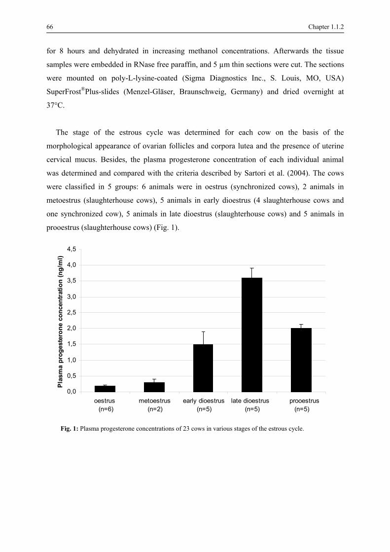
66 Chapter 1.1.2
for 8 hours and dehydrated in increasing methanol concentrations. Afterwards the tissue
samples were embedded in RNase free paraffin, and 5 µm thin sections were cut. The sections
were mounted on poly-L-lysine-coated (Sigma Diagnostics Inc., S. Louis, MO, USA)
SuperFrost®Plus-slides (Menzel-Gläser, Braunschweig, Germany) and dried overnight at
37°C.
The stage of the estrous cycle was determined for each cow on the basis of the
morphological appearance of ovarian follicles and corpora lutea and the presence of uterine
cervical mucus. Besides, the plasma progesterone concentration of each individual animal
was determined and compared with the criteria described by Sartori et al. (2004). The cows
were classified in 5 groups: 6 animals were in oestrus (synchronized cows), 2 animals in
metoestrus (slaughterhouse cows), 5 animals in early dioestrus (4 slaughterhouse cows and
one synchronized cow), 5 animals in late dioestrus (slaughterhouse cows) and 5 animals in
prooestrus (slaughterhouse cows) (Fig. 1).
0,0
0,5
1,0
1,5
2,0
2,5
3,0
3,5
4,0
4,5
oestrus (n=6)
metoestrus (n=2)
early dioestrus(n=5)
late dioestrus (n=5)
prooestrus (n=5)
Plas
ma
prog
este
rone
con
cent
ratio
n (n
g/m
l)
Fig. 1: Plasma progesterone concentrations of 23 cows in various stages of the estrous cycle.

Chapter 1.1.2 67
Immunohistochemical detection of ERα
For the detection of ERα, the assay described by Van den Broeck et al. (2002) was used.
The primary antibody was a monoclonal mouse-anti-human antibody clone 1D5 (DAKO,
Prosan, Merelbeke, Belgium) which is ERα-specific. The antigen-antibody complex was
visualized using biotinylated rabbit anti-mouse antibodies (DAKO) and diaminobenzidine
was used as chromogen substrate (DAKO). For each cell type, the presence of ERα was
assessed by counting the cells presenting a brown nuclear staining.
In every staining procedure, positive and negative controls were included. Bovine uterine
tissues known to be positive for ERα from a previous study served as positive controls. As
negative controls tissue sections incubated with dilution buffer instead of the primary
antibody, secondary antibody, and/or peroxidase-conjugated streptavidin (BioGenex, San
Ramon, CA, USA) were used.
In situ hybridization of ERβ mRNA
Probe multiplication
The ligated complementary DNA fragment containing the 602 base pairs fragment of ERβ
(D'Haeseleer et al., 2005) was purified and the plasmid DNA was linearized using restriction
enzymes SacII and SacI to allow in vitro run-off synthesis of sense- and antisense-oriented
RNA probes, respectively. After phenol-chloroform extraction and ethanol precipitation,
digoxigenin (DIG)-labelled sense and antisense riboprobes were generated by transcription
using the SP6-T7 DIG RNA Labelling Kit (Roche Molecular Biochemicals, Indianapolis,
USA) and purified with Mini quick Spin RNA columns (Roche Molecular Biochemicals,
Indianapolis, USA). The yield of the probes was estimated by DIG quantification on DIG
control test strips (Roche Diagnostics Corporation, Indianapolis, USA).
In situ hybridization
In situ hybridization was carried out in all ovarian tissue sections as described previously
(D'Haeseleer et al., 2005). The presence of ERβ mRNA was assessed by counting the cells
presenting a brown cytoplasmic staining. Two controls were performed to confirm that the
antisense riboprobe hybridized specifically to ERβ mRNA, viz., hybridization with the
68 Chapter 1.1.2
labelled sense probe and hybridization with hybridization buffer only. None of these
treatments showed in any detectable ribopobe binding.
Microscopic analysis
In each ovary the different follicle types, corpora hemorrhagica, corpora lutea, corpora
albicantia, deep stroma, superficial stroma, tunica albuginea and surface epithelium were
microscopically evaluated. All follicles were examined and classified by histological stage
into primordial, primary, secondary and tertiary follicles according to the criteria listed in the
Nomina Histologica (1994). Tertiary follicles containing less than 1 % apoptotic cells were
classified as vital tertiary follicles, whereas follicles containing more than 1% apoptotic cells
were regarded as atretic (D'Haeseleer et al., 2006a). The atretic follicles were further
classified as obliterative or cystic according to the criteria described by Dellman and Eurell
(1998).
At least 100 cells of every cell type were counted on digital micrographs taken randomly at
magnification 600 x with an Olympus DP 50 digital camera mounted on an Olympus BX61
light microscope (Olympus Belgium N.V., Aartselaar, Belgium). If too many cells were
present in one micrograph, cells were counted in 4 grids of 2500 µm² that were projected
randomly over the micrograph. Staining intensity scores ranged from 0 (no staining), 1 (weak
staining), 2 (moderate staining) to 3 (strong staining). The presence of ERα was evaluated
using a score SERα which was calculated according to the formula modified after Boos et al.
(1996): SERα = 0 n0 + 1 n1 + 2 n2 + 3 n3 with n0, n1, n2 and n3 are the percentage of cells
exhibiting a staining intensity 0, 1, 2 and 3, respectively. A similar formula (SERβ mRNA)
was used for calculating the score for ERβ mRNA.
Statistical analysis
Throughout the study, results were represented as mean values and variation was expressed
as standard error of the mean (SEM). Possible differences in the ER score between active and
inactive ovaries, between the various cell types and between the 5 stages of the estrous cycle
were analysed by means of the Kruskall-Wallis test. The Tukey's test was applied to
determine significant (P < 0.05) or highly significant (P < 0.01) differences between
particular cycle stages.

Chapter 1.1.2 69
The Spearman rank correlation test was used for determination of correlations between the
plasma progesterone levels and the scores for ERα and ERβ mRNA. Statistical analyses were
performed with procedures available in SPSS 12.0 (SPSS Inc., Chicago, Illinois, USA).
Results
Localization of ERα and ERβ mRNA in the bovine ovary
In all cows immunoreactivity of ERα was completely absent in primordial and primary
follicles, but was observed in low amounts in secondary follicles, vital and atretic tertiary
follicles, corpora lutea and corpora albicantia. Furthermore, immunostaining of ERα was
detected in cells of the deep and superficial stroma, tunica albuginea and surface epithelium
(Fig. 2a).
ERβ mRNA was demonstrated in all different ovarian cell types, viz., the various vital and
atretic follicles, corpora hemorrhagica, corpora lutea, corpora albicantia, deep stroma,
superficial stroma, tunica albuginea and surface epithelium (Fig. 2b).

70 Chapter 1.1.2
Fig. 2: Mean scores for estrogen receptors in ovarian tissues of 23 cows (pooled data of active and inactive
ovaries). Fig. 2a: Mean scores for estrogen receptor α in the different bovine ovarian tissues. Fig. 2b: Mean
scores for estrogen receptor β mRNA in the different bovine ovarian tissues.
PdF: primordial follicle, PF: primary follicle, SF: secondary follicle, VTF: vital tertiary follicle, OAF:
obliterative atretic follicle, CAF: cystic atretic follicle, CH: corpus hemorrhagicum, CL: corpus luteum, CA:
corpus albicans, DS deep stroma, SS: superficial stroma, TA: tunica albuginea, SE: surface epithelium, fc:
follicle cells, tf: theca follicularis, gc: granulosa cells, ti: theca interna, te: theca externa, ll: large lutein cells,
sl: small lutein cells, ps: perivascular stroma, is: internal stroma, cs: capsular stroma. Between the various
cell types of a particular ovarian structure, scores are significantly different when indicated by different
superscripts.
Fig. 2a ER-alfa
0
20
40
60
80
100
120
140
160
180
200
Mea
n ER
-alfa
sco
re
Fig. 2b ER-beta
0
20
40
60
80
100
120
140
160
180
200
fc fc fc tf gc ti te gc ti te gc ti te gc ti te ll sl ps cs is cs DS SS TA SE
Mea
n ER
-bet
a sc
ore
SF VTF OAF CAF CH CL CA
2a
PdF PF
a b cd d c e e e f f f g g g h h h h I I j jk l
a b c d d e e e f fg g h h h i i i i j j k k k
core
M
ean
ER
-alp
ha s
2b
Mea
n E
R-b
eta
scor
e

Chapter 1.1.2 71
Cell-specific variation of ERα and ERβ mRNA
The score for ERα varied from the score for ERβ mRNA, and for each receptor subtype the
score varied between the different ovarian cell types.
In general the immunoreactivity of ERα was low in all ovarian structures (Fig. 2a). It was
never observed in the follicle cells of primordial, primary and secondary follicles, apart from
a very weak immunostaining in the theca layers of the secondary follicles (P < 0.01). In vital
tertiary follicles the SERα was low in the granulosa cells and slightly higher in the theca
externa cells (P < 0.01), whereas in atretic tertiary follicles the score for ERα was low and
only observed in the theca externa cells. ERα was not detected in cells of the corpora
hemorrhagica. In corpora lutea scores for both large and small lutein cells were low, whereas
in the stroma cells the score was still lower. In corpora albicantia the ERα immunostaining
was stronger in the capsular stroma than in the internal stroma. ERα scores were high in cells
of the deep stroma and the superficial stroma, while low values were found in cells of the
tunica albuginea (P < 0.01). Finally, the score for ERα was very low in cells of the surface
epithelium.
The scores for ERβ mRNA in the different ovarian cell types are shown in fig. 2b. These
scores were moderate in the follicle/granulosa cells of primordial, primary, secondary and
vital tertiary follicles, and much higher in the granulosa cells of atretic follicles. In obliterative
atretic follicles a nearly similar SERβ mRNA was observed in the granulosa and theca layers,
whereas in cystic atretic follicles the score was much higher in the theca externa than in the
granulosa layers (P < 0.01). The SERβ mRNA in the theca cells was generally higher than in
the follicle/granulosa cells, but this difference was only significant for secondary follicles (P
< 0.01) and for vital tertiary follicles (P < 0.05). The granulosa cells of the corpora
hemorrhagica, the lutein cells of the corpora lutea and the internal stroma cells of the corpora
albicantia showed a similar moderate score for ERβ mRNA. Furthermore, the SERβ mRNA
was high in cells of the deep stroma and superficial stroma, and slightly lower in the tunica
albuginea and the surface epithelium.

72 Chapter 1.1.2
Ovary-specific variation
Comparison between active and inactive ovaries revealed no significant differences for
SERα, despite the distinct differences in the deep and superficial stroma (Fig. 3a).
The score for ERβ mRNA was generally higher in inactive than in active ovaries.
Significant differences were observed in the granulosa (P < 0.05) and theca interna layer (P <
0.05) of vital tertiary follicles and in the internal stroma of the corpora albicantia (P < 0.05)
(Fig. 3b).
Fig. 3
a ER-alfa
020406080
100120140160180200220
Mea
n ER
-alfa
sco
re
3a
ActiveInactive
Fig. 3b ER-beta
0
20
40
60
80
100
120
140
160
180
200
220
fc fc fc tf gc ti te gc ti te gc ti te gc ti te ll sl ps cs is cs DS SS TA SE
Mea
n ER
-bet
a sc
ore
SF VTF OAF CAF CH CL CA
Mea
n E
R-a
lpha
scor
e
3b
PF PdF
aa bb cc dd ef gh ii jj kk ll mm nn oo pp qq rr ss tu vv ww xx yy zz
Mea
n E
R-b
eta
scor
e
Fig. 3: Mean scores for estrogen receptors in the different cell types of the active and inactive ovaries
of 23 cows. Fig. 3a: Mean scores for ERα in the various cell types of the bovine active and inactive
ovary. Fig. 3b: Mean scores for ERβ mRNA in the various cell types of the bovine active and
inactive ovary. Abbreviations: See fig. 2. For a particular ovarian structure significant differences
between the active and inactive ovary were observed for ERβ mRNA and are indicated by different
superscripts.

Chapter 1.1.2 73
Cycle-specific variation
In all ovarian parenchymatous structures the immunoreactivity of ERα was very low in
each of the cycle stages (Fig. 4a). In contrast, statistically significant cycle-dependent
differences were seen in the deep and superficial stroma (Fig. 5a-c). In these cell types, the
levels were highest in oestrus, decreased in metoestrus and early dioestrus, increased in late
dioestrus and decreased again during prooestrus (P < 0.01).
High albeit statistically non-significant variations in the scores for ERβ mRNA during the
bovine cycle stages were observed in the different cell types (Fig. 4b). In both the primordial
and primary follicles similar patterns were seen, with the highest scores in early dioestrus and
the lowest score during metoestrus. In secondary follicles the follicle cells showed a gradually
increasing ERβ mRNA level from oestrus to prooestrus, whereas in the theca layers a nearly
similar level was observed in all cycle stages, apart from higher scores present during
metoestrus. In the cell layers of vital tertiary follicles the SERβ mRNA was moderate to high
during oestrus, increased in metoestrus, decreased during early dioestrus, and increased again
during late dioestrus and prooestrus. A similar cyclic pattern was seen in the superficial
stroma cells surrounding the follicles.
The staining pattern of ERβ mRNA in obliterative atretic follicles differed from that in
cystic atretic follicles. Obliterative follicles showed high levels in oestrus and early dioestrus,
while in cystic follicles the highest level was seen during metoestrus and prooestrus. The
corpora hemorrhagica, which were only observed during oestrus, metoestrus and early
dioestrus, showed moderate SERβ mRNA. In both the large and small lutein cells of corpora
lutea the score for ERβ mRNA was moderate in oestrus, increased during metoestrus,
decreased during early dioestrus and increased again during late dioestrus and prooestrus. In
the stroma cells of corpora lutea the score was generally lower than in the lutein cells in all
cycle stages, except during prooestrus.
In corpora albicantia nearly similar levels for ERβ mRNA were seen during all cycle
stages. In cells of the deep stroma and tunica albuginea a similar pattern for ERβ mRNA was
present, although the SERβ mRNA in the latter cell type was lower. In cells of the surface
epithelium moderate scores were detected with no cycle dependent variations (Fig. 5).

74 Chapter 1.1.2
In general, the correlations between the plasma progesterone levels and the SER of all
ovarian cell types were low and negative (Table 1). These correlations were significant for
ERα in the theca cells of vital tertiary follicles (p < 0.01), in cells of the corpora albicantia (p
< 0.05), in stroma cells (p < 0.01) and in the cells of the tunica albuginea (p < 0.01).
Significant correlations were found for ERβ mRNA in the corpora hemorrhagica.
Plasma Progesterone Level
ERα ERβ follicle cells / + 0,47 Secondary Follicle theca follicularis - 0,27 - 0,08 granulosa cells - 0,26 + 0,10 theca interna / - 0,36 Vital Tertiary Follicle theca externa - 0,55** - 0,23 granulosa cells / - 0,33 theca interna / - 0,27 Obliterative Atretic Follicle theca externa - 0,26 - 0,24 granulosa cells / - 0,13 theca interna / - 0,10 Cystic Atretic Follicle theca externa - 0,37 + 0,15 granulosa cells / / theca interna / + 0,87**Corpus Hemorrhagicum theca externa / + 0,87**
large lutein cells + 0,29 + 0,20 small lutein cells + 0,29 + 0,23 perivascular stroma cells + 0,29 - 0,05
Corpus Luteum
capsular stroma cells + 0,29 - 0,25 internal stroma cells - 0,44* + 0,04 Corpus Albicans capsular stroma cells - 0,45* - 0,35
Deep Stroma - 0,55** + 0,24 Superficial Stroma - 0,66** - 0,21 Tunica Albuginea - 0,63** + 0,24 Surface Epithelium - 0,26 + 0,03
Table 1: Correlations between the plasma progesterone levels and the progesterone receptor scores in the different ovarian cell types. “/” not detected; * P < 0.05; ** P < 0.01
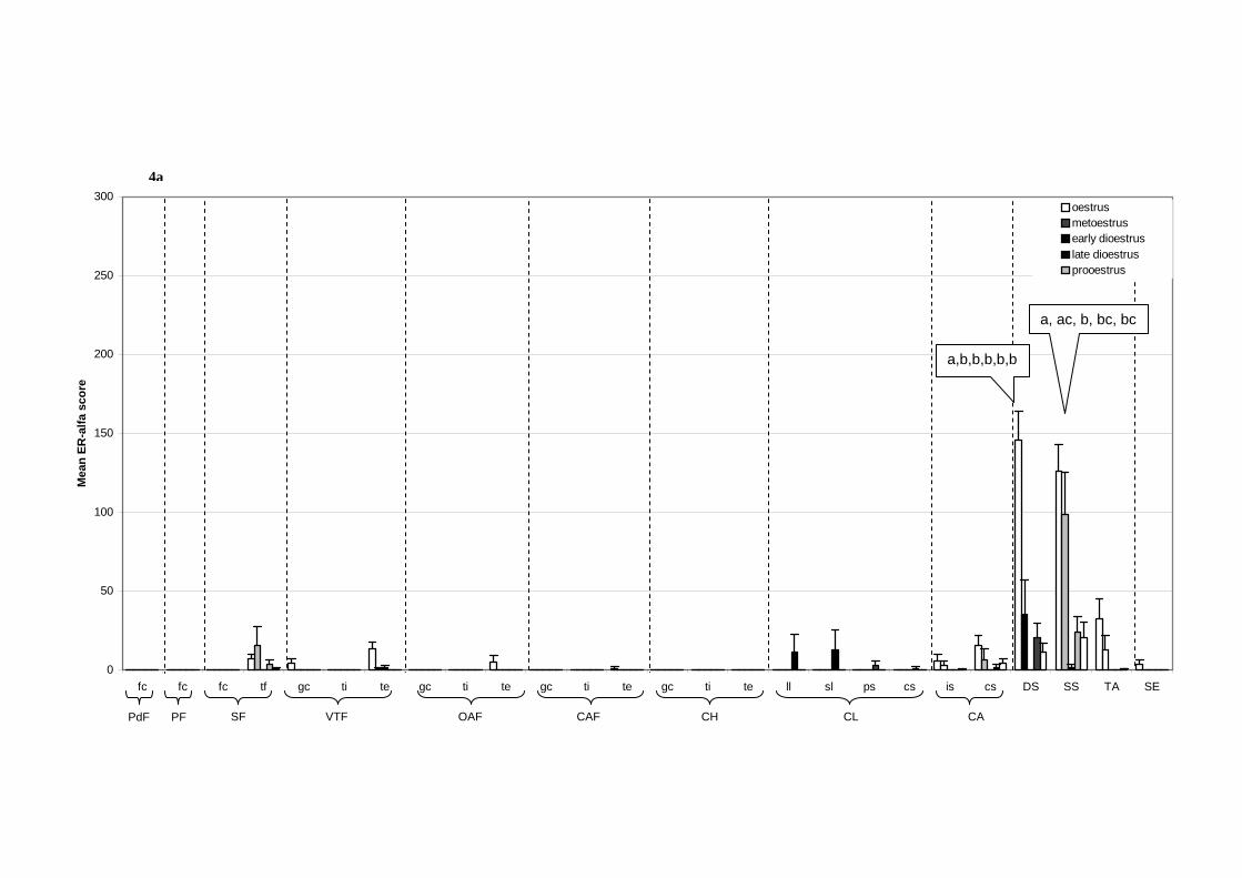
Fig. 4a ER-alfa
0
50
100
150
200
250
300
fc fc fc tf gc ti te gc ti te gc ti te gc ti te ll sl ps cs is cs DS SS TA SE
Mea
n ER
-alfa
sco
re
oestrusmetoestrusearly dioestruslate dioestrusprooestrus
SF VTF OAF CAF CH CL CA PdF
PF
a,b,b,b,b,b
a, ac, b, bc, bc
4a

Fig. 4b ER-beta
0
50
100
150
200
250
300
fc fc fc tf gc ti te gc ti te gc ti te gc ti te ll sl ps cs is cs DS SS TA SE
Mea
n ER
-bet
a sc
ore
oestrusmetoestrusearly dioestruslate dioestrusprooestrus
SF VTF OAF CAF CH CL CA PdF
PF
Fig. 4: Mean scores for estrogen receptors in various bovine ovarian cell types at different stages of the estrous cycle. Fig. 4a: Mean scores for ERα in the various cell types of the
bovine ovary at different cycle stages. Fig. 4b: Mean scores for ERβ mRNA in the various cell types of the bovine ovary at different cycle stages. Abbreviations: See fig. 2.
Significant differences between the cycle stages were observed in the deep and superficial stroma for ERα and are indicated by different superscripts.
4b

Chapter 1.1.2 77

78 Chapter 1.1.2
Fig. 5: Micrographs of different bovine ovarian cell types at various estrous stages showing immunostaining
of ERα (5a-c) and ERβ mRNA (5d-k). Fig. 5a: Outer region of the ovarian cortex during oestrus with low
scores for ERα (SERα) in the surface epithelium (1), higher scores in the superficial stroma (3), and no
expression in the tunica albuginea (2) nor in the primary follicles (4) (Bar = 100 µm). Fig. 5b: Secondary
follicle during prooestrus showing no expression for ERα in the granulosa cells (5), low SERα in the theca
cells (6) and high SERα in cells of the superficial stroma (7) (Bar = 100 µm). Fig. 5c: Overview of a corpus
albicans during oestrus with low SERα in the internal stroma cells (8) and higher SERα in the capsular
stroma cells (9) (Bar = 50 µm). Fig. 5d: Detail of the outer region of the bovine ovarian cortex during early
dioestrus with moderate scores for ERβ mRNA (SERβ mRNA) in cells of the surface epithelium (10),
higher scores in the tunica albuginea (11) and moderate scores in the superficial stroma (12) (Bar = 100
µm). Fig. 5e: Primary (13) and secondary (14) follicles at oestrus with moderate SERβ mRNA in the follicle
cells and high scores in the superficial stroma (15) (Bar = 50 µm). Fig. 5f: Vital tertiary follicle at
prooestrus with high SERβ mRNA in the granulosa cells (18) and cells of the cumulus oophorus (17). The
oocyte (16) is surrounded by the zona pellucida (Bar = 100 µm). Fig. 5g: Vital tertiary follicle at early
dioestrus with low to moderate SERβ mRNA in the granulosa layer (19), higher scores in the theca interna
(20) and still higher scores in the theca externa (21) (Bar = 100 µm). Fig. 5h: Cystic atretic tertiary follicle
at metoestrus with low to moderate SERβ mRNA in the granulosa layer (22) and higher scores in the theca
interna (23) and theca externa layers (24) (Bar = 100 µm). Fig. 5i: Overview of an obliterative atretic
tertiary follicle at oestrus with low SERβ mRNA in cells of the collapsed theca interna layers (25), and
higher scores in cells that are located in the thickened basal lamina (26) and in the theca externa layers (27)
(Bar = 200 µm). Fig. 5j: High magnification of a corpus luteum at early dioestrus with low to moderate
SERβ mRNA in the lutein cells (28) (Bar = 200 µm). Fig. 5k: Overview of a corpus albicans at oestrus with
low to moderate SERβ mRNA in the internal stroma cells (29) and high SERβ mRNA in cells of the deep
stroma (30) (Bar = 100 µm).
Discussion
In this study the presence of ERα and ERβ mRNA was demonstrated in the different
bovine ovarian cell types at different stages of the estrous cycle.
The results show that both types of ER have different tissue localization patterns,
suggesting that each receptor type has a specific function.
In the follicle cells of primordial, primary and secondary follicles moderate scores for ERβ
mRNA were observed, whereas ERα was not detected. This indicates that estrogens, which
play an important role in follicular growth (Hulshof et al., 1995) and development (Manikkam
et al., 2001a), act upon the early follicular stages via ERβ. These findings are in agreement
with previous studies in the mouse (Couse et al., 1997) and dog (Hatoya et al., 2003).

Chapter 1.1.2 79
In our study, the score for ERβ mRNA was lower in the granulosa cells of vital tertiary
follicles than in the follicle cells of secondary follicles. Moreover, the score for ERβ mRNA
in vital tertiary follicles was high in prooestrus, decreased during oestrus, to increase again
during metoestrus. These observations may reflect a down-regulation of ERβ mRNA by
pituitary gonadotrophins during oestrus as earlier described in the rat ovary (Byers et al.,
1997). In contrast, low levels of ERα were localized in the granulosa and theca externa layers
of vital tertiary follicles. This is in accordance with studies of mouse (Orimo et al., 1995), rat
(Byers et al., 1997), rabbit (Iwai et al., 1991) and baboon (Billiar et al., 1992). According to
Couse et al. (1997), the presence and interaction of both receptors within vital tertiary follicles
are required for the late stage of follicular growth. Because ERα and ERβ mRNA were
expressed in the same cells, both ERα and ERβ homodimers and/or ERα/ERβ heterodimers
could be formed (Pettersson et al., 1997; Pepe et al., 2002). These homo- and heterodimers
can also have affinities for other ligands that have estrogen-like activity (Sun et al., 1999).
ERβ mRNA is localized in vital tertiary follicles with moderate levels in the granulosa cells,
higher levels in the theca interna and still higher levels in the theca externa. This observation
is consistent with reports of ERβ mRNA or protein in the pig (Slomczynska et al., 2001) and
monkey (Pelletier et al., 1999) and it indicates that ERβ has functions that are specific in the
different cell layers of tertiary follicles.
The SERβ mRNA in the granulosa cells of both obliterative and cystic atretic follicles was
nearly two times higher than in vital tertiary follicles. This suggests that the level of ERβ
mRNA in the granulosa layers of tertiary follicles depends upon the physiological status of
the follicle. Studies of ERαKO and ERβKO mice have suggested that the presence of either
ERα or ERβ is sufficient to mediate the protective effects of estrogens against atresia of
tertiary follicles (Emmen et al., 2005). Our results suggest that in cattle ERβ is not sufficient
in conferring the protective capacity of estrogens. Another explanation, which is rather
speculative, is that the score for ERβ mRNA increases when follicular atresia occurred. It is
possible that other factors or another ER, maybe the recently detected ERγ, contributed to the
protective capacity of estrogens in bovine tertiary follicles.
In our study both ERα and ERβ mRNA were demonstrated in the corpora lutea. This
observation is in accordance with studies of mouse (Rosenfeld et al., 2001), rat (Wang et al.,
2000) and pig (Slomczynska et al., 2001). The presence of ERα and ERβ mRNA in the corpus
80 Chapter 1.1.2
luteum is suggestive of their involvement in the process of maintenance and luteinization of
the corpus luteum (Rosenfeld et al., 2001).
In all cell types of the corpus luteum ERβ mRNA had a similar localization pattern with
increasing levels after early dioestrus, while immunostaining of ERα was exclusively
observed during late dioestrus. These findings are suggestive of an up-regulation of both types
of ER by progesterone. It is of interest that the enhanced secretion of progesterone in the
corpus luteum only leads to up-regulation of ERs, whereas the progesterone receptors remain
unaltered (D'Haeseleer et al., 2006c). The simultaneous presence of ERα and ERβ mRNA in
the corpus luteum during dioestrus may lead to heterodimerisation and possible involvement
of ERα/β heterodimers in corpus luteum regression.
High scores for ERβ mRNA and relatively high amounts of ERα positive cells were found
in cells of the deep stroma and superficial stroma. Generally, an up-regulation of ERα and
ERβ mRNA was obvious around the time of oestrus. This may account for specific
compositional changes of the stromal cells surrounding the follicular structures during
oestrus, which has also been described in a study of estrogen receptors in the bovine oviduct
(Ulbrich et al., 2003). Furthermore, the high scores for ERα in the superficial stroma cells
which are in direct contact with the follicles and in the deep stroma cells which surround the
corpora albicantia are suggestive of possible stromal-epithelial interactions as reviewed by
Cunha et al. (2004).
For all ovarian structures the score for ERβ mRNA was higher in inactive than in active
ovaries, except during the early follicular stages where the highest levels for ERβ mRNA
were seen in the active ovaries. This implicates a cell specific involvement of ERβ mRNA in
the development of follicles in the active ovary.
Although the biological significance of ERα and ERβ has not yet been fully established,
the different tissue localization patterns and the cyclic alterations in the bovine ovarian tissues
suggest a specific cellular function of each receptor type. This is in agreement with the study
of Kuiper et al. (1997) who proved by analysis of mRNA distribution that ERβ expression
does not exactly match with the distribution of ERα, although colocalization of both types of
ER can occur. Further studies are needed to elucidate the precise role of the different types of
estrogen receptors, including ERγ, in the different organs and in particular in the ovary.

Chapter 1.1.2 81
References
Berisha B, Pfaffl MW, Schams D. Expression of estrogen and progesterone receptors in the bovine ovary
during estrous cycle and pregnancy. Endocrine 2002; 17: 207-214.
Billiar RB, Loukides JA, Miller MM. Evidence for the presence of the estrogen receptor in the ovary of the
baboon (Papio anubis). Journal of Clinical Endocrinology & Metabolism 1992; 75: 1159-1165.
Boos A, Meyer W, Schwarz R, Grunert E. Immunohistochemical assessment of oestrogen receptor and
progesterone receptor distribution in biopsy samples of the bovine endometrium collected throughout the
oestrous cycle. Animal Reproduction Science 1996; 44: 11-21.
Byers MG, Kuiper GGJM, Gustafsson J-Å, Park-Sarge O-K. Estrogen receptor-β mRNA expression in rat
ovary: down-regulation by gonadotrophins. Molecular Endocrinology 1997; 11: 172-182.
Coppens MT, Dhont MA, De Boever JG, Serreyn RF, Vandekerkhove DA, Roels HJ. The distribution of
oestrogen and progesterone receptors in the human endometrial basal and functional layer during the normal
menstrual cycle. Histochemistry 1993; 99: 121-126.
Couse JF, Hewitt SC, Bunch DO, Sar M, Walker VR, Davis BJ, Korach KS. Postnatal sex reversal of the
ovaries in mice lacking estrogen receptors alpha and beta. Science 1999; 286: 2328-2331.
Couse JF, Lindzey J, Grandien K, Gustafsson J-Ǻ, Korach KS. Tissue distribution and quantitative analysis
of estrogen receptor-α (ERα) and estrogen receptor-β (ERβ) messenger ribonucleic acid in the wild-type and
ERα knockout mouse. Endocrinology 1997; 138: 4613-4621.
Cunha GR, Cooke PS, Kurita T. Role of stromal-epithelial interactions in hormonal responses. Archives of
Histology and Cytology 2004; 67: 417-434.
Dall'Aglio C, Ceccarelli P, Gargiulo M, Pendine V, Monaci M. Immunohistochemical localization of
endometrial oestrogen and progesterone receptors in the cow. Anatomia Histologia Embryologia 1999; 28:
375-377.
Dellman HD, Eurell JA. Textbook of Veterinary Histology. Fifth edition Williams & Wilkins, London 1998;
pp. 252.
D'Haeseleer M, Van Poucke M, Van den Broeck W. Cell-specific localization of estrogen receptor beta
(ESR2) mRNA within various bovine ovarian cell types using in situ hybridization. Anatomia Histologia
Embryologia 2005; 34: 265-272.

82 Chapter 1.1.2
D'Haeseleer M, Cocquyt G, Van Cruchten S, Simoens P, Van den Broeck W. Cell specific localization of
apoptosis in the bovine ovary at different stages of the estrous cycle. Theriogenology 2006a; 65: 757-772.
D'Haeseleer M, Simoens P, Van den Broeck W. Cell specific localization of progesterone receptors in the
bovine ovary at different stages of the estrous cycle. Animal Reproduction Science 2006c; accepted.
Emmen JMA, Couse JF, Elmore SA, Yates MM, Kissling GE, Korach KS. In vitro growth and ovulation of
follicles from ovaries of estrogen (ER)-α and ERβ null mice indicate a role for ERβ in follicular maturation.
Endocrinology 2005; 146: 2817-2826.
Enmark E, Pelto-Huikko M, Grandien K, Lagercrantz S, Lagercrantz J, Fried G, Nordenskjöld M, Gustafsson
J-Å. Human estrogen receptor β-gene structure, chromosomal localization, and expression pattern. Journal of
Clinical Endocrinology & Metabolism 1997; 82: 4258-4265.
Enmark E, Gustafsson J-Ǻ. Oestrogen receptors - an overview. Journal of General Internal Medicine 1999;
246: 133-138.
Hatoya S, Torri R, Kumagai D, Sugiura K, Kawate N, Tamada H, Sawada T, Inaba T. Expression of estrogen
receptor α and β genes in the mediobasal hypothalamus, pituitary and ovary during the canine estrous cycle.
Neuroscience Letters 2003; 347: 131-135.
Hawkins MB, Thornton JW, Crews D, Skipper JK, Dotte A, Thomas P. Identification of a third distinct
estrogen receptor and reclassification of estrogen receptors in teleosts. Proceedings of the National Academy
of Sciences of the United States of America 2000; 97: 10751-10756.
Heard DJ, Norby PL, Holloway J, Vissing H. Human ERR-gamma, a third member of the estrogen receptor-
related receptor (ERR) subfamily of orphan nuclear receptors: tissue-specific isoforms are expressed during
development in the adult. Molecular Endocrinology 2000; 14: 382-392.
Henry M, Figueiredo AEF, Palhares MS, Corijn M. Clinical and endocrine aspects of the oestrous cycle in
donkeys (Equus asinus). Journal of Reproduction and Fertility. Supplement 1987; 35: 297-303.
Hulshof SCJ, Figueiredo JR, Beckers JF, Beyers MM, van der Donk JA, Van den Hurk R. Effects of fetal
bovine serum, FSH, and 17β-estradiol on the culture of bovine preantral follicles. Theriogenology 1995; 44:
217-226.
Ireland JJ, Murphee RL, Coulson PB. Accuracy of predicting stages of bovine estrous cycle by gross
appearance of the corpus luteum. Journal of Dairy Science 1980; 63: 155-160.

Chapter 1.1.2 83
Iwai T, Fujii S, Nanbu Y, Nonogaki H, Konishi I, Mori T, Okamura H. Effect of human chorionic
gonadotrophin on the expression of progesterone receptors and estrogen receptors in rabbit ovarian granulose
cells and the uterus. Endocrinology 1991; 129: 1840-1848.
Jansen HT, West C, Lehman MN, Padmanabhan V. Ovarian estrogen receptor-beta (ERP) regulation: I.
Changes in ER beta messenger RNA expression prior to ovulation in the ewe. Biology of Reproduction 2001;
65: 866-872.
Krege JH, Hodgin JB, Couse JF, Enmark E, Warner M, Mahler JF, Sar M, Korach KS, Gustafsson J-Å,
Smithies O. Generation and reproductive phenotypes of mice lacking estrogen receptor beta. Proceedings of
the National Academy of Sciences of the United States of America 1998; 95: 15677-15682.
Kuiper GGJM, Enmark E, Pelto-Huikko M, Nilsson S, Gustafsson J-Ǻ. Cloning of a novel estrogen receptor
expressed in rat prostate and ovary. Proceedings of the National Academy of Sciences of the United States of
America 1996; 93: 2925-2930.
Kuiper GGJM, Carlsson B, Grandien K, Enmark E, Häggblad J, Nilsson S, Gustafsson J-Ǻ. Comparison of
the ligand binding specificity and transcript tissue distribution of estrogen receptors α and β. Endocrinology
1997; 138: 863-870.
Lorke DE, Susens U, Borgmeyer U, Hermans-Borgmeyer I. Differential expression of the estrogen receptor-
related receptor gamma in the mouse brain. Brain Research. Molecular Brain Research 2000; 77: 277-280.
Lubahn DB, Moyer JS, Golding TS, Couse JF, Korach KS, Smithies O. Alteration of reproductive function
but not prenatal sexual development after insertional disruption of the mouse estrogen receptor gene.
Proceedings of the National Academy of Sciences of the United States of America 1993; 90: 11162-11166.
Manikkam M, Bao B, Rosenfeld CS, Yuan X, Salfen BE, Calder MD, Youngquist RS, Keisler DH, Lubahn
DB, Garverick HA. Expression of the bovine oestrogen receptor-β (bERβ) messenger ribonucleic acid
(mRNA) during the first ovarian follicular wave and lack of change in the expression of bERβ mRNA of
second wave follicles after LH infusion into cows. Animal Reproduction Science 2001a ; 67: 159-169.
Manikkam M, Calder MD, Salfen BE, Youngquist RS, Keisler DH, Garverick HA. Concentrations of
steroids and expression of messenger RNA for steroidogenic enzymes and gonadotrophin receptors in bovine
ovarian follicles of first and second waves and changes in second wave follicles after pulsatile LH infusion.
Animal Reproduction Science 2001b; 67: 189-203.
Nomina Histologica. International Committee on Veterinary Histological Nomenclature, 2nd edn, Cornell,
New York 1994; pp.38.

84 Chapter 1.1.2
Orimo A, Inoue S, Ikeda K, Noji S, Muramatsu M. Molecular cloning, structure, and expression of mouse
estrogen-receptor finger protein Efp. The Journal of Biochemistry 1995; 270: 24406-24413.
Pelletier G, Labrie C, Labrie F. Localization of oestrogen receptor α, oestrogen receptor β and androgen
receptors in the rat reproductive organs. Journal of Endocrinology 2000; 165: 359-370.
Pelletier G, Luu-The V, Charbonneau A, Labrie F. Cellular localization of estrogen receptor beta messenger
ribonucleic acid in cynomolgus monkey reproductive organs. Biology of Reproduction 1999; 61: 1249-1255.
Pepe GJ, Billiar RB, Leavitt MG, Zachos NC, Gustafsson J-Ǻ, Albrecht ED. Expression of estrogen
receptors alpha and beta in the baboon fetal ovary. Biology of Reproduction 2002; 66: 1054-1060.
Pettersson K, Grandien K, Kuiper GG, Gustafsson J-Ǻ. Mouse estrogen receptor β forms estrogen response
element-binding heterodimers with estrogen receptor α. Molecular Endocrinology 1997; 11: 1486-1496.
Revelli A, Pacchioni D, Cassoni P, Bussolati G, Massobrio M. In situ hybridization study of messenger RNA
for estrogen receptor and immunohistochemical detection of estrogen and progesterone receptors in the
human ovary. Gynecological Endocrinology 1996; 10: 177-182.
Rosenfeld CS, Yuan X, Manikkam M, Calder MD, Garverick HA, Lubahn DB. Cloning, sequencing, and
localization of bovine estrogen receptor-beta within the ovarian follicle. Biology of Reproduction 1999; 60:
691-697.
Rosenfeld CS, Wagner JS, Roberts RM, Lubahn DB. Intraovarian actions of oestrogen. Reproduction 2001;
122: 215-226.
Sartori R, Haughian JM, Shaver RD, Rosa GJM, Wiltbank MC. Comparison of ovarian function and
circulating steroids in estrous cycles of Holstein heifers and lactating cows. Journal of Dairy Science 2004;
87: 905-920.
Slomczynska M, Duda M, Galas J. Estrogen receptor alpha and beta expression in the porcine ovary.
Histochemica et Cytobiologica 2001; 39: 137-138.
Sun J, Meyers MJ, Fink BE, Rajendran R, Katsenellenbogen JA, Katsenellenbogen BS. Novel ligands that
function as selective estrogens or antiestrogens for estrogen receptor-alpha or estrogen receptor-beta.
Endocrinology 1999; 140: 800-804.
Tremblay GB, Tremblay A, Copeland NG, Gilbert DJ, Jenkins NA, Labrie F, Giguere V. Cloning,
chromosomal localization and functional analysis of the murine estrogen receptor β. Molecular
Endocrinology 1997; 11: 353-365.
Chapter 1.1.2 85
Ulbrich SE, Kettler A, Einspanier R. Expression and localization of estrogen receptor α, estrogen receptor β,
and progesterone receptor in the bovine oviduct in vivo and in vitro. Journal of Steroid Biochemistry and
Molecular Biology 2003; 84: 279-289.
Van den Broeck W, Corijn M, Simoens P, Lauwers H. Cell-specific distribution of estrogen receptor-alpha in
the bovine ovary. Reproduction in Domestic Animals 2002; 37: 291-293.
Verberckmoes S, Van Soom A, Dewulf J, De Kruif A. Assessment of a new utero-tubal junction
insemination device in dairy cattle. Theriogenology 2004; 61: 103-115.
Vermeirsch H. Immunohistochemical determination of estrogen receptor-α in the canine ovary and its
relation to sex steroid hormone levels. PhD thesis, Ghent University 2001.
Wang H, Eriksson H, Sahlin L. Estrogen receptors α and β in the female reproductive tract of the rat during
the estrous cycle. Biology of Reproduction 2000; 63: 1331-1340.


1.2 Cell-specific localization of progesterone receptors in the
bovine ovary at different stages of the estrous cycle
Modified from:
CELL-SPECIFIC LOCALIZATION OF PROGESTERONE RECEPTORS IN THE BOVINE OVARY AT DIFFERENT STAGES OF THE ESTROUS CYCLE D’Haeseleer M, Simoens P, Van den Broeck W Animal Reproduction Science 2006; in press


Chapter 1.2 89
Summary
This immunohistochemical study describes the localization of progesterone receptors (PR) in
the bovine ovary of 23 cows at different stages of the estrous cycle. In primordial, primary
and secondary follicles the score for PR in the follicle cells increased progressively with the
maturation of the follicle. In vital tertiary follicles and cystic atretic follicles a moderate score
for PR was found, while in obliterative atretic follicles the score was much lower. Scores
were high in corpora hemorrhagica, low in corpora lutea and still lower in corpora albicantia.
Low PR scores were also found in the tunica albuginea and surface epithelium. Cyclic
variations of PR immunoreactivity were manifest in most ovarian tissues. Follicular scores for
PR were high in oestrus and decreased during the following stages, whereas scores in corpora
lutea cells varied according to a characteristic pattern with high levels during oestrus and
metoestrus.
The variations in the scores for PR in the different ovarian cell types suggest a cell-specific
and cycle-dependent influence of progesterone. A negative correlation was found between the
PR scores and the plasma progesterone concentration.

90 Chapter 1.2
Introduction
Progesterone, synthesized and secreted by the corpus luteum, is one of the major regulators
of the reproductive cycle in mammals. It exerts its effects on the growth and differentiation of
ovarian structures and renders the endometrium receptive to the implantation of the embryo
(Pinter et al., 1996; Dellmann and Eurell, 1998). Progesterone acts on its target tissues after
binding to a specific intracellular progesterone receptor (PR). Two functionally different
receptor isoforms (PRA and PRB) have been identified (Vegeto et al., 1993; Graham and
Clarcke, 1997; Mangal et al., 1997). An additional progesterone binding protein PRC,
detected in the human breast cancer cell line T47D, may inhibit the activity of both PRA and
PRB in the presence of progesterone (Wei and Minner, 1994).
The presence of PR has been demonstrated in the ovary of a number of species, viz. in man
(Iwai et al., 1990), primates (Hild-Petito et al., 1988), cattle (Van den Broeck et al., 2002), pig
(Slomczynska et al., 2000), rabbit (Iwai et al., 1991a), dog (Vermeirsch et al., 2001) and
mouse (Lydon et al., 1995).
Although the distribution of PR has already been studied in the bovine ovary, it is not well
known whether the localization of PR varies during the estrous cycle stages. Therefore, the
aim of the present study was to determine the morphological localization of PR (PRA and
PRB) in various cell types of the bovine ovary at the different stages of the estrous cycle.
Putative differences between the scores of receptor immunoreactivity in active and inactive
ovaries were also examined. Furthermore, the correlation between the plasma progesterone
level and the receptor immunopresence in the different ovarian cell types was determined.
Materials and methods
Animals and samples
Samples of both ovaries and blood were obtained from 23 cows at the moment of slaughter
in a local slaughterhouse. All animals had been used in our previous study on the localization
of the estrogen receptors in the bovine ovary (D'Haeseleer et al., 2006b). The cows were
classified in 5 groups: 6 animals were in oestrus (synchronized cows), 2 animals in metoestrus

Chapter 1.2 91
(slaughterhouse cows), 5 animals in early dioestrus (4 slaughterhouse cows and one
synchronized cow), 5 animals in late dioestrus (slaughterhouse cows) and 5 animals in
prooestrus (slaughterhouse cows) (Fig. 1).
0,0
0,5
1,0
1,5
2,0
2,5
3,0
3,5
4,0
4,5
oestrus (n=6)
metoestrus (n=2)
early dioestrus(n=5)
late dioestrus (n=5)
prooestrus (n=5)
Plas
ma
prog
este
rone
con
cent
ratio
n (n
g/m
l)
Fig. 1: Plasma progesterone concentrations of 23 cows in various stages of the estrous cycle.
For immunohistochemical determination of PR the ovarian tissue samples were fixed for 6
hours in 3.5% phosphate-buffered formaldehyde (pH 6.7) at room temperature and
dehydrated in increasing concentrations of ethanol. Subsequently, all tissue samples were
embedded in paraffin and 5 µm thin sections were cut. The sections were mounted on 3-
aminopropyl-triethoxysilane-coated slides (APES, Sigma, St. Louis, MO, USA) and dried
overnight at 37°C.
Immunohistochemical detection of PR
The presence of PR was determined immunohistochemically as described by Van den
Broeck et al. (2002) by means of a monoclonal mouse-anti-human-PR antibody (clone 10A9,
ImmunoTech, Marseille, France) which binds to both PRA and PRB. Rabbit anti-mouse
antibodies conjugated to biotin (DAKO, Prosan, Merelbeke, Belgium) were used as secondary
92 Chapter 1.2
antibodies and peroxidase-conjugated streptavidine as enzyme label. Reaction of DAB
chromogen substrate (Liquid DAB+, DAKO, Prosan, Merelbeke, Belgium) with peroxidase
resulted in a brown nuclear staining. Positive and negative controls were included in every
staining procedure. Positive controls were bovine ovarian tissues known to be positive for PR,
whereas similar tissue sections incubated with dilution buffer instead of the primary antibody,
biotinylated secondary antibody, and/or peroxidase-conjugated streptavidine served as
negative controls.
Microscopic analysis
In each ovary the different follicle types, corpora hemorrhagica, corpora lutea, corpora
albicantia, deep stroma, superficial stroma, tunica albuginea and surface epithelium were
evaluated. All follicles were examined and classified by histological stage into primordial,
primary, secondary and tertiary follicles according to the criteria listed in the Nomina
Histologica (1994). Tertiary follicles containing less than 1 % apoptotic cells were classified
as vital tertiary follicles, whereas follicles containing more than 1% apoptotic cells were
regarded as atretic (D'Haeseleer et al., 2006a). The atretic follicles were further classified as
obliterative or cystic according to the criteria described by Dellmann and Eurell (1998). For
each cell type the presence of PR was assessed by counting the cells presenting a brown
nuclear staining.
At least 100 cells of every cell type were counted on digital micrographs taken randomly at
magnification 600 x with an Olympus DP 50 digital camera mounted on an Olympus BX61
light microscope (Olympus Belgium N.V., Aartselaar, Belgium). If too many cells were
present in one micrograph, cells were counted in 4 grids of 2500 µm² that were projected
randomly over the micrograph. Staining intensity scores ranged from 0 (no staining), 1 (weak
staining), 2 (moderate staining) to 3 (strong staining). The immunopresence of PR was
evaluated using a score (SPR) calculated according to the formula modified after Boos et al.
(1996): SPR= 0 n0 + 1 n1+ 2 n2 + 3 n3 with n0, n1, n2 and n3 are the percentage of cells
exhibiting a staining intensity 0, 1, 2 and 3, respectively.

Chapter 1.2 93
Statistical analysis
Throughout the study, results were represented as mean values and variation was expressed
as standard error of the mean (SEM). Possible differences in PR score between active and
inactive ovaries, between the various cell types and between the 5 stages of the estrous cycle
were analysed by means of the Kruskall-Wallis test. The Tukey's test was applied to
determine significant (P < 0.05) or highly significant (P < 0.01) differences between
particular cycle stages.
The Spearman rank correlation test was used for determination of correlations between the
plasma progesterone levels and the scores for PR. Statistical analyses were performed with
procedures available in SPSS 12.0.
Results
Localization of PR in the bovine ovary (Fig. 2a)
Immunoreactivity of PR was detected in the primordial, primary, secondary and tertiary
vital follicles, in cystic and obliterative atretic follicles, in corpora hemorrhagica, corpora
lutea and corpora albicantia, in cells of the deep and superficial stroma, and in the tunica
albuginea and surface epithelium.
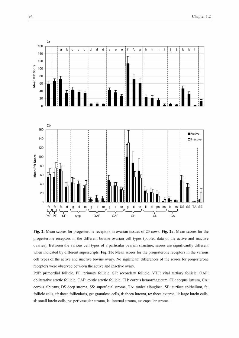
94 Chapter 1.2
2a
0
20
40
60
80
100
120
140
160
Mea
n PR
Sco
re
2b
0
20
40
60
80
100
120
140
160
fc fc fc tf g ti te g ti te g ti te g ti te ll sl ps cs is cs DS SS TA SE
Mea
n PR
Sco
re
Active
Inactive
SF
VTDF OAF CAF CH CL CA
a b c c c d d d e e e f fg g h h h i j j k k l
PF PdF VTF
Fig. 2: Mean scores for progesterone receptors in ovarian tissues of 23 cows. Fig. 2a: Mean scores for the
progesterone receptors in the different bovine ovarian cell types (pooled data of the active and inactive
ovaries). Between the various cell types of a particular ovarian structure, scores are significantly different
when indicated by different superscripts. Fig. 2b: Mean scores for the progesterone receptors in the various
cell types of the active and inactive bovine ovary. No significant differences of the scores for progesterone
receptors were observed between the active and inactive ovary.
PdF: primordial follicle, PF: primary follicle, SF: secondary follicle, VTF: vital tertiary follicle, OAF:
obliterative atretic follicle, CAF: cystic atretic follicle, CH: corpus hemorrhagicum, CL: corpus luteum, CA:
corpus albicans, DS deep stroma, SS: superficial stroma, TA: tunica albuginea, SE: surface epithelium, fc:
follicle cells, tf: theca follicularis, gc: granulosa cells, ti: theca interna, te: theca externa, ll: large lutein cells,
sl: small lutein cells, ps: perivascular stroma, is: internal stroma, cs: capsular stroma.

Chapter 1.2 95
Cell-specific variation of the PR expression (Fig. 2a)
Many differences were observed in the scores for PR of the different ovarian cell types.
The PR immunopresence in the follicle cells of primordial, primary and secondary follicles
was high and increased from primordial to secondary follicles (P < 0.05), whereas the
presence of PR in granulosa cells of vital tertiary follicles was clearly lower (P < 0.05). The
score for PR in the granulosa cells of cystic atretic tertiary follicles was similar to that of the
granulosa cells of vital tertiary follicles, whereas the score in the granulosa cells of
obliterative atretic follicles was much lower (P < 0.01). Granulosa cells of corpora
hemorrhagica showed a very high PR immunoreactivity. In the subsequent stage of corpora
lutea the score for PR decreased in both the large and small lutein cells (P < 0.01) and reached
the lowest level in internal stroma cells of corpora albicantia. In general, the score for PR in
theca cells was lower than in the follicle/granulosa cells of the corresponding structures, and
this difference was significant in secondary follicles (P < 0.01) and corpora hemorrhagica (P
< 0.05). Furthermore, theca externa cells always showed a lower score compared to theca
interna cells. The immunostaining for PR was moderate in cells of the deep stroma, whereas
superficial stroma cells had a lower staining intensity, nearly similar to that of the theca cells
in secondary and vital tertiary follicles. The surface epithelium showed a low score for PR,
while in the tunica albuginea the score was lower than in any other ovarian structure (P <
0.01).
Ovary-specific variation of the PR expression (Fig. 2b)
Comparison between active and inactive ovaries revealed no significant differences,
although the PR immunoreactivity in the majority of the ovarian cell types was slightly higher
in the active than in the inactive ovaries.
Cycle-specific variation of the PR expression (Fig. 2c, Fig. 3)
High variations in the PR immunoreactivity, concomitant with the bovine cycle stages,
were observed in the different ovarian cell types. In primordial, primary and secondary
follicles the score for PR was highest in oestrus, decreased during metoestrus, reached the
lowest levels in early dioestrus, and increased again to moderate scores during the subsequent
period of dioestrus and prooestrus. These differences between the various cycle stages were

96 Chapter 1.2
significant in the follicle cells (P < 0.05) and theca cells (P < 0.01) of secondary follicles. A
similar staining pattern was seen in the granulosa and theca layers of vital tertiary follicles,
although the PR immunoreactivity in theca interna and theca externa cells was slightly lower
in prooestrus than in late dioestrus.
In obliterative atretic tertiary follicles the PR immunostaining in granulosa and theca cell
was low in oestrus and metoestrus, and minimal in the other stages. In cystic atretic tertiary
follicles the granulosa and theca cells showed high levels during oestrus and the score for PR
decreased during the next stages in granulosa cells, whereas in the theca layers this score
decreased in metoestrus and early dioestrus to increase again during late dioestrus and
prooestrus (P < 0.05).
The corpora hemorrhagica, which were only observed during oestrus, metoestrus and early
dioestrus, showed a higher PR immunostaining than any other ovarian cell type.
In both the large and small lutein cell types of corpora lutea the score for PR was moderate
at oestrus, increased during metoestrus and decreased during the subsequent cycle stages.
Perivascular stroma cells showed a similar staining pattern, whereas in the capsular stroma
the cyclic differences were minimal. In corpora albicantia the score for PR was very low and
little variation was seen between the different cycle stages.
In both deep and superficial stroma, the PR immunostaining was high during oestrus and
metoestrus, very low during early dioestrus and higher again in the subsequent stage of
dioestrus and in prooestrus. The scores for PR were low in the surface epithelium and in the
tunica albuginea, with minimal cyclic variations in the latter structure. In general, the
correlation between the plasma progesterone levels and the PR score was negative for all
ovarian cell types (Table 1). These correlations were highly significant (P < 0.01) in the
obliterative follicles, the surface epithelium and the internal stroma cells of the corpora
albicantia.

Fig. 2c
0
50
100
150
200
250
fc fc fc tf gc ti te gc ti te gc ti te gc ti te ll sl ps cs is cs DS SS TA SE
Mea
n PR
sco
re
oestrusmetoestrusearly dioestruslate dioestrusprooestrus
a,ac,bcd,a,ad*
a,ac,bcd,ad,a*
a,ac,bcd,ad,a*
a,ab,b,b,ab**
a,ac,bcd,ad,a*
PdF PF SF VTF OAF CAF CH CL CA
Fig. 2c: Mean scores for progesterone receptors in various bovine ovarian cell types during the different estrous cycle stages. Abbreviations: See fig. 2. Between the various cell
types of a particular ovarian structure, PR scores are significantly different (* P < 0.05; ** P < 0.01) when indicated by different superscripts.

98 Chapter 1.2
Plasma Progesterone Level follicle cells - 0,34 Secondary Follicle theca follicularis - 0,52*
granulosa cells - 0,13 theca interna - 0,06 Vital Tertiary Follicle theca externa - 0,31 granulosa cells - 0,71**
theca interna - 0,55**Obliterative Atretic Follicle theca externa - 0,56**
granulosa cells - 0,24 theca interna - 0,39 Cystic Atretic Follicle theca externa - 0,46*
granulosa cells - 0,44 theca interna + 0,04 Corpus Hemorrhagicum theca externa - 0,12 large lutein cells - 0,40 small lutein cells - 0,38 perivascular stroma cells - 0,23
Corpus Luteum
capsular stroma cells - 0,16 internal stroma cells - 0,62**
Corpus Albicans capsular stroma cells - 0,24 Deep Stroma - 0,09 Superficial Stroma - 0,51*
Tunica Albuginea - 0,25 Surface Epithelium - 0,56 **
Table 1: Correlations between the plasma progesterone levels and the progesterone receptor scores in
the different ovarian cell types. * P < 0.05; ** P < 0.01
Fig. 3: Micrographs of different bovine ovarian cell types showing PR expression (arrows). Fig. 3a: Primary (1)
and early secondary (2) follicles at late dioestrus show immunoreaction for PR in the follicle cells (Bar = 50
µm). Fig. 3b: Secondary follicle at early dioestrus with low scores for PR (SPR) in the granulosa cells (3) and in
the theca cells (4) (Bar = 50 µm). Fig. 3c: Overview of a vital tertiary follicle at prooestrus with high SPR in
cells of the cumulus oophorus (5), granulosa layer (6), theca interna (7) and theca externa (8) (Bar = 200 µm).
Fig. 3d: Cystic atretic tertiary follicle at oestrus with high SPR in apoptotic granulosa cells (9) and in theca
interna cells (10), and moderate scores in cells of the theca externa (11) (Bar = 50 µm). Fig. 3e: Overview of an
obliterative atretic tertiary follicle at oestrus with low SPR in cells of the collapsed theca interna layer (12) and in
cells that are located in the thickened basal lamina (13) (Bar = 200 µm). Fig. 3f: High magnification of corpus
luteum lutein cells (14) during prooestrus with nuclear and cytoplasmic staining. (Bar = 50 µm). Fig. 3g:
Overview of a corpus albicans at oestrus with low SPR in the internal stroma cells (15) (Bar = 50 µm). Fig. 3h:
Cells of the deep stroma (16) at late dioestrus showing high SPR (Bar = 50 µm). Fig. 3i: Outer region of the
ovarian cortex during oestrus showing high immunoreactivity for PR in cells of the surface epithelium (17), low
scores in the tunica albuginea cells (18) and high scores in the superficial stroma (19) (Bar = 50 µm).

Chapter 1.2 99
100 Chapter 1.2
Discussion
The present study confirms previous data on the localization of PR in the bovine ovarian
structures (Van den Broeck et al., 2002) and additionally shows that the various ovarian cell
types exhibit different patterns of PR immunoreactivity during the estrous cycle. In the
follicle cells of primordial, primary and secondary follicles the scores for PR were high and
increased from primordial to secondary follicles. These data are in accordance with findings
in primates (Hild-Pepito et al., 1988) and dogs (Vermeirsch et al., 2001), and they indicate
that progesterone may regulate follicular growth during the early stages of follicular
development.
Studies in the monkey ovary (Hild-Petito et al., 1988) showed that PR was exclusively
located in the theca layers of secondary and tertiary follicles. In contrast, in our study PR
immunoreactivity was observed in the different cell types of secondary and tertiary follicles,
viz. the follicle/granulosa cells, theca interna and theca externa cells. Moreover, the scores for
PR in the theca layers of secondary and tertiary follicles were similar to the scores of the
surrounding superficial stroma cells. These data indicate that progesterone may regulate
follicular development in the bovine ovary not only via granulosa cells, but also through theca
and stroma cells. The possibility of such a paracrine action of ovarian steroids on follicles
mediated through stromal steroid receptors has also been suggested by Revelli et al. (1996).
In all bovine follicular structures examined, the follicle/granulosa cells showed the highest
PR immunostaining during oestrus, when ovulation occurs. These results are concomitant
with earlier observations in the dog (Vermeirsch et al., 2001) and with a study on PR mRNA
in the bovine ovary (Cassar et al., 2002). The crucial role of PR in the ovulatory process has
been demonstrated in PR-deficient mice, since such mice develop large follicles but fail to
ovulate (Lydon et al., 1995). All these findings emphasize the important role of progesterone
and its receptor in the ovulation process. The expression of PR in granulosa cells in tertiary
follicles is induced by the LH surge (Hild-Petito et al., 1988; Jo et al., 2002). The induction of
PR mRNA has also been observed in macaque granulosa cells during periovulatory stages
(Chandrasekher et al., 1994) and in porcine granulosa cells cultured in vitro after LH
stimulation (Iwai et al., 1991b).
Chapter 1.2 101
The finding that the PR expression in the bovine ovarian follicles presented a cyclic pattern
that was similar in all follicle types, indicates that the role of PR in follicle development is
specific in each stage of the ovarian cycle.
The score for PR in the bovine ovarian tissues was negatively correlated with the plasma
progesterone concentrations. This finding indicates that progesterone may downregulate the
expression of PR in the bovine ovary. A similar negative feed back process has also been
described for the distribution of PR in the bovine endometrium (Boos et al., 1996).
The progesterone receptors are reported to mediate the protective effects of progesterone
against apoptosis in the granulosa cells of bovine preovulatory follicles (Quirk et al., 2004). In
our study vital tertiary and cystic atretic follicles showed nearly similar PR levels. These
observations indicate that PR is sufficient in conferring the protective effects of progesterone
in vital tertiary follicles. If this PR expression decreases, the protective influence of
progesterone is lost. As a consequence, apoptosis can be induced in the vital tertiary follicle
so that the latter degenerates into an obliterative atretic follicle. This explains the low
expression level of PR in obliterative atretic follicles. However, in cystic atretic follicles the
PR score is as high as in vital tertiary follicles. Apparently, in some cases progesterone cannot
protect vital tertiary follicles to become atretic. A possible explanation is that in those cases
the effect of pro-apoptotic factors surpasses the protective effect of progesterone.
Alternatively, the presence of receptor-binding proteins in cells of cystic atretic follicles may
result in a lower affinity of PR for progesterone than in vital tertiary follicles, so that
apoptosis is induced despite the relatively high concentration of PR. Further studies are
needed to confirm these statements.
The presence of PR in corpora lutea reflects the role of progesterone in corpus luteum
activity (Revelli et al., 1996; Rueda et al., 2000). Progesterone regulates the proliferation and
development of luteinized granulosa and theca cells in an autocrine and paracrine way
(Sasano and Suzuki, 1997). The presence of PR in all lutein cells of the bovine corpora lutea
suggests the influence of progesterone in the luteinization process (Revelli et al., 1996; Duffy
et al., 1997). In the present study, however, the PR immunostaining in the corpus luteum was
lower than in most other ovarian structures, which can be due to a negative effect of the
locally produced high levels of progesterone to the PR production.

102 Chapter 1.2
In contrast to all other ovarian cells, the lutein cells of the corpora lutea showed PR
immunostaining not only in the nuclei, but in the cytoplasm as well. It is unlikely that this
cytoplasmic staining is due to an artefact, because it was absent in all other ovarian cell types
and in the negative controls. Therefore the idea is favoured that this cytoplasmic staining
reflects the cycle-dependent localization of PR in the cytoplasm of this specific ovarian cell
type. Whether this cytoplasmic PR reflects unbound and consequently inactive receptors in
cells that produce high concentrations of progesterone remains to be elucidated.
A low but manifest PR immunoreactivity was observed in cells of the tunica albuginea and
the surface epithelium. This corresponds with a study on ovine ovaries in which it has been
suggested that cells of the ovarian surface epithelium are enzymatically involved in the
ovulatory process by the influence of progesterone and its receptors (Murdoch, 1998). Further
investigations in cattle are necessary to verify the role of PR and progesterone in the ovarian
surface epithelium.
Chapter 1.2 103
References
Boos A, Meyer W, Schwarz R, Grunert E. Immunohistochemical assessment of oestrogen receptor and
progesterone receptor distribution in biopsy samples of the bovine endometrium collected throughout the
oestrous cycle. Animal Reproduction Science 1996; 44: 11-21.
Cassar CA, Dow MPD, Pursley JR, Smith GW. Effect of the preovulatory LH surge on bovine follicular
progesterone receptor mRNA expression. Domestic Animal Endocrinology 2002; 22: 179-187.
Chandrasekher YA, Melner MH, Nagalla SR, Stouffer RL. Progesterone receptor, but not estradiol receptor,
messenger ribonucleic acid is expressed in luteinizing granulosa cells and the corpus luteum in rhesus
monkeys. Endocrinology 1994; 135: 307-314.
Dellmann HD, Eurell JA. Textbook of Veterinary Histology. Fifth edition. Williams & Wilkins, London
1998, pp. 252, 325.
D'Haeseleer M, Cocquyt G, Van Cruchten S, Simoens P, Van Den Broeck W. Cell-specific localization of
apoptosis in the bovine ovary at different stages of the estrous cycle. Theriogenology 2006a; 65: 757-772.
D'Haeseleer M, Simoens P, Van den Broeck W. Localization of estrogen receptors within various bovine
ovarian cell types at different stages of the estrous cycle. Anatomia Histologia Embryologia 2006b; in press.
Duffy DM, Wells TR, Haluska GJ, Stouffer RL. The ratio of progesterone receptor isoforms changes in the
monkey corpus luteum during the luteal phase of the menstrual cycle. Biology of Reproduction 1997; 57:
693-699.
Graham JD, Clarke CL. Physiological action of progesterone in target tissues. Endocrine Reviews 1997; 18:
502-519.
Hild-Petito S, Stouffer RL, Bremmer RM. Immunocytochemical localization of estradiol and progesterone
receptors in the monkey ovary throughout the menstrual cycle. Endocrinology 1988; 123: 2896-2905.
Iwai T, Nanbu Y, Iwai M, Taii S, Fujii S, Mori T. Immunohistochemical localization of oestrogen receptors
and progesterone receptors in the human ovary throughout the menstrual cycle. Virchows Archiv A-
Pathological Anatomy and Histopathology 1990; 417: 369-375.
Iwai T, Fujii S, Nanbu Y, Nonogaki H, Konishi I, Mori T, Okamura H. Effect of human chorionic
gonadotrophin on the expression of progesterone receptors and estrogen receptors in rabbit ovarian granulosa
cells and the uterus. Endocrinology 1991a; 129: 1840-1848.

104 Chapter 1.2
Iwai M, Yasuda K, Fukuoka M, Iwai T, Takakura K, Taii S, Nakanishi S, Mori T. Luteinizing hormone
induces progesterone receptor gene expression in cultured porcine granulosa cells. Endocrinology 1991b;
129: 1621-1627.
Jo M, Komar CM, Fortune JE. Gonadotrophin surge induce two separate increases in messenger RNA for
progesterone receptor in bovine preovulatory follicles. Biology of Reproduction 2002; 67: 1981-1988.
Lydon JP, DeMayo FJ, Funk CR, Mani SK, Hughes AR, Jr Montgomery CA, Shyamala G, Conneely OM,
O'Malley BW. Mice lacking progesterone receptor exhibit pleiotropic reproductive abnormalities. Genes &
Development 1995; 9: 2266-2278.
Mangal RK, Wiehle RD, Poindexter AN, Weigel NL. Differential expression of uterine progesterone
receptor forms A and B during the menstrual cycle. Journal of Steroid Biochemistry and Molecular Biology
1997; 63: 195-202.
Murdoch WJ. Perturbation of sheep ovarian surface epithelial cells by ovulation: evidence for roles of
progesterone and poly (ADP-ribose) polymerase in the restoration of DNA integrity. Journal of
Endocrinology 1998; 157: 503-508.
Nomina Histologica. International Committee on Veterinary Histological Nomenclature, 2nd edition,
Cornell, New York 1994, pp. 38.
Pinter JH, Deep C, Park-Sarge OK. Progesterone receptors: expression and regulation in the mammalian
ovary. Clinical Obstetrics and Gynecology 1996; 39: 424-435.
Quirk SM, Cowan RG, Harman RM. Progesterone receptor and the cell cycle modulate apoptosis in
granulosa cells. Endocrinology 2004; 145: 5033-5043.
Revelli A, Pacchioni D, Cassoni P, Bussolati G, Massobrio M. In situ hybridization study of messenger RNA
for estrogen receptor and immunohistochemical detection of estrogen and progesterone receptor in the human
ovary. Gynecological Endocrinology 1996; 10: 177-186.
Rueda BR, Hendry III IRWJ, Stormshak F, Slayden OD, Davis JS. Decreased progesterone levels and
progesterone receptor antagonists promote apoptotic cell death in bovine luteal cells. Biology of
Reproduction 2000; 62: 269-276.
Sartori R, Haughian JM, Shaver RD, Rosa GJM, Wiltbank MC. Comparison of ovarian function and
circulating steroids in estrous cycles of Holstein heifers and lactating cows. Journal of Dairy Science 2004;
87: 905-920.

Chapter 1.2 105 Sasano H, Suzuki T. Localization of steroidogenesis and steroid receptors in human corpus luteum. Seminars
in Reproductive Endocrinology 1997; 15: 345-351.
Slomczynska M, Krok M, Pierscinski A. Localization of the progesterone receptor in the porcine ovary. Acta
Histochemica 2000; 102: 183-191.
SPSS Inc. Headquarters, 11th floor, 233 S Wacher Drive, Chicago, Illinois 60606, USA.
Van den Broeck W, D'Haeseleer M, Corijn M, Simoens P. Cell-specific distribution of progesterone
receptors in the bovine ovary. Reproduction in Domestic Animals 2002; 37: 164-170.
Vegeto E, Shahbaz MM, Wen DX, Goldman ME, O’Malley BW, McDonnell DP. Human progesterone
receptor A form is a cell- and promoter-specific repressor of human progesterone receptor B function.
Molecular Endocrinology 1993; 7: 1244-1255.
Vermeirsch H, Simoens P, Corijn M, Van den Broeck W. Immunolocalization of progesterone receptors in
the canine ovary and their relation to sex steroid hormone concentrations. Reproduction 2001; 122: 73-83.
Wei LL and Miner R. Evidence for the resistance of a third progesterone receptor protein in human breast
cancer line T47D. Cancer Research 1994; 54: 340-343.


CHAPTER 2
CELL DEATH IN THE CYCLIC BOVINE OVARY
Modified from:
CELL-SPECIFIC LOCALIZATION OF APOPTOSIS IN THE BOVINE OVARY AT DIFFERENT STAGES OF THE ESTROUS CYCLE D’Haeseleer M, Cocquyt G, Van Cruchten S, Simoens P, Van den Broeck W Theriogenology 2006; 65: 757-772


Chapter 2 109
Summary
Apoptosis was localized in all ovarian cell types of 23 cows in various stages of the estrous
cycle, using the detection of active caspase-3, in situ end labelling (TUNEL) and DNA
fluorescent staining (DAPI). Very few apoptotic cells were found in primordial, primary,
secondary and vital tertiary follicles. In contrast, apoptosis in atretic tertiary follicles was
much more frequent, and high apoptotic scores were recorded when using the TUNEL
technique and lower scores with the caspase-3 assay. Cystic atretic follicles showed in general
a higher apoptotic score than obliterative atretic follicles, with intermediate to high scores in
granulosa cells and lower scores in theca cells. In corpora lutea, large and small lutein cells
had intermediate to high scores using the caspase-3 assay, and intermediate to low scores
using the TUNEL assay. Irrespective of the detection method, the scores were higher in lutein
cells than in the capsular stroma cells. In all ovarian structures examined, variations in
apoptotic scores were seen in the different cycle stages, suggesting a cycle-dependent
influence on apoptosis, although correlations with plasma progesterone concentrations were
low.
110 Chapter 2
Introduction
Apoptosis in the mammalian ovary is involved in the mechanism of both follicular atresia
and luteal regression. Follicular atresia is the phenomenon through which the ovary eliminates
those follicles that are not selected for ovulation (Tilly et al., 1991; Yang and Rajamahendran,
2000a), while degeneration of corpora lutea is essential for maintaining the estrous or
menstrual cycle (Amsterdam et al. 2003). In general, apoptotic cells are identified by cellular
shrinking, ruffling of the plasma membrane, chromatin margination and condensation of the
cytoplasm and nucleus.
Subsequently, the cells become fragmented and break up into apoptotic bodies (Van
Cruchten et al., 2003). Ovarian apoptosis has already been documented abundantly in a
number of species, viz. in human (Depalo et al., 2003), baboon (Kugu et al., 1998), mouse
(Fenwick and Hurst, 2002), rat (Kaipia and Hsueh, 1997), sheep (Jolly et al., 1997), goat
(Sharma, 2000), pig (Berardinelli et al., 2004) and cattle (Manikkam and Rajamahendran,
1997; Porter et al., 2001). Despite data concerning the frequency and cyclic changes of
apoptosis in bovine ovarian follicles (Austin et al., 2001; Van Wezel et al., 1999; Zeuner et
al., 2003) and corpora lutea (Zheng et al., 1994; Juengel et al., 1993), data concerning
apoptosis in all different ovarian cell types are still incomplete. Therefore, in this study the
presence of apoptosis in the bovine ovarian cell types was examined using three techniques,
i.e., the detection of activated caspase-3, in situ DNA end labelling (TUNEL) and a DNA
fluorescent staining (DAPI). Furthermore, putative differences between the apoptotic scores
in active and inactive ovaries were studied. Finally, possible cyclic changes of apoptosis in
the different cell types and their correlation with the plasma progesterone level were
determined.
Materials and methods
Animals and samples
Samples of both ovaries and blood were obtained from 23 cows at the moment of slaughter
at a local slaughterhouse. All animals had been used in our previous studies on the

Chapter 2 111
localization of progesterone receptor and estrogen receptors (D’Haeseleer et al., 2006b; 2006
c).
The ovarian tissue samples were fixed for 6 hours in 3.5 % phosphate-buffered
formaldehyde (pH 6.7) at room temperature. Afterwards, the samples were embedded in
paraffin and from each sample three sections (5 µm thin) were cut, mounted on 3-
aminopropyl-triethoxysilane-coated slides (APES, Sigma, St. Louis, MO, USA) and dried
overnight at 37°C. These sections were used for TUNEL, caspase-3 assay and DAPI staining.
The stage of the estrous cycle of each animal was earlier determined (D’Haeseleer et al.,
2006b). The cows were classified in 5 groups: 6 animals in oestrus (synchronized cows), 2
animals in metoestrus (slaughterhouse cows), 5 animals in early dioestrus (4 slaughterhouse
cows and one synchronized cow), 5 animals in late dioestrus (slaughterhouse cows) and 5
animals in prooestrus (slaughterhouse cows).
Analysis of DNA cleavage by TUNEL
DNA breaks occurring during the apoptotic process were detected by means of the TACS TM TdT kit (R&D systems Europe Ltd, Abingdon, UK) a TUNEL (TdT-mediated dUTP Nick-
End Labelling) assay as described by Van Cruchten (2003). DAB chromogen substrate
(Liquid DAB +, DAKO, glostrup, Denmark) was administered for 5 min at room temperature,
resulting in a brown staining. Finally, sections were counterstained with Mayer's
haematoxylin for 10 sec and mounted. In every staining procedure positive and negative
controls were included.
Immunohistochemical detection of active caspase-3
The caspase-3 technique is an immunohistochemical assay for the detection of the enzyme
caspase-3 which can be activated during the apoptotic process and which in turn eventually
activates endonucleases that cause the characteristic morphology of apoptotic cells (Van
Cruchten et al., 2003). For the detection of activated caspase-3 a polyclonal rabbit-anti-mouse
caspase-3 antibody AF835 and the Cell and Tissue Staining Kit HRP-AEC System (R&D
Systems Europe Ltd, Abingdon, UK) were used. The protocol described by Van Cruchten
(2003) was followed with slight modification. After incubation of the sections with
112 Chapter 2
Streptavidin-HRP detection solution the slides were rinsed in PBS and 50 μl 3-amino-9-
ethylcarbazole (AEC) chromogenic substrate solution was administered for 6 min at room
temperature. The reaction of AEC with peroxidase resulted into a red coloured precipitate.
Sections were counterstained with Mayer's haematoxylin for 10 sec and mounted with an
aqueous mounting medium. In every staining procedure positive and negative controls were
included.
DAPI-staining
Nuclear DNA fragmentation was assessed using a cell permeable fluorescent DNA binding
probe, viz. 4', 6-Diamidino-2-Phenylindole (DAPI, Sigma-Aldrich Inc., St. Louis, USA).
Therefore, the ovarian tissue sections were rehydrated, rinsed in PBS and incubated with
DAPI (5µg/ml PBS) for 12 min at room temperature in a humidified chamber. After
incubation, sections were rinsed in PBS, mounted in a specific mounting medium for
fluorescence microscopy (Sigma-Aldrich Inc.) and stored at 4°C until observation using an
Olympus BX61 fluorescence microscope (Olympus Belgium N.V., Aartselaar, Belgium).
Microscopic analysis
Apoptosis was examined by using all of the above-mentioned assays applied in serial
tissue sections of every ovary. In each ovary, the different follicles, corpora hemorrhagica,
corpora lutea, corpora albicantia, stroma cells, tunica albuginea and surface epithelium were
evaluated. Follicles were classified as primordial, primary, secondary and tertiary follicles
according to the criteria listed in the Nomina Histologica (1994). Tertiary follicles containing
less than 1 % apoptotic cells were regarded as vital tertiary follicles, whereas follicles
containing more than 1% apoptotic cells were classified as atretic. The latter were further
classified as obliterative or cystic according to Dellmann and Eurell (1998).
For each cell type the presence of apoptosis demonstrated by the TUNEL assay and
caspase-3 assay was determined by counting the cells presenting a brown nuclear staining or
brown apoptotic bodies, and a red cytoplasmic staining, respectively. Using the DAPI
technique strongly fluorescent apoptotic nuclei or fragmented nuclear bodies were counted.
At least 100 cells of each cell type were counted on digital micrographs taken randomly at
magnification 600 x with an Olympus DP 50 digital camera mounted on an Olympus BX61

Chapter 2 113
light microscope (Olympus Belgium N.V., Aartselaar, Belgium). If too many cells were
present in one micrograph, cells were counted in 4 grids of 2500 µm² that were projected
randomly over the micrograph.
For the TUNEL and caspase-3 assay staining intensity scores ranged from 0 (no staining),
1 (weak staining), 2 (moderate staining) to 3 (strong staining). Apoptosis was evaluated using
an apoptotic score (AS) calculated according to the formula:
AS= 0 n0 + 1 n1+ 2 n2 + 3 n3
with n0, n1, n2 and n3 are the percentage of cells exhibiting a staining intensity 0, 1, 2 and 3,
respectively (Boos et al., 1996). For the DAPI staining procedure, the apoptotic score
indicated the mean percentage of apoptotic cells for each cell type.
Statistical analysis
Throughout the study, results were represented as means and variation was expressed as
standard error of the mean (SEM). Possible differences in the AS between active and inactive
ovaries, cystic and obliterative atretic follicles, and between the various cell types were
analysed using the Kruskall-Wallis test. This test was also used to compare the apoptotic
scores in the 5 stages of the estrous cycle. The Tukey's test was used to determine between
which particular cycle stages significant differences were present (P < 0.05, P < 0.01).
The Spearman rank correlation test was used for determination of correlations between the
plasma progesterone levels and the apoptotic score (Table 1). Statistical analyses were
performed using SPSS 12.0 (SPSS Inc., Chicago, Illinois, USA).

114 Chapter 2
TUNEL caspase-3 DAPI Secondary Follicle follicle cells - - - 0,20 theca follicularis - - - Vital Tertiary Follicle granulosa cells - 0,12 + 0,01 - 0,29 theca interna - 0,43* + 0,01 + 0,23 theca externa - 0,26 - + 0,03 Obliterative Atretic Follicle granulosa cells + 0,20 + 0,01 + 0,13 theca interna + 0,06 + 0,01 - 0,14 theca externa - 0,03 - - 0,15 Cystic Atretic Follicle granulosa cells - 0,14 + 0,34 - 0,18 theca interna - 0,56 - 0,10 + 0,22 theca externa + 0,42 - 0,03 + 0,20 Corpus Hemorrhagicum granulosa cells + 0,80 + 0,95* + 0,16 theca interna + 0,36 - - theca externa + 0,00 - - Corpus Luteum large lutein cells - 0,39 + 0,20 - 0,23 small lutein cells - 0,58** + 0,18 - 0,04 perivascular stroma cells - 0,20 - 0,24 + 0,06 capsular stroma cells + 0,13 - 0,32 + 0,04 Corpus Albicans internal stroma + 0,01 - 0,21 + 0,03 capsular stroma cells + 0,09 - 0,25 + 0,16 Deep Stroma - 0,31 + 0,02 + 0,03 Superficial Stroma - 0,32 - + 0,15 Tunica Albuginea - 0,13 - 0,35 - 0,26
Surface Epithelium - 0,42* - - 0,52*
Table 1: Correlations between the plasma progesterone levels and apoptotic scores detected by various
techniques (TUNEL, caspase-3 and DAPI) in the different ovarian cell types. "-" no correlation; * P < 0.05;
** P < 0.01.
Results
Localisation of apoptosis in the bovine ovary
In all animals TUNEL and caspase-3 positive cells were completely absent in primordial,
primary and secondary follicles (Figs. 1a, 1b, 1c). DAPI-staining showed a small amount of
apoptotic cells in the secondary follicles. In vital tertiary follicles low levels of apoptotic cells
were seen for the three assays. In contrast, apoptosis was obvious in atretic tertiary follicles.
Apoptotic cells were also noticed in corpora hemorrhagica, corpora lutea and corpora
albicantia. The deep stroma, superficial stroma, surface epithelium and tunica albuginea also
contained apoptotic cells.

Chapter 2 115
Fig. 1: Mean scores of apoptosis using the TUNEL (Fig. 1a), caspase-3 (Fig. 1b) and DAPI (Fig. 1c)
assay in the different groups of bovine ovarian cell types. Within the different cell groups, apoptotic
scores are significantly different (P < 0.05) when indicated by different superscripts.
PdF: primordial follicle, PF: primary follicle, SF: secondary follicle, VTF: vital tertiary follicle, OAF:
obliterative atretic follicle, CAF: cystic atretic follicle, CH: corpus hemorrhagicum, CL: corpus
luteum, CA: corpus albicans, DS deep stroma, SS: superficial stroma, TA: tunica albuginea, SE:
surface epithelium, fc: follicle cells, tf: theca follicularis, gc: granulosa cells, ti: theca interna, te:
theca externa, ll: large lutein cells, sl: small lutein cells, ps: perivascular stroma, is: internal stroma,
cs: capsular stroma.
1a TUNEL
0
5
10
15
20
25
30
35
40
45M
ean
Apo
ptot
ic S
core
1b Caspase-3
0
5
10
15
20
25
30
35
40
45
Mea
n A
popt
otic
Sco
re
1c DAPI
0
5
10
15
fc fc fc tf gc ti te gc ti te gc ti te gc ti te ll sl ps cs is cs DS SS TA SE
Mea
n A
popt
otic
Sco
re
SF VTF OAF CAF CH CL CA PF PdF
a a b b c d d e f g h i j k l l m m n n o p p
a a b c c d e e f g h i i i j j k l m m n n n
a a b c c d e e f g g h i i j j k k l m n o o
1a
1b
1c

116 Chapter 2
Cell-specific variation
Many differences were observed in the staining scores of the different ovarian cell types
(Figs. 1a, 1b, 1c). In vital tertiary follicles low apoptotic levels were found in granulosa and
theca cells. In contrast, apoptosis was clearly present in cystic and obliterative atretic tertiary
follicles with a high apoptotic score in the granulosa cells, an intermediate score in the theca
interna and a low score in the theca externa cells. These differences were statistically
significant (P < 0.01). Comparison between cystic and obliterative atretic follicles revealed
that cystic atretic follicles showed in general higher apoptotic scores for all cell types,
irrespective of the staining procedure.
Many variations in apoptotic levels were found in the corpora hemorrhagica, corpora lutea
and corpora albicantia. In corpora hemorrhagica the TUNEL assay revealed high to
intermediate apoptotic scores with significant differences between the granulosa and theca
cells (P < 0.05), whereas the caspase-3 assay and DAPI staining revealed a lower apoptotic
score in granulosa cells and no apoptotic score in theca cells. In contrast, the large and small
lutein cells of corpora lutea showed high scores using the caspase-3 assay, and lower scores
using the TUNEL assay. Apoptosis in capsular stroma cells of corpora lutea was only
demonstrated by TUNEL assay and DAPI staining. Furthermore, all assays revealed a
significantly higher apoptotic score in lutein cells than in stroma cells (P < 0.05). In corpora
albicantia, low to intermediate scores were seen irrespective of the staining protocol.
Comparing corpora hemorrhagica, corpora lutea and corpora albicantia with each other,
higher scores were observed in corpora lutea, apart from the very high value found in the
granulosa cells of the corpora hemorrhagica when using the TUNEL assay. Deep stroma cells
showed a low apoptotic score, comparable with capsular stroma cells of corpora lutea and
theca externa cells of atretic tertiary follicles. In the superficial stroma and tunica albuginea,
the apoptotic score was significantly lower than in the deep stroma for all assays (P < 0.01).
Intermediate apoptotic scores were seen in the surface epithelium following TUNEL assay.
Comparison between the different follicular stages revealed a significant increase in
apoptotic score for follicle/granulosa cells from secondary to vital tertiary to atretic tertiary
follicles (P < 0.05).

Chapter 2 117
Ovary-specific variation
Comparison between the active and inactive ovaries revealed no significant differences
(Figs. 2a, 2b, 2c), although, the DAPI-staining showed more apoptosis in the active ovary
than in the inactive ovary for almost all cell types.
2a TUNEL
0
5
10
15
20
25
30
35
40
45
Mea
n A
popt
otic
Sco
re
Fig. 2: Mean scores of apoptosis in the different cell types of the active and inactive bovine
ovary using the TUNEL (Fig. 2a), caspase-3 (Fig. 2b) and DAPI (Fig. 2c) assay.
Abbreviations: See fig. 1.
ActiveInactive
2b Caspase-3
0
5
10
15
20
25
30
35
40
45
Mea
n A
popt
otic
Sco
re
2 I
0
5
10
15
fc fc fc tf gc ti te gc ti te gc ti te gc ti te ll sl ps cs is cs DS SS TA SE
Mea
n A
popt
otic
Sco
re
c DAP
SF VTF OAF CAF CH CL CA PF PdF
2a
2b
2c
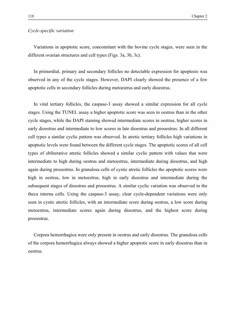
118 Chapter 2
Cycle-specific variation
Variations in apoptotic score, concomitant with the bovine cycle stages, were seen in the
different ovarian structures and cell types (Figs. 3a, 3b, 3c).
In primordial, primary and secondary follicles no detectable expression for apoptosis was
observed in any of the cycle stages. However, DAPI clearly showed the presence of a few
apoptotic cells in secondary follicles during metoestrus and early dioestrus.
In vital tertiary follicles, the caspase-3 assay showed a similar expression for all cycle
stages. Using the TUNEL assay a higher apoptotic score was seen in oestrus than in the other
cycle stages, while the DAPI staining showed intermediate scores in oestrus, higher scores in
early dioestrus and intermediate to low scores in late dioestrus and prooestrus. In all different
cell types a similar cyclic pattern was observed. In atretic tertiary follicles high variations in
apoptotic levels were found between the different cycle stages. The apoptotic scores of all cell
types of obliterative atretic follicles showed a similar cyclic pattern with values that were
intermediate to high during oestrus and metoestrus, intermediate during dioestrus, and high
again during prooestrus. In granulosa cells of cystic atretic follicles the apoptotic scores were
high in oestrus, low in metoestrus, high in early dioestrus and intermediate during the
subsequent stages of dioestrus and prooestrus. A similar cyclic variation was observed in the
theca interna cells. Using the caspase-3 assay, clear cycle-dependent variations were only
seen in cystic atretic follicles, with an intermediate score during oestrus, a low score during
metoestrus, intermediate scores again during dioestrus, and the highest score during
prooestrus.
Corpora hemorrhagica were only present in oestrus and early dioestrus. The granulosa cells
of the corpora hemorrhagica always showed a higher apoptotic score in early dioestrus than in
oestrus.
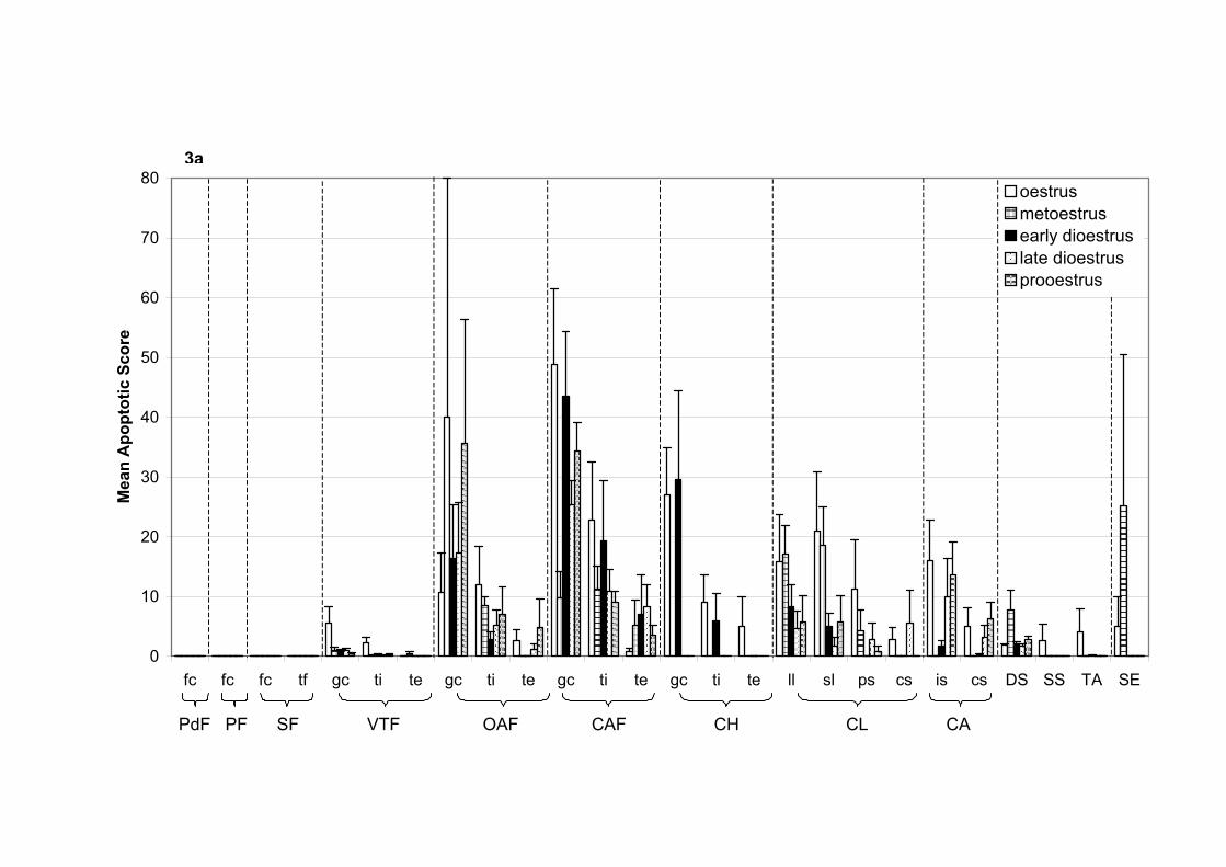
3a TU
NEL
0
10
20
30
40
50
60
70
80
fc fc fc tf gc ti te gc ti te gc ti te gc ti te ll sl ps cs is cs DS SS TA SE
Mea
n A
popt
otic
Sco
re
3a
oestrusmetoestrusearly dioestruslate dioestrusprooestrus
OAF CAF CH CL CAVTFSFPdF PF PdF PF SF VTF OAF CAF CH CL CA

3b Caspase-3
0
10
20
30
40
50
fc fc fc tf gc ti te gc ti te gc ti te gc ti te ll sl ps cs is cs DS SS TA SE
Mea
n A
popt
otic
Sco
re
3b
oestrusmetoestrusearly dioestruslate dioestrus
a b ab ab b prooestrus
PdF PF SF VTF OAF CAF CH CL CA PdF PF SF VTF OAF CAF CH CL CA
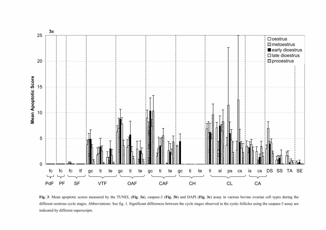
DAPI
0
5
10
15
20
25
fc fc fc tf gc ti te gc ti te gc ti te gc ti te ll sl ps cs is cs DS SS TA SE
Mea
n A
popt
otic
Sco
re
3c
oestrusmetoestrusearly dioestruslate dioestrusprooestrus
OAF CAF CH CL CAVTFSFPFPdF
Fig. 3: Mean apoptotic scores measured by the TUNEL (Fig. 3a), caspase-3 (Fig. 3b) and DAPI (Fig. 3c) assay in various bovine ovarian cell types during the
different oestrous cycle stages. Abbreviations: See fig. 1. Significant differences between the cycle stages observed in the cystic follicles using the caspase-3 assay are
indicated by different superscripts.
PdF PF SF VTF OAF CAF CH CL CA
3c

122 Chapter 2
In both the large and small lutein cells of corpora lutea, the highest apoptotic scores
following the TUNEL assay were seen in oestrus and metoestrus. These scores decreased in
early dioestrus and the subsequent period of dioestrus, and increased again in prooestrus. A
nearly similar pattern was observed in the stroma cells of corpora lutea. The caspase-3 assay
showed a different pattern, with the lowest apoptotic score in oestrus, higher scores in
metoestrus and both dioestrus stages, and the highest score in prooestrus. However, in stroma
cells the scores were extremely low in all stages. Using the DAPI staining, little variation was
seen in lutein cells, with scores that were relatively high in oestrus, lower in metoestrus and
both dioestrus stages, and high again in prooestrus. In stroma cells of corpora lutea, the scores
were low during oestrus, high during metoestrus, low again during both dioestrus stages, and
minimal during prooestrus.
In corpora albicantia, the apoptotic scores obtained by the TUNEL technique were
intermediate in oestrus, decreased in metoestrus to increase again in the next stages.
According to the caspase-3 assay and the DAPI staining the scores were low to intermediate,
with small differences between the various stages.
The apoptotic scores of deep stroma cells were low at oestrus, high in metoestrus, and low
again during the following stages. Using the TUNEL assay, apoptosis in superficial stroma
cells and tunica albuginea cells was clearly demonstrable during oestrus, and in surface
epithelial cells also at oestrus and during metoestrus (Fig. 4).
In addition to the cycle-specific variation, the correlations between plasma progesterone
levels and apoptotic scores were examined (Table 1). Positive correlations were found for the
granulosa cells of corpora hemorrhagica after using the TUNEL technique (0.80) and caspase-
3 assay (0.95, P < 0.05). Remarkably, the correlation with TUNEL scores in lutein cells of
corpora lutea was negative (-0.39 for large lutein cells and -0.58 (P < 0.01) for small lutein
cells) whereas the correlation was slightly positive following the caspase-3 assay (0.20 for
large and 0.18 for small lutein cells). In the surface epithelium, a significant and faintly
negative correlation was seen between the plasma progesterone levels and the apoptotic
scores determined by the TUNEL assay (-0.24, P < 0.05) and DAPI staining (-0.52, P < 0.05).
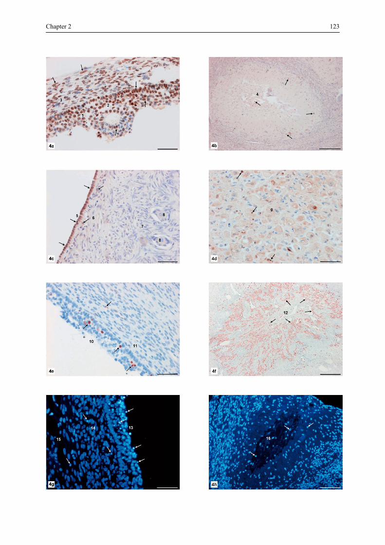
Chapter 2 123
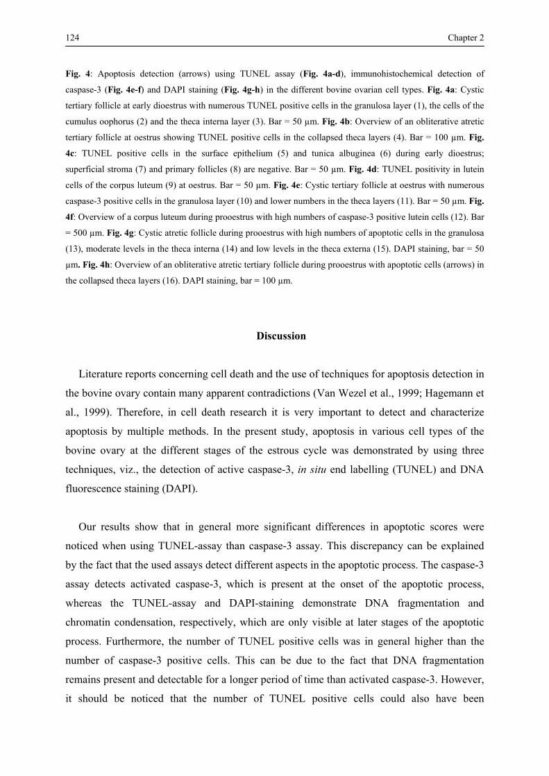
124 Chapter 2 Fig. 4: Apoptosis detection (arrows) using TUNEL assay (Fig. 4a-d), immunohistochemical detection of
caspase-3 (Fig. 4e-f) and DAPI staining (Fig. 4g-h) in the different bovine ovarian cell types. Fig. 4a: Cystic
tertiary follicle at early dioestrus with numerous TUNEL positive cells in the granulosa layer (1), the cells of the
cumulus oophorus (2) and the theca interna layer (3). Bar = 50 µm. Fig. 4b: Overview of an obliterative atretic
tertiary follicle at oestrus showing TUNEL positive cells in the collapsed theca layers (4). Bar = 100 µm. Fig.
4c: TUNEL positive cells in the surface epithelium (5) and tunica albuginea (6) during early dioestrus;
superficial stroma (7) and primary follicles (8) are negative. Bar = 50 µm. Fig. 4d: TUNEL positivity in lutein
cells of the corpus luteum (9) at oestrus. Bar = 50 µm. Fig. 4e: Cystic tertiary follicle at oestrus with numerous
caspase-3 positive cells in the granulosa layer (10) and lower numbers in the theca layers (11). Bar = 50 µm. Fig.
4f: Overview of a corpus luteum during prooestrus with high numbers of caspase-3 positive lutein cells (12). Bar
= 500 µm. Fig. 4g: Cystic atretic follicle during prooestrus with high numbers of apoptotic cells in the granulosa
(13), moderate levels in the theca interna (14) and low levels in the theca externa (15). DAPI staining, bar = 50
µm. Fig. 4h: Overview of an obliterative atretic tertiary follicle during prooestrus with apoptotic cells (arrows) in
the collapsed theca layers (16). DAPI staining, bar = 100 µm.
Discussion
Literature reports concerning cell death and the use of techniques for apoptosis detection in
the bovine ovary contain many apparent contradictions (Van Wezel et al., 1999; Hagemann et
al., 1999). Therefore, in cell death research it is very important to detect and characterize
apoptosis by multiple methods. In the present study, apoptosis in various cell types of the
bovine ovary at the different stages of the estrous cycle was demonstrated by using three
techniques, viz., the detection of active caspase-3, in situ end labelling (TUNEL) and DNA
fluorescence staining (DAPI).
Our results show that in general more significant differences in apoptotic scores were
noticed when using TUNEL-assay than caspase-3 assay. This discrepancy can be explained
by the fact that the used assays detect different aspects in the apoptotic process. The caspase-3
assay detects activated caspase-3, which is present at the onset of the apoptotic process,
whereas the TUNEL-assay and DAPI-staining demonstrate DNA fragmentation and
chromatin condensation, respectively, which are only visible at later stages of the apoptotic
process. Furthermore, the number of TUNEL positive cells was in general higher than the
number of caspase-3 positive cells. This can be due to the fact that DNA fragmentation
remains present and detectable for a longer period of time than activated caspase-3. However,
it should be noticed that the number of TUNEL positive cells could also have been

Chapter 2 125 overestimated, due to the fact that it is not always clear whether a cluster of apoptotic bodies
originated from a single cell or from different cells. Furthermore, DNA fragmentation is not
specific for apoptosis, and oncotic cells can be misinterpreted as apoptotic cells (Van
Cruchten et al., 2003). In corpora lutea, however, the number of caspase-3 positive cells was
two times higher than the number of TUNEL positive cells. This finding might indicate that
many caspase-3 positive cells disappear, probably by phagocytosis, before DNA
fragmentation occurs.
The present study demonstrates that apoptotic cells were extremely scarce in primordial,
primary and secondary follicles and very abundant in tertiary follicles in all cows at the
different cycle stages. Although it has been described that atresia may begin at any stage of
development of the follicle (Bloom and Fawcett, 1975), our findings would suggest that
atresia of tertiary follicles is of major importance for the reduction of the thousands of
follicles that will never ovulate. The very low frequency of atresia in the earlier follicular
stages has also been demonstrated in the human ovary in a recent immunohistochemical study
using TUNEL and active caspase-3 detection (Glamoclija et al., 2005). A possible
explanation for this observation could be that apoptosis in early stage follicles is caused by a
mechanism in which caspase-3 is not involved and which is not detectable by the TUNEL
assay. A more plausible reason might be the fact that for each cow in our study only a single
snapshot evaluation of ovarian structures was made. As follicular atresia starts already before
birth and continues for many years, the chance to detect atretic primordial and primary
follicles is clearly reduced when only a few histological sections are examined.
The finding of low apoptotic levels in secondary follicles is in line with a recent study on
apoptosis in human ovarian follicles, which demonstrates that, once the primary follicle
develops to a secondary follicle, the follicle cells of secondary follicles showed Bcl-2
expression and consequently were protected against apoptosis (Depalo et al., 2003). Another
study in rat ovaries demonstrated that apoptosis in the granulosa cells of early developing
follicles is prevented by progesterone (Peluso, 2003). However, in the present study no
correlations were found between apoptotic scores in secondary follicles and the plasma
progesterone concentration, indicating that progesterone acts via paracrine rather than via
endocrine route. In order to elucidate the putative influence of progesterone on secondary
126 Chapter 2 follicles, the localisation of progesterone-specific receptors in ovarian cells needs to be
investigated.
High numbers of atretic tertiary follicles were observed in all bovine ovaries at the
different cycle stages. This corresponds with the physiologic process of the bovine estrous
cycle in which only one tertiary follicle is selected for ovulation, while the remaining tertiary
follicles regress. In our study, the number of apoptotic cells in the different layers of atretic
tertiary follicles showed significant differences. These findings correspond with studies of
ovarian apoptosis in rats (Vickers et al., 2000), pigs (Tilly et al., 1992) and chicken (Tilly et
al., 1991). Cystic atretic follicles show higher apoptotic levels compared to obliterative
follicles, which can be due to their different morphology. Disruption of the intercellular
contacts in cystic atretic follicles may stimulate apoptosis (Zeuner et al., 2003). In contrast, in
obliterative follicles the granulosa and theca layers both collapse, while the cell-to-cell
contacts remain intact and the intercellular integrity inhibits the induction of apoptosis.
Therefore, apoptosis in obliterative tertiary follicles would be the result of morphological
changes during atresia, rather than the cause of follicular atresia. However, this hypothesis is
highly speculative and should be confirmed.
In many ovaries, cystic follicles with high apoptotic scores were located nearby cystic
follicles with low scores, suggesting that morphologically identical follicles showed a high
variability in apoptosis. In an earlier study we also found a variable expression of estrogen
receptor beta mRNA levels in neighbouring bovine follicles that were morphologically
identical (D’Haeseleer et al., 2005). These observations suggest that ovarian follicles are
mainly influenced by local factors produced by adjacent follicles or other ovarian cells, rather
than by systemic endocrine factors.
Earlier studies indicated that high progesterone levels trigger apoptosis in non-ovulatory
tertiary follicles during the bovine estrous cycle (Taylor et al., 1993; Yang and
Rajamahendran, 2000b). A similar observation was made in granulosa cells of obliterative
follicles in which the TUNEL assay demonstrated high apoptotic levels in early dioestrus,
when plasma progesterone concentrations are increasing. However, the apoptotic scores in
cystic follicles were always lower in early dioestrus, than in the other stages. Furthermore, no
significant correlations were found between apoptosis in tertiary follicles and plasma

Chapter 2 127 progesterone concentration. This suggests that the influence of progesterone on apoptosis in
tertiary follicles is complex and that other factors may also be involved.
In lutein cells, the apoptotic score detected by the caspase-3 assay increased from low
levels at oestrus to maximal levels at dioestrus / prooestrus, whereas the opposite was seen
using the TUNEL assay. This is in accordance with the positive and negative correlations
observed between the plasma progesterone concentrations and the caspase-3 and TUNEL
scores, respectively. This observation indicates that progesterone induces apoptosis in corpora
lutea by caspase-3 activation, whereas the DNA fragmentation occurs in a later stage,
explaining the negative correlation. This is in contrast with the results obtained from an in
vitro study on progesterone treated cultured bovine lutein cells, in which it was demonstrated
that progesterone prevents apoptosis of lutein cells (Rueda et al., 2000).
In general, apoptotic scores were low in cells of the deep stroma, superficial stroma, tunica
albuginea and surface epithelium with relative high levels during metoestrus. This indicates
that ovulation might have an influence on the regression of stroma and epithelial cells but data
to elucidate this process are still lacking.
128 Chapter 2
References
Amsterdam A, Sasson R, Keren-Tal I, Aharoni D, Dantes A, Rimon E, Land A, Cohen T, Dor Y, Hirsh L.
Alternative pathways of ovarian apoptosis: death for life. Biochemical Pharmacology 2003; 66: 1355-1362.
Austin EJ, Mihm M, Evans ACO, Knight PG, Ireland JLH, Roche JF. Alterations in intrafollicular regulatory
factors and apoptosis during selection of follicles in the first follicular wave of the bovine estrous cycle.
Biology of Reproduction 2001; 64: 839-848.
Berardinelli P, Russo V, Martelli A, Nardinocchi D, Di Giacinto O, Barboni B, Mattioli M. Colocalization of
DNA fragmentation and caspase-3 activation during atresia in pig antral follicles. Anatomia Histologia
Embryologia 2004; 33: 23-27.
Bloom W. and Fawcett W. A textbook of Histology. Tenth Edition. W.B. Saunders Company, 1975, pp. 873-
876.
Boos A, Meyer W, Schwarz R, Grunert E. Immunohistochemical assessment of oestrogen receptor and
progesterone receptor distribution in biopsy samples of the bovine endometrium collected throughout the
oestrous cycle. Animal Reproduction Science 1996; 44: 11-21.
Dellmann HD, Eurell JA. Textbook of Veterinary Histology. Fifth edition. Williams & Wilkins, London
1998, pp. 252.
Depalo R, Nappi L, Loverro G, Bettocchi S, Caruso ML, Valentini AM, Selvaggi L. Evidence of apoptosis in
human primordial and primary follicles. Human Reproduction 2003; 18: 2678-2682.
D'Haeseleer M, Van Poucke M, Van den Broeck W. Cell-specific localization of estrogen receptor beta
(ESR2) mRNA within various bovine ovarian cell types using in situ hybridisation. Anatomia Histologia
Embryologia 2005; 34: 265-272.
D'Haeseleer M, Simoens P, Van den Broeck W. Localization of estrogen receptors within various bovine
ovarian cell types at different stages of the estrous cycle. Anatomia Histologia Embryologia 2006b; in press.
D'Haeseleer M, Simoens P, Van den Broeck W. Cell specific localization of progesterone receptors in the
bovine ovary at different stages of the estrous cycle. Animal Reproduction Science 2006c; accepted.
Fenwick MA, Hurst PR. Immunohistochemical localization of active caspase-3 in the mouse ovary: growth
and atresia of small follicles. Reproduction 2002; 124: 659-665.

Chapter 2 129 Glamoclija V, Vilovic K, Saraga-Babic M, Baranovic A, Sapunar D. Apoptosis and active caspase-3
expression in human granulosa cells. Fertility and Sterility 2005; 83: 426-431.
Hagemann LJ, Beaumont SE, Berg M, Donnison MJ, Ledgard A, Peterson AJ, Schurmann A, Tervit HR.
Development during single IVP of bovine oocytes from dissected follicles: interactive effects of estrous cycle
stage, follicle size and atresia. Molecular Reproduction and Development 1999; 53: 451-458.
Jolly PD, Smith PR, Heath DA, Hudson NL, Lun S, Still LA, Watts CH, McNatty KP. Morphological
evidence of apoptosis and the prevalence of apoptotic versus mitotic cells in the membrana granulosa of
ovarian follicles during spontaneous and induced atresia in ewes. Biology of Reproduction 1997; 56: 837-
846.
Juengel JL, Garverick HA, Johnson AL, Youngquist RS, Smith MF. Apoptosis during luteal regression in
cattle. Endocrinology 1993; 132: 249-254.
Kaipia A, Hsueh AJW. Regulation of ovarian follicle atresia. Annual Review of Physiology 1997; 59: 349-
363.
Kugu K, Ratts VS, Piquette GN, Tilly KI, Tao XJ, Martimbeau S, Aberdeen GW, Krajewski S, Reed JC,
Pepe GJ, Albrecht ED, Tilly JL. Analysis of apoptosis and expression of bcl-2 gene family members in the
human and baboon ovary. Cell Death and Differentiation 1998; 5: 67-76.
Manikkam M., Rajamahendran R. Progesterone-induced atresia of the prooestrus dominant follicle in the
bovine ovary: changes in diameter, insulin-like growth factor system, aromatase activity, steroid hormones,
and apoptotic index. Biology of Reproduction 1997; 57: 580-587.
Nomina Histologica. International Committee on Veterinary Histological Nomenclature. Second edition,
Cornell, New York 1994, pp. 38.
Peluso JJ. Progesterone as a regulator of granulosa cell viability. Journal of Steroid Biochemistry and
Molecular Biology 2003; 85: 167-173.
Porter DA, Harman RM, Cowan RG, Quirk SM. Relationship of Fas ligand expression and atresia during
bovine follicle development. Reproduction 2001; 121: 561-566.
Rueda BR, Hendry IR, Hendry III WJ, Stormshak F, Slayden OD, Davis JS. Decreased progesterone levels
and progesterone receptor antagonists promote apoptotic cell death in bovine luteal cells. Biology of
Reproduction 2000; 62: 269-276.

130 Chapter 2 Sartori R, Haughian JM, Shaver RD, Rosa GJM, Wiltbank MC. Comparison of ovarian function and
circulating steroids in estrous cycles of Holstein heifers and lactating cows. Journal of Dairy Science 2004;
87: 905-920.
Sharma RK. Follicular atresia in goat: A review. Indian Journal of Animal Sciences 2000; 70: 1035-1046.
Taylor C, Rajamahendran R, Walton JS. Ovarian follicular dynamics and plasma luteinizing hormone
concentrations in norgestomet treated heifers. Animal Reproduction Science 1993; 32: 173-184.
Tilly JL, Kowalski KI, Johnson AL, Hsueh AJW. Involvement of apoptosis in ovarian follicular atresia and
postovulatory regression. Endocrinology 1991; 129: 2799-2801.
Tilly JL, Kowalski KI, Schomberg DW, Hsueh AJW. Apoptosis in atretic ovarian follicles is associated with
selective decreases in messenger ribonucleic acid transcripts for gonadotrophin receptors and cytochrome
P450 aromatase. Endocrinology 1992; 131: 1670-1676.
Van Cruchten S, Van den Broeck W, Duchateau L, Simoens P. Apoptosis in the canine endometrium during
the estrous cycle. Theriogenology 2003; 60: 1595-1608.
Van Wezel IL, Dharmarajan AM, Lavranos TC, Rodgers RJ. Evidence for alternative pathways of granulosa
cell death in healthy and slightly atretic bovine antral follicles. Endocrinology 1999; 140: 2602-2612.
Vickers SL, Cowan RG, Harman RM, Porter DA, Quirk SM. Expression and activity of the Fas Antigen in
bovine ovarian follicle cells. Biology of Reproduction 2000; 62: 54-61.
Yang MY, Rajamahendran R. Morphological and biochemical identification of apoptosis in small, medium,
and large bovine follicles and the effects of follicle-stimulating hormone and insulin-like growth factor-I on
spontaneous apoptosis in cultured bovine granulosa cells. Biology of Reproduction 2000a; 62: 1209-1217.
Yang MY, Rajamahendran R. Involvement of apoptosis in the atresia of non-ovulatory dominant follicle
during the bovine estrous cycle. Biology of Reproduction 2000b; 63: 1313-1321.
Zeuner A, Müller K, Reguszynski K, Jewgenow K. Apoptosis within bovine follicular cells and its effect on
oocyte development during in vitro maturation. Theriogenology 2003; 59: 1421-1433.
Zheng J, Fricke PM, Reynolds LP, Redmer DA. Evaluation of growth, cell proliferation and cell death in
bovine corpora lutea throughout the estrous cycle. Biology of Reproduction 1994; 51: 623-632.

CHAPTER 3
PROLIFERATION PATTERNS IN THE BOVINE OVARY AT
DIFFERENT STAGES OF THE ESTROUS CYCLE
Modified from:
PROLIFERATION PATTERNS IN THE BOVINE OVARY AT DIFFERENT STAGES OF THE ESTROUS CYCLE D’Haeseleer M, Simoens P, Van den Broeck W In preparation

Chapter 3 133
Summary
Proliferation was examined in the ovaries of 23 cows at different stages of the estrous cycle
using immunohistochemical detection of Ki-67. Ki-67 was not observed in primordial
follicles. In primary and secondary follicles the score for Ki-67 increased progressively with
the maturation of the follicle. In vital tertiary and cystic atretic follicles high to moderate
scores for Ki-67 were found, whereas in obliterative atretic follicles the score was much
lower. Scores were high in all cell types of corpora hemorrhagica and in the lutein cells of the
corpora lutea, moderate in the stroma cells of the corpora lutea and low in the cells of the
corpora albicantia. Low Ki-67 scores were also found in the deep and superficial stroma.
In vital tertiary and cystic atretic follicles the scores for Ki-67 were high at oestrus, decreased
during metoestrus, increased during dioestrus to decrease again during prooestrus. In corpora
lutea, corpora albicantia and deep stroma a different cyclic pattern was observed with high
scores during metoestrus and lower scores during dioestrus and prooestrus. These variations
suggest a cell-specific and cycle-dependent influence on cell proliferation, although
correlations with the plasma progesterone concentrations were low.

134 Chapter 3
Introduction
During the bovine estrous cycle, the ovary is subjected to extensive tissue remodelling that
consists of sequential phases of cell proliferation and cell death in the different ovarian
structures such as the follicles, the corpus hemorrhagic, the corpus luteum, the corpus
albicans, the stroma and the surface epithelium. Cell proliferation is morphological and
physiological event that is controlled by a combination of circulating steroid hormones and
locally produced growth factors (Quirk et al., 2004).
Cell proliferation in the ovary has been studied in a number of species such as human
(Funayama et al., 1996), the baboon (Wandji et al., 1997), the marmoset (Young et al., 2000),
the horse (Al-zi'abi et al., 2003), the ewe (Campbell et al., 2004; Jablonka-Shariff et al., 1993;
Jolly et al., 1997), the porcine (May et al., 1992), the dog (Spanel-Borowski a, b et al., 1984),
the rat (Gaytan et al., 1996; Richards et al., 1986; Uilenbroek et al., 1979; Oktay et al., 1995)
and the mouse (Britt et al., 2004). Many papers have been published on cell proliferation in
bovine ovarian follicles (Tajima et al., 2002; Kotsuyi et al., 1990; Isobe et al., 2000; van
Wezel, 1999) and in corpora lutea (Boos, 1998; Zheng et al., 1994).
However, the cyclic proliferation patterns in the different bovine ovarian cell types are not
well known. Therefore, the aim of the present study was to determine proliferation in the
different cell types of the bovine ovary at the different stages of the estrous cycle by using
immunohistochemical detection of Ki-67. Ki-67 is a nuclear antigen expressed by
proliferating cells and is occurring in all active parts of the cellular cycle, except in the resting
phase (G0) (Scholzen et al., 2000). Furthermore, putative differences between the scores of
Ki-67 in active and inactive ovaries were also examined. Finally, the correlation between the
plasma progesterone level and proliferation in the different ovarian cell types was determined.
Chapter 3 135
Materials and methods
Animals and samples
Samples of both ovaries and blood were obtained from 23 cows at the moment of slaughter
at a local slaughterhouse. All animals had been used in our previous study on apoptosis in the
bovine ovary (D'Haeseleer et al., 2005).
For immunohistochemical determination of PR the ovarian tissue samples were fixed for 6
h in 3.5% phosphate-buffered formaldehyde (pH 6.7) at room temperature and dehydrated in
increasing concentrations of ethanol. Subsequently, all tissue samples were embedded in
paraffin and 5 µm thin sections were cut. The sections were mounted on 3-aminopropyl-
triethoxysilane-coated slides (APES, Sigma, St. Louis, MO, USA) and dried overnight at
37°C.
The stage of the estrous cycle of each animal was earlier determined (D’Haeseleer et al.,
2006b). The cows were classified in 5 groups: 6 animals in oestrus (synchronized cows), 2
animals in metoestrus (slaughterhouse cows), 5 animals in early dioestrus (4 slaughterhouse
cows and one synchronized cow), 5 animals in late dioestrus (slaughterhouse cows) and 5
animals in prooestrus (slaughterhouse cows).
Immunohistochemical detection of Ki-67
For the detection of Ki-67, the assay described by Van Cruchten et al. (2004) was used.
The primary antibody was a monoclonal mouse-anti-human NCL-Ki-67-MM1 antibody
(Novocastra Laboratories Ltd., Newcastle upon Tyne, UK) which cross-reacts with the bovine
Ki-67. Rabbit anti-mouse antibodies conjugated to biotin (DAKO A/S, Glostrup, Denmark)
were used as secondary antibodies and peroxidase-conjugated streptavidine (DAKO A/S,
Glostrup, Denmark) as enzyme label. DAB (Liquid DAB+, DAKO, Prosan, Merelbeke,
Belgium) was used as chromogen substrate, resulting in a brown nuclear precipitate. Mayer's
haematoxylin was applied as a nuclear counterstain.
Positive and negative controls were included in every staining procedure. Canine uterine
tissues known to be positive for Ki-67 served as positive controls. As negative controls tissue

136 Chapter 3
sections incubated with dilution buffer instead of the primary antibody, secondary
antibody, and/or peroxidase-conjugated streptavidine were used.
Microscopic analysis
In each ovary the different follicle types, corpora hemorrhagica, corpora lutea, corpora
albicantia, deep stroma, superficial stroma, tunica albuginea and surface epithelium were
microscopically evaluated. All follicles were examined and classified by histological stage
into primordial, primary, secondary and tertiary follicles according to the criteria listed in the
Nomina Histologica (1994). Tertiary follicles containing less than 1 % apoptotic cells were
classified as vital tertiary follicles, whereas follicles containing more than 1% apoptotic cells
were regarded as atretic. The latter were further classified as obliterative or cystic according
to the criteria described by Dellmann and Eurell (1998). For each cell type, the presence of
Ki-67 was assessed by counting the cells presenting a brown nuclear staining.
At least 100 cells of every cell type were counted on digital micrographs taken randomly at
magnification 600 x with an Olympus DP 50 digital camera mounted on an Olympus BX61
light microscope (Olympus Belgium N.V., Aartselaar, Belgium). If too many cells were
present in one micrograph, cells were counted in 4 grids of 2500 µm² that were projected
randomly over the micrograph. Staining intensity scores ranged from 0 (no staining), 1 (weak
staining), 2 (moderate staining) to 3 (strong staining). The presence of Ki-67 was evaluated
using a score S(Ki-67) which was calculated according to the formula:
S(Ki-67) = 0 n0 + 1 n1 + 2 n2 + 3 n3 with n0, n1, n2 and n3 are the percentage of cells
exhibiting a staining intensity 0, 1, 2 and 3, respectively (Boos et al., 1996).
Statistical analysis
Throughout the study, results were represented as mean values and variation was expressed
as standard error of the mean (SEM). Possible differences between active and inactive
ovaries, between the various cell types and between the 5 stages of the estrous cycle were
analysed by means of the Kruskall-Wallis test. The Tukey's test was used to determine
significant (P < 0,05) or highly significant (P < 0,01) differences between particular cycle
stages. The Spearman rank correlation test was used for determination of correlations between
the plasma progesterone levels and the scores for Ki-67 (Table 1). Statistical analyses were

Chapter 3 137
performed with procedures available in SPSS 12.0 (SPSS Inc. Headquarters, Chicago, Illinois,
USA).
Results
Localization of Ki-67 in the bovine ovary (Fig. 1a)
Ki-67 was not detected in primordial follicles, tunica albuginea and surface epithelium but
was observed in low amounts in primary and secondary follicles, obliterative atretic follicles,
corpora albicantia, deep and superficial stroma. The expression of Ki-67 was high in vital
tertiary follicles, cystic atretic follicles, corpora hemorrhagica and corpora lutea (Fig. 1a).
Cell-specific variation of the Ki-67 expression (Fig. 1a)
Many differences were observed in the staining scores of Ki-67 between the different
ovarian cell types. The Ki-67 score in the follicle cells of primary follicles was low but
increased in secondary follicles (p < 0.05) to reach high numbers in vital tertiary follicles (p <
0.01). The score in the granulosa cells of obliterative atretic follicles was very low, while in
the granulosa cells of cystic atretic tertiary follicles a very high score was observed, similar to
that of the granulosa cells of vital tertiary follicles (p < 0.01). Granulosa cells of corpora
hemorrhagica showed a very high Ki-67 score and this score slowly decreased in both the
large and small lutein cell of corpora lutea and reached the lowest level in the internal stroma
cells of corpora albicantia.
The Ki-67 score in theca cells was always lower than that in the follicle / granulosa cells of
the corresponding structures, and this difference was significant in secondary follicles (P <
0.01), vital tertiary follicles (P < 0.05) and cystic atretic tertiary follicles (p < 0.01).
Furthermore, theca externa cells always showed a lower Ki-67 score compared to theca
interna cells.
Cells of the deep stroma and superficial stroma cells showed a lower Ki-67 expression,
similar to that of the theca cells in secondary follicles.

138 Chapter 3
Ovary-specific variation of the Ki-67 expression
Comparison between active and inactive ovaries revealed that the Ki-67 expression was
higher in the ovarian structures of the active than in the inactive ovaries, except in the cystic
atretic tertiary follicles and in the corpora hemorrhagica where higher scores in the inactive
ovaries were found. Significant differences were observed in the large lutein cells (P < 0.05)
and capsular stroma cells (P < 0.05) of corpora lutea (Fig. 1b).
1a
0
10
20
30
40
50
60
Mea
n K
I-67
Scor
e
a b c d e f fg g h i j k k k l l m m n o p q r
1b
0
10
20
30
40
50
60
70
fc fc fc tf g ti te g ti te g ti te g ti te ll sl ps cs is cs DS SS TA SE
Mea
n K
I-67
Scor
e
ActiveInactive
aa aa aa aa aa aa aa aa aa aa aa aa aa aa ab aa aa ab aa aa aa aa
OAF CAF CH CL CA PdF PF SF VTF
Chapter 3 139
Cycle-specific variation of the Ki-67 expression
High variations in the Ki-67 expression, concomitant with the bovine cycle stages, were
observed in the different ovarian cell types. However these differences were never significant
(Fig. 1c).
In primordial follicles Ki-67 expression was not detected in any of the cycle stages. In the
follicle cells of primary follicles the Ki-67 expression was low during oestrus, decreased
during metoestrus and early dioestrus and was not longer detectable at late dioestrus and
prooestrus.
In secondary follicles, the follicle cells showed low levels during oestrus which slightly
increased from metoestrus to late dioestrus and further decreased during prooestrus. In the
theca layers of latter follicles the expression was low and slightly changed during the
subsequent stages. In the granulosa layer of vital tertiary follicles the Ki-67 expression was
high in oestrus, slightly decreased during metoestrus, increased again during dioestrus to
decrease again in prooestrus. In the theca layers lower but similar cyclic patterns were
observed. In cystic atretic tertiary follicles the Ki-67 expression pattern was nearly similar to
that of vital tertiary follicles. However, in obliterative atretic tertiary follicles, the Ki-67
expression was very low and slightly differed between the cyclic stages.
The corpora hemorrhagica, which were only visible during oestrus, metoestrus and early
dioestrus showed moderate to high Ki-67 expression with the highest expression during early
dioestrus in the granulosa layer and during metoestrus in the theca layers.
In both the large and small lutein cells of corpora lutea, Ki-67 expression was moderate
during oestrus, increased during metoestrus and early dioestrus and decreased during the other
subsequent stages. A similar pattern with lower Ki-67 expression was observed in the stroma
cells of the corpora lutea, corpora albicantia and in the deep stroma cells. In the superficial
stroma, the Ki-67 expression was moderate during oestrus and low in the subsequent cycle
stages whereas in the tunica albuginea and surface epithelium no Ki-67 expression was
detected.

140 Chapter 3
ovarian cell types. “/” not detected; * P < 0.05; ** P < 0.01
Table 1: Correlations between the plasma progesterone levels and the Ki-67 scores in the different
Plasma Progesterone Level follicle cells - 0,35 Secondary Follicle theca follicularis - 0,47*
granulosa cells + 0,13 theca interna + 0,30 Vital Tertiary Follicle theca externa + 0,28 granulosa cells - 0,40 theca interna - 0,31 Obliterative Atretic Follicle theca externa - 0,44*
granulosa cells - 0,11 theca interna - 0,12 Cystic Atretic Follicle theca externa + 0,19 granulosa cells - 0,07 theca interna + 0,18 Corpus Hemorrhagicum theca externa + 0,25 large lutein cells - 0,09 small lutein cells - 0,39 perivascular stroma cells - 0,27
Corpus Luteum
capsular stroma cells - 0,53*
internal stroma cells - 0,37 Corpus Albicans capsular stroma cells - 0,66**
Deep Stroma - 0,10 Superficial Stroma - 0,72**
Tunica Albuginea / Surface Epithelium /

1c
0
10
20
30
40
50
60
70
fc fc fc l ps cs is SS TA SE
Mea
n K
I-67
Scor
e
oestrus
tf gc ti te gc ti te gc ti te gc ti te ll s cs DS
metoestrusearly dioestruslate dioestrusprooestrus
VTF OAF CAF CH CL CA PdF PF SF
142 Chapter 3
Discussion
In the present study, proliferation in various bovine ovarian cell types during the different
stages of the estrous cycle was examined using immunohistochemical detection of Ki-67. Ki-
67 was not observed in the primordial follicles, while increasing numbers of Ki-67 were
found in primary, secondary and tertiary follicles. These findings correspond with earlier data
on bovine (Kotsuji et al., 1990) and with observations in man (Funayama et al., 2005) and rat
(Oktay et al., 1995) and implicates that the primordial follicles are quiescent. The
transformation of primordial follicles from quiescence to growth is important in early
folliculogenesis (Funayama et al., 1996). It is of evidence that the significant increase in the
Ki-67 score of follicle/granulosa cells in primary, secondary and tertiary follicles reflects the
high number of cells in growing follicles.
Theca cells show a significant increase in secondary and tertiary follicles. These findings
are in accordance with a study on canine ovary and may indicate that thecal cell proliferation
is necessary for the development of vital follicles (Spanel-Borowski et al., 1974a).
Two proliferation peaks were seen in the vital tertiary follicles and in the cystic atretic
follicles during the estrous cycle. The first peak was observed during oestrus. Spannel-
Borowski (1974b) who found a similar observation in dog ovarian follicles ascribed this peak
to the synergetic action of both follicle stimulating hormone (FSH) and estrogens. It cannot be
excluded that the proliferative activity is higher during prooestrus because activation by
follicle stimulating hormone (FSH) starts at prooestrus.
The second proliferation peak, which is seen more or less in all ovarian structures except
obliterative atretic follicles, develops during metoestrus and early dioestrus to reach a high
level during late dioestrus. This proliferation activity may be ascribed to the increasing
plasma progesterone levels. It is most probable that rising levels of progesterone stimulated
the above-mentioned cell types sensitive for both estrogens and progesterone. The capacity of
progesterone to stimulate cell proliferation in cooperation with estrogens is distinctly shown
by studies in mouse (Bresciani et al., 1968) and dog (Spanel-Borowski et al., 1984a).
In our study the proliferative activity in cystic atretic follicles was nearly similar to that in
vital tertiary follicles. A similar observation was made in a study on human ovaries
(Funayama et al., 2006). These observations may suggest that in atretic tertiary follicles the
Chapter 3 143
effect of other factors, namely factors involved in cell turnover rather than cell proliferation
may be involved.
The corpus luteum undergoes extensive proliferation starting from metoestrus to late
dioestrus while this proliferating level decreased during prooestrus. Similar results were
obtained by studies in man (Funayama et al., 1996) and rat (Gaytan et al., 1996). These data
implicate that cells in the corpus luteum proliferate during their luteal phase, when the cells
actively produce and secrete hormones (Juengel et al., 1993). Furthermore, the present study
demonstrates that cell proliferation in the luteal phase of the corpus luteum is associated with
the circulating plasma progesterone levels.
Moreover, the low proliferation rate in the corpus luteum during prooestrus continued in
the cells of the corpora albicantia. This finding corresponds with earlier data on cell
proliferation in bovine luteal tissues of different reproduction states by Boos (1998), who
found that these proliferation was high in young corpora lutea and markedly reduced in the
corpora lutea regression stages. Cell proliferation in the bovine corpus luteum was therefore
limited to one cycle, whereas, morphological involutions takes several cycles (Zheng et al.,
1994).

144 Chapter 3
References
Al-zi'abi AO, Watson ED, Fraser HM. Angiogenesis and vascular endothelial growth factor expression in the
equine corpus luteum. Reproduction 2003; 125: 259-270.
Boos A, Meyer W, Schwarz R, Grunert E. Immunohistochemical assessment of oestrogen receptor and
progesterone receptor distribution in biopsy samples of the bovine endometrium collected throughout the
oestrous cycle. Animal Reproduction Science 1996; 44: 11-21.
Boos A. Cellular proliferation in bovine luteal tissues of different reproductive states as demonstrated by Ki-
67 immunocytochemistry. Italian Journal Of Anatomy And Embryology 1998; 103: 203-211.
Bresciani F. Topography of DNA synthesis in the mammary gland of C3H mouse and its control by ovarian
hormones: an autoradiographic study. Cell and Tissue Kinetics 1968; 1: 51-63.
Britt KL, Saunders PK, McPherson SJ, Misso ML, Simpson ER, Findlay JK. Estrogen actions on follicle
formation and early follicle development. Biology of Reproduction 2004; 71: 1712-1723.
Campbell BK, Telfer EE, Webb R, Baird DT. Evidence of a role for follicle-stimulating hormone in
controlling the rate of preantral follicle development in sheep. Endocrinology 2004; 145: 1870-1879.
Dellmann HD, Eurell JA. Textbook of Veterinary Histology. Fifth edition. Williams & Wilkins, London
1998.
D'Haeseleer M, Cocquyt G, Van Cruchten S, Simoens P, Van den Broeck W. Cell-specific localization of
apoptosis in the bovine ovary at different stages of the estrous cycle. Theriogenology 2006a; 65: 757-772.
Fraser HM, Wulff C. Angiogenesis in the corpus luteum. Reproductive Biology and Endocrinology 2003; 1:
88.
Funayama Y, Sasano H, Suzuki T, Tamura M, Fukaya T, Yajima A. Cell turnover in normal cycling human
ovary. Journal of Clinical Endocrinology and Metabolism 1996; 81: 828-834.
Gaytan F, Morales C, Bellido C, Aguilar E, Sanchez-Criado JE. Proliferative activity in the different ovarian
compartments in cycling rats estimated by the 5-bromodeoxyuridine technique. Biology of Reproduction
1996; 54: 1356-1365.
Isobe N, Yoshimura Y. Immunocytochemical study of cell proliferation in the cystic ovarian follicles in
cows. Theriogenology 2000; 15: 1159-1169.
Chapter 3 145
Jablonka-Shariff A, Grazul-Bilska AT, Redmer DA, Reynolds LP. Growth and cellular proliferation of ovine
corpora lutea throughout the estrous cycle. Endocrinology 1993; 133: 1871-1879.
Jolly PD, Smith PR, Heath DA, Hudson NL, Lun S, Still LA, Watts CH, McNatty KP. Morphological
evidence of apoptosis and the prevalence of apoptotic versus mitotic cells in the membrana granulosa of
ovarian follicles during spontaneous and induced atresia in ewes. Biology of Reproduction 1997; 56: 837-
846.
Juengel JL, Garverick HA, Johnson AL, Youngquist RS, Smith MF. Apoptosis during luteal regression in
cattle. Endocrinology 1993; 132: 249-254.
Kotsuji F, Kamitani N, Goto K, Tominaga T. Bovine theca and granulosa cell interactions modulate their
growth, morphology, and function. Biology of Reproduction 1990; 43: 726-732.
May JV, Bridge AJ, Gotcher ED, Gangrade BK. The regulation of porcine theca cell proliferation in vitro:
Synergistic actions of epidermal growth factor and platelet-derived growth factor. Endocrinology 1992; 131:
689-697.
Nomina Histologica: International Committee on Veterinary Histological Nomenclature. Cornell, New York
1994.
Oktay K, Schenken RS, Nelson JF. Proliferating cell nuclear antigen marks the initiation of follicular growth
in the rat. Biology of Reproduction 1995; 53: 295-301.
Quirk SM, Cowan RG, Harman RM, Hu CL, Porter DA. Ovarian follicular growth and atresia: The
relationship between cell proliferation and survival. Journal of Animal Science 2004; 82: 40-52.
Richards JS, Hedin L, Caston L. Differentiation of rat ovarian thecal cells: evidence for functional
luteinization. Endocrinology 1986; 118: 1660-1668.
Robker RL, Richards JS. Hormonal control of the cell cycle in ovarian cells: Proliferation versus
differentiation. Biology of Reproduction 1998; 59: 476-482.
Sartori R, Haughian JM, Shaver RD, Rosa GJM, Wiltbank MC. Comparison of ovarian function and
circulating steroids in oestrous cycles of Holstein heifers and lactating cows. Journal of Dairy Science 2004;
87: 905-920.
Sasano H, Suzuki T. Localization of steroidogenesis and steroid receptors in human corpus luteum. Seminars
in Reproductive Endocrinology 1997; 15: 345-351.

146 Chapter 3
Schams D, Berisha B. Steroids as local regulators of ovarian activity in domestic animals. Domestic Animal
Endocrinology 2002; 23: 53-65.
Scholzen T, Gerdes J. The Ki-67 protein: From the known and unknown. Journal of Cellular Physiology
2000; 182: 311-322.
Spanel-Borowski K, Schmalz V, Thor-Wiedemann S, Pilgrim C. Cell proliferation in the principal target
organs of the dog (Beagle) ovary during various periods of the estrous cycle. Acta Anatomica 1984a; 120:
207-213.
Spanel-Borowski K, Thor-Wiedemann S, Pilgrim C. Cell proliferation in the dog (Beagle) ovary during
proestrus and early estrus. Acta Anatomica 1984b; 118: 153-158.
Tajima K, Orisaka M, Hosokawa K, Amsterdam A, Kotsuji F. Effects of ovarian theca cells on apoptosis and
proliferation of granulosa cells: changes during bovine follicular maturation. Biology of Reproduction 2002;
66: 1635-1639.
Uilenbroek JThJ, Richards JS. Ovarian follicular development during the rat estrous cycle: gonadotrophin
receptors and follicular responsiveness. Biology of Reproduction 1979; 20: 1159-1165.
Van Cruchten S, Van den Broeck W, D'Haeseleer M, Simoens P. Proliferation patterns in the canine
endometrium during the estrous cycle. Theriogenology 2004; 62: 631-641.
Van Wezel IL, Krupa M, Rodgers RJ. Development of the membrana granulosa of bovine antral follicles:
structure, location of mitosis and pyknosis, and immunolocalization of involucrin and vimentin.
Reproduction, Fertility and Development 1999; 11: 37-48.
Wandji SA, Srsen V, Nathanielsz PW, Eppig JJ, Fortune JE. Initiation of growth of baboon primordial
follicles in vitro. Human Reproduction 1997; 12: 1993-2001.
Young FM, Rodger FE, Illingworth PJ, Fraser HM. Cell proliferation and vascular morphology in the
marmoset corpus luteum. Human Reproduction 2000; 15: 557-566.
Zheng J, Fricke PM, Reynolds LP, Redmer DA. Evaluation of growth, cell proliferation, and cell death in
bovine corpora lutea throughout the estrous cycle. Biology of Reproduction 1994; 51: 623-632.

GENERAL DISCUSSION

General Discussion 149
This study was undertaken to obtain a better insight into the role of estrogen receptors and
progesterone receptors in the different cell types of the bovine ovary, and to verify their
relation to cell proliferation and apoptosis throughout the estrous cycle of normal cycling
cows.
The first aim of this thesis was to examine the morphological localization of the estrogen
receptors (ERα and ERβ) and the progesterone receptor in the different cell types of both
ovaries of 23 cows. For the detection of ERα and PR, monoclonal antibodies were used, based
on previous studies (Van den Broeck et al., 2002a, 2002b) demonstrating that these antibodies
crossreact with bovine tissue. In contrast, for ERβ neither monoclonal nor polyclonal
antibodies useful for bovine studies are readily available and therefore, in a preliminary study,
a probe for in situ hybridisation to localize ERβ mRNA was generated.
The second aim of the present study was to examine whether ER and PR were
differentially localized throughout the estrous cycle. Therefore, quantitative differences in the
immunostaining for ER and PR were analysed during five cycle stages of the bovine estrous
cycle, namely prooestrus, oestrus, early metoestrus, late metoestrus and dioestrus.
Furthermore, a putative relation between the presence of the steroid receptors and the cellular
dynamics was studied. To achieve this, apoptosis and cell proliferation were quantified in all
different ovarian cell types during the five mentioned cycle stages. Apoptosis was
demonstrated by the use of three techniques which detect different aspects in the apoptotic
process. The caspase-3 assay detects activated caspase-3 which is demonstrated at the onset of
the apoptotic process, whereas the TUNEL-assay and the DAPI staining demonstrate DNA
fragmentation and chromatin condensation, respectively, which are only visible at later stages
of the apoptotic process. The proliferative activity in the bovine ovarian cell types was
assessed by immunohistochemical detection of the Ki-67 proliferation associated antigen.
In primordial follicles ERα was completely absent, while moderate levels of ERβ mRNA
and PR were detected. These findings are in accordance with other studies in cattle and
indicate that both estrogen and progesterone play a role in the early process of
folliculogenesis (Hulshof et al., 1995; Van den Broeck et al., 2002b). Alternatively, the
presence of ER and PR in the follicle cells of primordial follicles may be necessary to keep
the primordial follicle in the resting state. However, the exact influence of the steroid
hormones during initial recruitment remains to be elucidated.
150 General Discussion
No apoptosis was detected in primordial follicles. This finding however is in contrast with
earlier observations in human, rat and mouse ovaries (McGee and Hsueh, 2000; Depalo et al.,
2003), showing that the number of primordial follicles decreases over time due to apoptosis of
some of the follicles. This was confirmed by radioactive decay models of rats and mice,
which proposed a constant loss of a fixed number of primordial follicles from the resting pool
over time (Edwards et al., 1977). Furthermore, studies in humans showed that during lifetime,
the continuing recruitment of primordial follicles leads to a gradual decrease of the number of
these follicles. Additionally, an accelerated loss of follicles was seen during initial recruitment
in humans (McGee and Hsueh, 2000; Gougeon et al., 1994) while a decreasing loss of
follicles with increasing ages was seen in mouse ovaries (Jones and Krohn, 1961). The
continuous initial recruitment and the loss of primordial follicles through apoptosis leads to
depletion of the original follicle pool. In this way, the diminishing follicle reserve leads to the
onset of menopause (Richardson et al., 1987; Faddy et al., 1992; Faddy and Gosden, 1996). In
the present study, apoptosis was not detected in primary and secondary follicles. This is in
discordance with many reports in mammals, which show that atresia affects the early stages
of follicular development (Gougeon, 1996; Yang and Rajamahendran, 2000; Depalo et al.,
2003). At the beginning of reproductive life, the number of primordial follicles in a normal
cycling cow is ca 100 000 (Erickson, 1966). Only some 1000 of these follicles will develop to
the preovulatory stage when considering 180 cycles in a reproductive lifespan of 10 years in
cattle with in each cycle an average of 6 developing secondary follicles (Richards, 1994).
Earlier studies showed that only 2500 follicles were found in the ovaries of 10-year old cattle
(Käppeli, 1908). This implicates that more than 96 % of the early follicular stages, i.e.
primordial, primary and secondary follicles, become atretic. Despite our results, it is very
unlikely that atresia of primordial, primary and secondary follicles does not occur in cattle
and that depletion of the pool of primordial follicles is only caused by the continuous initial
recruitment during lifetime. A plausible explanation might be that the number of atretic
primordial, primary and secondary follicles is underestimated in our study by the fact that
only a single snapshot evaluation of ovarian structures was made for each cow. As follicular
atresia starts already before birth and continues for many years, the chance to detect these
atretic follicles is clearly reduced when only three histological sections are examined.
Elaborate studies in which the complete ovaries are analysed by means of stereology might
help to answer this question.

General Discussion 151
Increasing scores of Ki-67 were seen in primary and secondary follicles. In addition, the
follicle/granulosa cells of primary and secondary follicles showed moderate scores of ERβ
mRNA and PR whereas ERα was completely absent. The increasing scores for ERβ mRNA
and PR in the follicle/granulosa cells in the early follicle stages suggest a pivotal role of ERβ
and PR in follicular growth and development.
Although little is known about the estrogen action in the ovary, the constant presence of
ERβ mRNA in the primordial, primary, secondary and tertiary follicles suggests that
estrogens may modulate the proliferation and differentiation of the follicular cells. In vital
tertiary follicles, ERβ mRNA levels were higher than in primordial, primary and secondary
follicles. These findings may reflect the role of estrogens in the recruitment and dominance of
tertiary follicles. Scores for ERβ mRNA were different in the various cell layers of the tertiary
follicles. ERβ mRNA levels were significantly higher in the theca externa layer than in the
theca interna layer and significantly higher in the theca interna layer than in the granulosa
layer. This observation is consistent with reports in other species and indicates that ERβ
mRNA has cell-specific functions (Pelletier et al., 1999; Pelletier et al., 2000; Slomczynska et
al. 2001). Furthermore, it has been suggested that the locally produced estrogens by the
granulosa cells of the tertiary follicles may influence the presence of ERβ mRNA in the
granulosa cells itself (Rosenfeld et al., 1999). Additionally, in these tertiary follicles low
levels of ERα were found. According to Couse et al. (1997) the presence and interaction of
both receptors within vital tertiary follicles are required for the late stage of follicular growth.
In vital tertiary follicles the PR levels increased towards oestrus, while the ERβ mRNA
levels decreased. In this way it is suggestive that the PR expression is induced by the LH
surge (Hild-Petito et al., 1988; Jo et al., 2002) and that PR rather than ER, is involved in the
ovulatory process. The crucial role of PR in the ovulatory process has been demonstrated in
PR-deficient mice, since such mice develop large follicles but fail to ovulate (Lydon et al.,
1995). All these findings emphasize the pivotal role of progesterone and its receptor in the
ovulation process.
In the present study, high numbers of atretic tertiary follicles were observed in all bovine
ovaries during the different cycle stages. This observation supports the theory that follicle
recruitment is mainly situated at the level of the tertiary follicles and that a large number of
non-selected tertiary follicles regress (McGee and Hsueh, 2000). The number of apoptotic
152 General Discussion
cells in the different layers of atretic tertiary follicles showed significant differences. Cystic
atretic follicles showed higher apoptotic levels compared to obliterative follicles. In cystic
atretic follicles disruption of the intercellular contacts may stimulate apoptosis. In contrast, in
obliterative follicles the granulosa and theca layers both collapse, while the cell-to-cell
contacts remain intact and the intercellular integrity inhibits the induction of apoptosis. Ki-67
was detected at low levels in the obliterative atretic follicles, while in cystic atretic follicles
very high levels, similar to these of vital tertiary follicles were observed. This indicates that
cell proliferation continues even when the follicle becomes cystic atretic. Furthermore, it has
been shown that atretic follicles may continue to secrete considerable quantities of steroid
hormones (Jolly et al., 1994). Whether the cystic atretic follicles in the bovine ovary continue
to produce steroids and consequently play a functional role remains to be elucidated.
Estrogens and progesterone are reported to be protective against apoptosis in the bovine
tertiary follicles by binding to the specific receptors (Quirk et al., 2004; Emmen et al., 2005).
However, in atretic tertiary follicles low to intermediate levels of PR and high levels of ERβ
mRNA were observed. This implicates that despite the presence of ER and PR and their
putative protective effect, apoptosis is induced in these follicles. Therefore, it is suggestive
that the effect of pro-apoptotic factors involved in the process of follicular atresia (e.g., tumor
necrosis factor α, caspase-10 and β glycan) surpasses the protective effect of progesterone
and estrogens in atretic tertiary follicles (Evans et al., 2004).
In the corpora hemorrhagica high levels of PR and intermediate to high levels of ERβ
mRNA were found. According to earlier studies of Quirk (2004) this observation may
indicate that the protective activity of both hormones further continued in corpora
hemorrhagica maintaining the cells vital.
In the present study PR immunostaining in the corpora lutea was lower than in most other
ovarian structures. This can be due to the fact that the locally produced high levels of
progesterone may downregulate the PR expression in corpora lutea cells (Revelli et al., 1996;
Rueda et al., 2000). In the lutein cells and internal stroma cells a similar expression pattern
for PR was observed. Furthermore, the PR expression in these cell types was higher than in
the capsular cells. These observations suggest the influence of progesterone in the
development of luteinized granulosa and theca cells in an autocrine and paracrine way
(Sasano and Suzuki, 1997). In the corpus luteum ERα was exclusively observed during late

General Discussion 153
dioestrus and in this cycle stage the ERβ mRNA scores increased. These findings are
suggestive for an up-regulation of both types of ERs by progesterone. It is of interest that the
enhanced secretion of progesterone in the corpus luteum only leads to up-regulation of ER,
but not of PR. In the corpora lutea high numbers of apoptotic lutein cells were found.
Furthermore, the number of caspase-3 positive lutein cells was much higher than the number
of TUNEL positive cells. This finding may indicate that in corpora lutea many caspase-3
positive cells disappear, probably by phagocytosis, before DNA fragmentation occurs.
Additionally, the amount of caspase-3 positive cells increased from low numbers at oestrus to
maximal levels at dioestrus/prooestrus. This is in accordance with the positive correlations
observed between the plasma progesterone concentrations and the caspase-3 scores.
However, the possible influence of progesterone on apoptosis in the corpus luteum remains to
be elucidated.
In corpora albicantia intermediate to high levels of ERβ mRNA as well as low levels of
ERα were observed. Therefore, it is possible that the simultaneous presence of ERα and ERβ
might lead to heterodimerization and possible involvement of ERα/β in the formation of the
corpus albicans.
In both deep and superficial stroma the expression pattern of PR is similar to that in the
follicles, namely intermediate to high levels during oestrus, metoestrus and late dioestrus.
Furthermore, high scores for ERβ mRNA and ERα were found in cells of the deep and
superficial stroma. All these observations may account for possible stromal-epithelial
interactions and suggest that both ER and PR may mediate the regulation of follicular
development. The possibility that the paracrine action of ovarian steroids on follicles is
mediated through stromal steroid receptors has also been suggested by Revelli and co-
workers (1996). Both types of stroma cells frequently expressed a higher hormonal sensitivity
compared to the tunica albuginea cells. Furthermore, the relatively low numbers of apoptosis
and the low proliferation rate in cells of the stroma and the tunica albuginea suggest a low
turnover rate of the cells throughout the estrous cycle.
There seemed to be a temporal difference in receptor expression in the tunica albuginea
compared to cells of the surface epithelium. In the latter, the expression of PR, ERα and ERβ
mRNA was obvious during oestrus, which may be suggestive for the involvement of these
receptors and their hormones in the ovulatory process.

154 General Discussion
When verifying putative differences between the active and inactive ovaries, an ovary was
considered to be active when a Graafian follicle or a mature corpus luteum was present. In 14
out of the 23 cows analyzed in this study, the left ovary was the most active (Chapter 1.1.2).
This is in contrast to findings of Nation (1999) who found that in cows ovulation occurs
mostly in the right ovary. Nevertheless, in bovine the follicle populations in both ovaries are
the same (Erickson, 1966). Conclusions concerning the verification of the most active ovary
should be considered with caution, as the number of analysed cows in the present study is
very low.
Finally, correlations between the plasma progesterone levels on the one hand and the
immunostaining scores of their receptors, apoptosis and proliferation on the other hand, were
determined. Significant correlations between the plasma progesterone concentrations and PR
or ER were always negative. These findings are in accordance with a study in bovine
endometrium tissues (Boos et al., 1996) and indicate that progesterone may downregulate PR
and ER expression in the bovine ovarian cell types. Generally, the plasma progesterone
concentration was negatively correlated with the score for apoptosis and mitosis in the bovine
ovarian tissues. However, in the lutein cells the plasma progesterone concentration was
negatively correlated with the apoptotic score detected by the TUNEL technique, while
positive using the caspase-3 assay.
The abundant expression of ER and PR in the different bovine ovarian cell types and the
cyclic variations of ER and PR expression during the bovine estrous cycle stages indicate the
importance of estrogens and progesterone in the regulation of the ovarian activity.
The topographic and numerical data of the present study may be useful for further studies
regarding the role of estrogen and progesterone both in normal ovaries and in pathological
conditions such as cystic ovarian disease.

General Discussion 155
References
Boos A, Meyer W, Schwarz R, Grunert E. Immunohistochemical assessment of oestrogen receptor and
progesterone receptor distribution in biopsy samples of the bovine endometrium collected throughout the
oestrous cycle. Animal Reproduction Science 1996; 44: 11-21.
Couse JF, Lindzey J, Grandien K, Gustafsson J-Ǻ, Korach KS. Tissue distribution and quantitative analysis of
estrogen receptor-α (ERα) and estrogen receptor-β (ERβ) messenger ribonucleic acid in the wild-type and
ERα knockout mouse. Endocrinology 1997; 138: 4613-4621.
Depalo R, Nappi L, Loverro G, Bettocchi S, Caruso AL, Valentini AM, Selvaggi L. Evidence of apoptosis in
human primordial and primary follicles. Human Reproduction 2003; 18: 2678-2682.
Edwards RG, Fowler RE, Gore-Langton RE, Gosden RG, Jones EC, Readhead C, Steptoe PC. Normal and
abnormal follicular growth in mouse, rat and human ovaries. Journal of Reproduction and Fertility 1977; 51:
237-263.
Emmen JMA, Couse JF, Elmore SA, Yates MM, Kissling GE, Korach KS. In vitro growth and ovulation of
follicles from ovaries of estrogen (ER)-α and ERβ null mice indicate a role for ERβ in follicular maturation.
Endocrinology 2005; 146: 2817-2826.
Erickson BH. Development and senescence of the postnatal bovine ovary. Journal of Animal Science 1966;
25: 800-805.
Evans ACO, Ireland JLH, Winn ME, Lonergan P, Smith GW, Coussens PM, Ireland JJ. Identification of
genes involved in apoptosis and dominant follicle development during follicular waves in cattle. Biology of
Reproduction 2004; 70: 1475-1484.
Faddy MJ, Gosden RG, Gougeon A, Richardson SJ, Nelson JF. Accelerated disappearance of ovarian follicles
in mid-life: implications for forecasting menopause. Human Reproduction 1992; 7: 1342-1346.
Faddy MJ, Gosden RG. A model conforming the decline in follicle numbers to the age of menopause in
women. Human Reproduction 1996; 11: 1484-1486.
Gougeon A, Echochard R, Thalabard JC. Age-related changes of the population of human ovarian follicles:
increase in the disappearance rate of non-growing and early-growing follicles in aging women. Biology of
Reproduction 1994; 50: 653-663.
Gougeon A. Regulation of ovarian follicular development in primates: facts and hypotheses. Endocrine
Reviews 1996; 17: 121-155.

156 General Discussion
Hild-Petito S, Stouffer RL, Bremmer RM. Immunocytochemical localization of estradiol and progesterone
receptors in the monkey ovary throughout the menstrual cycle. Endocrinology 1988; 123: 2896-2905.
Hulshof SCJ, Figueiredo JR, Beckers JF, Beyers MM, van der Donk JA, Van den Hurk R. Effects of fetal
bovine serum, FSH, and 17β-estradiol on the culture of bovine preantral follicles. Theriogenology 1995; 44:
217-226.
Jo M, Komar CM, Fortune JE. Gonadotrophin surge induce two separate increases in messenger Rna for
progesterone receptor in bovine preovulatory follicles. Biology of Reproduction 2002; 67: 1981-1988.
Jolly PD, Tisdall TJ, Heath DA, Kun S, McNatty KP. Apoptosis in bovine granulosa cells in relation to
steroid synthesis, cyclic adenosine 3',5' monophosphate response to follicle-stimulating hormone and
luteinizing hormone, and follicular atresia. Biology of Reproduction 1994; 51: 934-944.
Jones EC, Krohn PL. The relationships between age, numbers of oocytes and fertility in virgin and
multiparous mice. Journal of Endocrinology 1961; 21: 469-495.
Käppeli J. 1908. Ovarien der Wiederkäuer und des Schweines. Inaug.-Diss. Bern.
Lydon JP, DeMayo FJ, Funk CR, Mani SK, Hughes AR, Jr Montgomery CA, Shyamala G, Conneely OM,
O'Malley BW. Mice lacking progesterone receptor exhibit pleiotropic reproductive abnormalities. Genes and
Development 1995; 9: 2266-2278.
McGee EA, Hsueh AJW. Initial and cyclic recruitment of ovarian follicles. Endocrine Reviews 2000; 21: 200-
214.
Nation DP, Burke CR, Rhodes FM. The inter-ovarian distribution of dominant follicles is influenced by the
location of the corpus luteum of pregnancy. Animal Reproduction Science 1999; 16: 69-76.
Pelletier, G., V. Luu-The, A. Charbonneau, and F. Labrie, 1999: Cellular localization of estrogen receptor
beta messenger ribonucleic acid in cynomolgus monkey reproductive organs. Biology of Reproduction 1999;
61: 1249-1255.
Pelletier G, Labrie C, Labrie F. Localization of oestrogen receptor α, oestrogen receptor β and androgen
receptors in the rat reproductive organs. Journal of Endocrinology 2000; 165: 359-370.
Pepe GJ, Billiar RB, Leavitt MG, Zachos NC, Gustafsson J-Ǻ, Albrecht ED. Expression of estrogen receptors
alpha and beta in the baboon fetal ovary. Biology of Reproduction 2002; 66: 1054-1060.

General Discussion 157
Peter AT. An update on cystic ovarian degeneration in cattle. Reproduction in Domestic Animals 2004; 39: 1-
7.
Pettersson K, Grandien K, Kuiper GG, Gustafsson J-Ǻ. Mouse estrogen receptor β forms estrogen response
element-binding heterodimers with estrogen receptor α. Molecular Endocrinology 1997; 11: 1486-1496.
Quirk SM, Cowan RG, Harman RM. Progesterone receptor and the cell cycle modulate apoptosis in granulosa
cells. Endocrinology 2004; 145: 5033-5043.
Revelli A, Pacchioni D, Cassoni P, Bussolati G, Massobrio M. In situ hybridization study of messenger RNA
for estrogen receptor and immunohistochemical detection of estrogen and progesterone receptor in the human
ovary. Gynecological Endocrinology 1996; 10: 177-186.
Rueda BR, Hendry III IRWJ, Stormshak F, Slayden OD, Davis JS. Decreased progesterone levels and
progesterone receptor antagonists promote apoptotic cell death in bovine luteal cells. Biology of Reproduction
2000; 62: 269-276.
Richards JS. Hormonal control of gene expression in the ovary. Endocrine Reviews 1994; 15: 725-751.
Richardson SJ, Senikas V, Nelson JF. Follicular depletion during the menopausal transition: evidence for
accelerated loss and ultimate exhaustion. The Journal of Clinical Endocrinology and Metabolism 1987; 65:
1231-1237.
Rosenfeld CS, Yuan X, Manikkam M, Calder MD, Garverick HA, Lubahn DB. Cloning, sequencing and
localization of bovine estrogen receptor-beta within the ovarian follicle. Biology of Reproduction 1999; 60:
691-697.
Sasano H, Suzuki T. Localization of steroidogenesis and steroid receptors in human corpus luteum. Seminars
in Reproductive Endocrinology 1997; 15: 345-351.
Slomczynska M, Duda M, Galas J. Estrogen receptor alpha and beta expression in the porcine ovary. Folia
Histochemica et Cytobiologica 2001; 39: 137-138.
Tremblay GB, Tremblay A, Labrie F, Giguère V. Dominant activity of activation function 1 (AF-1) and
differential stoichiometric requirements for AF-1 and -2 in the estrogen receptor α-β heterodimeric complex.
Molecular and Cellular Biology 1999; 19: 1919-1927.
Van den Broeck W, Corijn M, Simoens P, Lauwers H. Cell-specific distribution of estrogen receptor-alpha in
the bovine ovary. Reproduction in Domestic Animals 2002a; 37: 291-293.

158 General Discussion
Van den Broeck W, D'Haeseleer M, Corijn M, Simoens P. Cell-specific distribution of progesterone receptors
in the bovine ovary. Reproduction in Domestic Animals 2002b; 37: 164-170.
Yang MY, Rajamahendran R. Morphological and biochemical identification of apoptosis in small, medium,
and large bovine follicles and the effects of follicle-stimulating hormone and insulin-like growth factor-I on
spontaneous apoptosis in cultured bovine granulosa cells. Biology of Reproduction 2000; 62: 1209-1217.

SUMMARY

Summary 161
During the bovine estrous cycle the different ovarian structures are subject to extensive
tissue remodelling that consists of sequential phases of cell proliferation and cell death.
Although the cyclic ovarian processes have been studied intensively, the regulatory
mechanisms that are responsible for the cyclic changes remain unclear. A major role is
attributed to the sex steroid hormones including estrogens and progesterone. Both hormones
exert their functions by binding to specific receptors, i.e., the estrogen receptors (ERα and
ERβ) and the progesterone receptors (PRA and PRB).
The aim of the present study was to localize the estrogen receptors and progesterone
receptors in the different ovarian cell types of normally cycling cows and to examine the
variation of expression of ER and PR throughout the estrous cycle. Furthermore, the relation
between the presence of the steroid receptors and the cellular dynamics was examined by
determining the correlation between the plasma progesterone levels and the immunostaining
scores of their receptors, apoptosis and proliferation.
The study was performed in 23 cows. According to their ovarian morphological
characteristics and the corresponding plasma progesterone levels the animals were classified
in 5 different stages, viz. oestrus, metoestrus, early dioestrus, late dioestrus and prooestrus.
The first chapter of this thesis describes the localization of ER (ERα and ERβ mRNA) and
PR (PRA and PRB) in the bovine ovary at the different stages of the estrous cycle. In a
preliminary study ERβ mRNA was examined using in situ hybridization in the ovaries of
randomly selected cows, irrespective of the cycle stage of the animals. A 602-bp fragment of
ERβ mRNA was cloned, sequenced and digoxigenin-labelled. Semi-quantitative evaluation in
the different ovarian cell types demonstrated a cell-specific localization of ERβ mRNA in
several cell types.
The second section of the first chapter describes the localization of ERα and ERβ mRNA
in the various ovarian cell types during the 5 stages of the estrous cycle. ERα was detected by
immunohistochemistry and the localization of ERβ mRNA was examined using in situ
hybridization. Cytoplasmic staining for ERβ mRNA and nuclear staining for ERα were
observed in different cell types. The immunostaining of ERα was low in the ovarian follicles,
tunica albuginea and surface epithelium, but high in cells of the deep stroma and superficial

162 Summary
stroma. These findings indicate a functional role of ERα in the stroma cells surrounding the
follicles and suggest stromal-epithelial actions.
The predominant role of ERβ mRNA over ERα was manifestly present in the bovine
ovarian structures. The moderate scores of ERβ mRNA in primordial and primary follicles
and the high scores in tertiary follicles may indicate that estrogens are involved in follicular
growth and act via ERβ. In corpora lutea cyclic variations of ERβ mRNA and low levels of
ERα were observed. The simultaneous presence of both receptors in several ovarian cell types
suggests colocalization of both ERα and ERβ mRNA and may lead to ERα/ERβ
heterodimerisation. In general, the correlation between the plasma progesterone levels and the
scores for ERα and ERβ mRNA was low in all ovarian cell types.
The next section of the first chapter describes the immunolocalization of the progesterone
receptor in the various bovine ovarian tissues during the 5 cycle stages. The PR
immunostaining was observed in the nuclei of all ovarian cell types. Many differences were
observed in the scores of PR between the different cell types and high variations were seen in
the PR expression, concomitant with the bovine cycle stages. In primordial, primary and
secondary follicles the score for PR in the follicle cells increased along with the development
of the follicle. In both vital tertiary follicles and cystic atretic follicles a similar expression
pattern and moderate scores of PR were seen, whereas in obliterative atretic follicles the
expression pattern differed and the score was much lower. These observations might suggest
that the physiologic processes continue in cystic atretic follicles as in vital tertiary follicles. In
contrast, obliterative atretic follicles appear as a later stage or as a different type of follicular
atresia. Generally, the scores for PR in all follicular structures were high at oestrus and
decreased during the following stages. This finding is suggestive for a cell-specific and cycle-
dependent influence of progesterone. In corpora lutea cells the staining pattern of PR was
lower than in the other ovarian cell types, which can be due to a negative effect of the locally
produced high levels of
progesterone. Furthermore, a negative correlation was found between the PR scores and the
plasma progesterone concentration.
To examine the relation between the distribution of the estrogen and progesterone
receptors and the cell dynamics, the cellular proliferative activity and cell death have been
assessed in the ovaries of the cows.

Summary 163
Chapter 2 describes the cell-specific localization of apoptosis in the bovine ovary at
different stages of the estrous cycle. Apoptosis was localized in the different cell types during
the various stages of the estrous cycle, using the detection of active caspase-3, in situ end
labelling (TUNEL) and DNA fluorescent staining (DAPI). Each of the three techniques
demonstrates a different aspect of the apoptotic process. The caspase-3 assay detects activated
caspase-3 which is present at the onset of the apoptotic process, whereas the TUNEL assay
and the DAPI staining demonstrate DNA fragmentation and chromatin condensation,
respectively, which are only visible at later stages of the apoptotic process. In primordial,
primary and secondary follicles no apoptosis was detected. In contrast, high numbers of
atretic tertiary follicles were observed in all bovine ovaries during the different cycle stages.
This observation supports the fact that follicle recruitment is mainly situated at the level of
tertiary follicles and that a large number of non-selected tertiary follicles regress. Cystic
atretic follicles showed higher apoptotic levels compared to obliterative atretic follicles. In
cystic atretic follicles disruption of the intercellular contacts may stimulate apoptosis, whereas
in obliterative atretic follicles the granulosa and theca layers both collapse, while the cell-to-
cell contacts remain intact and the intercellular integrity inhibits the induction of apoptosis. In
all ovarian structures examined, variations in apoptotic scores were seen in the different cycle
stages, suggesting a cycle-dependent influence on apoptosis, although correlation with plasma
progesterone concentrations was low.
Chapter 3 describes the proliferation patterns in the bovine ovary at different stages of the
estrous cycle. Proliferation was examined using immunohistochemical detection of the Ki-67
proliferation associated antigen. Our results are in accordance with studies in humans and
mice showing that the recruited primordial follicles develop into primary follicles without
numerical proliferation of the follicle cells. Follicle cell proliferation is initiated in primary
follicles and increases progressively with the development of the follicle. A remarkable
finding of the present study is that the scores for Ki-67 in vital tertiary and cystic atretic
follicles were similar and high, whereas in obliterative atretic follicles the score was much
lower. This finding implicates that the proliferative activity of vital tertiary follicles further
continues in cystic atretic follicles but not in obliterative atretic follicles. In corpora lutea a
different cyclic pattern was observed with high levels during metoestrus and early dioestrus,
which might suggest that progesterone is involved in the proliferative activity of the lutein
cells. However, correlation with the plasma progesterone concentrations was low.

164 Summary
In the final chapter the conclusions of the different studies are summarized and discussed.
The abundance of estrogen receptors and progesterone receptors in the various bovine ovarian
cell types indicates the importance of estrogens and progesterone in the regulation of the
ovary activities. The topographic and numerical data of the present study may be useful for
further studies on the role of estrogen and progesterone in the regulation of ovarian activity.

SAMENVATTING

Samenvatting 167
Tijdens de oestrische cyclus van het rund zijn de verschillende ovariële structuren
onderworpen aan uitgebreide weefselveranderingen. Deze veranderingen bestaan uit
opeenvolgende fasen van celproliferatie (mitose) en celdood (apoptose). Alhoewel de ovariële
processen reeds uitvoerig bestudeerd zijn, blijven de regulatiemechanismen die
verantwoordelijk zijn voor de cyclische veranderingen onduidelijk. De steroïdale
geslachtshormonen oestrogeen en progesteron spelen hierbij een grote rol. Beide hormonen
oefenen hun functie uit door de binding aan specifieke receptoren, de oestrogeenreceptoren
(ERα en ERβ) en de progesteronreceptoren (PRA en PRB).
De eerste doelstellingen van deze studie bestonden uit het lokaliseren van de oestrogeen-
receptoren en de progesteronreceptoren in de verschillende ovariële celtypes van normaal
cyclerende runderen en het onderzoeken van mogelijke veranderingen in de
receptordistributie tijdens de oestrische cyclus. Een volgende doelstelling was het nagaan van
een mogelijke relatie tussen de aanwezigheid van de steroïdreceptoren en de cellulaire
activiteit, met name proliferatie en apoptose. Een laatste objectief was het onderzoeken van de
mogelijke correlatie tussen deze parameters en de progesteronconcentraties in het
bloedplasma.
Dit doctoraatsonderzoek is uitgevoerd op 23 koeien. De runderen zijn over vijf
verschillende groepen verdeeld naargelang hun progesteronconcentratie in het bloedplasma en
de morfologische ovariële kenmerken. De vijf stadia waarin de oestrische cyclus van het rund
werd opgesplitst zijn: oestrus, metoestrus, vroege dioestrus, late dioestrus en prooestrus.
Het eerste hoofdstuk van deze thesis beschrijft de lokalisatie van de oestrogeenreceptoren
(ERα en ERβ) en de progesteronreceptoren (PRA en PRB). In een voorafgaande studie is ERβ
mRNA onderzocht in de ovaria van een aantal koeien, ongeacht hun cyclusstadium. Ter
bepaling van ERβ mRNA werd een probe aangemaakt bestaande uit een ERβ mRNA-
fragment van 602 baseparen. Dit fragment werd gekloneerd, gesequeneerd en gemerkt met
digoxigenine. De in situ hybridisatietechniek is op punt gesteld en toegepast op een beperkt
aantal runderen, onafgezien hun cyclusstadium. Semi-kwantitatieve evaluatie van de
verschillende ovariële celtypes toonde aan dat de lokalisatie van ERβ mRNA celspecifiek
was.

168 Samenvatting
In het tweede deel van dit hoofdstuk is de lokalisatie van ERα en ERβ mRNA beschreven
in de verschillende ovariële celtypes gedurende de vijf stadia van de oestrische cyclus. ERα
werd bepaald door middel van immunohistochemie en de lokalisatie van ERβ mRNA is
onderzocht gebruik makende van de in situ hybridisatietechniek. ERα is in de celkernen
aangetroffen terwijl ERβ mRNA gelokaliseerd werd in het cytoplasma. De score voor ERα
was laag in de ovariële follikels, de tunica albuginea en het oppervlakte-epitheel, maar was
hoger in de cellen van het omringende diepe en oppervlakkige stroma. Deze bevindingen
duiden op een functionele rol van ERα in de stromacellen die de follikels omringen en kan
wijzen op stroma-epitheliale interacties. De middelmatige scores van ERβ mRNA in
primordiale en primaire follikels en de hoge scores in tertiaire follikels kunnen erop wijzen
dat oestrogenen en ERβ betrokken zijn in de follikulaire groei. In cellen van het corpus
luteum werden cyclische schommelingen voor ERβ mRNA gezien en is tevens de
aanwezigheid van ERα aangetoond. De aanwezigheid van ERβ mRNA was duidelijk groter
dan die van ERα in alle follikulaire structuren van het rund. Daarnaast zou de gelijktijdige
aanwezigheid van beide receptoren kunnen wijzen op co-lokalisatie van ERα en ERβ mRNA
wat kan leiden tot ERα/ERβ heterodimerisatie. In het algemeen was de correlatie tussen de
progesteronconcentraties in het bloedplasma en de scores voor ERα en ERβ mRNA zwak en
negatief in alle ovariële celtypes.
Het laatste deel van het eerste hoofdstuk beschrijft de immunolokalisatie van de
progesteronreceptor in de verschillende ovariële celtypes gedurende de vijf stadia van de
oestrische cyclus. De immunokleuring voor PR was zichtbaar in de celkernen van alle
ovariële structuren. De PR-scores varieerden sterk tussen de verschillende celtypes, doch
opmerkelijk was het steeds voorkomen van hetzelfde cycluspatroon. De score voor PR in
primordiale, primaire en secundaire follikels nam toe naargelang de ontwikkeling van de
follikel. Vitale en cysteuze tertiaire follikels vertoonden hetzelfde expressiepatroon voor PR
met middelmatige scores. Daarnaast was het expressiepatroon van PR in oblitererende
follikels verschillend van dat van de vitale en cysteuze follikels en was de score veel lager.
Deze bevindingen kunnen erop wijzen dat cysteuze follikels ontstaan na atresie van vitale
tertiaire follikels. Dit impliceert de continuïteit van de fysiologische processen zelfs wanneer
de follikel cysteus wordt. Anderzijds zou een oblitererende follikel kunnen beschouwd
worden als een laat stadium van een cysteuze follikel, of zoals beschreven in de literatuur, als
een ander type van atresie. Over het algemeen was de score voor PR hoog in alle follikulaire
structuren tijdens de oestrus en daalde deze score gedurende de volgende stadia. Deze

Samenvatting 169
bevinding zou kunnen wijzen op een celspecifieke en cyclusafhankelijke invloed van
progesteron. In de cellen van de corpora lutea was het expressiepatroon lager dan in de andere
ovariële celtypes. Dit kan te wijten zijn aan een negatief effect van de lokaal geproduceerde
hoge progesteronconcentraties in het corpus luteum. Daarenboven was de correlatie tussen de
PR-scores en de progesteronconcentraties in het bloedplasma negatief.
Om de relatie te bestuderen tussen de distributie van de oestrogeenreceptor en de
progesteronreceptor enerzijds en de cellulaire dynamiek anderzijds werden proliferatie en
apoptose nagegaan in de verschillende celtypes van de rundovaria.
Hoofdstuk 2 beschrijft de celspecifieke lokalisatie van apoptose in het rundovarium
gedurende de vijf verschillende stadia van de oestrische cyclus. Hiervoor werd gebruik
gemaakt van de detectie van actieve caspase-3, in situ end labelling (TUNEL) en de DNA
fluorescent staining techniek (DAPI). Elk van deze technieken toont een verschillend aspect
aan van het apoptotisch proces. De caspase-3 techniek detecteert actief caspase-3, dat
aanwezig is tijdens het apoptotisch proces. De TUNEL techniek en de DAPI-kleuring tonen
DNA-fragmentatie en chromatinecondensatie aan, die alleen waarneembaar zijn tijdens de
latere stadia van het apoptotisch proces.
Een belangrijke bevinding in deze thesis was de afwezigheid van apoptose in primordiale,
primaire en secundaire follikels. Daarentegen werden vele atretische tertiaire follikels
waargenomen in alle runderovaria tijdens de verschillende cyclusstadia. Deze observatie
bevestigt het feit dat follikelrekrutering voornamelijk plaatsgrijpt op het niveau van de
tertiaire follikels en dat een groot aantal niet-geselecteerde tertiaire follikels regresseren.
Cysteuze follikels vertonen hogere apoptotische scores in vergelijking met oblitererende
follikels. In cysteuze follikels zou het teloorgaan van intercellulaire contacten apoptose
stimuleren. In oblitererende follikels daarentegen blijven de celcontacten behouden en dit zou
de inductie van apoptose afremmen. In alle onderzochte ovariële structuren werden variaties
in de apoptotische scores gezien tijdens de verschillende cyclusstadia. Dit suggereert dat
apoptose een cyclusafhankelijk proces is, ook al was de correlatie tussen apoptose en de
progesteronconcentraties in het bloedplasma laag.
In hoofdstuk 3 werd de proliferatieve activiteit in het rundovarium tijdens de verschillende
ovariële stadia beschreven. Proliferatie is onderzocht door middel van immunohistochemische
170 Samenvatting
detectie van het Ki-67 nucleaire antigen. De resultaten bekomen uit dit onderzoek zijn in
overeenstemming met studies bij de mens en muis en impliceren dat de gerekruteerde
primordiale follikels zich ontwikkelen tot primaire follikels zonder toename van het aantal
follikelcellen. Proliferatie wordt geïnitieerd in de primaire follikels en neemt toe naarmate de
follikel zich verder ontwikkelt. Een opmerkelijke bevinding van de huidige studie is dat in
vitale en cysteuze tertiaire follikels even hoge scores voor Ki-67 werden gevonden, terwijl de
score in oblitererende follikels duidelijk lager was. Deze bevinding impliceert dat de
proliferatieve activiteit van de vitale tertiaire follikels aanhoudt in cysteuze follikels maar niet
in oblitererende follikels. In corpora lutea werd een verschillend cyclisch patroon
teruggevonden met hoge scores voor Ki-67 tijdens metoestrus en vroege dioestrus. Dit zou
kunnen betekenen dat progesteron de proliferatieve activiteit in de luteïnecellen beïnvloedt.
Nochtans is de correlatie tussen de score van Ki-67 en de progesteronconcentraties in het
bloedplasma laag.
De uitgebreide distributie van oestrogeen- en progesteronreceptoren in de verschillende
ovariële celtypes van het rund illustreert het belang van oestrogenen en progesteron in de
fysiologie van het rundovarium. De topografische en numerieke gegevens bekomen uit deze
studie kunnen van nut zijn voor verder onderzoek naar de specifieke rol van oestrogenen en
progesteron in de regulatie van de ovariële activiteit.

CURRICULUM VITAE
Mylène D'Haeseleer werd geboren op 5 december 1963 te Aalst.
Zij behaalde het diploma van Gegradueerde in de Farmaceutische en Biologische
Technieken aan het Paramedisch Hoger Instituut te Anderlecht in 1986 met onderscheiding.
Daarna trad zij achtereenvolgens in dienst bij de vakgroep Plantengenetica, de vakgroep
Algemene Plantkunde en Natuurbeheer en de vakgroep Ecologie aan de Vrije Universiteit
Brussel. Zij behaalde het diploma van Licentiaat in de Moleculaire Biologie aan de Vrije
Universiteit Brussel in 2000 met onderscheiding.
Geboeid door het wetenschappelijk onderzoek, startte zij in 2001 een doctoraatsstudie bij
de vakgroep Morfologie aan de Faculteit Diergeneeskunde. Deze studie werd gefinancierd
door het Bijzonder Onderzoeksfonds van de Universiteit Gent. Tevens behaalde zij het
diploma van de Doctoraatsopleiding in de Diergeneeskundige Wetenschappen aan de
Universiteit Gent in 2006.
Mylène D'Haeseleer is auteur of mede-auteur van meerdere wetenschappelijke publicaties
en rapporten en nam actief deel aan verschillende nationale en internationale congressen.

BIBLIOGRAPHY
Scientific papers in international journals with referee system
D'Haeseleer M, Simoens P, Van den Broeck W. Proliferation patterns in the bovine ovary
at different stages of the estrous cycle; in preparation.
D'Haeseleer M, Simoens P, Van den Broeck W. Cell-specific localization of progesterone
receptors in the bovine ovary at different stages of the estrous cycle. Animal Reproduction
Science 2006c; in press.
D'Haeseleer M, Simoens P, Van den Broeck W. Localization of estrogen receptors within
various bovine ovarian cell types at different stages of the estrous cycle. Anatomia
Histologia Embryologia 2006b; in press.
D'Haeseleer M, Cocquyt G, Van Cruchten S, Simoens P, Van den Broeck W. Cell-specific
localization of apoptosis in the bovine ovary at different stages of the estrous cycle.
Theriogenology 2006a; 65: 757-772.
D'Haeseleer M, Van Poucke M, Van den Broeck W. Cell-specific localization of estrogen
receptor beta (ESR2) mRNA within various bovine ovarian cell types using in situ
hybridisation. Anatomia Histologia Embryologia 2005; 34: 265-272.
D'Haeseleer M, Van Poucke M, Van den Broeck W. Bos taurus estrogen receptor beta
mRNA, partial sequence. NCBI 2005; GenBank Database Accession AY 690422.
Van Cruchten S, Van den Broeck W, D'Haeseleer M, Simoens P. Proliferation patterns in
the canine endometrium during the estrous cycle. Theriogenology 2004; 62: 631-641.
Van den Broeck W, D'Haeseleer M, Corijn M, Simoens P. Cell-specific distribution of
progesterone receptors in the bovine ovary. Reproduction in Domestic Animals 2002; 37:
164-170.
174 Bibliography
Oral presentations at international and national congresses
D'Haeseleer M, Van den Broeck W, Simoens P. Localization of estrogen receptor beta
(ERβ) mRNA within different bovine ovarian follicles. XXVth Congress of the European
Association of Veterinary Anatomists, 28-31 July 2004, Oslo, Norway.
D'Haeseleer M, Van den Broeck W. Localization of estrogen receptor beta (ERβ) mRNA
within various bovine ovarian cell types using in situ hybridisation. Reproduction, Abstract
series No 31, Joint European Conference in Reproduction and Fertility, 5-7 April 2004,
Ghent, Belgium.
Poster presentations at international and national congresses
D'Haeseleer M, Van Cruchten S, Simoens P, Van den Broeck W. Apoptosis in atretic
tertiary follicles and corpora lutea of the bovine ovary. Cell Cycle and Development
Symposium, 12 November 2004, Brussels, Belgium.
D'Haeseleer M, Van Poucke M, Van Cruchten S, Peelman L, Van den Broeck W.
Localisation of estrogen receptor beta (ERβ) mRNA within bovine ovarian cell types using
in situ hybridisation. Postersession Expovet, 12-14 December 2003, Ghent, Belgium.
Van Cruchten S, D'Haeseleer M, Simoens P, Van den Broeck W. Ki-67 positivity and
mitotic figures in the cyclic canine endometrium. Postersession Expovet, 12-14 December
2003, Ghent, Belgium.
Van Cruchten S, Van den Broeck W, D'Haeseleer M, Simoens P. Apoptosis detection in
canine endometrial cells using TUNEL assay, detection of active caspase-3 and DAPI
staining. EAVA Workshop on Cell Culture, 17-20 July 2003, Giessen, Germany.
Bibliography 175 D'Haeseleer M, Van Cruchten S, Simoens P, Van den Broeck W. Apoptosis in the bovine
ovary in relation to the plasma progesterone levels. Meeting "Apoptosis 2003: From
signalling pathway to therapeutic tools", 29 January-1 February 2003, Kirchberg,
Luxemburg.
Van Cruchten S, Van den Broeck W, D'Haeseleer M, Simoens P. Colocalization of active
caspase-3 with TUNEL positive cells in the canine endometrium. Meeting "Apoptosis
2003: From signalling pathway to therapeutic tools", 29 January-1 February 2003,
Kirchberg, Luxemburg.
D'Haeseleer M, Van Poucke M, Yuqing Y, Peelman L, Van den Broeck W. Cloning of a
bovine estrogen receptor-2 (ERbeta) fragment as a probe for in situ hybridisation.
Postersession Faculty of Verterinary Medicine Ghent University, 24 October 2001, Ghent,
Belgium.
Workshops
In Situ Hybridisation / IS-RT-PCR Workshop. 8-12 July 2002, Manchester, United
Kingdom.
Gamete function and interaction leading towards embryo development. 15 June 2001, Het
Pand, Ghent, Belgium.


DANKWOORD
Dit proefschrift werd voltooid met de onmiskenbare hulp van een aantal personen. Ik wil
hen dan ook oprecht danken.
In de eerste plaats wil ik mijn promotoren Prof. Dr. W. Van den Broeck en Prof. Dr. P.
Simoens bedanken. Het project waaruit dit proefschrift is ontstaan, werd voorgesteld door
Prof. Dr. W. Van den Broeck. Ik ben hem ten zeerste dankbaar voor zijn bijzondere interesse
en begeleiding tijdens deze doctoraatsstudie. Tevens gaat mijn oprechte dank uit naar Prof.
Dr. P. Simoens. Zijn kritische ingesteldheid bij het nalezen van de manuscripten en zijn
realiteitszin waren een bijzondere bijdrage tot de verwezenlijking van deze thesis.
Prof. Em. Dr. H. Lauwers ben ik zeer erkentelijk voor de mogelijkheid die hij mij bood om dit
doctoraatsonderzoek aan te vatten in de vakgroep Morfologie. Ik dank hem voor zijn
blijvende steun en zijn vertrouwen.
Ik dank de leden van de begeleidingscommissie, Prof. Dr. A. Van Soom, Prof. Dr. R.
Ducatelle en Prof. Dr. L. Peelman voor de interesse in dit thesisonderwerp en voor de
constructieve opmerkingen vanuit hun eigen vakdiscipline. Daarnaast wil ik de aansluitende
leden van de examencommissie, Prof. Dr. E. Meyer, Prof. Dr. A. de Kruif, Prof. Dr. F.
Haesebrouck en Prof. Em. Dr. M. Jacobs danken voor hun waardevolle opmerkingen tijdens
de laatste fase van deze thesis.
Prof. Dr. L. Duchateau ben ik ten zeerste dankbaar voor de raadgevingen bij de statistische
verwerking van de data. Prof. Dr. E. Cox en Dr. F. Verdonck dank ik voor het gebruik van
hun labo-apparatuur en het verstrekken van hun kennis betreffende DNA zuivering. Prof. Dr.
L. Peelman dank ik voor de mogelijkheid die hij mij geboden heeft om in het Laboratorium
van de vakgroep Voeding, Genetica en Ethologie de probe voor estrogen receptor β mRNA te
ontwikkelen. Mijn oprechte dank gaat uit naar Dr. M. Van Poucke voor de raadgevingen bij
het moleculaire aspect van dit onderzoek. Dank ook aan de enthousiaste en sportieve
medewerkers en ex-medewerkers van deze vakgroep, Dominique, Kathleen, Linda, Frank,
Andy en Marc. Dank aan Dr. S. Verberckmoes voor het synchroniseren van de runderen en

178 Dankwoord
aan Marc Coryn voor de bloedplasma-analyses. Uiteraard wil ik ook de directie, de
dierenartsen en het personeel van het slachthuis te Melle bedanken voor hun bereidwillige
medewerking bij de staalnames.
Veel hulp werd mij geboden door de collega’s en ex-collega’s van de vakgroep
Morfologie. Liliane, naast het attent opvolgen van de immunohistochemische kleuringen wil
ik je ook danken voor je morele steun. Lobke, dank voor je praktische raadgevingen en de
hulp bij de in situ-hybridisatietechniek. Bart en Eric, ik apprecieer jullie heel erg voor de
nauwgezette computerverwerking van tabellen, figuren en grafieken. Raf, bedankt voor alle
administratieve taken die je voor mij hebt volbracht. Dank aan Sofie, Monique, Christel,
Hilde, Pieter en Patrick voor jullie aanmoedigingen en jullie interesse in dit onderzoek. Griet
en Steven, dank voor jullie medewerking aan mijn onderzoek; jullie waren meer dan
collega’s!
Tevens is dit een gelegenheid om Prof. Em. Dr. M. Jacobs en Prof. Dr. N. Koedam te
danken. Zij hebben me met hun uitmuntend onderzoekstalent geboeid en aangemoedigd om
een doctoraatsstudie aan te vatten.
Daarnaast wil ik een woord van dank richten aan het Bijzonder Onderzoeksfonds van de
Universiteit Gent voor de financiering van het project waaruit deze thesis is ontstaan, en aan
het Facultair Onderzoeksfonds voor de financiële steun bij de deelname aan congressen. Ik
dank de Firma Roche Diagnostics Vilvoorde voor de gift die het mij mogelijk maakte om deel
te nemen aan de workshop te Manchester en voor de financiële tussenkomst bij het drukken
van deze thesis.
Een oprecht woord van dank aan mijn familie voor de indirecte bijdrage aan dit werk. Guy,
Elke, Ive, Katrien en Geert, ik wil jullie bedanken voor de interesse in mijn studies en in dit
werk. Dank aan mijn schoonouders voor de motivatie en het blijvend vertrouwen.
Mama en papa, aan de basis van mijn jeugd en mijn studies liggen jullie inspanningen en
goede zorgen. Op alle momenten kan ik op jullie rekenen, geen enkele moeite is jullie te veel.
Papa, dank voor je altijd luisterend oor, je raadgevingen, je vertrouwen in mijn kunnen.

Dankwoord 179
Mama, dank voor de attenties van elke dag, voor de naschoolse opvang van Annelien en voor
de extra stimulans tijdens het schrijven van deze thesis. Met jouw steun is dit doctoraat
realiteit geworden.
Annelien, je hebt veel geduld gehad met je studerende mama. Jouw studie-ijver en goede
schoolresultaten zijn een bewijs van je doorzettingskracht. Dank omdat je dit alles nog net iets
mooier maakt.
En tenslotte is er Peter, mijn lieve echtgenoot.
Peter, jij bent mijn drijfveer. Dank voor zoveel… Jouw begrip, aanmoediging en je bijdrage
tot de realisatie van deze thesis zijn van onschatbare waarde.