Diadenosine polyphosphate receptors
13
Pharmacology & Therapeutics 87 (2000) 103–115 0163-7258/00/$ – see front matter © 2000 Elsevier Science Inc. All rights reserved. PII: S0163-7258(00)00049-8 Associate editor: D. Shugar Diadenosine polyphosphate receptors from rat and guinea-pig brain to human nervous system Jesús Pintor*, Miguel Díaz-Hernández, Javier Gualix, Rosa Gómez-Villafuertes, Fernando Hernando, M. Teresa Miras-Portugal Departamento de Bioquímica y Biología Molecular IV, Facultad de Veterinaria, Universidad Complutense de Madrid, 28040 Madrid, Spain Abstract Diadenosine polyphosphates are a family of naturally occurring nucleotidic compounds present in secretory vesicles together with other chemical messengers. The exocytotic release of these compounds permits them to stimulate receptors termed “purinoceptors” or “ATP receptors.” Purinoceptors for nucleotides are named P2 in contrast with those sensitive to nucleosides (P1). P2 receptors are fur- ther subdivided into metabotropic P2Y receptors, further divided into 5 subtypes, and ionotropic P2X receptors, with 7 different sub- types. Diadenosine polyphosphates can activate recombinant P2Y 1 , P2Y 2 , and P2Y 4 and recombinant homomeric P2X 1 , P2X 2 , P2X 3 , P2X 4 , and P2X 6 . Heteromeric P2X receptors change their sensitivity to diadenosine polyphosphates when co-assembly between different subunits occurs. Diadenosine polyphosphates can activate specific receptors termed dinucleotide receptors or P4 receptors, which are in- sensitive to other nucleosides or nucleotides. The P4 receptor is a receptor-operated Ca 21 channel present in rat brain synaptic terminals, stimulated by diadenosine pentaphosphate and diadenosine tetraphosphate. This receptor is strongly modulated by protein kinases A and C and protein phosphatases. The dinucleotide receptor is present in different brain areas, such as midbrain (in rat and guinea-pig), cere- bellum (in guinea-pig), and cortex (in human). © 2000 Elsevier Science Inc. All rights reserved. Keywords: CNS; Diadenosine polyphosphates; Dinucleotide receptor; Homomeric P2X receptors; Heteromeric P2X receptors; P2Y receptors Abbreviations: Ap a A, P 1 ,P 2 -di(adenosine-59)triphosphate; Ap 3 A, P 1 ,P 3 -di(adenosine-59)triphosphate; Ap 4 A, P 1 ,P 4 -di(adenosine-59)tetraphosphate; Ap 5 A, P 1 ,P 5 - di(adenosine-59)pentaphosphate; Ap 6 A, P 1 ,P 6 -di(adenosine-59)hexaphosphate; Ap 7 A, P 1 ,P 7 -di(adenosine-59)heptaphosphate; e-Ap n A, the etheno derivatives of diadenosine polyphosphates; ATPgS, adenosine 59-O-(3-thiotriphosphate); a,b-meATP, a,b-methylene adenosine 59-triphosphate; Gp n G, diguanosine polyphos- phates; IP, inhibitory peptide; Ip n I, diinosine polyphosphates; Np n N, dinucleoside triphosphates; PKA, protein kinase A; PKC, protein kinase C; PL, phospholi- pase; PPADS, pyridoxalphosphate-6-azophenyl-29,49-disulphonic acid; VDCC, voltage-dependent calcium channel; VDSC, voltage-dependent sodium channels. Contents 1. Introduction . . . . . . . . . . . . . . . . . . . . . . . . . . . . . . . . . . . . . . . . . . . . . . . . . . . . . . . . . . . . . . 104 2. Receptors for extracellular nucleotides. . . . . . . . . . . . . . . . . . . . . . . . . . . . . . . . . . . . . . . . . 105 2.1. P2Y receptors . . . . . . . . . . . . . . . . . . . . . . . . . . . . . . . . . . . . . . . . . . . . . . . . . . . . . . . 105 2.2. P2X receptors . . . . . . . . . . . . . . . . . . . . . . . . . . . . . . . . . . . . . . . . . . . . . . . . . . . . . . . 105 3. Receptors activated by diadenosine polyphosphates . . . . . . . . . . . . . . . . . . . . . . . . . . . . . . 106 3.1. Activation of recombinant P2Y receptors by diadenosine polyphosphates . . . . . . . . 106 3.2. Activation of recombinant homomeric P2X receptors by diadenosine polyphosphates . . . . . . . . . . . . . . . . . . . . . . . . . . . . . . . . . . . . . . . . . . . . . . . . . . . . . . 106 3.3. Activation of heteromeric P2X receptors . . . . . . . . . . . . . . . . . . . . . . . . . . . . . . . . . . 107 3.4. Native P2 receptors stimulated by diadenosine polyphosphates in the central nervous system . . . . . . . . . . . . . . . . . . . . . . . . . . . . . . . . . . . . . . . . . . . . . . . . 107 4. Native receptors specifically stimulated by diadenosine polyphosphates in the central nervous system . . . . . . . . . . . . . . . . . . . . . . . . . . . . . . . . . . . . . . . . . . . . . . . . . . . . . 108 4.1. Rat brain receptors . . . . . . . . . . . . . . . . . . . . . . . . . . . . . . . . . . . . . . . . . . . . . . . . . . . 109 4.1.1. Pharmacology . . . . . . . . . . . . . . . . . . . . . . . . . . . . . . . . . . . . . . . . . . . . . . . . 109 4.1.2. Connection with voltage-dependent Ca 21 channels . . . . . . . . . . . . . . . . . . . 109 4.1.3. Modulation of the dinucleotide receptor by protein kinases and phosphatases . . . . . . . . . . . . . . . . . . . . . . . . . . . . . . . . . . . . . . . . . . . . . 109 * Corresponding author. Tel.: 134-91-3943890; fax: 134-91-3943909. E-mail address: [email protected] (J. Pintor).
Transcript of Diadenosine polyphosphate receptors

Pharmacology & Therapeutics 87 (2000) 103–115
0163-7258/00/$ – see front matter © 2000 Elsevier Science Inc. All rights reserved.PII:
S0163-7258(00)00049-8
Associate editor: D. Shugar
Diadenosine polyphosphate receptorsfrom rat and guinea-pig brain to human nervous system
Jesús Pintor*, Miguel Díaz-Hernández, Javier Gualix, Rosa Gómez-Villafuertes,Fernando Hernando, M. Teresa Miras-Portugal
Departamento de Bioquímica y Biología Molecular IV, Facultad de Veterinaria, Universidad Complutense de Madrid, 28040 Madrid, Spain
Abstract
Diadenosine polyphosphates are a family of naturally occurring nucleotidic compounds present in secretory vesicles together withother chemical messengers. The exocytotic release of these compounds permits them to stimulate receptors termed “purinoceptors” or“ATP receptors.” Purinoceptors for nucleotides are named P2 in contrast with those sensitive to nucleosides (P1). P2 receptors are fur-ther subdivided into metabotropic P2Y receptors, further divided into 5 subtypes, and ionotropic P2X receptors, with 7 different sub-types. Diadenosine polyphosphates can activate recombinant P2Y
1
, P2Y
2
, and P2Y
4
and recombinant homomeric P2X
1
, P2X
2
, P2X
3
,P2X
4
, and P2X
6
. Heteromeric P2X receptors change their sensitivity to diadenosine polyphosphates when co-assembly between differentsubunits occurs. Diadenosine polyphosphates can activate specific receptors termed dinucleotide receptors or P4 receptors, which are in-
sensitive to other nucleosides or nucleotides. The P4 receptor is a receptor-operated Ca
2
1
channel present in rat brain synaptic terminals,stimulated by diadenosine pentaphosphate and diadenosine tetraphosphate. This receptor is strongly modulated by protein kinases A andC and protein phosphatases. The dinucleotide receptor is present in different brain areas, such as midbrain (in rat and guinea-pig), cere-bellum (in guinea-pig), and cortex (in human). © 2000 Elsevier Science Inc. All rights reserved.
Keywords:
CNS; Diadenosine polyphosphates; Dinucleotide receptor; Homomeric P2X receptors; Heteromeric P2X receptors; P2Y receptors
Abbreviations:
Ap
a
A, P
1
,P
2
-di(adenosine-5
9
)triphosphate; Ap
3
A, P
1
,P
3
-di(adenosine-5
9
)triphosphate; Ap
4
A, P
1
,P
4
-di(adenosine-5
9
)tetraphosphate; Ap
5
A, P
1
,P
5
-di(adenosine-5
9
)pentaphosphate; Ap
6
A, P
1
,P
6
-di(adenosine-5
9
)hexaphosphate; Ap
7
A, P
1
,P
7
-di(adenosine-5
9
)heptaphosphate;
e
-Ap
n
A, the etheno derivatives ofdiadenosine polyphosphates; ATP
g
S, adenosine 5
9
-
O
-(3-thiotriphosphate);
a
,
b
-meATP,
a
,
b
-methylene adenosine 5
9
-triphosphate; Gp
n
G, diguanosine polyphos-phates; IP, inhibitory peptide; Ip
n
I, diinosine polyphosphates; Np
n
N, dinucleoside triphosphates; PKA, protein kinase A; PKC, protein kinase C; PL, phospholi-
pase; PPADS, pyridoxalphosphate-6-azophenyl-2
9
,4
9
-disulphonic acid; VDCC, voltage-dependent calcium channel; VDSC, voltage-dependent sodium channels.
Contents
1. Introduction. . . . . . . . . . . . . . . . . . . . . . . . . . . . . . . . . . . . . . . . . . . . . . . . . . . . . . . . . . . . . . 1042. Receptors for extracellular nucleotides. . . . . . . . . . . . . . . . . . . . . . . . . . . . . . . . . . . . . . . . . 105
2.1. P2Y receptors . . . . . . . . . . . . . . . . . . . . . . . . . . . . . . . . . . . . . . . . . . . . . . . . . . . . . . . 1052.2. P2X receptors . . . . . . . . . . . . . . . . . . . . . . . . . . . . . . . . . . . . . . . . . . . . . . . . . . . . . . . 105
3. Receptors activated by diadenosine polyphosphates . . . . . . . . . . . . . . . . . . . . . . . . . . . . . . 1063.1. Activation of recombinant P2Y receptors by diadenosine polyphosphates. . . . . . . . 1063.2. Activation of recombinant homomeric P2X receptors by diadenosine
polyphosphates . . . . . . . . . . . . . . . . . . . . . . . . . . . . . . . . . . . . . . . . . . . . . . . . . . . . . . 1063.3. Activation of heteromeric P2X receptors. . . . . . . . . . . . . . . . . . . . . . . . . . . . . . . . . . 1073.4. Native P2 receptors stimulated by diadenosine polyphosphates in the
central nervous system . . . . . . . . . . . . . . . . . . . . . . . . . . . . . . . . . . . . . . . . . . . . . . . . 1074. Native receptors specifically stimulated by diadenosine polyphosphates in the
central nervous system . . . . . . . . . . . . . . . . . . . . . . . . . . . . . . . . . . . . . . . . . . . . . . . . . . . . . 1084.1. Rat brain receptors . . . . . . . . . . . . . . . . . . . . . . . . . . . . . . . . . . . . . . . . . . . . . . . . . . . 109
4.1.1. Pharmacology. . . . . . . . . . . . . . . . . . . . . . . . . . . . . . . . . . . . . . . . . . . . . . . . 1094.1.2.
Connection with voltage-dependent Ca
2
1
channels . . . . . . . . . . . . . . . . . . . 1094.1.3. Modulation of the dinucleotide receptor by protein kinases
and phosphatases . . . . . . . . . . . . . . . . . . . . . . . . . . . . . . . . . . . . . . . . . . . . . 109
* Corresponding author. Tel.:
1
34-91-3943890; fax:
1
34-91-3943909.
E-mail address
: [email protected] (J. Pintor).

104
J. Pintor et al. / Pharmacology & Therapeutics 87 (2000) 103–115
4.2. Dinucleotide receptors in guinea-pig brain . . . . . . . . . . . . . . . . . . . . . . . . . . . . . . . . 1104.2.1. Cortical receptors . . . . . . . . . . . . . . . . . . . . . . . . . . . . . . . . . . . . . . . . . . . . . 1104.2.2. Midbrain receptors . . . . . . . . . . . . . . . . . . . . . . . . . . . . . . . . . . . . . . . . . . . . 1114.2.3. Cerebellar receptors . . . . . . . . . . . . . . . . . . . . . . . . . . . . . . . . . . . . . . . . . . . 111
4.3. Human brain receptors . . . . . . . . . . . . . . . . . . . . . . . . . . . . . . . . . . . . . . . . . . . . . . . . 1125. Could diadenosine polyphosphate derivatives be used as pharmacological tools
for P2X receptors? . . . . . . . . . . . . . . . . . . . . . . . . . . . . . . . . . . . . . . . . . . . . . . . . . . . . . . . . 1126. Conclusions. . . . . . . . . . . . . . . . . . . . . . . . . . . . . . . . . . . . . . . . . . . . . . . . . . . . . . . . . . . . . . 113
Acknowledgments. . . . . . . . . . . . . . . . . . . . . . . . . . . . . . . . . . . . . . . . . . . . . . . . . . . . . . . . . 113References. . . . . . . . . . . . . . . . . . . . . . . . . . . . . . . . . . . . . . . . . . . . . . . . . . . . . . . . . . . . . . . 113
1. Introduction
Diadenosine polyphosphates form a group of dinucleotidesstructurally related to ATP. These compounds are formed bytwo adenosine moieties linked by their ribose 5
9
-ends to avariable number of phosphates. The number of phosphates,which can change from 2 to 7, is the main structural featurethat differentiates the members of this family. Diadenosinepolyphosphates are also termed
a
,
v
-adenine dinucleotides, butthey are commonly abbreviated as Ap
n
A, where
n
representsthe number of phosphates (Fig. 1).
Diadenosine polyphosphates are naturally occurring sub-stances present in the cytoplasm of prokaryotic and eukary-otic cells, which are synthesised by some aminoacyl-tRNAsynthetases and other enzymes (Zamecnik et al., 1966; Bre-vet et al., 1989; Sillero et al., 1991; Plateau & Blanquet,1992; Sillero & Günther Sillero, 2000).
Several functions have been proposed for these dinucle-otides, including inhibition of adenosine kinase and adeny-late kinase, since they mimic the transition state of thoseenzymes (Lienhard & Secemski, 1973; Rotllán & Miras-
Portugal, 1985; Bone et al., 1986). Changes in cytosolicconcentrations of diadenosine polyphosphates can vary, de-pending on the proliferative state of the cell (Rapaport &Zamecnik, 1976).
Other functions of adenine dinucleotides may be relatedto environmental stress. In this sense, Ap
n
A concentrationsincrease under conditions such as hyperthermia or oxidativestress (Brevet et al., 1985; Baker & Jacobson, 1986). Inprokaryotic cells, it has been suggested that these com-pounds act as alarmones, low-molecular weight compoundssignalling to the cell a nonfavourable environment andprobably inducing some protective homeostatic mecha-nisms (Lee et al., 1983; Varshavsky, 1983; Bochner et al.,1984; McLennan, 2000).
Apart from intracellular roles, diadenosine polyphos-phates have extracellular effects, mediated by stimulatingmembrane receptors termed purinergic receptors. Adeninedinucleotides are stored in secretory vesicles from platelets,chromaffin cells, synaptic vesicles from
Torpedo
, and ratbrain synaptic terminals (Flodgaard & Klenow, 1982;
Fig. 1. Structure of diadenosine polyphosphates. (A) Schematic representation of diadenosine polyphosphates. (B) Space-fill model of Ap5A, a representativeadenine dinucleotide.
J. Pintor et al. / Pharmacology & Therapeutics 87 (2000) 103–115
105
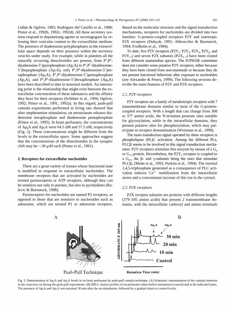
Lüthje & Ogilvie, 1983; Rodriguez del Castillo et al., 1988;Pintor et al., 1992b, 1992c, 1992d). All these secretory sys-tems respond to depolarising agents or secretagogues by re-leasing their vesicular content to the extracellular medium.The presence of diadenosine polyphosphates in the extracel-lular space depends on their presence within the secretoryvesicles under study. For example, while in platelets all thenaturally occurring dinucleotides are present, from P
1
,P
2
-di(adenosine-5
9
)pyrophosphate (Ap
2
A) to P
1
,P
7
-di(adenosine-5
9
)heptaphosphate (Ap
7
A), only P
1
,P
4
-di(adenosine-5
9
)tet-raphosphate (Ap
4
A), P
1
,P
5
-di(adenosine-5
9
)pentaphosphate(Ap
5
A), and P
1
,P
6
-di(adenosine-5
9
)hexaphosphate (Ap
6
A)have been described to date in neuronal models. An interest-ing point is the relationship that might exist between the ex-tracellular concentration of these substances and the affinitythey have for their receptors (Schlüter et al., 1994; Ogilvie,1992; Pintor et al., 1991, 1992a). In this regard, push-pullcannula experiments performed in living rats showed thatafter amphetamine stimulation, rat neostriatum releases dia-denosine tetraphosphate and diadenosine pentaphosphate(Pintor et al., 1995). In brain perfusates, the concentrationsof Ap
4
A and Ap
5
A were 64.5 nM and 57.5 nM, respectively(Fig. 2). These concentrations might be different from thelevels in the extracellular space. Some approaches suggestthat the concentrations of the dinucleotides in the synapticcleft may be
z
30
m
M each (Pintor et al., 1991).
2. Receptors for extracellular nucleotides
There are a great variety of tissues whose functional stateis modified in response to extracellular nucleotides. Themembrane receptors that are activated by nucleotides aretermed purinoceptors or ATP receptors, although they canbe sensitive not only to purines, but also to pyrimidines (Ra-levic & Burnstock, 1998).
Purinoceptors for nucleotides are named P2 receptors, asopposed to those that are sensitive to nucleosides such asadenosine, which are termed P1 or adenosine receptors.
Based on the molecular structure and the signal transductionmechanisms, receptors for nucleotides are divided into twofamilies: G-protein-coupled receptors P2Y and ionotropicP2X receptors (Dubyak, 1991; Abbracchio & Burnstock,1994; Fredholm et al., 1994).
To date, five P2Y receptors (P2Y
1
, P2Y
2
, P2Y
4
, P2Y
6
, andP2Y
11
) and seven P2X subunits (P2X
1–7
) have been clonedfrom different mammalian species. The IUPHAR committeedoes not consider some putative P2Y receptors, either becausethey have been cloned from non-mammals or because they donot present functional behaviour after exposure to nucleotides(see Alexander & Peters, 1999). The following sections de-scribe the main features of P2Y and P2X receptors.
2.1. P2Y receptors
P2Y receptors are a family of metabotropic receptors with 7transmembrane domains similar to most of the G-protein-coupled receptors. With a length that can change from 308to 377 amino acids, the N-terminus presents sites suitablefor glycosylation, while in the intracellular domains, theypresent putative sites for phosphorylation, which may par-ticipate in receptor desensitisation (Weisman et al., 1998).
The main transduction signal operated by these receptors isphospholipase (PL)C activation. Among the different PLs,PLC
b
seems to be involved in this signal transduction mecha-nism. P2Y receptors stimulate this enzyme by means of a G
q
or G
11
protein. Nevertheless, the P2Y
2
receptor is coupled toa G
i/o
, the
b
- and
g
-subunits being the ones that stimulatePLC
b
2
(Motte et al., 1993; Purkiss et al., 1994). The inositol1,4,5-triphosphate generated as a consequence of PLC acti-vation induces Ca
2
1
mobilisation from the intracellularstores and a concomitant increase of this ion in the cytosol.
2.2. P2X receptors
P2X receptor subunits are proteins with different lengths(379–595 amino acids) that present 2 transmembrane do-mains, with the intracellular carboxyl and amino terminals
Fig. 2. Determination of Ap4A and Ap5A levels in rat brain perfusates by push-pull cannula technique. (A) Schematic representation of the cannula insertionin the conscious rat during the push-pull experiments. (B) HPLC elution profiles of rat perfusates taken before stimulation (control) and at the indicated times.The presence of Ap4A and Ap5A was maximal 30 min after the rat stimulation, followed by a gradual return to control levels.

106
J. Pintor et al. / Pharmacology & Therapeutics 87 (2000) 103–115
separated by a large extracellular loop. They share between37 and 48% sequence similarity, the highest degree of ho-mology being in the extracellular domain. The extracellularloop also has 10 strictly conserved Cys residues that may beimplicated in disulphide bond formation necessary to formthe tertiary structure and for ATP binding. On the otherhand, the transmembrane domains show a high variabilityamong the different receptors (Soto et al., 1997). The topologyof two transmembrane domains, a long extracellular loopand the C- and N-terminals facing the cytosol, is present inother types of ion channels, such as the Na
1
channel in epi-thelium, the mechano-sensitive channel present in
Escherichiacoli
, and the inward rectifier K
1
channel (North, 1996).Functional P2X receptors are formed when two or more
subunits co-assemble. However, nothing is known about thenumber of subunits that form a receptor, and whether in nativetissues P2X receptors are formed by the same or different sub-units, called homomeric or heteromeric receptors, respectively(Kim et al., 1997; Nicke et al., 1998). There is evidence for thenecessity of heteromeric subunits to form a functional P2Xreceptor. For example, homomeric P2X
6
do not trigger cur-rents, as expected when they are expressed in oocytes (Colloet al., 1996; García-Guzmán et al., 1996; Soto et al., 1996).
Activation of P2X receptors by ATP produces a rapid in-ward flux of cations (Na
1
/K
1
and Ca
2
1
). The resultantchange in the membrane potential due to the ion flux trig-gers voltage-dependent Ca
2
1
channels (VDCCs), whichproduce a secondary Ca
2
1
entry.P2X receptors are further subdivided into two groups,
those that desensitise quickly (P2X
1
and P2X
3
) and othersthat do not. In the latter group, P2X
4
and P2X
6
maintain60% of their currents seconds after their activation, whileP2X
2
and P2X
5
maintain currents during long application ofagonists (Evans et al., 1996; Collo et al., 1996; García-Guzmán et al., 1996).
The P2X
7
subunit, with a long C-terminal domain, is dif-ferent from the others (Collo et al., 1997). Short ATP appli-cations produce the same ion fluxes as described in Section 2.2,but long applications of ATP induce pore formation permeableto metabolites up to 900 Da. This response can be reversed bywashing out the agonists, but in some cases, it is not reversibleand leads to cell death (Evans et al., 1998).
3. Receptors activated by diadenosine polyphosphates
3.1. Activation of recombinant P2Y receptors by diadenosine polyphosphates
Recombinant P2Y receptors expressed in heterologoussystems are activated by diadenosine polyphosphates. Thehuman P2Y
1
receptor cloned from a human genomic li-brary, and its avian homologue, are sensitive to Ap
4
A, withEC
50
values of 625 nM and 742 nM, respectively. This di-nucleotide behaves like a partial agonist, attaining 49% and67% of the effect produced by 2-methylthioadenosine tri-phosphate at human and turkey P2Y
1
receptors, respec-
tively. Other adenine dinucleotides tested showed an effectonly at millimolar concentrations (Schachter et al., 1996).
The P2Y2 receptor is stimulated by Ap4A, with an EC50
value in the same range as that described for ATP or UTP(Lazarowski et al., 1995). On the other hand, P1,P3-di(adeno-sine-59)triphosphate (Ap3A) is a good agonist at the P2Y1
receptor, while Ap4A behaves like a partial agonist. None of theother adenine dinucleotides exert any effect on this receptor.
The P2Y4 receptor, cloned from a human source and de-fined as a pyrimidinoreceptor, is sensitive to Ap3A, Ap4A,Ap5A, and Ap6A, with EC50 values between 3 and 7 mM,but their maximal effect is only 20–25% that of UTP(Communi et al., 1996). At the putative P2Y5 receptor, theseries of diadenosine polyphosphates was assayed with nopositive results (Janssens et al., 1997).
3.2. Activation of recombinant homomeric P2X receptors by diadenosine polyphosphates
Diadenosine polyphosphates can activate at least four re-combinant homomeric P2X receptors (P2X1–4). ApnA com-pounds show different patterns of pharmacological activity atthese receptors. At P2X1, only Ap6A is a full agonist, comparedwith ATP, although the potency order is Ap4A . Ap6A 5Ap5A, Ap2A and Ap3A being almost inactive. The effect of di-adenosine polyphosphates on the P2X2 receptor showed thatonly Ap4A behaves like an agonist, 5-fold less potent thanATP. The other ApnA compounds were not agonists of this ho-momeric receptor (Pintor et al., 1996; Wildman et al., 1999).
Four dinucleotides are as potent as ATP at the P2X3 re-ceptor. The dinucleotides showed the following potency or-der: Ap4A 5 Ap3A . Ap5A 5 Ap6A (based on EC50 val-ues). At this receptor, Ap3A is a partial agonist, while theother three behaved like full agonists.
The P2X4 homomeric receptor responds only to Ap4Aand Ap6A, the potency order being Ap4A > ATP ... Ap6A(by EC50 values). It is important to note that neither of thetwo dinucleotides behaved like a full agonist, presentingonly 30% of the maximal activity of ATP (Pintor et al.,1996; Wildman et al., 1999).
It is of interest that some dinucleotides, which do not ex-ert any agonistic effect, can potentiate responses triggeredby ATP, e.g., nanomolar concentrations of Ap5A potentiateATP responses at the recombinant P2X2 receptor, withoutaltering the Hill coefficient for ATP (nH 5 2), indicatingthat Ap5A is not occupying any of the two ATP sites at theP2X2 receptor and thus, is probably acting on an allostericsite. Ap2A is also a reversible positive modulator of the ho-momeric P2X3 receptor. When the same experiments wereperformed on P2X4 receptors, both Ap2A and Ap3A potenti-ated the responses elicited by ATP, but in this case, in themicromolar range (Pintor et al., 1996; Wildman et al., 1999).
The ability of some diadenosine polyphosphates to po-tentiate the effect of ATP does not occur only on ionotropicreceptors. Indeed, potentiation of ATP responses has beendemonstrated on metabotropic P2Y receptors from rat cere-bellar astrocytes. One of the main differences between the effect

J. Pintor et al. / Pharmacology & Therapeutics 87 (2000) 103–115 107
of dinucleotides in the potentiation of the P2X and P2Y re-ceptors is that at the metabotropic receptor, the potentiation waspermanent, being observed for up to 6 hr. This fact indicatesthat ATP and Ap5A may play an important role in astroglialdifferentiation and proliferation (Jiménez et al., 1998).
3.3. Activation of heteromeric P2X receptors
As reviewed by Buell et al. (1996), virtually all ionotropicP2X receptors have been reported to form functional homo-meric channels when they are expressed in heterologoussystems. These studies proved that in some tissues, the na-tive channel contains an homomeric assembly of one P2Xreceptor subunit. An example is the P2X1 receptor found invas deferens (Valera et al., 1994). However, some nativeP2X receptor phenotypes do not appear to correspond withhomomeric recombinant P2X receptors. In addition, in situhybridisation and immunohistochemical experiments haveshown co-localisation of different receptor subunits withinthe same population of cells in a variety of tissues (Collo etal., 1996; Kanjhan et al., 1999; Le et al., 1998; Nori et al.,1998; Vulchanova et al., 1997; Xiang et al., 1998). Thesefindings suggest that native receptors might also act as het-eromeric assemblies of members of this family. However,the exact subunit composition still remains to be elucidated.In this regard, only one study has shown a functional chan-nel in dorsal root sensory neurons, which appears to be dueto a hetero-oligomer containing P2X2 and P2X3 (Lewis etal., 1995). Subsequently, Le et al. (1998) demonstrated thatco-expression of P2X4 and P2X6 receptors in Xenopus oocytesleads to the generation of a novel ATP receptor, not foundin native tissues. More recently, Le et al. (1999) demon-strated another functional heteromeric channel formed withP2X1 and P2X5 subunits, when co-transfected in human em-bryonic kidney 293 cells. Again, a native receptor has notbeen found yet with the same pharmacological profile. In asearch for potential heteromeric ATP-gated channels,Torres et al. (1999) studied the possible subunit co-assem-bly in the P2X family, using an immunoprecipitation assay.Interestingly, P2X6 was unable to form a homomeric com-plex, indicating that it assembles with other subunits to forma functional channel. On the other hand, the P2X7 subunitforms only homomeric receptors. In the light of these find-ings, it has become apparent that P2X receptors might formboth homo- and heteromeric functional channels. Furtherexperiments are necessary to elucidate the subunit compositionof the native ATP-gated channel.
In synaptosomal preparations obtained from rat mid-brain, diadenosine polyphosphates activate P2X receptorsand an as yet uncharacterised novel receptor (Pintor et al.,1992b; Pintor & Miras-Portugal, 1995) called the diadenos-ine polyphosphate receptor (P4 receptor), since it is selec-tively activated by ApnA (see Section 4). Currently, we arefocusing on the molecular cloning of this new receptor, butcannot rule out the possibility that it may be a heterodimerof two or more P2X subunits. It is relevant that a recentstudy described a new pharmacological phenotype when
different opioid receptors are heterologously co-expressed(Jordan & Devi, 1999). This is also the case for the g-ami-nobutyric acid-B receptors (Jones et al., 1998). In addition,some of the physiological effects of ApnA may be due to ac-tivation of heteromeric P2X receptors. In line with this hy-pothesis, we have started to study the pharmacological pro-file of ApnA on heteromeric channels composed of differentP2X subunits. Initially, we studied the effect of these com-pounds on intracellular Ca21 levels from the rat glioma tu-mour cell line C6BU-1, transiently transfected with eitherthe P2X4 or P2X6 receptors alone or by a combination ofboth subunits. In our laboratory, this cell line has proven tobe a useful tool for such studies, since it does not respond toany mono- or dinucleotide. Fig. 3 shows the different phar-macological responses to ATP, Ap4A, and Ap5A of thesecells, differentially transfected with P2X4 and/or P2X6 re-ceptor subunits. Interestingly, an increase in intracellularCa21 levels is observed in C6BU-1 cells transfected with thehomomeric P2X4 or P2X6 receptors alone after stimulationwith 100 mM Ap4A. However, this response disappearswhen the cells are transfected with the heteromeric receptorP2X4/P2X6, indicating that ApnA may behave with a differ-ent pharmacological phenotype on heteromeric P2X recep-tors, compared with homomeric P2X receptors. With theseexperiments, it may be possible to elucidate the possible re-lationship with the P4 receptor.
3.4. Native P2 receptors stimulated by diadenosine polyphosphates in the central nervous system
There is substantial evidence that diadenosine polyphos-phates bind to native P2 receptors in the CNS. Ap4A is ableto displace the binding of [3H]a,b-methylene adenosine 59-triphosphate (a,b-meATP) in rat brain slices, suggestingthat both a,b-meATP and Ap4A compete for the same P2Xreceptor (Balcar et al., 1995).
Additional binding and functional studies indicate thatdiadenosine polyphosphates can activate P2 receptors in theCNS. Binding studies carried out in rat brain cortical synap-tosomes by means of [35S]adenosine 59-O-(3-thiotriphos-phate) ([35S]ATPgS) (a putative P2Y agonist) demonstratedthat Ap5A and Ap6A gradually enhanced the binding of[35S]ATPgS by up to 60% in a concentration range from 1to 50 mM. Ap4A produce only a 15% of increase in the sameconcentration range. Diadenosine polyphosphates assayedat concentrations above 50 mM inhibited the binding of[35S]ATPgS to synaptosomal membranes, but with very lowpotency (Schäfer & Reiser, 1997).
Ap4A and Ap5A mimic the effect of ATP on P2X recep-tors expressed in rat sensory neurons (Krishtal et al., 1983),but Ap3A and Ap2A are nearly ineffective (Marchenko etal., 1987). The effect produced by diadenosine tetraphos-phate and diadenosine pentaphosphate was 15% of the max-imum amplitude of the ATP-activated current. The esti-mated Kd value for Ap4A was z40 mM, and it could beconcluded that both the dinucleotide and ATP are acting viathe same receptor (Krishtal et al., 1988).

108 J. Pintor et al. / Pharmacology & Therapeutics 87 (2000) 103–115
It has been suggested that noradrenergic neurones lo-cated in the locus coeruleus express both P2X and P2Y re-ceptors (Tschöpl et al., 1992; Illes et al., 1995). The firingrate of these neurones is increased by Ap3A, Ap4A, andAp5A, with the potency order Ap5A . Ap4A . Ap3A (Illeset al., 1996). The effects of the dinucleotides are partiallyblocked by suramin and pyridoxalphosphate-6-azophenyl-29,49-disulphonic acid (PPADS), and are insensitive to reac-tive blue 2, suggesting that adenine dinucleotides act prefer-entially through P2X receptors (Fröhlich et al., 1996).
4. Native receptors specifically stimulated by diadenosine polyphosphates in the central nervous system
The existence of specific receptors for adenine dinucle-otides has been described in the CNS. Although diadenosine
polyphosphates could exert their actions on P2 purinocep-tors in different neural and non-neural preparations, as shownin Section 3.4, evidence accumulated during recent years sup-ports the possible existence of specific receptors for diadenos-ine polyphosphates (see Miras-Portugal et al., 1998), but notmononucleotides. The concept “specific” means not sensitiveto ATP, UTP, adenosine, or their respective pharmacologicalanalogues, but in most cases, sensitive only to dinucleosideoligophosphates. This receptor has been termed dinucleotidereceptor or P4 receptor (Pintor & Miras-Portugal, 1995).
Hilderman et al. (1991) first identified a receptor forAp4A in rat brain homogenates, with a Kd value for Ap4A of0.71 mM. Subsequently, Pivorun and Nordone (1996) dem-onstrated that Ap4A was able to increase intrasynaptosomalCa21 levels in deermouse brain. This effect was not pro-duced by ATP and was not sensitive to P2 antagonists, sug-gesting the presence of a dinucleotide receptor.
Fig. 3. Effect of 100 mM ATP, Ap4A, and Ap5A on intracellular Ca21 levels in C6BU-1 cells transiently transfected with P2X4 or P2X6 receptors, or a combina-tion of both. Cells grown in cover slips were transfected using liposomes, and 48 hr later were loaded with fura-2 AM in a buffer containing CaCl2 (1.33 mM)for 45 min. Cover slips were then mounted in a perfusion chamber, and intracellular Ca21 levels were analysed after stimulation with the different drugs, usinga multiple excitation microfluorescence system. Cells were illuminated alternately at 380 and 400 nm. The emitted fluorescence was collected at 510 nm.Results are shown as a normalised ratio of F380/F400 signals that increases as [Ca21]i increases. (A) Homomeric P2X6 receptor-expressing cells respond veryweakly to ATP and Ap5A, with Ap4A as a potent agonist. (B) Homomeric P2X4 receptor respond to Ap4A as intensely as ATP, but there is cross-desensitisation.The figure shows the time course of fluorescence changes in a representative cell initially stimulated by Ap4A. P2X6 receptor-expressing cells primarily stimu-lated with ATP respond with an increase in intracellular Ca21 level, but are insensitive to further stimulation with Ap4A or Ap5A. (not shown). (C) The responseto Ap4A disappears when cells are co-transfected with P2X4 and P2X6 receptor subunits, although the heteromeric receptor formed is still sensitive to ATP.
J. Pintor et al. / Pharmacology & Therapeutics 87 (2000) 103–115 109
These features, and the fact of being coupled to VDCCs,makes this receptor very similar to that described by ourgroup in rat midbrain (Pintor & Miras-Portugal, 1995; seethe next section).
4.1. Rat brain receptors
Dinucleotide receptors, also termed P4 receptors, werepharmacologically described in 1995 in rat midbrain synap-tic terminals, but the molecular structure remains unknown.These receptors are sensitive to diadenosine tetraphosphateand diadenosine pentaphosphate and elicited intracellularCa21 increases within presynaptic rat brain terminals, actingas receptor-operated Ca21 channels (Pintor & Miras-Portu-gal, 1995).
Some facts point to these receptors being receptor-oper-ated Ca21 channels. The global functioning of the dinucle-otide receptor in the presence of the G-protein modulatorsdoes not fit with a model in which the receptor is coupled toa Ca21 channel via a G-protein mechanism (Pintor et al.,1997b). For example, in the absence of extracellular Ca21,the responses to diadenosine polyphosphates are abolished.Another point in favour of this hypothesis is that the receptor isfunctional in the presence of G-protein modulators. Substancessuch as GDPbS or GTPgS severely changed the activity ofthe dinucleotide receptor. For instance, the application of thenonhydrolysable GDP analogue, which inactivates G-proteins,enhanced the Ca21 response elicited by diadenosine poly-phosphates. On the other hand, GTPgS produced the oppositeeffect, since a clear reduction in the Ca21 response was seenin rat midbrain synaptosomes. Nevertheless, the functioningof the receptor in the presence of these GTP/GDP analoguescan be explained on the basis of activation of protein kinases(Pintor et al., 1997b; see the following section).
4.1.1. PharmacologyAs mentioned in Section 4, the dinucleotide receptor is
stimulated by diadenosine polyphosphates, being insensi-tive to ATP, adenosine, and their respective analogues. Thisfact generates the need for new antagonists because classi-cal antagonists for P2 receptors cannot be used on this re-ceptor. Pharmacological approaches have been carried outwith the available diadenosine polyphosphates. Experi-ments performed in rat midbrain synaptic terminals showedthat diadenosine pentaphosphate was the most powerful naturaldinucleotide, followed by Ap4A, with Ap3A being almostinactive. Ap2A, recently described as a naturally occurringdinucleotide in platelets, was also a good agonist on this re-ceptor. The EC50 values for Ap5A, Ap4A, and Ap2A were al-most identical, z55 mM (Pintor & Miras-Portugal, 1995).
More recent experiments with additional dinucleosidepolyphosphates, such as e-ApnA (the etheno derivatives ofdiadenosine polyphosphates) or GpnG (diguanosine poly-phosphates), showed the following pharmacological orderin terms of EC50 values: Gp5G . Ap5A 5 Ap4A . Gp4G 5e-Ap4A 5 e-Ap5A . Gp3G . Ap3A 5 e-Ap3A. As a gen-
eral rule, the following potency order prevails: Np5N .Np4N . Np3N (Miras-Portugal et al., 1999).
With the help of different fluorescent dyes, it has beenpossible to determine whether this receptor is permeable toNa1 and Ca21 or only to Ca21. Measurement of intracellularCa21 with fura-2 and Na1 with SBFI have demonstrated thatafter stimulation with diadenosine pentaphosphate, Ca21 entryoccurs, depolarising the terminals and opening voltage-dependent ion channels (Pintor & Miras-Portugal, 1995).
4.1.2. Connection with voltage-dependent Ca21 channelsAfter stimulation of the synaptic terminal with adenine
dinucleotides through the P4 receptor, Ca21 entry depolarisesthe terminal activating voltage-dependent Na1 channels(VDSC) and VDCCs (Panchenko et al., 1996; Pintor &Miras-Portugal, 1995). The activation of VDSC was confirmedby pretreatment of the synaptic terminals with the blockertetrodotoxin. A lack of Na1 entry was measured with SBFIwhen the toxin was present.
Concerning Ca21 channels, three different substanceswere assayed to block the different VDCCs: nifedipine, toantagonise the L-type; ω-conotoxin G-VI-A, to block theN-type; and ω-agatoxin, to antagonise the P-type. Ni21 wasalso used to rule out involvement of the T-type Ca21 chan-nel. Only ω-conotoxin G-VI-A was able to partially reduceCa21 entry induced by diadenosine polyphosphates. A care-ful analysis of the Ca21 entry in the presence of this toxinreveals the existence of an initial transient, suggesting thatthe dinucleotide receptor elicits an initial Ca21 entry, whichis voltage-insensitive. There is a clear decrease in Ca21 en-try after the initial peak when w-conotoxin G-VI-A is tested.This indicates that the total Ca21 increase is the sum of avoltage-independent transient, followed by a voltage-depen-dent one mediated by an N-type Ca21 channel (Fig. 4). Thiswas also observed when the synaptic terminals were pre-stimulated with 60 mM K1. Under these conditions, the initialentry after stimulation with Ap5A remains unchanged, but thesecondary phase disappears (Pintor & Miras-Portugal., 1995).
Experiments performed with the patch-clamp techniquehave demonstrated that hippocampal CA3 neurones arestimulated by diadenosine polyphosphates, showing re-sponses that are partially mediated by a VDCC N-typechannel (Panchenko et al., 1996).
4.1.3. Modulation of the dinucleotide receptor by proteinkinases and phosphatases
Many receptors for neurotransmitters and hormones arecoupled to second messenger systems that produce activa-tion or inhibition of adenylate cyclase or PLC. Activation ofthe mentioned second messenger systems triggers the syn-thesis of cyclic AMP and diacylglycerol that activate pro-tein kinases A and C (PKA and PKC), respectively. Proteinkinases can phosphorylate enzymes and membrane proteinssuch as receptors, channels, and transporters. Protein phos-phatases reverse the action of protein kinases, keeping a bal-ance of phosphorylation in many proteins.

110 J. Pintor et al. / Pharmacology & Therapeutics 87 (2000) 103–115
The dinucleotide receptor present in rat midbrain synap-tic terminals is strongly modulated by PKA and PKC, aswell as by protein phosphatases. Activation of adenylate cy-clase by forskolin, or PKA by dibutyryl cyclic AMP, blocksthe Ap5A intrasynaptosomal Ca21 transients. Compoundsblocking the activation of PKA, such as the PKA inhibitorypeptide (PKA-IP), clearly potentiate the Ca21 responseselicited by Ap5A in rat midbrain synaptosomes. Substancesacting on PKC generate similar results. For example, activa-tion of PKC with phorbol esters such as 12,13-dibutyratedramatically inhibits the Ca21 transients elicited by Ap5A.Like the PKA inhibitors, the PKC-IP and staurosporine bothpotentiated the responses elicited by Ap5A, as shown in Fig. 5(Pintor et al., 1997b).
The possible involvement of a phosphorylation processwas further confirmed by the effect of different proteinphosphatase inhibitors (Cohen, 1989). Substances such asokadaic acid, a broad protein phosphatase inhibitor, pro-
duced a reduction in the Ca21 responses elicited by Ap5A.This effect was also observed with other protein phos-phatase inhibitors, such as mycrocistin (a protein phos-phatase A2A inhibitor) or cyclosporin A (a calcineurin in-hibitor). The action of these inhibitory substances producinga decrease in the Ca21 transients elicited by Ap5A indicatesthat different protein phosphatases could reverse the phos-phorylation process carried out by PKA and PKC (Fig. 5).The high concentration of calcineurin in neural tissues, andespecially in neurones, may indicate a potential role for thisenzyme in modulating neural activity, also reversing thephosphorylation of the dinucleotide receptor by protein ki-nases (Pintor et al., 1997b). Calcineurin is implicated in theregulation of ionotropic receptors and channels in mamma-lian and nonmammalian neural tissues (Cohen, 1989; Kunz &Hall, 1993).
The dinucleotide receptor seems to be controlled by abalance between phosphorylation and dephosphorylationthat depends on the degree of activation by those receptorspositively coupled to PKA and PKC and the activity of pro-tein phosphatases (Fig. 5).
4.2. Dinucleotide receptors in guinea-pig brain
Several areas of the brain in different species have beeninvestigated in a search for the existence of dinucleotide re-ceptors. As described in the previous paragraphs, rat mid-brain was the first tissue where this receptor was well char-acterised. Nevertheless, guinea-pig brain was chosen tostudy the distribution of the dinucleotide receptor by inves-tigating three different brain areas: cortex, midbrain, andcerebellum.
4.2.1. Cortical receptorsThe receptors present in guinea-pig cortical synaptic ter-
minals respond both to ATP and diadenosine polyphos-phates by acting through the same P2 receptor subtype. Theeffect of the whole series of adenine dinucleotides gave si-
Fig. 4. Schematic diagram of the relationship between the dinucleotidereceptor and the VDCC. The activation of the dinucleotide receptor permitsCa21 entry into the synaptic terminal, which generates a depolarisationthreshold that activates a VDCC N-type.
Fig. 5. Modulation of the dinucleotide receptor by protein kinases and phosphatases. The activation of PKA and PKC inhibits the Ca21 transients elicited byAp5A. Stimulation of protein phosphatases produces the opposite effect, showing higher Ca21 transients than those in the absence of phosphatase activation.Stimulation of neurotransmitter (NT) receptors coupled to PLC or adenylate cyclase (AC) may modify dinucleotide response via protein kinase activation.

J. Pintor et al. / Pharmacology & Therapeutics 87 (2000) 103–115 111
milar EC50 values for all of them. For maximal effects pro-duced, Ap4A was the best (29 nM Ca21 increase above thebasal value) followed by Ap3A and Ap2A, Ap5A and Ap6Abeing the poorest (20 nM Ca21 increase).
Cross-desensitisation studies carried out with ATP ana-logues, such as a,b-meATP, abolished the responses elic-ited by diadenosine polyphosphates. Moreover, preincuba-tion of the cortical synaptic terminals with suramin, a P2antagonist, also markedly attenuated the responses pro-duced by ApnA compounds. These two aspects clearly indi-cate that in the cortical guinea-pig synaptic terminals, bothdiadenosine polyphosphates and ATP share the same recep-tor, presumably a P2X (see Fig. 6; Pintor et al., 1997c).
4.2.2. Midbrain receptorsIn midbrain guinea-pig synaptic terminals, diadenosine
polyphosphates produce an intracellular Ca21 increaseabove the basal cytosolic values, which was concentration-dependent. As with cortical synaptosomes, there were nodifferences in the EC50 values obtained for each of the as-sayed compounds. At the highest concentration assayed, therank order for evoking the Ca21 signal was Ap2A 5 Ap4A 5Ap6A . Ap3A . Ap5A.
In these synaptic terminals, ATP, as well as the syntheticanalogues, elicited Ca21 transients that in some cases, werecomparable with those produced by ApnA compounds.
Nevertheless, in clear contrast to the cortical synaptic terminals,the cross-desensitisation studies performed by applyinga,b-meATP prior to the diadenosine polyphosphatesshowed an inability of this ATP analogue to block the effectof the adenine dinucleotides. Moreover, the P2 antagonistsuramin, applied before ApnA, was unable to block the Ca21
transients elicited by adenine dinucleotides. Taking intoconsideration these two facts, it can be concluded that inguinea-pig midbrain synaptic terminals, diadenosine poly-phosphates and ATP act via independent receptors, a dinu-cleotide and a P2X receptor, respectively (Fig. 6; Pintor et al.,1997c).
4.2.3. Cerebellar receptorsApnA compounds were able to increase the intracellular
Ca21 levels in cerebellar isolated synaptic terminals withdifferent EC50 values, the potency order being Ap2A .Ap6A 5 Ap3A $ Ap5A 5 Ap4A. The best was Ap2A, whichshowed not only a lower EC50, but also maximal Ca21 in-flux (34 nM above the basal cytosolic value).
Cross-desensitisation studies performed with ATP ana-logues demonstrated that the mononucleotides did not mod-ify the Ca21 transient elicited by ApnA, suggesting that ATPand diadenosine polyphosphates are acting through differ-ent receptors. To further confirm this, the P2 receptor antag-onist suramin was used to see whether or not the responses
Fig. 6. Distribution of P4 and P2X receptors in the guinea-pig brain. (A) Responses produced by diadenosine polyphosphates mediated by different receptors.(B) In the midbrain and cerebellum, ApnA activated P4 receptors and ATP P2X receptors. (C) In the cortex, all the nucleotides activated the same P2X recep-tor. The antagonists Ip5I, suramin, and PPADS helped to identify the receptors involved in each region.

112 J. Pintor et al. / Pharmacology & Therapeutics 87 (2000) 103–115
elicited by diadenosine polyphosphates were blocked bythis substance. Suramin could block the responses elicitedby ATP, but did not change the responses elicited by ApnAcompounds. This supports the idea that ATP and ApnA actthrough independent receptors, as in guinea-pig midbrain(Fig. 6; Pintor et al., 1997c).
4.3 Human brain receptors
Receptors for diadenosine polyphosphates have alsobeen investigated in human brain isolated synaptic termi-nals. Healthy human brain tissue is not readily accessible,hence the number of experiments has been limited. Never-theless, the main questions, such as whether there are receptorsfor diadenosine polyphosphates or whether they resemblesome of the animal models, have been positively answered.
Application of ApnA or ATP to human cortical synapticterminals produces a concentration-dependent intracellularCa21 increase. Analysis of the dose-response curves yieldedEC50 values of 11.5 6 2.1 mM and 23.4 6 3.7 mM for Ap5Aand ATP, respectively. It is interesting to note that both ago-nists, although mobilising only a small amount of Ca21, pro-duced a clear change in the membrane potential of the syn-aptic terminals (Pintor et al., 1999). This suggests that undercertain conditions, these two nucleotides could induce achange in the synaptic terminal sufficient to induce thetransmitter release.
As in animal studies, the question whether Ap5A andATP act via the same or different receptors was answeredby performing cross-desensitisation studies, as well as stud-ies with antagonists. Cross-desensitisation studies indicatedthat responses to ATP were not blocked by pretreatmentwith Ap5A or vice versa. Experiments performed with an-tagonists PPADS (a P2 antagonist) and Ip5I (a P4 antago-nist) demonstrated that the effects elicited by ATP were an-tagonised by PPADS, but not by Ip5I. In contrast, Ap5Aresponses were not blocked by PPADS, and only partially(50%) with Ip5I. The effect of Ap5A on intrasynaptosomalCa21 increase was partially blocked by Ip5I, in clear contrastto the effect obtained in rat synaptosomes, where Ip5I wasable to abolish the effect of Ap5A (Pintor & Miras-Portugal,1995; Pintor et al., 1999). This discrepancy is subject to dif-ferent interpretations. It is possible that the dinucleotide re-ceptors in rat and human brain are different. Another possi-bility is the existence of ectonucleotidases in the humanbrain, with a different selectivity for nucleotides. In thiscase, the difference in the activity of the antagonists may bedue to a selective cleavage, significantly reducing the con-centration of Ip5I. Whether this or the previous hypothesis istrue remains to be resolved.
As in rat brain synaptic terminals, dinucleotide receptorsare coupled to VDCCs in the human brain. The activation ofthis receptor by Ap5A produces an increase in the ω-conotoxin-sensitive component of the Ca21 channels in human corticalsynaptic terminals. The specificity of this toxin (Miller &Fox, 1990; Tsien & Tsien, 1990) and the lack of effect by
the other blockers indicate that Ap5A facilitates only the ac-tivity of N-type Ca21 channels, the others being uninvolvedin the dinucleotide action. As commented in Section 4.1.2,this result is in agreement with those described in rat mid-brain synaptosomes (Pintor & Miras-Portugal, 1995) andCA3 hippocampal neurones (Panchenko et al., 1996).
The non-association of Ap5A stimulation with the activa-tion of an L-type Ca21 channel has been pointed out for ratbrain synaptosomes, hippocampal neurones, and also deer-mouse synaptosomes. L-type channel blockers, such as ver-apamil or nifedipine, do not modify the Ca21 responses in-duced by diadenosine polyphosphates in these neuralmodels (Pintor & Miras-Portugal, 1995; Panchenko et al.,1996; Pivorun & Nordone, 1996).
5. Could diadenosine polyphosphate derivatives be used as pharmacological tools for P2X receptors?
In 1995, the existence of a nonspecific adenylic aciddeaminase in the snail Helix pomatia was described (Gura-nowski et al., 1995). The authors showed that this enzyme isable to transform the adenine rings of ApnA compounds intohypoxanthine, thus transforming the diadenosine polyphos-phates into diinosine polyphosphates (IpnI).
When instead of the H. pomatia enzyme the commercialadenylic acid deaminase (from Aspergillus sp.) was used,the same results were obtained. From Ap3A, Ap4A, andAp5A, it was possible to obtain Ip3I, Ip4I, and Ip5I, respec-tively. These compounds were assayed for their ability toact as agonists or antagonists in rat midbrain synaptosomes.IpnI compounds did not behave as agonists, since they didnot modify the cytosolic Ca21 levels of the synaptic termi-nals under study. Nevertheless, when they were tested asantagonists on the dinucleotide receptor, the three hypoxan-thine compounds blocked the actions of Ap4A and Ap5A.There was a clear difference among the three dinucleotides.While Ip3I and Ip4I were antagonists at the dinucleotide re-ceptor, with IC50 values in the micromolar range (4.9 mMand 8.3 mM, respectively), Ip5I behaved as a very potent an-tagonist, with an IC50 value of 4.2 nM (Pintor et al., 1997c).
Apart from the dinucleotide receptor, rat midbrain syn-aptosomes possess P2X receptors. The three diinosine com-pounds, assayed for responses mediated through P2X recep-tors, presented IC50 values of 100 mM, 29.5 mM, and 27.5mM for Ip3I, Ip4I, and Ip5I, respectively. From these values,with an z6000 times greater affinity for the dinucleotide re-ceptor, it can be seen that Ip5I is a useful tool to discriminatebetween the activation of dinucleotides versus P2 receptorsby diadenosine polyphosphates (Pintor et al., 1997a).
In the periphery, IpnI compounds have been assayed inguinea-pig vas deferens. This tissue, considered a classicalone for studying P2X1 receptors, displayed an interestingbehaviour towards the hypoxanthine dinucleotides. In par-ticular, Ip5I was a good antagonist of the P2X1 receptor,with an IC50 value of 0.30 mM. This effect seems to be se-

J. Pintor et al. / Pharmacology & Therapeutics 87 (2000) 103–115 113
lective since in other models, such as taenia coli (with P2Yreceptors), or guinea-pig left atrium (with P1 and P2 recep-tors), Ip5I did not significantly modify the responses medi-ated by ATP (Hoyle et al., 1997).
More recently, it proved possible to study the effects ofIp3I, Ip4I, and Ip5I on recombinant P2X receptors expressedin Xenopus laevis oocytes. At the P2X1 receptor, the threediinosine polyphosphates presented the following antago-nistic potency: Ip5I . Ip4I . Ip3I, with IC50 values of 3.16mM, 0.5 mM, and 31.6 mM, respectively. On the P2X2 re-ceptor, none of the diinosine polyphosphates had any effect.On the P2X3 receptor, the three IpnI behaved as antagonistspresenting the following potency order and IC50 values: Ip4I(1 mM) . Ip5I (2.5 mM) . Ip3I (.30 mM). The agonist re-sponses mediated by the P2X4 receptor were potentiated byIp4I and Ip5I in a reversible fashion (King et al., 1999).
These results showed that Ip5I is a good antagonist at theGroup 1 P2X receptors (P2X1 and P2X3), being more selec-tive for the P2X1 than for the P2X3. Ip5I is 900-fold less po-tent at the P2X3, where the required concentration to blockATP action is in the micromolar range, compared with thenanomolar concentrations necessary to antagonise ATP viathe P2X1 receptor (King et al., 1999).
6. Conclusions
Diadenosine polyphosphates can stimulate different sub-types of purinergic receptors after their release into the ex-tracellular space from synaptic vesicles or secretory gran-ules. The physiological role of these dinucleotides remainsunclear, although we already know the receptors that can beactivated by these compounds. Ionotropic responses medi-ated by P2X and P4 receptors suggest an involvement insynaptic transmission in the CNS, facilitating neurotrans-mitter release from the synaptic terminals, as does ATP (Ed-wards et al., 1992; Pankratov et al., 1999). In this sense, it isnecessary to establish the possible relationship between dia-denosine polyphosphates and their receptors and other clas-sical neurotransmitters in order to confirm this hypothesis.
Acknowledgments
This work has been supported by research grants fromthe Universidad Complutense de Madrid, PR49/98-7786,Comunidad Autónoma de Madrid No. 8.5/18/1998, andDGICYT PM98-0089. M.D.-H., J.G., R.G.-V., and F.H. arefellowship holders of the Universidad Complutense, Minis-terio de Educación, Comunidad Autonoma de Madrid, andBeca de Reincorporación de la Comunidad de Madrid, re-spectively. We thank Charles H. V. Hoyle for help in pre-paring this manuscript.
References
Abbracchio, M. P., & Burnstock, G. (1994). Purinoceptors: are there fami-lies of P2X and P2Y purinoceptors? Pharmacol Ther 64, 445–475.
Alexander, S. P. H., & Peters, J. A. (1999). Receptors and ion channels: no-menclature supplement. Trends Pharmacol Sci 20, 64–67.
Baker, J. C., & Jacobson, M. K. (1986). Alteration of adenyl dinucleotide me-tabolism by enviromental stress. Proc Natl Acad Sci USA 83, 2350–2352.
Balcar, V. J., Li, Y., Killinger, S., & Bennett, M. R. (1995). Autoradiogra-phy of P2X ATP receptors in rat brain. Br J Pharmacol 115, 302–306.
Bochner, B. R., Lee, P. C., Wilson, S. W., Cutler, C. W., & Ames, B. N.(1984). AppppA and related adenylylated nucleotides are synthesizedas a consequence of oxidation stress. Cell 37, 225–232.
Bone, R., Cheng, Y. C., & Wolfenden, R. (1986). Inhibition of adenosineand thymidylate kinases by bisubstrate analogs. J Biol Chem 261,16410–16413.
Brevet, A., Plateau, P., Best-Belpomme, M., & Blanquet, S. (1985). Varia-tion of Ap4A and other dinucleoside polyphosphates in stressed Droso-phila cells. J Biol Chem 260, 15566–15570.
Brevet, A., Chen, J., Lévêque, F., Plateau, P., & Blanquet, S. (1989). Invivo synthesis of adenylylated bis(59-nucleosidyl)tetraphosphates(Ap4N) by Escherichia coli aminoacyl-tRNA synthetases. Proc NatlAcad Sci USA 86, 8275–8279.
Buell, G., Collo, G., & Rassendren, F. (1996). P2X receptors: an emergingchannel family. Eur J Pharmacol 8, 2221–2228.
Cohen, P. (1989). The structure and regulation of protein phosphates. AnnuRev Biochem 58, 453–508.
Collo, G., North, R. A., Kawashima, E., Merlo-Pich, E., Neidhart, S., Sur-prenant, A., & Buell, G. (1996). Cloning of P2X5 and P2X6 receptorsand the distribution and properties of an extended family of ATP-gatedion channels. J Neurosci 16, 2495–2507.
Collo, G., Neidhart, S., Kawashima, E., Kosco-Vilbois, M., North, R. A., &Buell, G. (1997). Tissue distribution of the P2X7 receptor. Neurophar-macology 36, 1227–1283.
Communi, D., Motte, S., Boeynaems, J. M., & Pirotton, S. (1996). Pharma-cological characterisation of the human P2Y4 receptors. Eur J Pharma-col 317, 383–389.
Dubyak, G. R. (1991). Signal transduction by P2-purinergic receptors forextracellular ATP. Am J Respir Cell Mol Biol 4, 295–300.
Edwards, F. A., Gibb, A. J., & Colquhoun, D. (1992). ATP receptor-mediatedsynaptic currents in the central nervous system. Nature 359, 144–147.
Evans, R. J., Lewis, C., Virginio, C., Lundstrom, K., Buell, G., Surprenant,A., & North, A. (1996). Ionic permeability of, and divalent cation ef-fects on, two ATP-gate cation channels (P2X receptors) expressed inmammalian cells. J Physiol 497, 413–422.
Evans, R. J., Suprenant, A., & North, A. (1998). P2X receptors, cloned andexpressed. In J. T. Turner, G. A. Weisman, & J. S. Fedan (Eds.), TheP2 Nucleotide Receptors (pp. 43–61). Totowa: Humana Press, Inc.
Flodgaard, H., & Klenow, H. (1982). Abundant amounts of diadenosine59,5999 P1,P4-tetraphosphate are present and releasable, but metaboli-cally inactive in human platelets. Biochem J 208, 737–742.
Fredholm, B. B., Abbrachio, M. P., Burnstock, G., Daly, J. W., Harden,T. K., Jacobson, K. A., Leff, P., & Williams, M. (1994). Nomenclatureand classification of purinoceptors. Am Soc Pharmacol Exp Ther 46,143–156.
Fröhlich, R., Boehm, S., & Illes, P. (1996). Pharmacological characteriza-tion of P2 purinoceptor types in rat locus coeruleus neurons. Eur JPharmacol 315, 255–261.
García-Guzmán, M., Soto, F., Laube, B., & Stühmer, W. (1996). Molecularcloning and functional expression of a novel heart P2X receptor. FEBSLett 388, 123–127.
Guranowski, A., Starzynska, E., Sillero, M. A. G., & Sillero, A. (1995).Conversion of adenosine(59) oligophospho(59) adenosines intoinosine(59) oligophospho (59) inosines by non-specific adenylate deam-inase from the snail Helix pomatia. Biochim Biophys Acta 1243, 78–84.
Hilderman, R. H., Martin, M., Zimmerman, J. K., & Pivorun, E. B. (1991).Identification of a unique membrane receptor for adenosine 59,5999-P1-P4-tetraphosphate. J Biol Chem 266, 6915–6918.
Hoyle, C. H. V., Pintor, J., Gualix, J., & Miras-Portugal, M. T. (1997). An-tagonism of P2X receptors in guinea-pig vas deferens by diinosine pen-taphosphate. Eur J Pharmacol 333, R1–R2.

114 J. Pintor et al. / Pharmacology & Therapeutics 87 (2000) 103–115
Illes, P., Nieber, N., & Nörenberg, W. (1995). Neuronal ATP receptors. InL. Belardinelli, & A. Pelleg (Eds.), Adenosine and Adenine Nucle-otides: Molecular Biology to Integrative Physiology (pp. 77–84). Nor-well: Kluwer Academic.
Illes, P., Nieber, N., & Nörenberg, W. (1996). Electrophysiological effectsof ATP on brain neurones. J Auton Pharmacol 16, 407–411.
Janssens, R., Boeynaems, J. M., Godart, M., & Communi, D. (1997). Clon-ing of a human heptahelical receptors closely related to the P2Y5 recep-tor. Biochem Biophy Res Commun 236, 106–112.
Jiménez, A. I., Castro, E., Delicado, E. G., & Miras-Portugal, M. T. (1998).Potentiation of adenosine 59-triphosphate calcium responses by diade-nosine pentaphosphate in individual rat cerebellar astrocytes. NeurosciLett 246, 109–111.
Jones, K. A., Borowsky, B., Tamm, J. A., Craig, D. A., Durkin, M. M.,Dai, M., Yao, W. J., Johnson, M., Gunwaldsen, C., Huang, L. Y., Tang,C., Shen, Q., Salon, J. A., Morse, K., Laz, T., Smith, K. E., Nagarath-nam, D., Noble, S. A., Branchek, T. A., & Gerald, C. (1998). GABAbreceptors function as a heteromeric assembly of the subunitsGABAbR1 and GABAbR2. Nature 396, 674–687.
Jordan, B. A., & Devi, L. A. (1999). G-protein-coupled receptor het-erodimerization modulates receptor function. Nature 399, 697–700.
Kanjhan, R., Housley, G. D., Burton, L. D., Christie, D. L., Kippenberger,A., Thorne, P. R., Luo, L., & Ryan, A. F. (1999). Distribution of theP2X2 receptor subunit of the ATP gated ion channels in the rat centralnervous system. J Comp Neurol 28, 11–32.
Kim, M., Yoo, O. J., & Choe, S. (1997). Molecular assembly of the extra-cellular domain of P2X2, an ATP-gated ion channel. Biochem BiophysRes Commun 240, 618–622.
King, B. F., Liu, M., Pintor, J., Gualix, J., Miras-Portugal, M. T., & Burn-stock, G. (1999). Diinosine pentaphosphate (Ip5I) is a potent antagonistat recombinant rat P2X1 receptors. Br J Pharmacol 128, 981–988.
Krishtal, O. A., Marchenko, S. M., & Pidoplichko, V. J. (1983). Receptorsfor ATP in the membrane of mammalian sensory neurones. NeurosciLett 35, 41–45.
Krishtal, O. A., Marchenko, S. M., Obukhov, A. G., & Volkova, T. M.(1988). Receptors for ATP in rat sensory neurones: the structure-func-tions relationship for ligands. Br J Pharmacol 95, 1057–1062.
Kunz, J., & Hall, M. N. (1993). Cyclosporine A, FK506 and rapamycin:more than just immunosuppression. Trends Biochem Sci 18, 334–338.
Lazarowski, E. R., Watt, W. C., Stutts, M. J., Boucher, R. C., & Harden,T. K. (1995). Pharmacological selectivity of the cloned human P2U-purinoceptor: potent activation by diadenosine tetraphosphate. Br JPharmacol 116, 1619–1627.
Le, K. T., Villeneuve, P., Ramjaun, A. R., McPherson, P. S., Beaudet, A., &Seguela, P. (1998). Sensory presynaptic and widespread somatoden-dritic immunolocalization of central ionotropic P2X ATP receptors.Neuroscience 83, 177–190.
Le, K. T., Boué-Grabot, E., Archambault, V., & Seguela, P. (1999). Functionaland biochemical evidence for heteromeric ATP-gated channels composedof P2X1 and P2X5 subunits. J Biol Chem 274, 15415–15419.
Lee, P. C., Bochner, B. R., & Ames, B. N. (1983). AppppA, heat-shockstress and cell oxidation. Proc Natl Acad Sci USA 80, 7496–7500.
Lewis, C., Neidhart, S., Holy, C., North, R. A., Buell, G., & Suprenant, A.(1995). Coexpression of P2X2 and P2X3 receptor subunits can accountfor ATP-gated currents in sensory neurons. Nature 377, 432–435.
Lienhard, G. E., & Secemski, I. I. (1973). P1,P5-Di(adenosine-59)penta-phosphate, a potent multisubstrate inhibitor of adenylate kinase. J BiolChem 248, 1121–1123.
Lüthje, J., & Ogilvie, A. (1983). Presence of diadenosine 59,5999-p1,p4-triphosphate (Ap3A) in human platelets. Biochem Biophys Res Com-mun 115, 253–260.
Marchenko, S. M., Volkova, T. M., & Fedorov, O. I. (1987). ATP-acti-vated ionic permeability in the isolated smooth muscle cells from uri-nary bladder of the guinea pig. Neirofiziologiya 19, 95–100.
McLennan, A. C. (2000). Dinucleoside polyphosphates—friend or foe?Pharmacol Ther, this issue.
Miller, R. J., & Fox, A. P. (1990). Voltage-sensitive calcium channels. In
F. Bronner (Ed.), Intracellular Calcium Regulation (pp. 97–138). NewYork: Wiley-Liss.
Miras-Portugal, M. T., Gualix, J., & Pintor, J. (1998). The neurotransmitterrole of diadenosine polyphosphates. FEBS Lett 430, 78–82.
Miras-Portugal, M. T., Gualix, J., Mateo, J., Díaz-Hernández, M., Gómez-Vil-lafuertes, R., Castro, E., & Pintor, J. (1999). Diadenosine polyphosphates,extracellular function and catabolism. Prog Brain Res 120, 397–409.
Motte, S., Pirotton, S., & Boeynaems, J. M. (1993). Heterogeneity of ATPreceptors in aortic endothelial cells: involvement of P2Y and P2U re-ceptors in inositol phosphate response. Circ Res 172, 504–510.
Nicke, A., Baumert, H. G., Rettinger, J., Eichele, A., Lambrecht, G., Mut-schler, E., & Schmalzing, G. (1998). P2X1 and P2X3 receptors formstable trimers: a novel structural motif of ligand-gated ion channels.EMBO J 17, 3016–3028.
Nori, S., Fumagalli, L., Bo, X., Bogdanov, Y., & Burnstock, G. (1998).Coexpression of mRNAs for P2X1, P2X2 and P2X4 receptors in ratvascular smooth muscle: an in situ hybridization and RT-PCR study. JVasc Res 35, 179–185.
North, R. A. (1996). Families of ion channels with two hydrophobic seg-ments. Curr Opin Cell Biol 8, 474–483.
Ogilvie, A. (1992). Extracellular functions for ApnA. In A. C. McLennan(Ed.), Ap4A and Other Dinucleoside Polyphosphates (pp. 229–273).London: CRC Press.
Panchenko, V. A., Pintor, J., Tsyndrenko, A. Y., Miras-Portugal, M. T., &Krishtal, O. A. (1996). Diadenosine polyphosphates selectively potentiateN-type Ca21 channels in rat central neurons. Neuroscience 70, 353–360.
Pankratov, Y., Lalo, U., Castro, E., Miras-Portugal, M. T., & Krishtal, O.(1999). ATP receptor-mediated component of the excitatory synaptictransmission in the hippocampus. Prog Brain Res 120, 237–250.
Pintor, J., & Miras-Portugal, M. T. (1995). A novel receptor for diadeno-sine polyphosphates coupled to calcium increase in rat midbrain synap-tosomes. Br J Pharmacol 115, 895–902.
Pintor, J., Torres, M., Castro, E., & Miras-Portugal, M. T. (1991). Characteri-sation of diadenosine tetraphosphate (Ap4A) binding sites in cultured chro-maffin cells: evidence for a P2Y site. Br J Pharmacol 103, 1980–1984.
Pintor, J., Díaz-Rey, M. A., & Miras-Portugal, M. T. (1992a). Ap4A andADP-b-S binding to P2 purinoceptors present on rat brain synaptic ter-minals. Br J Pharmacol 108, 1094–1099.
Pintor, J., Díaz-Rey, M. A., Torres, M., & Miras-Portugal, M. T. (1992b).Presence of diadenosine polyphosphates- Ap4A and Ap5A- in rat brainsynaptic terminals. Ca21 dependent release evoked by 4-aminopyridineand veratridine. Neurosci Lett 136, 141–144.
Pintor, J., Kowalewski, H. J., Torres, M., Miras-Portugal, M. T., & Zimmer-mann, H. (1992c). Synaptic vesicle storage of diadenosine polyphosphatesin the Torpedo electric organ. Neurosci Res Commun 10, 9–15.
Pintor, J., Rotllán, P., Torres, M., & Miras Portugal, M. T. (1992d). Char-acterization and quantification of diadenosine hexaphosphate in chro-maffin cells: granular storage and secretagogue-induced release. AnalBiochem 200, 296–300.
Pintor, J., Porras, A., Mora, F., & Miras-Portugal, M. T. (1995). Dopaminereceptor blockade inhibits the amphetamine-induced release of diade-nosine polyphosphates, diadenosine tetraphosphate and diadenosinepentaphosphate, from neostriatum of the conscious rat. J Neurochem64, 670–676.
Pintor, J., King, B. F., Miras-Portugal, M. T., & Burnstock, G. (1996). Se-lectivity and activity of adenine dinucleotides at recombinant P2X2 andP2Y1 purinoceptors. Br J Pharmacol 119, 1006–1012.
Pintor, J., Gualix, J., & Miras-Portugal, M. T. (1997a). Diinosine poly-phosphates, a group of dinucleotides with antagonistic effects on diade-nosine polyphosphate receptor. Mol Pharmacol 51, 277–284.
Pintor, J., Gualix, J., & Miras-Portugal, M.T. (1997b). Dinucleotide recep-tor modulation by protein kinases (protein kinases A and C) and proteinphosphatases in rat brain synaptic terminals. J Neurochem 68, 2552–2557.
Pintor, J., Puche, J. A., Gualix, J., Hoyle, C. H. V., & Miras-Portugal,M. T. (1997c). Diadenosine polyphosphates evoke Ca21 transients inguinea-pig brain via receptors distinct from those for ATP. J Physiol504, 327–335.
J. Pintor et al. / Pharmacology & Therapeutics 87 (2000) 103–115 115
Pintor, J., Díaz-Hernández, M., Bustamante, C., Gualix, J., Gomez de Ter-reros, F. J., & Miras-Portugal, M. T. (1999). Presence of dinucleotideand ATP receptors in human cerebrocortical synaptic terminals. Eur JPharmacol 366, 159–165.
Pivorun, E. B., & Nordone, A. (1996). Brain synaptosomes display a diaden-osine tetraphosphate (Ap4A)-mediated Ca21 influx distinct from ATP-mediated influx. J Neurosci Res 44, 478–489.
Plateau, P., & Blanquet, S. (1992). Synthesis of NpnN9 (n 5 3 or 4) in vitroand in vivo. In A. G. McLennan (Ed.), Ap4A and Other DinucleosidePolyphosphates (pp. 63–79). Boca Raton: CRC Press.
Purkiss, J. R., Wilkinson, G. F., & Boarde, M. R. (1994). Differential regu-lation of inositol 1,4,5-triphosphate by co-existing P2Y-purinoceptorsand nucleotide receptors on bovine aortic endothelial cells. Br J Phar-macol 111, 723–728.
Ralevic, V., & Burnstock, G. (1998). Receptors for purines and pyri-midines. Am Soc Pharmacol Exp Ther 50, 413–492.
Rapaport, E., & Zamecnik, P. C. (1976). Presence of diadenosine 59, 5999-P1,P4-tetraphosphate (Ap4A) in mammalian cells in levels varyingwidely with proliferative activity of the tissue: a possible positive“pleiotypic activator.” Proc Natl Acad Sci USA 73, 3984–3988.
Rodriguez del Castillo, A., Torres, M., Delicado, E. G., & Miras-Portugal,M. T. (1988). Subcellular distribution studies of diadenosine polyphos-phates—Ap4A and Ap5A—in bovine adrenal medulla: presence inchromaffin granules. J Neurochem 51, 1696–1703.
Rotllán, P., & Miras-Portugal, M. T. (1985). Adenosine kinase from bovineadrenal medulla. Eur J Biochem 151, 365–371.
Schachter, J. B., Li, Q., Boyer, L., Nicholsa, R. A., & Harden, K. (1996).Second messenger cascade specificity and pharmacological selectivityof the human P2Y1-purinoceptor. Br J Pharmacol 118, 167–173.
Schäfer, R., & Reiser, G. (1997). Characterisation of [35S]-ATPaS and [3H]-a,b-MeATP binding sites in rat brain cortical synaptosomes: regulation ofligand binding by divalent cations. Br J Pharmacol 121, 913–922.
Schlüter, H., Offers, E., Brüggemann, G., van der Giet, M., Tepel, M., Nor-dhoff, E., Karas, M., Spieker, C., Witzel, H., & Zidek, W. (1994). Dia-denosine phosphates and the physiological control of blood pressure.Nature 367, 186–188.
Sillero, A., & Günther Sillero, M. A. (2000). Synthesis of dinucleosidepolyphosphates catalyzed by firefly luciferase and several ligases.Pharmacol Ther 87(2–3), 91–102.
Sillero, M. A. G., Guranowski, A., & Sillero, A. (1991). Synthesis of dinu-cleoside polyphosphates catalyzed by firefly luciferase. Eur J Biochem202, 507–513.
Soto, F., Garcia-Guzman, M., Karschin, C., & Stühmer, W. (1996). Clon-ing and tissue distribution of novel P2X receptor from rat brain. Bio-chem Biophys Res Commun 223, 456–460.
Soto, F., Garcia-Guzman, M., & Stühmer, W. (1997). Cloned ligand-gatedchannels activated by extracellular ATP (P2X receptors). J Membr Biol160, 91–100.
Torres, G. E., Egan, T. M., & Voigt, M. M. (1999). Hetero-oligomeric as-sembly of P2X receptor subunits. J Biol Chem 274, 6653–6659.
Tschöpl, M., Harms, L., Nörenberg, W., & Illes, P. (1992). Excitatory ef-fects of adenosine 59-triphosphate on rat locus coeruleus neurons. Eur JPharmacol 213, 71–77.
Tsien, T. W., & Tsien, R. Y. (1990). Calcium channels, stores and oscilla-tions. Annu Rev Cell Biol 6, 715–760.
Valera, S., Hussy, N., Evans, R. J., Adami, N., North, R. A., Suprenant, A.,& Buell, G. (1994). A new class of ligands-gated ion channel definedby P2X receptor for extracellular ATP. Nature 373, 516–519.
Varshavsky, A. (1983). Diadenosine 59, 5999-P1,P4-tetraphosphate: a pleio-tropically acting alarmone? Cell 34, 711–712.
Vulchanova, L., Riedl, M. S., Shuster, S. J., Buell, G., Suprenant, A.,North, R. A., & Elde, R. (1997). Immunohistochemical study of theP2X2 and P2X3 receptor subunits in rat and monkey sensory neuronsand their central terminals. Neuropharmacology 36, 1229–1242.
Weisman, G. A., Gonzalez, F. A., Erb, L., Garrad, R. C., & Turner, J. T.(1998). The cloning and expression of G protein-coupled P2Y nucle-otide receptors. In J. T. Turner, G. A. Weisman, & J. S. Fedan (Eds.),The P2 Nucleotide Receptors (pp. 63–79). Totowa: Humana Press, Inc.
Wildman, S. S., Brown, S. G., King, B. F., & Burnstock, G. (1999). Selec-tivity of diadenosine polyphosphates for rat P2X receptors subunits.Eur J Pharmacol 367, 119–123.
Xiang, Z., Bo, X., & Burnstock, G. (1998). Localization of ATP-gated P2Xreceptor immunoreactivity in rat sensory and sympathetic ganglia. Neu-rosci Lett 256, 105–108.
Zamecnik, P. C., Stephenson, M. L., Janeway, C. M., & Randerath, K.(1966). Enzymatic synthesis of diadenosine tetraphosphate and diade-nosine triphosphate with a purified lysyl-sRNA synthetase. BiochemBiophys Res Commun 24, 91–97.