“Synthesis of macrocyclic receptors with intrinsic fluorescence ...
32
1 “Synthesis of macrocyclic receptors with intrinsic fluorescence featuring quinizarin moities“ M. Bürger, a F. Katzsch, a E. Brendler, b T. Gruber a, * a Institute of Organic Chemistry, Technische Universität Bergakademie Freiberg, Leipziger Straße 29, Freiberg/Sachsen, Germany b Institute of Analytical Chemistry, Technische Universität Bergakademie Freiberg, Leipziger Straße 29, Freiberg/Sachsen, Germany * Corresponding author. Tel.: +49 3731 392390. E-mail address: [email protected] (T. Gruber). Keywords: Fluorescent cyclophane, intrinsic fluorescence, macrocyclic receptor, molecular recognition, X-ray structure, quinizarin
-
Upload
khangminh22 -
Category
Documents
-
view
1 -
download
0
Transcript of “Synthesis of macrocyclic receptors with intrinsic fluorescence ...

1
“Synthesis of macrocyclic receptors with intrinsic fluorescence featuring
quinizarin moities“
M. Bürger,a F. Katzsch,
a E. Brendler,
b T. Gruber
a,*
a Institute of Organic Chemistry, Technische Universität Bergakademie Freiberg, Leipziger Straße
29, Freiberg/Sachsen, Germany
b Institute of Analytical Chemistry, Technische Universität Bergakademie Freiberg, Leipziger Straße
29, Freiberg/Sachsen, Germany
* Corresponding author. Tel.: +49 3731 392390.
E-mail address: [email protected] (T. Gruber).
Keywords:
Fluorescent cyclophane, intrinsic fluorescence, macrocyclic receptor, molecular recognition, X-ray
structure, quinizarin

2
Abstract:
An unprecedented class of macrocycles with intrinsic fluorescence consisting of phenolic trimers and
quinizarin is developed. Though they are lacking of strong hydrogen bonds as observed in
calixarenes, the two examples introduced here adopt each a vase-like conformation with all four
aromatic units pointing in one direction (syn orientation). This ‘cone’ conformation had been
confirmed by NMR spectroscopy, molecular modelling and X-ray crystallography. The laminar,
electron-rich fluorophore as part of the macrocycle allows additional contacts to enclosed guest
molecules.

3
INTRODUCTION
Due to their high sensitivity, selectivity and response time, fluorescence-based analytical techniques
are extensively used in different fields of science. Respective sensors have been employed to detect a
wide range of chemical and biochemical species as for example cations, anions, neutral molecules,
biochemical analytes and gases.1 Thereby, the quality of the complexation equilibrium is indicated by
a change in the intensity of the emitted light.2 In many cases, the used sensor-active materials consist
of a macrocycle connected to a pendant fluorophore. Much less studied are examples, where the
fluorophore is part of the recognition unit. The respective literature employs very often condensed
aromatic compounds like naphthalene3, anthracene
4, pyrene
5, fluorene
6 or related fluorophores
7.
During our research on artificial hosts for methyl ammonium ions, we focused on cyclophanes as
the macrocyclic recognition unit. They are a well-known class of supramolecular receptors, with the
name being derived from their general constitution as “cyclic phenyl alkanes”.8 Especially [1n]meta-
cyclophanes, so-called calixarenes, have attracted considerable attention over the last decades and are
one of the best investigated cyclophane sub-family9 as they have a rather stable cavity and
advantageous host/guest properties.10
Moreover, they are easily prepared and allow a wide range of
derivatization reactions, which make them ideal candidates for the preparation of sensor-active
compounds. In most cases, fluorophores were introduced at the upper or lower rim or in lateral
positions,11
hence bearing the responsive unit more or less in the periphery of the molecule. A rather
less developed approach is the replacement of the phenolic units of a calixarene against fluorogenic
moieties, which so far had only been shown for a complete exchange in the case of the
calixnaphthalenes.12
To the best of our knowledge, the replacement of only one of the phenolic units
of a calixarene has not been endeavored yet, though the combination of conformational flexibility on
the one hand and rigidity on the other seems a rather auspicious concept (Scheme 1). By inserting a

4
condensed aromatic system, the resulting receptor is envisaged to enable better C-H··· and
cation···interactions with a methyl ammonium ion guest and at the same time allow an efficient
quantification of host/guest interactions.
Scheme 1. The exchange of a phenolic unit of a calix[4]arene against a fluorophore leads to a receptor with
intrinsic fluorescence.
As a proof of concept to our approach, we advanced the synthesis of two rather simple
representatives (1 and 2), featuring tert-butyl group at the upper and methoxy units at the lower rim.
As a fluorophore we chose quinizarin (1,4-dihydroxyanthraquinone)13,14
delivering a receptor with a
well-balanced combination of soft -electrons and hard H-bond acceptors (C=O, OR), which is
joined with putative functionalized phenolic trimers via ether bridges (Scheme 2).

5
Scheme 2. (a) Synthesis of monodemethylated trimer 5. (b) Syntheses of macrocycles 1 (58 %) and 2 (18 %). a
a)
b)
a Reaction conditions: (i) 1. NaH, THF/DMF, 2. MeI; (ii) (CH2O)n, ZnBr2, HBr/AcOH; (iii) K2CO3, acetone.
RESULTS AND DISCUSSION
Syntheses of fluorescophanes 1 and 2. The key step during the synthesis of the two title compounds
is the formation of ether bridges between quinizarin and bisbromomethylated trimers (5 or 6,
respectively). As the preparation of 615
has been achieved by a Blanc-analogue reaction on 316
as
described for a related compound,17
we want to focus here on the synthesis of 5. For similar partially

6
methylated oligomers two different procedures have been applied so far: partial methylation of
phenolic trimers with dimethylsulfate18
on the one hand and isoaromatization of cyclohexanone
derivatives19
on the other. We perceived another route via selective demethylation of a permethylated
trimer as starting material. For that purpose, phenolic trimer 3 has been fully methylated in the first
step to yield trimethoxy derivative 4, which was subsequently treated with paraformaldehyde, zinc
bromide and hydrogen bromide in acetic acid (Scheme 2). In the first step of the reaction, two CH2Br
groups are introduced into the molecule, similar to a Blanc reaction. For the originating compound,
we assume an interaction of the CH2Br bromine atoms with the methoxy groups located at the same
aromatic unit, withdrawing electron density from the respective ether oxygen atom. Suggested by
molecular modelling, C-H···Br-contacts are about 2.92 and 2.96 Å, respectively, which is shorter
than the sum of the van der Waals radii of hydrogen and bromine (3.05 Å). Hence, the electrophilic
attack of a proton, viz. the first reaction step of an ether scission, will preferably occur at the central
anisole moiety (Scheme 3).
Scheme 3. a) Selective ether scission induced by C-H···Br-contacts of the double bisbromomethylated intermediate in the
synthesis of 5. b) The energy-minimized structure delivers C-H···Br-distances of 2.92 and 2.96 Å, respectively. (Only
one of the bromomethylated anisole units is shown in detail.)
a) b)

7
With all particular trimeric fragments in hand, the synthesis of macrocycles 1 and 2 was
implemented. Potassium carbonate has been applied as a base to deprotonate the two phenolic
hydroxyl group of quinizarin and facilitates a nucleophilic substitution analogously to a Williamson
ether synthesis. As trimer 5 contains phenolic protons like quinizarin, we examined the calculated
pKa values: 7.5 for quinizarin and 10.3 for a monophenolic trimer20
similar to 5. Thus, quinizarin is
expected to be deprotonated over hundred times faster than trimer 5. In order to yield the smallest
possible cycle during reaction, we used a high dilution apparatus21
applying the principle of Ziegler
and Ruggli.22
With this concept, title compounds 1 and 2 were synthesized in moderate to good
yields (58 % and 18 %, respectively). The somewhat lower yields in the cyclization step of 2 may be
attributed to the phenolic hydrogen at the middle aromatic unit of trimer 5: it is feasible to assume
that it is also abstracted to a certain extent during the reaction delivering side products.
Conformational studies. The given similarity of the target compounds with calixarenes suggests an
analogue conformational behavior. As described for permethylated calix[4]arenes,23
we expected
rather complicated 1H and
13C NMR spectra for 1 and 2. Surprisingly, both compounds delivered
very clear NMR data (Figs. S4-S7, ESI). In the 13
C NMR spectra the signals for Ar-CH2-Ar where
found at 31.5 (1) and 30.3 ppm (2) suggesting a syn orientation for the three aromatic units
involved.24
Together with the 1H-NMR data for the aromatic protons this points on the face of it at a
stable, highly symmetric conformation (cone) at room temperature. In both macrocycles two
constitutional different methylene bridges can be identified: Ar-CH2O-Ar and Ar-CH2-Ar, producing
different chemical shifts. The OCH2 group in 1 gives a broad singlet at 5.37 ppm; in 2 we found a
broad doublet at 5.42 ppm. Whereas 2 delivers two broad singlets for the Ar-CH2-Ar protons at 3.43
and 4.30 ppm, the analogue signals in 1 are extremely broadened due to coalescence, underlying the
signals of the OMe- groups. These findings question stable conformations at room temperature, as

8
this may be explained from conformational conversions in 1 and 2. These do not affect the chemical
and magnetic environments of the aromatic nuclei, however, significantly those for the methylene
bridges.
a)

9
b)
c)
Figure 1. Details of the 1H NMR spectra of 1 (a)
and 2 (b, c) at different temperatures in CDCl3.
10
The broad signals for the bridging methylene groups stimulated 1D and 2D NMR studies at lower
temperatures, with typical spectra displayed in Figure 1. Both title molecules are rather flexible
indicating a lower coalescence temperature than observed for the tert-butylcalix[4]arene (52 °C25
in
CDCl3). As the temperature had been lowered, the broad singlets of Ar-CH2O-Ar split into a pair of
doublets with 2J coupling constants of 15 Hz for 1 and 2. Lacking of strong hydrogen acceptors, 1
shows a higher flexibility than 2 as also proven by the behavior of the Ar-CH2-Ar units: even at -40
°C the conformational conversion in compound 1 does not allow the complete resolution of the
couplings between the axial (endo) and equatorial (exo) protons (2J = 15 Hz, each). Interestingly, in 2
the aromatic protons H3 (H19) and H5 (H17) of ring A (C), H37 (H38) at quinizarin ring D and the
hydroxyl proton are submitted to a considerable downfield shift during cooling. We attribute this to a
decreasing thermal dynamic mobility of the pending hydroxyl and methoxy groups. It is feasible to
assume that at the same time hydrogen bonding of H37 (H38) and the hydroxyl proton with the two
oxygen atoms of the methoxy groups is strengthened. Hence, the aromatic protons H3 (H19) and H5
(H17) of ring A (C) are turning slightly away from the shielding cyclophane cavity. For the aromatic
region of cycle 1 we only found insignificant shifts during cooling for H5 (H17), H10 (H12) and H37
(H38), though not for H3 (H19). Noteworthy, all aromatic signals exhibit broadening as the
temperature was lowered, suggesting dynamic conformational exchange (Fig. S8, ESI).
As the pending methoxy groups are not directly involved in the coalescence, we anticipated sharp
1H NMR signals for OCH3 at room temperature, which proved to be true for cycle 2. A respective
singlet at 3.91 ppm for the OCH3 groups indicates a more or less rigid cavity, supposively fixed by
strong O-H···O hydrogen bonds between the hydroxyl and the methoxy moieties. In 1 only the
central methoxy group delivered sharp singlet (3.67 ppm). However, the methyl groups of the two
outer anisole units in 1 gave a rather broad singlet at 3.72 ppm. Obviously, the methoxy group at ring

11
B is much more flexible at room temperature than the ones at ring A and C. The OCH3 signal on the
B ring was broadened and upfield-shifted (3.67 ppm 3.18 ppm) with the temperature lowering;
this is typical for methoxy groups pointing into a cyclophane cavity.26
As the conformation of the
macrocyclic backbone is getting more stable while cooling to -40 °C, the free rotation of methoxy
group B is obstructed and slowed down to the NMR time scale. Owing to the obstructed rotation of
the methoxy group(s), a rather low resolution of the two doublets of Ar-CH2-Ar and the broadened
OCH3 singlet(s) even at -45 °C is observed.
In principle, the anthraquinone units in the target molecules can adopt two positions: opposite the
anisole moieties or away from them. To solve this issue, we performed 2D NOESY spectra of 1 and
2 at room temperature and -40 °C (for 2), elucidating the proximity of the different aromatic units of
the molecule (Fig. 2 and Fig. S9-S10, ESI). For both macrocycles, we found NOEs between the
aromatic protons H5/H10 and H12/H17 (1: 6.81/7.25 ppm; 2: 6.89/7.19 ppm) indicating that all three
aromatic moieties are in a syn conformation even at room temperature. This is also confirmed by
interactions between the tert-butyl groups of rings A and C with the one of ring B (0.86/1.38 ppm for
1 and 0.88/1.34 ppm for 2). The arrangement of the anthraquinone unit has been determined from
interactions of quinizarin ring D (H37) with other regions of the molecule. For 1, we observed NOEs
between H37 and OCH2 (6.78/5.37) as well as H37 to the methoxy group of ring B (6.78/3.63),
which is only possible when all four aromatic units are in a syn arrangement, i.e. the macrocycle is in
a cone conformation. The same conformation can be assumed in 2, as its OH group is involved in
dipolar interactions with H37/38 of ring D (6.62/7.13 ppm). At -40 °C we were able to identify
additional NOEs between H5 (H17) / H10 (H12) and the axial proton of the adjacent methylene
bridges.

12
a)
b)
Figure 2. Observed NOEs, indicated by dotted lines, for title compounds 1 (a) and 2 (b) in CDCl3 at 20 °C. No NOE
contacts indicating other conformations than a cone were observed for either cyclophane.
Molecular modeling studies. For supplementing the spectroscopy studies on the conformational
properties of both macrocycles we performed molecular modeling studies. With exception of a strong
hydrogen bond between the phenolic OH and a neighbouring methoxy moiety in 2, no strong
hydrogen bonds have been found. However, the quinizarin units in both target molecules are engaged
into invers-bifurcated C-H···O-contacts (Fig. 3), stabilizing the cone conformation of both cycles
(Fig. S1, ESI).

13
Figure 3. Common H-bonding motif in 1 and 2 found by
molecular modeling studies. 1: a = 2.721 Å, 2.692 Å; b =
2.729 Å, 2.752 Å; 2: a = 2.543 Å, 2.638 Å; b = 2.583 Å,
2.630 Å.
Single X-ray studies. In order to verify the results from the spectroscopic and the modeling studies,
we investigated the molecular structure of the title compounds by X-ray crystallography. When
crystallized from acetonitrile/chloroform and acetonitrile, resp., compounds 1 and 2 gave the
inclusion compounds 1a [1 · acetonitrile · chloroform (2:2:1)] and 2a [2 · acetonitrile (3:1)] (Table
S1, ESI). Perspective views of the asymmetric units and the crystal packings are displayed in Figures
4-7. For the description of the molecular conformations of the host compounds we determined the
inclination of the aromatic rings with respect to the mean plane of the four methylene groups of the
molecules. Complementary, the dihedral angles between pairs of opposite arene rings have been
designated. These parameters together with structural details on the quinizarin units are summarized
in Table 1; specifics on hydrogen bonds and other contacts are listed in Table S2 (ESI).

14
Table 1. Selected conformational parameters of compounds
1a and 2a
compound 1a(1) 1a(2) 2a
interplanar angles (°)a
mpla A/mpla C 41.9 35.9 33.4
mpla B/mpla Qb 79.2 68.5 76.4
mpla Mc/mpla A 73.1 78.1 79.5
mpla M/mpla B 41.8 36.5 51.4
mpla M/mpla C 72.8 74.0 72.4
mpla M/mpla Q 37.7 32.1 52.3
mpla D/mpla F 11.0 11.0 8.6
ACN/mpla M 45.0 40.1 46.8
a Aromatic rings: ring A: C1…C6; ring B: C8…C13; ring C:
C15…C20; ring D: C36…C49; ring F: C42…C47. b Best plane
through atoms of the quinizarin unit: C36…C49. c Best plane through
the carbon atoms of the methylene bridges C7, C14, C35, C50.
Crystallization of trimethoxy receptor 1 from acetonitrile/chloroform (1:1) gives inclusion
compound 1a in the monoclinic space group C2/c. The asymmetric unit consists of two
crystallographically independent host molecules, two acetonitrile (ACN) molecules and one molecule
of chloroform. The CH3CN guests are incorporated within the host cavities, the chloroform is
situated clathrate-like in lattice voids. (In total, the structure contains a solvent accessible void of ca
40 ų). In both independent hosts of 1a, viz. molecule 1 and molecule 1’, the four aromatic units of
the macrocycle point in the same direction with corresponding interplanary angles A/C and
B/quinizarin of 41.9 ° (35.9 °) and 79.2 ° (68.5 °), respectively (Fig. 4a, Table 1). For the centroid-to-
centroid distance between the facing arenes we found 6.83 Å (6.75 Å) and 7.00 Å (7.18 Å), which is
considerably larger than for pinched cone calixarenes.27
By way of interest, the bulky quinizarin
moiety points towards the cavity. This conformation seems to be stabilized by intramolecular C-
H···O-contacts [d(O···H) = 2.32 - 2.64 Å] between two aromatic protons of the quinizarin (H37,

15
H38; H87, H88) and the oxygen atoms of two outer methoxy groups (O1, O3; O8, O10). In Figure 4b
the trapezoidal form of the cavity gets obvious, being a result of the ring extension by two oxygen
atoms compared to a calix[4]arene. The anthraquinone moieties are both bended by 11 °; a
phenomenon which is also observed in the structures of quinizarin28
(1.5 °), dimethoxyquinizarin29
(22.0 °) and quinizarin diacetate30
(8.1 °). As it is typical for tert-butyl groups, one in each
independent host molecule is disordered (SOF = 94.4 and 5.6 %).
a)
b)
Figure 4. a) Molecular structure of macrocycle 1 in the 2:2:1 inclusion with acetonitrile and chloroform (1a). Only host
molecule 1 is displayed; all guest molecules and hydrogen atoms have been omitted for clarity. The disorder of the two

16
tert-butyl groups concerned is not displayed. b) Asymmetric unit of 1a shown with 50 % displacement probability. Only
hydrogen atoms involved in intra- and intermolecular contacts are presented.
The acetonitrile molecules within the cavities of molecules 1 and 1’ are tilted by 40.0 ° and 45.1 °,
respectively, against the mean plane of the methylene bridges; they are more or less collinear with the
aromatic rings B and B’. The ACN methyl groups point into the cavity; each of these endo CH3CN
guests is fixed by one C-H···-contact to the aromatic unit A (A’) [d(centroid···H) = 2.88 and 2.29
Å, resp.] and one C-H···O-contact to the methoxy oxygen of ring B (B’) [d(O···H) = 2.56 and 2.57
Å, resp.]. In contrast, the slightly acidic H atom of the exo guest chloroform is engaged in a three-
centered hydrogen bond.31
Additionally, we found a Cl···-contact32
to the ring B’ in molecule 1’
[d(centroid···Cl) = 3.505(2) Å].
The two independent molecules 1 and 1’ are connected via their anthraquinone units developing a
handshake-like motif. The distance of rings F and F’ is 3.411 Å, which suggests aromatic face-to-
face-contacts. By way of interest, also the quinone moiety is engaged into these interactions. In the
overall packing, we found slightly displaced stacks of quinizarin residues units in direction of the
crystallographic c axis featuring ···-stacking [d(D···D) = 3.454(2) Å] (Fig. 5). Thereby, two
dependent molecules are additionally connected via C-H···O-contacts involving methylene and
methoxy groups and quinizarin oxygen atoms [d(centroid···H) = 2.53 - 2.46 Å].
17
Figure 5. Packing details in the inclusion compound 1a. The
characteristic stacking of the anthraquinone moieties is highlighted
in grey. Disordered tert-butyl groups, guest molecules and
hydrogen atoms are left out for clarity.
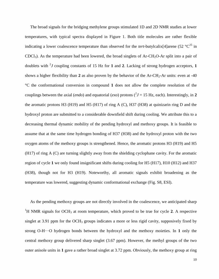
When crystallized from acetonitrile, title compound 2 was found to form a 1:3 inclusion
compound (2a) in the monoclinic space group P21/n (Figure 6). One of the guest molecules is
accommodated inside the cavity (endo) and the two others outside the cavity (exo) of the host
molecule. Similar to 1a, the macrocyclic framework adopts a pinched cone conformation with
interplanary angles of 33.4° (A/C) and 76.4° (B/quinizarin), respectively (Table 2). The cup-shape of
the molecule is stabilized by a strong O-H···O hydrogen bond, which links the phenolic hydroxyl

18
group to one of the neighbouring methoxy atoms. Like in 1a, the sterically demanding anthraquinone
moiety is turned towards the cavity (mpla M/mpla Q = 52.3°), and again we found stabilizing C-
H···O-contacts involving the two methoxy oxygens and quinizarine ring D [d(O···H) = 2.49 and 2.75
Å, resp.] The enlarged cavity is able to incorporate one acetonitrile molecule which is fixed by three
C-H···-interactions33
to the aromatic rings A, B and C. Noteworthy, it is tilted by an angle of 48.7°
against the mean plane of the four carbon atoms of the methylene bridges and thereby parallel to the
quinizarin unit. Very likely, the reasons for this is a kind of ···-stacking, involving quinizarin ring
D and the CN triple bond in the acetonitrile [d = 3.384(2) Å]. Additionally, the latter is engaged in an
intramolecular C-H···-contact with a tert-butyl H atom H30 (d = 2.83 Å).
Figure 6. Molecular structure of 2a, showing 50 % probability displacement
representation.
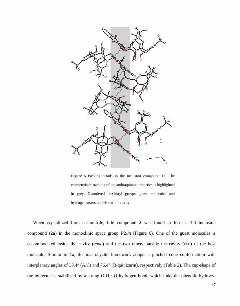
In the packing of 2a, two hosts are paired via their quinizarin units developing C-H···-contacts
[d(H35A···centroid F) = 2.93 Å]. Interestingly, we also observed a close proximity between bridge
19
oxygen O4 and a neighboring quinone unit in ring E [d = 3.215(2) Å] (Figure 7). These contacts
resemble charge-transfer-like complexes which are already known from quinones and sulfur.34
Unlike molecule 1, we observe no continuous aromatic stacking. In the overall packing all three of
the included solvent molecules take part with the exo oriented acetonitrile molecules mediating the
connection of the host molecules.
a) b)
Figure 7. a) Dimer formation in the packing of 2 in its 1:3 acetonitrile inclusion compound (2a). Guest molecules and
hydrogen atoms are left out for clarity. b) The two paired anthraquinone units in 2a resemble a charge-transfer complex.
Fluorescence Studies. In order to extensively characterize the new receptors 1 and 2, we studied
their luminescent behavior in DMSO solution in comparison to that of quinizarin (1,4-
dihydroxyanthraquinone) and 1,4-dimethoxyanthraquinone.35
The absorption and fluorescence
spectra are presented in Figure 8. Absolute excitation maxima, (ex)max, absolute emission maxima,
(em)max, and the associated Stokes shifts are summarized in Table 2. The Lambert-Beer law is valid
in the entire concentration interval. The fluorescence spectra have been collected at the respective
absorption maxima.

20
Table 2. Fluorescence data for 1, 2, quinizarin and 1,4-dimethoxy-
anthraquinone in DMSO (20 °C)
compound 1 2 quinizarin 1,4-dimethoxy-
anthraquinone
(ex)max (nm) 423 438 481 426
(em)max (nm) 539 548 560 540
Stokes shift (nm) 116 110 79 114
a) concentration: 0.007 mM; 0.01 mM; 0.02 mM; 0.05
mM; 0.1 mM; 0.2 mM
b) concentration: 0.007 mM; 0.01 mM; 0.02 mM;
0.05 mM; 0.1 mM; 0.2 mM
c) concentration: 0.005 mM; 0.007 mM; 0.01 mM;
0.02 mM; 0.05 mM; 0.07 mM; 0.1 mM
d) concentration: 0.005 mM; 0.007 mM; 0.01 mM;
0.02 mM; 0.05 mM; 0.07 mM; 0.1 mM
Figure 8. Excitation and fluorescence emission spectra of macrocycles 1 (a), 2 (b), quinizarin (c) and 1,4-
dimethoxyanthraquinone (d) at different concentrations in DMSO.

21
Quinizarin shows a broad absorption maximum at 481 nm, which corresponds to the red colour of
its solution. Macrocycles 1 and 2 as well as 1,4-dimethoxyanthraquinone experience a hypsochromic
shift to 423, 438 and 426 nm, respectively, being in coherence with their yellow solutions. The color
change is obviously a result of the altered electronic situation connected with the etherification of the
hydroxyl groups of the quinizarin. The fluorescence maxima for 1 and 2 have been determined with
539 and 548 nm, which is slightly lower than for quinizarin (560 nm) and similar to 1,4-
dimethoxyanthraquinone (540 nm). The resulting stokes shifts are considerable higher for the
macrocycles and 1,4-dimethoxyanthraquinone than for quinizarin. By way of interest, the excitation
and emission spectra of quinizarin develop a rather pronounced plateau; this may result from a higher
aggregation of the molecules.
CONCLUSION
Two unprecedented cyclophanes (1 and 2) with intrinsic fluorescence have been synthesized from
putative linear trimers and quinizarin applying the principle of Ziegler and Ruggli [22]. The
constitution of the molecules is related to the one of methylated calixarenes, though we observed a
different conformational behavior. In general, trimethoxy cycle 1 shows a higher flexibility than the
monophenolic cycle 2, which is stabilized by a strong hydrogen bond. For each of the title
compounds temperature-dependent 1H NMR spectroscopy revealed a typical sharpening of the
methylene resonances and a conformational stabilization with decreasing temperatures. These
findings justify the assumption of ‘cone’ conformations with all four aromatic units pointing in one
direction for both cycles. This hypothesis is supported by respective shifts of the methylene carbons
in the 13
C NMR spectra and pertinent NOEs. The cup-shape of both title molecules has also been
confirmed by molecular modelling and X-ray crystallography. It seems opportune to assume that C-
H···O-contacts between the methoxy oxygen atoms and the quinizarin fulfill here the function of the

22
strong O-H···O-bonds found in calixarenes to stabilize the cone conformation. Morever, fast
exchange processes in the NMR spectra of 1 and 2 suggest two rather similar cone forms for each
cycle similar to pinched cone calixarenes.
As demonstrated by excitation and fluorescence emission spectra the incorporation of an
anthraquinone unit into a cyclophane does not change its luminescent behavior. The additional plane,
electron-rich fluorophore in 1 and 2 allows additional interactions towards potential guest molecules
as shown for acetonitrile complexes. We found a better fit between host and guest with more
stabilizing C-H···O- and C-H···-contacts in comparison to the somewhat smaller calix[4]arenes.
This is especially true for cycle 2, where all three hydrogen atoms of the acetonitrile molecule are
involved in the complexation. By regarding acetonitrile as a simple model of methyl amines, these
findings are encouraging the further development of this promising new class of macrocycles. Our
next step in this respect will be the introduction of hydrophilic groups to the molecules resulting in
water soluble receptors.
EXPERIMENTAL SECTION
Materials and Methods. Melting points have been determined on a microscope heating stage and
are uncorrected. IR spectra were measured as KBr pellets and with the ATR method. The UV/VIS
and fluorescence measurements have been carried out using quartz cuvettes (10 x 10 mm) and
diluting the respective stock solutions in DMSO (1: c = 1.3 mmol/l; 2: c = 1.2 mmol/l; quinizarin: c =
1.2 mmol/l; 1,4-dimethoxyanthraquinone: c = 1.2 mmol/l) to the concentrations specified. NMR
spectra were recorded at 500.1 (1H NMR) and 125.7 MHz (
13C NMR), respectively, with sample
temperatures regulated at 293 K, unless otherwise stated. Spectra have been assigned using COSY,
DEPT-135, HSQC and HMBC. Chemical shifts are reported in parts per million relative to the

23
internal reference TMS. Multiplicity is abbreviated as follows: s (singlet), d (doublet), t (triplet) and
m (multiplet). Analytical TLC was performed on precoated silica gel plates (60 F254). The reagents
and solvents were used as purchased from the chemical suppliers, with the exception of those for
cyclisation reaction (dried dynamically over molecular sieve, 3 Å). The trimers 316
and 615
as well as
quinizarin36
were prepared according to literature procedures. 1,4-Dimethoxyanthraquinone is
commercially available. For the energy minimizations we used the program MacroModel V.9.8
(OPLS_2001 forcefield, MCMM, 1,000 steps).
5-tert-Butyl-1,3-bis-(5-tert-butyl-2-methoxy-benzyl)-2-methoxybenzene (4)37
. The phenolic
trimer 3 (5.50 g, 11.6 mmol) was dissolved in THF (130 ml) and DMF (10 ml). Subsequently, NaH
(60% in paraffin, 3.00 g, 127.6 mmol) and MeI (8.7 ml, 139.50 mmol) were added carefully. Heating
under reflux for 4 h produced a white precipitate. Subsequently, the solvent was evaporated and the
residue taken up in chloroform and water. The organic phase was separated and the solvent removed
almost completely. The white precipitate of 4 formed after addition of MeOH was filtered, washed
first with MeOH, then with ether and dried afterwards. Yield: 5.85 g (97 %). Mp. 138-139°C (Lit.38
:
140-141 °C). 1H-NMR (CDCl3): 1.17 (s, 9H, C(CH3)); 1.22 (s, 18H, C(CH3)); 3.61 (s, 3H, ArOCH3);
3.81 (s, 6H, ArOCH3); 4.03 (s, 4H, ArCH2Ar); 6.80 (d, 2H, ArCH, 3JHH=8.5 Hz); 6.98 (s, 2H,
ArCH); 7.06 (d, 2H, ArCH, 4JHH=2.5 Hz); 7.18 (dd, 2H, ArCH,
3JHH=8.5 Hz,
4JHH=2.5 Hz);
13C-
NMR (CDCl3): 30.0 (CH2), 31.4, 31.5 (C(CH3)); 34.0, 34.2 (C(CH3)); 55.4, 61.0 (ArOCH3); 109.7,
123.3, 126.1, 127.7, 129.1, 132.5, 142.8, 145.9, 154.9, 155.4 (ArC). m/z (ESI): Calc.: 516.36, found:
555.26 (M+K+) IR (cm
-1): 2951, 2901, 2865, 2836, 1609, 1505, 1461, 1245, 1135, 1011, 816.
Elemental analysis calculated for C35H48O3: C, 81.35 %; H, 9.36 %. Found: C, 81.72 %; H, 9.82 %.
24
1,3-Bis{[3-(bromomethyl)-5-(tert-butyl)-2-methoxy-phenyl]methyl}]-5-(tert-butyl)-2-
hydroxybenzene (5). Methyl ether 4 (3.00 g, 5.8 mmol), paraformaldehyde (0.42 g, 14.0 mmol) and
glacial acetic acid (40 ml) were stirred for 1 h at 70 °C in a bomb tube. To the white suspension, zinc
bromide (1.30 g, 5.8 mmol) and hydrogen bromide (33 % in glacial acetic acid, 5.8 ml) were added
and stirring was continued for 5 h at 70 °C. After cooling down, the clear brown solution was poured
into water, whereupon a white cloudy suspension appeared. After extraction with CH2Cl2, the organic
phase was dried over MgSO4 und the solvent was evaporated under reduced pressure. The resulting
brown oil was column chromatographed on SiO2 (eluent: n-hexane/ethyl acetate, 14:1) to yield 0.38 g
(9 %) of a beige solid. Mp. 158-160 °C. TLC: Rf = 0.19 (SiO2; n-hexane/chloroform, 1:1). 1H-NMR
(CDCl3): 1.20 (s, 27H, C(CH3)); 3.93 (s, 4H, ArCH2Ar); 3.95 (s, 6H, ArOCH3); 4.58 (s, 4H,
ArCH2OAr); 6.98 (s, 2H, ArCH); 7.15 (d, 2H, ArCH, 4JHH=2.5 Hz); 7.24 (d, 2H, ArCH,
4JHH=2.5
Hz); 7.34 (s, 1H, ArOH); 13
C-NMR (CDCl3): 28.8, 31.0 (CH2); 31.3, 31.5 (C(CH3)); 34.0, 34.3
(C(CH3)); 62.2 (ArOCH3); 125.6, 126.5, 126.7, 128.9, 130.3, 133.3, 142.7, 147.8, 149.9, 153.2
(ArC). m/z (APCI): Calc.: 688.19, found: 687.50 [M-]. IR (cm
-1): 3374, 2956, 2901, 2866, 1600,
1481, 1434, 1218, 1203, 988, 880, 751. Elemental analysis calculated for C36H48Br2O3 · 2 H2O: C,
59.67 %; H, 7.23 %. Found: C, 59.17 %; H, 7.06 %. Additionally to 5, we isolated 0.15 g (4 % yield)
of the fully methylated trimer 6.
1,3-Bis{[3-(bromomethyl)-5-(tert-butyl)-2-methoxy-phenyl]methyl}]-5-(tert-butyl)-2-
methoxybenzene (6). Under an argon atmosphere methyl ether 4 (4.75 g, 9.2 mmol), dissolved in
TFA (20 ml), was treated with bromomethyl methyl ether (1.0 ml, 1.53 g, 12.3 mmol). After 24 h the
brownish solution was quenched with water, followed by extraction with CHCl3. The organic phase
was dried over MgSO4 und the solvent was evaporated under reduced pressure. The resulting green
oil was column chromatographed on SiO2 (eluent: n-hexane/chloroform, 1:1 1:2) to yield 0.70 g

25
(11 %) of a colorless oil, which crystallized later. Mp. 120-122 °C. TLC: Rf = 0.25 (SiO2; n-
hexane/chloroform, 1:1). 1H-NMR (CDCl3): 1.16 (s, 9H, C(CH3)); 1.21 (s, 18H, C(CH3)); 3.59 (s,
3H, ArOCH3); 3.84 (s, 6H, ArOCH3); 4.08 (s, 4H, ArCH2Ar); 4.61 (s, 4H, ArCH2Br); 6.94 (s, 2H,
ArCH); 7.02 (d, 2H, ArCH, 4JHH=2.5 Hz); 7.25 (d, 2H, ArCH,
4JHH=2.6 Hz);
13C-NMR (CDCl3):
29.1, 29.9 (CH2); 31.3, 31.4 (C(CH3)); 34.2, 34.3 (C(CH3)); 60.8, 61.3 (ArOCH3); 126.2, 126.2,
128.9, 130.3, 132.5, 133.9, 146.5, 147.0, 154.5, 154.7 (ArC). m/z (ESI): Calc.: 702.21, found: 725.30
[M+Na+], 741.3 [M+K
+]. IR (cm
-1): 3672, 2955, 2826, 1583, 1480, 1220, 1203, 1006, 882. Elemental
analysis calculated for C36H48Br2O3 · H2O: C, 62.45 %; H, 7.22 %. Found: C, 62.13 %; H, 7.41 %.
General Synthetic Procedures for Macrocycles 1 and 2. Under an argon atmosphere the
respective trimer 5 or 6 and quinizarin, each dissolved in dry acetone (250 ml per 0.55 mmol of the
trimer), are slowly dropped into a refluxing suspension of K2CO3 (4 eq) in dry acetone (100 ml per
0.55 mmol trimer) using a high-dilution apparatus.21
The color of the reaction mixture changes from
dark blue to dark green. After 48 h of refluxing, solid components are removed by filtration. The
solvent was evaporated and the resulting reddish brown oil was column chromatographed on SiO2
(eluent for 1: chloroform/ethyl acetate, 24:1; eluent for 2: n-hexane/ethyl acetate, 2:1) to yield
macrocycles 1 and 2 as orange solids.
2,10-Dioxa-44,6
4,8
4-tri-tert-butyl-4
1,6
1,8
1-trimethoxy-1(1,4)-anthraquinona-4,6,8(2,6)-
tribenzenacyclodecaphane (1). Yield 435 mg (58 %). Mp. 280-282 °C. TLC: Rf = 0.44 (SiO2;
chloroform/ethyl acetate, 24:1).1H-NMR (CDCl3): 0.86 (s, 18H, C(CH3)); 1.38 (s, 9H, C(CH3)); 3.50
(s, 2H, ArCH2Ar);39
3.63 (s, 3H, ArOCH3); 3.72 (s, 6H, ArOCH3); 4.31 (s, 2H, ArCH2Ar);39
5.37 (s,
4H, ArCH2OAr); 6.78 (s, 2H, 37-, 38-ArCH); 6.81 (s, 2H, 5-, 17-ArCH); 7.11 (d, 2H, 3-, 19-ArCH,
4JHH=1.5 Hz); 7.25 (s, 2H, 10-, 12-ArCH); 7.69 (m, 2H, 44-, 45-ArCH); 8.18 (m, 2H, 43-, 46-ArCH);
13C-NMR (CDCl3): 31.0 (C(CH3)); 31.5 (CH2); 31.6 (C(CH3)); 33.9, 34.2 (C(CH3)); 60.3, 62.3

26
(ArOCH3); 64.4 (OCH2); 122.7 (40-, 49- ArC); 123.0 (37-, 38-ArC); 124.0 (3-, 19-ArC); 126.3 (43-
, 46-ArC); 126.8 (10-, 12-ArC); 127.0 (5-, 17-ArC); 128.3 (2-, 20-ArC); 132.9 (9-, 13-ArC); 133.0
(44-, 45-ArC); 134.3 (42-, 47-ArC); 134.5 (6-, 16-ArC); 145.8 (11-ArC); 146.7 (4-, 18-ArC); 151.9
(36-, 39-ArC); 153.8 (1-, 15-ArC); 155.0 (8-ArC); 183.4 (C=O). m/z (ESI): Calc.: 780.40, found:
803.37 (M+Na+). IR (cm
-1): 3442, 2960, 2905, 2870, 2826, 1668, 1595, 1567, 1483, 1465, 1255,
1238, 1216, 1014, 727. Elemental analysis calculated for C51H56O7 · ½ CH3COOCH2CH3: C, 77.16
%; H, 7.33 %. Found: C, 77.01 %; H, 7.36 %.
41,8
1-Dimethoxy-2,10-dioxa-6
1-hydroxy-4
4,6
4,8
4-tri-tert-butyl-1(1,4)-anthraquinona-
4,6,8(2,6)-tribenzenacyclodecaphane (2). Yield: 75 mg (18 %). Mp. 234-236 °C. TLC: Rf = 0.22
(SiO2; n-hexane/ethyl acetate, 2:1). 1H-NMR (CDCl3): 0.88 (s, 18H, C(CH3)); 1.34 (s, 9H, C(CH3));
3.44 (br s, 2H, ArCH2Ar); 3.91 (s, 6H, ArOCH3); 4.30 (br s, 2H, ArCH2Ar); 5.38 (br s, 2H,
ArCH2OAr); 5.45 (br s, 2H, ArCH2OAr); 6.62 (s, 1H, ArOH); 6.89 (d, 2H, 5-, 17-ArCH, 4JHH=2.5
Hz); 7.13 (s, 2H, 37-, 38-ArCH); 7.19 (s, 2H, 10-, 12-ArCH); 7.23 (d, 2H, 3-, 19-ArCH, 4JHH=2.5
Hz); 7.68 (m, 2H, 44-, 45-ArCH); 8.15 (m, 2H, 43-, 46-ArCH); 13
C-NMR (CDCl3): 30.3 (CH2); 30.9,
31.7 (C(CH3)); 33.9, 34.0 (C(CH3)); 63.3 (OCH2); 63.8 (ArOCH3); 121.7 (37-, 38-ArC); 122.7 (40-,
49-ArC); 125.0 (3-, 19-ArC); 126.2 (44-, 45-ArC); 126.4 (10-, 12-ArC); 126.5 (9-, 13-ArC); 127.1
(5-, 17-ArC); 128.2 (2-, 20-ArC); 132.0 (6-, 16-ArC); 132.9 (43-, 46-ArC); 134.5 (42-, 47-ArC);
142.1 (11-ArC); 147.8 (21-, 29-ArC); 150.0 (8-ArC); 151.5 (36-, 39-ArC); 152.5 (1-, 15-ArC); 183.3
(C=O). m/z (ESI): Calc.: 766.39, found: 789.40 (M+Na+). IR (cm
-1): 2953, 1667, 1635, 1591, 1568,
1483, 1237, 1210, 981, 795, 726. Elemental analysis calculated for C50H54O7 · ½ CH3COOCH2CH3:
C, 77.01 %; H, 7.21 %. Found: C, 76.91 %; H, 7.60 %.
X-ray structure determination. Crystals of compounds 1a, 2a and 4 suitable for X-ray
diffraction have been obtained by slow evaporation of the respective solution (1 in

27
acetonitrile/chloroform (1:1), 2 in acetonitrile and 4 in ethyl acetate). The intensity data were
collected at 100 K on a Bruker Kappa diffractometer equipped with an APEX II CCD area detector
and graphite-monochromatized Mo Kα radiation (λ = 0.71073 Å) employing φ and ω scan modes.
The data were corrected for Lorentz and polarization effects. Semiempirical absorption correction
was applied using the SADABS program.40
The SAINT program40
was used for the integration of the
diffraction profiles. The crystal structures were solved by direct methods using SHELXS-9741
and
refined by full-matrix least-squares refinement against F2 using SHELXL-97.
41 All non-hydrogen
atoms were refined anisotropically. Hydrogen atoms were positioned geometrically and allowed to
ride on their parent atoms. Geometrical calculations were performed using PLATON and molecular
graphics were generated using SHELXTL.41
The crystallographic data for the structures in this paper
have been deposited with the Cambridge Crystallographic Data Centre; CCDC numbers: 1042786
(1), 1042787 (2) and 1042788 (4).
ASSOCIATED CONTENT
Supporting Information
Energy-minimized structures of 1 and 2. 1H und
13C NMR spectra of compounds 1, 2, 4, 5 and 6;
NOESY spectra of 1 and 2. Selected details of the data collection; table of distances and angles of
inter- and intramolecular contacts in the structures of 1a, 2a and 4; description of the X-ray structure
of 4. X-ray crystallographic data files (CIF) for 1, 2 and 4. This material is available free of charge
via the Internet at http://pubs.acs.org.
AUTHOR INFORMATION
Corresponding Author
*E-mail: [email protected]

28
Notes
The authors declare no competing financial interest.
ACKNOWLEDGEMENTS
Financial support from ‘Fonds der Chemischen Industrie’ and the Institute of Organic Chemistry,
University of Freiberg, is gratefully acknowledged. Furthermore, we thank M. Stapf for his helpful
advice.

29
REFERENCES AND NOTES
(1) B. Valeur, Molecular Fluorescence. Principles and Applications, 2nd
ed., Wiley-VCH, Weinheim,
2013.
(2) Parkesh, R.; Veale, E. B.; Gunnlaugsson, T.; edited by Wang, B.; Anslyn, E. V. Chemosensors
2011, 229-252.
(3) (a) Azadbakht, R.; Khanabadi, J. Spectrochim. Acta 2014, A124, 249-255. (b) Nativi, C.;
Francesconi, O.; Gabrielli, G.; De Simone, I.; Turchetti, B.; Mello, T.; Di Cesare Manelli, L.;
Ghelardini, C.; Buzzini, P., Roelens, S. Chem. Eur. J. 2012, 18, 5064-5072. (c) Alfonso, I.;
Burguete, M. I.; Galindo, F.; Luis, S. V.; Vigara, L. J. Org. Chem. 2009, 74, 6130–6142.
(4) (a) Li, Z.; Sei, Y.; Akita, M.; Yoshizawa, M. Chem. Asian J. 2014, 9, 1016-1019. (b) Ahmed, N.;
Shirinfar, B.; Youn, I. S.; Yousuf, M.; Kim, K. S. Org. Biomol. Chem. 2013, 6407-6413. (c) Ghosh,
K.; Sarkar, A. R. Tetrahedron Lett. 2009, 50, 85-88.
(5) (a) Halling, M. D.; Unikela, K. S.; Bodwell, G. J.; Grant, D. M.; Pugmire, R. J. J. Phys. Chem. A
2012, 116, 5193-5198. (b) Nandaluru, P. R.; Dongare, P.; Kraml, C. M.; Pascal Jr., R. A.; Dawe, L.
N.; Thompson, D. W.; Bodwell, G. J. Chem. Commun. 2012, 48, 7747-7749. (c) Franz, D.; Robbins,
S. J.; Boeré, R. T.; Dibble, P. W. J. Org. Chem. 2009, 74, 7544-7547. (d) Tsuge, A.; Otsuka, M.;
Moriguch, T.; Sakata, K. Org. Biomol. Chem. 2005, 3, 3590-3593. (e) Abe, H; Mawatari, Y.;
Teraoka, H.; Fujimoto, K.; Inouye, M. J. Org. Chem. 2004, 69, 495-504.
(6) (a) Rajakumar, P.; Anandhan, R. Synth. Commun. 2013, 43, 882-892. (b) Rajakumar, P.;
Kanagalatha, R Tetrahedron Lett. 2007, 48, 2761-2764. (c) Lukyanenko, N. G.; Lyapunov, A. Y.;
Kirichenko, T. I.; Botoshansky, M. M.; Simonov, Y. A.; Fonari, M. S. Tetrahedron Lett. 2005, 46,
2109-2112. (d) Matsumoto, K.; Minami, H.; Kawase, T.; Oda, M. Org. Biomol. Chem. 2004, 2, 2323-
2326.
(7) (a) Chu, M.; Scioneaux, A. N.; Hartley, C. S. J. Org. Chem., 2014, 79, 9009–9017. (b)
Balakrishnan, K.; Datar, A.; Zhang, W.; Yang, X.; Naddo, T.; Huang, J.; Zuo, J.; Yen; M., Moore, J.
S.; Zang, L. J. Am. Chem. Soc. 2006, 128, 6576–6577. (c) Yasuhide, I.; Takahiro, M.; Noboru, O.;
Hidemitsu, U.; Atsuhiro, O. Angewandte Chemie, Int. Ed. 2005, 44, 1856-1860.
(8) Gleiter, R.; Hopf, H. (eds.) Modern Cyclophane Chemistry, Wiley, Weinheim, 2004.
(9) (a) Gutsche, C. D. Calixarenes, The Royal Society of Chemistry, Cambridge, UK, 2008. (b)
Vicens, J.; Harrowfield, J. Calixarenes in the Nanoworld, Springer-Verlag, Dordrecht, 2007. (c)
Asfari, M. Z.; Böhmer, V.; Harrowfield, J.; Vicens, J. Calixarenes 2001, Kluwer Academic,
Dordrecht, 2001. (d) Mandolini, L.; Ungaro, R. Calixarenes in Action, Imperial College Press,
London, 2000
(10) Steed, J. W.; Atwood, J. L. Supramolecular Chemistry, 2nd
ed., John Wiley & Sons, Chichester,
2009.

30
(11) (a) Song, M.; Sun, Z.; Han, C.; Tian, D.; Li, H.; Kim, J. S. Chem. Asian J. 2014, 9, 2344-2357.
(b) Ocak, U.; Ocak, M.; Bartsch, R. A. Inorg. Chim. Acta 2012, 381, 44-57. (c) Sharma, K.; Cragg, P.
J. Chem. Sensors 2011, 1, 9/1-9/18. (d) Fischer, C.; Gruber, T.; Seichter, W.; Weber, E. Org. Biomol.
Chem. 2011, 9, 4347-4352. (e) Talanova, G. G.; Talanov, V. S. Supramol. Chem. 2010, 22, 838-852.
(f) Chawla, H. M.; Pant, N.; Kumar, S.; Kumar, N.; Black, D. StC. Calixarene-based materials for
Chemical Sensors, in Chemical Sensors: Fundamentals of Sensing Materials , ed. G. Korotcenkov,
Momentum Press, New York, 2010, vol. 3. (g) Lo, P. K.; Wong, M. S. Sensors 2008, 8, 5313-5335.
(12) Georghiou, P. E.; Li, Z.; Ashram, M.; Chowdhury, S.; Mizyed, S.; Tran, A. H.; Al-Saraierh, H.;
Miller, D. O. Synlett 2005, 6, 879-891.
(13) (a) Moreno-Corral, R.; Lara, K. O. Supramol. Chem. 2008, 20, 427-435. (b) Gu, S.-J.; Qin, D.-
B.; Jin, L.-H. Chin. J. Struct. Chem. 2008, 27, 1035-1038. (c) Kitamura, C.; Fujimoto, J.; Kawatsuki,
N.; Yoneda, A. Heterocycles 2006, 68, 2171-2175. (d) Haeg, M. E.; Whitlock, B. J.; Whitlock, H.
W., Jr. J. Amer. Chem. Soc. 1989, 111, 692-696. (e) Buhleier, E.; Vögtle, F. Chem. Ber. 1978, 111,
2729-2731.
(14) Bhattacharya, D.; Sathiyendiran, M.; Wu, J.-Y.; Chang, C.-H.; Huang, S.-C.; Zeng, Y.-L.; Lin,
C.-Y.; Thanasekaran, P.; Lin, B.-C.; Hsu, C.-P.; Lee, G. H.; Peng, S. M.; Lu, K. L. Inorg. Chem.
2010, 49, 10264-10272
(15) (a) Lin, C.-X.; Kong, X.-F.; Li, Q.-S.; Zhang, Z.-Z.; Yuan, Y.-F.; Xu, F.-B. CrystEngComm
2013, 15, 6948-6962. (b) Qin, D.-B.; Xu, F.-B.; Li, Q.-S.; Song, H.-B.; Zhang, Z.-Z. Synlett 2005, 19,
2987-2989.
(16) Dhawan, B.; Gutsche, C. D. J. Org. Chem. 1983, 48, 1536-1539.
(17) Li, H.; Homan, E. A.; Lampkins, A. J.; Ghiviriga, I.; Castellano, R. K. Org. Lett. 2005, 7, 443-
446.
(18) (a) Kämmerer, H.; Schweikert, H.; Haub, H.-G. Makromol. Chem. 1963, 67, 173–181. (b)
Kämmerer, H.; Gros, G.; Schweikert, H. Makromol. Chemie 1971, 143, 135–152.
(19) (a) Hornung, E. J. Org. Chem. 1945, 10, 263-266. (b) Pickholtz, Y.; Sasson, Y.; Blum, J.
Tetrahedron Lett. 1974, 14, 1263-1266. (c) Aizenshtat, Z.; Hausmann, M.; Pickholtz, Y.; Tal, D.;
Blum, J. J. Org. Chem. 1977, 42, 2386-2394.
(20) We used 4-tert-Butyl-2,6-bis[(5-tert-butyl-2-methoxyphenyl)methyl]phenol as the
monophenolic trimer. The pKa have been calculated using Advanced Chemistry Development
(ACD/Labs) Software V11.02 (© 1994-2014 ACD/Labs) as employed in SciFinder.
(21) Vögtle, F. Chem.-Ztg. 1972, 96, 396-403.
(22) (a) Ziegler, K.; Eberle, H.; Ohlinger,H. Justus Liebigs Ann. Chem. 1933, 504, 94-130; (b)
Ziegler, K.; Aurnhammer, R. Justus Liebigs Ann. Chem. 1934, 513, 43-64; (c) Knops, P.; Sendhoff,
N.; Mekelburger, H.-B.; Vögtle, F. Top. Curr. Chem. 1991, 161, 1–36.

31
(23) (a) Kuno, L.; Biali, S. E. J. Org. Chem. 2011, 76, 3664-3675. (b) Gruner, M.; Fischer, C.;
Gruber, T.; Weber, E. Supramol. Chem. 2010, 22, 256-266. (c) Choe, J.-I; Lee, S. H.; Oh, D.-S. Bull.
Korean Chem. Soc. 2004, 25, 55-58.
(24) Jaime, C.; de Mendoza, J.; Prados, P.; Nieto, P. M.; Sánchez, C. J. Org. Chem. 1991, 56, 3372-
3376.
(25) Gutsche, C. D.; Bauer, L. J. Tetrahedron Lett. 1981, 22, 4763-4766.
(26) Gruber, T.; Gruner, M.; Fischer, C.; Seichter, W.; Bombicz, P.; Weber, E. New J. Chem. 2010,
34, 250-259.
(27) Gruber, T.; Bombicz, P.; Seichter, W.; Weber, E. J. Struct. Chem. 2009, 50, 522-531.
(28) (a) Nigam, G. D.; Deppisch, B. Z. Kristallogr. 1980, 151, 185-191. (b) Swaminathan, S.; Nigam,
G. D. Current Sci. 1967, 36, 541. (c) Murty, B. V. R. Z. Kristallogr. 1959, 111, 238-239.
(29) Cao, L.-P.; Wang, Y.-Z.; Gao, M.; Zhou, B.-H. Acta Cryst. 2007, E63, o1876–o1877.
(30) Zhang, J.-J.; Yina, C.-X.; Huob, F.-J. Acta Cryst. 2013, E69, o788.
(31) G. A. Jeffrey, An introduction to hydrogen bonding, Oxford University Press, 1997.
(32) (a) Saraogi, I.; Vijay, V. G.; Das, S.; Sekar, K.; Guru Row, T. N. Cryst. Eng. 2003, 6, 69-77. (b)
Yumi, N. I.; Inoue, Y.; Nakanishi, I.; Kitaura. K. Protein Sci. 2008, 17, 1129-1137.
(33) Nishio, M; Umezawa, Y; Honda, K.;Tsuboyama, S.; Suezawa, H. CrystEngComm 2009, 11,
1757-1788
(34) Aly, A. A.; Elkanzi, N. A. A.; Brown, A. B. Phosphorus Sulfur Silicon Relat. Elem. 2014, 189,
440-452.
(35) For the respective spectra in methylcyclohexane see: Shcheglova, N. A.; Shigorin, D. N.;
Sergeev, A. M. Zhurn. Fizich. Khim. 1979, 53, 1749-1752.
(36) Gattermann, L; Wieland, H.; Wieland, T. Die Praxis des organischen Chemikers, 43rd ed., de
Gruyter, Berlin, 1982.
(37) Similar to: Yamagishi, T.; Enoki, M.; Inui, M.; Furukawa, H.; Nakamoto, Y.; Ishida, S. J.
Polym. Sci., Part A: Polym. Chem. 1993, 31, 675-682.
(38) Zinke, A.; Ott, R.; Garrana, F. H. Monatsh. Chem. 1958, 89, 135-142.
(39) The respective signal is not visible at room temperature. We display here the chemical shift
at -35 °C.
(40) Bruker, SAINT, SADABS, AXS Inc., Madison, Wisconsin, USA, 2007.

32
(41) Sheldrick, G. M. Acta Cryst. 2008, A64, 112-122.