Fluorescence Quenching by Colloidal Heavy Metals Nanoparticles: Implications for Correlative...
10
SCANNING VOL. 29, 152–161 (2007) Wiley Periodicals, Inc. Fluorescence Quenching by Colloidal Heavy Metals Nanoparticles: Implications for Correlative Fluorescence and Electron Microscopy Studies IRAWATI K. KANDELA 1 and RALPH M. ALBRECHT 1,2 1 Department of Pharmaceutical Sciences, 777 Highland Avenue, Madison, WI 53705, USA 2 Department of Animal Science, 1675 Observatory Drive, Madison, WI 53706, USA Summary: Labels for correlative immunolabeling in light (LM) and electron microscopy (EM) employing colloidal metal nanoparticles (gold or palladium) and fluorescent dyes (Alexa Fluor, AF) were investigated. The fluorescence signals from direct conjugates (cAu- IgG-AF) and from an indirect label system (cAu-IgG- anti IgG-AF) were studied using scanning spectrofluo- rometry and fluorescence light microscopy. Direct conjugation of protein — AF, IgG-AF or FGN- AF to 18 and 5 nm colloidal gold (cAu 18 and cAu 5 ) or 12 nm colloidal palladium particles (cPd 12 ) resulted in nearly completely quenched fluorescence signals (>99 %) at excitation wavelengths of 488, 546 and 594 nm. In contrast, indirect conjugation, when col- loidal metal particles and AF were conjugated to primary or secondary antibody, respectively (cAu- IgG-antiIgG-AF), sufficient fluorescence signal was detected. Commercially available conjugates, consisting of IgG-AF-cAu 5 and IgG-AF-cAu 10 , were also tested and proved to be a mixture of IgG-AF (unbound to cAu) and cAu-IgG-AF. SCANNING 29: 152 – 161, 2007. 2007 Wiley Periodicals, Inc. Key words: correlative labeling, fluorescent dyes, multiple labeling, Alexa Fluor, electron microscopy, light microscopy PACS: code: 78.67.Bf Nanocrystals and nanoparticles Introduction Correlative microscopy often involves the use of wide field or confocal fluorescence light microscopy Address for reprints: Ralph M. Albrecht, 1675 Observatory Drive Madison, WI 53706 USA, E-mail: [email protected] Received 24 December 2006; Accepted with revision 18 March 2007 (LM) coupled with scanning or transmission electron microscopy, (SEM or TEM). The examination of a single specimen by two or more imaging modes is often effective in obtaining complimentary sets of information which may otherwise not be attained by a single mode alone (Wetzel and Albrecht, 1989). Furthermore, LM enables real time imaging of live specimens and can also serve as a screening tool to determine which samples merit subsequent preparation for observation by EM at higher levels of spatial resolution. In LM, fluorophores are commonly used as reporters (Figure 1(b)). A variety of fluorophores, having a wide range of excitation and emission wavelengths, is commercially available, enabling multiple labeling for detection of different antigenic sites at the LM lev- els of spatial resolution. Most fluorophores are small, compatible with biological samples and easily conju- gated to different proteins or ligands. However, resolu- tion attainable with LM is limited (Birrell et al , 1985). Therefore, subsequent EM imaging may be beneficial to obtain higher spatial resolution for structural studies (Nisman et al , 2004; Albrecht et al , 1993). In EM, electron dense materials, such as colloidal metal nanoparticles, are useful as immunolabeling reporters (Figure 1(c)) because of their ability to scatter electrons, their increased secondary and backscattered electron emission properties and their distinct spheri- cal shapes. Colloidal gold, cAu, nanoparticles can be produced in sizes ranging from 1–150 nm (Simmons and Albrecht, 1989). For labeling, cAu particles can be conjugated to biological molecules, including anti- bodies or ligands via hydrophobic bonding. In gen- eral, the conjugation does not impair the activity and specificity of most antibodies or ligands. Conjugates are stable for 6 months and beyond if properly stored (Slot and Geuze, 1985). Simultaneous multiple labeling and co-localization for EM can be accomplished using colloidal particles of different elemental compositions, different shapes and different sizes (Meyer et al , 2006).
-
Upload
independent -
Category
Documents
-
view
1 -
download
0
Transcript of Fluorescence Quenching by Colloidal Heavy Metals Nanoparticles: Implications for Correlative...

SCANNING VOL. 29, 152–161 (2007) Wiley Periodicals, Inc.
Fluorescence Quenching by Colloidal Heavy Metals Nanoparticles:Implications for Correlative Fluorescence and Electron MicroscopyStudies
IRAWATI K. KANDELA1 and RALPH M. ALBRECHT1,2
1 Department of Pharmaceutical Sciences, 777 Highland Avenue, Madison, WI 53705, USA2 Department of Animal Science, 1675 Observatory Drive, Madison, WI 53706, USA
Summary: Labels for correlative immunolabeling inlight (LM) and electron microscopy (EM) employingcolloidal metal nanoparticles (gold or palladium) andfluorescent dyes (Alexa Fluor, AF) were investigated.The fluorescence signals from direct conjugates (cAu-IgG-AF) and from an indirect label system (cAu-IgG-anti IgG-AF) were studied using scanning spectrofluo-rometry and fluorescence light microscopy.
Direct conjugation of protein—AF, IgG-AF or FGN-AF to 18 and 5 nm colloidal gold (cAu18 and cAu5)
or 12 nm colloidal palladium particles (cPd12) resultedin nearly completely quenched fluorescence signals(>99 %) at excitation wavelengths of 488, 546 and594 nm. In contrast, indirect conjugation, when col-loidal metal particles and AF were conjugated toprimary or secondary antibody, respectively (cAu-IgG-antiIgG-AF), sufficient fluorescence signal wasdetected.
Commercially available conjugates, consisting ofIgG-AF-cAu5 and IgG-AF-cAu10, were also tested andproved to be a mixture of IgG-AF (unbound to cAu) andcAu-IgG-AF. SCANNING 29: 152–161, 2007. 2007Wiley Periodicals, Inc.
Key words: correlative labeling, fluorescent dyes,multiple labeling, Alexa Fluor, electron microscopy,light microscopy
PACS: code: 78.67.Bf Nanocrystals and nanoparticles
Introduction
Correlative microscopy often involves the use ofwide field or confocal fluorescence light microscopy
Address for reprints: Ralph M. Albrecht, 1675 ObservatoryDrive Madison, WI 53706 USA,E-mail: [email protected]
Received 24 December 2006; Accepted with revision18 March 2007
(LM) coupled with scanning or transmission electronmicroscopy, (SEM or TEM). The examination of asingle specimen by two or more imaging modes isoften effective in obtaining complimentary sets ofinformation which may otherwise not be attained bya single mode alone (Wetzel and Albrecht, 1989).Furthermore, LM enables real time imaging of livespecimens and can also serve as a screening tool todetermine which samples merit subsequent preparationfor observation by EM at higher levels of spatialresolution.
In LM, fluorophores are commonly used as reporters(Figure 1(b)). A variety of fluorophores, having awide range of excitation and emission wavelengths, iscommercially available, enabling multiple labeling fordetection of different antigenic sites at the LM lev-els of spatial resolution. Most fluorophores are small,compatible with biological samples and easily conju-gated to different proteins or ligands. However, resolu-tion attainable with LM is limited (Birrell et al , 1985).Therefore, subsequent EM imaging may be beneficialto obtain higher spatial resolution for structural studies(Nisman et al , 2004; Albrecht et al , 1993).
In EM, electron dense materials, such as colloidalmetal nanoparticles, are useful as immunolabelingreporters (Figure 1(c)) because of their ability to scatterelectrons, their increased secondary and backscatteredelectron emission properties and their distinct spheri-cal shapes. Colloidal gold, cAu, nanoparticles can beproduced in sizes ranging from 1–150 nm (Simmonsand Albrecht, 1989). For labeling, cAu particles canbe conjugated to biological molecules, including anti-bodies or ligands via hydrophobic bonding. In gen-eral, the conjugation does not impair the activity andspecificity of most antibodies or ligands. Conjugatesare stable for 6 months and beyond if properly stored(Slot and Geuze, 1985). Simultaneous multiple labelingand co-localization for EM can be accomplished usingcolloidal particles of different elemental compositions,different shapes and different sizes (Meyer et al , 2006).

I. K. Kandela and R. M. Albrecht: Correlative fluorescence and electron microscopy studies 153
Fig 1. Different types of immunolabeling. A. Antigenic sitesto be detected. B. A typical immunolabeling in LM wherefluorophores are conjugated to antibodies specific for antigenicsites. C. A typical immunolabeling in EM where colloidal metalparticles are conjugated to antibody specific for antigenic sites.D. Ideal label, for correlative studies in LM and EM, containsfluorophore and colloidal metal particles, which are reporters forLM and EM, respectively. E. Commercially available label forcorrelative immunolabeling, containing mixed conjugates, whichare fluorophore-IgG-cAu and fluorophore-IgG. F. An indirectlabel system containing colloidal metal particles conjugatedto primary antibody and fluorophore conjugated to secondaryantibody.
Colloidal heavy metal particles have a high extinc-tion coefficient and produce a visible, inflated diffrac-tion image and thus can be identified and tracked inseveral forms of interference based LM (Goodman andAlbrecht, 1987). However, the use of heavy metal col-loids as light microscopy reporters, mainly in correl-ative studies, is often problematic. Particle size andspecimen geometry limit the information attainable byinterference based imaging systems. Individual parti-cles can be tracked and followed but not resolved.Enlarged smaller particles via silver enhancement mightprovide visibility in LM, but the spatial resolutionnecessary for subsequent EM of the same sample islost. Therefore, using reporters suitable for LM (fluo-rophores) together with those suitable for EM (colloidalmetal particles) is often a practical approach to labelsamples for correlative studies. For simultaneous mul-tiple labeling, colloidal metal particles with differentsizes and shapes or with different metal compositions,conjugated to primary antibodies or active antibodyfragments, can be linked to secondary antibodies, whichare in turn, bound to different fluorophores with differ-ent emission wavelengths. In ideal probes or labels forcorrelative studies, both reporters would be attached tothe same primary IgG (Figure 1(d)). However, previousstudies have shown that direct conjugation of cAu parti-cles to fluorophore-bound primary antibodies results inquenching and insufficient fluorescence signal. Fluores-cence quenching has been observed when other ligands
such as albumin (De Brabander et al , 1985) or avidin(Horisberger, 1988) are directly conjugated to both flu-orophores and cAu nanoparticles. In the other studies,the fluorescence signal was only weakly detected evenwhen both fluorescein isothiocyanate (FITC) and cAu6were conjugated to tertiary antibodies (Pombo, et al ,1999).
However, a new generation of fluorophores withhigher quantum yields, a wide range of excitation andemission wavelengths and greater resistance to bleach-ing are currently available. Whether the improved flu-orescence properties would result in improved fluores-cence in cases where the dyes are located near colloidalmetal particles of smallest size (5–15 nm) or decreaseddensity, Pd vs. Au is not clear.
In this study, we investigated the fluorescence sig-nal of labels where colloidal metal particles of dif-ferent sizes and different elemental compositions weredirectly or indirectly conjugated to fluorophores. Dif-ferent Alexa Fluors (AF), having a range of excitationand emission energies, were bound to primary IgG withor without colloidal nanoparticles. This was comparedto the signal obtained from an indirect immunolabel-ing (Figure 1(f)) with colloidal metal particles conju-gated to primary IgG, which was then labeled withAF-bound secondary IgG. Scanning spectrofluorometryand fluorescence microscopy was used to measure andobserve the differences in signal strength. AF of sev-eral excitation and emission wavelengths was selectedfor the current studies. For direct conjugates, both col-loidal nanoparticles of cAu and cPd were used. Varioussizes were also compared. Antibodies, IgG, and ligand,Fibrinogen, (FGN) systems were also investigated. Acommercially available direct conjugate of cAu parti-cles to AF-bound primary antibodies was also tested(Figure 1(e)).
Materials and Methods
Colloidal Gold Nanoparticles
Colloidal gold particles having an average diameterof 18 nm (cAu18) were prepared by reducing 0.5 mLof 4% HAuCl4 in boiling deionized distilled waterby adding 4 mL of 1% trisodium citrate. The mixturewas refluxed for 30 min. The change in color to redindicates the formation of cAu particles. The particleswere usually stored at 4◦C before use (Olorundare et al ,1992; Goodman et al , 1991; Park et al , 1987). Usingthe same procedure except that 4.5 mL of 1% trisodiumcitrate was added, cAu15 was produced.
Five nanometer colloidal gold nanoparticles, cAu5,were prepared by reduction of 0.75 mL HAuCl4 in240 mL deionized distilled water by adding 2 mLof white phosphorous solution (1 mL of saturatedphosphorous was diluted with 4 mL of ether). After
154 SCANNING Vol. 29, 4 (2007)
adjusting the pH to 7.0–7.2 with 0.2 M of K2CO3, theflask was agitated gently for 15 min. The mixture wasrefluxed for 20 min (Faulk and Taylor, 1971).
Colloidal Palladium Nanoparticles
Twelve nanometer colloidal palladium nanoparticleswere produced by reduction of 500 µM of K2PdCl4by 0.005% sodium ascorbate at 100oC. Reduction wasperformed in the presence of 0.04% of trisodium citrateas a stabilizing agent (Meyer and Albrecht, 2003).
Label Preparations
Goat antirabbit IgG conjugated to either Alexa Fluor(AF) 488, 546 and 594 and conjugates of humanFGN conjugated to Alexa Fluor (AF) 488, 546 and594 were purchased from Molecular Probes (Eugene,Oregon). Commercially available conjugates of goatantirabbit antibody conjugated to both Alexa Fluor488 and also directly conjugated to either cAu5 orcAu10 were purchased from Molecular Probes (Eugene,Oregon). Rabbit IgG was purchased from Sigma (St.Luois, Missouri). IgG and FGN were conjugated tocAu at pH 7.2 and 6.5 respectively, slightly above
1600
0
1400
1200
1000
800
600
400
200
1600
0
1400
1200
1000
800
600
400
200
30
25
20
15
10
05
00
Inte
nsity
Inte
nsity
Inte
nsity
Inte
nsity
510 520 530 540 550
Wavelength (nm)
570 580575 585 590 595 600
Wavelength (nm)
605 615 620610 625 630 635 640
Wavelength (nm)
605 615 620610 625 630 635 640
Wavelength (nm)
605 615 620610 625 630 635 640
Wavelength (nm)
570 580575 585 590 595 600
Wavelength (nm)
570 580575 585 590 595 600
Wavelength (nm)
510 520 530 540 550
Wavelength (nm)
510 515 520 525 530 535 540Wavelength (nm)
1000
800
600
400
200
0
Inte
nsity
250
200
150
100
50
0
Inte
nsity
250
200
150
100
50
0
Inte
nsity
1000
800
600
400
200
0
Inte
nsity
5
4
3
2
1
0
Inte
nsity
5
4
3
2
1
0
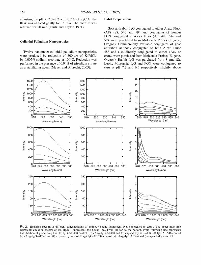
Fig 2. Emission spectra of different concentrations of antibody bound fluorescent dyes conjugated to cAu18. The upper most linerepresents emission spectra of 100 µg/mL fluorescent dye bound IgG. From the top to the bottom, every following line representshalf dilution of proceeding line. (a) IgG-AF 488 control, (b) cAu18-IgG-AF488 and (c) expanded y axis of B; (d) IgG-AF 546 control(e) cAu18-IgG-AF546 and (f) expanded y axis of E; (g) IgG-AF 594 control (h) cAu18-IgG-AF594 and (i) expanded y axis of H.

I. K. Kandela and R. M. Albrecht: Correlative fluorescence and electron microscopy studies 155
the pI of IgG and FGN (Simmons and Albrecht,1989). Since cAu or cPd clogs the conventional pHelectrode, pH adjustment was done with 0.2 N K2CO3,using a gel-filled combination electrode (#9115, OrionResearch, Cambridge, Massachusetts). The minimumamount of IgG or FGN necessary to stabilize thecAu was determined by adsorption isotherms (DeRoe et al , 1987; Slot and Geuze, 1985; Horisbergerand Rosset, 1977) obtained by testing if flocculationoccured, indicated by a change in color from red toblue in the case of cAu and precipitated colloid inthe case of cPd, when one drop of saturated Nacl wasadded to 0.2 mL of conjugated particle solution. ThecAu-IgG and cAu-FGN conjugates were sedimentedat 9,500× G for 30 min (cAu18) or 115,000× G for75 min (cAu5). Unbound protein was removed and softpellets were re-suspended in 0.1 M Hepes buffer at pH7.3.
Indirect label systems were assessed by conju-gation of rabbit IgG to cAu. Unbound IgG wasremoved by centrifugation followed by addition ofgoat anti-rabbit IgG-Alexa Fluor as a secondary label.Unbound secondary antibody was removed via a secondcentrifugation.
Spectrofluorometer Studies
Solutions of different concentrations of IgG or FGNconjugated to Alexa Fluor (AF) were measured using
a fluorescence spectrophotometer (Hitachi F3010) cou-pled with IBM analytical calculation software. AF 488,546 and 594 controls and conjugates were excited at495, 564 and 594 nm and emission was measured at523, 582 and 615 nm respectively. Fluorescence mea-surement was done over a range of excitation and emis-sion wavelengths, to be sure that there was no shift inexcitation or emission frequencies due to the presenceof colloidal nanoparticles.
Light Microscopy Studies
Conjugates were observed in a water in oil emulsionusing LM. 7 µL of suspension of conjugates in Hepesbuffer were mixed with 0.5 mL of oil phase (Zeissimmersion oil). The droplet emulsion diameter rangedfrom 5–25 µm. Neither oil nor the Hepes buffergave an autofluorescence signal in the excitation andemission wavelength ranges tested. The emulsions wereplaced on slides, sealed with nail polish and observedwith a Zeiss Axiovert 200 M microscope equippedwith mercury arc lamp (HBO-103W) using a 100×differential interference contrast (DIC) oil immersionobjective. Images were captured with a cooled CCDmonochrome camera, Axiocam HRm, at the frame sizeof 1388 × 1040 pixels in grey scale mode with 16 bitinformation depth with an exposure time of 20 s. Themicroscope and camera were controlled by Axio Visionsoftware version 4.4 (Zeiss, Germany).
IgG-AF 488
IgG-AF 546
IgG-AF 594
cAu5 cAu18
Fig 3. Light microscopy studies of emulsion systems with IgG-AFs as controls and direct conjugation of both AF and cAu5 or 18attached to the IgG. The conjugates were suspended in Hepes buffer and then were emulsified in oil (w/o). AF-488 emitted at 520 nm,AF-546 was emitted at 575 nm and AF 594 emitted at 615 nm.

156 SCANNING Vol. 29, 4 (2007)
1600
0
1400
1200
1000
800
600
400
200
1600
0
1400
1200
1000
800
600
400
200
10
8
6
4
2
0
Inte
nsity
Inte
nsity
Inte
nsity
Inte
nsity
510 520 530 540 550
Wavelength (nm)
570 580575 585 590 595 600
Wavelength (nm)
605 615 620610 625 630 635 640
Wavelength (nm)
605 615 620610 625 630 635 640
Wavelength (nm)
605 615 620610 625 630 635 640
Wavelength (nm)
570 580575 585 590 595 600
Wavelength (nm)
570 580575 585 590 595 600
Wavelength (nm)
510 520 530 540 550
Wavelength (nm)
510 515 520 525 530 535 540
Wavelength (nm)
1000
800
600
400
200
0
Inte
nsity
250
200
150
100
50
0
Inte
nsity
250
200
150
100
50
0
Inte
nsity
1000
800
600
400
200
0
Inte
nsity
5
4
3
2
1
0
Inte
nsity
5
4
3
2
1
0
A B C
D E F
G H I
Fig 4. Emission spectra of different concentrations of antibody bound fluorescent dyes conjugated to cPd12. The upper most linerepresents emission spectra of 100 µg/mL fluorescent dye bound IgG. From the top to the bottom, every following line represents halfdilution of proceeding line. (a) IgG-AF-488, (b) cPd12-IgG-AF488 and (c) expanded y axis of B; (d) IgG-AF-546, (e) cPd12-IgG-AF546and (f) expanded y axis of E; (g) IgG-AF-594, (h) cPd12-IgG-AF549 and (i) expanded y axis of H.
Results
Spectrofluorometry studies showed that direct conju-gation of different concentrations of IgG-AF 488, 546and 594 to cAu18 (Figure 2) and to cAu5, as well as theconjugates of fibrinogen-alexa fluor (FGN-AF) 488,546 and 594 to cAu18 and cAu5 resulted in an almostcompletely quenched fluorescence signal. The concen-trations of IgG-AF used started at 100 µg/mL in theseries of 1 : 1 dilution steps up to dilution of 1 : 1000.The studies demonstrated that, in all cases, there was adecrease in signal that was more than 99% comparedto the same concentrations of proteins-AF, which werenot conjugated to cAu particles. Results were similar at
all protein-AF concentrations and no shift of excitationor emission wavelength was observed.
Results obtained from spectrofluorometry showedthat while the fluorescence signal quenching is greaterthan 99%, it is not completely diminished. A residualfluorescence signal can be demonstrated after sufficientexpansion of the y-axis (Intensity) (Figure 2(c), (e),(i)). The fundamental question for actual imaging isapplication, whether or not a sensitive CCD camerain LM unambiguously detects a residual signal fromthe conjugate. This was tested by suspending cAu-IgG-AF conjugates in a Hepes buffer in an oil emulsion.Additional exposure time did not improve detection.The results demonstrated that signals from conjugates

I. K. Kandela and R. M. Albrecht: Correlative fluorescence and electron microscopy studies 157
35
30
25
20
15
10
5
0
Inte
nsity
35
30
25
20
15
10
5
0
Inte
nsity
35
30
25
20
15
10
5
0
Inte
nsity
14
12
10
8
6
4
0
2
Inte
nsity
35
30
25
20
15
10
5
0
Inte
nsity
20
15
10
05
00
Inte
nsity
510 515 520 525 530 535 540
Wavelength (nm)
510 515 520 525 530 535 540
Wavelength (nm)
510 520 530 550540
Wavelength (nm)
510 520 530 540 550
Wavelength (nm)
510 520 530 540 550
Wavelength (nm)
510 515 520 525 530 535 540
Wavelength (nm)
A B C
FED
Fig 5. Emission spectra of indirect label systems where primary antibody was conjugated to either cAu5 or 18 and further labeledby secondary antibody bound fluorescent dyes. The upper most line represents emission spectra of 8 µg/mL fluorescent dye boundAF. From the top to the bottom, every following line represents half dilution of proceeding line. (a) IgG-AF 488, (b) cAu18-IgG-antiIgG-AF 488, (c) expanded scale of B. (d) IgG-AF 488, (e) cAu5-IgG-anti IgG-AF 488, (f) expanded scale of E.
were not visualized (Figure 3). Similar results wereobtained after direct conjugation of cPd12 to IgG-AF488, 546 and 594 nm. The results showed that 99% ofthe signal was lost compared to the same concentrationof IgG-AF, which was not conjugated to cPd particles(Figure 4).
To determine if quenching can be overcome bylocating the fluorescent molecules farther from cAu(10–20 nm with indirect labeling, as opposed to1–6 nm, with direct labeling), primary IgG was conju-gated to cAu5 or cAu18 and then subsequently labeledwith secondary anti-IgG-AF. With AF on the secondantibody, the results showed that >50 and 20% ofthe fluorescence signal (50 and 80 % quenching) wasdetected, with cAu5 or cAu18 respectively. This is incomparison to IgG-AF, which was not conjugated tocAu (Figure 5). These labels could be readily observedusing fluorescence LM, even with this degree of signalloss (Figure 6).
Commercially available direct conjugates, whereboth fluorescent molecules and metal nanoparticles areconjugated to the same antibody molecules, (cAu5-IgG-AF and cAu10-IgG-AF), were also tested. Although theoriginal conjugates showed a strong fluorescence sig-nal in LM and colloidal gold nanoparticles were seen in
IgG–cAu18–anti IgG Alexa 488 IgG–cAu5–anti IgG Alexa 488
Fig 6. Light microscopy of secondary antibody labeling systemswith cAu5 or cAu18 directly conjugated to primary antibodies,followed by labeling of the primary antibody where fluorescencemolecules (anti IgG-AF 488) were conjugated to secondaryantibodies.
EM, after ultracentrifugation, IgG-AF unconjugated toAu still existed at high concentration in the supernatant.The pellet was resuspended in original volume of Hepesbuffer while the supernatant was collected and mea-sured with a spectrofluorometer. The centrifugation wasperformed several times to ensure complete removal ofthe unbound, IgG-AF. The results from spectrofluorom-etry studies showed that the labels contained unboundor free IgG-AF in solution. Apparently, labels con-sisted of mixed conjugates with some of the fluorescent

158 SCANNING Vol. 29, 4 (2007)
25
20
15
10
5
0
Inte
nsity
25
20
15
10
5
0
Inte
nsity
510 520 530 540 550
Wavelength (nm)
510 520 530 540 550
Wavelength (nm)
Supernatant after firstcentrifugationSupernatant after secondcentrifugation
Original conjugates
Pellet after secondcentrifugation
Supernatant after firstcentrifugation
Original conjugates
Pellets uponcentrifugation
(A) cAu5-IgG-AF488 (B) cAu10-IgG-AF488
Fig 7. Commercially available direct conjugates of (a) cAu5-IgG-AF488; (b) cAu10-IgG-AF488.
antibodies conjugated to cAu (cAu5-IgG-AF or cAu10-IgG-AF) and others were not conjugated to colloidalnanoparticles (IgG-AF). It was shown that after cen-trifugation, unbound IgG-AF was still present in thesupernatant and gave a high fluorescence reading whilethe pellet contained cAu5-IgG-AF or cAu10-IgG-AFand showed only a low fluorescence reading (Figure 7).
Discussion
Correlative immunolabeling is an important tech-nique in LM and EM to obtain two sets of data fromone sample. It can be used to track cells while they arestill alive and to preserve them for EM for studies atsubmolecular levels of resolution. Fluoresceinated anti-bodies or active fragment of antibodies are commonlyused for LM studies. Fluorophores are usually nontoxicand commercially available in different excitation andemission wavelengths. In EM, electron dense materialssuch as colloid gold nanoparticles conjugated to anti-bodies or ligands are often used.
Previous studies demonstrated that when fluoropho-res (fluorescein or rhodamine) and cAu30 are attachedto the same molecule (such as IgG or ligand), thefluorescence signal was quenched (Goodman, et al ,1991; Birrell, et al , 1985).
Alexa-Fluor (AF) and Cys dyes are a new generationof fluorophores, which have a higher quantum yield andgreater resistance to bleaching. These properties haveprovided advantages for their use as reporters in LM.Whether or not the improved fluorescence propertieswould translate to reduce quenching or greater visibil-ity when located near colloidal heavy metal nanoparti-cles was not clear. Sufficient fluorescence signal could
potentially enable simultaneous conjugation of fluores-cent dyes and colloidal metal nanoparticles to the sameantibody.
The ideal probe for correlative immunolabeling is aconjugate where colloidal metal particles are directlyattached to fluoresceinated antibodies or ligands. Inthis condition, the fluorophore will be seen in LM andthe colloidal metal particles are observed in EM, thusproviding a 1 : 1 correlation.
However, all direct conjugations sized 5 or 18 nmof cAu particles to IgG-AF or FGN-AF resulted inalmost complete quenching of fluorescence signal. Onlyresidual fluorescence signal (<1%) could be detected.Repeated washing with buffer and centrifugation canremove most of the residual signal which is thereforemost probably due to a very small amount of non-Auconjugated fluoresceinated antibody. This tiny amountof IgG-AF will always exist in equilibrium with theconjugates (Dulkeith et al , 2002). Even in well pre-pared conjugates, the possibility of free AF-IgG disso-ciating from conjugates over time is minimal but notzero. Previous studies have demonstrated that conju-gates linked by hydrophobic interaction showed lessthan 1% dissociation of cAu from antibody or lig-and within 6 months if properly stored (Warchol et al ,1982). Different sizes of the cAu particles did not affectthe degree of quenching when IgG-AF was directlyconjugated to cAu particles.
Another possible avenue to reduce quenching is theuse of a different metal composition for the colloidalparticle. We hypothesized that the fluorescence quench-ing may be reduced with reduced metal density. Pal-ladium, Pd, is another relatively inert element withsufficient density to be useful as an EM label. It alsocan be readily conjugated to antibodies and ligands.

I. K. Kandela and R. M. Albrecht: Correlative fluorescence and electron microscopy studies 159
The density of Pd is almost 1.6 times less that thedensity of Au. However, it was found that direct conju-gates of cPd12-IgG-AF showed a nearly similar degreeof quenching as did direct conjugates of cAu5or18-IgG-AF. The use of a light element, such as Fe, is alsoa possibility but greater reactivity in biological sys-tems and relative density become problems with thismetal.
Different fluorophores with different excitation andemission wavelengths (IgG-AF 488, 546 and 594) weretested. Theoretically, quenching occurs if the excitationor emission wavelength of the fluorophore overlapsthe absorption spectrum of cAu particles. Since cAu5and cAu18 absorb in the visible light around 515and 523 nm respectively, quenching would perhapsbe strongest for IgG-AF 488 because the fluorophoreis emitting at 520 nm. In contrast, for AF 546 and594 with emission wavelengths of 575 and 620 nmrespectively, quenching may be reduced. However, AF546 and 594 were nearly completely quenched whendirectly conjugated to either cAu5 or cAu18.
The number of AF-IgG molecules bound per parti-cles is different when AF-IgG is conjugated to eithercAu5 or cAu18. The number can be 1–2 for cAu5and can be as many as 70–85 for cAu18 dependingon binding and packaging (Simmons and Albrecht,1989). As the number of IgG molecules increases,the number of fluorophores increases as well, andthe increased fluorophore concentration could improveemission. However, the mass of Au increases substan-tially with increased particle size. The overall effectis that the fluorescence signal does not show anyimprovement. The residual fluorescence signal wasstill less than 1% compared to the unconjugated AF-IgG. The binding capacity of the ligand, fibrinogen,FGN (340 kD), which has dimensions of 9 × 9 ×46 nm, to the fluorophore is twice as much as thatof IgG (150 kD) (Murthy et al , 1987). However, flu-orescence signal quenching is still more than 99%,when compared to the unconjugated AF-FGN. How-ever, previous studies demonstrated that the numberof fluorophores is roughly the same and the thick-ness of the protein shell around the Au particles isonly slightly greater with FGN than with IgG. Thus,the maximal distances of ligand bound fluorophoresfrom the Au nanoparticles is similar to that seenwith the antibody bound fluorophores described pre-viously.
To overcome this problem, an indirect-labeling sys-tem was employed. This system consists of colloidalmetal particles conjugated to a primary IgG, whichis subsequently labeled with fluorophore conjugatedto a secondary IgG. This approach provides sufficientdistance (around 12–20 nm) between the cAu parti-cles and the fluorophore resulting in less quenchingof the fluorescence signal (Sari et al ., 2004). It wasdemonstrated by spectrofluorometry that 20% of the
fluorescence signal was not quenched if fluorophoreswere conjugated to secondary IgG: in the case of cAu5,50% of the signal remained. Placing the fluorophore ona secondary or tertiary IgG will not degrade LM resolu-tion while conjugation of the colloidal metal nanoparti-cles to the primary antibody produces maximal spatialresolution for EM. Observation of this system alsoresults in an increase in the number of fluorophoremolecules per labeled antigenic site or epitope. Simulta-neous multiple labeling studies or colocalization can beperformed. Correlative studies require only two label-ing steps. It should be noted that this system includesthe need for a separate secondary antibody for each pri-mary antibody, the absence of cross reactivity betweensecondary antibodies and the need for the secondaryantibodies to rapidly access primary antibody, particu-larly in live cell studies with LM.
The results from spectrofluorometry studies comple-ment the fluorescence microscopy data. The resultsshow that a single label system gives no signal whereboth fluorophore and colloidal metal nanoparticles areon the primary antibody, whereas a relatively good sig-nal can be seen with the primary antibody-secondaryantibody fluorophore combinations. Thus, the LM stud-ies are consistent with the results from spectrofluorom-etry studies.
The phenomenon of quenching is not well under-stood. Quenching of the fluorescence signal dependson several factors, such as the distance between thecAu particle and the fluorophore, orientation of themolecular dipole, and overlapping of the absorptionspectra of the fluorophore molecules and nanoparticles(Dulkeith et al , 2002). Distance plays an important rolein the lifetime of the excited fluorophores. At a shortdistance, the interference effects are greater due to anonradiative energy transfer from the fluorophore tothe colloidal metal, which decreases the lifetime of theexcited state of fluorophore (Ruppin, 1982). Anotherpossibility could be due to Fluorescence ResonanceEnergy Transfer (FRET). Dulkeith, et al . investigatedlissamine dye molecules that were placed 1 nm awayfrom cAu particles of different radii sizes (1–30 nmin diameter) via thioether groups. It was shown thatthe signal quenching reached up to 95%, the remain-ing 5% signal came from unbound fluorophore, whichwas in equilibrium with the conjugates in the solution.Further investigation via lifetime fluorescence showedthat the quenching efficiency can reach up to 99.8%with 1 nm cAu particles attached to lissamine (Dulkeithet al , 2002).
Direct conjugates containing two reporters, fluo-rophore and cAu particles (5 and 10 nm), are commer-cially available. Our results showed the system to be amixture of AF-IgG and AF-IgG-cAu. Spectrofluorom-etry studies showed that after centrifugation, the pelletcontaining AF-IgG-cAu, gave a low fluorescence sig-nal. On the other hand, the supernatant gave a strong

160 SCANNING Vol. 29, 4 (2007)
fluorescence signal implying that the system containeda substantial amount of unconjugated AF-IgG and thuswas a mixed conjugate.
There are several limitations to the use of mixedconjugates. AF-IgG labeled epitopes can only be seenin LM but not in EM while AF-IgG-cAu will beseen only in EM but not LM. Therefore, 1 : 1colocalization in correlative studies at high resolutioncannot be achieved. However, mixed conjugates canstill be useful for systems if the epitopes are plentiful.In this situation, some epitopes are labeled by AF-IgG only and some epitopes are labeled by AF-IgG-cAu. Another consideration is that penetrationrates for both AF-IgG and AF-IgG-cAu should becomparable, otherwise the epitopes are likely to belabeled more heavily by one of the complexes. As aresult, samples may be preferentially visible in LMor in EM. Mixed conjugates are useful if AF orcAu is directly conjugated to a primary ligand orIgG. It is important that the IgG’s affinity is highand conjugation to cAu or AF does not alter theantibody’s affinity.
Another commercially available conjugate contain-ing fluorophores and cAu particles is fluoronanoprobes(FNP). FNP is a multifunction label for correlativelabeling in LM and EM. This system contains a 1.4 nmgold cluster compound to which antibodies and flu-orophores are covalently conjugated (Takizawa, et al ,1998). Owing to a smaller size and a fewer gold atomsin the particles, the fluorophore and gold cluster canbe conjugated the same IgG molecule. However, FNPrequire silver enhancement for visualization in mosttypes of EM instrumentation. Therefore, allowing betterpenetration. The disadvantage of FNP is that it requiressilver enhancement for visualization in the EM. There-fore, for simultaneous multiple immunolabeling in EM,FNP is impractical, because different metals cannot bedifferentiated. Another disadvantage is that the FNPis usually placed on a secondary IgG, therefore thespatial resolution at the EM level is further reduced.Unless FNP is conjugated directly to Fab fragments,the penetration of labels is, in part, dependent on thesize of the primary IgG and not the size of the metalnanoprobe.
Therefore, labels consisting of primary antibodies,or Fab fragments of primary antibodies, conjugated tocolloidal metal nanoparticles and secondary antibodiesconjugated to fluorescent dyes provides a straightforward and practical approach to perform correlativeimmunolabeling in LM and EM. This provides asufficient fluorescent signal, and appropriate resolutionfor LM and also permits a high spatial resolution forlocalization of labeled epitopes or binding sites in EM.Thus, information at both the cellular and molecularlevels of resolution can be obtained using LM and EMrespectively on a single specimen.
Acknowledgements
The authors gratefully acknowledge support fromNIH/NIGMS grants 63001 and 67244.
References
Albrecht RM, Simmons S, Pawley J: Correlative video-enhanced light microscopy, high voltage transmissionelectron microscopy, and field emission scanning electronmicroscopy for the localization of colloidal goldlabels. In Immunocytochemistry: A Practical Approach(Ed.Beesley J), pp. 151–176. New York: Press at OxfordUniversity Press, (1993).
Birrell GB, Habliston DL, Nadakavukaren KK, Griffith OH:Immunophotoelectron microscopy: the electron opticalanalog of immunofluorescence microscopy. Proc Natl AcadSci U S A 82(1), 109–113 (1985).
De Brabander M, Geuens G, Nuydens R, Moeremans M, De MeyJ: Probing microtubule-dependent intracellular motilitywith nanometre particle video ultramicroscopy (nanovidultramicroscopy). Cytobios 43(174S), 273–283 (1985).
De Roe C, Courtoy PJ, Baudhuin P: A model of protein-colloidal gold interactions. J Histochem Cytochem 35(11),1191–1198 (1987).
Dulkeith E, Morteani AC, Niedereichholz T, Klar TA, FeldmannJ, et al .: Fluorescence quenching of dye molecules near goldnanoparticles: radiative and nonradiative effects. Phys RevLett 89(20), 203002–203004 (2002).
Faulk W, Taylor G: An immunocolloid method for electronmicroscope. Immunochemistry 8, 1081–1083 (1971).
Goodman SL, Albrecht RM: Correlative light and electronmicroscopy of platelet adhesion and fibrinogen receptorexpression using colloidal-gold labeling. Scanning Microsc1(2), 727–734 (1987).
Goodman SL, Park K, Albrecht RM: A correlative approachto colloidal gold labeling with video-enhanced lightmicroscopy, low voltage scanning electron microscopy,and high voltage electron microscopy. In Colloidal Gold:Principles, Methods, and Applications (Ed.Hayat MA),1991; New York: Academic Press, pp 369–405.
Horisberger M: Colloidal gold: a cytochemical marker forlight and fluorescent microscopy and for transmissionand scanning electron microscopy. In: Bioapplications andbiotechnology of colloidal gold. Albrecht RM and HodgesGM, eds. Scan Microsc Intern 19–40 (1988).
Horisberger M, Rosset J: Colloidal gold, a useful markerfor transmission and scanning electron microscopy.J Histochem Cytochem 25(4), 295–305 (1977).
Meyer D, Bleher R, Kandela I, Oliver J, Albrecht R:The development of alternative markers for transmissionelectron microscopy and correlative transmission electonand light microscopies. Microsc Microanal 12 (Suppl 2),32–33 (2006).
Meyer DA, Albrecht RM: Sodium ascorbate method for thesynthesis of colloidal palladium particles of different sizes.Microsc Microanal 9, 1190–1191 (2003).
Murthy K, Diwan A, Simmons S, Albrecht R, Cooper S:Utilization of immunogold labeling to compare theadsorption behavior of fibrinogen, fibronectin and albuminon polymers. Scanning Microsc 1, 765–773 (1987).
Nisman R, Dellaire G, Ren Y, Li R, Bazett-Jones DP:Application of quantum dots as probes for correlativefluorescence, conventional, and energy-filtered transmissionelectron microscopy. J Histochem Cytochem 52(1),13–18 (2004).

I. K. Kandela and R. M. Albrecht: Correlative fluorescence and electron microscopy studies 161
Olorundare OE, Simmons SR, Albrecht RM: Evidencefor two mechanisms of ligand-receptor movement onsurface-activated platelets.: Cytochalasin D and E:effects on fibrinogen receptor movement and cytoskeletalreorganization in fully spread, surface-activated platelets:a correlative light and electron microscopic investigation.Blood 79(1), 99–109 (1992).
Park K, Simmons SR, Albrecht RM: Surface characterization ofbiomaterials by immunogold staining–quantitative analysis.Scanning Microsc 1, 339–350 (1987).
Pombo A, Hollinshead M, Cook PR: Bridging the resolution gap:imaging the same transcription factories in cryosections bylight and electron microscopy. J Histochem Cytochem 47(4),471–480 (1999).
Richard DP, Carol MR, Hainfeld JF: Combined fluorescent andgold immunoprobes: reagents and methods for correlativelight and electron microscopy. Microsc Res Tech 42,2–12 (1998).
Ruppin R: Decay of an excited molecule near a small metalsphere. J Chem Phys 76(4), 1681–1684 (1982).
Sari M, Debouttiere P, Lamartine R, Vocanson F, Dujardin C,Ledoux G, Roux S, Tilament O, Perriat P 2004; Grafting ofcolloidal stable gold nanoparticles with lissamine rhodamine
B: An original procedure for counting the number of dyemolecules attached to the particles. Journal of MaterialsChemistry 14, 402–407.
Simmons SR, Albrecht RM: Probe size and bound labelconformation in colloidal gold-ligand labels andgold-immunolabels. Scan Electron Suppl 3, 27–34(1989).
Slot JW, Geuze HJ: A new method of preparing gold probesfor multiple-labeling cytochemistry. Eur J Cell Biol 38,87–93 (1985).
Takizawa T, Suzuki K, Robinson JM: Correlative microscopyusing fluoronanogold on ultrathin cryosections: proofof principle. J Histochem Cytochem 46(10), 1097–1102(1998).
Warchol JB, Brelinska R, Herbert DC: Analysis of colloidalgold methods for labelling proteins. Histochemistry 76,567–575 (1982).
Wetzel BK, Albrecht RM: The Evolution of CorrelativeTechniques for Electron Microscopy–An Overview. In TheScience of Biological Specimen Preparation for Microscopyand Microanalyisis Scanning Microscopy, Supplement 3(Eds. Albrecht RM, Ornberg RL). 1989; Chicago: ScanningMicroscopy International. 1–6.