THE ROLE OF HIPPOCAMPAL NMDA RECEPTORS IN ...
153
THE ROLE OF HIPPOCAMPAL NMDA RECEPTORS IN ENCODING AND CONSOLIDATION OF SPATIAL INFORMATION ON THE SPATIAL VERSION OF THE WATER TASK Cameron M. Bye B.Sc. of Psychology, University of Lethbridge, 2013 A thesis Submitted to the School of Graduate studies Of the University of Lethbridge In Partial Fulfillment of the Requirements of the degree MASTER OF SCIENCE Department of Neuroscience University of Lethbridge LETHBRIDGE, ALBERTA, CANADA © Cameron M. Bye 2017
-
Upload
khangminh22 -
Category
Documents
-
view
3 -
download
0
Transcript of THE ROLE OF HIPPOCAMPAL NMDA RECEPTORS IN ...

THE ROLE OF HIPPOCAMPAL NMDA RECEPTORS IN ENCODING
AND CONSOLIDATION OF SPATIAL INFORMATION ON THE SPATIAL
VERSION OF THE WATER TASK
Cameron M. Bye
B.Sc. of Psychology, University of Lethbridge, 2013
A thesis
Submitted to the School of Graduate studies
Of the University of Lethbridge
In Partial Fulfillment of the
Requirements of the degree
MASTER OF SCIENCE
Department of Neuroscience
University of Lethbridge
LETHBRIDGE, ALBERTA, CANADA
© Cameron M. Bye 2017

ii
The role of hippocampal NMDA receptors in encoding and consolidation of
spatial information on the spatial version of the water task
Cameron M. Bye
Oral Defense Date: April 19, 2017
Dr. Robert J. McDonald Professor PhD
Supervisor
Dr. Robert J. Sutherland Professor PhD
Thesis examination committee member
Dr. David R. Euston Associate Professor PhD
Thesis examination committee member
Dr. Aaron Gruber Associate Professor PhD
Moderator
iii
Abstract
The NMDA receptor is a proposed molecular mechanism responsible for the
structural changes that occur in neurons during learning and memory formation. I
investigate the role that NMDA receptors have in hippocampal spatial learning and
memory. Three projects were done, in which hippocampal NMDA receptors were
pharmacologically blocked in groups of rats. They were pre-trained on a spatial version of
the Morris water task with mass reversal training occurring in same or different training
environments as pre-training. I measured expression of Arc protein throughout the main
hippocampal subfields, CA1, CA3, and dentate gyrus, after training. I observed that
NMDA receptor blockade allowed spatial learning but not consolidation when using
previously acquired environmental information, and impaired learning when this
information was novel. Arc protein expression in the dentate gyrus followed this pattern
of NMDA receptor dependent spatial behavior. These results implicate dentate NMDA
receptors in the acquisition of novel environmental information.

iv
Acknowledgements
I would like to thank Dr. Robert McDonald for his supervision on these projects
as well as Dr. David Euston and Dr. Robert Sutherland for being on my committee and
giving me sound advice. Dr. David Bannerman for his insights into hippocampal and
NMDA receptor theory. I would like to thank every one in the McDonald lab for all of
your help over the years.
v
Table of contents
Abstract…………………………………………………………………………………II
Acknowledgements…………………………………………………………………….IV
List of figures………………………………………………………………………......VII
Abbreviations………………………………………………………………………..…IX
Chapter 1 General Introduction……………………………………………………..10
NMDA receptors and LTP……………………………………………………..………11
NMDA receptors and spatial learning……………………………………………….....14
Pre-training……………………………………………………………………………...16
NMDA receptors and memory consolidation……………………………………….…18
Other molecular mechanisms…………………………………………………………..19
Purpose and projects……………………………………………………………………22
Chapter 2 The effects of full hippocampal NMDA receptor blockade on spatial
learning and consolidation in the Morris water task..................................................25
Methods…………………………………………………………………………………31
Results…………………………………………………………………………………..36
Discussion……………………………………………………………………………….41
Chapter 3 The effects of full hippocampal NMDA receptor blockade on spatial
learning across environments in the Morris water task ……………………………48
Methods ………………………………………………………………………………...51
Results…………………………………………………………………………………..52
Discussion………………………………………………………………………………56
Chapter 4 The effect of NMDA receptor dependent learning behaviors on
Arc expression in the hippocampus ………………………………………………….61
Methods…………………………………………………………………………………64
Results…………………………………………………………………………………..66
Discussion………………………………………………………………………………68
Chapter 5 General Discussion ………………………………………………………..72
Novel contributions and support for hypotheses………………………………………..73

vi
NMDA receptors and memory retrieval………………………………………………..74
NMDA receptors and spatial learning………………………………………………….77
Immediate early genes and the dentate gyrus…………………………………………..83
Limitations and considerations of my work…………………………………………….88
Final contributions……………………………………………………………………...92
References………………………………………………………………………………93
Figures………………………………………………………………………………….112

vii
List of Figures
Experiment 1 figures
Fig. 1.1 Pre-training cohort 1 latency…………………………………………………..112
Fig. 1.2 Pre-training cohort 1 path length………………………………………………113
Fig. 1.3 Mass training cohort 1 latency…………………………………………………114
Fig. 1.4 Mass training cohort 1 path length……………………………………………..115
Fig. 1.5 Trial 1 probe cohort 1…………………………………………………………..116
Fig. 1.6 Immediate probe cohort 1……………………………………………………...117
Fig 1.7 8 hour probe cohort 1…………………………………………………………...118
Fig 1.8 Pre-training cohort 2 latency…………………………………………………....119
Fig 1.9 Pre-training cohort 2 path length……………………………………………….120
Fig. 1.10 Mass training cohort 2 latency………………………………………………..121
Fig. 1.11 Mass training cohort 2 path length……………………………………………122
Fig. 1.12 24 hour probe cohort 2………………………………………………………..123
Fig. 1.13 Dorsal and ventral cannulations ……………………………………………...124
Experiment 2 figures
Fig. 2.1 Pre-training cohort 1 latency………………………………………………...…125
Fig. 2.2 Pre-training cohort 1 path length……………………………………………....126
Fig. 2.3 Mass training cohort 1 latency…………………………………………………127
Fig. 2.4 Mass training cohort 1 path length……………………………………………..128
Fig. 2.5 Trial 1 probe cohort 1…………………………………………………………..129
Fig. 2.6 24 probe cohort 1………………………………………………………………130
Fig 2.7 Pre-training cohort 2 latency…………………………………………………...131
Fig 2.8 Pre-training cohort 2 path length……………………………………………….132
Fig. 2.9 Mass training cohort 2 latency………………………………………………...133
Fig. 2.10 Mass training cohort 2 path length…………………………………………...134
Fig. 2.11 Immediate probe cohort 2……………………………………………………135
Fig. 2.12 Dorsal and ventral cannulations ……………………………………………..136
Experiment 3 figures
Fig. 3.1 Pre-training latency…………………………………………………………....137
viii
Fig. 3.2 Pre-training path length……………………………………………………....138
Fig. 3.3 Mass training latency………………………………………………………...139
Fig. 3.4 Mass training path length…………………………………………………….140
Fig. 3.5 Hippocampal cell counts……………………………………………………..141
Fig. 3.6 Dentate Arc protein expression………………………………………………142
Fig 2.7 Dorsal and ventral cannulations………………………………………………143

ix
List of abbreviations
AMPA receptor - α-amino-3-hydroxy-5-methyl-4-isoxazolepropionic acid receptor
APV - 2-amino-5-phosphonovalerate
Arc – Activity related cytoskeletal protein
CA1 – Cornu ammonis 1
CA3 – Cornu ammonis 3
Cav1.2 – Voltage gated calcium channel 1.2
CamkII – Calmodulin dependent protein kinase II
CREB - cAMP response element-binding protein
CPP - 3-(2-Carboxypiperazin-4-yl) propyl-1-phosphonic acid
LTP – Long term potentiation
NMDA receptor - N-methyl-D-aspartate receptor
1
Chapter 1
General Introduction
The mammalian central nervous system appears to contain multiple memory sub-
systems for acquiring information about an animal’s environment and storing that
information in a way that can be retrieved for future use. One such memory system
centers on the hippocampal formation, a medial temporal lobe structure (Scoville &
Millner, 1957; O’Keefe & Nadel, 1978). Through its extensive connections with cortical
and other limbic structures, the hippocampus is thought to form associative
representations between stimuli in the environment and use these representations to
influence a variety of autonomic and voluntary movements (Gruber & McDonald, 2012;
McDonald & Hong, 2013). The internal circuitry of the hippocampus and
parahippocampal structures, combined with compelling behavioural/neurophysiological
correlates characteristic of hippocampal principal neurons, has led to the idea that it
encodes information about space and context as well as the temporal ordering of events.
The discovery of place cells in the hippocampus, cells whose activity is related to where
the organism is positioned in its environment, has reinforced this idea (O’Keefe &
Dostrovsky, 1971). This combination of spatial, contextual and temporal information
provides evidence for the hippocampus’ primary hypothesized role: episodic memory.
The behavioural effects of damage to the hippocampus in humans and rats provide further
evidence for this claim (Morris, Garrud, Rawlins, & O’Keefe, 1982; Scoville & Millner,
1957; Sutherland, Whishaw, & Kolb, 1983).
The neurobiology and physiology underlying hippocampal learning and memory
is an exciting and ever growing field of study. One widely studied neural property
proposed to allow for the formation of memories is synaptic plasticity. Synaptic plasticity
2
is the strengthening or weakening of synaptic connections between neurons and is the
result of what is called long term potentiation (LTP) and long term depression (LTD)
respectively. LTP is an amplification of the excitatory post synaptic potential as a result
of simultaneous high frequency inputs (Bliss & Lømo, 1973; Levy & Steward, 1979).
Typically, LTP is induced artificially through electrical stimulation by an experimenter,
however, some of the bio-markers for LTP have been found in freely learning animals
providing evidence that LTP also occurs naturally (Whitlock, Heynen, Shuler & Bear,
2006)
NMDA receptors and LTP
N-methyl-D-aspartate receptors (NMDA receptor) are a class of postsynaptic
glutamate receptors located in many regions of the brain, and are highly expressed in the
hippocampus (Dingledine, 1983). Although not necessary for normal synaptic
transmission of signals between neurons, NMDA receptors have been thought to play an
important role in mediating synaptic plasticity (Collingridge, Kehl & McLennan, 1983;
Harris, Ganong & Cotman, 1984). By manipulating extracellular ion concentrations that
are critically involved in NMDA receptor function like calcium or magnesium, LTP can
be inhibited (Dunwiddie & Lynch, 1979; Herron, Lester, Coan, & Collingridge, 1985).
LTP can also be inhibited with NMDA receptor antagonists like o-2-amino-5-
phosphonovalerate (APV) that block the receptor (Abraham & Kairiss, 1988; Herron,
Lester, Coan, & Collingridge, 1986). Genetic knockouts for the NMDA receptor in mice
have been associated with reduced synaptic plasticity and LTP in the hippocampus
(Sakimura et al, 1995). A significant body of evidence over the past decades has shown a
strong relationship between the expression of LTP and the NMDA receptor.
3
One brain area in which LTP and NMDA receptors have been studied extensively
is the hippocampus because of its links with memory functions. Research has consistently
shown that LTP and the NMDA receptor fit many of the requirements necessary for a
proposed neural mechanism for associative memory (Hebb, 1949). LTP is induced
rapidly after stimulation and because most NMDA receptors are present in the
postsynaptic membrane, they are activated immediately following neurotransmitter
release. Like long term memory, LTP also persists over time. Once stimulated, LTP can
last anywhere from 3 days (Bliss & Gardner-Medwin, 1973) to several months (Abraham,
Logan, Greenwood & Dragunow, 2002), although it is subject to decay. LTP and NMDA
receptors also have associative and cooperative properties (McNaughton, Douglas &
Goddard, 1978). When neurons that are coactive cooperate in depolarizing a target
neuron, the connections of those neurons will be strengthened, i.e., associated, while other
connections will not. The NMDA receptor possesses similar associative properties.
Glutamate release from the active presynaptic neuron must bind to the receptor while the
postsynaptic neuron is currently depolarized, causing a release of its magnesium plug
allowing for calcium influx (Herron, Lester, Coan, & Collingridge, 1985). This has led to
the receptor often being labelled as a “coincidence detector”. This necessary coactivity
property is what makes LTP and the postsynaptic NMDA receptor prime candidates for
synaptic mechanisms of associative memory (Bashir et al., 1993).
The biochemical connection between NMDA receptors and LTP is thought to be
mediated by calcium regulated second messenger systems located in the dendritic space
of the postsynaptic neuron. Calcium is introduced to the intracellular space via NMDA
receptors and the second messenger systems involved can cause immediate changes in the

4
proteins present or cause transcriptional changes within the nucleus. When calcium enters
the dendrite, it binds to the proteins Calmodulin and Protein Kinase C (PKC). These
proteins phosphorylate several other proteins in a series of cascades thought to mediate
LTP. One of the most widely studied of these proteins is calcium/Calmodulin-dependent
protein kinase CaMKII which is phosphorylated by Calmodulin to alter its structure
(Leonard et al., 1999). If enough calcium, and hence Calmodulin, is present, CaMKII will
alter its structure in such a way that it will auto-phosphorylate itself and persist in its
activated state. The activated CaMKII plays a critical role in the regulation of α-amino-3-
hydroxy-5-methyl-4-isoxazolepropionic acid receptors (AMPA receptor) in the
postsynaptic membrane (Barria et al., 1997). AMPA receptors depolarize the cell under
normal neural activity and if more are present then neurotransmitter release from the
presynaptic neuron will result in more stimulation of the post-synapse, in other words, a
strengthening of the connection. LTP cannot be induced in CaMKII knockout mice
(Silva, Stevens, Tonegawa & Wang, 1992). By manipulating intracellular levels of PKC,
LTP can be induced or inhibited (Wang & Feng, 1992). It is important to note that many
other molecules are involved in the potentiation of synapses. Another mechanism behind
synaptic plasticity is Immediate Early Genes (IEG). These are genes that are transcribed
and translated into functional proteins immediately following high levels of neural
activity. Because the transcription of genes takes a much longer time than it takes to
simply induce LTP, some IEGs are thought to regulate proteins involved in the
maintenance and consolidation of LTP. The molecular biology behind LTP induction and
consolidation is complex and extensive.
5
NMDA receptors and spatial learning
In a breakthrough study linking NMDA receptor function, LTP, and hippocampal
based memory, Morris, Anderson, Lynch, and Baudry (1986) bilaterally infused APV
into the ventricles of rats and observed a blocking of LTP in the hippocampus. When
subjected to a hippocampal-dependent spatial water task that requires the rats to locate
and learn the position of a hidden platform under a pool of milky water, they failed to
accurately find the hidden escape platform location. These results are similar to those of
rats with hippocampal lesions (Sutherland, Whishaw, & Kolb, 1983). NMDA receptor
inactivation has been associated with impairments in other learning tasks as well. Intra-
ventricular administration of APV has been shown to impair performance on operant
conditioning (Tonkiss, Morris & Rawlins, 1988). The use of other NMDA receptor
antagonists such as 3-(2-Carboxypiperazin-4-yl)propyl-1-phosphonic acid (CPP) and
Dizocilpine (MK-801) also produce impairments in rats on other types of hippocampus-
dependent tasks like the 8 arm radial maze (Ward, Mason & Abraham, 1990). NMDA
receptor antagonists like APV and MK 801 have not been found to influence the retrieval
of already formed memories. If rats acquire spatial information and are given NMDA
receptor antagonists 24 hours later before being tested for retention of that learned
information there is no effect (Shapiro & Caramanos, 1990). In other words, these drugs
do not affect already formed memories or retrieval processes. The results of this early
research using NMDA receptor antagonists combined with electrophysiological and
behavioral techniques has generated the popular theory that these receptors are critical for
6
inducing synaptic plasticity, and therefore are critical for the formation of learned
associations and memories. (Collingridge & Bliss, 1987).
However, separate lines of research suggest that this story is not as
straightforward as it might seem. NMDA receptor antagonists may have multiple effects
on behavior because when administered intraperitoneally or intraventricularly, which are
common methods (Morris, Anderson, Lynch & Baudry, 1986; Morris, 1989: Ward,
Mason & Abraham, 1990), NMDA receptors are blocked throughout the entire central
nervous system. NMDA receptors are located in the cerebral cortex, hippocampus,
amygdala, striatum, thalamus, and brainstem (Monaghan & Cotman, 1985). Not all of the
brain regions that express NMDA receptors are primarily involved in memory functions
and so blocking receptor function in these areas may produce confounding behavioral
effects. NMDA receptor antagonists have been shown to induce a wide variety of
electrophysiological and behavioral impairments outside associative learning involving
sensory (Salt, 1986), motor coordination, (Cain et al., 1996), and anxiolytic responses
(Stephens et al., 1986). Some claim that NMDA receptor antagonists only affect the rats’
ability to learn new information by impairing their sensorimotor skills. In the spatial
water task, motor impairments like poor swimming performance and speed, deflecting,
jumping off, or falling off the platform, can severely limit the rats’ ability to learn the
location of the platform (Cain et al., 1996). In fact, many of the studies examining the role
of NMDA receptors in spatial learning have also observed motor problems in their rats
when subjected to antagonists (Morris, Steele, Bell, Martin, 2013; Shapiro & Caramanos,
1990; Robinson, Crooks, Shinkman & Gallagher 1989). These factors bring into question
the validity of much of the past research on NMDA receptor based memory.

7
These impairments present a potential confound that begs the question: When
applied universally, are the behavioral effects that these drugs have on learning and
memory tasks the result of their effects on memory systems or sensorimotor, attentional,
and motivational systems? Several behavioral methodologies have been proposed to
avoid these potential confounds. Non-spatial visual discrimination tasks can be used to
dissociate the effects that NMDA receptor antagonists might have on sensori-motor
processes from their effects on learning. Typically, rats are impaired on the spatial task
and successful at the visual discrimination task. Although this task can be learned
normally without a hippocampus, the visual discrimination task still requires learning and
rats with APV in their brains can learn this task successfully, indicating that NMDA
receptors may not be a universal plasticity mechanism in the brain (Morris, 1989).
Pre-training
More surprisingly, pre-training laboratory rats on the spatial water task has been
shown to eliminate the learning deficits associated with NMDA receptor antagonists
(Bannerman et al., 1995; Saucier & Cain 1995). Briefly, if a rat is procedurally trained to
learn the spatial water task, prior to drug administration and standard training, the rat is
capable of learning new spatial information independent of NMDA receptor function and
can perform perfectly compared to controls. It is argued that by making the rat
procedurally skilled in a task, the potential effects that the antagonist has on the sensory
or motor functions of the subject is diminished. If the sensori-motor impairments
disappear then whatever effects the antagonist has on learning can be independently
observed. Pre-training can take the form of standard training involving finding a hidden
platform (McDonald et al., 2005), training in an entirely different context (Bannerman et
8
al., 1995) or navigating the pool in a non-spatial way with either curtains drawn or a non-
fixed platform position (Hoh et al., 1999, Cain et al., 1996). Hoh et al. (1999) pre-trained
rats in a non-spatial version of the water task while they were exposed to a NMDA
receptor antagonist, and then successfully trained rats to find a fixed hidden platform with
NMDA receptors blocked. These results showed that not only are NMDA receptors
unnecessary for the rats to learn place information, but they are potentially unnecessary
for learning the behavioral strategies required to navigate the task during pre-training.
Studies utilizing pre-training have produced results very different from the earlier
research and suggest that the proposed role of NMDA receptors in the acquisition of
information may be incorrect.
The functional significance of the NMDA receptor is not as clear in other aspects
of associative learning either. NMDA receptor antagonists prevent rats from retaining
contextual information and associative fear memories 24 hours after exposure when
administered to the appropriate memory system, the dorsal hippocampus and amygdala
respectively (Matus-Amat et al., 2007). However, expression of fear memories does not
seem to be affected when tested immediately after exposure (Kim, DeCola, Landeira, &
Fanselow, 1991; Kim, Fanselow, DeCola, & Landeira, 1992). Tonkiss and Rawlins
(1991) used a T maze to analyze reference memory and working memory deficits. Rats
had to correctly choose a baited arm from an un-baited one using memory from previous
trials. If they were familiarized with the task during pre-training, the NMDA receptor
antagonist APV did not influence the rats ability to make correct arm choices. However,
if a 20 second time delay was added, rats made more errors and failed to learn the task.
Steele & Morris (1999) added time delays to the training trial intervals in the spatial water

9
task. Pre-trained rats could rapidly learn new spatial positions if the inter-trial intervals
were kept short. When the intervals were increased the rats’ performance worsened and
eventually the subjects failed to learn altogether.
NMDA receptors and memory consolidation
Much of the spatial training that rats undergo while being administered NMDA
receptor antagonist occurs over multiple days, however, if training occurs rapidly within a
short time period then the rats may be able to acquire the spatial information (McDonald
et al., 2005). Research using time delays suggests that the NMDA receptors role in
memory may be in the acquisition of associative information, but in the consolidation of
that information, a process that occurs later in time. The distinction between rapidly
acquired short term memory and long term memory that is acquired over the course of
days may explain the inability of rats to learn in several NMDA receptor antagonist
studies, even those utilizing pre-training. Indeed, the majority of research testing the role
of NMDA receptors and spatial memory are done over several days of drug
administration (Morris, 1989; Inglis, Martin & Morris, 2013; Robinson, Crooks,
Shinkman & Gallagher 1989)
Some of the most critical support for the consolidation idea came from Kentros et
al., (1998). They examined the effects that a NMDA receptor antagonist would have on
the formation and stability of place fields in the hippocampus. What they found was that
the drug did not prevent the formation of new place field representations in the
hippocampus when the rat was located in a new environment, and that this new
representation lasted for approximately 1.5 hours. The antagonist did however prevent the
long term stability of the representation as it disappeared the following day. This study
10
provided electrophysiological support for behavioral results showing that the acquisition
of new spatial information is possible independent of NMDA receptor function.
Further support came from Santini, Muller, and Quirk (2001) and McDonald et
al., (2005). Santini, Muller, and Quirk (2001) found that when rats were given CPP
peripherally they could acquire extinction memories on a conditioned fear task but could
not remember the extinction 24 hours later. Similarly, when given CPP in the rest period
directly after acquiring extinction memory, the rats also could not remember what they
had learned at a later time. This means that the availability of NMDA receptors after
training had an impact on memory. In 2005, McDonald et al., pre-trained rats to find a
hidden location in the spatial water task, then later rapidly trained to a new location while
given intraperitoneal or intrahippocampal injections of CPP. The rats successfully learned
the new location during the training period showing acquisition without NMDA receptor
function. When tested 24 hours, the rats did not remember what they had learned. They
preferentially swam towards and spent most of their time in the location of the original
platform location, showing a complete forgetting of the new, rapidly acquired location.
This means that their ability to learn was intact while their ability to consolidate the
information into long term memory was impaired. The McDonald et al., (2005) study will
be the foundation for much of the proposed research below.
Other molecular mechanisms
If NMDA receptors are necessary for the consolidation of spatial information in
the hippocampus and not acquisition, then what might a possible mechanism for
acquisition be? Or conversely, is there a non-LTP form of plasticity that supports
hippocampal learning? Instead of throwing out LTP all together, I propose the simpler
11
explanation, that a NMDA receptor independent form of LTP induction is possible and
that it can support certain forms of spatial learning. LTP can be induced in hippocampal
neurons without NMDA receptor function if the frequency of stimulation is higher than
what is normally used for induction. Usually, a 100 Hz stimulation is used but a 200 Hz
stimulation can induce LTP in hippocampal principle cells when NMDA receptors are
blocked (Grover & Teyler, 1990). NMDA receptor independent forms of LTP have been
induced in many synapses of the hippocampus such as those at mossy fibres (Harris &
Cotman, 1986), the perforant path (Bramham, Milgram & Srebro, 1991), and Schaffer
collaterals (Grover, 1998). This means that some other non-NMDA mediated synaptic
mechanism is potentially responsible for acquisition of spatial information in tasks like
the water maze.
Some forms of LTP may not be NMDA receptor dependent but something that
LTP is dependent on is intracellular calcium. There exist other mechanisms by which
calcium can enter the intracellular space following synaptic activity that are non-NMDA
receptor based. Voltage gated calcium channels located on the postsynaptic membrane
can provide this calcium to the intracellular space. These channels are located throughout
the central and peripheral nervous systems with the Cav1.2 being the most abundant in the
brain. Work done with Cav1.2 channels has shown that they may play an important role in
the induction of LTP. LTP can be induced with a 200 Hz stimulation in CA1 pyramidal
neurons when NMDA receptors are blocked but not when Cav1.2 channels are blocked
using nipefidine, a calcium channel antagonist (Grover & Teyler, 1990). Similarly,
genetic knockout mice missing the Cav1.2 receptor can exhibit normal NMDA receptor
dependent LTP in CA1 neurons, but when an NMDA receptor antagonist is administered

12
LTP cannot be induced (Moosmang, et al., 2005). Behaviorally, these mice expressed
impaired performance on a variety of hippocampal dependent spatial tasks. Other
behavioral research has produced mixed results. Bauer, Schafe and LeDoux (2002) found
that blocking NMDA receptors impaired learning contextual fear learning and that
blocking calcium channels impaired retention of this information. The complex
relationship that Cav1.2s and NMDA receptors have in the production of synaptic
plasticity may explain the contradictory behavioral results (Freir & Herron 2003) found in
the research literature. Intracellular signaling cascades involved in LTP induction such as
ERK/MAPK and CREB phosphorylation have been found to be affected by manipulation
of postsynaptic calcium channels (Dolmetsch et al., 2001). Postsynaptic voltage gated
calcium channels are a prime candidate mechanism for NMDA receptor independent LTP
as they can support spatial learning and trigger its necessary molecular signals. Therefore,
the Cav1.2 channel should be examined as a potential mechanism behind spatial learning
in the hippocampus in the pre-training paradigm.
Another potential mechanism behind the NMDA receptors memory properties are
immediate early genes (IEG). Activity-regulated cytoskeleton-associated protein (Arc) is
an IEG that is upregulated by NMDA receptor activation (Lyford et al., 1995) and is
limited to cells expressing NMDA receptors. Because IEG activation and subsequent
protein synthesis takes at least 15 minutes to induce, their expression is associated with
the later stages of LTP. Performance on the spatial water task has been shown to induce
Arc and zif268 in the hippocampus (Guzowski, Setlow, Wagner, & McGaugh, 2001) and
blocking NMDA receptors with various antagonists can eliminate this expression
(Wisden et al., 1990). Conversely, when rats are treated with Arc antisense
13
oligonucleotides, special DNA and RNA strands that disrupt protein translation, they are
able to acquire an inhibitory avoidance association but unable to consolidate this
association into memories, a pattern of results very similar to McDonald et al., (2005) and
Santini, Muller, and Quirk, (2001) (McIntyre et al., 2005). Analyzing the expression of
IEG activity as a direct result from behavioral training will provide molecular evidence
for the involvement of NMDA receptors in memory consolidation.
Purpose of this thesis and research questions
The purpose of this thesis is to provide an answer to the general question “what
role does the NMDA receptor have in learning in memory?” I attempted to answer this
question with a series of more specific questions regarding its involvement in
hippocampal spatial memory. This was accomplished by the use of animal learning
behavioral procedures, neuropharmacology, and molecular imaging techniques. Based on
previous research, I hypothesized that the hippocampal NMDA receptor will have a
critical role in the consolidation of spatial memory.
Project 1: Does full hippocampal NMDA receptor blockade impair spatial memory
acquisition or consolidation?
In the McDonald (2005) study, the NMDA receptor antagonist CPP was only
administered to the dorsal hippocampus, leaving the ventral portion unaffected. It is
possible that ventral hippocampal NMDA-based plasticity could support place learning
which could account for the lack of effect of the NMDA manipulation in this study.
Further, there are functional and anatomical differences between the dorsal and ventral
regions of the hippocampus (Ferbinteanu, Ray, & McDonald, 2003; McDonald, Jones,

14
Richards & Hong, 2006). Lesions of the dorsal segment have been associated with poor
performance on the spatial water task whereas lesions of the ventral segment were not
(Bannerman et al., 1999). However, more recent evidence has pointed to different spatial
roles of the dorsal and ventral segments across training (Ruediger, Spirig, Donato &
Caroni, 2012). From an experimental design perspective, intracranial injections of
NMDA directly into the entire hippocampus is also important because this procedure
leaves NMDA receptors in other brain regions unaffected allowing the isolation of the
mnemonic effects of this manipulation from any other potential behavioral effects.
Experiment 1 was a replication of the McDonald et al, (2005) study using entire
hippocampal blockade. I hypothesized that full hippocampal blockade will result in
identical results as the McDonald et al., (2005) study, showing that rats should be able to
learn new spatial information in the water maze yet fail to consolidate this information,
and soon forget it.
Project 2: Does full hippocampal NMDA receptor blockade impair novel contextual
learning?
It has been argued that in studies where the same training context is used for both
pre-training and standard training (McDonald et al., 2005; Hoh et al., 1999), most of the
learning that is crucial for completing the task occurs during the pre-training phase and
that later training in the task can be completed without engaging plasticity mechanisms in
the hippocampus. This means that even though the platform is in a different position
during regular training, the rat can locate and “learn” the new position without needing
hippocampal plasticity mediated by NMDA receptors and may either rely on previously
acquired hippocampal memories or cortical plasticity (Inglis, Martin & Morris, 2013).
15
This presents a potential confound because if learning the new platform position doesn’t
actually require hippocampal plasticity then administering NMDA receptor antagonists
will be irrelevant and the rat will “learn” the new position regardless. However, even
when two completely different contexts are used (Bannerman et al., 1995), replications
have failed to reproduce identical results (Inglis, Martin & Morris, 2013). To determine
whether NMDA receptors only encode novel spatial information, and not further learning
dependent on previously acquired spatial information, a second version of experiment 1
will be done but using two different contexts for pre-training and rapid training. If the rats
can still rapidly acquire spatial information in an entirely different context, then the idea
that NMDA receptors are involved in the acquisition of novel spatial information can be
ruled out. Experiment 2 will be identical to experiment 1 except that pre-training and
NMDA receptor blocked training will occur in two different spatial contexts.
Project 3: Is contextual learning induced Arc expression in the hippocampus NMDA
receptor dependent?
Immediate early gene activity is a molecular product of learning and memory
related behaviors. Spatial memory tasks such as the water maze have been shown to
induce expression of Arc in the hippocampus. Both water maze performance and
hippocampal Arc expression can be affected by NMDA receptor antagonism. Project 3
explored this relationship further. Rats were subject to a water maze pre-training
procedure, and then trained again in either the same room, a new room, or a new room
with an NMDA receptor blocker. The expression of Arc was analyzed in the
hippocampus after this experience. I hypothesize that Arc expression will follow the
behavioral results of experiments 1 and 2. Namely, that whichever learning scenario, if

16
any, is found to be NMDA receptor dependent, this will be the scenario that results in the
highest Arc expression and that NMDA receptor blockade will inhibit this expression.
17
Chapter 2
The effects of full hippocampal NMDA receptor blockade on spatial
learning and consolidation in the Morris water task
The NMDA receptor is a post-synaptic glutamate receptor expressed both on
excitatory and inhibitory synapses in principle cells throughout the central nervous
system (Moreau & Kullman, 2013). One of the primary roles in which this receptor is
studied is as a molecular mechanism that supports synaptic plasticity and learning new
information. The NMDA receptor is indeed an attractive candidate for the neurobiology
of learning and memory due to its various properties and associations with other learning
and memory phenomena (Hunt & Castillo, 2012). It is distributed throughout the central
nervous system, in the spinal cord, brainstem, neocortex, and cerebellum, but
concentrated most heavily in brain regions associated with learning and memory, such as
the striatum, amygdala, and hippocampal formation (Monaghan & Cotman, 1985). The
NMDA receptor is often referred to as a coincidence detector, only being fully opened
when both its mg+ plug and channel pore have been opened by simultaneous pre- and
post-synaptic activity. Similarly, this type of electrophysiological activity that activates
NMDA receptors also produces long term potentiation (LTP) (Nicoll, 2003).
LTP is the strengthening of synaptic connections between neurons and is the most
prominent model of physiological memory formation. Blocking NMDA receptors has
been shown to prevent the maintenance of LTP in hippocampal neurons. (Abraham &
Mason, 1988; Lu et al., 1991) The ability to link two separate but related stimuli together
make both the NMDA receptor and LTP prime candidates for Hebbian associative
learning (Cotman & Monaghan, 1988). In the opposite direction, long term depression

18
(LTD) is the weakening of synaptic connections, usually due to repetitive, low frequency
stimulations. Both LTP and LTD are typically NMDA receptor dependent processes
(Luscher & Malenka 2012). Within the hippocampus, NMDA receptor dependent LTP
can be induced at all synaptic connections of the tri-synaptic loop; Schaffer collaterals
projections to CA1 (Bashir & Alford, 1991), Mossy fibre projections to CA3 (Kwon &
Castillo, 2008), and entorhinal projections neurons that make up the perforant path that
project to the granule cells of the dentate gyrus (Xie, Berger, & Barrionuevo, 1992).
The NMDA receptor, although permeable to both NA+ and K+, is primarily a Ca+
channel. Ca+ is a second messenger molecule critical to several intracellular processes
thought to underlie the biological formation of memory such as CaMKII, CREB, IEG
activation, and AMPA receptor cycling (Leonard et al., 1999; Alberini, 2009). Along with
the several biomolecular processes associated with learning and memory, NMDA
receptor function can also be studied at the behavioral level. An animal will either be bred
as a genetic knockout, missing a key component of the receptor complex in some
specified brain region such as CA1 (McHugh et al., 1996) or dentate gyrus (Niewoehner
et al., 2007), or will be given an NMDA receptor antagonist and then trained on some
learning and memory task. In their original study, Morris, Anderson, Lynch & Baudry
(1986) found that intracerebroventricular injection of APV blocked both LTP in the
perforant pathdentate gyrus circuits and spatial learning in the Morris water task.
Similar studies in his lab and by others have found comparable results (Morris, 1989;
Davis, Butcher & Morris 1992; Abraham & Mason, 1988). When injected directly into
the amygdala, NMDA receptor blockade prevents the learning of associative fear
memories (Matus-Amat et al., 2007; Lee & Kim 1998). And when injected directly into
19
the striatum, impairs instrumental learning in conditioning experiments (Yin, Knowlton &
Balleine 2005; Smith-Row, Sadeghian & Kelley, 1999). The use of genetic mouse models
has produced similar results (Cravens, Vargas-Pinto, Christian & Nakazawa, 2006;
Sakamura et al., 1995). Studies of these kinds have reinforced the theory that NMDA
receptors have a critical role in LTP and memory formation.
However, there exists an extensive contrary literature detailing opposite or
different effects: that NMDA receptors are either not necessary for learning at all, (Cain
et al. 1996), necessary for learning depending on the parameters of the task (Steele &
Morris, 1999, Bannerman et al., 2008), or necessary for different aspects of the learning
and memory process (i.e., cellular consolidation, memory decay, resolve memory
ambiguity) (Bannerman et al., 2012; McDonald 2005; Roesler et al., 2005; Shinohara &
Hata, 2014). First evidence of this kind was found by Bannerman et al., (1995) and
Saucier & Cain, (1996), showing that when animals were pre-trained to be procedurally
proficient at the water task, that learning new information on the task could be done
independent of NMDA receptor function. It was argued that blocking NMDA receptors in
naïve rats masked any learning effect by impairing sensori-motor processes. NMDA
receptor blockade has been shown to impair visual processing (Salt, 1986), anxiety
responses (Stephens et al., 1986) and motor function (Cain et al., 1996).
Motor impairments in the water task can be observed to increase in a dose
dependent way in response to NMDA receptor antagonist drugs, regardless of injection
site, making the dissociation between learning and motor impairments very difficult
(Ahlander, Misane, Schott & Ogren, 1999; Inglis, Martin & Morris, 2013). Not only this,
but several aspects of successful water task performance must be learned during training,
20
such as swimming, strategy, and platform size and stability, not just spatial location. Pre-
training ensures that these other aspects, many of which are not related to hippocampal
spatial navigation, do not occlude the learning and memory behaviors of further testing.
The pre-training effect has also been observed in other tasks like inhibitory avoidance
(Roesler et al., 1998) and context fear conditioning (Taylor et al., 2010; although see Lee
& Kim, 1998). Extensive reviews of these two streams of literature can be found in
Bannerman, Rawlins & Good (2006), Nakazawa, McHugh, Wilson & Tonegawa (2004),
and Morris (2013).
There also exist alternative molecular mechanisms that when NMDA receptors are
inactive, can support learning and its proposed physiological and molecular correlates like
LTP (Bauer, Schafe & LeDoux, 2002; Anwyl, 2006), MAPK/CREB signaling cascade
and immediate early gene activation (Moosmang et al., 2005). Voltage Gated Calcium
Channels (VGCC) can allow for high levels of calcium influx into synapses in amounts
capable of inducing LTP (Freir & Herron, 2003). VGCCs are also capable of supporting
learning and memory processes in the presence of NMDA receptor antagonists (Borroni,
Fichtenhotlz, Woodside, & Teyler 2000; Woodside, Borroni, Hammonds &Teyler, 2004).
These two bodies of work, those detailing NMDA receptor dependent learning
and memory, and those detailing NMDA receptor independent learning and memory
leave the question of the mnemonic role of NMDA receptors open. Previous work in our
lab has shown that when an animal has been procedurally pre-trained in the Morris water
task, that these animals can learn new information in this task when an NMDA receptor
antagonist is injected into the dorsal hippocampus, yet fail to consolidate that information
when tested at a later time (McDonald et al., 2005).
21
This current work is an extension of that previous work. The McDonald (2005)
work observed the effects of infusing the NMDA receptor antagonist CPP into the dorsal
hippocampus on a rapid learning version of the Morris water task. In order to isolate the
type of learning required by the animal during the task as purely spatial, rats were given
spatial water task pre-training. Pre-training ensures that the only thing that is required for
the rat to learn during the drug influenced spatial training is a novel spatial location,
because all other aspects of the task are learned during this pre-training period. In order to
avoid any potential sensorimotor impairments or anxiolytic responses, bilateral
hippocampal cannulations were used to limit the receptor blockade to only the
hippocampal formation. NMDA receptors are located in several places throughout the
central nervous system, administering an antagonist intra-peritoneally or intra-
cerebroventricularly means that the drug will be widely distributed throughout the central
nervous system, which adds a potential confound. NMDA receptor antagonism in other
brain regions is not only unnecessary but may induce unwanted behavioral effects that
might occlude or interfere with learning and memory processes.
In McDonald (2005) only the dorsal segment of the hippocampus was blocked
with a receptor antagonist. Although the dorsal portion of the hippocampus is usually
recognized as being the portion necessary for spatial navigation and memory (Moser,
Moser & Anderson, 1993; Moser & Moser 1998; Ferbinteanu & McDonald, 2001), the
ventral portion may still support memory functions or navigation in spatial tasks (Kim &
Levin, 1996; Rudy & Matus-Amat, 2005; Ferbinteanu, Ray & McDonald, 2003). Apart
from the conditions of global blockade (Morris, Steele, Bell & Martin, 2013), or
hippocampal receptor knockouts that are thought to span the entire septo-temporal axis
22
(Bannerman et al, 2012) very few pharmacological studies have been done using full
hippocampal NMDA receptor blockade in the Morris water task (Steele & Morris, 1999;
Inglis, Martin & Morris 2013). To determine if the reason why rats with NMDA receptor
blockade limited to the dorsal hippocampus could learn new information in the McDonald
(2005) study was because of an intact ventral hippocampus NMDA receptor function, rats
with full hippocampal NMDA receptor blockade manipulations were trained in the rapid
acquisition version of the Morris water task. This study also sought to shed light on
comparable studies of its kind, observing what alterations of learning and memory
process occur following pharmacological blockade of the NMDA receptor spanning the
entire hippocampus.
Rats were given 4 days of spatial pre-training, followed by NMDA receptor
antagonist drug infusion 2-Carboxypiperazin-4-yl)-propyl-1-phosphonic acid (CPP) and
mass spatial training to a new platform location. This was followed by a series of probe
tests, one immediately after training to test the hypothesis that NMDA receptors are
necessary for the acquisition of novel spatial information, and two others done 8 hours
and 24 hours later to examine the effect of NMDA receptor blockade on short term
memory consolidation. Based on previous results (McDonald et al, 2005), we
hypothesized that rats should be able to acquire new information without the use of
NMDA receptors, but that this information would not be consolidated and would
disappear after a short period of time.
23
Methods
Subjects, Acclimation, and Handling
Subjects were male Long Evans rats aged 90 days upon arrival to the facility
(n=16). The weight range at the start of the experiment was between 300 – 350g. They
were housed in pairs and were kept on a 12h light/12h dark cycle with lights turning on at
7:30 and turning off at 19:30. The rats had ad libitum access to both water and food. Rats
were allowed 7 days of acclimatization in their home cages to reduce stress induced from
travel. After this period all rats were handled for 5 minutes a day for 5 days to familiarize
them with the experimenters and being manipulated.
Training room/pool/ apparatus
The training apparatus was a large white circular fibreglass pool 46 cm in height
and 127 cm in diameter. The pool was placed roughly in the centre of the room. The pool
was filled with water low enough that the rats could not escape by climbing onto the walls
but high enough that the rats could see the extra maze cues on the laboratory walls. Water
was made non-transparent with non-toxic white paint. The pool was emptied, cleaned,
and refilled with fresh water daily. The escape platform was located approximately 2 cm
below the surface of the water. It was a white plastic circle 13 cm in diameter and made
up approximately 1% of the total surface area of the pool. It had several small holes
drilled into the surface to allow the rats to grip for balance. The platform was held down
with a small weight to ensure that it did not move out of place between trials. Posters of
simple colored geometric shapes (ex. Black square, red triangle) were placed on the walls

24
of the laboratory room to serve as visual cues along with the computer, experimenter, a
large black shelf, and a visible door frame.
Surgery
Permanent guide cannulae were implanted bilaterally into both the dorsal and
ventral hippocampi of all rats. Rats received subcutaneous injections of buprenorphine
(Temgesic®, Schering-Plough, Hertfordshire, UK) at 0.03 mg/kg prior to surgery to
avoid pain wind up and offer post-surgical analgesia. Rats were anesthetized using 4%
isoflurane gas (Benson Medical Industries, Inc., ON, Canada) in oxygen with a flow of
1.5 l/min. Surgical anesthetic plane was maintained using 1–2% isoflurane throughout the
surgery. The rats were positioned in a standard stereotaxic apparatus (Kopf Instruments,
Tujunga, CA, USA). An incision was made in the scalp, the skin retracted, and seven 0.7
mm holes were drilled into the skull. Three pilot holes were drilled for anchor screws
(Small Parts, USA), and 4 holes for guide cannulae. Two 23-gauge stainless steel guide
cannulae were lowered bilaterally into the dorsal (A/P -3.5, M/L: ±2.0, D/V:-3.2) and
ventral (A/P:-5.8, M/L:±5.2, D/V:-6.0) hippocampus and were held in position using
dental acrylic. The guide cannulae were plugged using 30-gauge wire obturators, which
stayed inside until infusion. Following surgery, rats were injected with Metacam®, 5
mg/ml, 0.5 mg/kg (Buehringer Integelheim, Burlington, ON, Canada) and monitored for
24 h, then returned to their home cages.
Data collection and statistical analysis
Data was collected with a movement tracking software (Noldus Ethovision 3.1)
and a ceiling mounted camera. Statistical analysis was performed using IBM SPSS
25
Statistic Version 22. Acquisition and probe test data was analyzed with two way repeated
measures ANOVA. When a significant interaction occurred, planned post hoc pair wise
comparisons were done between the pre- and mass trained quadrants on the trial 1 and
immediate probes in experiment 1 as I expected differences to occur. Planned post hoc
pairwise comparisons were done for the consolidation probes in experiment 1 comparing
% time spent in the pre- vs mass trained quadrants. For the trial 1 probe in experiment 2,
post hoc pairwise comparisons were done within groups with Bonferroni correction. For
cell count data in experiment 3, post hoc pairwise comparisons were done between groups
with Bonferroni correction.
Training/testing
The training procedure consisted of a three phase version of the Morris water task.
All training and testing occurred in the same room and occurred between 0700 and 1200,
except for phase 3 of experiment 1. Two cohorts of rats were run in this experiment,
differing only in the amount of training and time between phases.
PHASE 1
Rats were brought into the testing room in individual cages on a wheeled cart and
placed into the north-east corner of the room. Animals were run in squads of 4, one right
after the other. For this phase, all rats were trained to find a hidden platform located in the
south-west quadrant of the pool. Each rat was given 8 trials a day for 4 days, for a total of
32 trials. The starting position of each trial was randomly assigned to arbitrarily
equidistant points labelled NE, SE, SW, and NW. (ex. NE, SW, NW, SE, SW, NE, NW,
SW). The sequence of start points varied each day. The rat was placed in the pool at one
26
of the start positions facing the pool wall. Rats swam until they reached the hidden
platform or until 60 seconds had elapsed. If after 60 seconds the rat had not found the
platform it would be led to the platform by hand. After every trial the rat would be left on
the platform for 10 seconds and then removed and placed back in its transport cage while
the next rat was being trained. No drugs were administered during this phase of training.
Each training session took approximately 30-40 minutes with an average inter-trial
interval of 5 minutes.
PHASE 2
24 hours after completing phase 1 rats began phase 2 (day 5) of training. The
platform was moved to the north-east quadrant, opposite to that of phase 1 pre-training.
For cohort 1, training consisted of 16 trials within a two-hour period, all on day 5. For
cohort 2, training was 20 trials. Similar to phase 1, rats were placed in the pool at one of
the cardinal positions in random order, were allowed 60 seconds to find the platform, and
remained on it for 10 seconds. The north-east starting point was eliminated during phase
2 because it was closest to the platform. Prior to training, rats were brought into a novel
room and assigned to either the treatment group or a control group. The assignment to
treatment groups was designed in such a way that there was no difference in the pre-
training acquisition between pre-treatment groups. The treatment group (cohort 1:n=6,
cohort 2:n=7) received bilateral dorsal and ventral intrahippocampal infusion CPP (0.32
ng/µl). The control group (cohort1:n=7, cohort 2:n=7) received artificial cerebral spinal
fluid. Obturators were removed and infusions were done at a rate of 0.25 µl/minute for 4
minutes, for a total of 1 µl per infusion site. This dose is the same used in the McDoanld

27
et al (2005) study and is a dose of CPP that has been shown to impair spatial water maze
performance in hippocampal injection (Riekkinen & Riekkinen, 1997).
The infusion cannulae were left inside the permanent guide cannula for an extra
minute to allow for diffusion of the drug and to prevent any drug being pulled out with
the infusion cannula when removed. After this 5 minute procedure, cleaned, new
obturators were placed into the permanent guide cannula and rats were returned to their
home cage. Training began 30 minutes after infusion.
For cohort 1, the platform was removed after the 16 massed training trials and a
30 second probe test was given. The interval between the last trial and the probe test was
5 minutes. Cohort 2 did not receive a probe test during this time. All training occurred
within a two-hour period, a time frame that CPP has been shown to block prime-burst
potentiation in the hippocampus (Kentros et al., 1998). The purpose of phase 2 is to
determine whether rats can learn a new platform location while their NMDA receptors are
blocked across the dorsal and ventral aspects of the hippocampus.
PHASE 3
8 hours after completing the phase 2 probe test, cohort 1 received a second probe
test. This was done to determine if what was learned during mass training would be
remembered. The rat was placed in the pool in the exact same way, in the same start
location, as the prior probe test. After 30 seconds the rat was removed from the pool.
Cohort 2 received a probe test 24 hours after completing phase 2. This difference in the
two cohorts was used to examine potential differences in periods of consolidation.
28
Perfusion and Euthanization
The day after phase 3 the rats were euthanized with an intraperitoneal injection of
Sodium Pentobarbitol (300mg/kg) and then transcardially perfused with 4%
Paraformaldehyde solution and 5% phosphate buffered ACSF. The tissue was left in the
4% paraformaldehyde solution for 24 hours for cryoprotection, then placed into a 30%
sucrose + 0.2% sodium azide solution for 5 days. Brain tissue was sliced on a freezing
microtome and sections of the hippocampus were stained with a cresyl violet stain. Proper
cannulation placement was analyzed and all subjects with cannulation points outside of
the hippocampal formation were excluded from statistical analysis.
Results
Cohort 1
Pre-training
The measures of learning and memory used during acquisition were latency to
find the platform and path length to the platform. These graphs represent the average of 4
trials across 4 days of training. The average latency of the first trial block was 35.8
seconds for controls and 42.1 seconds for pre-CPP rats. By the end of pre-training, the
latency for controls on the last trial block was 4.7 seconds and for pre-CPP rats 4.1
seconds (fig. 1.1). Similarly, the average path length of the first trial block was 11.6
metres for controls and 17.1 metres for pre-CPP rats. By the end of pre-training, the path
length for controls on the last trial block was 1.4 metres and for pre-CPP rats 1.3 metres
(fig. 1.2). Two way repeated measures ANOVA showed that there was a significant effect
of trial (F7,77 = 63.307, p < 0.001) on latency but no effect of group (F1,11 = 0.016, P >
29
0.05) and no interaction (F7,77 = 1.571, P > 0.05). There is a similar effect of trial on path
length (F7,77 = 29.344, P < 0.001) with no effect of group (F1,11 = 1.09, P > 0.05) and no
interaction (F7,77 = 1.615, P > 0.05). Over the 4-day pre-training period, all rats from both
groups learned the platform position in the pool.
Mass training
Mass training was analyzed in two trial average blocks. Because the platform was
in the opposite quadrant of the pool, the latencies and path lengths start high and decrease
throughout training. The average latency of the first trial block was 25.6 seconds for
ACSF infused controls and 42.4 seconds for CPP infused rats. Despite this difference at
the start of mass training, by the end of mass training, the latency for ACSF infused
controls on the last trial block was 6.3 seconds and for CPP infused rats 6.9 seconds (fig.
1.3). The average path length of the first trial block was 7.4 metres for ACSF infused
controls and 14.9 metres for CPP infused rats. However, by the end of mass training this
differenced reduced to 2.0 metres ACSF infused controls and 2.3 metres for CPP infused
rats (fig. 1.4).
Two way repeated measures ANOVA revealed a significant effect of trial on
latency (F7,77 = 22.829, P < 0.001) but not of group (F1,11 = 4.008, P > 0.05) and no
interaction (F7,77 = 1.984, P > 0.05). For Path length, there was a significant effect of trial
(F7,77 = 24.062, P < 0.001) as well as group (F1,11 = 8.34, P = 0.015) and an interaction
(F7,77 = 4.294, P < 0.001). With or without hippocampal NMDA receptor function, rats
successfully learned a new platform position over a 2-hour training period.
30
Probes
To determine if the rats had successfully acquired a spatial memory during pre-
training, as well as test the effects of CPP on the expression of already formed memories,
the first trial of mass training was analyzed as a probe trial (fig. 1.5). Comparisons were
made between the percentage of time spent in the target quadrant where rats were trained
during pre-training and an average of the percentage of time spent in the other three
quadrants. Because the platform was present during this trial, not all animals had equal
latencies on trial one of mass training and so not all animals spent an equal amount of
time searching within the pool. The percent of time spent in each quadrant given the total
time each animal spent in the pool was used. ACSF infused controls spent an average
39.6 % search time in the target quadrant and an average of 20.1% in all other quadrants.
CPP infused rats spent an average 37.8% search time in the target quadrant and an
average of 20.7% in all other quadrants. Two-way repeated ANOVA revealed that there
was a significant effect of quadrant (F1,11 = 55.660, P < 0.001) but not of group (F1,11 =
0.247, P > 0.05) and no interaction (F1,11 = 0.227. P > 0.05). These data show that rats had
developed a spatial memory during pre-training and that CPP did not interfere with the
expression of this memory.
After 16 trials of mass training to the new quadrant location, the platform was
removed and rats were put through a 30 second probe test to determine if a new spatial
preference had been learned (fig. 1.6). Comparisons were made between the percentage
of time spent in the new target quadrant where rats were trained during mass training and
an average of the percentage of time spent in the other three quadrants. ACSF infused
controls spent an average of 42% in the new target quadrant and an average of 19.3% in

31
the all other quadrants. CPP infused rats spent an average of 36.1% in the new target
quadrant and an average of 21.2% in all others. Two way repeated measures ANOVA
revealed a significant effect of quadrant (F1,11 = 52.568, P < 0.001) but not of group (F1,11
= 2.289, P > 0.05) with no interaction (F1,11 = 2.280, P > 0.05).
Probe data was also analyzed comparing percentage of time spent in a small
region surrounding the platform covering 2% of the total surface area of the pool, to
contrast with the 25% surface area of the quadrant. This type of analysis provides
information about the spatial specificity of what was learned during mass training, as the
region of interest is limited to the area immediately surrounding the platform location.
ACSF infused controls spent an average of 10.2% in the new target location and an
average of 1.5% in the pre-trained location. CPP infused rats spent an average of 7.9% in
the new target quadrant and an average of 2.2% in all others. (fig. 1.7). Two way repeated
measures ANOVA revealed a significant effect of quadrant (F2,22 = 51.402, P < 0.001) but
not of group (F1,12 = 0.355, P > 0.05) with no interaction (F2,22 = 2.090, P > 0.05).
Along side with this, first quadrant entered was also measured as a rough measure
of heading direction. First quadrant entered is the first quadrant that rats swim to when
placed into the pool during the probe. Rats were placed into the SW quadrant and so had
two options, they could either enter the pretrained or the mass trained quadrant first. 6 out
of 7 rats in the control group went to the mass trained quadrant first and 5 out of the 6
CPP rats went to the mass trained quadrant first (fig. 1.8). This indicates that the majority
of rats in each group went to the mass trained quadrant immediately after being placed
into the pool. Together, these results indicate that both groups had learned a spatial
preference over mass training, they both went immediately to the quadrant where they

32
were trained and both displayed a high degree of spatial preference for the location of the
platform.
Previous work has shown that hippocampal NMDA receptors may have a critical
role in the consolidation of newly acquired memories (McDonald et al., 2005; Roesler et
al., 2005). For cohort 1, a probe test was administered 8 hours after the end of mass
training to determine if what was learned during mass training would remain or be
forgotten due to a lack of consolidation.
The percentage of time spent in the two trained target quadrants, pre-training and
mass training, as well as the average percentage of time spent in the other two non-trained
quadrants, were compared within and between groups (fig.1.9). Two way repeated
measures ANOVA, showed no effect of quadrant (F2,22 = 2.043, P > 0.05), no effect of
group (F1,11 = 11.373, P > 0.05), but a significant group x quadrant interaction (F2,22 =
7.256, P < 0.05). Post hoc pairwise comparisons revealed a significant difference within
the CPP group, rats spending more time in the pre-trained quadrant (avg = 37.35%) then
in the mass trained quadrant (avg = 17.18%) (P = 0.03) No difference was found within
the control group between any of the quadrants.
Probe data for the 8 hour consolidation probe was also analyzed comparing
percentage of time spent in the 2% area surrounding the pre-trained and mass trained
platform locations. ACSF infused controls spent an average of 4.8% in the mass trained
location and an average of 1.3% in the pre-trained location. However, CPP infused rats
spent an average of 2.3% in the pre-trained location and an average of 5.4% in the mass
trained location. (Fig. 1.10). Two way repeated measures ANOVA revealed no significant
effect of quadrant (F2,24 = 1.029, P < 0.001) and not of group (F1,12 = 0.001, P > 0.05).

33
However, there was a significant interaction (F2,22 = 10.936, P < 0.001). Post hoc pairwise
comparisons revealed a significant difference within the CPP group, rats spending more
time in the pre-trained location then in the mass trained location (P = 0.012). Control rats
spent significantly more time in the mass trained location than in the pre-trained location
(P = 0.006).
First quadrant entered was also measured for the 8 hour probe. as a rough measure
of heading direction. 4 out of 7 rats in the control group went to the mass trained quadrant
first and the other 3 went to the pre-trained quadrant first, indicating that this group
displayed no preference in total for either of the quadrants immediately. All of the 6 CPP
rats went to the pre-trained quadrant first (Fig. 1.11). Together, these results indicate that
control rats had maintained some spatial preference for the mass trained platform
location, but overall displayed no preference for either quadrant. CPP rat however
displayed a strong preference for the pretrained quadrant as well as a preference for the
pre-trained platform location. This all happened 8 hours after mass training, indicating
that whatever was learned during mass training was not consolidated in the CPP rats.
Cohort 2
Pre-training
During pre-training the average latency of the first trial block was 44.0 seconds
for controls and 44.4 seconds for pre-CPP rats. By the end of pre-training, the latency for
controls on the last trial block was 7.4 seconds and for pre-CPP rats 6.8 seconds (fig.
1.12). Similarly, the average path length of the first trial block was 12.0 metres for
controls and 14.2 metres for pre-CPP rats. By the end of pre-training, the path length for
34
controls on the last trial block was 2.1 metres and for pre-CPP rats 1.9 metres (fig. 1.13).
Two way repeated measures ANOVA showed that there was a significant effect of trial
(F7,84 = 26.965, p < 0.001) on latency but no effect of group (F1,12 = 0.925, P > 0.05) and
no interaction (F7,84 = 0.537, P > 0.05). There is a similar effect of trial on path length
(F7.84 = 28.479, P < 0.001) with no effect of group (F1,12 = 0.051, P > 0.05) and no
interaction (F7,84 = 1.151, P > 0.05). Over the 4-day pre-training period, all rats from both
groups learned the platform position in the pool.
Mass training
The average latency of the first trial block for mass training was 39.3 seconds for
ACSF infused controls and 32.7 seconds for CPP infused rats. By the end of mass
training, the latency for ACSF infused controls on the last trial block was 7.2 seconds and
for CPP infused rats 9.8 seconds (fig. 1.13). The average path length of the first trial
block was 11.8 metres for ACSF infused controls and 9.8 metres for CPP infused rats.
However, by the end of mass training this differenced reduced to 2.3 metres ACSF
infused controls and 3.0 metres for CPP infused rats (fig. 1.15). Two way repeated
measures ANOVA revealed a significant effect of trial on latency (F9,108 = 17.898, P <
0.001) but not of group (F1,12 = 4.106, P > 0.05) and no interaction (F9,108 = 1.412, P >
0.05). For Path length, there was a significant effect of trial (F9,108 = 16.457, P < 0.001)
but no effect of group (F1,12 = 4.387, P > 0.05) and no interaction (F9,108 = 1.519, P >
0.05). With or without hippocampal NMDA receptor function, rats successfully learned a
new platform position over a 2 hour training period.
Probes
35
For cohort 2, the probe test for memory consolidation was done 24 hours after
completion of mass training. The percentage of time spent in the two trained target
quadrants, pre-training and mass training, as well as the average percentage of time spent
in the other two non-trained quadrants, were compared within and between groups (fig.
1.16). Two way repeated measures ANOVA, showed a significant effect of quadrant
(F2,24 = 4.408, P < 0.05), a significant effect of group (F1,12 = 7.681, P < 0.05), and no
group x quadrant interaction (F2,24 = 1.366, P > 0.05). Post hoc pairwise comparisons
revealed a significant difference within the CPP group, rats spending more time in the
pre-trained quadrant (avg = 33.1%) then in the mass trained quadrant (avg = 18.1%) (P =
0.05) No differences were found within any of the control group percentages or between
groups. These probe results are similar to those found in cohort 1 and indicate that despite
rats learning a new spatial position during mass training, CPP rats reversed their
preference while controls did not maintain a preference over 24 hours. This implicates
NMDA receptors in the consolidation of spatial memory.
Discussion
In this experiment, rats with bilateral dorsal and ventral cannulations (fig. 1.17A-
D) were pre-trained on the spatial version of the Morris water task. After 4 days of pre-
training, they were given either the NMDA receptor antagonist CPP or ACSF via intra-
hippocampal infusion and mass trained to a new platform location in the task. Three
probe tests were done across 2 cohorts. The first was immediately after mass training to
assess new learning, and the second was done 8 hours later, and the third 24 hours later,
to assess consolidation. There were no significant differences between the two groups
during the pre-training phase. For the measures of both latency and swim path length, all

36
animals started relatively high, and by the end of training significantly reduced their swim
times and path lengths.
After intra-hippocampal drug infusion, rats were mass trained to a reversal
platform position in the same pool and room. The first trial of this mass training revealed
a significant preference for the quadrant where the platform was located during pre-
training. This spatial preference shows that the pre-training was sufficient to induce a
spatial memory in the animals. It also shows that NMDA receptor blockade does not
interfere with previously acquired spatial memories. During the first trial of mass training,
both groups latency to find the platform and swim path length drastically increase from
their final pre-training measures. This is because the platform is located in a novel
position in the pool. Over the course of training, swim path length and latency
significantly decrease for both groups. The probe test done immediately after mass
training revealed a significant spatial preference for the quadrant that the new platform
was located in compared to all others. All groups performed equally well on this probe
test.
However, when tested again on a second probe test 8 hours later or 24 hours later,
the spatial preference in CPP administered rats reversed back to the pre-training location.
CPP rats spent significantly more time in the quadrant where the pre-training platform
was located, despite having learned a novel location as indicated by the acquisition curve
and probe results 8-24 hours earlier. The control animals in contrast, still spent more time
in the mass trained quadrant during the 8 or 24 hour retention probes, but this preference
was not statistically significant.
37
These results provide three insights into the role of NMDA receptors in learning
and memory. Firstly, NMDA receptors are not necessary for the expression of previously
acquired memories, and the blockade of NMDA receptors does not extinguish or alter
previously acquired memories. This effect is in line with most prior research showing
NMDA receptor independent memory expression (Matus-Amat et al., 2007; Kim et al.,
1991; Shapiro & Caramanos, 1990). NMDA receptor blockade will interfere with types
of neural activity associated with plasticity, such as LTP and LTD, yet leave basal neural
activity unaltered. Second, NMDA receptors are not necessary for the rapid acquisition of
novel information. Despite being under pharmacological NMDA receptor blockade, rats
learned the new platform position just as well as controls did in the rapid acquisition
water task. This effect has been explored in different ways in the past (Bannerman et al.,
1995, Inglis, Martin & Morris, 2013; Otnæss, Brun, Moser & Moser, 1999) and
compliments the previous work done in our lab (McDonald et al., 2005; Holahan et al.,
2005).
Third, NMDA receptors have a role in the consolidation of recently acquired
memory. Santini, Muller & Quirk (2001) observed that during extinction of conditioned
fear and bar pressing responses, rats could successfully extinguish these memories under
the effects of CPP, but could not recall extinction when tested 24 hours later. In a dry-
land version of the water task, Kesner & Dakis (1995) injected Phenylciclidine directly
into the dentate gyrus of rats. Using multiple training trials per day, over multiple training
days, they observed that rats could learn over many trials within a day. However,
performance would always return back to its initial level the next day, and learning over
the course of several days was impaired. Regarding hippocampal spatial processes, the
38
formation of place cells has been found to form independent of NMDA receptor function,
while its long-term maintenance is impaired without them (Kentros et al., 1998). These
results and the results of Experiment 1 supports the idea that rapid memory acquisition in
the hippocampus may be an NMDA receptor independent process while its consolidation
or maintenance may be NMDA receptor dependent.
This may explain the effect of NMDA receptor antagonism on the ability of rats to
learn tasks which require multiple training days such as the Morris water task (Morris,
1989; Davis, Butcher & Morris 1992; Abraham & Mason, 1988). Most of the past
research using this task involves several training days and it is possible that the learning
impairments reported are actually impairments in memory consolidation. If rats cannot
consolidate what they have learned after each training session, then they would never be
able to learn over several days. Similarly, if individual learning trials are sufficiently
spaced out over time, a lack of consolidation may also appear to be a lack of learning. For
example, in a delay matching-to-place (DMP) version of the water task, Steele & Morris
(1999) gave intracerebroventricular infusions of APV to rats. Utilizing a pre-training
procedure, when rats were given the drug for the DMP training, they were impaired when
ITI’s were 20 minutes or 2 hours, but not when they were 15 seconds. In other words,
when training trials are spaced close together the drug had no effect on learning and only
when they were spaced relatively far apart was learning impaired. In our experiment the
ITI averaged 2-4 minutes during mass training.
However, pre-training can allow for rats to learn new spatial information in the
presence of NMDA receptor blockers, even over the course of several days (Bannerman
et al., 1995, Saucier & Cain, 1995, Inglis Martin & Morris, 2013), as well as rapidly in

39
our experiment. Many possible explanations exist for this effect. During the pre-training
phase, rats learn to be procedurally proficient at the water task. This phase of training can
be said to be NMDA receptor dependent because receptor antagonists impair novel task
acquisition (Morris, 1989; Davis, Butcher & Morris 1992; Abraham & Mason, 1988). It is
possible that all NMDA receptor dependent learning processes occur during the pre-
training phase, and that mass reversal training does not require new learning that is
NMDA receptor dependent.
What exactly is learned during the pre-training phase may also influence its ability
to rescue drug induced learning impairments later on. A “spatial strategy” account of pre-
training is that what is learned is how to use environmental cues as a means of navigation
(Bannerman, Rawlins & Good, 2006). If rats learn a spatial strategy during pre-training,
then they can simply employ this previously acquired strategy during later training
without drug-induced impairment, since NMDA receptors are not necessary for memory
expression. Different types of pre-training have been used before to examine this
hypothesis. Hoh (1999) trained rats to learn novel spatial search strategies under NMDA
receptor blockade, showing that even if rats could utilize previously learned strategies
during later training, the strategies themselves could be learned even without NMDA
receptor function. Bannerman et al., (1995) used non-spatial pre-training in one of their
cohorts. A black curtain surrounded the pool during the pre-training phase. APV impaired
these rats in subsequent spatial water task learning whereas rats pre-trained in the spatial
task, or given ASCF, were not. However, the use of a curtain also prevents the formation
of a coherent spatial representation of the environment as all the features of the room are
occluded. Instead of a spatial strategy being learned, it is possible that hippocampal
40
NMDA receptors are necessary for rapidly forming a spatial representation of the testing
room, and that this is what is learned during pre-training. This might allow for new
platform locations to be learned independent of NMDA receptor function as long as it
occurs in the same spatial context. The processes and biochemical mechanisms behind
learning a new spatial location in the same context is still up for debate. Past experiments
have observed no learning impairment in novel contexts (Bannerman et al., 1995) and
learning impairments in the same context (Morris, Davis & Butcher, 1990). Inglis, Martin
& Morris (2013) propose the possibility that spatial learning can be supported by NMDA
receptor dependent plasticity that occurs in the cortex when hippocampal plasticity is not
involved. In this account, all learning is molecularly the same, but that different stages of
training depend on different brain regions, namely, the hippocampus for novel spatial
memory (pre-training), and the cortex for utilizing this information in very similar
learning situations (mass training). Given the number of experiments relying on
intracerebroventricular or intraperitoneal injection methods, this explanation seems
unlikely as cortical NMDA receptors would also be blocked along with hippocampal ones
(McDonald et al., 2005, Hoh et a 1999, Bannerman et al., 1995).
One aspect of this idea is intriguing though and that is the potential role of NMDA
receptor in rapidly acquiring a global spatial representation when first exposed to a new
environment but once formed NMDA receptor function is not required to use that
representation to learn to navigate to any location within that environment. These
explanations may provide an account for why the rats in our experiment where able to
rapidly learn a new spatial location in the same spatial environment without the use of
NMDA receptors: The mass reversal training occurs in the same environment as pre-
41
training, utilizes the same type of spatial search strategy, and occurs rapidly with a very
low ITI. Despite this, the relationship of NMDA receptors to novel versus same
environment learning and to that of consolidation is still unclear at this time. The
hypothesis of an NMDA receptor dependent formation of a spatial representation of a
novel environment will be explored in the next experiment.
42
Chapter 3
The effects of full hippocampal NMDA receptor blockade on spatial
learning across environments in the Morris water task
In the first experiment, rats were capable of learning new spatial information in a
water task with their NMDA receptors blocked across the dorsal and ventral
hippocampus. Although the new platform location was learned as evidenced by the
acquisition learning curve and spatial preference in the probe test results, this spatial
preference was quickly lost. When tested for this preference again 8 hours or 24 hours
later, the rats whose NMDA receptors were blocked during mass training showed a
spatial preference to a previously learned position, while the control animals did not.
These results implicate NMDA receptors in the consolidation of newly acquired
memories, even though the acquisition of that memory may occur independent of NMDA
receptor function.
One possible explanation for the occurrence of NMDA receptor independent
learning during rapid acquisition of a new platform location in the same room may be that
most of the learning that is actually NMDA receptor dependent has already occurred
during prior training. A lot of information needs to be learned for successful navigation of
the Morris water task (location, direction, cue orientation, functional swimming, platform
stability, task strategy, reinforcement contingencies, etc.) and this information is all
learned during a pre-training phase. The purpose of pre-training is to create a situation in
which the only thing that is required for an animal to learn during mass training is a new
spatial location in the same pool, in the same room. NMDA receptors can be blocked
during the mass training phase of the experiment to assess their involvement in spatial
43
learning. However, if all of the NMDA receptor dependent learning occurs during pre-
training, then new location training may simply involve using the already learned
information to complete the task, with no actual hippocampal NMDA receptor dependent
plasticity required.
Many explanations as to what aspects of an initial learning experience are NMDA
receptor dependent have been proposed for the hippocampus. Things like behavioral
search strategies (Hoh et al, 1999), contextual setup (Taylor et al., 2010) or a region
specific change in NMDA receptor plasticity depending on task parameters (Inglis,
Martin & Morris, 2013). NMDA receptor antagonists have been shown to interfere with
neural plasticity associated with learning, but not basal level activity associated with
memory recall and behavioral expression (Bast, Silva & Morris, 2005). Rapid acquisition
of a new location in an environment in which rats had already learned a spatial location
may be such that it is functionally different from novel spatial learning, and so may
involve a different set of neural circuits or molecular mechanisms.
If a behavioral search strategy account is taken, then rats learn how to find the
platform during pre-training, and this same strategy can be used during drug administered
reversal training without impairment. Saucier & Cain (1995) provided non-spatial pre-
training to rats that later received a NMDA receptors antagonist during spatial training
and performed as well as controls. Hoh et al., (1999) successfully pre-trained rats during
receptor blockade as well as during later training. The ability of rats to learn search
strategies without NMDA receptor use, as well as perform on spatial versions of the water
task despite non-spatial pre-training counters the behavioral strategy account.
44
One proposed hypothesis is that the all spatial learning is NMDA receptor
dependent but where in the brain this plasticity occurs changes throughout training. Initial
learning of novel spatial information will take place in the hippocampus, and subsequent
learning involving the use of this previously learned information will rely on the
neocortex. This would explain why when NMDA receptor antagonists are injected
directly into the hippocampus, they produce learning impairments in novel tasks, but not
when animals have previously learned the task. (Inglis, Martin & Morris, 2013) This has
been called the “pre-training effect”. However, this hippocampal-to-cortical switch
hypothesis does not account for when global receptor inactivation produces similar
results, such as in Bannerman et al., (1995) or McDonald et al., (2005). During i.c.v. or
i.p. injection, cortical NMDA receptors would be blocked as well as hippocampal ones.
A third account is that of spatial representation of novel environments. This is the
idea that what is learned during pre-training is the layout of the training environment, i.e.,
useful information about the environment that can be used for navigation, such as
proximal and distal cues, pool shape, and room geometry. In a series of experiments using
fear conditioning to context, Taylor et al., (2010) showed that learning about
environmental cues could be made NMDA receptor independent if it was similar enough
to previously learned ones. The more dissimilar a novel training environment is from
previously experienced ones, the more it required NMDA receptors to be learned.
However, pre-training has been shown to allow new spatial learning in novel
environments such as in Bannerman et al., (1995) as well as its replication Inglis, Martin
& Morris (2013) making conclusions about NMDA receptor dependent learning less
straight forward.
45
The spatial environment account will be explored in this experiment. Rats were
pre-trained over 4 days in room A in a spatial version water task. After procedural
proficiency and learning of the initial platform position, mass training under
pharmacological NMDA receptor blockade limited to the dorsal and ventral hippocampus
was performed in room B. Probe tests were completed immediately after and 24 hours
after mass training to assess memory formation. Much of the procedures were identical to
that of experiment 1, the only difference being a change in training rooms across training
phases.
If the reason why rats were able to rapidly learn the new platform location in the
same room in experiment 1 was because of previously acquired, NMDA receptor
dependent spatial information during pre-training, then providing novel environmental
information during blockade should result in learning impairments.
Methods
In the multiple room version of the rapid acquisition water task procedure,
animals were pre-trained in room A: a separate room on the other end of the facility, with
distinct extra maze cues and relationships between constant inter-room cues. These
included the door, computer, and experimenter. This training consisted of 4 days of
training, 8 trials a day, to the same platform location each day, similar to experiment 1.
For the second phase of training, animals were mass trained to a new platform location in
room B over 2 hours. Room B was the same room used in experiment 1. The animals
were also brought to the room in a different style of transport cage between pre-training
and mass training phase. The drug administration process was identical to experiment 1,

46
either CPP or ASCF was infused bilaterally into the both the dorsal and ventral
hippocampus 30 minutes prior to mass training in room B.
Two cohorts of rats were trained on this task. For the cohort 1 (n=13), mass
training consisted of 20 trials in Room B. In cohort 2 (n=11), mass training was only 16
trials. Trial one of mass training was analyzed in a similar way as experiment 1 to assess
if spatial pre-training in room A had any effects on novel spatial navigation. Previous
work from our lab has shown that some aspects of spatial navigation can transfer between
training environments (Clark et al., 2015). A 30 second probe test was run 24 hours after
mass training to determine if what was learned during mass training was consolidated into
long term memory. In experiment 1, control rats did not maintain a spatial preference for
very long, as evidenced by a probe test run 8 hours after mass training. This may be due
to the two different memories for trained platform locations competing with each other,
not allowing for a preference to be maintained. In a novel environment, only 1 platform
location is learned and so training may reliably form a stable spatial memory. The second
cohort (n=11) received a probe test immediately after mass training to assess whether
mass training induced a spatial preference.
Surgical procedure, data and statistical analysis, and histology was identical to that of
experiment 1.
Results
Cohort 1
Pre-training
For all training procedures, Latency to find the platform and path length were used
as measures of learning and memory. The average latency of the first trial block was 41.6
47
seconds for controls and 48.1 seconds for pre-CPP rats. By the end of pre-training, the
latency for controls on the last trial block was 6.7 seconds and for pre-CPP rats 7.2
seconds (fig. 2.1). Similarly, the average path length of the first trial block was 10.7
metres for controls and 13.9 metres for pre-CPP rats. By the end of pre-training, the path
length for controls on the last trial block was 1.8 metres and for pre-CPP rats 1.9 metres
(fig. 2.2). Two way repeated measures ANOVA showed that there was a significant effect
of trial (F7,77 = 47.696, p < 0.001) on latency but no effect of group (F1,11 = 0.423, P >
0.05) and no interaction (F7,77 = 0.587, P > 0.05). There is a similar effect of trial on path
length (F7,77 = 43.133, P < 0.001) with no effect of group (F1,11 = 1.260, P > 0.05) and no
interaction (F7,77 = 1.046, P > 0.05). This means that all rats from each group successfully
learned the platform position over the 4 days of training and that no group differences
were present.
Mass training
Mass training was analyzed in two-trial average blocks. The average latency of
the first trial block was 28.5 seconds for ACSF infused controls and 24.3 seconds for CPP
infused rats. By the end of mass training, the latency for ACSF infused controls on the
last trial block was 6.0 seconds and for CPP infused rats, latency remained high at 20.4
seconds (fig. 2,3). The average path length of the first trial block was 7.9 metres for
ACSF infused controls and 8.2 metres for CPP infused rats. However, by the end of mass
training, ACSF infused controls swam on average 1.5 metres whereas CPP infused rats
swam on average 6.2 metres (fig. 2.4).
Two way repeated measures ANOVA revealed a significant effect of trial on
latency (F9,99 = 4.192, P < 0.001) and of group (F1,11 = 11.5, P < 0.05) but no interaction

48
(F9.99 = 1.842, P > 0.05). For Path length, there was a significant effect of trial (F9,99 =
4.084, P < 0.001) as well as group (F1,11 = 12.499, P = 0.005) but no interaction (F9,99 =
1.850, P > 0.05). This means that control rats successfully learned over the course of mass
training whereas rats given CPP did not.
Probes
To examine if spatial pre-training in Room A effects spatial training in Room B,
trial 1 of mass training was analyzed as a probe. The percent of time spent in each
quadrant given the total time each animal spent in the pool was measured (fig. 2.5). Two-
way repeated ANOVA revealed that there was a significant effect of quadrant (F3,33 =
9.606, P < 0.001) but not of group (F1,11 = 0.035, P > 0.05) and no interaction (F3,33 =
0.701. P > 0.05). Within groups post hoc pairwise comparisons (LSD) revealed
significant differences between percentage swim time spent in some of the quadrants. The
only quadrant in either group that was significantly different from the others was quadrant
2. The amount of time spent in quadrant 2 significantly differed from all other quadrants
(P < 0.05) for the CPP rats. In the controls, amount of time spent in quadrant 2 different
from quadrant 1 (p < 0.05) but not quadrant 3 or 4. This probe indicated that despite
NMDA receptor blockade, rats expressed a spatial preference when they first entered the
pool in room B. An possible explanation of this effect will be provided in the discussion,
For the 24 hour probe, the percentage time swam in the quadrant where the
platform was located during mass training was compared to an average of the other three
quadrants (fig. 2.6). ANOVA revealed an effect of quadrant (F1,11 = 8.565, P < 0.05) but
not of group (F1,11 = 0.414, P > 0.05). and no interaction (F1,11 = 0.074, P > 0.05. These
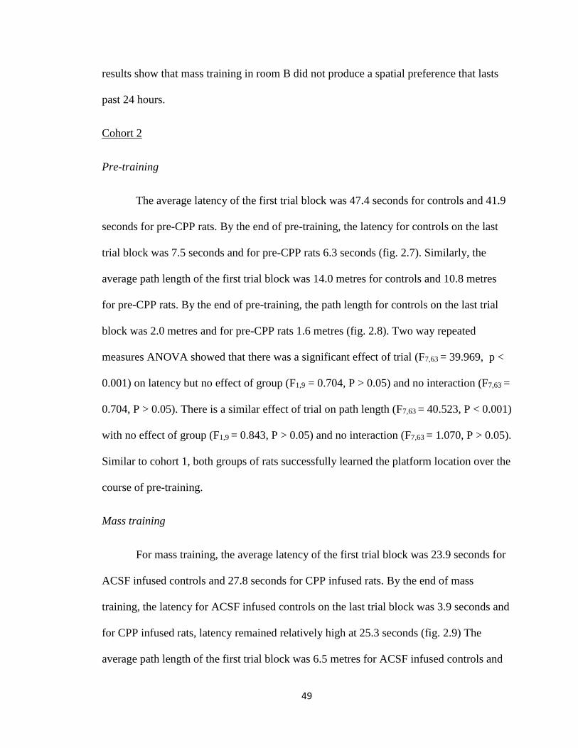
49
results show that mass training in room B did not produce a spatial preference that lasts
past 24 hours.
Cohort 2
Pre-training
The average latency of the first trial block was 47.4 seconds for controls and 41.9
seconds for pre-CPP rats. By the end of pre-training, the latency for controls on the last
trial block was 7.5 seconds and for pre-CPP rats 6.3 seconds (fig. 2.7). Similarly, the
average path length of the first trial block was 14.0 metres for controls and 10.8 metres
for pre-CPP rats. By the end of pre-training, the path length for controls on the last trial
block was 2.0 metres and for pre-CPP rats 1.6 metres (fig. 2.8). Two way repeated
measures ANOVA showed that there was a significant effect of trial (F7,63 = 39.969, p <
0.001) on latency but no effect of group (F1,9 = 0.704, P > 0.05) and no interaction (F7,63 =
0.704, P > 0.05). There is a similar effect of trial on path length (F7,63 = 40.523, P < 0.001)
with no effect of group (F1,9 = 0.843, P > 0.05) and no interaction (F7,63 = 1.070, P > 0.05).
Similar to cohort 1, both groups of rats successfully learned the platform location over the
course of pre-training.
Mass training
For mass training, the average latency of the first trial block was 23.9 seconds for
ACSF infused controls and 27.8 seconds for CPP infused rats. By the end of mass
training, the latency for ACSF infused controls on the last trial block was 3.9 seconds and
for CPP infused rats, latency remained relatively high at 25.3 seconds (fig. 2.9) The
average path length of the first trial block was 6.5 metres for ACSF infused controls and
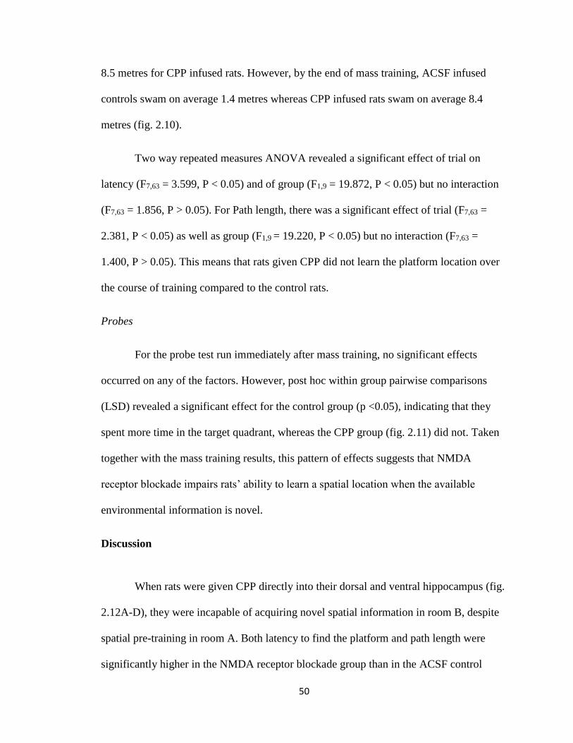
50
8.5 metres for CPP infused rats. However, by the end of mass training, ACSF infused
controls swam on average 1.4 metres whereas CPP infused rats swam on average 8.4
metres (fig. 2.10).
Two way repeated measures ANOVA revealed a significant effect of trial on
latency (F7,63 = 3.599, P < 0.05) and of group (F1,9 = 19.872, P < 0.05) but no interaction
(F7,63 = 1.856, P > 0.05). For Path length, there was a significant effect of trial (F7,63 =
2.381, P < 0.05) as well as group (F1,9 = 19.220, P < 0.05) but no interaction (F7,63 =
1.400, P > 0.05). This means that rats given CPP did not learn the platform location over
the course of training compared to the control rats.
Probes
For the probe test run immediately after mass training, no significant effects
occurred on any of the factors. However, post hoc within group pairwise comparisons
(LSD) revealed a significant effect for the control group (p <0.05), indicating that they
spent more time in the target quadrant, whereas the CPP group (fig. 2.11) did not. Taken
together with the mass training results, this pattern of effects suggests that NMDA
receptor blockade impairs rats’ ability to learn a spatial location when the available
environmental information is novel.
Discussion
When rats were given CPP directly into their dorsal and ventral hippocampus (fig.
2.12A-D), they were incapable of acquiring novel spatial information in room B, despite
spatial pre-training in room A. Both latency to find the platform and path length were
significantly higher in the NMDA receptor blockade group than in the ACSF control
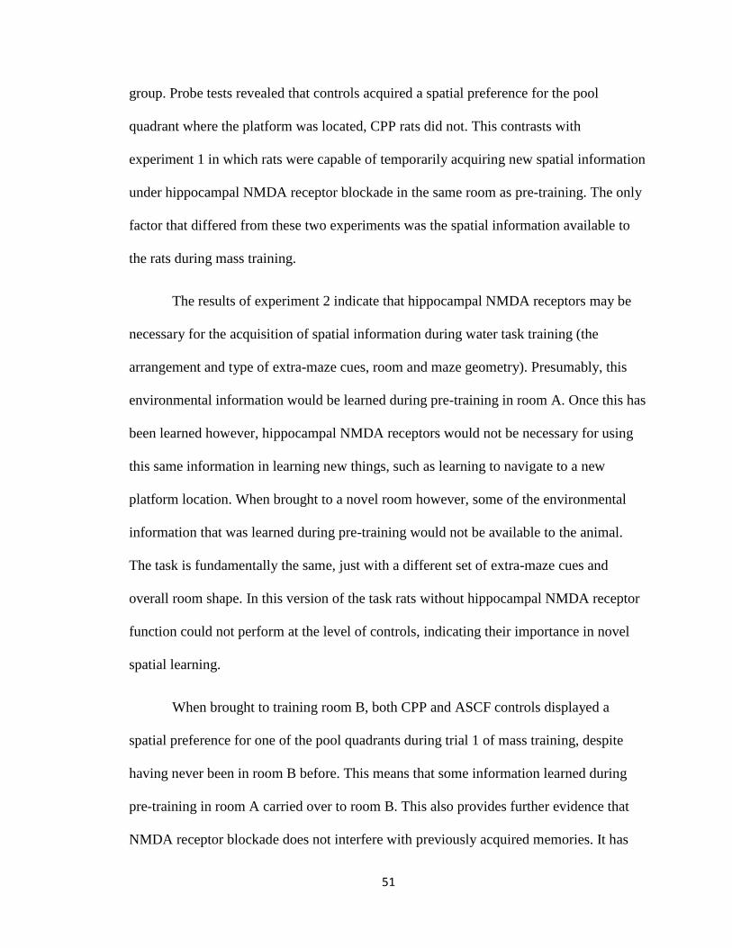
51
group. Probe tests revealed that controls acquired a spatial preference for the pool
quadrant where the platform was located, CPP rats did not. This contrasts with
experiment 1 in which rats were capable of temporarily acquiring new spatial information
under hippocampal NMDA receptor blockade in the same room as pre-training. The only
factor that differed from these two experiments was the spatial information available to
the rats during mass training.
The results of experiment 2 indicate that hippocampal NMDA receptors may be
necessary for the acquisition of spatial information during water task training (the
arrangement and type of extra-maze cues, room and maze geometry). Presumably, this
environmental information would be learned during pre-training in room A. Once this has
been learned however, hippocampal NMDA receptors would not be necessary for using
this same information in learning new things, such as learning to navigate to a new
platform location. When brought to a novel room however, some of the environmental
information that was learned during pre-training would not be available to the animal.
The task is fundamentally the same, just with a different set of extra-maze cues and
overall room shape. In this version of the task rats without hippocampal NMDA receptor
function could not perform at the level of controls, indicating their importance in novel
spatial learning.
When brought to training room B, both CPP and ASCF controls displayed a
spatial preference for one of the pool quadrants during trial 1 of mass training, despite
having never been in room B before. This means that some information learned during
pre-training in room A carried over to room B. This also provides further evidence that
NMDA receptor blockade does not interfere with previously acquired memories. It has
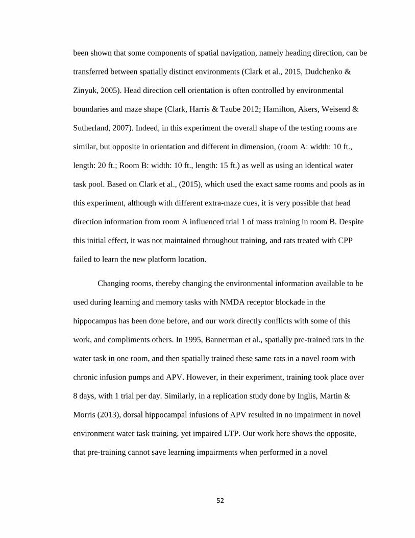
52
been shown that some components of spatial navigation, namely heading direction, can be
transferred between spatially distinct environments (Clark et al., 2015, Dudchenko &
Zinyuk, 2005). Head direction cell orientation is often controlled by environmental
boundaries and maze shape (Clark, Harris & Taube 2012; Hamilton, Akers, Weisend &
Sutherland, 2007). Indeed, in this experiment the overall shape of the testing rooms are
similar, but opposite in orientation and different in dimension, (room A: width: 10 ft.,
length: 20 ft.; Room B: width: 10 ft., length: 15 ft.) as well as using an identical water
task pool. Based on Clark et al., (2015), which used the exact same rooms and pools as in
this experiment, although with different extra-maze cues, it is very possible that head
direction information from room A influenced trial 1 of mass training in room B. Despite
this initial effect, it was not maintained throughout training, and rats treated with CPP
failed to learn the new platform location.
Changing rooms, thereby changing the environmental information available to be
used during learning and memory tasks with NMDA receptor blockade in the
hippocampus has been done before, and our work directly conflicts with some of this
work, and compliments others. In 1995, Bannerman et al., spatially pre-trained rats in the
water task in one room, and then spatially trained these same rats in a novel room with
chronic infusion pumps and APV. However, in their experiment, training took place over
8 days, with 1 trial per day. Similarly, in a replication study done by Inglis, Martin &
Morris (2013), dorsal hippocampal infusions of APV resulted in no impairment in novel
environment water task training, yet impaired LTP. Our work here shows the opposite,
that pre-training cannot save learning impairments when performed in a novel
53
environment. The experiments above utilized training protocols over several days
whereas ours was a rapid single session acquisition.
Taylor et al., (2010) has shown that in a context fear conditioning paradigm, rats
previously trained to fear one environment can be trained to fear a novel environment
without the use of hippocampal NMDA receptors. This effect was only seen when the
two training environments were very similar. If they were made to be very distinct, prior
training did not make subsequent learning NMDA receptor independent, implicating their
role in learning environmental cues. Interestingly, it was also found that NMDA receptor
independent learning occurred in the hippocampus, and that the expression of Arc protein
depended on the similarity of the two training environments. In similar experiments, Lee
& Kim (1998) have shown that context fear pre-training does not make subsequent novel
fear training NMDA receptor independent. They also showed that learning tone-shock, or
light –shock pairings was also shown to be NMDA receptor dependent.
Given the variability in results from different experiments, the role of
hippocampal NMDA receptors in spatial learning is uncertain. The data from our
experiment is in line with the traditional view of hippocampal NMDA receptors. Namely,
that they have a critical role in the acquisition of spatial information. However, the aspect
of spatial learning found to be dependent on hippocampal NMDA receptors was learning
about the features of the external environment to guide spatial navigation. They may still
have a role in memory consolidation though. Evidence from experiment 1 also indicates
NMDA receptors role in the consolidation of spatial memories as well. These findings do
not discount the behavioral strategy or cortical plasticity accounts of the pre-training
effect, since those accounts were not explored in this experiment. It is clear that reversal
54
learning in using the previously acquired information and novel learning are different
phenomena, involving different task requirements because of the differences in available
information. Indeed, the effects of environmental information on reversal learning,
inhibition, and extinction have been studied for decades in associative learning paradigms
(Bouton, 1993; McDonald, Ko & Hong, 2002). It may be the case that because same
room reversal and novel room acquisition depend on different types of information, they
will potentially depend on different neural mechanisms, either molecular, or regional. If
NMDA receptors are necessary for the consolidation of reversal learning information but
the acquisition of novel information, then it is possible that the ultimate function of
hippocampal NMDA receptors, as well as what hippocampal plasticity represents
mnemonically, is dependent on the type of behavior to be learned and the environmental
information available to the animal.
In the next experiment, we examined the effect of NMDA receptor blockade and
spatial learning on the expression of Arc protein in the hippocampus.
55
Chapter 4
The effect of NMDA receptor dependent learning behaviors on
Arc expression in the hippocampus
In experiments 1 and 2, the effects of hippocampal specific NMDA receptor
blockade on spatial learning was examined. In this final experiment, I set out to determine
if there are observable molecular, neurobiological changes in the hippocampus that are
associated with the behavioral effects seen in those experiments. The type of impairment
seen in experiment 1, in which NMDA receptors were blocked during rapid acquisition of
a new spatial position in the same environment, was the consolidation of long term
memory formation and not encoding or short-term retention. Accordingly, I chose to
examine the immediate early gene (IEG) Activity related cytoskeletal protein (Arc) as it
has been implicated in long-term memory processes (Czerniawski et al., 2011).
First described by Lyford et al., (1995), Arc is an IEG, a gene whose mRNA is
rapidly increased by high levels of neural activity, with many IEGs being activated by
LTP in particular. Arc mRNA is delivered to dendrites of heavily active synapses,
implicating its use in synaptic changes associated with plasticity and learning. Expression
of Arc in the hippocampus has been reported to be enhanced following novel spatial
learning in both the dorsal and ventral segments (Guzowski, Setlow, Wagner & McHugh,
2001; Czerniawski et al., 2011). The NMDA receptor has been heavily implicated in the
activation of Arc, with the same type of LTP that is NMDA receptor dependent inducing
ARC expression (Lyford et al., 1995). This was later reinforced by Steward & Worley
(2001), showing that NMDA receptors are necessary for Arc trafficking and expression in
dendrites. Administering Arc antisense oligodeoxynucleotides interferes with both
56
learning and memory behaviors as well as the molecular processes associated with them
(Guzowski et al., 2000; Ploski et al., 2008). Similar effects have been observed with Arc
genetic knockout animals (Plath et al., 2006).
IEGs are thought to mediate the molecular consolidation of memory because of
their time course of action and the types of subsequent molecular mechanisms they
induce. It is proposed that there are two general processes involved in the molecular
formation of memory. The first utilizes transient increases in local calcium resulting in
the short term heightened reactivity of synapses and shorter forms of LTP. The second
involves protein synthesis via genetic activation, namely an increase in immediate early
gene protein synthesis resulting in synaptic membrane AMPA receptor insertion. Because
of the time involved in gene transcription and translation, and molecular trafficking, it is
believed that these latter processes are critical for memory consolidation, and not its
initial formation. (Steward & Worley, 2002)
Stable LTP requires structural changes of the synapse relying on F-actin and
protein synthesis. When Arc antisense is applied to tissue to disrupt LTP, this disruption
can be reversed by F-actin stabilizing drugs. Reciprocally, F-actin is necessary for the
localization of Arc at synapses (Haung, Chotiner & Steward, 2007). Arc has also been
shown to be critically related to AMPA receptor trafficking, a process by which new
AMPA receptors are brought to and inserted in the post synaptic membrane. This process
is thought to be one of the primary mechanisms by which the heightened calcium influx
seen in LTP is maintained. (Chowdhurry et al., 2006)
When applied to hippocampal tissue, Arc antisense will allow for the induction of
LTP, yet its long-term maintenance will disappear (Guzowski, Setlow, Wagner &

57
McHugh, 2001). Knockout mice similarly express heightened early phase LTP and
reduced late phase LTP (Plath et al., 2006). In a fear conditioning paradigm, Czerniawski
et al., (2011) showed that an NMDA receptor antagonist could prevent arc expression in
both the dorsal and ventral hippocampus and that arc antisense would impair memory
consolidation but spare initial learning, linking NMDA receptors, Arc, and memory
consolidation. Taken together, these electrophysiological, molecular, and behavioral data,
all suggest a role for IEGs, and NMDA receptor dependent Arc in particular, in the long-
term consolidation of memory.
This link between NMDA receptors and Arc protein with regards to molecular
mechanisms of plasticity and learning behaviors is further explored in this experiment.
These two molecules’ involvement in the consolidation of activity induced synaptic
changes is exactly the type of process we think is involved in experiment 1, in which the
consolidation of newly acquired memories was impaired by NMDA receptor antagonism.
In the present experiment, rats were trained in two versions of the water task: one
involving rapid mass training in the same spatial environment as pre-training similar to
experiment 1, and the other rapid mass training in a novel spatial environment separate
from pre-training, similar to experiment 2. The NMDA receptor antagonist CPP was
injected into the dorsal and ventral hippocampus of rats trained to swim to a hidden
platform in a novel context. After training, levels of Arc protein expression throughout
the hippocampus were measured and compare between groups. In our previous
experiment, animals could not learn novel spatial information without NMDA receptors
but could learn a new platform location using previously learned environmental cues.
Because of these results, we hypothesized that Arc expression would be much higher after

58
novel spatial learning compared to reversal learning in the same spatial environment, and
that NMDA receptor blockade would simultaneously impair learning behaviors and
reduce Arc protein expression.
Methods
Behavioral training and testing was very similar to that of experiments 1 and 2.
Pre-training was done in context A for all groups except cage controls. There were four
groups in this experiment, differing only in how their mass training was done in reference
to pre-training: Same room, New room, New room + CPP, and cage controls (n= 5). For
the Same room group (n= 5), mass reversal training consisted of 16 trials and was
performed identically to experiment 1. For the New room group (n= 5) and New room +
CPP group (n= 5) mass training consisted of 16 trials and was identical to experiment 2.
Drug infusion for the New room + CPP rats was done 30 minutes before mass training
and was performed identical to the CPP group in experiment 2. The rats were run one at a
time during mass training to avoid variation in timing of procedures. After completing
mass training, rats were brought back to their home cage until perfusion. Perfusion
occurred approximately 70-80 minutes after trial 8 of mass training. This is a time period
in which IEG protein expression after learning is active (Lonergan, Gafford, Jarome &
Helmstetter, 2010). Euthanization and perfusion procedures were identical to experiments
1 and 2.
Tissue collection and immunohistochemistry
After extraction, the brains were placed in 4% PFA solution for 24 hours, at which
point they were then placed in a 30 % sucrose + sodium Azide solution for a 3 day period

59
of cryoprotection. The brains were then sliced on a freezing microtome in 12 series, to
include the entire hippocampal formation. Tissue was stained using a 3 day
immunohistochemical protocol that fluorescently stained Arc protein.
On day 1, tissue was pre-washed in 1% PBS solution, 3 x 10 minutes. Tissue was
then placed in 2 ml of a 0.3 % triton and PBS solution with a 1:1000 ratio of primary
antibody. For Arc protein staining, Arc (c-7) sc-17839 mouse monoclonal IgG (Santa
Cruz Biotechnologies) was used. The tissue well was placed on a rotating belly dancer
and left to incubate for 24 hours. On day 2, the tissue went through a second wash in 1%
PBS solution, 3 x 7 minutes. After the wash, the tissue was placed in a 1:500 solution of
PBS and Alexa fluor 488 donkey antimouse IgG (H+L) (Thermo Fisher Scientific). The
tissue well was then covered in tin foil to prevent light from affecting the staining
process. The well was placed back on the belly dancer and the room light turned off and
left for a second 24 hour incubation period. On day 3, the tissue was put through another
1% PBS wash, 3 x 7 minutes and then wet mounted onto 1 % gelatin and 0.2 % chromatin
coated slides. The slides were covered and placed in a refrigerator at 3° Celsius for 24
hours.
Microscopy and cell counts
Cell counts were done on a Zeiss AxioImager M1 microscope (Zeiss, Jena,
Germany) using the program Stereo Investigator® (MicroBrightField Inc., 2013, Version
10). A constant light intensity exposure was set at 50%. Light was projected through a
FITC filter. A 20X magnification was used when counting cells. All labelled granule cells
were individually counted in the granule cell layer of the dentate gyrus. All labelled
pyramidal cells were individually counted in the pyramidal cell layers of areas CA1 and

60
CA3. Cell counts in each hippocampal slice for each animal were summed and multiplied
by 12 (12 series sections were sliced) to get an approximation of total cell number in each
region. Tissue photographs were taken and colored with ImageJ® (ImageJ, R. W., & US
National Institutes of Health. (2012). Bethesda, Maryland, USA.)
Results
Water task
The measures of learning and memory used during acquisition were latency to
find the platform and path length to the platform. These graphs represent groups of 4 trial
averages across 4 days of training for the 3 groups that did water task training. The
average latency of the first trial block was 48.0 seconds for Same room group, 41.9
seconds for the new room group, and 30.2 seconds for the New+CPP group. By the end
of pre-training, the latency for the Same room group on the last trial block was 7.3
seconds, 6.3 seconds for New room group, and 5.2 seconds and for New+CPP group (fig.
3.1). Similarly, the average path length of the first trial block was 13.7 metre for Same
room group, 10.8 metres for New room group and 9.0 metres for New+CPP group. By the
end of pre-training, the path length for controls on the last trial block was 1.8 metres for
Same room group, 1.6 metres for the New room group, and 1.6 metres for the New+CPP
group (fig 3.2). This indicates that Latency and path length follow identical patterns and
that all groups reduced these measures over training. Two way repeated measures
ANOVA showed that there was a significant effect of trial on latency (F7,84 = 47.373, p <
0.001), no effect of group (F2,12 = 3.636, P > 0.05) but there was an interaction (F7,14 =
2.312, P < 0.05). There was a similar effect of trial on path length (F7,77 = 48.977, P <
0.001) with no effect of group (F2,12 = 2.529, P > 0.05) and no interaction (F7,14 = 1.782, P
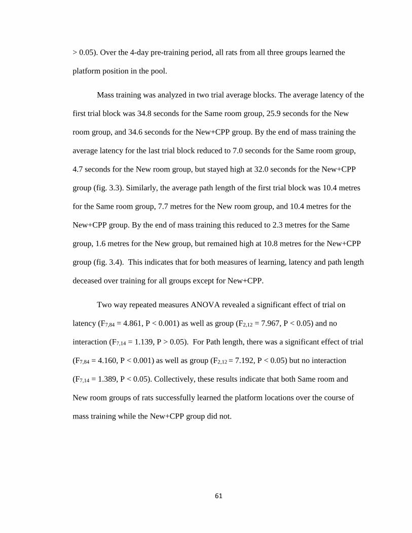
61
> 0.05). Over the 4-day pre-training period, all rats from all three groups learned the
platform position in the pool.
Mass training was analyzed in two trial average blocks. The average latency of the
first trial block was 34.8 seconds for the Same room group, 25.9 seconds for the New
room group, and 34.6 seconds for the New+CPP group. By the end of mass training the
average latency for the last trial block reduced to 7.0 seconds for the Same room group,
4.7 seconds for the New room group, but stayed high at 32.0 seconds for the New+CPP
group (fig. 3.3). Similarly, the average path length of the first trial block was 10.4 metres
for the Same room group, 7.7 metres for the New room group, and 10.4 metres for the
New+CPP group. By the end of mass training this reduced to 2.3 metres for the Same
group, 1.6 metres for the New group, but remained high at 10.8 metres for the New+CPP
group (fig. 3.4). This indicates that for both measures of learning, latency and path length
deceased over training for all groups except for New+CPP.
Two way repeated measures ANOVA revealed a significant effect of trial on
latency (F7,84 = 4.861, P < 0.001) as well as group (F2,12 = 7.967, P < 0.05) and no
interaction (F7,14 = 1.139, P > 0.05). For Path length, there was a significant effect of trial
(F7,84 = 4.160, P < 0.001) as well as group (F2,12 = 7.192, P < 0.05) but no interaction
(F7,14 = 1.389, P < 0.05). Collectively, these results indicate that both Same room and
New room groups of rats successfully learned the platform locations over the course of
mass training while the New+CPP group did not.
62
Immunohistochemistry
Cell counting was done for all rats in areas CA1, CA3, and dentate gyrus and
repeated measures ANOVAs were completed on the data. All three areas were compared
between all four behavior groups (fig. 3.5). There were significant effects of area (F2,32 =
28.818, P < 0.001), of group (F1,16 = 3.733, P < 0.001) as well as an interaction (F6,32 =
10.653, P < 0.001). Post hoc pairwise comparisons were made between all the groups and
areas. Significant differences were found between cell counts in the dentate gyrus of the
new room group. They expressed more Arc positive cells than cage controls (p < 0.001),
the same room group (p < 0.001), and the new+CPP group as well (p < 0.05). The
New+CPP group also expressed more Arc positive granule cells than cage controls (p <
0.05) and but did not differ from the Same room group. For area CA1, the Same room
group expressed more Arc positive cells than did the New room rats (p < 0.05). no
differences were found in area CA3 between any of the groups. This indicates that New
room training induces more dentate Arc expression than Same room and control groups,
and that CPP diminishes this effect. Images of Arc expression in the dentate gyrus across
groups are seen in fig 3.6 and cannulation coordinates are seen in fig 3.7.
Discussion
In this experiment, rats were trained in a version of the Morris water task that
involved multiple training rooms. The effect of the NMDA receptor antagonist CPP on
Arc protein expression in hippocampal sub-regions in response to learning experiences
was examined. When pre-trained in the water task in room A, rats could easily learn a
new platform position in room A or in room B. However, when CPP was administered to
both the dorsal and ventral hippocampus prior to the second training phase, rats could not

63
learn a platform position in room B, despite pre-training. These results are identical to
those found in experiment 2. They further support the idea that hippocampal NMDA
receptors are critical for the acquisition of novel environmental information used in
spatial navigation.
The expression of the protein for the immediate early gene Arc differed between
group and region, depending on the training conditions. In the dentate gyrus, expression
was high for the New group compared to the equally low cage controls and Same room
group. When CPP was given, this corresponded with both a reduction in dentate Arc
expression and an impairment in learning compared to the New room group. The same
room rats expressed a significant increase in Arc in area CA1 compared to New room
rats. No differences were observed in area CA3 between any of the groups.
Despite a lack of a defined pattern of behavior related activity in areas CA1 and
CA3, the pattern of Arc expression in the dentate gyrus fits with the behaviors exhibited
by the rats in their respective learning conditions, both in this experiment and experiments
1 and 2. When rats had to learn a new spatial platform location in the same environment
as pre-training, dentate arc expression was not significantly different from cage controls.
Learning in the new room however produced a large increase in dentate Arc expression.
This would indicate that if Arc is necessarily tied to learning and plasticity, that the same
room reversal training in the water task is not the same as novel room training,
behaviorally or molecularly.
The results from experiments 1 and 2 also show that depending on the task
requirements, learning can be made NMDA receptor dependent. Learning novel
environmental information is an NMDA receptor dependent process while utilizing
64
previously learned environmental information for new navigational purposes is not. The
results from this experiment show that Arc expression follows a similar pattern. In a task
that was not found to be NMDA receptor dependent (experiment 1), dentate Arc
expression was no different from controls. In a task found to be NMDA receptor
dependent (experiment 2), Arc expression was high in the dentate gyrus, and reduced in
the same condition that produced behavioral impairments (hippocampal NMDA receptor
blockade).
This is a specific pattern of results that support the idea that Arc expression is both
a plasticity based IEG activated by novel learning behaviors, and that this molecular and
behavioral expression is NMDA receptor dependent (Czerniawski et al., 2011; Steward
and Worley 2001). From these 3 experiments, our results associate both molecules with
learning novel environmental information in the hippocampus, particularly in the dentate
gyrus.
Another IEG proposed to be related to learning based plasticity and potentially
NMDA receptor dependent is the regulator IEG Zif268 (Richardson et al., 1992; Cole,
Saffen, Baraban & Worley, 1989). In a multiple training room water task experiment very
similar to this one, rats were trained to multiple platform locations over multiple days in
room A, and then trained in room B just once. Zif268 expression was recorded across
hippocampal sub-regions after training. After extensive spatial training in one room, new
room learning induced Zif268 expression in the dentate gyrus and CA1 much higher
compared to same room trained rats.
Guzowski, Wagner, Setlow & McGaugh (2001) have shown that same room water
task reversal training induces Arc mRNA in the hippocampus at higher rates than
65
controls. When spatial water task training was done in a room separate from pre-training,
a sub-region analysis showed that Arc mRNA expression increases in the dentate and area
CA3 relative to controls, however, this sub-region analysis was not done in same room
reversal and so does not speak to reversal learning. Dentate Arc expression has also been
observed in object place learning. When learning to match objects to places in a maze,
moving the position of the objects while maintaining the extra maze cues induced Arc
mRNA in the dentate gyrus of rats (Soule et al., 2009). These conditions are similar to our
water task experiments but different in the type of changes that induce dentate Arc
expression. These contrasts and others will be discussed in the general discussion section.
66
Chapter 5
General discussion
In these experiments, we assessed the role that hippocampal NMDA receptors
have in spatial memory and the molecular processes associated with it. NMDA-blockade
across the dorsal and ventral hippocampus was accomplished using the receptor
antagonist CPP and direct cannulations. In each experiment, all animals were pre-trained
to become procedurally efficient at the Morris water task. After this, a mass training
procedure was done in one of two conditions: in the same training room as pre-training,
or in a new training room, different from pre-training. In this way, we could observe how
NMDA receptors mediate spatial learning dependent on previously acquired or novel
environmental information.
Without the use of NMDA receptor function in the hippocampus, rats were
capable of expressing what they had learned previously, and they could also learn new
spatial information in the same environment as pre-training. However, they forgot this
information as soon as 8 hours or 24 hours later. When brought to a completely novel
training environment, rats could not learn new spatial information at all without the use of
hippocampal NMDA receptors. These results lead to the conclusion that NMDA receptors
mediate the acquisition of novel environmental information to be used in spatial
navigation.
This type of NMDA receptor dependent behavior was also found to induce
specific patterns of Arc expression in the hippocampus, namely the dentate gyrus. In the
task in which NMDA receptors were not found to be necessary for learning (same room),
expression was low. In the task in which NMDA receptors were found to be necessary for
67
learning (new room), expression was high. CPP blocked both learning and reduced Arc
expression in animals trained in the new room. This pattern of results points to a link
between these three phenomena: NMDA receptor function, Arc expression, and novel
spatial learning. The relationship between these will be explored below.
Novel contributions and support for hypotheses
The results from the three experiments above support a series of hypotheses
regarding the biological nature of learning and memory behaviors thought to be
dependent on the hippocampus and related circuits. First, that depending on what has to
be learned and the information available to do so, learning can be NMDA receptor
independent. Second, NMDA receptors are not involved in the retrieval of previously
learned information. Third, NMDA receptors have a critical role in the consolidation of
spatial memory. Fourth, NMDA receptors in the hippocampus also have a critical role in
the acquisition of novel environmental information. Fifth, NMDA receptor dependent
learning also activates the immediate early gene Arc in the dentate gyrus of the
hippocampus. Finally, some aspects of the information acquired during spatial navigation
training in the MWT can be applied outside of where it was originally learned. However,
much of the scientific research done in the past on these issues does not give support to
some of these claims. Here I will discuss the discrepancies between the results of my
present experiments and previous work done by others, as well as what novel
contributions this work provides.
68
NMDA receptor blockade in the dorsal versus full hippocampus
The inspiration and design of my experiments was from McDonald et al, (2005).
In that series of experiments only the dorsal hippocampus was blocked for the intracranial
experiments and no effect of NMDA receptor blockade on mass training of a new spatial
position was reported. Experiment 1, reported in the current thesis, was completed to
determine if the pattern of results observed in the McDonald et al., (2005) study was due
to a functional ventral hippocampus. The ventral portion of the hippocampus has been
shown to support certain aspects of spatial learning (Ferbinteanu, Ray & McDonald,
2003). Very few water task studies have been done with full hippocampal NMDA
receptor blockade (Steele & Morris, 1999; Inglis, Martin & Morris 2013). Both the
experiments cited above utilized pre-training procedures followed further training under
receptor blockade, and both experiments resulted in successful spatial learning under
specific circumstances, despite full blockade. These experiments will be explained in
more detail below.
As a stated hypothesis in experiment 1, the full hippocampal NMDA blockade
discounts the possibility that the reason why animals were capable of learning novel
spatial locations in the McDonald et al., (2005) study was due to a functional ventral
hippocampus. Comparably, some rats in that experiment that were given I.P. injections
also learned the novel platform location. Due to the injection method, I.P. necessarily
results in global antagonism, and so would presumably cover both dorsal and ventral
hippocampus. This leads to a further conclusion that the dorsal and ventral hippocampal
NMDA receptors do not play a critical role in this task. With regards to novel
environmental learning as assessed in experiment 2 of this thesis, only full hippocampal

69
blockade was done, and so the respective contributions of the dorsal and ventral segments
cannot be elaborated on.
NMDA receptors and memory retrieval
The lack of NMDA receptor involvement in memory retrieval has long been
accepted after several experiments outlining rats’ ability to use previously acquired
information during NMDA receptor blockade (Shapiro & Caramanos, 1990; Bast, Silva,
Morris, 2005). Biologically this would make sense, if NMDA receptors are necessary for
LTP induction and learning based plasticity, then once the circuit connections
representing the memory have been formed and consolidated, plasticity mechanisms
should not be involved in simple circuit activation during retrieval. However, recent
evidence has shown the contrary; that NMDA receptor blockade can prevent the
expression of previously established fear memories (Lopez, Gamache, Schneider &
Nader, 2015). Our results in experiment 1 further support the claim that NMDA receptors
are not involved in memory retrieval and expression. When analyzed as a probe, trial 1 of
mass training revealed that rats from both groups displayed a spatial preference for the
pre-training platform location, indicating that it was both successfully learned and
retrieved. Trial 1 of mass training in experiment 2 (room B) also supports NMDA
receptor independent retrieval, as well as providing some interesting insights into spatial
processing.
In experiment 2, rats were brought into room B after being pre-trained in room A.
Trial 1 of mass training was analyzed as a probe to see if any spatial preferences could be
observed. This was done to see if any information learned from training in room A could
be used during training in room B. Past research from our lab has shown that this may be

70
the case (Clark et al, 2015), and so we hypothesized that rats would show some kind of
directed spatial behavior during trial 1, although the specific expression of which was
undetermined.
Both ASCF controls and CPP infused groups spent most of their swim time in
quadrant 2. This indicates that despite NMDA receptor blockade, both groups behaved
identically after spatial training in room A. However, rats placed in a novel water task for
the first time almost never display spatial preferences but instead display a type of
thigmotaxis characterized by hovering near the pool wall and circling quickly around the
perimeter of the pool. The fact that these rats went through pre-training in room A, and
then displayed a spatial preference in room B, despite having never been in room B
before raises the question of what exactly is learned in the water task that can be
transferred between rooms. There are environmental cues that exist in both rooms and
both pools are identical in shape, color and size. The geometrical structure of the room
has similar features such as wall shape and length. The door and experimenter are also
present in both rooms. Indeed, the spatial preference was displayed by both groups for the
quadrant which was closest to the door, which was also the quadrant where the platform
was located in the pre-training in room A. (fig 2.5).
Our results from experiment 2 indicate that some spatial information learned in
one environment can be used in other environments. The specific nature of transferable
information between environments or what common environmental features may be used
in navigation remains unclear, but its expression is NMDA receptor independent.

71
NMDA receptors and spatial learning
Starting with Bannerman et al. (1995) and Saucier & Cain (1995), the idea of
NMDA receptor independent learning began to be studied. Novel water task training in
the presence of NMDA receptor antagonist drugs resulted in learning impairments, but by
changing certain parameters of the task, these impairments could be eliminated. Since
then, many studies have examined this phenomenon, through NMDA receptor
independent extinction (Santini, Muller & Quirk, 2001), place cell formation (Kentros et
al., 1998), LTP (Grover & Teyler, 1990), and molecular cascades associated with learning
and plasticity (Moosmang, et al., 2005). How exactly learning processes can be made
NMDA receptor independent is not certain. Some have shown that behavioral strategies
are learned once, and then can be applied across multiple learning scenarios independent
of NMDA receptor plasticity (Hoh et al., 1999). Others have argued that where in the
brain learning takes place changes over training (Inglis, Martin & Morris, 2013). Some of
these issues were discussed in ch2.
Almost all studies examining NMDA receptor independent spatial learning have
done so using pre-training, in some form or another. A review of the literature on
experiments using NMDA receptor blockade, pre-training, and spatial learning reveals
two overall findings. The first is spatial learning can be made NMDA receptor
independent when done with the same information available to prior learning, and is also
done rapidly. This is made most clear by Steele & Morris (1999) where rats were pre-
trained in the water task on a version that involved a new platform location every day.
After several days, rats continued training in the same room but with hippocampal
NMDA receptor blockade. The manipulated variable was inter-trial interval, and it was
72
found that when it was kept short at 15 seconds rats learned successfully. However, when
it was lengthened to 20 minutes, they failed to learn. This means that rapid learning was
possible while learning over longer periods of time was not. Because the platform
changes every day, the effect of receptor blockade on long term training cannot be
assessed.
Work done by Saucier & Cain (1995) and Hoh et al., (1999) have shown that non-
spatial pre-training (NSP) can lead to rapid NMDA receptor independent spatial learning.
The argument put forth by these researchers is that NSP allows for behavioral search
strategies to be developed prior to spatial training and that this is what leads to NMDA
receptor independence later on. However, behavioral search strategies themselves were
also found to be learned independent of NMDA receptors. This contrasts with earlier
work done (Morris, Davis & Butcher, 1990) in which NSP led to impairments in same
room spatial training that was done over the course of several days. NSP is performed
with a curtain around the pool and so may potentially conflict with our explanation of
previously learned environmental information hypothesis. However, the complete
environmental repertoire that contributes to spatial navigation behaviors goes beyond wall
cues like posters.
The water task environment can be divided into two separate spaces: the intra-
maze (local) environment which contains everything inside the pool walls, and the extra-
maze (global) environment which contains everything outside the pool walls but inside
the experiment room. Some evidence shows that aspects of spatial navigation can even
transfer between multiple contexts and so would constitute potentially larger,
environment independent navigation strategies as well, such as heading direction (Taube
73
& Burton, 1995, Clark et al., 2015). Given the continued use of the maze and global
location of the maze throughout training, NSP does not necessarily mean that no usable
environmental information is learned. With this in mind, in the studies using NSP, rats
successfully learn the spatial water task when trained rapidly (Saucier & Cain, 1995; Hoh
et al., 1999).
Our work reinforces these conclusions. It shows that in the absence of NMDA
receptors in the hippocampus, rapid learning is possible when the information used to
learn a new escape location is identical to what has been previously learned. After both an
8 hour and a 24 hour retention period, everything that was learned during mass training
was forgotten. Our specific pattern of results indicates that whatever was learned was not
consolidated. A lack of consolidation may explain why rats in these types of tasks can
learn rapidly but not with increased ITIs or over several days. NMDA receptor
involvement in memory consolidation has been shown on multiple occasions (McDonald
et al, 2005; Santini, Muller & Quirk (2001).
The second finding is that novel spatial learning can be made NMDA receptor
independent when done over several days. This comes primarily from two experiments.
Bannerman et al., (1995) was able to get rats to learn the spatial water task in a room
different from that of pre-training, and did so over multiple training days. A similar effect
was found in that experiments replication (Inglis, Martin & Morris, 2013). In the
Bannerman et al., (1995) study, a NSP experiment was also done, and when done in room
1, it did not allow for spatial learning in room 2 across multiple days. NSP is a very
specific type of pre-training that eliminates extra-maze cues. Given that there is no
environmental information outside of the pool, it is likely that the rats given NSP pre-

74
training treated room 2 in the Bannerman study in the same way that the rats in the Cain
and Hoh studies treated their respective training rooms. Stated differently, without
previous environmental information to compare to new information, all information is
new, and so new training done in a different or same room will not matter. A similar
intra-maze environment and lack of extra-maze specific cues means that there is no such
thing as same room or different room (dependent on extra-maze cues) following NSP, and
that all subsequent spatial training will follow the same principles. This would also
explain the multiple day learning impairments observed in Morris, Davis & Butcher
(1990).
However, multiple day NMDA receptor independent learning does not apply to
naïve rats in the water task. This may be because novel water task training does not just
involve spatial learning but also developing successful motor patterns for swimming,
trying out search strategies, as well as reducing fear. The water task is an aversive
learning task and given NMDA receptors involvement in motor function and anxiety,
some have argued that learning deficits cannot be dissociated from other types of effects
(Ahlander, Misane, Schott & Ogren, 1998, Keith & Rudy, 1990). Because of the
ubiquitous nature of the NMDA receptor, it may have a role in many neural and
behavioral processes outside of spatial navigation and learning. This is why pre-training
is used, it allows for only certain aspects of the task to be isolated and examined.
In all of the work involving water task training that relies on previously learned
environmental information, NMDA receptor independent learning is successful when
done rapidly but not when done with increased ITIs or over several days. However, when
training is done with novel environmental information and the potentially confounding

75
influence of behavioral strategies, anxiety, and sensori-motor impairments are removed,
learning may be NMDA receptor independent when done over several days, but not
rapidly. To our knowledge, no one has examined the role of NMDA receptors in rapid
novel spatial learning after pre-training. Our work provides support these conclusions by
showing that when transferred to a new room with novel environmental cues rats without
NMDA receptor function cannot rapidly learn.
The shift in NMDA receptor dependence may have to do with the requirements of
the task. Reversal learning in the water task consists of fundamentally different task
parameters and learning requirements than novel learning. When put through the water
task for the first time, rats must learn many things ranging from behavioral search
strategies, spatial locations, and interacting with the maze, the experimenter, and the
environment. These include, but are not limited to, swimming away from the pool wall,
the size and stability of the hidden platform, the arrangement of extra-maze cues, etc. One
of the main reasons why pre-training procedures are used is to limit the type of learning
that must occur in subsequent training procedures for successful maze navigation. When
put through reversal learning in the same spatial context after a pre-training procedure,
the only thing required for the rat to learn is a new platform position. The dimensions and
details of the maze and room, as well as a successful search strategy, have already been
learned. Also, rats have to extinguish the previously learned spatial information, so that
reversal doesn’t just involve new learning, but extinction as well. The rat must do this
using information and strategies it is already familiar with.
Further training with a completely new set of information such as in experiment 2
is also different from both naïve training and same room reversal. Several components of
76
training are already familiar to the rat such as swimming and an expectation of the rules
of further training. Yet some things are completely unfamiliar like where the animal is
and what exists in its environment. In this way, three different stages of water task
training, naïve, new room, same room reversal, all have increasing amounts of familiarity
to them, respectively.
This means that all three stages of training may involve different search strategies,
or the same strategies but based on different types of available information. Given the
different task requirements during each stage, it is not unlikely that the role plasticity has
will vary across stages. Our own results, as well as a review of the relevant literature,
reveal that NMDA receptors in the hippocampus will be heavily involved in several
processes during naïve water task training, resulting in an inability to dissociate between
learning, anxiety, and sensori-motor processes just from behavioral data alone. After
extensive pre-training and task-familiarization, NMDA receptors consolidate changes to
existing spatial information. And that when processing completely novel environmental
information, are necessary for its rapid learning.
Hippocampal remapping
A potential electrophysiological correlate to these behavioral patterns may be
remapping. Remapping occurs when changes to the external information that guides
internal place cell activity produces changes to that cell activity. (Bostock, Muller &
Kubie, 1991) Slight changes such as single, distinct environmental cues result in rate
remapping, in which the population of active cells and their representative spatial location
remain the same but their firing rate changes. Large changes to environmental
information or even a complete switch in environments results in global remapping, in
77
which the entire population of active place cells changes its spatial representations and
firing rates (Leutgeb et al, 2005).
Neither the initial formation of place fields nor the maintenance of already present
place fields is an NMDA receptor dependent process. However, their long-term stability
(Kentros et al, 1998) and changes in their representative properties is impaired by
pharmacological and genetic manipulation (McHugh et al., 2007; Ekstrom, Metzel,
McNaughton & Barnes, 2001) and so rate and global remapping to a novel context would
likely involve them as well. Rate remapping in CA3 was impaired between environments
in an NMDA receptor knockout mouse (McHugh et al., 2007). LTP also appears to play a
critical role in global remapping as well (Dragoi, Hariss & Buzsaki, 2003).
In our experiments, it could be argued that same room reversal might produce
rate remapping because of the small change in platform position. While new room
training could produce global remapping because of the training room change. Given the
link between NMDA receptors, LTP, and remapping, the inability to learn due to blocked
NMDA receptors in our experiment 2, could be due to an inability to remap place fields
in the hippocampus. However, in our experiments, this remains completely hypothetical.
Given the overlap of global and rate remapping across different types of spatial
manipulations (training rooms, color, size, shape), what exact features of the environment
lead to remapping, and whether these changes are behind pattern separation and
navigational behaviors remains to be determined (Jeffery, Gilbert, Burton & Strudwick,
2003; Speirs et al., 2013; Leutgeb et al., 2005; Kentros et al, 1998).

78
Immediate early genes and the dentate gyrus
The use of IEGs in neuroscience research is commonly used to map behaviorally
relevant neural activity. The IEG Arc is thought to be dependent on LTP related activity
and so is used to map neurobiological mechanisms of learning and memory. Learning and
memory behaviors are known to reliably induce Arc mRNA and protein expression in
behaviorally relevant brain regions. Activation of the NMDA receptor has been identified
as a critical mechanism that leads to Arc expression. Blocking NMDA receptors with
antagonist drugs reduces behavior induced Arc mRNA (Czerniawski et al., 2011).
Furthermore, NMDA receptor hypofunction leads to reduced Arc expression (Balu &
Coyle, 2014). The links between learning behaviors, NMDA receptors, LTP, and Arc
activity is well supported by research (Guzowski et al., 2001; Shepard & Bear, 2011).
My results from experiment 3 show that in one version of a water task that did not
require NMDA receptors to learn, Arc protein expression was not significantly different
from cage controls. In a version of the task that did require NMDA receptors to learn, Arc
protein expression was high compared to cage controls and the NMDA receptor
independent task. When an NMDA receptor antagonist was introduced to the
hippocampus, it impaired learning as well as reduced Arc protein expression. The level of
expression in the CPP group was significantly below the New room group but also
significantly higher than the cage control group. No observable patterns emerged in areas
CA1 or CA3, however, the pattern of expression described above was observed in the
dentate gyrus.
Two important conclusions can be reached from these results. One is that NMDA
receptor dependent learning of novel environmental information activates Arc in the

79
dentate gyrus. The other is that NMDA receptor blockade reduces Arc expression in the
dentate gyrus. Because we have did not determine the binding efficacy of CPP
administered, it is impossible to know if the heightened Arc expression seen in the
CPP+New group relative to cage controls was because of NMDA receptor independent
processes or due to still active NMDA receptors. Given the specific pattern of results our
experiments produced, as well as the contributions of other work on this topic, we can
also make hypotheses regarding the function of dentate gyrus plasticity as it pertains to
spatial learning.
Because of the relatively small size of the dentate gyrus, region specific
manipulations are limited to specialized techniques. Dentate specific lesions of granule
cells can be accomplished with intra-dentate Colchicine injections (Sutherland & Rudy,
1988). This type of lesion has been shown to impair both reference memory across
training days and working memory within training days in spatial water tasks (Xavier,
Oliveira-filho & Santos, 1999; McNaughton, Barnes, Meltzer & Sutherland, 1989).
Adrenalectomy also produces granule cells loss in the dentate gyrus and results in similar
impairments in reference and working memory spatial water tasks. (Spanswick, Keith,
Epp & Sutherland, 2007). However, spatial learning impairments are not always seen
with this type of manipulation (McCormick, McNamara, Mukhopadhyay & Kelsey,
1997).
In a study of object-context associations, adrenalectomized rats with dentate
damage were placed in small boxes with objects placed in them. When objects were
moved, or new objects were placed inside, ADX rats displayed the same degree of
novelty preference as control rats. However, in a scenario where the objects and the
80
context were both changed, ADX rats displayed no novelty preference, whereas control
rats did (Spanswick & Sutherland, 2016). This is not a general spatial learning
impairment like the studies mentioned above but a more specific impairment related to
objects in a novel spatial environment.
Other forms of dentate manipulation, like NMDA receptor genetic manipulations,
produce more nuanced spatial impairments similar to Spanswick & Sutherland (2010).
Colchicine and ADX lesions have several other consequences such as hormone changes,
damage to neighboring tissue, as well as eliminating entire hippocampal circuits in which
the dentate is a key component. Selective genetic knockouts have the advantage of
maintaining dentate morphology while eliminating plasticity mechanisms. Selective
genetic knockout of NMDA receptor NR1 subunit exclusively in the dentate gyrus of
mice produces working memory impairments while maintaining reference memory in an
8 arm radial maze (Niewoehner et al., 2007). In a mouse model of fragile X syndrome,
NMDA receptor hypofunction occurs in the dentate gyrus. This sub-region specific
receptor loss results in impaired context discrimination (Eadie et al., 2012). Similarly,
McHugh et al., (2007) produced a dentate specific knockout mouse that could learn
context fear conditioning. However, the mice could not learn context discrimination,
supporting the dentates proposed role in pattern separation (Kesner, 2013, Luetgeb,
Luetgeb, Moser & Moser, 2007).
In a BAX-KO mouse, overexpression of newly generated granule cells disrupts
dentate function. These mice perform as well as controls in learning the spatial version of
the water task over several days, however, when the extra maze cues were rotated,
controls went to the location relative to the cues and the BAX-KO mice did not. No
81
impairments were observed in a similarly designed radial maze experiment over several
days. The researchers indicate that the dentate gyrus has a role in aligning internal spatial
maps to external landmarks, and when not functional, other methods of navigation take
over, such as dead reckoning (Lee, Kim sun & Jung 2009). Other genetic studies have
provided even more insight into the function of dentate NMDA receptors. Chen et al.,
(2009) produced a mutant NMDA receptor mouse with GluN1R subunits being expressed
in roughly 10% of total NMDA receptors in the dentate gyrus. Receptors that contain the
GluN1R mutant allele do not act as coincidence detectors and so provides a method for
examining subtle changes to specific populations of NMDA receptor functions. The
behavioral phenotype of these mice was that of impaired water task performance over
multiple days, as well as impaired same room reversal learning over multiple days.
The results from lesion and knockout experiments points to three functions of the
dentate gyrus: 1) its role in the hippocampal circuits mediating spatial learning; 2) a role
in both working memory with a sparing of reference memory (Niewoehner et al., 2007;
Lee, Kim Sun & Jung, 2009); 3) supporting discriminations between multiple learned
environments (McHugh, 2007; Eadie et al., 2010). The results from the Chen et al.,
(2009) experiments suggest differently, as these rats failed to learn anything, on any
spatial task. However, as mentioned by the authors themselves, this was not an NMDA
receptor knockout, or dentate gyrus lesion, but an altering of the functional capacity of
certain NMDA receptors.
Interestingly, genetic experiments show a very specific type of behavioral learning
deficit after NMDA receptor manipulation in the dentate gyrus, namely, the inability to
discriminate environmental information and learn rapidly where as in our experiment,
82
rapid novel environmental learning resulted in NMDA receptor dependent activity in the
dentate gyrus. The previous work produces behavioral effects after the molecular
manipulation, our work shows similar molecular effects after similar behavioral
manipulations. Although more work would necessarily have to be done to prove it, this
bidirectional effect demonstrates a link between NMDA receptor plasticity in the dentate
gyrus and the rapid learning of novel environmental information.
Limitations and considerations of my work
One of the primary problems with pharmacological blockade is the difficulty in
directly quantifying drug-receptor binding efficacy. Many techniques have been used
such as electrophysiological recordings of LTP (Inglis, Martin & Morris, 2013) given that
NMDA receptor dependent LTP has been well characterized and has a unique and
identifiable form. Another technique that has been explored is autoradiography (Steele &
Morris, 1999).
However, in our work we did no direct measure so there is no way of knowing for
sure how much CPP was released or how far its effect spread in the nervous tissue. Even
though 1 µl was injected through each cannula, they sometimes clog, and equipment
setup and performance does not always go as planned. The only measure we have is of
Arc protein expression and the behavioral results. In the first two experiments, all rats
were treated as equally as possible and the only difference between groups being whether
CPP was present in the ACSF hippocampal infusions. The behavioral effects demonstrate
the presence and effect of CPP, although not its anatomical spread.
83
A similar issue is that NMDA receptor antagonists have been shown to reduce the
excitability and complex spike firing of hippocampal neurons in addition to its effects on
LTP induction (Abraham & Kairiss, 1988). This leaves open the possibility that the
behavioral results of my experiments were not due to blocked LTP but instead of reduced
hippocampal excitability. For example, it may be the case that NMDA receptors are not
necessary for memory consolidation as shown in experiment 1, but instead that the overall
activity of the hippocampus was reduced, leading to less activation of consolidation
mechanisms. If hippocampal excitability was reduced artificially in some other way,
similar results may be expected. As with the drug-binding problem, this research did not
address this potential confound and so remains speculative.
Another drug issue is that CPP may still be present in the brains of the rats during
the 8 hour retention probe in experiment 1. CPP has been shown to fully maintain its
antagonist effects for up to 6-8 hours after administration (Abraham & Mason 1988). The
first trial of mass training reveals a spatial preference that supports the idea that NMDA
receptors do not interfere with the expression of previously formed memories. The probe
done immediately after mass training also reveals no differences between the two groups,
even though NMDA receptor antagonism is still present in the drug group. Because
NMDA receptor antagonism does not interfere with memory expression, any difference
observed in the 8 hour retention probe would not be due to non-mnemonic effects of the
drug. If NMDA receptors have no role in the learning and memory process, then CPP
administered rats should perform identical to controls even during the 8 hour retention
probe, despite still having CPP, and hence blocked NMDA receptors, in their hippocampi.
84
CPP rats showed a clear reversal in spatial preference during this probe, indicating that
what was learned during mass training was forgotten or not consolidated.
One neural process important in spatial navigation is the orientation of head
direction cells outsid of the hippocampal formation, namely the thalamus and post
subiculum (Taube, 1995; Taube, Muller & Ranck, 1990). In experiment 1, mass training
involved locating a novel platform location in the pool. This type of training also
demands that the rats head in a different direction in the room. Heading direction is an
important part of navigation, and some research indicates that it plays a primary role in
water maze performance (Hamilton et al, 2007). It is possible that because only
hippocampal blockade occurred, that the head direction system, with intact NMDA
receptor dependent plasticity, may account for the rats ability to learn the novel location.
Previous work using mice with a genetic knockout of NR1 subunit in Ca1 indicate that
heading direction processes remain intact (McGugh et al., 1996). Many other studies
using NMDA receptor antagonists have done I.P. or I.C.V. injections with varying
directional requirements in the same training environment (McDonald et al., 2005; Steele
& Morris, 1999). This means that the neurophysiology behind spatial behavior in the head
direction system may not depend on NMDA receptors, although direct, brain-region
specific receptor blockade has not yet been done. Procedures such as spatial
disorientation (Clark et al, 2015) prior to training may reveal whether a functional head
direction system can account for the lack of learning impairments in my experiments.
Other potential confounds regard consolidation. This first concerns the time
course of cellular consolidation and the time we waited between mass training and
probes. In experiments 1 and 2, we used a 24 hour retention probe, and in experiment 1
85
we used an additional 8 hour retention probe. Applying arc antisense 2 hours after the
induction of LTP in hippocampal tissue eliminates it, however, applying Arc antisense at
4 hours post induction has no effect (Messauodi et al., 2007), indicating that Arc has a
very time specific role in LTP consolidation. PKC inhibitors will disrupt LTP if
administered 10-60 minutes after induction but not at 240 minutes (Lovinger et al., 1987).
Similar patterns of early post training disruption, but not disruption at later times, can
seen with learning behaviors as well. In a fear conditioning experiment, protein synthesis
disruption (cAMP) impairs memory retention at 4 hour but not 24 hours after training
(Guzowski & McGaugh ,1997).
These results and others indicate that the consolidation of memory is dependent on
protein synthesis activated by IEGs, and that the most critical time of activity is in the
immediate hours following training. From this it can be assumed that a 24 hour retention
probe in our water task experiments is a sufficient time for memory consolidation.
Consistent with this claim, the spatial preferences we observed in the experiment 1 8 hour
probe were no different from the 24 hour probe. The pattern of behavior was identical
across the two probe tests.
The second issue relating to consolidation is differentiating consolidation from
extinction. Rats given CPP during mass training in the same room showed a spatial
preference for the pre-trained platform location during the 24-hour probe. It could be
argued that this happens because NMDA receptor blockade prevented extinction learning
from occurring towards the original platform. In a platform reversal training session,
presumably two types of learning or going on: new spatial learning and extinction
learning for the old platform position. However, it is not consistent that rats would be able

86
to learn a new spatial location during training while also not being able to learn extinction
for an old spatial location, at the same time. This would necessitate novel learning and
extinction learning to be molecularly different. Not only this but the mechanisms
underlying cellular consolidation of extinction memories are proposed to be very similar
to the cellular consolidation of other memories (Quirk, Gregory & Mueller, 2008, Santini,
Quirk & Muller, 2001).
A third consolidation problem is that the results of the 8 and 24 hour probes in
experiment 1 may be due to state dependent learning during mass training. State
dependent learning suggests that when learning is done in a certain psychological state (in
this case under the influence of a drug), memory retrieval is most efficient when done in a
similar state. State dependent learning has been shown to occur with a number of
different psychoactive substances (Overton, 1966, Overton, 1972). The fact that CPP rats
in experiment 1 behaved differently 24 hours after mass training compared to
immediately after suggests that what was learned was state dependent. Indeed, state
dependent learning effects were not controlled for in these experiments. CPP was not
administered before any of the consolidation probes, and this leaves open the possibility
that if it were, the CPP rats may have behaved similar to controls. However, prior work
investigating NMDA receptor involvement in consolidation suggests that it is not state
dependent (Santini, Muller & Quirk, 2001; Kentros et al., 1998).
Another potential problem is the pattern of expression of Arc in the hippocampus
in experiment 3. Although an explanation for the results is given above, design limitations
should also be explored. Many studies examining the effects of spatial learning on IEG
expression have found it in all three sub-regions of the hippocampus proper: Dentate

87
gyrus, CA1, and CA3. (Guzowski. Setlow, Wagner & McGaugh, 2001; Guzowski,
McNaughton, Barnes & Worley, 1999). However, in our experiment, same room reversal
training did not induce Arc expression any different from cage controls. New room
training did induce higher Arc expression but was limited to the dentate gyrus.
An alternative explanation of this effect could be the time course of protein action.
The time scales used to detect Arc protein were borrowed from previous research
(Lonergan, Gafford, Jarome & Helmstetter, 2010). In our experiment, rats were
euthanized and perfused approximately 70-80 minutes after trial 8 of mass training. It is
possible, although has not been explored to our knowledge, that IEG protein expression
varies throughout the hippocampus after water task training in a time dependent manner.
Unlike fear conditioning, water task training takes more time to complete successfully. If
animals were euthanized 70 minutes after trial 1 versus after trial 16, then sub-region
expression differences may be seen. Consistent with this idea is evidence showing that
rats change their search strategies throughout the course of training (Grazziano, Petrosini
& Bartoletti, 2003). Different strategies may induce activity in different hippocampal sub-
regions depending on what the animal is learning.
For example, early on in training the rat may be learning environmental
information to set up a context representation, which may be dependent on the dentate
gyrus (Kesner, 2013), and further on in training, when the exact location of the platform
is being learned, place cell formation in area CA1 may be more dominant. Activation of
area CA1 has been shown in water maze tasks using measuring IEGS (Guzowski,
McNaughton, Barnes & Worley, 1999). However, given the robust time course of Arc
protein action (30-90 minutes), sub-region activation dependent on training time would
88
likely be observed regardless of which trial the rat was perfused in our experimental
design.
Contributions of the present work
In these experiments, we examined what role hippocampal NMDA receptors have
in spatial learning and memory and the expression of memory related IEGs. Several
conclusions can be drawn from this work including the role of these receptors in
hippocampus for memory consolidation of spatial information and the lack NMDA
receptor involvement in memory retrieval. However, the novel contributions of this work
centre on three main findings. The first is that hippocampal blockade across both
dorsal/ventral aspects results in similar behavioral patterns in the spatial water task as a
dorsal-limited blockade. This suggests that the lack of an impairment of NMDA dorsal
hippocampal blockade on rapid acquisition of a new spatial location, reported in
McDonald et al., 2005 was not due to intact ventral hippocampal NMDA function.
Second, NMDA receptors appear to be necessary for encoding and consolidation of new
environmental information that will be utilized for spatial navigational behaviours but not
new spatial navigational behaviours in a previously trained context. Finally, NMDA
receptor dependent Arc activation occurs in the dentate gyrus after rapid spatial learning
in a new environment. These three novel contributions to the understanding of the
neurobiology of learning and memory will hopefully lead to more discoveries in the
future.

89

90
References
Abraham, W. C., & Kairiss, E. W. (1988). Effects of the NMDA antagonist 2AP5 on
complex spike discharge by hippocampal pyramidal cells. Neuroscience letters,
89(1), 36-42.
Abraham, W. C., Logan, B., Greenwood, J. M., & Dragunow, M. (2002). Induction and
experience dependent consolidation of stable long-term potentiation lasting
months in the hippocampus. Journal of Neuroscience, 22(21), 9626-9634.
Åhlander, M., Misane, I., Schött, P. A., & Ögren, S. O. (1999). A behavioral analysis of
the spatial learning deficit induced by the NMDA receptor antagonist MK-801
(dizocilpine) in the rat. Neuropsychopharmacology, 21(3), 414-426.
Alberini, C. M. (2009). Transcription factors in long-term memory and synaptic
plasticity. Physiological reviews, 89(1), 121-145.
Anwyl, R. (2006). Induction and expression mechanisms of postsynaptic NMDA receptor
independent homosynaptic long-term depression. Progress in neurobiology, 78(1),
17-37.
Balu, D. T., & Coyle, J. T. (2014). Chronic D-serine reverses arc expression and partially
rescues dendritic abnormalities in a mouse model of NMDA receptor
hypofunction. Neurochemistry international, 75, 76-78.
Bannerman, D. M., Bus, T., Taylor, A., Sanderson, D. J., Schwarz, I., Jensen, V., ... &
Sprengel, R. (2012). Dissecting spatial knowledge from spatial choice by
hippocampal NMDA receptor deletion. Nature neuroscience, 15(8), 1153-1159.
Bannerman, D. M., Good, M. A., Butcher, S. P., Ramsay, M., & Morris, R. G. M. (1995).
Distinct components of spatial learning revealed by prior training and NMDA
receptor blockade. Nature, 378(6553), 182.
Bannerman, D. M., Niewoehner, B., Lyon, L., Romberg, C., Schmitt, W. B., Taylor, A.,
... & Köhr, G. (2008). NMDA receptor subunit NR2A is required for rapidly
acquired spatial working memory but not incremental spatial reference memory.
Journal of Neuroscience, 28(14), 3623-3630.

91
Bannerman, D. M., Rawlins, J. N. P., & Good, M. A. (2006). The drugs don’t work—or
do they? Pharmacological and transgenic studies of the contribution of NMDA
and GluR-A containing AMPA receptors to hippocampal-dependent memory.
Psychopharmacology, 188(4), 552-566.
Bannerman, D. M., Yee, B. K., Good, M. A., Heupel, M. J., Iversen, S. D., & Rawlins, J.
N. P. (1999). Double dissociation of function within the hippocampus: a
comparison of dorsal, ventral, and complete hippocampal cytotoxic lesions.
Behavioral neuroscience, 113(6), 1170.
Barria, A., Muller, D., Derkach, V., Griffith, L. C., & Soderling, T. R. (1997). Regulatory
phosphorylation of AMPA-type glutamate receptors by CaM-KII during long
term potentiation. Science, 276(5321), 2042-2045.
Bashir, Z. I., & Alford, S. (1991). Long-term potentiation of NMDA receptor-mediated
synaptic transmission in the hippocampus. Nature, 349(6305), 156.
Bashir, Z. I., Bortolotto, Z. A., Davies, C. H., Berretta, N., Irving, A. J., Seal, A. J., ... &
Collingridge, G. L. (1993). Induction of LTP in the hippocampus needs synaptic
activation of glutamate metabotropic receptors. Nature, 363(6427), 347.
Bast, T., da Silva, B. M., & Morris, R. G. (2005). Distinct contributions of hippocampal
NMDA and AMPA receptors to encoding and retrieval of one-trial place memory.
Journal of Neuroscience, 25(25), 5845-5856.
Bauer, E. P., Schafe, G. E., & LeDoux, J. E. (2002). NMDA receptors and L-type
voltage-gated calcium channels contribute to long-term potentiation and different
components of fear memory formation in the lateral amygdala. Journal of
Neuroscience, 22(12), 5239-5249.
Bliss, T. V., & Lømo, T. (1973). Long‐lasting potentiation of synaptic transmission in the
dentate area of the anaesthetized rabbit following stimulation of the perforant path.
The Journal of physiology, 232(2), 331-356.
Bliss, T. V., & Gardner-Medwin, A. R. (1973). Long-lasting potentiation of synaptic
transmission in the dentate area of the unanaesthetized rabbit following
stimulation of the perforant path. The Journal of physiology, 232(2), 357.
92
Bramham, C. R., Milgram, N. W., & Srebro, B. (1991). Activation of AP5‐sensitive
NMDA Receptors is Not Required to Induce LTP of Synaptic Transmission in the
Lateral Perforant Path. European Journal of Neuroscience, 3(12), 1300-1308.
Borroni, A. M., Fichtenholtz, H., Woodside, B. L., & Teyler, T. J. (2000). Role of voltage
dependent calcium channel long-term potentiation (LTP) and NMDA LTP in
spatial memory. Journal of Neuroscience, 20(24), 9272-9276.
Bostock, E., Muller, R. U., & Kubie, J. L. (1991). Experience‐dependent modifications of
hippocampal place cell firing. Hippocampus, 1(2), 193-205.
Bouton, M. E. (1993). Context, time, and memory retrieval in the interference paradigms
of Pavlovian learning. Psychological bulletin, 114(1), 80.
Cain, D. P., Saucier, D., Hall, J., Hargreaves, E. L., & Boon, F. (1996). Detailed
behavioral analysis of water maze acquisition under APV or CNQX: contribution
of sensorimotor disturbances to drug induced acquisition deficits. Behavioral
neuroscience, 110(1), 86.
Chen, P. E., Errington, M. L., Kneussel, M., Chen, G., Annala, A. J., Rudhard, Y. H., ...
& Morris, R. G. (2009). Behavioral deficits and subregion-specific suppression of
LTP in mice expressing a population of mutant NMDA receptors throughout the
hippocampus. Learning & Memory, 16(10), 635-644.
Chowdhury, S., Shepherd, J. D., Okuno, H., Lyford, G., Petralia, R. S., Plath, N., ... &
Worley, P. F. (2006). Arc/Arg3. 1 interacts with the endocytic machinery to
regulate AMPA receptor trafficking. Neuron, 52(3), 445-459.
Clark, B. J., Harris, M. J., & Taube, J. S. (2012). Control of anterodorsal thalamic head
direction cells by environmental boundaries: comparison with conflicting distal
landmarks. Hippocampus, 22(2), 172-187.
Clark, B. J., Hong, N. S., Bettenson, D. J., Woolford, J., Horwood, L., & McDonald, R. J.
(2015). Maintained directional navigation across environments in the Morris water
task is dependent on vestibular cues. Journal of Experimental Psychology:
Animal Learning and Cognition, 41(3), 301.
93
Cole, A. J., Saffen, D. W., Baraban, J. M., & Worley, P. F. (1989). Rapid increase of an
immediate early gene messenger RNA in hippocampal neurons by synaptic
NMDA receptor activation. Nature, 340(6233), 474-476.
Collingridge, G. L., & Bliss, T. V. P. (1987). NMDA receptors-their role in long-term
potentiation. Trends in Neurosciences, 10(7), 288-293.
Collingridge, G. L., Kehl, S. J., & McLennan, H. (1983). The antagonism of amino acid
induced excitations of rat hippocampal CA1 neurones in vitro. The Journal of
physiology, 334, 19.
Cotman, C. W., & Monaghan, D. T. (1988). Excitatory amino acid neurotransmission:
NMDA receptors and Hebb-type synaptic plasticity. Annual review of
neuroscience, 11(1), 61-80.
Cravens, C. J., Vargas‐Pinto, N., Christian, K. M., & Nakazawa, K. (2006). CA3 NMDA
receptors are crucial for rapid and automatic representation of context memory.
European Journal of Neuroscience, 24(6), 1771-1780.
Czerniawski, J., Ree, F., Chia, C., Ramamoorthi, K., Kumata, Y., & Otto, T. A. (2011).
The importance of having Arc: expression of the immediate-early gene Arc is
required for hippocampus-dependent fear conditioning and blocked by NMDA
receptor antagonism. Journal of Neuroscience, 31(31), 11200-11207.
Davis, S., Butcher, S. P., & Morris, R. G. (1992). The NMDA receptor antagonist D-2
amino-5 phosphonopentanoate (D-AP5) impairs spatial learning and LTP in vivo
at intracerebral concentrations comparable to those that block LTP in vitro.
Journal of Neuroscience, 12(1), 21 34.
De Lima, M. N. M., Laranja, D. C., Bromberg, E., Roesler, R., & Schröder, N. (2005).
Pre-or post training administration of the NMDA receptor blocker MK-801
impairs object recognition memory in rats. Behavioural brain research, 156(1),
139-143.
Dingledine, R. (1983). N-methyl aspartate activates voltage-dependent calcium
conductance in rat hippocampal pyramidal cells. The Journal of physiology, 343,
385.
94
Dolmetsch, R. E., Pajvani, U., Fife, K., Spotts, J. M., & Greenberg, M. E. (2001).
Signaling to the nucleus by an L-type calcium channel-calmodulin complex
through the MAP kinase pathway. Science, 294(5541), 333-339.
Dragoi, G., Harris, K. D., & Buzsáki, G. (2003). Place representation within hippocampal
networks is modified by long-term potentiation. Neuron, 39(5), 843-853.
Dudchenko, P. A., & Zinyuk, L. E. (2005). The formation of cognitive maps of adjacent
environments: evidence from the head direction cell system. Behavioral
neuroscience, 119(6), 1511.
Dunwiddie, T. V., & Lynch, G. (1979). The relationship between extracellular calcium
concentrations and the induction of hippocampal long-term potentiation. Brain
research, 169(1), 103-110.
Eadie, B. D., Cushman, J., Kannangara, T. S., Fanselow, M. S., & Christie, B. R. (2012).
NMDA receptor hypofunction in the dentate gyrus and impaired context
discrimination in adult Fmr1 knockout mice. Hippocampus, 22(2), 241-254.
Ekstrom, A. D., Meltzer, J., McNaughton, B. L., & Barnes, C. A. (2001). NMDA receptor
antagonism blocks experience-dependent expansion of hippocampal “place
fields”. Neuron, 31(4), 631-638.
Ferbinteanu, J., & McDonald, R. J. (2001). Dorsal/ventral hippocampus, fornix, and
conditioned place preference. Hippocampus, 11(2), 187-200.
Ferbinteanu, J., Ray, C., & McDonald, R. J. (2003). Both dorsal and ventral hippocampus
contribute to spatial learning in Long–Evans rats. Neuroscience letters, 345(2),
131-135.
Freir, D. B., & Herron, C. E. (2003). Inhibition of L-type voltage dependent calcium
channels causes impairment of long-term potentiation in the hippocampal CA1
region in vivo. Brain research, 967(1), 27-36.
95
Graziano, A., Petrosini, L., & Bartoletti, A. (2003). Automatic recognition of explorative
strategies in the Morris water maze. Journal of neuroscience methods, 130(1), 33
44.
Grover, L. M. (1998). Evidence for postsynaptic induction and expression of NMDA
receptor independent LTP. Journal of neurophysiology, 79(3), 1167-1182.
Grover, L. M., & Teyler, T. J. (1990). Two components of long-term potentiation induced
by different patterns of afferent activation. Nature, 347(6292), 477.
Gruber, A. J., & McDonald, R. J. (2012). Context, emotion, and the strategic pursuit of
goals: Interactions among multiple brain systems controlling motivated behavior.
Frontiers in behavioral neuroscience, 6, 50.
Guzowski, J. F., Lyford, G. L., Stevenson, G. D., Houston, F. P., McGaugh, J. L.,
Worley, P. F., & Barnes, C. A. (2000). Inhibition of activity-dependent arc protein
expression in the rat hippocampus impairs the maintenance of long-term
potentiation and the consolidation of long-term memory. Journal of Neuroscience,
20(11), 3993-4001.
Guzowski, J. F., & McGaugh, J. L. (1997). Antisense oligodeoxynucleotide-mediated
disruption of hippocampal cAMP response element binding protein levels impairs
consolidation of memory for water maze training. Proceedings of the National
Academy of Sciences, 94(6), 2693-2698.
Guzowski, J. F., McNaughton, B. L., Barnes, C. A., & Worley, P. F. (1999).
Environment-specific expression of the immediate-early gene Arc in hippocampal
neuronal ensembles. Nature neuroscience, 2(12), 1120-1124.
Guzowski, J. F., Setlow, B., Wagner, E. K., & McGaugh, J. L. (2001). Experience
dependent gene expression in the rat hippocampus after spatial learning: a
comparison of the immediate-early genesArc, c-fos, and zif268. Journal of
Neuroscience, 21(14), 5089-5098.

96
Hamilton, D. A., Akers, K. G., Weisend, M. P., & Sutherland, R. J. (2007). How do room
an apparatus cues control navigation in the Morris water task? Evidence for
distinct contributions to a movement vector. Journal of Experimental Psychology:
Animal Behavior Processes, 33(2), 100.
Harris, E. W., & Cotman, C. W. (1986). Long-term potentiation of guinea pig mossy fiber
responses is not blocked by N-methyl D-aspartate antagonists. Neuroscience
letters, 70(1), 132-137.
Harris, E. W., Ganong, A. H., & Cotman, C. W. (1984). Long-term potentiation in the
hippocampus involves activation of N-methyl-D-aspartate receptors. Brain
research, 323(1), 132-137.
Hebb, D. O. (1949). The organization of behavior: A neuropsychological approach. John
Wiley & Sons.
Herron, C. E., Lester, R. A. J., Coan, E. J., & Collingridge, G. L. (1985). Intracellular
demonstration of an N-methyl-D-aspartate receptor mediated component of
synaptic transmission in the rat hippocampus. Neuroscience letters, 60(1), 19-23.
Herron, C. E., Lester, R. A., Coan, E. J., & Collingridge, G. L. (1986). Frequency
dependent involvement of NMDA receptors in the hippocampus: a novel synaptic
mechanism.
Hoh, T., Beiko, J., Boon, F., Weiss, S., & Cain, D. P. (1999). Complex behavioral
strategy and reversal learning in the water maze without NMDA receptor
dependent long-term potentiation. Journal of Neuroscience, 19, RC2-1.
Holahan, M. R., Taverna, F. A., Emrich, S. M., Louis, M., Muller, R. U., Roder, J. C., &
McDonald, R. J. (2005). Impairment in long-term retention but not short-term
performance on a water maze reversal task following hippocampal or mediodorsal
striatal N-methyl-D-aspartate receptor blockade. Behavioral neuroscience, 119(6),
1563.
Huang, F., Chotiner, J. K., & Steward, O. (2007). Actin polymerization and ERK
phosphorylation are required for Arc/Arg3. 1 mRNA targeting to activated
synaptic sites on dendrites. Journal of Neuroscience, 27(34), 9054-9067.
97
Hunt, D. L., & Castillo, P. E. (2012). Synaptic plasticity of NMDA receptors:
mechanisms and functional implications. Current opinion in neurobiology, 22(3),
496-508.
Inglis, J., Martin, S. J., & Morris, R. G. (2013). Upstairs/downstairs revisited: spatial pre
training induced rescue of normal spatial learning during selective blockade of
hippocampal N methyl‐d‐aspartate receptors. European Journal of Neuroscience,
37(5), 718-727.
Jeffery, K. J., Gilbert, A., Burton, S., & Strudwick, A. (2003). Preserved performance in a
hippocampal‐dependent spatial task despite complete place cell remapping.
Hippocampus, 13(2), 175-189.
Kim, J. J., DeCola, J. P., Landeira-Fernandez, J., & Fanselow, M. S. (1991). N-methyl-D
aspartate receptor antagonist APV blocks acquisition but not expression of fear
conditioning. Behavioral neuroscience, 105(1), 126.
Kim, J. J., Fanselow, M. S., DeCola, J. P., & Landeira-Fernandez, J. (1992). Selective
impairment of long term but not short-term conditional fear by the N-methyl-D
aspartate antagonist APV. Behavioral neuroscience, 106(4), 591.
Kim, J. S., & Levin, E. D. (1996). Nicotinic, muscarinic and dopaminergic actions in the
ventral hippocampus and the nucleus accumbens: effects on spatial working
memory in rats. Brain research, 725(2), 231-240.
Keith, J. R., & Rudy, J. W. (1990). Why NMDA-receptor-dependent long-term
potentiation may not be a mechanism of learning and memory: Reappraisal of the
NMDA-receptor blockade strategy. Psychobiology, 18(3), 251-257.
Kentros, C., Hargreaves, E., Hawkins, R. D., Kandel, E. R., Shapiro, M., & Muller, R. V.
(1998). Abolition of long-term stability of new hippocampal place cell maps by
NMDA receptor blockade. Science, 280(5372), 2121-2126.
Kesner, R. P. (2013). An analysis of the dentate gyrus function. Behavioural brain
research, 254, 1-7.
98
Kesner, R. P., & Dakis, M. (1995). Phencyclidine injections into the dorsal hippocampus
disrupt long-but not short-term memory within a spatial learning task.
Psychopharmacology, 120(2), 203-208.
Kwon, H. B., & Castillo, P. E. (2008). Long-term potentiation selectively expressed by
NMDA receptors at hippocampal mossy fiber synapses. Neuron, 57(1), 108-120.
Lee, H., & Kim, J. J. (1998). Amygdalar NMDA receptors are critical for new fear
learning in previously fear-conditioned rats. Journal of Neuroscience, 18(20),
8444-8454.
Lee, J. W., Kim, W. R., Sun, W., & Jung, M. W. (2009). Role of dentate gyrus in aligning
internal spatial map to external landmark. Learning & Memory, 16(9), 530-536.
Leonard, A. S., Lim, I. A., Hemsworth, D. E., Horne, M. C., & Hell, J. W. (1999).
Calcium/calmodulin dependent protein kinase II is associated with the N-methyl
D aspartate receptor. Proceedings of the National Academy of Sciences, 96(6),
3239-3244.
Leutgeb, J. K., Leutgeb, S., Moser, M. B., & Moser, E. I. (2007). Pattern separation in the
dentate gyrus and CA3 of the hippocampus. science, 315(5814), 961-966.
Leutgeb, J. K., Leutgeb, S., Treves, A., Meyer, R., Barnes, C. A., McNaughton, B. L., ...
& Moser, E. I. (2005). Progressive transformation of hippocampal neuronal
representations in “morphed” environments. Neuron, 48(2), 345-358.
Levy, W. B., & Steward, O. (1979). Synapses as associative memory elements in the
hippocampal formation. Brain research, 175(2), 233-245.
Lopez, J., Gamache, K., Schneider, R., & Nader, K. (2015). Memory retrieval requires
ongoing protein synthesis and NMDA receptor activity-mediated AMPA receptor
trafficking. Journal of Neuroscience, 35(6), 2465-2475.
Lovinger, D. M., Wong, K. L., Murakami, K., & Routtenberg, A. (1987). Protein kinase
C inhibitors eliminate hippocampal long-term potentiation. Brain research,
436(1), 177-183.
99
Lu, W. Y., Man, H. Y., Ju, W., Trimble, W. S., MacDonald, J. F., & Wang, Y. T. (2001).
Activation of synaptic NMDA receptors induces membrane insertion of new
AMPA receptors and LTP in cultured hippocampal neurons. Neuron, 29(1), 243
254.
Lüscher, C., & Malenka, R. C. (2012). NMDA receptor-dependent long-term potentiation
and long term depression (LTP/LTD). Cold Spring Harbor perspectives in
biology, 4(6), a005710.
Lonergan, M. E., Gafford, G. M., Jarome, T. J., & Helmstetter, F. J. (2010). Time
dependent expression of Arc and zif268 after acquisition of fear conditioning.
Neural plasticity, 2010.
Lyford, G. L., Yamagata, K., Kaufmann, W. E., Barnes, C. A., Sanders, L. K., Copeland,
N. G., ... & Worley, P. F. (1995). Arc, a growth factor and activity-regulated gene,
encodes a novel cytoskeleton-associated protein that is enriched in neuronal
dendrites. Neuron, 14(2), 433-445.
Matus-Amat, P., Higgins, E. A., Sprunger, D., Wright-Hardesty, K., & Rudy, J. W.
(2007). The role of dorsal hippocampus and basolateral amygdala NMDA
receptors in the acquisition and retrieval of context and contextual fear memories.
Behavioral neuroscience, 121(4), 721.
McCormick, C. M., McNamara, M., Mukhopadhyay, S., & Kelsey, J. E. (1997). Acute
corticosterone replacement reinstates performance on spatial and nonspatial
memory tasks 3 months after adrenalectomy despite degeneration in the dentate
gyrus. Behavioral neuroscience, 111(3), 518.
McDonald, R. J., & Hong, N. S. (2013). How does a specific learning and memory
system in the mammalian brain gain control of behavior?. Hippocampus, 23(11),
1084-1102.
McDonald, R. J., Hong, N. S., Craig, L. A., Holahan, M. R., Louis, M., & Muller, R. U.
(2005). NMDA receptor blockade by CPP impairs post‐training consolidation of a
rapidly acquired spatial representation in rat hippocampus. European Journal of
Neuroscience, 22(5), 1201-1213.
100
McDonald, R. J., Jones, J., Richards, B., & Hong, N. S. (2006). A double dissociation of
dorsal and ventral hippocampal function on a learning and memory task mediated
by the dorso lateral striatum. European Journal of Neuroscience, 24(6), 1789
1801.
McDonald, R. J., Ko, C. H., & Hong, N. S. (2002). Attenuation of context-specific
inhibition on reversal learning of a stimulus–response task in rats with neurotoxic
hippocampal damage. Behavioural brain research, 136(1), 113-126.
McHugh, T. J., Blum, K. I., Tsien, J. Z., Tonegawa, S., & Wilson, M. A. (1996). Impaired
hippocampal representation of space in CA1-specific NMDAR1 knockout mice.
Cell, 87(7), 1339-1349.
McHugh, T. J., Jones, M. W., Quinn, J. J., Balthasar, N., Coppari, R., Elmquist, J. K., ...
& Tonegawa, S. (2007). Dentate gyrus NMDA receptors mediate rapid pattern
separation in the hippocampal network. Science, 317(5834), 94-99.
McIntyre, C. K., Miyashita, T., Setlow, B., Marjon, K. D., Steward, O., Guzowski, J. F.,
& McGaugh, J. L. (2005). Memory-influencing intra-basolateral amygdala drug
infusions modulate expression of Arc protein in the hippocampus. Proceedings of
the National Academy of Sciences of the United States of America, 102(30),
10718-10723.
McNaughton, B. L., Barnes, C. A., Meltzer, J., & Sutherland, R. J. (1989). Hippocampal
granule cells are necessary for normal spatial learning but not for spatially
selective pyramidal cell discharge. Experimental Brain Research, 76(3), 485-496.
McNaughton, B. L., Douglas, R. M., & Goddard, G. V. (1978). Synaptic enhancement in
fascia dentata: cooperativity among coactive afferents. Brain research, 157(2),
277-293.
Messaoudi, E., Kanhema, T., Soulé, J., Tiron, A., Dagyte, G., da Silva, B., & Bramham,
C. R. (2007). Sustained Arc/Arg3. 1 synthesis controls long-term potentiation
consolidation through regulation of local actin polymerization in the dentate
gyrus in vivo. Journal of Neuroscience, 27(39), 10445-10455.
101
Monaghan, D. T., & Cotman, C. W. (1985). Distribution of N-methyl-D-aspartate
sensitive L-[3H] glutamate-binding sites in rat brain. J neurosci, 5(11), 2909
2919.
Moosmang, S., Haider, N., Klugbauer, N., Adelsberger, H., Langwieser, N., Müller, J., ...
& Goebbels, S. (2005). Role of hippocampal Cav1. 2 Ca2+ channels in NMDA
receptor independent synaptic plasticity and spatial memory. Journal of
Neuroscience, 25(43), 9883-9892.
Moreau, A. W., & Kullmann, D. M. (2013). NMDA receptor-dependent function and
plasticity in inhibitory circuits. Neuropharmacology, 74, 23-31.
Morris, R. G. (2013). NMDA receptors and memory encoding. Neuropharmacology, 74,
32-40.
Morris, R. G. (1989). Synaptic plasticity and learning: selective impairment of learning
rats and blockade of long-term potentiation in vivo by the N-methyl-D-aspartate
receptor antagonist AP5. Journal of Neuroscience, 9(9), 3040-3057.
Morris, R. G. M., Anderson, E., Lynch, G. A., & Baudry, M. (1986). Selective
impairment of learning and blockade of long-term potentiation by an N-methyl-D
aspartate receptor antagonist, AP 5. Nature, 319(6056), 774-776.
Morris, R. G. M., Davis, S., & Butcher, S. P. (1990). Hippocampal synaptic plasticity and
NMDA receptors: a role in information storage?. Philosophical Transactions of
the Royal Society of London B: Biological Sciences, 329(1253), 187-204.
Morris, R. G. M., Garrud, P., Rawlins, J. N. P., & O'Keefe, J. (1982). Place navigation
impaired in rats with hippocampal lesions. Nature, 297(5868), 681-683.
Morris, R. G. M., Steele, R. J., Bell, J. E., & Martin, S. J. (2013). N‐methyl‐d‐aspartate
receptors, learning and memory: chronic intraventricular infusion of the NMDA
receptor antagonist d AP5 interacts directly with the neural mechanisms of spatial
learning. European Journal of Neuroscience, 37(5), 700-717.
102
Moser, M. B., & Moser, E. I. (1998). Functional differentiation in the hippocampus.
Hippocampus, 8(6), 608-619.
Moser, E., Moser, M. B., & Andersen, P. (1993). Spatial learning impairment parallels
the magnitude of dorsal hippocampal lesions, but is hardly present following
ventral lesions. Journal of Neuroscience, 13(9), 3916-3925.
Moser, M. B., Moser, E. I., Forrest, E., Andersen, P., & Morris, R. G. (1995). Spatial
learning with a minislab in the dorsal hippocampus. Proceedings of the National
Academy of Sciences, 92(21), 9697-9701.
Nakazawa, K., McHugh, T. J., Wilson, M. A., & Tonegawa, S. (2004). NMDA receptors,
place cells and hippocampal spatial memory. Nature Reviews Neuroscience, 5(5),
361-372.
Nicoll, R. A. (2003). Expression mechanisms underlying long-term potentiation: a
postsynaptic view. Philosophical Transactions of the Royal Society of London B:
Biological Sciences, 358(1432), 721-726.
Niewoehner, B., Single, F. N., Hvalby, Ø., Jensen, V., Meyer zum Alten Borgloh, S.,
Seeburg, P. H., ... & Bannerman, D. M. (2007). Impaired spatial working memory
but spared spatial reference memory following functional loss of NMDA receptors
in the dentate gyrus. European Journal of Neuroscience, 25(3), 837-846.
O'Keefe, J., & Dostrovsky, J. (1971). The hippocampus as a spatial map. Preliminary
evidence from unit activity in the freely-moving rat. Brain research, 34(1), 171
175.
Otnæss, M. K., Brun, V. H., Moser, M. B., & Moser, E. I. (1999). Pre-training prevents
spatial learning impairment after saturation of hippocampal long-term
potentiation. J Neurosci, 19(RC49), 1-5.
Overton, D. A. (1966). State-dependent learning produced by depressant and atropine-like
drugs. Psychopharmacology, 10(1), 6-31.
103
Overton, D. A. (1972). State-dependent learning produced by alcohol and its relevance to
alcoholism. In The biology of alcoholism (pp. 193-217). Springer US.
Plath, N., Ohana, O., Dammermann, B., Errington, M. L., Schmitz, D., Gross, C., ... &
Kobalz, U (2006). Arc/Arg3. 1 is essential for the consolidation of synaptic
plasticity and memories. Neuron, 52(3), 437-444.
Ploski, J. E., Pierre, V. J., Smucny, J., Park, K., Monsey, M. S., Overeem, K. A., &
Schafe, G. E. (2008). The activity-regulated cytoskeletal-associated protein
(Arc/Arg3. 1) is required for memory consolidation of pavlovian fear
conditioning in the lateral amygdala. Journal of Neuroscience, 28(47), 12383
12395.
Quirk, G. J., & Mueller, D. (2008). Neural mechanisms of extinction learning and
retrieval. Neuropsychopharmacology, 33(1), 56-72.
Richardson, C. L., Tate, W. P., Mason, S. E., Lawlor, P. A., Dragunow, M., & Abraham,
W. C. (1992). Correlation between the induction of an immediate early gene,
zif/268, and long term potentiation in the dentate gyrus. Brain research, 580(1),
147-154.
Riekkinen, M., & Riekkinen Jr, P. (1997). Dorsal hippocampal muscarinic acetylcholine
and NMDA receptors disrupt water maze navigation. Neuroreport, 8(3), 645-648.
Roesler, R., Vianna, M., Sant’Anna, M. K., Kuyven, C. R., Kruel, A. V., Quevedo, J., &
Ferreira, M. B. C. (1998). Intrahippocampal infusion of the NMDA receptor
antagonist AP5 impairs retention of an inhibitory avoidance task: protection from
impairment by pre-training or preexposure to the task apparatus. Neurobiology of
learning and memory, 69(2), 87-91.
Rodrigues, S. M., Bauer, E. P., Farb, C. R., Schafe, G. E., & LeDoux, J. E. (2002). The
group I metabotropic glutamate receptor mGluR5 is required for fear memory
formation and long term potentiation in the lateral amygdala. Journal of
Neuroscience, 22(12), 5219-5229.
104
Robinson Jr, G. S., Crooks Jr, G. B., Shinkman, P. G., & Gallagher, M. (1989).
Behavioral effects of MK 801 mimic deficits associated with hippocampal
damage. Psychobiology, 17(2), 156-164.
Rudy, J. W., & Matus-Amat, P. (2005). The ventral hippocampus supports a memory
representation of context and contextual fear conditioning: implications for a
unitary function of the hippocampus. Behavioral neuroscience, 119(1), 154.
Ruediger, S., Spirig, D., Donato, F., & Caroni, P. (2012). Goal-oriented searching
mediated by ventral hippocampus early in trial-and-error learning. Nature
neuroscience, 15(11), 1563-1571.
Sakimura, K., Kutsuwada, T., Ito, I., & Manabe, T. (1995). Reduced hippocampal LTP
and spatial learning in mice lacking NMDA receptor epsilon1 subunit. Nature,
373(6510), 151.
Salt, T. E. (1986). Mediation of thalamic sensory input by both NMDA receptors and
non-NMDA receptors.
Santini, E., Muller, R. U., & Quirk, G. J. (2001). Consolidation of extinction learning
involves transfer from NMDA-independent to NMDA-dependent memory.
Journal of Neuroscience, 21(22), 9009-9017.
Saucier, D., & Cain, D. P. (1995). Spatial learning without NMDA receptor-dependent
long-term potentiation. Nature, 378(6553), 186.
Scoville, W. B., & Milner, B. (1957). Loss of recent memory after bilateral hippocampal
lesions. Journal of Neurology, Neurosurgery & Psychiatry, 20(1), 11-21.
Shapiro, M. L., & Caramanos, Z. (1990). NMDA antagonist MK-801 impairs acquisition
but not performance of spatial working and reference memory. Psychobiology,
18(2), 231-243.
Shinohara, K., & Hata, T. (2014). Post-acquisition hippocampal NMDA receptor
blockade sustains retention of spatial reference memory in Morris water maze.
Behavioural brain research, 259, 261-267.
105
Shepherd, J. D., & Bear, M. F. (2011). New views of Arc, a master regulator of synaptic
plasticity. Nature neuroscience, 14(3), 279-284.
Silva, A. J., Stevens, C. F., Tonegawa, S., & Wang, Y. (1992). Deficient hippocampal
long-term potentiation in alpha-calcium-calmodulin kinase II mutant mice
Science, 257(5067), 201-207.
Smith-Roe, S. L., Sadeghian, K., & Kelley, A. E. (1999). Spatial learning and
performance in the radial arm maze is impaired after N-methyl-{d}-aspartate
(NMDA) receptor blockade in striatal subregions. Behavioral neuroscience,
113(4), 703.
Soulé, J., Penke, Z., Kanhema, T., Alme, M. N., Laroche, S., & Bramham, C. R. (2009).
Object place recognition learning triggers rapid induction of plasticity-related
immediate early genes and synaptic proteins in the rat dentate gyrus. Neural
plasticity, 2008.
Spanswick, S. C., Epp, J. R., Keith, J. R., & Sutherland, R. J. (2007). Adrenalectomy
induced granule cell degeneration in the hippocampus causes spatial memory
deficits that are not reversed by chronic treatment with corticosterone or
fluoxetine. Hippocampus, 17(2), 137 146.
Spanswick, S. C., & Sutherland, R. J. (2010). Object/context-specific memory deficits
associated with loss of hippocampal granule cells after adrenalectomy in rats.
Learning & Memory, 17(5), 241-245.
Spiers, H. J., Hayman, R. M., Jovalekic, A., Marozzi, E., & Jeffery, K. J. (2015). Place
field repetition and purely local remapping in a multicompartment environment.
Cerebral Cortex, 25(1), 10-25.
Steele, R. J., & Morris, R. G. M. (1999). Delay‐dependent impairment of a matching‐to place task with chronic and intrahippocampal infusion of the NMDA‐antagonist
D‐AP5. Hippocampus, 9(2), 118-136.
Stephens, D. N., Meldrum, B. S., Weidmann, R., Schneider, C., & Grützner, M. (1986).
Does the excitatory amino acid receptor antagonist 2-APH exhibit anxiolytic
activity?. Psychopharmacology, 90(2), 166-169.
106
Steward, O., & Worley, P. F. (2001). Selective targeting of newly synthesized Arc mRNA
to active synapses requires NMDA receptor activation. Neuron, 30(1), 227-240.
Steward, O., & Worley, P. (2002). Local synthesis of proteins at synaptic sites on
dendrites: role in synaptic plasticity and memory consolidation?. Neurobiology of
learning and memory, 78(3), 508-527.
Sutherland, R. J., & Rudy, J. W. (1988). Place learning in the Morris place navigation
task is impaired by damage to the hippocampal formation even if the temporal
demands are reduced. Psychobiology, 16(2), 157-163.
Sutherland, R. J., Whishaw, I. Q., & Kolb, B. (1983). A behavioural analysis of spatial
localization following electrolytic, kainate-or colchicine-induced damage to the
hippocampal formation in the rat. Behavioural brain research, 7(2), 133-153.
Taube, J. S. (1995). Head direction cells recorded in the anterior thalamic nuclei of freely
moving rats. Journal of Neuroscience, 15(1), 70-86.
Taube, J. S., & Burton, H. L. (1995). Head direction cell activity monitored in a novel
environment and during a cue conflict situation. Journal of Neurophysiology,
74(5), 1953-1971.
Taube, J. S., Muller, R. U., & Ranck, J. B. (1990). Head-direction cells recorded from the
postsubiculum in freely moving rats. I. Description and quantitative analysis.
Journal of Neuroscience, 10(2), 420-435.
Tayler, K. K., Lowry, E., Tanaka, K., Levy, B., Reijmers, L., Mayford, M., & Wiltgen, B.
J. (2011). Characterization of NMDAR-independent learning in the hippocampus.
Frontiers in behavioral neuroscience, 5, 28.
Tonkiss, J., Morris, R. G. M., & Rawlins, J. N. P. (1988). Intra-ventricular infusion of the
NMDA antagonist AP5 impairs performance on a non-spatial operant DRL task in
the rat. Experimental Brain Research, 73(1), 181-188.
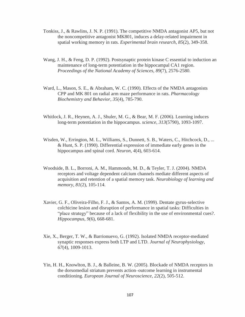
107
Tonkiss, J., & Rawlins, J. N. P. (1991). The competitive NMDA antagonist AP5, but not
the non competitive antagonist MK801, induces a delay-related impairment in
spatial working memory in rats. Experimental brain research, 85(2), 349-358.
Wang, J. H., & Feng, D. P. (1992). Postsynaptic protein kinase C essential to induction an
maintenance of long-term potentiation in the hippocampal CA1 region.
Proceedings of the National Academy of Sciences, 89(7), 2576-2580.
Ward, L., Mason, S. E., & Abraham, W. C. (1990). Effects of the NMDA antagonists
CPP and MK 801 on radial arm maze performance in rats. Pharmacology
Biochemistry and Behavior, 35(4), 785-790.
Whitlock, J. R., Heynen, A. J., Shuler, M. G., & Bear, M. F. (2006). Learning induces
long-term potentiation in the hippocampus. science, 313(5790), 1093-1097.
Wisden, W., Errington, M. L., Williams, S., Dunnett, S. B., Waters, C., Hitchcock, D., ...
& Hunt, S. P. (1990). Differential expression of immediate early genes in the
hippocampus and spinal cord. Neuron, 4(4), 603-614.
Woodside, B. L., Borroni, A. M., Hammonds, M. D., & Teyler, T. J. (2004). NMDA
receptors and voltage dependent calcium channels mediate different aspects of
acquisition and retention of a spatial memory task. Neurobiology of learning and
memory, 81(2), 105-114.
Xavier, G. F., Oliveira‐Filho, F. J., & Santos, A. M. (1999). Dentate gyrus‐selective
colchicine lesion and disruption of performance in spatial tasks: Difficulties in
“place strategy” because of a lack of flexibility in the use of environmental cues?.
Hippocampus, 9(6), 668-681.
Xie, X., Berger, T. W., & Barrionuevo, G. (1992). Isolated NMDA receptor-mediated
synaptic responses express both LTP and LTD. Journal of Neurophysiology,
67(4), 1009-1013.
Yin, H. H., Knowlton, B. J., & Balleine, B. W. (2005). Blockade of NMDA receptors in
the dorsomedial striatum prevents action–outcome learning in instrumental
conditioning. European Journal of Neuroscience, 22(2), 505-512.

108
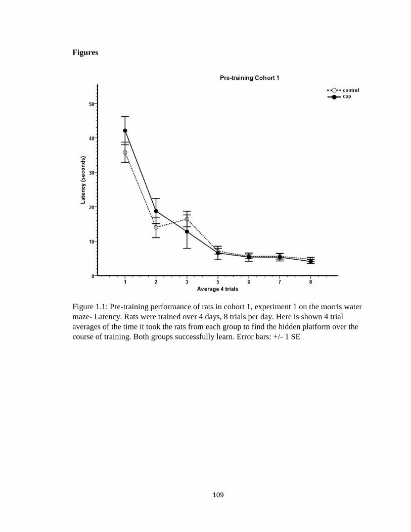
109
Figures
Figure 1.1: Pre-training performance of rats in cohort 1, experiment 1 on the morris water
maze- Latency. Rats were trained over 4 days, 8 trials per day. Here is shown 4 trial
averages of the time it took the rats from each group to find the hidden platform over the
course of training. Both groups successfully learn. Error bars: +/- 1 SE

110
Figure 1.2: Pre-training performance of rats in cohort 1, experiment 1 on the morris water
maze- Path length. Rats were trained over 4 days, 8 trials per day. Here is shown 4 trial
averages of the length of the swim paths it took the rats from each group to find the
hidden platform over the course of training. Both groups successfully learn. Error bars:
+/- 1 SE
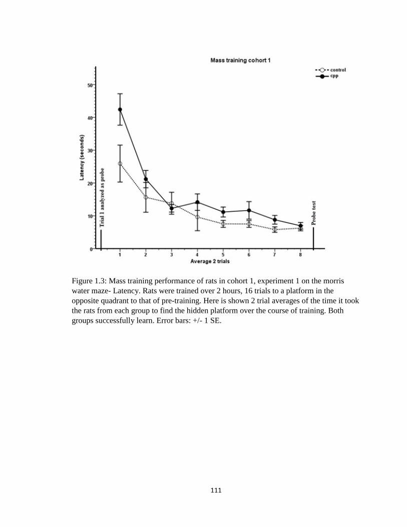
111
Figure 1.3: Mass training performance of rats in cohort 1, experiment 1 on the morris
water maze- Latency. Rats were trained over 2 hours, 16 trials to a platform in the
opposite quadrant to that of pre-training. Here is shown 2 trial averages of the time it took
the rats from each group to find the hidden platform over the course of training. Both
groups successfully learn. Error bars: +/- 1 SE.

112
Figure 1.4: Mass training performance of rats in cohort 1, experiment 1 on the morris
water maze- Path length. Rats were trained over 2 hours, 16 trials to a platform in the
opposite quadrant to that of pre-training. Here is shown 2 trial averages of the length of
the swim paths it took the rats from each group to find the hidden platform over the
course of training. Both groups successfully learn. Error bars: +/- 1 SE

113
Figure 1.5: Trial 1 of mass training analyzed as a 30 second probe to test pre-training
spatial memory in cohort 1, experiment 1. Comparison between time spent in pre-trained
quadrant vs an average of the other three shows that both groups show a spatial
preference to the pretrained quadrant. * p < 0.05, Error bars: +/- 1 SE
114
Figure 1.6: 30 second probe test done immediately after massed reversal training
comparing time spent in mass trained quadrant to average of the other three in experiment
1, cohort 1. Both groups show preference for the mass trained quadrant immediately after
training * p < 0.05, Error bars: +/- 1 SE

115
Fig 1.7: 30 second probe test done immediately after massed reversal training comparing
time spent in an area immediately surrounding the mass training platform location (2% of
total pool surface area) to the pre-training platform location in experiment 1, cohort 1.
Both groups show preference for the mass trained location immediately after training * p
< 0.05, Error bars: +/- 1 SE
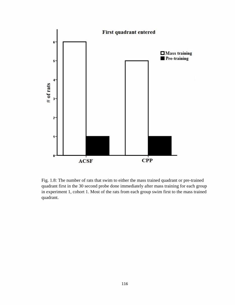
116
Fig. 1.8: The number of rats that swim to either the mass trained quadrant or pre-trained
quadrant first in the 30 second probe done immediately after mass training for each group
in experiment 1, cohort 1. Most of the rats from each group swim first to the mass trained
quadrant.
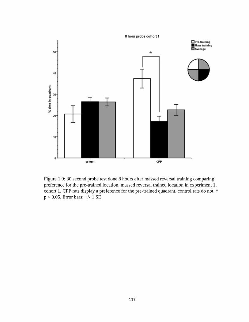
117
Figure 1.9: 30 second probe test done 8 hours after massed reversal training comparing
preference for the pre-trained location, massed reversal trained location in experiment 1,
cohort 1. CPP rats display a preference for the pre-trained quadrant, control rats do not. *
p < 0.05, Error bars: +/- 1 SE

118
Fig. 1.10: 30 second probe test done 8 hours after massed reversal training comparing
time spent in an area immediately surrounding the mass training platform location (2% of
total pool surface area) to the pre-training platform location in experiment 1, cohort 1.
The CPP rats show preference for the pre-trained location while the control rats show the
opposite, a preference for the mass trained location compared to the pre-trained location.
* p < 0.05, Error bars: +/- 1 SE
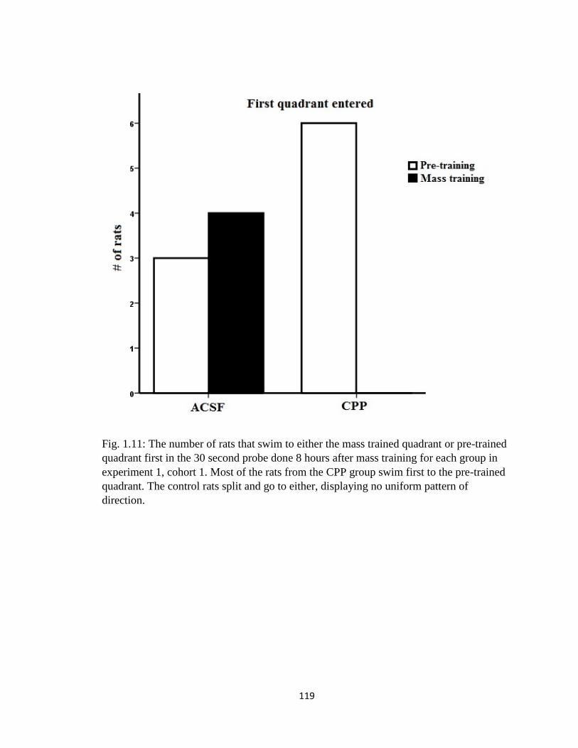
119
Fig. 1.11: The number of rats that swim to either the mass trained quadrant or pre-trained
quadrant first in the 30 second probe done 8 hours after mass training for each group in
experiment 1, cohort 1. Most of the rats from the CPP group swim first to the pre-trained
quadrant. The control rats split and go to either, displaying no uniform pattern of
direction.
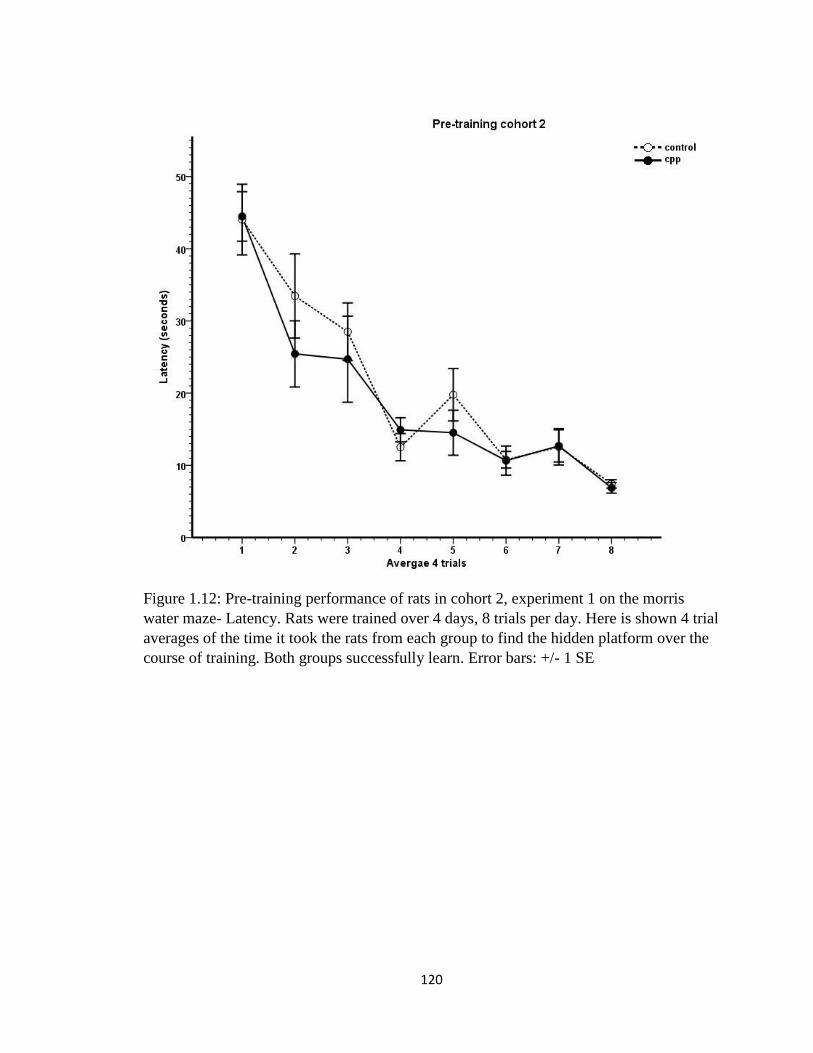
120
Figure 1.12: Pre-training performance of rats in cohort 2, experiment 1 on the morris
water maze- Latency. Rats were trained over 4 days, 8 trials per day. Here is shown 4 trial
averages of the time it took the rats from each group to find the hidden platform over the
course of training. Both groups successfully learn. Error bars: +/- 1 SE
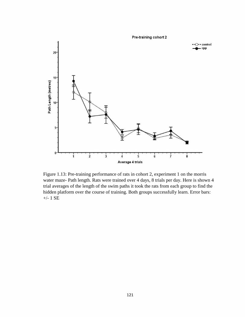
121
Figure 1.13: Pre-training performance of rats in cohort 2, experiment 1 on the morris
water maze- Path length. Rats were trained over 4 days, 8 trials per day. Here is shown 4
trial averages of the length of the swim paths it took the rats from each group to find the
hidden platform over the course of training. Both groups successfully learn. Error bars:
+/- 1 SE
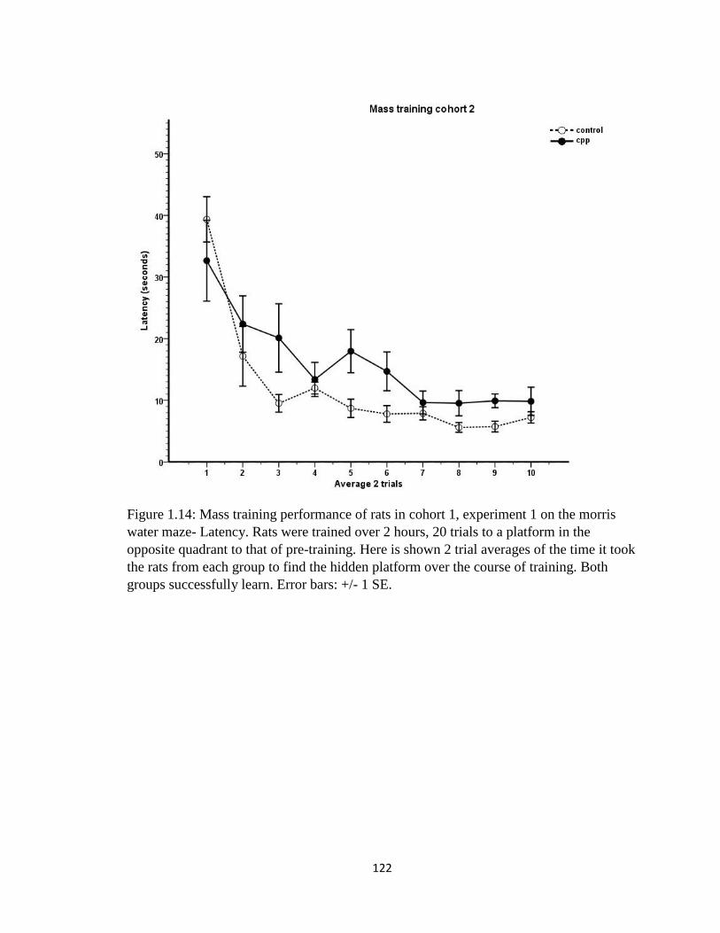
122
Figure 1.14: Mass training performance of rats in cohort 1, experiment 1 on the morris
water maze- Latency. Rats were trained over 2 hours, 20 trials to a platform in the
opposite quadrant to that of pre-training. Here is shown 2 trial averages of the time it took
the rats from each group to find the hidden platform over the course of training. Both
groups successfully learn. Error bars: +/- 1 SE.

123
Figure 1.15: Mass training performance of rats in cohort 2, experiment 1 on the morris
water maze- Path length. Rats were trained over 2 hours, 20 trials to a platform in the
opposite quadrant to that of pre-training. Here is shown 2 trial averages of the length of
the swim paths it took the rats from each group to find the hidden platform over the
course of training. Both groups successfully learn. Error bars: +/- 1 SE
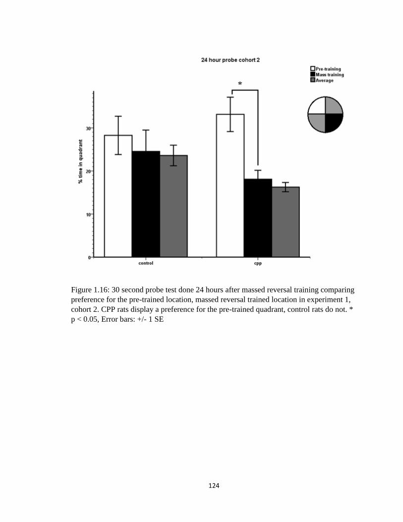
124
Figure 1.16: 30 second probe test done 24 hours after massed reversal training comparing
preference for the pre-trained location, massed reversal trained location in experiment 1,
cohort 2. CPP rats display a preference for the pre-trained quadrant, control rats do not. *
p < 0.05, Error bars: +/- 1 SE

125
Figure 1.17: Dorsal and ventral hippocampal cannulation placements for CPP group rats,
experiment 1. Cannulation end points were taken from hippocampal slices and imposed
onto images of the dorsal and ventral hippocampi. A. dorsal cohort 1, B. ventral cohort 1,
C. dorsal cohort 2, D. ventral cohort 2.

126
Figure 2.1: Pre-training performance of rats in cohort 1, experiment 2 on the morris water
maze in room A- Latency. Rats were trained over 4 days, 8 trials per day. Here is shown 4
trial averages of the time it took the rats from each group to find the hidden platform over
the course of training. Both groups successfully learn. Error bars: +/- 1 SE
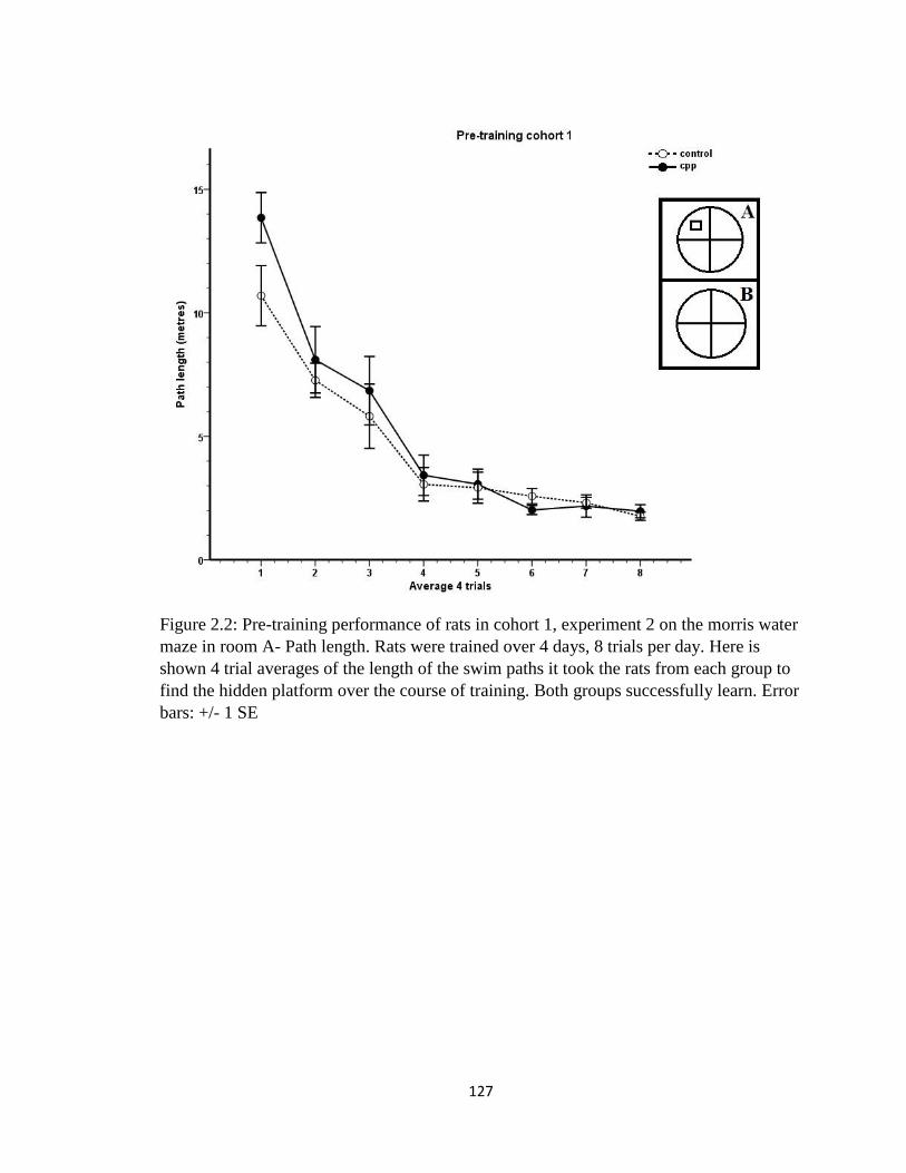
127
Figure 2.2: Pre-training performance of rats in cohort 1, experiment 2 on the morris water
maze in room A- Path length. Rats were trained over 4 days, 8 trials per day. Here is
shown 4 trial averages of the length of the swim paths it took the rats from each group to
find the hidden platform over the course of training. Both groups successfully learn. Error
bars: +/- 1 SE
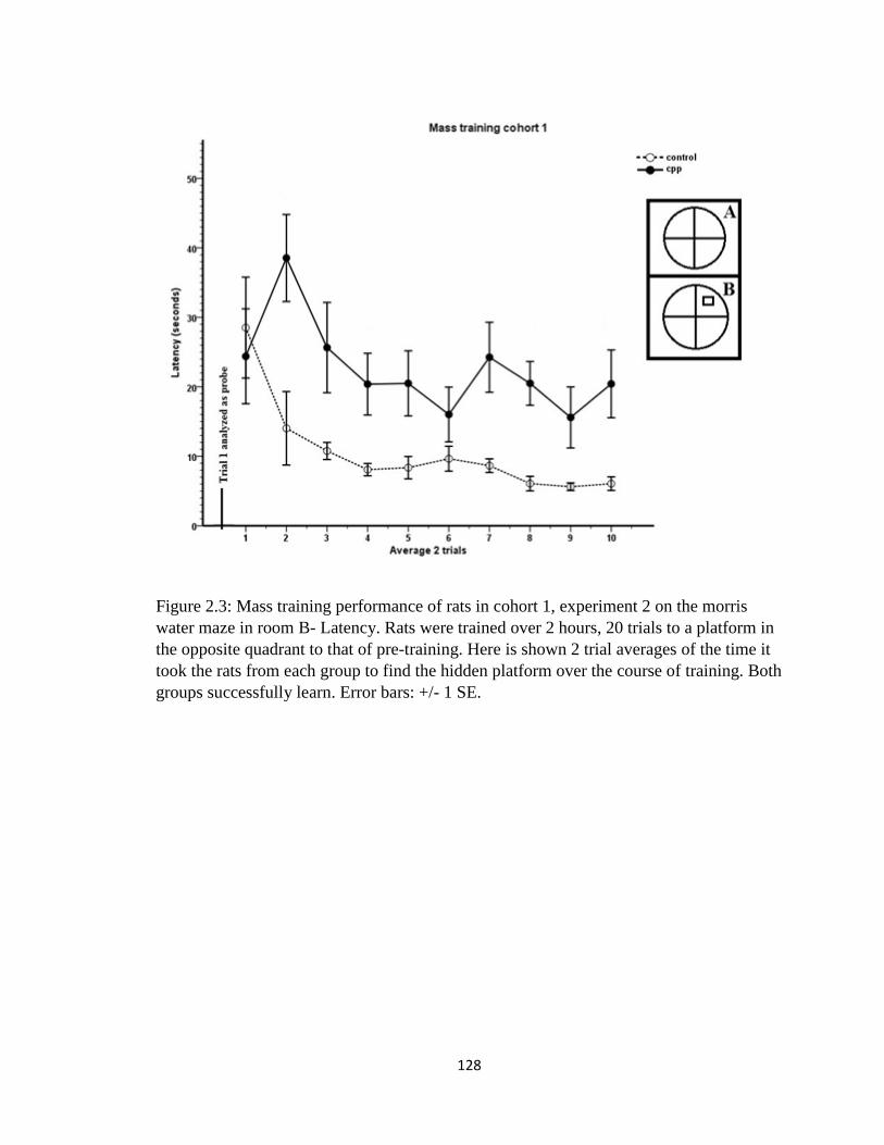
128
Figure 2.3: Mass training performance of rats in cohort 1, experiment 2 on the morris
water maze in room B- Latency. Rats were trained over 2 hours, 20 trials to a platform in
the opposite quadrant to that of pre-training. Here is shown 2 trial averages of the time it
took the rats from each group to find the hidden platform over the course of training. Both
groups successfully learn. Error bars: +/- 1 SE.
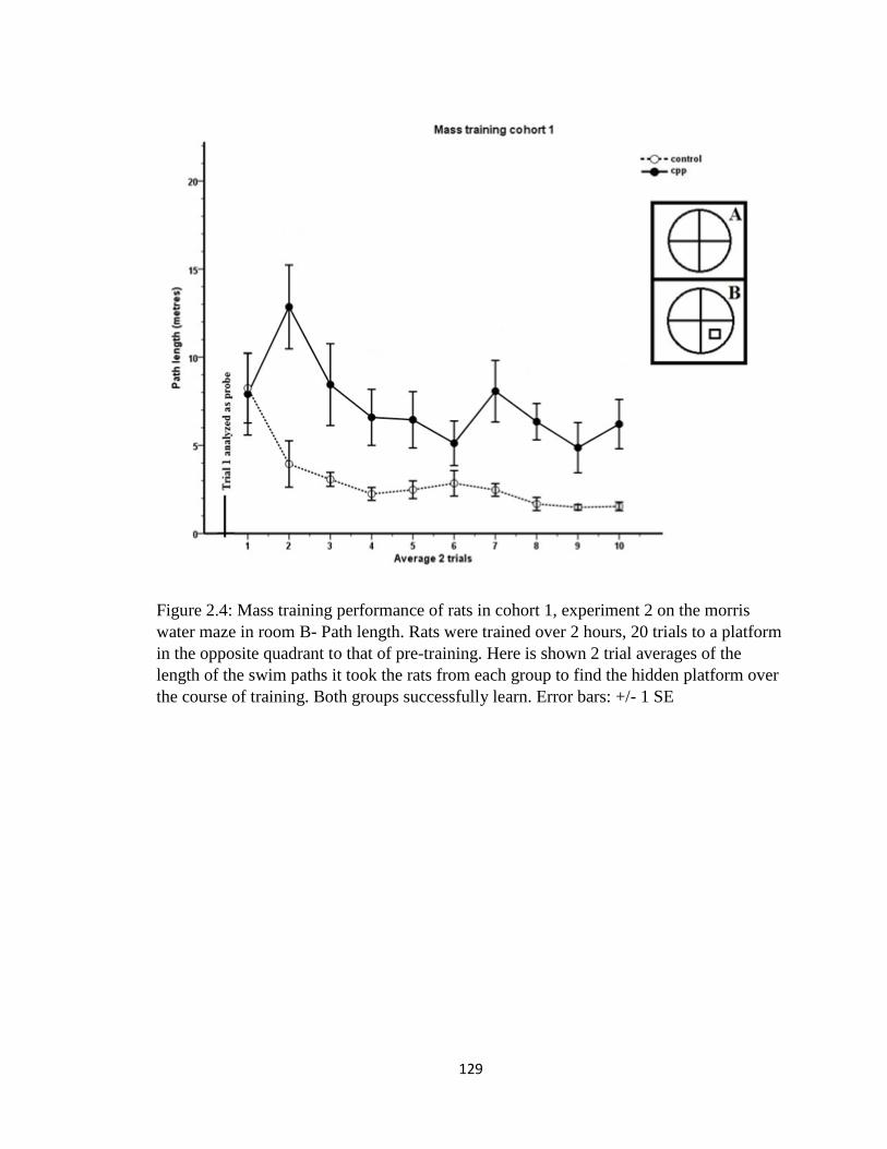
129
Figure 2.4: Mass training performance of rats in cohort 1, experiment 2 on the morris
water maze in room B- Path length. Rats were trained over 2 hours, 20 trials to a platform
in the opposite quadrant to that of pre-training. Here is shown 2 trial averages of the
length of the swim paths it took the rats from each group to find the hidden platform over
the course of training. Both groups successfully learn. Error bars: +/- 1 SE
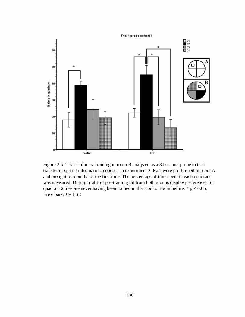
130
Figure 2.5: Trial 1 of mass training in room B analyzed as a 30 second probe to test
transfer of spatial information, cohort 1 in experiment 2. Rats were pre-trained in room A
and brought to room B for the first time. The percentage of time spent in each quadrant
was measured. During trial 1 of pre-training rat from both groups display preferences for
quadrant 2, despite never having been trained in that pool or room before. * p < 0.05,
Error bars: +/- 1 SE
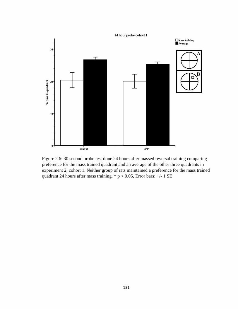
131
Figure 2.6: 30 second probe test done 24 hours after massed reversal training comparing
preference for the mass trained quadrant and an average of the other three quadrants in
experiment 2, cohort 1. Neither group of rats maintained a preference for the mass trained
quadrant 24 hours after mass training. * p < 0.05, Error bars: +/- 1 SE

132
Figure 2.7: Pre-training performance of rats in cohort 2, experiment 2 on the morris water
maze in room A- Latency. Rats were trained over 4 days, 8 trials per day. Here is shown 4
trial averages of the time it took the rats from each group to find the hidden platform over
the course of training. Both groups successfully learn. Error bars: +/- 1 SE
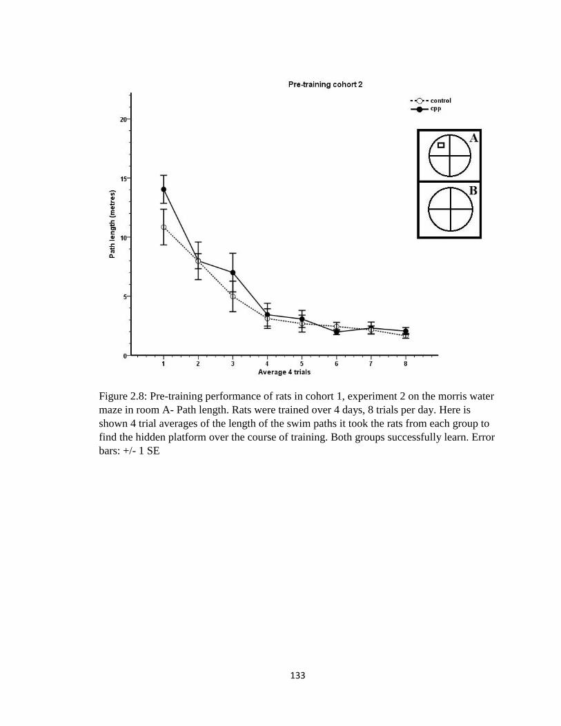
133
Figure 2.8: Pre-training performance of rats in cohort 1, experiment 2 on the morris water
maze in room A- Path length. Rats were trained over 4 days, 8 trials per day. Here is
shown 4 trial averages of the length of the swim paths it took the rats from each group to
find the hidden platform over the course of training. Both groups successfully learn. Error
bars: +/- 1 SE
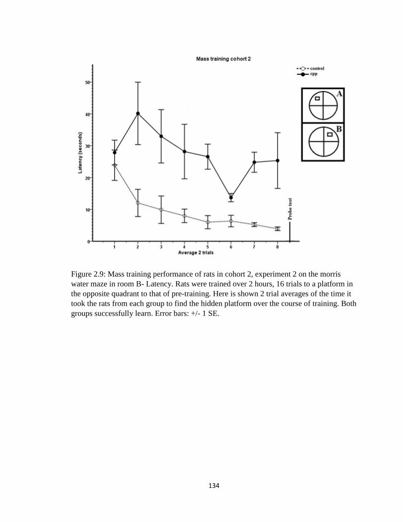
134
Figure 2.9: Mass training performance of rats in cohort 2, experiment 2 on the morris
water maze in room B- Latency. Rats were trained over 2 hours, 16 trials to a platform in
the opposite quadrant to that of pre-training. Here is shown 2 trial averages of the time it
took the rats from each group to find the hidden platform over the course of training. Both
groups successfully learn. Error bars: +/- 1 SE.
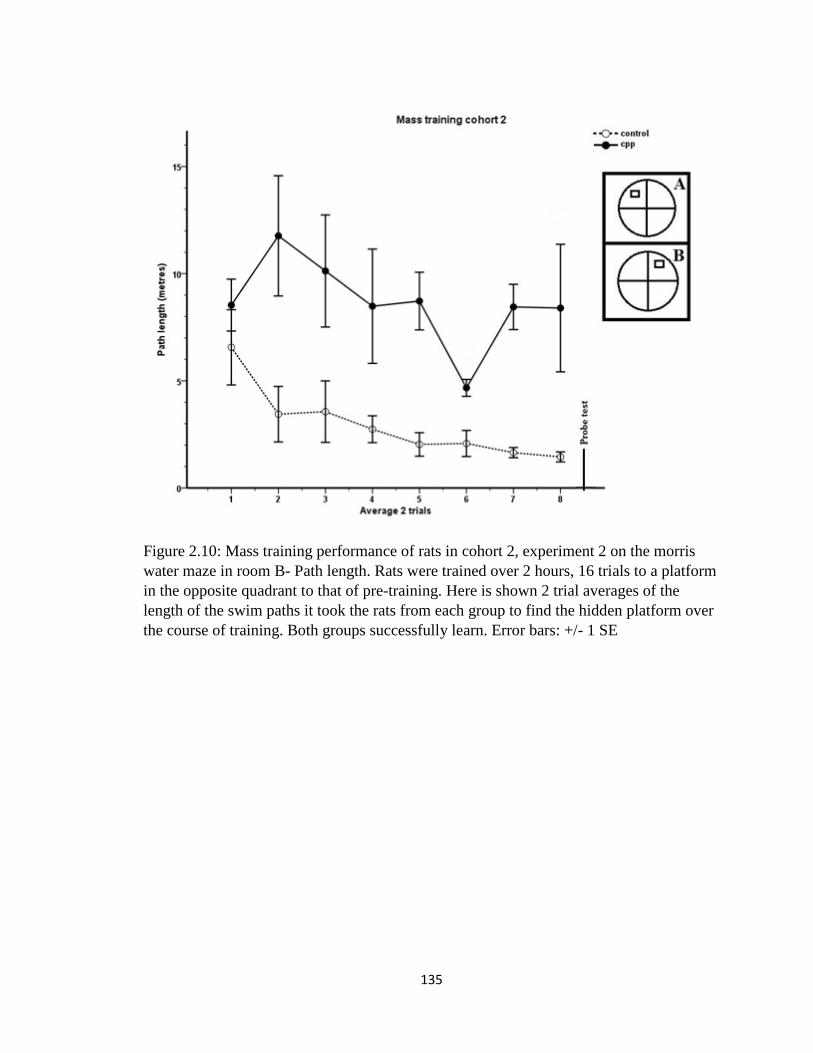
135
Figure 2.10: Mass training performance of rats in cohort 2, experiment 2 on the morris
water maze in room B- Path length. Rats were trained over 2 hours, 16 trials to a platform
in the opposite quadrant to that of pre-training. Here is shown 2 trial averages of the
length of the swim paths it took the rats from each group to find the hidden platform over
the course of training. Both groups successfully learn. Error bars: +/- 1 SE
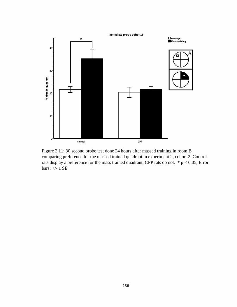
136
Figure 2.11: 30 second probe test done 24 hours after massed training in room B
comparing preference for the massed trained quadrant in experiment 2, cohort 2. Control
rats display a preference for the mass trained quadrant, CPP rats do not. * p < 0.05, Error
bars: +/- 1 SE
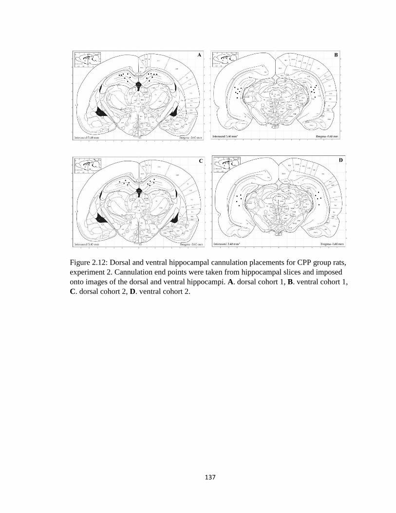
137
Figure 2.12: Dorsal and ventral hippocampal cannulation placements for CPP group rats,
experiment 2. Cannulation end points were taken from hippocampal slices and imposed
onto images of the dorsal and ventral hippocampi. A. dorsal cohort 1, B. ventral cohort 1,
C. dorsal cohort 2, D. ventral cohort 2.

138
Figure 3.1: Pre-training performance of rats in experiment 3 on the morris water maze in
room A- Latency. Rats were trained over 4 days, 8 trials per day. Here is shown 4 trial
averages of the time it took the rats from each group to find the hidden platform over the
course of training. All groups successfully learn. Error bars: +/- 1 SE
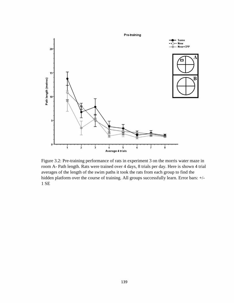
139
Figure 3.2: Pre-training performance of rats in experiment 3 on the morris water maze in
room A- Path length. Rats were trained over 4 days, 8 trials per day. Here is shown 4 trial
averages of the length of the swim paths it took the rats from each group to find the
hidden platform over the course of training. All groups successfully learn. Error bars: +/-
1 SE
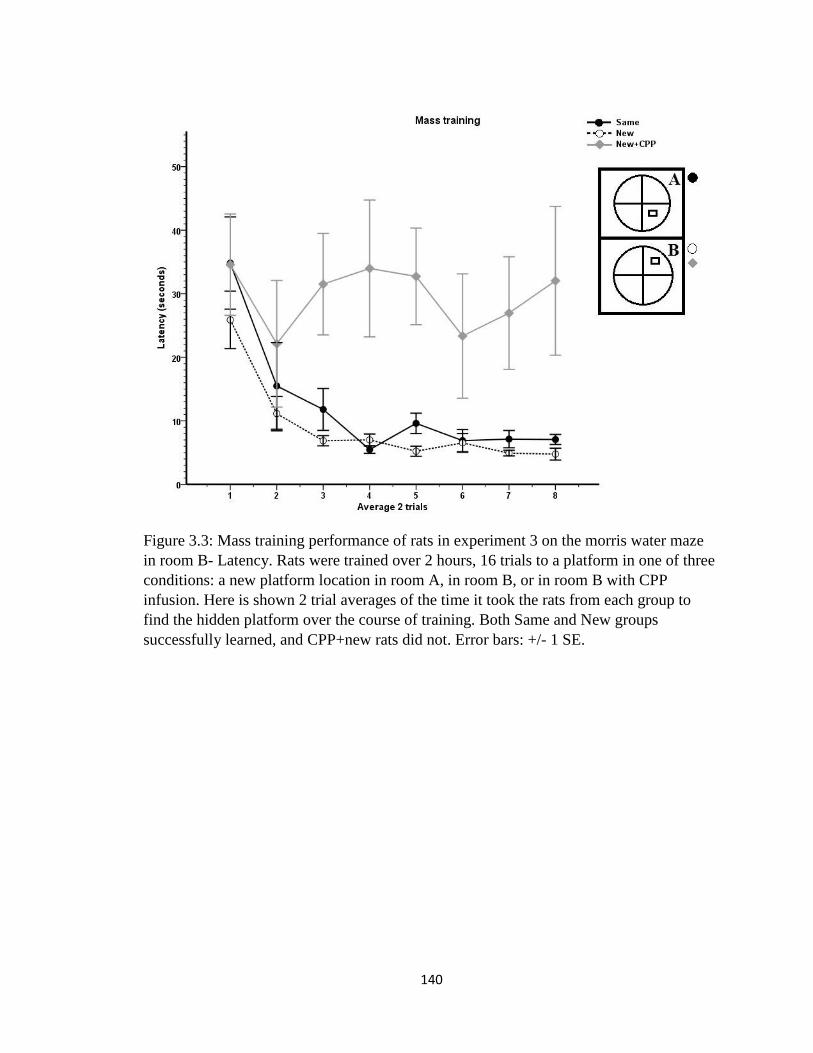
140
Figure 3.3: Mass training performance of rats in experiment 3 on the morris water maze
in room B- Latency. Rats were trained over 2 hours, 16 trials to a platform in one of three
conditions: a new platform location in room A, in room B, or in room B with CPP
infusion. Here is shown 2 trial averages of the time it took the rats from each group to
find the hidden platform over the course of training. Both Same and New groups
successfully learned, and CPP+new rats did not. Error bars: +/- 1 SE.

141
Figure 3.4: Mass training performance of rats in experiment 3 on the morris water maze
in room B- Path length. Rats were trained over 2 hours, 16 trials to a platform in one of
three conditions: a new platform location in room A, in room B, or in room B with CPP
infusion. Here is shown 2 trial averages of the length of the swim path it took the rats
from each group to find the hidden platform over the course of training. Both Same and
New groups successfully learned, and CPP+new rats did not. Error bars: +/- 1 SE.
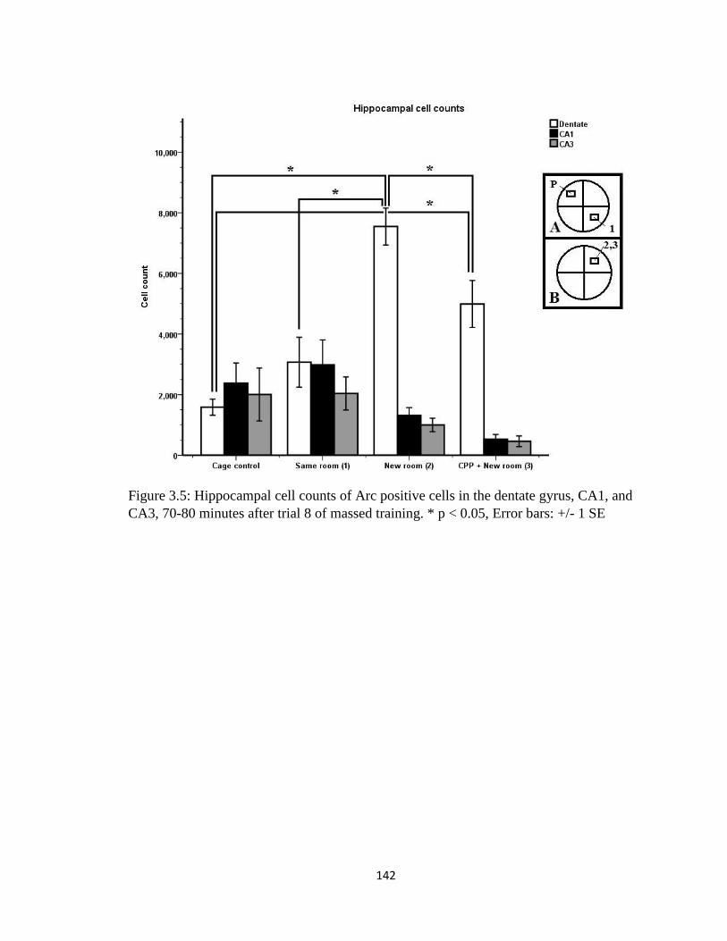
142
Figure 3.5: Hippocampal cell counts of Arc positive cells in the dentate gyrus, CA1, and
CA3, 70-80 minutes after trial 8 of massed training. * p < 0.05, Error bars: +/- 1 SE
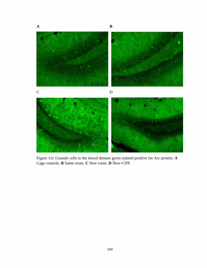
143
A B
C D
Figure 3.6: Granule cells in the dorsal dentate gyrus stained positive for Arc protein. A
Cage controls. B Same room. C New room. D New+CPP.

144
Figure 3.7: Dorsal and ventral hippocampal cannulation placements for CPP group rats,
experiment 3. Cannulation end points were taken from hippocampal slices and imposed
onto images of the dorsal and ventral hippocampi. A. dorsal, B. ventral.


![Role of desensitization of AMPA receptors on the neuronal viability and on the [Ca2+]i changes in cultured rat hippocampal neurons](https://static.fdokumen.com/doc/165x107/63230bd664690856e1096f90/role-of-desensitization-of-ampa-receptors-on-the-neuronal-viability-and-on-the-ca2i.jpg)


