Ca2+ Oscillations Mediated by the Synergistic Excitatory Actions of GABAA and NMDA Receptors in the...
13
Neuron, Vol. 18, 243–255, February, 1997, Copyright 1997 by Cell Press Ca 21 Oscillations Mediated by the Synergistic Excitatory Actions of GABA A and NMDA Receptors in the Neonatal Hippocampus Xavier Leinekugel, Igor Medina, Ilgam Khalilov, stages of development when the activation of GABA A receptors provides depolarization instead of hyperpo- Yehezkel Ben-Ari, and Roustem Khazipov INSERM Unite ´ 29 larization (Ben-Ari et al., 1989; 1994; Fiszman et al., 1990; Wu et al., 1992; Hales et al., 1994; Reichling et al., 1994; Ho ˆ pital de Port-Royal 123, Bd de Port-Royal LoTurco et al., 1995; Serafini et al., 1995; Chen et al., 1996; Rohrbough and Spitzer, 1996). In several types of 75014 Paris France neonatal neurons, activation of GABA A R triggers action potentials and activates voltage-dependent Ca 21 chan- nels, producing rises of [Ca 21 ] i (Yuste and Katz, 1991; Hales et al., 1994; Reichling et al., 1994; Leinekugel et Summary al., 1995; LoTurco et al., 1995; Obrietan and van den Pol, 1995; Chen et al., 1996). If GABA is the principal We asked whether GABA A and NMDA receptors may fast-acting excitatory transmitter during early postnatal act in synergy in neonatal hippocampal slices, at a life in the hippocampus as suggested from earlier stud- time when GABA exerts a depolarizing action. The ies from this laboratory (Cherubini et al., 1991; Ben-Ari GABAA receptor agonist isoguvacine reduced the volt- et al., 1994), it may, in contrast to adult neurons, act age-dependent Mg 21 block of single NMDA channels in synergy with NMDA R, providing the depolarization recorded in cell-attached configuration from P2–5 CA 3 required to release their voltage-dependent Mg 21 block. pyramidal neurons and potentiated the Ca 21 influx If so, GABA A R would play in neonatal neurons the role through NMDA channels. The synaptic response conferred to AMPA R in more mature neurons. We have evoked by electrical stimulation of stratum radiatum now tested this hypothesis in P 2–5 neonatal hippocampal was mediated by a synergistic interaction between slices. GABA A and NMDA receptors. Network-driven Giant Depolarizing Potentials, which are a typical feature of the neonatal hippocampal network, provided coacti- Results vation of GABA A and NMDA receptors and were asso- ciated with spontaneous and synchronous Ca 21 in- We used cell-attached recordings and noninvasive Ca 21 creases in CA 3 pyramidal neurons. Thus, at the early imaging techniques (that do not affect [Cl 2 ] i ) to study stages of development, GABA is a major excitatory the interaction between GABAA and NMDA receptor– transmitter that acts in synergy with NMDA receptors. mediated signals in hippocampal slices at postnatal This provides in neonatal neurons a hebbian stimula- days 2–5 (P 2–5 ). tion that may be involved in neuronal plasticity and network formation in the developing hippocampus. Isoguvacine Reduces the Mg 21 Block Introduction of Single NMDA Channels We examined the effects of the GABA A -R agonist isogu- The NMDA subtype of glutamate receptors plays an vacine on single NMDA channel activity recorded in cell- important role in adult and developmental neuronal plas- attached configuration from P 2–5 pyramidal cells (Figure ticity via increases in intracellular [Ca 21 ] i (Constantine- 1). Action potential–dependent synaptic transmission Paton et al., 1990; Goodman and Shatz, 1993; Malenka was blocked by TTX (1 mM in the bath). The activity and Nicoll, 1993; Fox, 1995; Durand et al., 1996). Since of NMDA channels in presence of Mg 21 was voltage the voltage-dependent Mg 21 block of NMDA channels dependent with characteristic flickering at resting mem- (Mayer et al., 1984; Nowak et al., 1984) operates not brane potential (estimated to be 282 6 3 mV from the only in adult but also in neonatal neurons (LoTurco et apparent [V pipette ] reversal potential of NMDA R–mediated al., 1991; Strecker et al., 1994; Crair and Malenka, 1995; currents, n 5 5). The affinity of Mg 21 for NMDA channels Khazipov et al., 1995), their activation during synaptic was estimated using the model of open-channel block activity requires external sources of depolarization. In (Nowak et al., 1984), as described elsewhere (Khazipov adult neurons, this is largely provided by glutamate act- et al., 1995). Bath application of isoguvacine (10 mM) ing on AMPA receptors that mediate most of the excit- strongly and reversibly reduced the flickering of NMDA atory drive throughout the mammalian central nervous channels (Figure 1A), decreasing the Mg 21 affinity for system. In contrast, GABA, the primary inhibitory trans- NMDA channels (K Mg 21 : control: 16.3 6 5.3 mM; isogu- mitter, acting via ionotropic GABA A receptors (GABA A vacine: 118 6 36 mM; wash: 19.1 6 4 mM; n 5 5; Figure R), increases a chloride conductance that usually hyper- 1B). Because of the configuration used (cell-attached polarizes adult neurons, thus preventing the activation recording), the effect of isoguvacine cannot be due to of NMDA R (Agmon and O’Dowd, 1992; Kanter et al., direct action on the NMDA channels recorded. It was in 1996). Thus, the induction of NMDA R–dependent forms fact entirely due to depolarization of the cell from 282 6 of long-term potentiation or depression (LTP, LTD) are 3 mV to 259 6 4 mV (n 5 5) as indicated by the positive facilitated by GABA A -R antagonists (Wigstrom and Gus- shift of the apparent (V pipette ) reversal potential of NMDA tafsson, 1983; Artola and Singer, 1987; Kanter and Ha- berly, 1993). An opposite situation may prevail at early receptor–mediated currents (Figure 1C).
Transcript of Ca2+ Oscillations Mediated by the Synergistic Excitatory Actions of GABAA and NMDA Receptors in the...

Neuron, Vol. 18, 243–255, February, 1997, Copyright 1997 by Cell Press
Ca21 Oscillations Mediated by theSynergistic Excitatory Actions of GABAA
and NMDA Receptors in the Neonatal Hippocampus
Xavier Leinekugel, Igor Medina, Ilgam Khalilov, stages of development when the activation of GABAA
receptors provides depolarization instead of hyperpo-Yehezkel Ben-Ari, and Roustem KhazipovINSERM Unite 29 larization (Ben-Ari et al., 1989; 1994; Fiszman et al., 1990;
Wu et al., 1992; Hales et al., 1994; Reichling et al., 1994;Hopital de Port-Royal123, Bd de Port-Royal LoTurco et al., 1995; Serafini et al., 1995; Chen et al.,
1996; Rohrbough and Spitzer, 1996). In several types of75014 ParisFrance neonatal neurons, activation of GABAA R triggers action
potentials and activates voltage-dependent Ca21 chan-nels, producing rises of [Ca21]i (Yuste and Katz, 1991;Hales et al., 1994; Reichling et al., 1994; Leinekugel etSummaryal., 1995; LoTurco et al., 1995; Obrietan and van denPol, 1995; Chen et al., 1996). If GABA is the principalWe asked whether GABAA and NMDA receptors mayfast-acting excitatory transmitter during early postnatalact in synergy in neonatal hippocampal slices, at alife in the hippocampus as suggested from earlier stud-time when GABA exerts a depolarizing action. Theies from this laboratory (Cherubini et al., 1991; Ben-AriGABAA receptor agonist isoguvacine reduced the volt-et al., 1994), it may, in contrast to adult neurons, actage-dependent Mg21 block of single NMDA channelsin synergy with NMDA R, providing the depolarizationrecorded in cell-attached configuration from P2–5 CA3
required to release their voltage-dependent Mg21 block.pyramidal neurons and potentiated the Ca21 influxIf so, GABAA R would play in neonatal neurons the rolethrough NMDA channels. The synaptic responseconferred to AMPA R in more mature neurons. We haveevoked by electrical stimulation of stratum radiatumnow tested this hypothesis in P2–5 neonatal hippocampalwas mediated by a synergistic interaction betweenslices.GABAA and NMDA receptors. Network-driven Giant
Depolarizing Potentials, which are a typical feature ofthe neonatal hippocampal network, provided coacti-
Resultsvation of GABAA and NMDA receptors and were asso-ciated with spontaneous and synchronous Ca21 in-
We used cell-attached recordings and noninvasive Ca21creases in CA3 pyramidal neurons. Thus, at the earlyimaging techniques (that do not affect [Cl2]i) to studystages of development, GABA is a major excitatorythe interaction between GABAA and NMDA receptor–transmitter that acts in synergy with NMDA receptors.mediated signals in hippocampal slices at postnatalThis provides in neonatal neurons a hebbian stimula-days 2–5 (P2–5).tion that may be involved in neuronal plasticity and
network formation in the developing hippocampus.
Isoguvacine Reduces the Mg21 BlockIntroductionof Single NMDA ChannelsWe examined the effects of the GABAA-R agonist isogu-The NMDA subtype of glutamate receptors plays anvacine on single NMDA channel activity recorded in cell-important role inadult and developmental neuronal plas-attached configuration from P2–5 pyramidal cells (Figureticity via increases in intracellular [Ca21]i (Constantine-1). Action potential–dependent synaptic transmissionPaton et al., 1990; Goodman and Shatz, 1993; Malenkawas blocked by TTX (1 mM in the bath). The activityand Nicoll, 1993; Fox, 1995; Durand et al., 1996). Sinceof NMDA channels in presence of Mg21 was voltagethe voltage-dependent Mg21 block of NMDA channelsdependent with characteristic flickering at resting mem-(Mayer et al., 1984; Nowak et al., 1984) operates notbrane potential (estimated to be 282 6 3 mV from theonly in adult but also in neonatal neurons (LoTurco etapparent [Vpipette] reversal potential of NMDA R–mediatedal., 1991; Strecker et al., 1994; Crair and Malenka, 1995;currents, n 5 5). The affinity of Mg21 for NMDA channelsKhazipov et al., 1995), their activation during synapticwas estimated using the model of open-channel blockactivity requires external sources of depolarization. In(Nowak et al., 1984), as described elsewhere (Khazipovadult neurons, this is largely provided by glutamate act-et al., 1995). Bath application of isoguvacine (10 mM)ing on AMPA receptors that mediate most of the excit-strongly and reversibly reduced the flickering of NMDAatory drive throughout the mammalian central nervouschannels (Figure 1A), decreasing the Mg21 affinity forsystem. In contrast, GABA, the primary inhibitory trans-NMDA channels (KMg
21: control: 16.3 6 5.3 mM; isogu-mitter, acting via ionotropic GABAA receptors (GABAA
vacine: 118 6 36 mM; wash: 19.1 6 4 mM; n 5 5; FigureR), increases a chloride conductance that usually hyper-1B). Because of the configuration used (cell-attachedpolarizes adult neurons, thus preventing the activationrecording), the effect of isoguvacine cannot be due toof NMDA R (Agmon and O’Dowd, 1992; Kanter et al.,direct action on the NMDA channels recorded. It was in1996). Thus, the induction of NMDA R–dependent formsfact entirely due to depolarization of the cell from 282 6of long-term potentiation or depression (LTP, LTD) are3 mV to 259 6 4 mV (n 5 5) as indicated by the positivefacilitated by GABAA-R antagonists (Wigstrom and Gus-shift of the apparent (Vpipette) reversal potential of NMDAtafsson, 1983; Artola and Singer, 1987; Kanter and Ha-
berly, 1993). An opposite situation may prevail at early receptor–mediated currents (Figure 1C).

Neuron244
Figure 1. The GABAA Agonist Isoguvacine Decreases the Voltage-Dependent Mg21 Block of NMDA Channels
Single NMDA channel recordings in the cell-attached configuration from neonatal CA3 hippocampal pyramidal neurons.(A) Traces of single NMDA-channels activity recorded at different membrane potentials in control (left) and (right) in presence of isoguvacine(10 mM in the bath). Note the reduction in flickering in presence of isoguvacine.(B) Plot of Mg21 affinity (mean 6 SE; n 5 5) at different membrane potentials estimated using the model of open-channel block. Control, opencircles; isoguvacine, closed circles; wash, triangles.(C) Current–voltage relationships of single NMDA channels in control (open circles), in presence (closed circles), and after wash (triangles) ofisoguvacine.
Isoguvacine Increases Ca21 Influx through conditions, isoguvacine did not affect NMDA responses(180 6 37 pA in control, 178 6 37 pA in presence ofNMDA Channels
If activation of GABAA receptors facilitates NMDA recep- isoguvacine; Vh 5 250 mV; n 5 5) (data not shown),suggesting that the effects of isoguvacine on NMDAtors’ activity, it should also increase the Ca21 influx
through NMDA channels. In an earlier study, we showed R–mediated Ca21 increase are mediated by the GABAer-gic depolarization. In keeping with this hypothesis,that isoguvacine increases [Ca21]i via D600 sensitive
voltage-dependent Ca21 channels (Leinekugel et al., blocking the isoguvacine-mediated depolarization byvoltage clamping the cell at 280 mV during whole-cell1995). We therefore routinely added D600 (50 mM in the
bath) to study changes in [Ca21]i mediated by NMDA R. recording (Figure 2B, lower lanes) prevented the risein [Ca21]i (12 6 6%; n 5 3) that was observed duringAs shown in Figure 2A, D600 prevented the rise in Ca21
(top lanes) but not the depolarization (lower traces) pro- coapplication of isoguvacine and NMDA in current-clampmode (133 6 8%; n 5 3) (Figure 2B, upper lanes). There-duced by isoguvacine (100 mM, focal application by
pressure ejection) in current-clamp whole-cell recording fore, the depolarization produced by GABAA receptors inconditions that do not affect [Cl2]i reduces the Mg21(internal solution 6: ECl2 around 210 mV). We then used
confocal microscopy with the permeant dye Fluo3-AM block of NMDA channels, thus facilitating NMDA-medi-ated current and Ca21 influx. We then investigated theto measure the Ca21 influx through NMDA channels from
nondialyzed P2–5 CA3 pyramidal cells (Figure 2C). In the interaction between GABAA and NMDA R–mediated sig-nals in synaptic activity.presence of D600, neither NMDA (20 mM, bath applied)
nor isoguvacine (100 mM, focal application by pressureejection) alone increased [Ca21]i (0 6 3% and 14 6 3%, Synaptically Activated GABAA
and NMDA Receptorsrespectively; n 5 12). In contrast, coapplication of bothagonists produced a large [Ca21]i increase (11586 33%, The neonatal hippocampal network is characterized by
the presence of spontaneous and evoked network-n 5 12). These observations suggest that thedepolariza-tion induced by isoguvacine removes the voltage- driven Giant Depolarizing Potentials (GDPs: Ben-Ari et
al., 1989; Gaıarsa et al., 1990). GDPs were described asdependent Mg21 block and thus potentiates Ca21 influxthrough NMDA R. To exclude possible direct effect of primarily GABAergic based on their sensitivity to the
GABAA-R antagonist bicuculline and their reversal po-isoguvacine on NMDA channels, we measured NMDA-induced whole-cell currents from freshly dissociated tential (Ben-Ari et al., 1989). Yet, GDPs are readily
blocked by the NMDA-R antagonist APV, suggesting ahippocampal neurons (P3–5), in presence of the noncom-petitive GABAA antagonist picrotoxin (10 mM). In these possible contribution of GABAA and NMDA R in their

GABA and NMDA Drive Ca21 Oscillations in Neonates245
Figure 2. Isoguvacine Potentiates Ca21 Influx through NMDA Receptors Via Depolarization
(A–B) A CA3 pyramidal cell was loaded with the impermeant dye Fluo-3 through a patch pipette and recorded in whole-cell configuration(internal solution 6: ECl2 around 210 mV).(A) Electrophysiological responses (downward traces: current-clamp mode, Vr 5 275 mV) to pressure application of isoguvacine in control(left trace) and in presence of D600 (50 mM in the bath) (right trace) and associated intracellular [Ca21]i levels (upper lanes: correspondingpseudocolored photomicrographs of the fluorescence; left, control; middle, isoguvacine; and right, isoguvacine in presence of D600). Notethat D600 blocked the Ca21 rise but not the depolarization produced by isoguvacine.(B) Successive (from left to right) pseudocolored photomicrographs of the fluorescence collected in the presence of D600 (50 mM in the bath)and NMDA (20 mM in the bath) before (left), during (middle), and after (right) pressure application of isoguvacine in current clamp ([B] upperlane: Vr 5 275 mV) and voltage clamp ([B] lower lane: Vh5 280 mV) modes. Note that blocking the isoguvacine-mediated depolarization byvoltage clamping the cell prevents the Ca21 rise produced by the coapplication of isoguvacine and NMDA.(C) A P5 CA3 pyramidal cell was loaded extracellularly with the Ca21-sensitive dye Fluo3-AM. The slice was continuously superfused with ACSFcontaining the voltage-dependent Ca21 channels antagonist D600 (50 mM). The effects of NMDA (20 mM in the bath) and of isoguvacine (100mM; 100 ms; focal pressure ejection) on [Ca21]i in the cell presented in the upper lanes (pseudocolored photomicrographs of the fluorescencecollected at the corresponding time points, arrows a–d; scale bar, 10 mm) are quantified as changes in DF/F over time (lower traces) fromthree consecutive acquisition frames (left and right traces, 2 images/s; middle trace, 1 image/4 s).

Neuron246
expression (Corradetti et al., 1988; Ben-Ari et al., 1989).With cell-attached recordings (Figure 3), the electricalstimulation of the s. radiatum evoked with a variablelatency (in the range of 20–150 ms) a burst of actionpotentials (3.9 6 0.4 a.p.; n 5 13) that correspondedto GDP when the patch was subsequently broken intowhole-cell configuration (not shown). Bath applicationof CNQX (10 mM) at a concentration that fully blocksAMPA R in these neurons (McBain and Dingledine, 1992;McLean et al., 1995; I. Khalilov, unpublished data)slightly reduced the response (3.6 6 0.5 a.p.; n 5 13)that was abolished by further addition of bicuculline(not shown). Application of APV (50 mM), in presence ofCNQX, significantly reduced the response to 1.4 6 0.1a.p. (n 5 13), which were abolished by further additionof bicuculline (0 a.p.; n 5 13) (Figure 3). These resultssuggest that GABAA and NMDA R may act in synergyto generate GDPs and bursts of action potentials.
To determine whether GABAA and NMDA R are synap-tically coactivated during GDPs in CA3 pyramidal neu-rons, we selectively blocked GABAA R in the recordedneuron by whole-cell dialysis with CsF2 1 DIDS,MgATP-free pipette solution (solution 5) as describedby Nelson et al. (1996) (Figure 4). Synaptic responseswere recorded in presence of CNQX (10 mM). At thebeginning of recording, the evoked GDP was largelymediated by Cl2 permeable GABAA R as shown by theirnegative reversal potential (254 6 4 mV; n 5 7). After20–30 min of dialysis, we observed that this responsereversed around 0 mV (4 6 2 mV; n 5 7) and rectified atnegative potentials, as expected for NMDA R–mediatedcurrents (Figure 4). These results suggest that GDPsprovide coactivation of GABAA and NMDA R. Similarresults (n 5 7; not shown) could be obtained duringlonger dialysis (about 1 hr), in absence of DIDS in thepipette (internal solution 4),as recently reported by Khal-ilov et al. (1996, Soc. Neurosci. abstract). Since GDPsalso occur spontaneously (Ben-Ari et al., 1989; Gaıarsaet al., 1990), we suggest that synergistic actions of GA-BAA and NMDA R occur during spontaneous synapticactivity in neonatal CA3 pyramidal cells.
GABAA and NMDA R–Dependent GDPsProvide Synchronized Neuronal ActivityAssociated to Ca21 OscillationsIf GDPs provide a coactivation of GABAA and NMDAreceptors, they should be associated with increases inintracellular Ca21. Moreover, since GDPs were synchro-nously generated in pairs of simultaneously recordedCA3 pyramidal neurons (cell-attached and whole-cellconfigurations; n 5 7 pairs; Figure 5) and associatedwith a burst of 2–7 action potentials (3.4 6 0.3 a.p.; n 5
7; Figures 5A and 5B), these rises in [Ca21]i should beFigure 3. Synaptic Interaction between GABAA and NMDA Receptors
synchronous. P2–5 CA3 pyramidal cells were loaded by(A) Synaptic responses of a CA3 pyramidal neuron from a neonatalfocal pressure application of the fluorescent dye Fluo3-(P5) hippocampal slice were evoked by electrical stimulation of s.
AM to monitor spontaneous changes in [Ca21]i. As de- radiatum and recorded in the cell-attached configuration. Each largescribed in earlier studies (Leinekugel et al., 1995), this downward deflection corresponds to the firing of an action potentialtechnique allows loading of several adjacent cells in neo- by the recorded cell. Drugs were added to the bath to the following
concentrations: CNQX (10 mM), APV (50 mM), and bicuculline (10natal slices without modifying the intracellular milieu.mM).We observed that these cells had spontaneous syn-(B) Plot of mean results (mean 6 SE; asterisk, p < 0.05) obtainedchronized [Ca21]i increases (n 5 97 cells from 14 slices)by the same procedures as in (A) in 13 different cells (P2–5).(Figures 6A and 6B). Subsequent whole-cell recordings

GABA and NMDA Drive Ca21 Oscillations in Neonates247
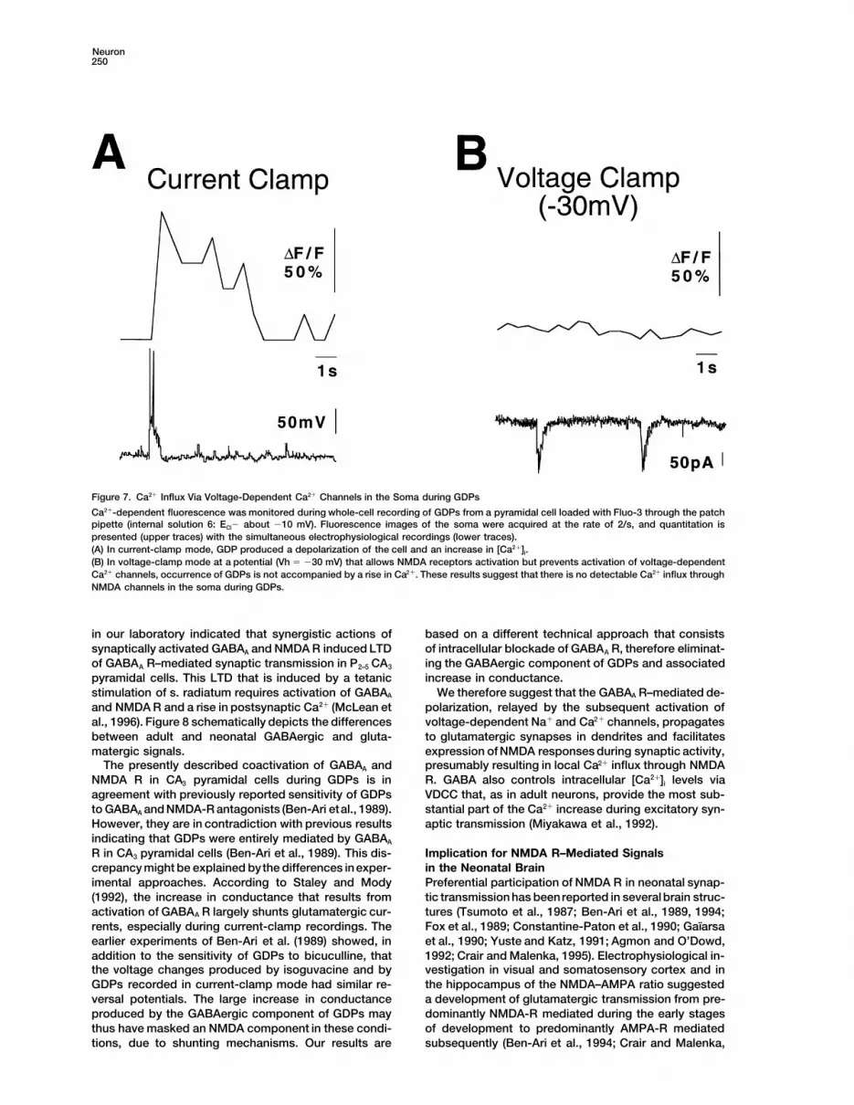
The coactivation of GABAA and NMDA R during spon-taneous and evoked GDPs should provide a Ca21 influxthrough NMDA channels. However, exogenous and syn-aptic activation of GABAA receptors in P2–5 slices alsoincreases (Ca21)i via VDCC (Leinekugel et al., 1995). Todistinguish between these two sources of Ca21 influx,we loaded cells with Fluo3 in the whole-cell configura-tion and analyzed [Ca21]i changes in the soma either (i)in current-clamp mode that allows the activation ofVDCC and NMDA R or (ii) in voltage-clamp mode atdepolarized potentials (230 mV) to allow the activationof NMDA R but not VDCC that rapidly inactivate. DuringGDPs, large Ca21 increase was observed in current-clamp conditions (Vh 5 275 mV: 161 6 19%, n 5 8;internal solution 6: ECl- around 210 mV) but not in volt-age-clamp conditions (Vh 5 230 mV: 12 6 1%; n 5 8)(Figure 7). Therefore, VDCC but not NMDA R provideCa21 influx to the soma during GDPs. We suggest thatNMDA R that are activated during GDPs might howeverprovide local Ca21 influx at the location of glutamatergicsynapses in dendrites (Muller and Connor, 1991; Regherand Tank, 1992; Durand et al., 1996).
Discussion
Our results allow the following conclusions to be drawn.First, during early postnatal life, activation of GABAA Rreduces the voltage-dependent Mg21 block of NMDAchannels in CA3 pyramidal neurons and increases[Ca21]i. At these early stages of development, GABAA
and NMDA R act synergistically, GABAA R playing theFigure 4. Coactivation of GABAA and NMDA Receptors during Syn- role conferred to AMPA R in more adult neurons (Figureaptic Responses
8). Second, the synergistic interaction between GABAA(A) CA3 pyramidal cells were dialyzed with a pipette solution con-and NMDA R plays an important role in the generationtaining CsF1DIDS (whole-cell recording; internal solution 5). Synap-of network-driven GDPs and associated synchronoustic responses that correspond to GDPs were evoked by electricalCa21 oscillations that are a major feature of the neonatalstimulation in the s. radiatum in the presence of the AMPA-R antago-
nist CNQX (10 mM). Synaptic responses evoked in one neuron at hippocampal network.different membrane potentials are presented at the beginning (left)and after 30 min dialysis (right). GABA–NMDA Interaction in the(B) The relationship between the current charge of these events and
Neonatal Hippocampusthe membrane potential is plotted at the beginning (left) and afterIn P2–5 CA3 pyramidal cells, in conditions that affect nei-(right) dialysis (n 5 7). Note that during dialysis, the C–V curvether [Cl2]i nor resting-membrane potential, synapticallychanged from nearly linear to strongly rectificating at negative po-
tentials while the reversal potential shifted toward 0 mV, revealing activated GABAA R generate action potentials. Thesethe NMDA component of GDPs. results confirm and extend previous studies reporting
depolarizing effects of GABA in neonatal neurons (Ben-(with Lucifer Yellow in the pipette solution) of the cells Ari et al., 1989; Fiszman et al., 1990; Wu et al., 1992;that displayed these Ca21 increases (n 5 17 cells from Hales et al., 1994; Reichling et al., 1994; LoTurco et al.,11 experiments) revealed typicalmorphological (charac- 1995; Serafini et al., 1995; Rohrbough and Spitzer, 1996;teristic shape of soma and initial dendrites) and electro- Chen et al., 1996). The mechanisms involved in this neo-physiological properties (discharge of action potentials natal GABAA R–mediated depolarization are not com-and presence of synaptic activity including GDPs) of pletely understood. The GABAA receptor–channel com-pyramidal neurons (Figure 6C). plex is primarily Cl2 permeable (Sivilotti and Nistri, 1991;
Recording of [Ca21]i transients in groups of CA3 pyra- Kaila and Voipio, 1994), and there is a number of evi-midal cells simultaneously with whole-cell recording dences for an elevated [Cl2]i in neonatal neurons (Luh-from an additional neuron (pyramidal cells: n 5 17 exper- mann and Prince, 1991; Zhang et al., 1991; Hara et al.,iments; or CA3 s. radiatum interneurons: n 5 4 experi- 1992; Serafini et al., 1995; Rohrbough and Spitzer, 1996).ments) showed that the [Ca21]i increases were synchro- Depolarization due to HCO32 permeability of GABAA
nized with the GDPs recorded in that neuron (DF/F: 151 R (Kaila and Voipio, 1994; Staley et al., 1995) is not6 4% at the peak; n 5 81 cells from 14 different slices necessarilly involved in immature neurons since werecorded during 3–20 GDPs), returning to control values found that during perfusion with HCO32-free HEPES
buffer, isoguvacine still increased Ca21 in neonatal pyra-about 1–4 s later (Figures 6D–6F). In the vast majorityof slices, these GDP-induced Ca21 increases were quite midal cells loaded extracellularly with Fluo3-AM (X.
Leinekugel, unpublished data).regular, occurring at a rate of 0.05–0.2 s21.

Neuron248
Figure 5. GDPs Are Synchronous in CA3 Pyramidal Neurons
(A–B) Simultaneous recording of two CA3 pyramidal cells in the cell-attached (upper trace, cell 1) and whole-cell (lower trace, cell 2) configura-tions. Note that bursts of action potentials are generated in cell 1 during GDPs in cell 2 (GDP marked by the asterisk in [A] is presented in[B] in expanded time scale) but that spontaneous action potentials can also occur between GDPs ([A]: arrow).(C–D) Entry into the whole-cell mode in cell 1 shows that GDPs are synchronous in both cells (GDP marked by the asterisk in [C] is presentedin [D] in expanded time scale). Cell 1: solution 3, Vh 5 260 mV; Cell 2: solution 7, Vh 5 280 mV.
The fact that blocking AMPA R only slightly affected et al., 1994). Release of the Mg21 block of NMDA chan-nels by the depolarizing effect of GABAA R during synap-the synaptic responses evoked by electrical stimulation
of s. radiatum is in agreement with other studies sug- tic activity is suggested by the following observations:(i) the depolarization induced by the GABAA agonist iso-gesting a limited participation of AMPA R in the early
synaptic drive to neonatal pyramidal cells (Ben-Ari et guvacine potentiates NMDA R–mediated signals; (ii)synaptically activated GABAA R provide neuronal excita-al., 1989, 1994; Durand et al., 1996), confirming that
GABA is the main fast-acting excitatory transmitter dur- tion; (iii) GABAA and NMDA R are coactivated duringsynaptic activity; and (iv) a parallel study conducteding early postnatal life (Cherubini et al., 1991; Ben-Ari
(Figure 6 legend continued)
(C) After Ca21 measurements, the three cells were loaded one by one with Lucifer Yellow using patch pipettes (whole-cell mode; internalsolution 3), andspontaneous electrophysiological activities were recorded. Note that morphological (picture) and electrophysiological (recordingfrom cell 2; upper trace: current clamp, Vr 5 265 mV; lower trace: voltage clamp, Vh 5 260 mV) properties (pyramidal neuronal shape, firingof action potentials, and presence of spontaneous synaptic activity including GDPs) are typical of neonatal hippocampal neuronal cells. Scalebars, 15 mm.(D–F) In another series of experiments, Ca21-dependent fluorescence changes from a group of CA3 pyramidal cells loaded with Fluo 3-AM([D]: pseudocolor fluorescence image from this group of pyramidal cells; CA3 PYR: CA3 pyramidal layer; S.R., s. radiatum; scale bar, 15 mm)were monitored during simultaneous whole-cell recording (internal solution 2) of an additional pyramidal cell (not represented). Fluorescenceimages were collected at the rate of 2/s. Ca21-dependent fluorescence from this group of pyramidal cells was quantified and is presented([E], upper trace) with the corresponding electrophysiological trace recorded simultaneously from an additional pyramidal cell ([E] downwardtrace, voltage clamp 260 mV). Note that Ca21 increased synchronously with GDPs.(F) Mean 6 E.S. Ca21-dependent fluorescence responses (upper traces) from three visually identified pyramidal cells from the group presentedin (D) (cells 1–3 delimited by white lines) recorded during 20 GDPs (mean trace: downward trace).

GABA and NMDA Drive Ca21 Oscillations in Neonates249
Figure 6. Synchronous Spontaneous Ca21 Oscillations in CA3 Pyramidal Neurons
(A–C) Three CA3 pyramidal cells were visually selected and loaded with Fluo 3-AM.(A) Ca21-dependent fluorescence images of these three cells (upper lanes: left, resting level; right, peak of Ca21 transient) were acquired atthe rate of 2/s, and quantitation is presented in (B). Note that Ca21 transients are synchronous in the three cells.
(Figure 6 legend continued on previous page)
Neuron250
Figure 7. Ca21 Influx Via Voltage-Dependent Ca21 Channels in the Soma during GDPs
Ca21-dependent fluorescence was monitored during whole-cell recording of GDPs from a pyramidal cell loaded with Fluo-3 through the patchpipette (internal solution 6: ECl2 about 210 mV). Fluorescence images of the soma were acquired at the rate of 2/s, and quantitation ispresented (upper traces) with the simultaneous electrophysiological recordings (lower traces).(A) In current-clamp mode, GDP produced a depolarization of the cell and an increase in [Ca21]i.(B) In voltage-clamp mode at a potential (Vh 5 230 mV) that allows NMDA receptors activation but prevents activation of voltage-dependentCa21 channels, occurrence of GDPs is not accompanied by a rise in Ca21. These results suggest that there is no detectable Ca21 influx throughNMDA channels in the soma during GDPs.
in our laboratory indicated that synergistic actions of based on a different technical approach that consistsof intracellular blockade of GABAA R, therefore eliminat-synaptically activated GABAA and NMDA R induced LTD
of GABAA R–mediated synaptic transmission in P2–5 CA3 ing the GABAergic component of GDPs and associatedincrease in conductance.pyramidal cells. This LTD that is induced by a tetanic
stimulation of s. radiatum requires activation of GABAA We therefore suggest that the GABAA R–mediated de-polarization, relayed by the subsequent activation ofand NMDA R and a rise in postsynaptic Ca21 (McLean et
al., 1996). Figure 8 schematically depicts the differences voltage-dependent Na1 and Ca21 channels, propagatesto glutamatergic synapses in dendrites and facilitatesbetween adult and neonatal GABAergic and gluta-
matergic signals. expression of NMDA responses during synaptic activity,presumably resulting in local Ca21 influx through NMDAThe presently described coactivation of GABAA and
NMDA R in CA3 pyramidal cells during GDPs is in R. GABA also controls intracellular [Ca21]i levels viaVDCC that, as in adult neurons, provide the most sub-agreement with previously reported sensitivity of GDPs
to GABAA and NMDA-R antagonists (Ben-Ari etal., 1989). stantial part of the Ca21 increase during excitatory syn-aptic transmission (Miyakawa et al., 1992).However, they are in contradiction with previous results
indicating that GDPs were entirely mediated by GABAA
R in CA3 pyramidal cells (Ben-Ari et al., 1989). This dis- Implication for NMDA R–Mediated Signalsin the Neonatal Braincrepancy might be explained bythe differences inexper-
imental approaches. According to Staley and Mody Preferential participation of NMDA R in neonatal synap-tic transmission has beenreported in severalbrain struc-(1992), the increase in conductance that results from
activation of GABAA R largely shunts glutamatergic cur- tures (Tsumoto et al., 1987; Ben-Ari et al., 1989, 1994;Fox et al., 1989; Constantine-Paton et al., 1990; Gaıarsarents, especially during current-clamp recordings. The
earlier experiments of Ben-Ari et al. (1989) showed, in et al., 1990; Yuste and Katz, 1991; Agmon and O’Dowd,1992; Crair and Malenka, 1995). Electrophysiological in-addition to the sensitivity of GDPs to bicuculline, that
the voltage changes produced by isoguvacine and by vestigation in visual and somatosensory cortex and inthe hippocampus of the NMDA–AMPA ratio suggestedGDPs recorded in current-clamp mode had similar re-
versal potentials. The large increase in conductance a development of glutamatergic transmission from pre-dominantly NMDA-R mediated during the early stagesproduced by the GABAergic component of GDPs may
thus have masked an NMDA component in these condi- of development to predominantly AMPA-R mediatedsubsequently (Ben-Ari et al., 1994; Crair and Malenka,tions, due to shunting mechanisms. Our results are

GABA and NMDA Drive Ca21 Oscillations in Neonates251
Figure 8. Major Developmental Changes inthe GABA–Glutamate Interactions
In adult neurons, activation of AMPA recep-tors provides the excitatory drive necessaryto remove the voltage-dependent Mg21 blockof NMDA channels while GABA, acting onGABAA and GABAB receptors, provideshyperpolarization and inhibits NMDA recep-tor–mediated signals. In neonatal neurons,postsynaptic GABAB receptors are not func-tional, and depolarizing effects of GABA, act-ing via GABAA receptors, reduce the voltage-dependent Mg21 block of NMDA channels,providing synergy between GABA and gluta-mate. AMPA receptor–mediated transmis-sion is weak in the early periods of devel-opment.
1995; Durand et al., 1996). Several factors are implicated 1996), and a shift from de- to hyperpolarizing effects ofGABA is also observed in neurons in cultures (Wang etin the preferential participation of NMDA R in neonatal
neurons: delayed development of G protein–mediated al., 1994; Obrietan and van den Pol, 1995; Chen et al.1996). Therefore, the excitatory effects of GABA as well(including GABAB) postsynaptic inhibition (Fukuda et al.,
1993; Gaıarsa et al., 1995), higher density of NMDA R as the GABA–NMDA synergy may represent a funda-mental property of developing networks.in humans and rats (Tremblay et al., 1988; Represa et al.,
1989), and slower kinetics of NMDA responses (Hestrin,1992; Carmignoto and Vicini, 1992; Fox, 1995; Khazipov Implications for Developmental Plasticity
GABA and glutamate have multiple morphogenic andet al., 1995) that may be due to different expression ofNMDA-R subunits in neonatal neurons (Pollard et al., trophic effects in developing neurons (Redburn and
Schousboe, 1987; Spoerri, 1988; Belhage et al., 1988;1993; Monyer et al., 1994). However, the voltage-depen-dent Mg21 block of NMDA channels seems to beefficient Scherer and Udin, 1989; Constantine-Paton et al., 1990;
Goodman and Shatz, 1993; Barbin et al., 1993; Rakicat all stages of development (LoTurco et al., 1991;Strecker et al., 1994; Crair and Malenka, 1995; Khazipov and Komuro, 1995; Behar et al., 1996) via changes in
the intracellular Ca21, control of DNA synthesis, andet al., 1995; Durand et al., 1996), suggesting that a pow-erful excitatory drive is required to activateNMDA recep- growth factors expression (Lipton and Kater, 1989; Lau-
der, 1993; LoTurco et al., 1995; Marty et al., 1996). Thetors. We suggest that GABA exerts this role in the neo-nates. This may, in addition to the above-mentioned present study describes how the changes in Ca21 ho-
meostasis and possibly related trophic effects of thesemechanisms, explain the large participation of NMDA Rin the neonatal synaptic drive. The depolarizing effects neurotransmitters occur during physiological patterns
of activity in the neonatal hippocampus.of GABA have been reported during the early periodsof development in all brain structures studied thus far Synchronous neuronal activity represents a funda-
mental property of the developing neuronal networks(Ben-Ari et al., 1989; Yuste and Katz, 1991; Gaıarsa etal., 1995; LoTurco et al., 1995; Obrietan and van den (Ben-Ari et al., 1989; Gaıarsa et al., 1990; O’Donovan et
al., 1992; Kandler and Katz, 1995; Gu and Spitzer, 1995;Pol, 1995; Serafini et al., 1995; Rohrbough and Spitzer,

Neuron252
MgCl2, 10 HEPES, 10 glucose, and 0.01 glycine, pH 7.4. The bestYuste etal., 1995; Feller et al., 1996),but themechanismsresults were obtained when slices were allowed to recover for 2–5 hrof synchronization differ. While in the neocortex it isafter protease treatment. For whole-cell recordings, internal solutiongenerated by gap junctions (Kandler and Katz, 1995;(pH 7.3; osmolarity 300 mmol/kg) of the following composition was
Yuste et al., 1995), synchronization of neuronal activity used (in mM): 80 CsCl, 80 Cs-Gluconate, 0.06 CaCl2, 1.1 BAPTA-clearly implies synaptic mechanisms in retina, via cholin- Cs4, 10 HEPES, and 2 MgATP, pH 7.2.ergic transmission (Katz, 1993; Wong et al., 1993; Feller
Electrophysiological Recordingset al., 1996) and in the hippocampus, via the excitatoryRecordings were performed using the patch-clamp technique in theactions of GABAA and glutamate receptors (Corradetticell-attached and whole-cell configurations. Microelectrodes had aet al., 1988; Ben-Ari et al., 1989; Gaıarsa et al., 1990;resistance of 7–10 MV. Internal solutions (pH 7.3; osmolarity 270–
Hanse et al., Soc. Neurosci. abstract, 1996). We ob- 280 mmol/kg measured by a Knauer osmometer, Berlin) of the fol-served that GDPs can also be recorded from inter- lowing composition were used (in mM): for recordings of singleneurons and that they are synchronous with GDPs in NMDA channels in cell-attached configuration, 1) ACSF with 0.05
Mg21, 1 EGTA, 0.01 NMDA, and 0.01 glycine; for whole-cell re-pyramidal cells, indicating that GDPs result from thecordings in slice: 2) 140 CsCl, 1 CaCl2, 10 EGTA, and 10 HEPES; orsynchronous discharge of pyramidal cells and GABAer-3) 100 KCl, 10 NaCl, 0.25 CaCl2, 5 EGTA, 10 HEPES, 10 Glucose, 2gic interneurons (Khazipov et al., 1997). The neonatalMgATP, and 0.2 GTP; or 4) 140 CsF, 1 CaCl2, 10 EGTA, and 10
hippocampal network is organized in recurrent excit- HEPES; or 5) solution 4 1 0.5–1 4,4’-diisothiocyanatostilbene-2,2’-atory loops, pyramidal cells and interneurons being ex- disulfonic acid (DIDS); for Ca21-imaging whole-cell recordings: 6)cited by the depolarizing effects of GABA and glutamate solution 3 1 0.01 Fluo3; for recordings of spikes in cell-attached
configuration: 7) 135 K gluconate, 2 MgCl2, 0.1 CaCl2, 1 EGTA, 2 Na2(Leinekugel et al., 1995), which provide synchronousATP, and 10 HEPES. Occasionally, Lucifer Yellow (0.1–1%) wasneuronal discharges (GDPs) largely mediated by GABAAadded to solution 3 for morphological monitoring. To isolate thereceptors. These synchronous activities may be impli-glutamate receptor–mediated component of the evoked synaptic
cated in the control of neuronal growth and formation of response, the cells were dialyzed, as suggested by Nelson et al.the neuronal networks via changes in Ca21 homeostasis (1996), with internal solution that contained CsF, DIDS, and that(Constantine-Paton et al., 1990; Goodman and Shatz, did not contain MgATP (solution 5). In some experiments, internal
dialysis was performed in absence of DIDS (solution 4). As described1993). Although the consequences of the Ca21 influxelsewhere (Khalilov et al., Soc. Neurosci. abstract, 1996), this proce-during GDPs via voltage-gated Ca21 channels anddure completely suppresses exogenous and synaptic GABAA recep-NMDA channels are presently unknown, activation oftor–mediated responses in the cell under investigation, with only
voltage-gated Ca21 channels promotes differentiation moderate effects on the exogenous and synaptic AMPA and NMDAof neurons in several preparations (Desarmenien and receptor–mediated responses (10%–30% reduction of NMDA re-Spitzer, 1991; Gu and Spitzer, 1995; LoTurco et al., 1995; sponses). Slices were stimulated by a bipolar tungsten electrode
(30–80 V; 10–30 ms; 0.02–0.05 Hz) placed in the s. radiatum of theRusanescu et al., 1995). Moreover, localized Ca21 influxCA3 region of the hippocampus.through NMDA receptors can provide a hebbian modu-
lation of developing synapses (Komatsu and Iwakiri,Data Analysis1993; Fox, 1995; Crair and Malenka, 1995; Kirkwood etSynaptic-evoked responses were recorded using an Axopatch 200
al., 1995; Durand et al., 1996; McLean et al., 1996) and (Axon Instrument, USA) amplifier, stored into the memory of anmay be involved in activity-dependent synaptogenesis 80486 personal computer using TL1 DMA Labmaster A/D converterand network formation. (USA) and then analyzed using Acquis Software (Gerard Sadoc,
France). Axotape and SE04 (USA) programs were used for the acqui-sition and analysis of spontaneous events. Single-channel currentsExperimental Procedureswere recorded at 5 kHz on a tape recorder DTR 1201 (Bio-Logic,France) and then digitized at a sampling frequency of 10 kHz forSlice Preparationanalysis (p-CLAMP programs, Axon Instruments, USA). Mg21 blockSlices were prepared as described previously (Ben-Ari et al., 1989)of NMDA channels was estimated using the model of open-channelfrom 2- to 5-day-old male Wistar rat pups. In brief, after sacrificingblock (Nowak et al., 1984), as described elsewhere (Khazipov et al.,the rat by decapitation, the brain was rapidly removed and placed1995). Group measurements were expressed as means 6 SEM.in oxygenated, ice-cooled artificial cerebrospinal fluid (ACSF); hip-Statistical significance of differences between means was assessedpocampal transverse slices (thickness 400–500 mm) were cut withwith the Student’s t-test, with the aid of statistical software StatVieweither a McIIwain tissue chopper or a vibroslicer (FTB Vibracut) andSE1 Graphics. The level of significance was set at p , 0.05.kept in oxygenated (95% O2 and 5% CO2) ACSF (in mM: 126 NaCl;
3.5 KCl; 2 CaCl2; 1.3 MgCl2; 25 NaHCO3; 1.2NaH2PO4; and 11 glucose,pH 7.3) at room temperature at least 1 hr before use. Individual Fluorescence Measurements
Fluorescence measurements were performed as described (Leine-slices were then transferred to the recording chamber where theywere fully submerged and superfused with oxygenated ACSF at kugel et al., 1995) on neurons loaded with the Ca21-sensitive dye
Fluo-3 either in the impermeant form (whole-cell configuration, inter-30–328C at a rate of 2–3 ml/min.nal solution 6) or in the esterified form (Fluo-3 AM: 3.3 mM, appliedfocally from a micropipette during 5–30 min by 0.3–1 s, 0.2 Hz pres-Isolated Neurons Preparation
Neurons were freshly isolated from P2–5 hippocampal slices as de- sure pulses), using a confocal laser scanning microscope (MRCBIORAD 600) equipped with argon–krypton laser and photomulti-scribed (Medina et al., 1994). In brief, slices were incubated at 308C
for 25–30 min in an O2 atmosphere, in a solution containing (in plier. Excitation was delivered at 488 nm, and emission intensitywas measured at wavelength .500 nm. Images were acquired everymM): 115 NaCl, 10 KCl, 1.2 NaH2PO4, 10 MgCl2, 26 NaHCO3, and 10
glucose, pH 7.4 (solution A), 1 1.5 mg/ml protease XXIII. Slices were 0.5–4 s using the program SOM (BIORAD, USA) and analyzed off-line with the program Fluo (IMSTAR, France). All results were ex-then washed with solution A for 15 min. The concentration of CaCl2
was slowly increased up to 1 mM. For dissociation of cells, slices pressed as DF/F0, with F 5 fluorescence from the defined portionof the image corresponding to the cell(s) under investigation andwere again transferred to solution A without CaCl2, and the CA3
region was dissected and gently agitated by sharp glass needles F0 5 mean baseline fluorescence in the selected area(s) from at leastfive consecutive images. Because Fluo-3 is a single-wavelengthto release individual cells. Cells were placed in the recording cham-
ber and solution A was replaced slowly (over 10 min) by a solution chromophore, and fluorescence is a function of the concentrationof Ca21 and dye (Kao et al., 1989), we have used this dye only forof the following composition (in mM): 150 NaCl, 1 KCl, 2 CaCl2, 1

GABA and NMDA Drive Ca21 Oscillations in Neonates253
approximate estimation of [Ca21]i and included for analysis only Corradetti, R., Gaıarsa, J.L., and Ben-Ari, Y. (1988). D-Aminophos-phonovaleric acid-sensitive spontaneous giant EPSPs in immatureexperiments in which the fluorescence level recovered to control
value after cell excitation. As described earlier (Leinekugel et al., rat hippocampal neurones. Eur. J. Pharmacol. 154, 221–222.1995), individual neurons were selected using the optics of an axio- Crair, M.C., and Malenka, R.C. (1995). A critical period for long-termscope Karl Zeiss microscope (water immersion objective 340) that potentiation at thalamocortical synapses. Nature 375, 325–328.allows recognition of neurons in slices. The neurons were then ap- Desarmenien, M.G., and Spitzer, N.C. (1991). Role of calcium andproached with the Fluo3-AM containing pipette for loading. The use protein kinase C in development of the delayed rectifier potassiumof a calibrated map to correlate fluorescence spots and the visual current in xenopus spinal neurons. Neuron 7, 797–805.optical field verified that the fluorescence signals monitored origi-
Durand, G.M., Kovalchuk, Y., and Konnerth, A. (1996). Long-termnated from the selected neurons.potentiation and functional synapse induction in developing hippo-Solutions and Drugscampus. Nature 381, 71–75.Fluo 3-AM and DIDS were first dissolved in DMSO (,0.1% final)Feller, M.B., Wellis, D.P., Stellwagen, D., Werblin, F.S., and Shatz,and just before use in either standard ACSF (Fluo 3-AM) or internalC.J. (1996). Requirement for cholinergic synaptic transmission in thepipette solution (DIDS). NMDA and isoguvacine were dissolved inpropagation of spontaneous retinal waves. Science 272, 1182–1187.ACSF and applied by bath. In some experiments, isoguvacine was
applied locally by pressure ejection from a micropipette using a Fiszman, M.L., Novotny, E.A., Lange, G.D., and Barker, J.L. (1990).Picospritzer II (General Valve, USA). Drugs used were purchased Embryonic and early postnatal hippocampal cells respond to nano-from Sigma (NMDA, tetrodotoxin, D-600, DIDS, and protease XXIII), molar concentrations of muscimol. Dev. Brain Res. 0, 1–8.Tocris Neuramin (isoguvacine, bicuculline, CNQX [6-Cyano-7-nitro- Fox, K. (1995). The critical period for long-term potentiation in pri-quinoxaline-2,3 dione], and glycine), APV ([d-2-Amino-5-phospho- mary sensory cortex. Neuron 15, 485–488.pentanoate]) and Molecular Probes (Fluo3 and Fluo3-AM).
Fox, K., Sato, H., and Daw, N. (1989). The location and function ofNMDA receptors in cat and kitten visual cortex. J. Neurosci. 9,
Acknowledgments 2443–2454.
Fukuda, A., Mody, I., and Prince, D.A. (1993). Differential ontogenesisWe thank Drs. R. Miles, K. Krnjevic, J. L. Gaıarsa, S. Shorte, G.of presynaptic and postsynaptic GABAB inhibition in the rat somato-Holmes, and H. A.McLean for useful discussions and critical readingsensory cortex. J. Neurophysiol. 70, 448–452.of the manuscript; Dr. A. Bakhramov for the help in single channelsGaıarsa, J.L., Corradetti, R., Cherubini, E., and Ben-Ari, Y. (1990).analysis; and B. Martin and S. Weiler for technical assistance inModulation of GABA-mediated synaptic potentials by glutamatergicCa21 images processing. This work was supported by grants fromagonists in neonatal CA3 rat hippocampal neurons. Eur. J. Neurosci.INSERM, Ministere de la Recherche et de l’Espace, Societe de Sec-3, 301–309.ours des Amis des Sciences, and Association Nationale de Recher-
che sur le SIDA (AIDS). Gaıarsa, J.L., McLean, H., Congar, P., Leinekugel, X., Khazipov, R.,Tseeb, V., and Ben-Ari, Y. (1995). Postnatal maturation of Gamma-Aminobutyric acid A and B-mediated inhibition in the CA3 hippocam-Referencespal region of the rat. J. Neurobiol. 26, 339–349.
Goodman, C.S., and Shatz, C.J. (1993). Developmental mechanismsAgmon, A., and O’Dowd, D.K. (1992). NMDA receptor–mediated cur-that generate precise patterns of neuronal connectivity. Cell/Neuronrents are prominent in the thalamocortical synaptic response before72/10 (suppl.), 77–98.maturation of inhibition. J. Neurophysiol. 68, 345–349.Gu, X., and Spitzer, N.C. (1995). Distinct aspects of neuronal differen-Artola, A., and Singer (1987). Long-term potentiation and NMDAtiation encoded by frequency of spontaneous Ca21 transients. Na-receptors in rat visual cortex. Nature 330, 649–652.ture 375, 784–787.Barbin, G., Pollard, H., Gaıarsa, J.L., and Ben-Ari, Y. (1993). Involve-Hales, T.G., Sanderson, M.J., and Charles, A.C. (1994). GABA hasment of GABAA receptors in the outgrowth of cultured hippocampalexcitatory actions on GnRH-secreting immortalized hypothalamicneurons. Neurosci. Lett. 152, 150–154.(GT1–7) neurons. Neuroendocrinology 59, 297–308.Behar, T.B., Li, Y.-X., Tran, H.T., Ma, W., Dunlap, V., Scott, C., andHanse, E., Garaschuk, O., and Konnerth, A. (1996). Early networkBarker, J.L. (1996). GABA stimulates chemotaxis and chemokinesisoscillations in the developing hippocampus. Soc. Neurosci., 26,of embryonic cortical neurons via calcium-dependent mechanisms.774.6.J. Neurosci. 16, 1808–1818.Hara, M., Inoue, M., Yasukura, T., Ohnishi, S., Mikami, Y., and Ina-Belhage, B., Hansen, G.H., Schousboe, A., and Meier, E. (1988).gaki, C. (1992). Uneven distribution of intracellular Cl2 in rat hippo-GABA agonist promoted formation of low affinity GABA receptorscampal neurons. Neurosci. Lett. 143, 135–138.on cerebellar granule cells is restricted to early development. Int.
J. Dev. Neurosci. 6, 125–128. Hestrin, S. (1992). Developmental regulation of NMDA receptor–mediated synaptic currents at a central synapse. Nature 357,Ben-Ari, Y., Cherubini, E., Corradetti, R., and Gaıarsa, J.L. (1989).686–689.Giant synaptic potentials in immature rat CA3 hippocampal neu-Kaila, K.,and Voipio, J. (1994). Ionicbasis of GABAA receptor channelrones. J. Physiol. (Lond.) 416, 303–325.function in the nervous system. Prog. Neurobiol. 42, 489–537.Ben-Ari, Y., Tseeb, V., Raggozzino, D., Khazipov, R., and Gaıarsa,Kandler, K., and Katz, L.C. (1995). Neuronal coupling and uncouplingJ.L. (1994). g-Aminobutyric acid (GABA): a fast excitatory transmitterin the developing nervous system. Curr. Opin. Neurobiol. 5, 98–105.which may regulate the development of hippocampal neurones in
early postnatal life. Prog. Brain Res. 102, 261–273. Kanter, E.D., and Haberly, L.D. (1993). Associative long-term potenti-ation in piriform cortex slices requires GABAA blockade. J. Neurosci.Carmignoto, G., and Vicini, S. (1992). Activity-dependentdecrease in13, 2477–2482.NMDA receptor responses during development of the visual cortex.
Science 258, 1007–1011. Kanter, E.D., Kapur, A., and Haberly, L.B. (1996). A dendritic GABAA-mediated IPSP regulates facilitation of NMDA-mediated responsesChen, G., Trombley, P., and van den Pol, A.N. (1996). Excitatoryto burst stimulation of afferent fibers in piriform cortex. J. Neurosci.actions of GABA in developing rat hypothalamic neurones. J. Phys-16, 307–312.iol. (Lond.) 494, 451–464.Kao, J.P.Y., Harootunian, A.T., and Tsien, R.Y. (1989). Photochemi-Cherubini, E., Gaıarsa, J.L., and Ben-Ari, Y. (1991). GABA: an excit-cally generated cytosolic calcium pulses and their detection byatory transmitter in early postnatal life. Trends Neurosci. 14,Fluo-3. J. Biol. Chem. 264, 8179–8184.515–519.Katz, L.C. (1993). Coordinate activity in retinal and cortical develop-Constantine-Paton, M., Cline, H.T., and Debski, E. (1990). Patternedment. Curr. Opin. Neurobiol. 3, 93–99.activity, synaptic convergence, and the NMDA receptor in devel-
oping visual pathways. Annu. Rev. Neurosci. 13, 129–154. Khalilov, I., Khazipov, R., Leinekugel, X., Feger, J., and Ben-Ari,

Neuron254
Y. (1996). Intracellular blockade of GABAA receptors as a tool to neuronal compartments for synaptic Ca11 responses. Nature 354,73–76.investigate neuronal network activity. Soc. Neurosci. 26 221.1.
Nowak, L., Bregestovski, P., Ascher, P., Herbert, A., and Prochiantz,Khazipov, R., Ragozzin, D., and Bregestovski, P. (1995). KineticsA. (1984). Magnesium gates glutamate-activated channels in mouseand Mg21 block of N-Methyl-D-Aspartate receptor channels duringcentral neurons. Nature 307, 462–465.postnatal development of hippocampal CA3 pyramidal neurons.
Neuroscience 69, 1057–1065. O’Donovan, M.J., Sernagor, E., Sholomenko, G., Ho, S., Antal, M.,and Yee, W. (1992). Development of spinal motor networks in theKhazipov, R., Leinekugel, X., Khalilov, I., Gaıarsa, J.L., and Ben-Ari,chick embryo. J. Exp. Zool. 261, 261–273.Y. (1997). Synchronization of GABAergic interneuronal network in
CA3 subfield of neonatal rat hippocampal slices. J. Physiol. (Lond.), Obrietan, K., and van den Pol, A.N. (1995). GABA neurotransmissionin press. in the hypothalamus: developmental reversal from Ca11 elevating
to depressing. J. Neurosci. 15, 5065–5077.Kirkwood, A., Lee, H., and Bear, M.F. (1995). Co-regulation of long-Pollard, H., Khrestchatisky M., Moreau J., and Ben-Ari Y. (1993).term potentiation and experience-dependent synaptic plasticity inTransient expression of the NR2C subunit of the NMDA receptor invisual cortex by age and experience. Nature 375, 328–331.developping rat brain. Neuroreport 4, 411–414.Komatsu, Y., and Iwakiri, M. (1993). Long-term modification of inhibi-Rakic, P., and Komuro, H. (1995). The role of receptor-channel activ-tory synaptic transmission in developing visual cortex. Neuroreportity in neuronal cell migration. J. Neurobiol. 26, 299–315.4, 907–910.Redburn, D., and Schousboe, A. (1987). Neurotrophic activity ofLauder, J.M. (1993). Neurotransmitters as growth regulatory signals:GABA during development. Neurol. Neurobiol. 32, 1–227.role of receptors and second messengers. Trends Neurosci. 16,
233–240. Regher, W.G., andTank, D.W. (1992). Calcium concentration dynam-ics produced by synaptic activation of CA1 hippocampal pyramidalLeinekugel, X., Tseeb, V., Ben-Ari, Y., and Bregestovski, P. (1995).cells. J. Neurosci. 12, 4202–4223.Synaptic GABAA activation induces Ca11 rise in pyramidal cellsReichling, D.B., Kyrozis, A., Wang, J., and MacDermott, A.B. (1994).and interneurons from rat neonatal hippocampal slices. J. Physiol.Mechanisms of GABA and glycine depolarization-induced calcium(Lond.) 487, 319–329.transients in rat dorsal horn neurons. J. Physiol. (Lond.) 476,Lipton, S.A., and Kater, S.B. (1989). Neurotransmitter regulation of411–421.neuronal outgrowth, plasticity and survival. Trends Neurosci. 12,Represa, A., Tremblay, E., and Ben-Ari, Y. (1989). Transient increase265–270.of NMDA-binding sites in human hippocampus during development.LoTurco, J.J., Blanton, M.G., and Kriegstein, A.R. (1991). Initial ex-Neurosci. Lett. 99, 61–66.pression and endogenous activation of NMDA channels in earlyRohrbough, J., and Spitzer, N.C. (1996). Regulation of intracellularneocortical development. J. Neurosci. 11, 792–799.Cl2 levels by Na1-dependent Cl2 cotransport distinguishes depolar-
LoTurco, J.J., Owens, D.F., Heath, M.J.S., Davis, M.B.E., andizing from hyperpolarizing GABAA receptor-mediated responses in
Kriegstein, A.R. (1995). GABA and glutamate depolarize cortical pro-spinal neurons. J. Neurosci. 16, 82–91.
genitor cells and inhibit DNA synthesis. Neuron 15, 1287–1298.Rusanescu, G., Qi, H., Thomas, S.M., Brugge, J.S., and Halegoua,
Luhmann, H.J., and Prince, D.A. (1991). Postnatal maturation of the S. (1995). Calcium influx induces neurite growth through a Src-RasGABAergic system in rat neocortex. J. Neurophysiol. 65, 247–263. signaling cassette. Neuron 15, 1415–1425.Malenka, R.C., and Nicoll, R.A. (1993). NMDA-receptor-dependent Scherer, W.S., and Udin, S.B. (1989). N-methyl-D-aspartate antago-synaptic plasticity: multiple forms and mechanisms. Trends Neu- nists prevent interaction of binocular maps in Xenopus tectum. J.rosci. 16, 521–527. Neurosci. 9, 3837–3843.Marty, S., Berninger, B., Carroll, P., andThoenen, H. (1996). GABAer- Serafini, R., Valeyev, A.Y., Barker, J.L., and Poulter, M.O. (1995).gic stimulation regulates the phenotype of hippocampal interneur- Depolarizing GABA-activated Cl2 channels in embryonic rat spinalons through the regulation of Brain-Derived Neurotrophic Factor. and olfactory bulb cells. J. Physiol. (Lond.) 488, 371–386.Neuron 16, 565–570.
Sivilotti, L., and Nistri, A. (1991). GABA receptor mechanism in theMayer, M.L., Westbrook, G.L., and Guthrie, P.B. (1984). Voltage- central nervous system. Prog. Neurobiol. 36, 35–92.dependent block by Mg11 of NMDA responses in spinal cord neu- Spoerri, P.E. (1988). Neurotrophic effects of GABA in cultures ofrons. Nature 309, 261–263. embryonic chic brain and retina. Synapse 2, 11–22.McBain, C., and Dingledine, R. (1992). Dual-component miniature Staley, K.J., and Mody, I. (1992). Shunting of excitatory input toexcitatory synaptic currents in rat hippocampal CA3 pyramidal neu- dentate gyrus granule cells by a depolarizing GABAA receptor–rons. J. Neurophysiol. 68, 16–27. mediated postsynaptic conductane. J. Neurophysiol. 68, 197–212.McLean, H.A., Rovira, C., Ben-Ari, Y., and Gaıarsa, J.L. (1995). Staley, K.J., Soldo, B.L., and Proctor, W.R. (1995). Ionic mechanismsNMDA-dependent GABA-A-mediated polysynaptic potentials in the of neuronal excitation by inhibitory GABAA receptors. Science 269,neonatal rat hippocampal CA3 region. Eur. J. Neurosci. 7, 1442– 977–981.1448. Strecker, G.J., Jackson, M.B., and Dudek, F.E. (1994). BlockadeMcLean, H.A., Caillard, O., Ben-Ari Y., and Gaıarsa, J.L. (1996). Bidi- of NMDA-activated channels by magnesium in the immature ratrectional plasticity expressed by GABAergic synapses in the neona- hippocampus. J. Neurophysiol. 72, 1538–1548.tal rat hippocampus. J. Physiol. (Lond.) 496, 471–477. Tremblay, E., Roisin, M.-P., Represa, A., Charriaut-Marlange, C.,Medina, I., Filippova, N., Barbin, G., Ben-Ari, Y., and Bregestovski, and Ben-Ari, Y. (1988). Transient increased density of NMDA bindingP. (1994). Intracellular Ca21 inactivates NMDA currents. J. Neuro- sites in the developing rat hippocampus. Brain Res. 461, 393–396.physiol. 72, 456–465. Tsumoto, T., Hagihara, K., Sato, H., and Hata, Y. (1987). NMDA
receptors in the visual cortex of young kittens are more effectiveMiyakawa, H., Ross, W.N., Jaffe, D., Callaway, J.C., Lasser-Ross,than those of adult cats. Nature 327, 513–514.N., Lisman, J.E., and Johnston, D. (1992). Synaptically activated
increases in Ca11 concentration in hippocampal CA1 pyramidal Wang, J., Reichling, D.B., Kyrozis, A., and MacDermott, A.B. (1994).cells are primarily due to voltage-gated Ca11 channels. Neuron 9, Developmental loss of GABA- and glycine-induced depolarization1163–1173. and Ca11 transients in embryonic rat dorsal horn neurons in culture.
Eur. J. Neurosci. 6, 1275–1280.Monyer, H., Burnashev, N., Laurie, D., Sakmann, B., and Seeburg,P.H. (1994). Developmental and regional expression in the rat brain Wigstrom, H., and Gustafsson, B. (1983). Facilitated induction ofand functional properties of four NMDA receptors. Neuron 12, hippocampal long-lasting potentiation during blockade of inhibition.529–540. Nature 270, 356–357.
Wong, R.O.L., Meister, M., and Shatz, C.J. (1993). Transient periodMuller, W., and Connor, J.A. (1991). Dendritic spines as individual

GABA and NMDA Drive Ca21 Oscillations in Neonates255
of correlated bursting activity during development of the mammalianretina. Neuron 11, 923–938.
Wu, W.L., Ziskind-Conhaim, L., and Sweet, M.A. (1992). Early devel-opment of glycine- and GABA-mediated synapses in rat spinal cord.J. Neurosci. 12, 3935–3945.
Yuste, R., and Katz, L.C. (1991). Control of postsynaptic Ca11 influxin developing neocortex by excitatory and inhibitory neurotransmit-ters. Neuron 6, 333–344.
Yuste, R., Nelson, D.A., Rubin, W.W., and Katz, L.C. (1995). Neuronaldomains in developping neocortex: mechanisms of coactivation.Neuron 14, 7–17.
Zhang, L., Spigelman, I., and Carlen, P.L. (1991). Development ofGABA-mediated, chloride-dependent inhibition in CA1 pyramidalneurones of immature rat hippocampal slices. J. Physiol. (Lond.)444, 25–49.