Inhibitory synapses are repeatedly assembled and removed at ...

Trans-synaptic adhesion between NGL-3 and LARregulates the formation of excitatory synapses
Jooyeon Woo1,4, Seok-Kyu Kwon1,4, Seungwon Choi1, Seho Kim1, Jae-Ran Lee1, Anthone W Dunah2,Morgan Sheng3 & Eunjoon Kim1
Synaptic adhesion molecules regulate multiple steps of synapse formation and maturation. The great diversity of neuronal
synapses predicts the presence of a large number of adhesion molecules that control synapse formation through trans-synaptic
and heterophilic adhesion. We identified a previously unknown trans-synaptic interaction between netrin-G ligand–3 (NGL-3),
a postsynaptic density (PSD) 95–interacting postsynaptic adhesion molecule, and leukocyte common antigen-related (LAR), a
receptor protein tyrosine phosphatase. NGL-3 and LAR expressed in heterologous cells induced pre- and postsynaptic
differentiation in contacting axons and dendrites of cocultured rat hippocampal neurons, respectively. Neuronal overexpression of
NGL-3 increased presynaptic contacts on dendrites of transfected neurons. Direct aggregation of NGL-3 on dendrites induced
coclustering of excitatory postsynaptic proteins. Knockdown of NGL-3 reduced the number and function of excitatory synapses.
Competitive inhibition by soluble LAR reduced NGL-3–induced presynaptic differentiation. These results suggest that the trans-
synaptic adhesion between NGL-3 and LAR regulates excitatory synapse formation in a bidirectional manner.
Synaptogenesis involves a number of molecular processes, includingaxon-dendrite recognition, formation of nascent synapses andsynapse maturation through recruitment of synaptic proteins. Synapticcell adhesion molecules have been implicated in each of theseprocesses1–6. Adhesion molecules capable of inducing early synapticdifferentiation include neuroligin7,8, neurexin7,9, SynCAM10,11,NGL12,13 and EphB receptors14.
The heterophilic and trans-synaptic adhesion between postsynapticneuroligins and presynaptic neurexins is one of the most extensivelystudied synaptic adhesions7. Neuroligin and neurexin expressed innon-neural cells induce pre- and postsynaptic differentiation, respec-tively, in a bidirectional manner8,9. The interaction between neuroliginand neurexin is regulated by alternative splicing15–17. Neuroligininteracts with the postsynaptic scaffolding protein PSD-95 (ref. 18),which is thought to couple synaptic adhesion to postsynaptic proteinclustering. Consistent with these important characteristics of theneuroligin-neurexin interaction, defects in neuroligin function areassociated with human autism19,20. However, considering the greatdiversity of neuronal synapses, it is probable that additional celladhesion molecule interactions that regulate synapse formation andfunctions remain to be discovered.
Recent studies have identified the NGL family of PSD-95–interactingpostsynaptic adhesion molecules, which contains three known mem-bers, NGL-1, NGL-2 and NGL-3 (refs. 12,13). NGL associates withnetrin-G/laminet12, a family of glycosylphosphatidylinositol (GPI)-anchored adhesion molecules21–23, in an isoform-specific manner;
NGL-1 and NGL-2 associate with netrin-G1 and netrin-G2, respec-tively12,13. In transgenic mice with netrin-G1 deficiency, NGL-1, but notNGL-2, shows a diffuse dendritic distribution; likewise, NGL-2 isselectively dispersed in netrin-G2–deficient mice24, suggesting thatthese interactions regulate axon-dependent localization of NGL-1 andNGL-2 in specific segments of dendrites. In support of the role for NGLsin synapse formation, NGL-2 induces presynaptic differentiation incontacting axons when expressed in non-neural cells13. Netrin-Gs showextensive alternative splicing21–23, which may regulate the netrin-G–NGL-1/2 interaction. Mice that are deficient in netrin-G2 or NGL-2show abnormal auditory responses25. Single-nucleotide polymorphismanalysis associated both netrin-G1 and netrin-G2 with schizophrenia26.In contrast with the increasing studies of NGL-1/2 and their ligands, thethird member of the NGL family (NGL-3), which does not bind netrin-G1 or netrin-G2, has remained an orphan receptor.
LAR is well-known for its involvement in axon guidance andpresynaptic differentiation27,28. LAR contains three Ig-like domainsand eight fibronectin type III domains in the extracellular region andtwo phosphatase domains in the intracellular region. The membrane-proximal phosphatase domain is catalytically active, whereas themembrane-distal phosphatase domain is inactive and associates withvarious cytoplasmic proteins, including liprin-a29 and b-catenin30.C. elegans and Drosophila homologs of LAR are involved in theregulation of synaptic growth and active zone assembly at the neuro-muscular junction (NMJ)31–33. Mammalian LAR has been shown toregulate excitatory synaptic development and maintenance34.
Received 23 December 2008; accepted 22 January 2009; published online 1 March 2009; doi:10.1038/nn.2279
1National Creative Research Initiative Center for Synaptogenesis and Department of Biological Sciences, Korea Advanced Institute of Science and Technology, Daejeon,Korea. 2MassGeneral Institute for Neurodegenerative Disease, Massachusetts General Hospital and Harvard Medical School, Charlestown, Massachusetts, USA. 3ThePicower Institute for Learning and Memory, Howard Hughes Medical Institute, Massachusetts Institute of Technology, Cambridge, Massachusetts, USA. 4These authorscontributed equally to this work. Correspondence should be addressed to E.K. ([email protected]).
428 VOLUME 12 [ NUMBER 4 [ APRIL 2009 NATURE NEUROSCIENCE
ART ICLES
©20
09 N
atu
re A
mer
ica,
Inc.
All
rig
hts
res
erve
d.

Several extracellular ligands for LAR have been identified. LARassociates with the laminin-nidogen complex, a major componentof the extracellular matrix, and regulates extracellular matrix–dependent morphological changes in non-neural cells35. InDrosophila, LAR associates with the heparan sulfate proteoglycansSyndecan and Dallylike at the NMJ36,37. Syndecan is a transmem-brane protein that regulates LAR-dependent presynaptic growthand Dallylike is a GPI-anchored protein that inhibits LAR’s abilityto regulate active zone size at the NMJ36,37. LAR associates with asmall (B11 kDa) ectodomain isoform of LAR, LARFN5C, in ahomophilic manner to promote neurite outgrowth in mousehippocampal neurons38. However, transmembrane ligands ofLAR that adhere to LAR in a trans-synaptic manner at inter-neuronal synapses have not been identified in either invertebratesor vertebrates.
Here, we identified a previously unknown interaction betweenNGL-3 and LAR. NGL-3 and LAR induced pre- and postsynapticdifferentiation in contacting axons and dendrites, respectively, whenthey were expressed in non-neural cells. Data from direct aggregation,knockdown and competitive inhibition experiments suggest that thetrans-synaptic adhesion between NGL-3 and LAR regulates excitatorysynapse formation.
RESULTS
NGL-3 in HEK293 cells induces presynaptic differentiation
The synaptogenic activity of an adhesion molecule can be studied in amixed-culture or coculture assay in which an adhesion moleculeexpressed in non-neural cells is tested for its ability to induce pre-synaptic differentiation in contacting axons of cocultured neurons8,39.Accordingly, we used this mixed-culture assay to test for possibleNGL-3 synaptogenic activity. NGL-3–expressing HEK293T cells thatwere cocultured with hippocampal neurons induced a strong clusteringof synapsin I, a presynaptic vesicle marker, in contacting axons, whereascontrol cells expressing enhanced green fluorescent protein (EGFP)alone induced minimal synapsin I clustering (Fig. 1a,b). The extent ofNGL-3–induced synapsin I clustering was much greater than thatinduced by NGL-1 or NGL-2 (Fig. 1a,b). This difference was notattributable to differential expression of the three NGL isoformsbecause all three were expressed on the surface of HEK293T cells atsimilar levels (Supplementary Fig. 1 online).
NGL-3–expressing HEK293T cells also induced the clustering ofboth vGlut1 and VGAT, which are excitatory and inhibitory presynapticmarkers, respectively (Fig. 1c–f). NGL-3 was more efficient in inducingvGlut1 clustering than VGAT clustering; the extent of vGlut1 clusteringrelative to the EGFP control was about sixfold greater than that of
80
a
e f g h
b
c d70
60
50
40
Inte
nsity
of s
ynap
sin
I clu
ster
s (a
u)In
tens
ity o
f VG
ATcl
uste
rs (
au)
30
20
20
***
***
***
***
***
**
16
12
8
4
0
10
EGFP
NGL-1–
EGFP
NGL-2–
EGFP
NGL-3–
EGFP EGFP
NGL-3–
EGFP
EGFP
NGL-3–
EGFPEGFP
NGL-3
0
60
50
40
Inte
nsity
of v
Glu
t1 c
lust
ers
(au)
Inte
nsity
of S
ynTa
gcl
uste
rs (
au)
30
20
10
0
35
30
25
20
15
10
0
5
Figure 1 NGL-3 expressed in non-neural cells induces functional presynaptic differentiation in contacting axons. (a) Induction of presynaptic differentiation
by NGL family proteins. HEK293T cells expressing EGFP alone, NGL-1–EGFP (C-terminally tagged), NGL-2–EGFP or NGL-3–EGFP were cocultured with
hippocampal neurons (10–13 DIV) and stained for synapsin I. (b) Quantification of the integrated intensity of synapsin I clusters induced by NGLs (mean ±
s.e.m.; n ¼ 32 for EGFP, 29 for NGL-1–EGFP, 32 for NGL-2–EGFP and 29 for NGL-3–EGFP; ** P o 0.01, *** P o 0.001, Student’s t test; au, arbitrary
units). (c,d) NGL-3 induced clustering of vGlut1, an excitatory presynaptic marker. HEK293T cells expressing NGL-3–EGFP or EGFP alone were coculturedwith neurons (10–13 DIV) and stained for vGlut1 (mean ± s.e.m.; n ¼ 34 for EGFP and 37 for NGL-3, ***P o 0.001, Student’s t test). (e,f) NGL-3 induced
clustering of VGAT, an inhibitory presynaptic marker. Cocultured cells prepared as in c and d were stained for VGAT (mean ± s.e.m.; n ¼ 31 for EGFP and 33
for NGL-3, ***P o 0.001, Student’s t test). (g,h) NGL-3 induced functional presynaptic differentiation. Cocultured cells were incubated with antibodies to
synaptotagmin I (SynTag) luminal domain to visualize functional presynaptic nerve terminals (mean ± s.e.m.; n ¼ 27 for EGFP and NGL-3, ***P o 0.001,
Student’s t test). Scale bars represent 20 mm.
NATURE NEUROSCIENCE VOLUME 12 [ NUMBER 4 [ APRIL 2009 429
ART ICLES
©20
09 N
atu
re A
mer
ica,
Inc.
All
rig
hts
res
erve
d.

VGAT clustering (Fig. 1d,f). NGL-3 did not induce clustering of PSD-95 or gephyrin, which are excitatory and inhibitory postsynapticmarkers, respectively (Supplementary Fig. 2 online), indicating thatthe NGL-3–dependent presynaptic protein clustering was not inducedby interneural synapses. In contacting axons of cocultured neurons,NGL-3 induced the uptake of antibodies that were directed against theluminal domain of the synaptic vesicle protein synaptotagmin I, whichis present during the recycling of presynaptic vesicles (Fig. 1g,h), thusindicating that NGL-3 induces functional presynaptic differentiation.These results suggest that NGL-3 can induce functional presynapticdifferentiation at both excitatory and inhibitory synapses.
Expression patterns of NGL-3 proteins
Previous northern blot and in situ hybridization results have shownthat NGL-3 mRNAs are mainly expressed in the brain and are wide-spread in various brain regions13. The expression patterns of NGLproteins have been studied using an antibody that recognizes all threeNGL isoforms (pan-NGL)13. To study the specific expression patternsof NGL-3 proteins, we generated an NGL-3 antibody that does notcross-react with NGL-1 or NGL-2 (Fig. 2a). The NGL-3 antibodydetected a single band (B115 kDa) in rat brain (Fig. 2a). The apparentmolecular weight (115 kDa) of NGL-3, which falls into the upper endof the size range of the three NGL proteins (95–115 kDa)13, isconsistent with the longer length of NGL-3 (709 amino acids in rat)relative to NGL-1 (640 amino acids) and NGL-2 (652 amino acids).NGL-3 expressed in heterologous cells showed two bands (B140 and100 kDa), which differed from the size of NGL-3 expressed in the brain.This may reflect differential postsynaptic modification (that is,glycosylation) because NGL-3 proteins expressed in heterologouscells (140 and 100 kDa) showed higher and lower levels of glycosyla-tion, respectively, compared with NGL-3 in the brain (SupplementaryFig. 3 online).
Expression of NGL-3 proteins was gradually increased during thefirst 3 weeks of postnatal rat brain development (Fig. 2b) and mainlydetected in the brain, but not in other tissues (Fig. 2c). NGL-3 wasmainly detected in synaptic fractions, including the crude synapto-somal and synaptic plasma membrane fractions (Fig. 2d). NGL-3 was
detected in PSD fractions, including the most detergent-resistant PSDIII fraction (Fig. 2e), indicative of a tight association of NGL-3 with thePSD. PNGase F, O-glycosidase and neuramidase reduced the apparentmolecular weight of NGL-3, indicating that NGL-3 is N- andO-glycosylated and sialylated (Fig. 2f,g).
Neuronal NGL-3 overexpression induces presynaptic contacts
We next tested the effects of NGL-3 overexpression in cultured neurons.NGL-3 overexpression in cultured hippocampal neurons substantiallyincreased presynaptic contacts, as measured by the integrated intensityof synapsin I clusters on dendrites (Fig. 3a,b). NGL-3 also induced astrong increase in excitatory presynaptic contacts, as measured byvGlut1 clusters (Fig. 3c,d). In contrast, inhibitory presynapticcontacts (VGAT clusters) were not induced by NGL-3 overexpression(Fig. 3e,f). These results suggest that NGL-3 expression in neuronsselectively induces excitatory, but not inhibitory, presynaptic differ-entiation in contact axons, which contrasts with our findings that bothexcitatory and inhibitory presynaptic differentiation are induced byNGL-3 in the mixed-culture experiments (Fig. 1).
However, the number of excitatory synapses, defined as vGlut1-positive PSD-95 clusters, was reduced by NGL-3 overexpression,mainly as the result of a decrease in the number of PSD-95 clusters(Supplementary Fig. 4 online). It is possible that NGL-3 overexpres-sion causes the dispersal of NGL-3–associated proteins, including PSD-95, to extrasynaptic sites in a dominant-negative manner, which havebeen observed in neurons overexpressing neuroligin-2 and SALM2(refs. 9,40).
NGL-3 aggregation clusters postsynaptic proteins
Because NGL-3 has the ability to induce presynaptic differentiation, wereasoned that it may also promote postsynaptic differentiation byrecruiting various postsynaptic proteins. To test this hypothesis, weexpressed N-terminally EGFP-tagged NGL-3 in cultured hippocampalneurons and induced direct clustering of EGFP–NGL-3 on the den-dritic surface by incubating the neurons with beads coated withantibodies to EGFP. Direct NGL-3 aggregation induced secondaryclustering of postsynaptic proteins, including PSD-95, GKAP (a post-synaptic scaffold), Shank (a postsynaptic scaffold), GluR2 (an AMPAreceptor subunit) and NR1 (an NMDA receptor subunit), but notgephyrin (Fig. 4). Direct aggregation of a control membrane proteincontaining EGFP alone in the extracellular region (EGFP-pDis) did not
200
(kDa)
a b
c
e f g
d
(kDa)E18 1 d 1 2 3 4 6 weeksUnt
rans
Myc
–NGL-
1
Myc
–NGL-
2
Myc
–NGL-
3
S2 P2
11697
66
45
200
11697
66
45
31
NGL-3
NGL-3
α-tubulin
NGL-3
NGL-3
10
P2 (µg) PSD
2 I II III
NGL-3
(kDa)
– –
–
+
++
– –
–
–
––
+
+++ + +
116
97
(kDa)116
97
PSD-95
PNGase FO-glycosidase
Neuramidase37°C 37°C
SynPhy
Heart
Brain
Spleen
Lung
Liver
Sk. m
uscle
Kidney
Testi
s H P1 LP1
LP2
LS2
P2 P3S2 S3
PSD-95
PSD-95
Myc
**
**
Figure 2 Expression patterns of NGL-3 proteins in rat brain. (a) NGL-3
antibodies selectively recognized NGL-3, but not NGL-1 or NGL-2 (lanes
1–4), and detected a 115-kDa band (arrow) in brain samples. The
immunoblot was also probed with antibody to Myc for normalization. P2,
crude synaptosomes; S2, supernatant after P2 precipitation. Asterisks
indicate nonspecific bands. (b) NGL-3 protein expression gradually increased
during the first 3 weeks of postnatal rat brain development. Whole rat brain
extracts from different developmental stages were used. a-tubulin wasvisualized for normalization. Asterisks indicate nonspecific bands. (c) NGL-3
proteins were mainly expressed in the brain. PSD-95 was visualized for
comparison. Sk. muscle, skeletal muscle. (d) Distribution of NGL-3 in
subcellular fractions of adult rat brain. Note that NGL-3 proteins were mainly
detected in synaptic fractions, including P2 and LP1. PSD-95 and
synaptophysin (SynPhy) were probed for comparison. H, homogenates; LP1,
synaptosomal membranes; LP2, synaptic vesicle-enriched fraction; LS2,
synaptosomal cytosol; P1, crude nuclear fraction; P3, light membranes; S3,
cytosol. (e) NGL-3 was detected in PSD fractions of rat brain (3 weeks), with
a strong enrichment in the PSD III fraction. (f) N-glycosylation of NGL-3. The
crude synaptosomal fraction of adult rat brain was subjected to PNGase F
digestion, followed by immunoblot assay. (g) O-glycosylation and sialylation
of NGL-3, evidenced by O-glycosidase and neuramidase digestion.
430 VOLUME 12 [ NUMBER 4 [ APRIL 2009 NATURE NEUROSCIENCE
ART ICLES
©20
09 N
atu
re A
mer
ica,
Inc.
All
rig
hts
res
erve
d.

induce PSD-95 coclustering (Supplementary Fig. 5 online). A quanti-tative analysis showed that a large fraction of NGL-3 clusters coloca-lized with excitatory postsynaptic protein clusters, but not withgephyrin clusters (95.8 ± 2.7%, n ¼ 41 for PSD-95; 0.0%, n ¼ 38 forgephyrin; 94.6 ± 2.6%, n ¼ 45 for GKAP; 89.6 ± 5.4%, n ¼ 25 forShank; 83.9 ± 6.8%, n ¼ 30 for GluR2; 98.5 ± 4.5%, n ¼ 44 for NR1;0.0%, n ¼ 33 for PSD-95 by EGFP-pDis). In addition, the fluorescentintensities of the colocalized proteins normalized to nearby dendriteswere 2.36 ± 0.18 for PSD-95 (n ¼ 46), 0.93 ± 0.04 for gephyrin(n ¼ 35), 2.06 ± 0.07 for GKAP (n ¼ 47), 3.26 ± 0.27 for Shank(n¼ 24), 1.64 ± 0.09 for GluR2 (n¼ 44), 2.42 ± 0.10 for NR1 (n¼ 40)and 0.95 ± 0.99 for PSD-95 by EGFP-pDis (n ¼ 33). Postsynapticprotein clusters induced by NGL-3 aggregation were negative forsynapsin I or synaptophysin (Fig. 4), indicating that the postsynapticprotein clusters were not induced by interneuronal synapses. Theseresults suggest that NGL-3 clustering on dendrites induces excitatory,but not inhibitory, postsynaptic differentiation.
In additional experiments, we induced dendritic NGL-3 clusteringby preclustered EGFP antibodies, instead of by EGFP antibody–coated beads. This primary NGL-3 clustering on dendrites induced
coclustering of excitatory postsynaptic pro-teins (Supplementary Fig. 6 online), similarto our results with bead-induced proteinclustering (Fig. 4). In quantitative analysis, alarge fraction of NGL-3 clusters colocalizedwith excitatory postsynaptic protein clusters,but minimally colocalized with gephyrinclusters (Supplementary Fig. 6). In addition,the fluorescence intensities of the coloca-lized excitatory postsynaptic protein clusters,normalized to nearby dendrites, were greaterthan that of gephyrin (SupplementaryFig. 6). Collectively, these results suggest thatNGL-3 is sufficient to induce dendritic clus-tering of excitatory, but not inhibitory, post-synaptic proteins.
NGL-3 knockdown reduces excitatory
synapse number
We next tested whether knockdown of NGL-3in neurons would lead to a loss of excitatorysynapses by RNA interference. We gene-rated two independent shRNA constructsfor NGL-3 knockdown (shNGL-3 #1 andshNGL-3 #2), which reduced NGL-3 expres-sion in HEK293T cells by 61% and 46%,
respectively (Supplementary Fig. 7 online). In hippocampal neurons,shNGL-3 #1 and shNGL-3 #2 also reduced the expression of exogenousNGL-3 by 53% and 69%, respectively (Supplementary Fig. 7).
Knockdown of endogenous NGL-3 expression in cultured neuronscould not be tested because we lacked NGL-3 antibodies that aresuitable for immunostaining. In cultured hippocampal neurons,NGL-3 knockdown by the two shNGL-3 constructs reduced thenumber of excitatory synapses, defined as vGlut1-positive PSD-95clusters, compared with control neurons transfected with emptyshRNA vector (sh-vec) (Fig. 5a,b). In contrast, NGL-3 knockdowndid not affect the number of inhibitory synapses, defined as VGAT-positive gephyrin clusters (Fig. 5c,d). A variant of shNGL-3 #1 withpoint mutations that do not induce NGL-3 knockdown in hetero-logous cells and neurons (shNGL-3 #1*; Supplementary Fig. 7) didnot reduce excitatory synapse number relative to sh-vec (Fig. 5a,b).The reduction in excitatory synapse number by shNGL-3 #1 could berescued by coexpression of a NGL-3 expression construct that isresistant to shNGL-3 #1 (NGL-3*) (Fig. 5a,b and SupplementaryFig. 7), further supporting the specific action of shNGL-3 #1. Func-tionally, NGL-3 knockdown reduced the frequency, but not the
100
**
***
80
a
c
e
b
d
f
Inte
nsity
of
syna
psin
I cl
uste
rs (
au)
Inte
nsity
of
vGlu
t1 c
lust
ers
(au)
Inte
nsity
of
of V
GAT
clu
ster
s (a
u)
60
40
20
0
EGFP
NGL-3
EGFP
NGL-3
EGFP
NGL-3
80
60
25
15
10
5
0
20
40
20
0
Figure 3 Overexpression of NGL-3 in cultured
neurons increases excitatory, but not inhibitory,
presynaptic contacts on dendrites of transfected
neurons. (a–f) Cultured hippocampal neurons were
transfected with NGL-3 and EGFP or EGFP alone
(12–15 DIV) and immunostained for synapsin I
(a), vGlut1 (c), VGAT (e) or EGFP (a,c,e). For
quantification, integrated fluorescence intensitiesof presynaptic protein clusters along the dendrites
were normalized to the dendrite area (synapsin I:
n ¼ 10 for EGFP and 11 for NGL-3, **P o 0.01,
Student’s t test, b; vGlut1: n ¼ 12 for EGFP and
10 for NGL-3, ***P o 0.001, d; VGAT: n ¼ 9 for
EGFP and 10 for NGL-3, P ¼ 0.3, f). Data are
presented as mean ± s.e.m. Scale bar represents
20 mm.
NATURE NEUROSCIENCE VOLUME 12 [ NUMBER 4 [ APRIL 2009 431
ART ICLES
©20
09 N
atu
re A
mer
ica,
Inc.
All
rig
hts
res
erve
d.

amplitude, of miniature excitatory postsynaptic currents (mEPSCs) toa greater extent than the morphological reduction (Fig. 5e–g). Theseresults suggest that NGL-3 is required for the morphological andfunctional maintenance of excitatory synapses.
NGL-3 interacts with LAR
NGL-3 probably induces presynaptic differentiation in contactingaxons by interacting with a presynaptic ligand. To identify a specificligand for NGL-3, we systematically screened NGL-3–expressing L cellsfor their ability to coaggregate with a panel of L cells expressing specificsynaptic membrane proteins (examples in Supplementary Fig. 8online). This led us to the identification of LAR as a specific ligandfor NGL-3.
In additional experiments, LAR-expressing cells selectively aggre-gated with NGL-3–expressing cells, but not with those expressingNGL-1 or NGL-2 (Fig. 6a,b). Removal of extracellular calcium didnot reduce the aggregation between NGL-3– and LAR-expressing cells(Fig. 6c,d). There was no homophilic adhesion between cells expressingNGL-3 or LAR (Supplementary Fig. 8). To further confirm theadhesion of NGL-3 and LAR, we incubated HEK293T cells expressing
the three NGL isoforms with LAR-ecto-Fc, asoluble LAR protein formed by fusion ofthe ectodomain of LAR with the immuno-globulin Fc domain. LAR-ecto-Fc selectivelybound to NGL-3, but not to NGL-1 orNGL-2 (Fig. 6e). These results suggest thatLAR selectively interacts with NGL-3 in acalcium-independent manner and that thisinteraction can mediate cell adhesion.
Deletion of the leucine-rich repeat (LRR)domain in the extracellular region of NGL-3(NGL-3DLRR), but not the Ig domain (NGL-3DIg), abolished the interaction betweenNGL-3 and LAR in the cell adhesion andsoluble LAR binding assays (SupplementaryFig. 9 online). In addition, NGL-3DLRR, butnot NGL-3DIg, failed to induce presynapticdifferentiation in contacting axons of co-cultured neurons when expressed in hetero-logous cells (Supplementary Fig. 9). Surfaceexpression levels of these NGL-3 variantswere comparable, as determined by biotinyla-tion assays (wild type, 22.0 ± 3.0; DLRR,20.1 ± 2.6; DIg, 24.3 ± 1.5 arbitrary units).These results indicate that the LRR domainof NGL-3 is important for the NGL-3–LARinteraction.
We further tested the interaction betweenNGL-3 and LAR in a mixed-culture assay, inwhich NGL-3–expressing HEK293T cells werecocultured with neurons expressing exo-genous LAR. NGL-3–expressing HEK293Tcells induced a strong aggregation of Flag-tagged LAR (LAR-Flag) in contacting axons,whereas EGFP-expressing HEK293T cells(control) induced minimal LAR clustering(average fluorescence intensities of LAR clus-ters normalized to the HEK293T cell area:NGL-3 induced, 24.22 ± 5.98, n ¼ 12; EGFPinduced, 6.34 ± 0.95; n ¼ 14; P o 0.005,Student’s t test; Fig. 6f). Conversely,
HEK293T cells expressing LAR, but not EGFP, induced clustering ofFlag-tagged NGL-3 (NGL-3–Flag) in contacting dendrites (averagefluorescence intensities of NGL-3 clusters: LAR induced, 62.27 ±9.48, n¼ 11; EGFP induced, 21.37 ± 2.47, n¼ 11; Po 0.001, Student’st test; Fig. 6g). These results suggest that the interaction betweenNGL-3 and LAR may occur in a trans-synaptic manner.
We next determined the binding affinity for the interaction betweenNGL-3 and LAR. NGL-3–expressing HEK293T cells were incubatedwith increasing amounts of LAR-ecto-Fc and proteins bound to thecell surface were quantified by ELISA assays. The Kd value for theNGL-3–LAR interaction that we calculated by Scatchard analysis was37.4 ± 2.1 nM (Fig. 6h).
Soluble LAR inhibits NGL-3–dependent presynaptic induction
If NGL-3–induced presynaptic differentiation in the mixed-cultureassay is dependent on the interaction of NGL-3 with LAR expressedon the surface of axons, exogenously added soluble LAR fusionproteins (LAR-ecto-Fc), which compete with endogenous LAR forNGL-3 binding, should inhibit the NGL-3–induced presynaptic differ-entiation. Indeed, LAR-ecto-Fc reduced NGL-3–induced synapsin I
a
b
c
d
e
f
Figure 4 Direct aggregation of NGL-3 on the surface of dendrites induces coclustering of excitatory
postsynaptic proteins. (a–f) Cultured hippocampal neurons expressing N-terminally EGFP-tagged
NGL-3 (14–16 DIV) were incubated with EGFP-coated beads and visualized at 17 DIV by triple
immunofluorescence staining for EGFP (NGL-3), synapsin I (a–e) or synaptophysin (f) and the indicated
postsynaptic proteins. sGluR2, surface GluR2. Arrowheads indicate enlarged beads and protein clusters.
Scale bar represents 10 mm.
432 VOLUME 12 [ NUMBER 4 [ APRIL 2009 NATURE NEUROSCIENCE
ART ICLES
©20
09 N
atu
re A
mer
ica,
Inc.
All
rig
hts
res
erve
d.
clustering in contacting axons of cocultured neurons comparedwith Fc alone (control) (Fig. 7a,b).
However, when we added LAR-ecto-Fc to cultured neurons, it didnot reduce the number of excitatory synapses (synapsin I–positivePSD-95 clusters; Supplementary Fig. 10 online). It is possible thatother trans-synaptic adhesions unaffected by LAR may be maintainingnormal excitatory synapses when the LAR–NGL-3 interaction isinhibited, whereas the synapses formed in the mixed-culture assay,which rely on the LAR–NGL-3 interaction, but not other synapticadhesions, may be more easily inhibited. These results suggest that theadhesion between NGL-3 and LAR is required for NGL-3–inducedpresynaptic differentiation.
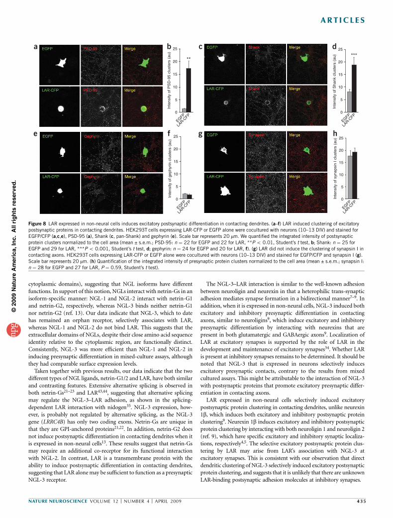
LAR in HEK293T cells induces postsynaptic differentiation
If the trans-synaptic interaction between NGL-3 and LAR is involved inthe formation of excitatory synapses, presynaptic LAR may also inducepostsynaptic differentiation in contacting dendrites. Indeed, in amixed-culture assay, LAR-expressing HEK293T cells induced the clus-tering of excitatory postsynaptic proteins PSD-95 and Shank,
whereas EGFP alone induced minimal postsynaptic protein clustering(Fig. 8a–d). In addition, LAR expressed in HEK 293T cells did notinduce gephyrin clusters in contacting dendrites (Fig. 8e,f). Theseresults suggest that presynaptic LAR induces excitatory, but notinhibitory, postsynaptic protein clustering on contacting dendrites.
LAR regulates excitatory synaptic development and maintenancepartly through its association with AMPA receptors34, suggesting thatLAR is also present on the postsynaptic plasma membrane. We there-fore tested whether LAR expressed in HEK293T cells is capable ofinducing presynaptic differentiation in contacting axons. LAR-expres-sing HEK293T cells did not induce synapsin I clustering in contactingaxons (Fig. 8g,h). This suggests that postsynaptic LAR has a differentrole, which is unrelated to the induction of presynaptic differentiation.
DISCUSSION
Here, we identified a previously unknown trans-synaptic adhesionbetween NGL-3 and LAR. In support of the synaptogenic functionof this interaction, NGL-3 and LAR expressed in non-neural cellsinduced pre- and postsynaptic differentiation in contacting axons and
3.5
vGlu
t1-p
ositi
veP
SD
-95
clus
ters
per
10
µmV
GAT
-pos
itive
geph
yrin
clu
ster
s pe
r 10
µm
Freq
uenc
y (H
z)
Am
plitu
de (
pA)
3.0
2.0
0.5
0
sh-ve
c
shNGL-
3 #2
shNGL-
3 #1
*
NGL-3*
shNGL-
3 #1
+
shNGL-
3 #1
sh-ve
c
shNGL-
3 #2
shNGL-
3 #1
*
NGL-3*
sh-ve
c
shNGL-
3 #2
sh-ve
c
shNGL-
3 #2
shNGL-
3 #1
+
shNGL-
3 #1
2.5 ****
1.5
1.0
2.0
sh-vec shNGL-3 #1 shNGL-3 #2 shNGL-3 #1*shNGL-3 #1 +
NGL-3*
sh-veca b
e f g
c d
shNGL-3 #1 shNGL-3 #2
shNGL-3 #2
sh-vec
10 pA
1 s
shNGL-3 #1*shNGL-3 #1 +
NGL-3*
0.5
0
2.5
0.8 15
10
5
0
0.6
0.4
0.2 **
0
1.5
1.0
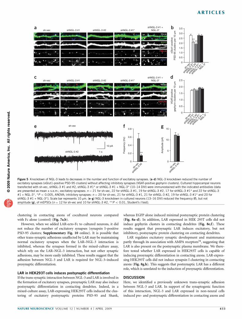
Figure 5 Knockdown of NGL-3 leads to decreases in the number and function of excitatory synapses. (a–d) NGL-3 knockdown reduced the number of
excitatory synapses (vGlut1-positive PSD-95 clusters) without affecting inhibitory synapses (VGAT-positive gephyrin clusters). Cultured hippocampal neurons
transfected with sh-vec, shNGL-3 #1 and #2, shNGL-3 #1* or shNGL-3 #1 + NGL-3* (10–14 DIV) were immunostained with the indicated antibodies (data
are presented as mean ± s.e.m.; excitatory synapses: n ¼ 21 for sh-vec, 22 for shNGL-3 #1, 19 for shNGL-3 #2, 17 for shNGL-3 #1* and 22 for shNGL-3
#1 + NGL-3*, *P o 0.005, ANOVA; inhibitory synapses: n ¼ 20 for sh-vec, 21 for shNGL-3 #1, 21 for shNGL-3 #2, 19 for shNGL-3 #1* and 20 for
shNGL-3 #1 + NGL-3*). Scale bar represents 10 mm. (e–g) NGL-3 knockdown in cultured neurons (13–16 DIV) reduced the frequency (f), but notamplitude (g), of mEPSCs (n ¼ 12 for sh-vec and 10 for shNGL-3 #2, **P o 0.01, Student’s t test).
NATURE NEUROSCIENCE VOLUME 12 [ NUMBER 4 [ APRIL 2009 433
ART ICLES
©20
09 N
atu
re A
mer
ica,
Inc.
All
rig
hts
res
erve
d.

dendrites, respectively. Neuronal NGL-3 expression increased presy-naptic contacts. Dendritic NGL-3 aggregation induced excitatorypostsynaptic protein clustering. NGL-3 knockdown reduced excitatorysynapse number and function. Soluble LAR inhibited NGL-3–inducedpresynaptic differentiation. These results suggest that the trans-synapticadhesion between NGL-3 and LAR regulates excitatory synapse for-mation in a bidirectional manner.
We were unable to determine the ultrastructural synaptic localiza-tion of NGL-3 because of the lack of suitable NGL-3 antibodies.However, our previous electron microscopy results, obtained using apan-NGL antibody, indicate that NGL isoforms are mainly postsynap-tically localized at excitatory synapses13. The presynaptic localization of
LAR in C. elegans and Drosophila has been well characterized28,32,33,36,although the pre- or postsynaptic localization of mammalian LARremains to be determined34. On the basis of the widespread distribu-tion of both mRNAs, determined by in situ hybridization13,41, theadhesion between NGL-3 and LAR probably occurs in various brainregions. Notably, NGL-3 proteins are mainly expressed in the brain(Fig. 2b), whereas LAR mRNAs, determined by Northern analysis, areexpressed in various tissues42, suggesting that the interaction betweenNGL-3 and LAR occurs primarily in the brain.
The three NGL isoforms share an identical domain structure andinteract with PSD-95 through their extreme C termini12,13. However,their primary sequences are substantially different (especially in their
***
0
20
40
60
Inte
nsity
of s
ynap
sin
Icl
uste
rs (
au)
80
100
120
140
160
Fc alon
e
LAR-e
cto-F
c
Fc alone
LAR-ecto-Fc
a bFigure 7 Soluble LAR reduces NGL-3–induced
presynaptic differentiation. (a) Reduced NGL-3–
induced presynaptic differentiation in the
presence of soluble LAR. HEK293T cells
expressing NGL-3–EGFP or EGFP alone were
cocultured with hippocampal neurons (10–13
DIV) in the presence of LAR-ecto-Fc or Fc alone
(10 mg ml�1) and stained for EGFP (for NGL-3),
synapsin I and human Fc (for LAR-ecto-Fc). Scale
bar represents 20 mm. (b) Quantification of the
integrated intensity of synapsin I clusters
normalized to the cell area (mean ± s.e.m.,
n ¼ 20 for Fc alone and 23 for LAR-ecto-Fc,
***P o 0.001, Student’s t test).
NGL-1
LAR
-CF
P
LAR-ecto-Fc
LAR
-CF
P
NGL-2 NGL-3 NGL-3
NG
L-2
NG
L-1
NG
L-37
6
Num
ber
of c
ell a
ggre
gate
spe
r fr
ame
5
4
3
2
NGL-3
+ LA
R
NGL-3
+ LA
R
+ 10
mM
EGTA
NGL-1
+ LA
R
NGL-2
+ LA
R
NGL-3
+ LA
R
0
1
7***
a d eb c
f g h
6
Num
ber
of c
ell a
ggre
gate
spe
r fr
ame
Bou
nd (
OD
450
)
Bou
nd p
er fr
ee(O
D 4
50 p
er n
M)
5
4
3
2
0 50 100
LAR-ecto-Fc (nM)
150
1
0.10
0.05
0.000 1 2 3 4
0
3
2
0
1
Bound (OD 450)
Figure 6 NGL-3 interacts with LAR. (a) NGL-3, but not NGL-1 or NGL-2, interacted with LAR in cell-aggregation assays. L cells cotransfected with NGLs
and EGFP or LAR (C-terminally cyan fluorescent protein (CFP)-tagged) and dsRed were mixed to induce cell aggregation. Scale bar represents 50 mm.
(b) Quantification of the cell aggregation in a. Cell aggregates were defined by four or more clustered cells containing at least one red or green cell (mean ±
s.e.m., n ¼ 10, ***P o 0.001, ANOVA). (c,d) Calcium-independent adhesion between NGL-3 and LAR. L cells expressing NGL-3 + EGFP or LAR-CFP +
dsRed were mixed in the presence or absence of 10 mM EGTA (mean ± s.e.m., n ¼ 10, ***P o 0.001, Student’s t test). Scale bar represents 50 mm.
(e) Selective association of LAR-ecto-Fc with NGL-3, but not with NGL-1 and NGL-2 expressed in HEK293T cells. Scale bar represents 10 mm. (f) NGL-3–
expressing HEK293T cells induced LAR-Flag clustering on contacting axons of cocultured neurons. HEK293T cells expressing NGL-3–EGFP or EGFP alone
were cocultured (14–15 DIV) with hippocampal neurons expressing LAR-Flag C1522S (C-terminally Flag-tagged, 13–14 DIV), followed by EGFP and Flag
staining. The phosphatase-dead LAR C1522S mutant was used to minimize possible effects of phosphatase activity on transfected neurons. (g) LAR-
expressing HEK293T cells induce NGL-3–Flag clustering on contacting dendrites of cocultured neurons. HEK293T cells expressing LAR-CFP or EGFP alonewere cocultured (14–15 DIV) with hippocampal neurons expressing NGL-3–Flag (13–14 DIV). Scale bar represents 20 mm. (h) Saturation curve of LAR-ecto-Fc
binding to NGL-3 expressed in HEK293T cells. A Scatchard plot analyzed by linear regression of the data is shown in the inset. The calculated Kd for the
interaction was 37.4 ± 2.1 nM.
434 VOLUME 12 [ NUMBER 4 [ APRIL 2009 NATURE NEUROSCIENCE
ART ICLES
©20
09 N
atu
re A
mer
ica,
Inc.
All
rig
hts
res
erve
d.
cytoplasmic domains), suggesting that NGL isoforms have differentfunctions. In support of this notion, NGLs interact with netrin-Gs in anisoform-specific manner: NGL-1 and NGL-2 interact with netrin-G1and netrin-G2, respectively, whereas NGL-3 binds neither netrin-G1nor netrin-G2 (ref. 13). Our data indicate that NGL-3, which to datehas remained an orphan receptor, selectively associates with LAR,whereas NGL-1 and NGL-2 do not bind LAR. This suggests that theextracellular domains of NGLs, despite their close amino acid sequenceidentity relative to the cytoplasmic region, are functionally distinct.Consistently, NGL-3 was more efficient than NGL-1 and NGL-2 ininducing presynaptic differentiation in mixed-culture assays, althoughthey had comparable surface expression levels.
Taken together with previous results, our data indicate that the twodifferent types of NGL ligands, netrin-G1/2 and LAR, have both similarand contrasting features. Extensive alternative splicing is observed inboth netrin-Gs21–23 and LAR43,44, suggesting that alternative splicingmay regulate the NGL-3–LAR adhesion, as shown in the splicing-dependent LAR interaction with nidogen35. NGL-3 expression, how-ever, is probably not regulated by alternative splicing, as the NGL-3gene (LRRC4B) has only two coding exons. Netrin-Gs are unique inthat they are GPI-anchored proteins21,22. In addition, netrin-G2 doesnot induce postsynaptic differentiation in contacting dendrites when itis expressed in non-neural cells13. These results suggest that netrin-Gsmay require an additional co-receptor for its functional interactionwith NGL-2. In contrast, LAR is a transmembrane protein with theability to induce postsynaptic differentiation in contacting dendrites,suggesting that LAR alone may be sufficient to function as a presynapticNGL-3 receptor.
The NGL-3–LAR interaction is similar to the well-known adhesionbetween neuroligin and neurexin in that a heterophilic trans-synapticadhesion mediates synapse formation in a bidirectional manner7–9. Inaddition, when it is expressed in non-neural cells, NGL-3 induced bothexcitatory and inhibitory presynaptic differentiation in contactingaxons, similar to neuroligins9, which induce excitatory and inhibitorypresynaptic differentiation by interacting with neurexins that arepresent in both glutamatergic and GABAergic axons9. Localization ofLAR at excitatory synapses is supported by the role of LAR in thedevelopment and maintenance of excitatory synapses34. Whether LARis present at inhibitory synapses remains to be determined. It should benoted that NGL-3 that is expressed in neurons selectively inducesexcitatory presynaptic contacts, contrary to the results from mixedcultured assays. This might be attributable to the interaction of NGL-3with postsynaptic proteins that promote excitatory presynaptic differ-entiation in contacting axons.
LAR expressed in non-neural cells selectively induced excitatorypostsynaptic protein clustering in contacting dendrites, unlike neurexin1b, which induces both excitatory and inhibitory postsynaptic proteinclustering9. Neurexin 1b induces excitatory and inhibitory postsynapticprotein clustering by interacting with both neuroligin 1 and neuroligin 2(ref. 9), which have specific excitatory and inhibitory synaptic localiza-tions, respectively4,5. The selective excitatory postsynaptic protein clus-tering by LAR may arise from LAR’s association with NGL-3 atexcitatory synapses. This is consistent with our observation that directdendritic clustering of NGL-3 selectively induced excitatory postsynapticprotein clustering, and suggests that it is unlikely that there are unknownLAR-binding postsynaptic adhesion molecules at inhibitory synapses.
0
Inte
nsity
of P
SD
-95
clus
ters
(au
)
25
20 **
15
10
5
EGFP
LAR-C
FP
b
0
Inte
nsity
of g
ephy
rin c
lust
ers
(au)
25
20
15
10
5
EGFP
LAR-C
FP
f
a
e
c
g
0
Inte
nsity
of S
hank
clu
ster
s (a
u)
25
20
***
15
10
5
EGFP
LAR-C
FP
d
0Inte
nsity
of s
ynap
sin
I clu
ster
s (a
u) 25
20
15
10
5
EGFP
LAR-C
FP
h
Figure 8 LAR expressed in non-neural cells induces excitatory postsynaptic differentiation in contacting dendrites. (a–f) LAR induced clustering of excitatory
postsynaptic proteins in contacting dendrites. HEK293T cells expressing LAR-CFP or EGFP alone were cocultured with neurons (10–13 DIV) and stained for
EGFP/CFP (a,c,e), PSD-95 (a), Shank (c, pan-Shank) and gephyrin (e). Scale bar represents 20 mm. We quantified the integrated intensity of postsynaptic
protein clusters normalized to the cell area (mean ± s.e.m.; PSD-95: n ¼ 22 for EGFP and 22 for LAR, **P o 0.01, Student’s t test, b; Shank: n ¼ 25 for
EGFP and 29 for LAR, ***P o 0.001, Student’s t test, d; gephyrin: n ¼ 24 for EGFP and 20 for LAR, f). (g) LAR did not induce the clustering of synapsin I in
contacting axons. HEK293T cells expressing LAR-CFP or EGFP alone were cocultured with neurons (10–13 DIV) and stained for EGFP/CFP and synapsin I (g).
Scale bar represents 20 mm. (h) Quantification of the integrated intensity of presynaptic protein clusters normalized to the cell area (mean ± s.e.m.; synapsin I:
n ¼ 28 for EGFP and 27 for LAR, P ¼ 0.59, Student’s t test).
NATURE NEUROSCIENCE VOLUME 12 [ NUMBER 4 [ APRIL 2009 435
ART ICLES
©20
09 N
atu
re A
mer
ica,
Inc.
All
rig
hts
res
erve
d.
How might NGL-3 and LAR lead to post- and presynaptic differ-entiation, respectively? One possible mechanism by which synapticadhesion molecules contribute to synapse formation is by interactingwith and promoting the synaptic localization of specific membrane andcytoplasmic proteins. PSD-95, which interacts with NGL-3, is anabundant postsynaptic protein that interacts with a variety of mem-brane, signaling and scaffolding/adaptor proteins of excitatorysynapses. Therefore, PSD-95 may couple NGL-3–dependent synapticadhesion to the localization of various postsynaptic proteins.
Liprin-a, which binds to the membrane-distal phosphatase domainof LAR, may act together with LAR to mediate NGL-3–dependentpresynaptic differentiation. Liprin-a has been implicated in the regula-tion of active zone assembly and presynaptic development through itsinteractions with synaptic proteins, including ERC/ELKS, RIM andCASK28. Studies on liprin-a homologs in C. elegans (SYD-2) andDrosophila (Dliprin-a) have firmly established the roles of liprin-a inpresynaptic differentiation31,32,45,46.
Another LAR-interacting cytoplasmic protein that may contribute toLAR-dependent presynaptic differentiation is b-catenin, a componentof the cadherin-catenin complex30. Presynaptic b-catenin localizes thereserve pool of vesicles to presynaptic sites through mechanismsinvolving interaction of its C terminus with synaptic PDZ proteins47.Notably, brain-derived neurotrophic factor activation of the TrkBreceptor tyrosine kinase regulates tyrosine phosphorylation ofb-catenin, which disrupts the cadherin–b-catenin interaction andpromotes synaptic vesicle splitting and synapse formation48.Considering that LAR dephosphorylates b-catenin30,34, LAR andTrkB may reciprocally regulate b-catenin–dependent presynapticvesicle clustering.
One possibility is that the binding of NGL-3 to LAR may regulate thetyrosine phosphatase activity of LAR. Receptor tyrosine phosphatasescan be negatively regulated by dimerization49. However, a study on thecrystallographic structure of the phosphatase domain of LAR hassuggested that the phosphatase activity of LAR is probably notregulated by dimerization50.
In conclusion, our results suggest that the trans-synaptic interactionbetween NGL-3 and LAR regulates excitatory synapse formation in abidirectional manner. Future investigations will aim to explorepossible functional interactions between NGL-1/2–netrin-Gs andNGL-3–LAR adhesions.
METHODSDNA constructs and antibodies. Full-length human NGL-1 (NM_020929,
amino acids 1–641) and rat NGL-3 (XM_218615, amino acids 1–709) were
subcloned into pEGFP-N1 (Clontech). Human LAR (amino acids 1–1881) was
subcloned into pECFP-N1 (Clontech). For small interfering RNA knockdown,
nucleotides 359–377 of rat NGL-3 (GCA AGA ATC TGG TGC GCA A), its
point mutant (GCA AGT CTC TTG TGC GCA A) and nucleotides 1,254–1,272
(GCA CGA TGG CAC ACT CAA T) were subcloned into pSuper.gfp/
neo (OligoEngine) to generate shNGL-3 #1, shNGL-3* and shNGL-3 #2,
respectively. The extracellular domain of LAR (amino acids 1–1,235) was
subcloned into pEGFP-N1, in which EGFP was replaced with human Fc. Other
constructs, antibodies and reagents are described in the Supplementary
Methods online.
Mixed-culture assay. Mixed-culture assays were carried out as described39.
Briefly, primary hippocampal neuron cultures at 10 d in vitro (10 DIV)
prepared from embryonic day 18–19 (E18–19) rats were cocultured with
HEK293T cells expressing NGLs, LAR or EGFP, followed by immunostaining
at 13 DIV. For the synaptotagmin I antibody uptake assay, neurons were
incubated with antibodies to the synaptotagmin luminal domain at the dilution
of 1:10 in isotonic depolarizing solution for 5 min. For competitive inhibition,
LAR-ecto-Fc proteins (10 mg ml�1) were added to NGL-3–expressing HEK293T
cells that were cocultured with neurons for 3 d (10–13 DIV).
Transfection of neurons and immunocytochemistry. Cultured neurons were
transfected using a mammalian transfection kit (Clontech) and fixed with 4%
paraformaldehyde/4% sucrose (vol/vol), permeabilized with 0.2% Triton X-100
(vol/vol) in phosphate-buffered saline, incubated with primary antibodies and
then incubated with Cy3-, Cy5- or FITC-conjugated secondary antibodies
(Jackson ImmunoResearch). For the inhibition of endogenous synapses
with soluble LAR, neurons were treated with LAR-ecto-Fc for 3 d
(20 mg ml�1, 7/8–10/11 DIV) and transfected with pEGFP-N1 for visualization
(9/10–10/11 DIV).
Bead and antibody aggregation assays. Bead aggregation assays were per-
formed as described previously13. Briefly, neutravidin-conjugated FluoSphere
beads (Molecular Probes) were pre-incubated with biotin-conjugated antibo-
dies to EGFP. The antibody-coated beads were added onto neurons expressing
EGFP–NGL-3 (14–16 DIV) and cultured for 24 h. For antibody aggregation,
antibodies to EGFP were clustered by FITC-conjugated antibodies to guinea pig
in complete neurobasal medium and added to neurons at 16 DIV, followed by
24-h culture and immunofluorescence staining at 17 DIV.
Image acquisition and quantification. All z-stacked images were randomly
acquired by confocal microscopy (LSM510, Zeiss) and analyzed with Meta-
Morph image analysis software (Universal Imaging). The density of synaptic
protein clusters was measured from 15–30 neurons; the primary dendritic
lengths of B50 mm from the cell body were measured for each neuron. For
quantification of images from coculture assays, all captured images were
thresholded and the integrated intensity of the clusters on transfected
HEK293T cells was normalized to the cell area. All values are presented as
mean ± s.e.m. and analyzed by Student’s t test or ANOVA Tukey’s test.
Quantitative cell surface–binding assay. HEK293T cells transfected with
NGL-3 were transferred to 96-well plates and grown for 24 h. After fixation
in 4% paraformaldehyde/4% sucrose, cells were incubated with increasing
concentrations of LAR-ecto-Fc for 1 h, incubated with horseradish peroxidase–
conjugated antibodies to human Fc (Sigma, 1:10,000) and color reacted with
TMB (Sigma).
Electrophysiology. Cultured pyramidal neurons from the hippocampus trans-
fected with shNGL-3 (13–16 DIV) were whole-cell voltage clamped at �60 mV
using an Axopatch 200B amplifier (Axon Instruments). The extracellular
solution contained 145 mM NaCl, 2.5 mM KCl, 10 mM HEPES, 1.25 mM
NaH2PO4, 2 mM CaCl2, 1 mM MgCl2, 10 mM glucose and 0.4 mM sodium
ascorbate. The intracellular solution contained 100 mM potassium gluconate,
20 mM KCl, 10 mM HEPES, 8 mM NaCl, 4 mM magnesium ATP, 0.3 mM
sodium GTP and 0.5 mM EGTA. For mEPSC measurement, tetrodotoxin
(10 nM, Tocris) and bicuculline (10 mM, Tocris) were added into the
extracellular solution. Synaptic currents were analyzed using a custom-written
macro in Igor Pro (WaveMetrics).
Note: Supplementary information is available on the Nature Neuroscience website.
ACKNOWLEDGMENTSWe would like to thank A.M. Craig for neurexin 1a, 2a, 3a and 1b cDNAs,T. Biederer for SynCAM 1, Y.-P. Hsueh for Syndecan-2, P. Maness forNCAM-140, J. Ko for quantitative analysis, J. Nam for SALM4, M.-H. Kimfor the mini analysis program, and Y.-G. Oh, M.-S. Baek, M. Ryu and J.-G. Jungfor the help with antibody generation. This work was supported by the NationalCreative Research Initiative Program of the Korean Ministry of Science andTechnology (E.K.).
AUTHOR CONTRIBUTIONSJ.W. and S.-K.K. carried out the experiments, analyzed the data and wrote themanuscript. S.C. helped with the shRNA knockdown experiments. S.K., J.-R.L.,A.W.D. and M.S. contributed reagents. E.K. supervised the project and wrote themanuscript. All authors discussed the results and commented on the manuscript.
436 VOLUME 12 [ NUMBER 4 [ APRIL 2009 NATURE NEUROSCIENCE
ART ICLES
©20
09 N
atu
re A
mer
ica,
Inc.
All
rig
hts
res
erve
d.

Published online at http://www.nature.com/natureneuroscience/
Reprints and permissions information is available online at http://www.nature.com/
reprintsandpermissions/
1. Yamagata, M., Sanes, J.R. & Weiner, J.A. Synaptic adhesion molecules. Curr. Opin. CellBiol. 15, 621–632 (2003).
2. Dalva, M.B., McClelland, A.C. & Kayser, M.S. Cell adhesion molecules: signalingfunctions at the synapse. Nat. Rev. Neurosci. 8, 206–220 (2007).
3. Washbourne, P. et al. Cell adhesion molecules in synapse formation. J. Neurosci. 24,9244–9249 (2004).
4. Akins, M.R. & Biederer, T. Cell-cell interactions in synaptogenesis.Curr. Opin. Neurobiol.16, 83–89 (2006).
5. Craig, A.M. & Kang, Y. Neurexin-neuroligin signaling in synapse development.Curr. Opin. Neurobiol. 17, 43–52 (2007).
6. McAllister, A.K. Dynamic aspects of CNS synapse formation. Annu. Rev. Neurosci. 30,425–450 (2007).
7. Ichtchenko, K. et al. Neuroligin 1: a splice site–specific ligand for beta-neurexins.Cell 81, 435–443 (1995).
8. Scheiffele, P., Fan, J., Choih, J., Fetter, R. & Serafini, T. Neuroligin expressed in non-neuronal cells triggers presynaptic development in contacting axons. Cell 101,657–669 (2000).
9. Graf, E.R., Zhang, X., Jin, S.X., Linhoff, M.W. & Craig, A.M. Neurexins inducedifferentiation of GABA and glutamate postsynaptic specializations via neuroligins.Cell 119, 1013–1026 (2004).
10. Biederer, T. et al. SynCAM, a synaptic adhesion molecule that drives synapse assembly.Science 297, 1525–1531 (2002).
11. Fogel, A.I. et al. SynCAMs organize synapses through heterophilic adhesion. J. Neurosci.27, 12516–12530 (2007).
12. Lin, J.C., Ho, W.H., Gurney, A. & Rosenthal, A. The netrin-G1 ligand NGL-1 promotes theoutgrowth of thalamocortical axons. Nat. Neurosci. 6, 1270–1276 (2003).
13. Kim, S. et al. NGL family PSD-95–interacting adhesion molecules regulate excitatorysynapse formation. Nat. Neurosci. 9, 1294–1301 (2006).
14. Kayser, M.S., McClelland, A.C., Hughes, E.G. & Dalva, M.B. Intracellular and trans-synaptic regulation of glutamatergic synaptogenesis by EphB receptors. J. Neurosci. 26,12152–12164 (2006).
15. Boucard, A.A., Chubykin, A.A., Comoletti, D., Taylor, P. & Sudhof, T.C. A splice code fortrans-synaptic cell adhesion mediated by binding of neuroligin 1 to alpha- and beta-neurexins. Neuron 48, 229–236 (2005).
16. Chih, B., Gollan, L. & Scheiffele, P. Alternative splicing controls selective trans-synapticinteractions of the neuroligin-neurexin complex. Neuron 51, 171–178 (2006).
17. Graf, E.R., Kang, Y., Hauner, A.M. & Craig, A.M. Structure function and splice siteanalysis of the synaptogenic activity of the neurexin-1 beta LNS domain. J. Neurosci.26, 4256–4265 (2006).
18. Irie, M. et al. Binding of neuroligins to PSD-95. Science 277, 1511–1515 (1997).19. Tabuchi, K. et al. A neuroligin-3 mutation implicated in autism increases inhibitory
synaptic transmission in mice. Science 318, 71–76 (2007).20. Jamain, S. et al. Reduced social interaction and ultrasonic communication in a mouse
model of monogenic heritable autism. Proc. Natl. Acad. Sci. USA 105, 1710–1715(2008).
21. Nakashiba, T. et al. Netrin-G1: a novel glycosyl phosphatidylinositol–linked mammaliannetrin that is functionally divergent from classical netrins. J. Neurosci. 20, 6540–6550(2000).
22. Nakashiba, T., Nishimura, S., Ikeda, T. & Itohara, S. Complementary expression andneurite outgrowth activity of netrin-G subfamily members. Mech. Dev. 111, 47–60(2002).
23. Yin, Y., Miner, J.H. & Sanes, J.R. Laminets: laminin- and netrin-related genes expressedin distinct neuronal subsets. Mol. Cell. Neurosci. 19, 344–358 (2002).
24. Nishimura-Akiyoshi, S., Niimi, K., Nakashiba, T. & Itohara, S. Axonal netrin-Gstransneuronally determine lamina-specific subdendritic segments. Proc. Natl. Acad.Sci. USA 104, 14801–14806 (2007).
25. Zhang, W. et al. Netrin-G2 and netrin-G2 ligand are both required for normal auditoryresponsiveness. Genes Brain Behav. 7, 385–392 (2008).
26. Aoki-Suzuki, M. et al. A family-based association study and gene expression analyses ofnetrin-G1 and -G2 genes in schizophrenia. Biol. Psychiatry 57, 382–393 (2005).
27. Johnson, K.G. & Van Vactor, D. Receptor protein tyrosine phosphatases in nervoussystem development. Physiol. Rev. 83, 1–24 (2003).
28. Stryker, E. & Johnson, K.G. LAR, liprin alpha and the regulation of active zonemorphogenesis. J. Cell Sci. 120, 3723–3728 (2007).
29. Serra-Pages, C. et al. The LAR transmembrane protein tyrosine phosphatase anda coiled-coil LAR-interacting protein colocalize at focal adhesions. EMBO J. 14,2827–2838 (1995).
30. Kypta, R.M., Su, H. & Reichardt, L.F. Association between a transmembrane proteintyrosine phosphatase and the cadherin-catenin complex. J. Cell Biol. 134, 1519–1529(1996).
31. Zhen, M. & Jin, Y. The liprin protein SYD-2 regulates the differentiation of presynaptictermini in C. elegans. Nature 401, 371–375 (1999).
32. Kaufmann, N., DeProto, J., Ranjan, R., Wan, H. & Van Vactor, D. Drosophila liprin-alphaand the receptor phosphatase Dlar control synapse morphogenesis. Neuron 34, 27–38(2002).
33. Ackley, B.D. et al. The two isoforms of the Caenorhabditis elegans leukocyte-commonantigen related receptor tyrosine phosphatase PTP-3 function independently in axonguidance and synapse formation. J. Neurosci. 25, 7517–7528 (2005).
34. Dunah, A.W. et al. LAR receptor protein tyrosine phosphatases in the development andmaintenance of excitatory synapses. Nat. Neurosci. 8, 458–467 (2005).
35. O’Grady, P., Thai, T.C. & Saito, H. The laminin-nidogen complex is a ligand for a specificsplice isoform of the transmembrane protein tyrosine phosphatase LAR. J. Cell Biol.141, 1675–1684 (1998).
36. Johnson, K.G. et al. The HSPGs Syndecan and Dallylike bind the receptor phosphataseLAR and exert distinct effects on synaptic development. Neuron 49, 517–531 (2006).
37. Fox, A.N. & Zinn, K. The heparan sulfate proteoglycan syndecan is an in vivo ligand forthe Drosophila LAR receptor tyrosine phosphatase. Curr. Biol. 15, 1701–1711(2005).
38. Yang, T. et al. Leukocyte antigen-related protein tyrosine phosphatase receptor: a smallectodomain isoform functions as a homophilic ligand and promotes neurite outgrowth.J. Neurosci. 23, 3353–3363 (2003).
39. Biederer, T. & Scheiffele, P. Mixed-culture assays for analyzing neuronal synapseformation. Nat. Protoc. 2, 670–676 (2007).
40. Ko, J. et al. SALM synaptic cell adhesion–like molecules regulate the differentiation ofexcitatory synapses. Neuron 50, 233–245 (2006).
41. Zhang, J.S., Honkaniemi, J., Yang, T., Yeo, T.T. & Longo, F.M. LAR tyrosine phosphatasereceptor: a developmental isoform is present in neurites and growth cones and itsexpression is regional- and cell-specific. Mol. Cell. Neurosci. 10, 271–286 (1998).
42. Pulido, R., Serra-Pages, C., Tang, M. & Streuli, M. The LAR/PTP delta/PTP sigmasubfamily of transmembrane protein-tyrosine- phosphatases: multiple human LAR, PTPdelta and PTP sigma isoforms are expressed in a tissue-specific manner and associatewith the LAR- interacting protein LIP.1. Proc. Natl. Acad. Sci. USA 92, 11686–11690(1995).
43. O’Grady, P., Krueger, N.X., Streuli, M. & Saito, H. Genomic organization of the humanLAR protein tyrosine phosphatase gene and alternative splicing in the extracellularfibronectin type-III domains. J. Biol. Chem. 269, 25193–25199 (1994).
44. Zhang, J.S. & Longo, F.M. LAR tyrosine phosphatase receptor: alternative splicing ispreferential to the nervous system, coordinated with cell growth and generates novelisoforms containing extensive CAG repeats. J. Cell Biol. 128, 415–431 (1995).
45. Patel, M.R. et al. Hierarchical assembly of presynaptic components in definedC. elegans synapses. Nat. Neurosci. 9, 1488–1498 (2006).
46. Dai, Y. et al. SYD-2 Liprin-alpha organizes presynaptic active zone formation throughELKS. Nat. Neurosci. 9, 1479–1487 (2006).
47. Bamji, S.X. et al. Role of beta-catenin in synaptic vesicle localization and presynapticassembly. Neuron 40, 719–731 (2003).
48. Bamji, S.X., Rico, B., Kimes, N. & Reichardt, L.F. BDNF mobilizes synaptic vesicles andenhances synapse formation by disrupting cadherin-beta-catenin interactions. J. CellBiol. 174, 289–299 (2006).
49. Majeti, R. & Weiss, A. Regulatory mechanisms for receptor protein tyrosine phospha-tases. Chem. Rev. 101, 2441–2448 (2001).
50. Nam, H.J., Poy, F., Krueger, N.X., Saito, H. & Frederick, C.A. Crystal structure of thetandem phosphatase domains of RPTP LAR. Cell 97, 449–457 (1999).
NATURE NEUROSCIENCE VOLUME 12 [ NUMBER 4 [ APRIL 2009 437
ART ICLES
©20
09 N
atu
re A
mer
ica,
Inc.
All
rig
hts
res
erve
d.
Copyright © 2022 FDOKUMEN