Steroid hormone receptors: an update
12
Human Reproduction Update 2000, Vol. 6 No. 3 pp. 225–236 © European Society of Human Reproduction and Embryology Steroid hormone receptors: an update M.Beato * and J.Klug Institut für Molekularbiologie und Tumorforschung, IMT, Philipps-Universität, 35033 Marburg, Germany Received on August 26, 1999; accepted on February 28, 2000 Steroid hormones (SHs) are lipophilic molecules derived from cholesterol and synthesized in the adrenal cortex (glucocorticoids, mineralocorticoids, and adrenal androgens), the testes (testicular androgens, oestrogen), and the ovary and placenta (oestrogens and progestagens or progestins). SHs reach their target cells via the blood, where they are bound to carrier proteins, and because of their lipophilic nature pass the cell membrane by simple diffusion. Within the target cells SHs bind to steroid hormone receptors (SHRs), the key mediators of SH action, which are complexed to chaperones, e.g. heat shock protein 90 (Hsp90), that help other proteins to fold and prevent aggregation. SHRs are intracellular transcription factors that can be activated, among other possibilities, by the specific and high affinity binding of ligand to exert positive or negative effects on the expression of target genes. Binding of agonistic or antagonistic ligands leads to different allosteric changes of SHRs making them competent to exert positive or negative effects on the expression of target genes by different mechanisms. (i) After dissociation of chaperones the liganded SHR–complexes can bind to chromatin organized DNA sequences in the vicinity of target genes, termed hormone response elements (HREs). The HRE-recruited hormone-receptor-complexes are then able to initiate chromatin remodelling and to relay activating or repressing signals to the target genes transcription machinery; (ii) through protein–protein interactions with other sequence-specific transcription factors, SHRs can also regulate the activity of many genes that are switched on, for instance, during stress or an inflammatory response; (iii) the SH response can also be integrated in the intracellular signalling network via cross-talk of SHRs with signal transduction pathways that transmit extracellular signals via membrane receptors and activation of protein kinase cascades to nuclear transcription factors that activate various target genes. By all these different mechanisms SHRs modulate numerous and specific responses in a large variety of cells, whereby their particular effect depends on the physiological, cellular and genetic context. Key words: chromatin/cross-talk/steroid hormones/steroid hormone receptors/transcription factors TABLE OF CONTENTS Introduction 225 Domain structure of steroid hormone receptors 226 Receptor isoforms and variants 227 DNA and chromatin binding 228 Ligand binding 230 Activation of transcription 230 Cross-talk with other signal transduction pathways 233 Conclusions 233 Acknowledgements 234 References 234 Introduction In mammals the gonads and adrenal gland produce five major groups of steroid hormones (SHs): oestrogens, progestins, androgens, glucocorticoids and mineralocorticoids. All these SHs regulate a large number of physiological processes in target cells equipped with the corresponding steroid hormone receptors (SHRs). The concept of target cells has been extended in the last years following the demonstration of functional SHRs in a large variety of cell types. For these, and many other crucial experiments, the availability of radioactively labelled SHs in the late 1950s was a key development. Using these compounds it was possible to follow the fate of the steroid hormones from their site of synthesis in the endocrine glands, through the blood circulation, up to their target tissues (Jensen and Jacobsen, 1962). Although SHs are extensively metabolized, particularly in the liver, it could be shown that in most cases the hormone itself, not a metabolite, produced the response via the modulation of gene expression mechanisms. The concept that steroid hormones are involved in transcriptional control was triggered by the observation that the insect steroid hormone ecdysone induces puffs in giant chromosomes (Clever and Karlson, 1960). A few years later, a two-step model was established that involved binding of the hormone to specific high-affinity SHRs within the target cells, followed by activation of the hormone–receptor complex in order to induce expression of hormone responsive genes (Noteboom and * To whom correspondence should be addressed at Institut für Molekularbiologie und Tumorforschung, IMT, Philipps-Universität, 35033 Marburg, Germany. Phone: +49 6421 286 62 86; Fax: +49 6421 286 53 98; E-mail: [email protected]
-
Upload
khangminh22 -
Category
Documents
-
view
1 -
download
0
Transcript of Steroid hormone receptors: an update

Human Reproduction Update 2000, Vol. 6 No. 3 pp. 225–236 © European Society of Human Reproduction and Embryology
Steroid hormone receptors: an update
M.Beato* and J.Klug
Institut für Molekularbiologie und Tumorforschung, IMT, Philipps-Universität, 35033 Marburg, Germany
Received on August 26, 1999; accepted on February 28, 2000Steroid hormones (SHs) are lipophilic molecules derived from cholesterol and synthesized in the adrenal cortex(glucocorticoids, mineralocorticoids, and adrenal androgens), the testes (testicular androgens, oestrogen), and theovary and placenta (oestrogens and progestagens or progestins). SHs reach their target cells via the blood, where theyare bound to carrier proteins, and because of their lipophilic nature pass the cell membrane by simple diffusion.Within the target cells SHs bind to steroid hormone receptors (SHRs), the key mediators of SH action, which arecomplexed to chaperones, e.g. heat shock protein 90 (Hsp90), that help other proteins to fold and preventaggregation. SHRs are intracellular transcription factors that can be activated, among other possibilities, by thespecific and high affinity binding of ligand to exert positive or negative effects on the expression of target genes.Binding of agonistic or antagonistic ligands leads to different allosteric changes of SHRs making them competent toexert positive or negative effects on the expression of target genes by different mechanisms. (i) After dissociation ofchaperones the liganded SHR–complexes can bind to chromatin organized DNA sequences in the vicinity of targetgenes, termed hormone response elements (HREs). The HRE-recruited hormone-receptor-complexes are then ableto initiate chromatin remodelling and to relay activating or repressing signals to the target genes transcriptionmachinery; (ii) through protein–protein interactions with other sequence-specific transcription factors, SHRs canalso regulate the activity of many genes that are switched on, for instance, during stress or an inflammatory response;(iii) the SH response can also be integrated in the intracellular signalling network via cross-talk of SHRs with signaltransduction pathways that transmit extracellular signals via membrane receptors and activation of protein kinasecascades to nuclear transcription factors that activate various target genes. By all these different mechanisms SHRsmodulate numerous and specific responses in a large variety of cells, whereby their particular effect depends on thephysiological, cellular and genetic context.
Key words: chromatin/cross-talk/steroid hormones/steroid hormone receptors/transcription factors
TABLE OF CONTENTS
Introduction 225Domain structure of steroid hormone receptors 226Receptor isoforms and variants 227DNA and chromatin binding 228Ligand binding 230Activation of transcription 230Cross-talk with other signal transduction pathways 233Conclusions 233Acknowledgements 234References 234
Introduction
In mammals the gonads and adrenal gland produce five majorgroups of steroid hormones (SHs): oestrogens, progestins,androgens, glucocorticoids and mineralocorticoids. All these SHsregulate a large number of physiological processes in target cellsequipped with the corresponding steroid hormone receptors
(SHRs). The concept of target cells has been extended in the lastyears following the demonstration of functional SHRs in a largevariety of cell types. For these, and many other crucialexperiments, the availability of radioactively labelled SHs in thelate 1950s was a key development. Using these compounds it waspossible to follow the fate of the steroid hormones from their siteof synthesis in the endocrine glands, through the blood circulation,up to their target tissues (Jensen and Jacobsen, 1962). AlthoughSHs are extensively metabolized, particularly in the liver, it couldbe shown that in most cases the hormone itself, not a metabolite,produced the response via the modulation of gene expressionmechanisms. The concept that steroid hormones are involved intranscriptional control was triggered by the observation that theinsect steroid hormone ecdysone induces puffs in giantchromosomes (Clever and Karlson, 1960). A few years later, atwo-step model was established that involved binding of thehormone to specific high-affinity SHRs within the target cells,followed by activation of the hormone–receptor complex in orderto induce expression of hormone responsive genes (Noteboom and
*To whom correspondence should be addressed at Institut für Molekularbiologie und Tumorforschung, IMT, Philipps-Universität, 35033 Marburg, Germany.Phone: +49 6421 286 62 86; Fax: +49 6421 286 53 98; E-mail: [email protected]
226 M.Beato and J.Klug
Gorski, 1965; Jensen et al., 1968). Almost 20 years later thereceptors for glucocorticoids and oestrogens were cloned and thusbecame the first molecularly-defined transcription factors forRNA polymerase II (Hollenberg et al., 1985; Walter et al., 1985;Weinberger et al., 1985a,b). At around the same time cloned SHRtargets, e.g. human metallothionein gene and the mouse mammarytumour virus (MMTV) were used in DNA binding and genetransfer experiments to identify the first hormone responseelements (HREs) (Chandler et al., 1983; Payvar et al., 1983;Scheidereit et al., 1983; Karin et al., 1984). HREs are short DNAsequence elements that convey direct transcriptionalresponsiveness to adjacent genes. During the last decade manydetails of the SHR signal transduction pathway, including newmechanisms, have been discovered. In this review we willsummarize our present view of the various pathways by whichSHRs modulate gene expression.
Domain structure of SHRs
Following the cloning of the receptors for glucocorticoids (GR)and oestrogens (ERα), receptors for androgens (AR), progestins
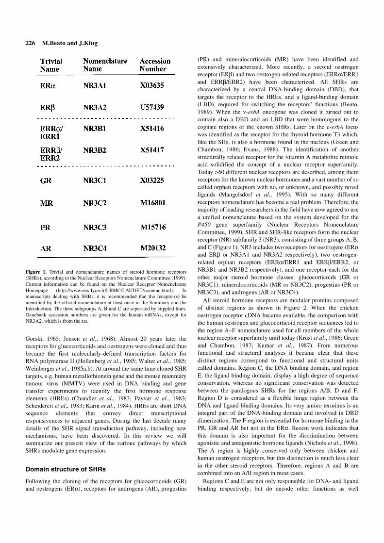
(PR) and mineralocorticoids (MR) have been identified andextensively characterized. More recently, a second oestrogenreceptor (ERβ) and two oestrogen-related receptors (ERRα/ERR1and ERRβ/ERR2) have been characterized. All SHRs arecharacterized by a central DNA-binding domain (DBD), thattargets the receptor to the HREs, and a ligand-binding domain(LBD), required for switching the receptors’ functions (Beato,1989). When the v-erbA oncogene was cloned it turned out tocontain also a DBD and an LBD that were homologous to thecognate regions of the known SHRs. Later on the c-erbA locuswas identified as the receptor for the thyroid hormone T3 which,like the SHs, is also a hormone found in the nucleus (Green andChambon, 1986; Evans, 1988). The identification of anotherstructurally related receptor for the vitamin A metabolite retinoicacid solidified the concept of a nuclear receptor superfamily.Today >60 different nuclear receptors are described, among themreceptors for the known nuclear hormones and a vast number of socalled orphan receptors with no, or unknown, and possibly novelligands (Mangelsdorf et al., 1995). With so many differentreceptors nomenclature has become a real problem. Therefore, themajority of leading researchers in the field have now agreed to usea unified nomenclature based on the system developed for theP450 gene superfamily (Nuclear Receptors NomenclatureCommittee, 1999). SHR and SHR-like receptors form the nuclearreceptor (NR) subfamily 3 (NR3), consisting of three groups A, B,and C (Figure 1). NR3 includes two receptors for oestrogens (ERαand ERβ or NR3A1 and NR3A2 respectively), two oestrogen-related orphan receptors (ERRα/ERR1 and ERRβ/ERR2, orNR3B1 and NR3B2 respectively), and one receptor each for theother major steroid hormone classes: glucocorticoids (GR orNR3C1), mineralocorticoids (MR or NR3C2), progestins (PR orNR3C3), and androgens (AR or NR3C4).
All steroid hormone receptors are modular proteins composedof distinct regions as shown in Figure 2. When the chickenoestrogen receptor cDNA became available, the comparison withthe human oestrogen and glucocorticoid receptor sequences led tothe region A–F nomenclature used for all members of the wholenuclear receptor superfamily until today (Krust et al., 1986; Greenand Chambon, 1987; Kumar et al., 1987). From numerousfunctional and structural analyses it became clear that thesedistinct regions correspond to functional and structural unitscalled domains. Region C, the DNA binding domain, and regionE, the ligand binding domain, display a high degree of sequenceconservation, whereas no significant conservation was detectedbetween the paralogous SHRs for the regions A/B, D and F.Region D is considered as a flexible hinge region between theDNA and ligand binding domains. Its very amino terminus is anintegral part of the DNA-binding domain and involved in DBDdimerization. The F region is essential for hormone binding in thePR, GR and AR but not in the ERα. Recent work indicates thatthis domain is also important for the discrimination betweenagonistic and antagonistic hormone ligands (Nichols et al., 1998).The A region is highly conserved only between chicken andhuman oestrogen receptors, but this distinction is much less clearin the other steroid receptors. Therefore, regions A and B arecombined into an A/B region in most cases.
Regions C and E are not only responsible for DNA- and ligandbinding respectively, but do encode other functions as well
Figure 1. Trivial and nomenclature names of steroid hormone receptors(SHRs), according to the Nuclear Receptors Nomenclature Committee (1999).Current information can be found on the Nuclear Receptor NomenclatureHomepage (http://www.ens-lyon.fr/LBMC/LAUDET/nomenc.html). Inmanuscripts dealing with SHRs, it is recommended that the receptor(s) beidentified by the official nomenclature at least once in the Summary and theIntroduction. The three subgroups A, B and C are separated by stippled lines.Genebank accession numbers are given for the human mRNAs, except forNR3A2, which is from the rat.

Steroid hormone receptors 227
(Figure 2). The intracellular distribution of steroid receptors is theresult of nuclear–cytoplasmic diffusion and ATP-dependentcytoplasmic–nuclear shuttling (Guiochon-Mantel et al., 1991). Atequilibrium the majority of ER, AR and PR is in the nucleus dueto the presence of so-called nuclear localization signals (NLSs)that are believed to be required for nuclear pore recognition. Thenumber and location of NLSs varies among SHRs but often aconstitutive NLS is located at the border of region C–D, whereasa second NLS in the LBD is ligand dependent. Intracellularlocalization is less clear for the GR and MR because hormone-induced nuclear translocation has been reported in these cases.
All unliganded SHRs are associated with a large multiproteincomplex of chaperones, including heat shock protein 90 (Hsp90),which maintains the receptor in an inactive state but keeps it wellprepared for hormone binding (Pratt and Toft, 1997). Again theregion at the border of region C to D together with the LBD arerequired for an SHR–Hsp90 interaction to take place. Most likelythese chaperones play an active role in keeping the SHRsfunctional (Godowski and Picard, 1989).
Ligand binding confers transcriptional competence onto SHRsthat is exerted in most receptors by two independenttransactivation functions, a constitutively active one in the A/B-region located close to the DBD, referred to as activation function 1(AF-1, also called τ-1 or enh-1) and a ligand-inducible activationfunction in the LBD, called AF-2. The two AFs act synergisticallyand connect the receptor to the transcription apparatus via directinteractions with basal transcription factors, sequence-specifictranscription factors and/or transcriptional co-activators (seebelow).
Receptor isoforms and variants
PR was the first SHR shown to exist in two common isoformsgenerated by differential promoter usage (Kastner et al., 1990).One promoter initiates transcription at positions +1 and +15 of thegene which gives rise to the longer isoform B, PR-B (Figure 3).The second promoter initiates human PR transcripts between +737and +842 encoding the 164 amino acid residues shorter hPR formA, PR-A. Both PR isoforms show high affinity for the naturalligand progesterone and the synthetic agonist R5020. Only PR-Bharbours a third activation function at its specific amino terminus,termed AF-3, that functions in a promoter and cell-specificmanner (Sartorius et al., 1994). Both isoforms, therefore, display
differential target gene specificity. First regarded as an exceptionin the family, differential promoter usage and alternative splicingare now the rule for all members.
The human ERα (hERα, Figure 3) was cloned from a cDNAexpression library produced from the breast cancer cell line MCF-7(Walter et al., 1985). Recently, a second oestrogen receptor, ERβ,was characterized which is very similar to ERα in terms ofstructure and function but also shows subtle and important
Figure 2. Steroid hormone receptor (SHR) domain structure and structure–function relationships. Domains are numbered A to F, originally based on thecomparison of human oestrogen and glucocorticoid receptor sequences. Domain C (dark grey box) is the DNA-binding domain (DBD) and domain E (light greybox) is the ligand-binding domain (LBD). AF-1 and -2 are the transcription activation functions 1 and 2. Indicated regions are required for receptor dimerization(symbolized by hooked lines), nuclear localization (arrows) and heat shock protein (Hsp)90 binding (dashed lines).
Figure 3. The human steroid hormone receptor family including isoforms andvariants. The numbers refer to amino acid positions. Highlighted are the DNAbinding domain (dark grey box) and the ligand binding domain (light greybox). The A/B- and F-domains are drawn to scale. The name of each receptoris indicated: h = human; ER = oestrogen receptor; ERR = oestrogen-relatedreceptor; GR = glucocorticoid receptor; MR = mineralocorticoid receptor; PR-A/PR-B = progesterone receptor form A and B; AR-A/AR-B = androgenreceptor form A and B.

228 M.Beato and J.Klug
functional differences (Kuiper et al., 1996; Mosselman et al.,1996; Ogawa et al., 1998). Both receptors bind the ligands,oestradiol, diethylstilboestrol, oestriol, and oestrone with highaffinity (Paech et al., 1997) and can form heterodimers on HREs(Pettersson et al., 1997). cDNA clones for the oestrogen-relatedreceptors α and β were isolated from a human testis cDNA library,using the human (h) ERα DNA-binding domain as a probe.Moreover, breast cancer cells as well as other transformedoestrogen target cells contain truncated variants of hERα, whoserelation to the progression of the malignancy is still unclear(Pfeffer et al., 1995; Vladusic et al., 1998).
For the glucocorticoid receptor, two different classes of cDNAshave been described in humans, hGRα and β, which are the resultof alternative splicing from a single gene transcript (Hollenberget al., 1985; Bamberger et al., 1995; Oakley et al., 1996). Bothisoforms are identical up to amino acid 727 and then diverge withhGRα being slightly larger (777 amino acids) than hGRβ (742amino acids). In contrast to hGRα, that is commonly referred to asthe bona fide hGR, hGRβ was long dismissed as a cloning artefactbut is now shown to be expressed at modest but varying levels ina range of tissues. As hGRβ does not bind hormone and istranscriptionally inactive, it acts as a ligand-independent negativeregulator of glucocorticoid action in transfection experiments.However, the β-isoform is not conserved across species and therelative expression of both isoforms is not known. hGRα showshigh affinity for the artificial glucocorticoid dexamethasone,moderate affinity for the physiological steroids cortisol andcorticosterone, and low affinity for mineralocorticoids andprogesterone. In contrast to all other SHRs, GRα dimerizes onlyweakly, which is generally believed to be due to the lack of astrong dimerization interface within the LBD. The monomericform of GRα is involved in gene repression via interaction withother sequence-specific transcription factors, such as AP1 (seebelow).
The human mineralocorticoid and androgen receptors werecloned 2–3 years after GR (Arriza et al., 1987; Chang et al., 1988;Lubahn et al., 1988; Patel et al., 1989; Faber et al., 1991). hARexists in two isoforms hAR-A and hAR-B which are structurallyanalogous to the two hPR isoforms (Wilson and McPhaul, 1996).However, in contrast to hPR-A/B, hAR-A is expressed atsubstantially lower levels than the B-form, and its contribution toandrogen action is not known. The hAR binds the two naturallyoccurring ligands dihydrotestosterone and testosterone with highaffinity whereas the hMR shows high and equivalent affinity foraldosterone, physiological corticosteroids, and progesterone.
DNA and chromatin binding
Initially all members of the SHR subfamily were thought to bindas homodimers to the palindromic HREs within the promoters oftarget genes (Figure 4), in contrast to the other members of thenuclear receptor family which bind as heterodimers to theircognate sequences (Beato, 1989). But with the identification ofthe oestrogen-related receptors ERRα and ERRβ the family nowcontains two orphan members that have been reported to bind toDNA as monomers (Johnston et al., 1997) as well as homodimers(Vanacker et al., 1999). Moreover, for the two closely relatedoestrogen receptors ERα and ERβ it could be shown that they
form heterodimers (Pettersson et al., 1997). It is also well knownthat the predominant form of the GRα is monomeric in solutionand that dimerization occurs only after binding to an HRE,accounting for the co-operativity observed during DNA-binding.Responsible for dimerization are a weak dimerization regionencoded within the DBD and a dimerization interface provided bythe LBD, the strength of which varies among receptors and seemsto be weaker for GR.
The receptors for progestins, glucocorticoids,mineralocorticoids, and androgens bind to the same HREs, whichoriginally were described as glucocorticoid responsive element(GRE) (Scheidereit et al., 1983; Karin et al., 1984). GREs arecomposed of hexanucleotide halves (TGTYCT) arranged asinverted (palindromic) repeats and separated by three non-conserved base pairs, abbreviated as inverted repeat-3 (IR-3)(Beato, 1989). As the sixth base pair of each half-palindrome isnot well conserved and its identity is not essential for specificbinding (Truss et al., 1991), a more proper description would be toview each half palindrome as a pentanucleotide TGTYC (where Ystands for a pyrimidine base, C or T) with the half-sites separatedby three non-conserved base pairs (IR-3). The oestrogen
Figure 4. Illustration of a steroid hormone receptors (SHR) homodimer boundto its cognate hormone response element (HRE). The HRE of all SHRs (toppanel) is of an inverted repeat (IR) type. The half-sites (arrows) are separatedby three unspecified (N) nucleotides (IR3-HRE). The hormone ligand isindicated by a sterane formula. The HRE sequences recognized by the differentSHRs are given underneath the DNA bar. Both ERRs can also bind to an ERE,but, together with ERα, can also bind to one extended oestrogen responseelement half-site, called an oestrogen-related response element, ERRE(bottom panel).

Steroid hormone receptors 229
responsive element (ERE) recognized by both oestrogen receptorscontains the half-site TGRCC (where R stands for a purine base,A or G), which deviates only in the third position from the GREhalf-site (Figure 4). Each HRE half-site is recognized by onereceptor monomer (Luisi et al., 1991). GR and PR discriminatetheir half-site from that of an ERE mainly by a hydrophobicinteraction with the methyl group of the thymine in position 3 ofeach pentanucleotide half (TGTYC) that is not present in EREs(Truss et al., 1990, 1992). The half-site spacing of five base pairsis such that an SHR-homodimer in head-to-head orientation bindsboth half-sites on the same face of the DNA double helixcontacting a narrow sector of the helix circumference (Scheidereitet al., 1983).
The two oestrogen-related receptors ERRα and ERRβ wereinitially reported to bind to an oestrogen-related response element(ERRE) as monomers (Johnston et al., 1997). An ERRE can beviewed as a 5′-extended ERE half-site (Figure 4) that is also
recognized by ERα but not by ERβ (Vanacker et al., 1999).Conversely, the two ERRs recognize also an ERE, with both half-sites being required for binding of homodimers. Whereas ERαand ERβ can bind to an ERE as heterodimers, ERα is only able tobind to an ERRE as a homodimer, as it was also shown for ERRα.Therefore, ERs and ERRs have common DNA-binding but notligand-binding properties.
The DNA-binding domain of SHRs comprises ~80 amino acidsencoded by region C plus some 14 N-terminal amino acids ofregion D (Figure 5). Domain C contains two patterns that arereminiscent of, but clearly distinguishable from, the zinc fingermotifs first observed in the Xenopus laevis transcription factorIIIA (Beato, 1989). The SHR zinc fingers are also able totetrahedrally co-ordinate a zinc atom but are of the type (Cys2-Cys2) (Luisi et al., 1991; Schwabe et al., 1993). Only very fewamino acids, termed the proximal (P)-box, within the first SHRzinc finger are responsible for specific recognition of the cognateHRE (Figure 5). Another set of amino acids, called the distal (D)-box within the second SHR zinc finger, forms the weakdimerization interface of the DNA-binding domain (Figure 5).
The nature of DNA binding, i.e. the interaction with a narrowsector of one side of the DNA double helix, should enable SHRsto bind to HREs organized in chromatin, provided the region ofthe major groove contacted by the receptor is exposed on thesurface of nucleosomes (Beato and Eisfeld, 1997). This predictionhas been confirmed experimentally (Piña et al., 1990a,b). Aprecise rotational orientation of some HREs in chromatin has beendescribed in vitro and in vivo and has been shown to be the maindeterminant of SHR binding to chromatin (Piña et al., 1990a;
Figure 5. (A) The DNA-binding domain of human oestrogen receptor (ER)αis characterized by two steroid hormone receptor-specific zinc fingers. Fourcysteines each tetrahedrally co-ordinate two zinc ions (grey). The proximalbox (P-box) responsible for specific DNA-recognition is shown in red, thedistal box (D-box) mediating DBD-dimerization is shown in green. (B) Thesecondary structure explodes into a view of the crystal structure of the hERα’sDBD (blue) bound to DNA (black). The four amino acid side chains of the P-box interacting with DNA bases are again shown in red. These residues are partof an α-helix responsible for specific DNA-sequence recognition which ispositioned within the DNAs major groove (perpendicular to the paper plane).A second amphipathic α-helix can be seen to cross the recognition helix withthe red P-box residues at a right angle (within the paper plane). Locatedbetween the two helices are the green D-box residues promoting DBDdimerization.
Figure 6. Crystal structure of the ligand binding domain of human oestrogenreceptor (ER)α complexed with the natural ligand 17β-oestradiol (E2, yellow,left) and the anti-hormone raloxifene (RAL, green, right). Decisive for aproductive interaction with co-activators is the proper positioning of helix 12(blue) over the ligand binding pocket. (Figure courtesy of R.Hubbard andA.Pike, University of York, UK). N = N-terminus.

230 M.Beato and J.Klug
Truss et al., 1995). This property is not shared by othertranscription factors, such as nuclear factor 1 (NF1), which exhibithigher affinity for DNA and a larger number of contacts withDNA. As these proteins embrace the DNA double helix theycannot access their target sites in chromatin, independently oftheir rotational orientation (Beato and Eisfeld, 1997; Eisfeld et al.,1997). Therefore, the DNA nucleotide sequence and its specificpackaging in chromatin determine the interaction of certainregulatory sites with their cognate factors.
Ligand binding
Although not proven formally, it is believed that the lipophilicsteroid hormones as well as synthetic compounds with agonisticor antagonistic effects enter the target cell by simple diffusion andbind, within the cytoplasm, to a multiprotein complex ofchaperones and SHR. Hormone binding induces a transformationof the SHR complexes that is associated with an increase inaffinity for DNA and a decrease in complex size. For instance, theunliganded hERα migrates as an 8S complex on low-salt sucrosegradients but can be activated with high salt, temperature orhormone to yield a more compact and proteinase resistant 4Sform. Hormone-induced transformation of the 8S receptorcomplex appears to reflect loss of the associated chaperone Hsp90followed by a tight association with the nuclear compartment. The3D-structure of the LBDs of hERα and hPR complexed with theirnatural ligands have been determined (Brzozowski et al., 1997;Tanenbaum et al., 1998). Like for other known nuclear receptorsthe LBDs of SHRs are folded into a three-layered antiparallelα-helical sandwich that creates a wedge-shaped molecularscaffold with the ligand binding cavity at the narrower end of thedomain (Figure 6). This cavity is completely partitioned from theexternal environment and is closed by helix 12 of the LBD,operating as a ‘lid’ after hormone has entered the binding pocket.The relocation of this amphipathic helix 12 over the hormonebinding site generates (a) new surface(s) that allows co-activators(see below) to bind to the LBD, thereby mediating the activity ofactivation function 2 which forms the core of helix 12. Binding ofanti-oestrogens, such as Raloxifen, to ERα generates a differentconformational change leading to an orientation of helix 12incompatible with co-activator binding (Figure 6). The LBDdimerization interfaces are formed by α-helices that line up and/orintertwine and differ slightly between ERα and PR. Knowledge ofthe atomic structure of the LBD of SHRs and of the structuralchanges required for receptor activation is being heavily exploitedby the pharmaceutical industry to design specific and efficientagonistic and antagonist ligands.
Activation of transcription
To regulate transcription, agonist-liganded SHRs must talk to thegeneral transcription machinery which has to gain access to thechromatin-organized target promoters to form the transcriptionpreinitiation complex at the transcription start point. This can beachieved either by a direct contact between SHRs and the generaltranscription factors (GTFs), or by means of co-activators, alsocalled transcription intermediary factors (TIFs), mediators orbridging factors. Though direct interactions of SHRs with
components of the GTFs have been described (Beato andSánchez-Pacheco, 1996), their physiological significance remainsobscure and recent efforts have been mainly devoted to identifyand characterize SHR co-activators. Interactions with othersequence-specific transcription factors or chromatin factors canalso function as interpreters between SHRs and the generaltranscription machinery.
Co-activators
Co-activators are supposed to bridge between DNA-boundsequence-specific transcription factors and GTFs. The suspicionthat SHRs may require such co-activators for activation wasderived from the ‘squelching’ phenomenon (Gill and Ptashne,1988). Squelching refers to the observation that excess receptorcan inhibit its own transactivation as well as transcription by othertransactivators (Wright et al., 1991). One possible explanation forthis behaviour is that additional factors, which are present inlimited amounts and required for transactivation, are trapped byexcess SHRs in unproductive complexes.
Today many of these transcription intermediary factors areknown. One of the best characterized examples is the steroidreceptor co-activator-1 (SRC-1) (Onate et al., 1995). SRC1interacts with the AF-2 of PR, GR and ERα in a ligand-dependentfashion and enhances their hormone-dependent transcriptionalactivities without altering the basal activity of a target promoter.Moreover, by co-expressing SRC-1 it is possible to reactivate atarget promoter that is squelched by excess SHR. Anotherimportant and general co-activator shown to interact with SHRs
Figure 7. Overview over nuclear partners of steroid hormone receptorsrequired for activation or repression of transcription. For each class (generaltranscription factors, sequence-specific transcription factors, co-activators, co-repressors and chromatin factors) typical examples are shown. See text forabbreviations.

Steroid hormone receptors 231
and SRC-1 is the CREB (for cAMP responsive element bindingprotein) binding protein (CBP), and the related protein p300(Kamei et al., 1996), which both interact with another potentialco-activator, i.e. p300/CBP-associated factor (PCAF) (Yang et al.,1996; Blanco et al., 1998). Interestingly, SRC-1, as well as CBPand p300 exhibit intrinsic histone acetyltransferase (HAT) activity(Bannister and Kouzarides, 1996; Ogryzko et al., 1996; Spenceret al., 1997).
For the binding of at least a subset of these co-activators toagonist-liganded SHRs the presence of one or more nuclearreceptor (NR) boxes with the amino acid signature motif, LXXLL,are necessary and sufficient (Heery et al., 1997). After binding ofan agonistic ligand to the SHR, helix 12, containing the AF-2 coreof the receptor, is relocated (see above), thus creating a newinterface that is poised for interaction with the signature motif of aco-activator. Binding of an antagonistic ligand leads to a differentorientation of helix 12 incompatible with efficient binding of co-activators (Shiau et al., 1998).
Recently the co-activator field has entered a new dimensionwith the discovery of a number of multiprotein complexesinvolved in general transcriptional activation. Two main classes ofglobal co-activators can be distinguished: (i) those interactingwith the sequence-specific transcription factors; and (ii) thoseinteracting with the general transcriptional machinery on corepromoter elements. To the latter class belong the TATA-boxbinding protein associated factors (TAFs), which are found in atleast two different multiprotein complexes, the generaltranscription factor D for RNA polymerase II (TFIID) complexand the Spt, Ada, Gcn5 activation (SAGA) complex (Struhl andMoqtaderi, 1998). In the TFIID complex, TAFs are associatedwith the TATA-box binding protein (TBP), whereas in the SAGAcomplex they interact with several other polypeptides involved inchromatin remodelling, including HATs, e.g. PCAF and generalcontrol non-repressed (GCN5). A similar redundancy orpromiscuity is found within the first class of co-activators, whichinteract directly with SHRs. The large multicomponent thyroidhormone receptor associated protein (TRAP) complex, wasisolated a few years ago via its stable intracellular association withligand-activated thyroid hormone receptor (TR) (Fondell et al.,1996; Ito et al., 1999). TRAP also interacts with SHRs and is verysimilar or identical to several other co-activator complexes: (i)suppressor of RNA polymerase b (Srb)/mediator co-activatorcomplex (SMCC), identified by independent affinity purificationmethods and functional assays with viral protein 16 from theHerpes simplex virus (VP16) and p53 (Gu et al., 1999); (ii)vitamin D receptor interacting protein (DRIP) isolated via itsinteraction with the vitamin D3 receptor (Rachez et al., 1999;Rachez et al., 1998); (iii) activator-recruited co-factor (ARC) andco-factor required for specificity protein 1 (Sp1) activation(CRSP), described as co-activators of other transcription factors,e.g. Sp1 and sterol regulating element binding protein (SREBP)(Naar et al., 1999) (Ryu et al., 1999); (iv) the histone deacetylasecontaining negative regulator of activated transcription (NAT)complex (Sun et al., 1998); (v) the recently identified human 30polypeptide mediator complex containing suppressor of ras(Sur2)/DRIP130 (Boyer et al., 1999); (vi) and the general positiveco-factor (PC2) (Kretzschmar et al., 1994). All these complexesshare a small subset of components of the mediator (Kim et al.,
1994) and Srb complexes (Thompson et al., 1993), both originallyidentified in yeast, that are partly associated with RNApolymerase II holoenzyme complexes. Thus, some of theconfusion generated by the multiplicity of co-activator complexesis reduced by the observation that all these complexes share acommon and stable core, but differ with respect to various subsetsof other components. Most likely these variable componentsprovide the necessary specificity of co-activator function.
The components within the co-activator complexes interactingwith some of the sequence-specific transcription factors have beendefined. A region of TRAP220/DRIP205 has been identified thatcontains two conventional NR boxes, NR1 and NR2, which sharethe LXXLL motifs but differ in the flanking amino acid sequence(Yuan et al., 1998). This region interacts with the AF-2 region ofthyroid receptor (TR)α, VDR and SHRs in a ligand-dependentmanner. On the other hand, the N-terminal AF-1 transactivationfunction of GR interacts with DRIP150, thus offering a possibleexplanation for the transcriptional synergism between the twotransactivation domains of GR in response to hormone (Hittelmanet al., 1999). Neither purified DRIP complex nor the TRAPcomplex (Fondell et al., 1999; Rachez et al., 1999) exhibit HATactivity. Along with observations that the TRAP and SMCCcomplexes mediate activator functions on DNA templates, theseresults support an earlier model (Yuan et al., 1998; Fondell et al.,1999) suggesting that these complexes act in steps of thetransactivation process subsequent to chromatin remodelling(Freedman, 1999).
The recently discovered steroid receptor RNA co-activator(SRA) (Lanz et al., 1999), interacts with the AF-1 of SHRs andalso with SRC-1, suggesting a connection between SHRs andfurther steps in RNA processing. Another potential co-activatorfor the AF-1of SHRs is p68, an RNA helicase that appears to bespecific for ERα (Endoh et al., 1999). Its in-vitro interaction withthe N-terminal domain was potentiated when the domain wasphosphorylated by mitogen-activated protein kinase (MAPK)implying that it may play a role in cross-coupling betweenoestrogen and epidermal growth factor (EGF)/insulin-like growthfactor I (IGF-I) signalling pathways which have been shown tophosphorylate and enhance the activity of AF-1 (Kato et al.,1995).
Co-repressors
For some members of the nuclear receptor family, e.g. TR and theretinoid acid receptors (RARs), forming heterodimers with theretinoic X receptor (RXR), it is well known that they repress basaltranscription in the unliganded state. Recently, it could be shownthat this silencing effect is mediated by co-repressors that werecalled nuclear receptor co-repressor (N-CoR) and silencingmediator for retinoid and thyroid hormone receptors (SMRT)(Chen and Evans, 1995; Hörlein et al., 1995). N-CoR and SMRTassociate with DNA-bound TR/RXR or RAR/RXR heterodimersin the absence of hormone, but not in its presence. SMRT and N-CoR are multiprotein complexes that exhibit histone deacetylaseactivity. The same proteins are also implicated in SH action as itwas demonstrated that overexpression of N-CoR and SMRTrepresses the partial agonistic activity of both ERα bound to theanti-hormone tamoxifen and of PR bound to the antagonist RU486(Jackson et al., 1997; Lavinsky et al., 1998). Because up to nowall investigations in this field have used artificial assays, e.g. yeast

232 M.Beato and J.Klug
two-hybrid screens and transfection experiments, these resultshave to be considered with caution in terms of their in-vivorelevance.
A novel co-repressor, Alien, first identified in Drosophila, isdistinct from N-CoR/SMRT and has a different expression profile(Dressel et al., 1999). The protein potentiates silencing by TR inthe absence of hormone, but not by RAR, and harbours anautonomous silencing domain. It may also regulate the activity ofa number of nuclear receptors in Drosophila since it also interactswith the ecdysone receptor and seven-up. Evidence isaccumulating to suggest that the receptor interacting 140 kDaprotein (RIP140), which interacts with activated SHRs, alsofunctions as a repressor (Cavailles et al., 1995). RIP140antagonizes the ability of the GR to stimulate transcription fromboth GRE- and AP1-based reporter genes (Subramaniam et al.,1999).
Transactivation by SHRs not only appears to be a simple netactivation but the sum of relief from repression by co-repressorsand activation by co-activators. The switch from the repressed tothe activated state is promoted by the hormone ligand through anallosteric change in the SHR structure. SHRs can exist in amultitude of conformations depending on the nature of boundligand. The various interaction surfaces are mainly but notexclusively determined by helix 12, and most likely the C-terminus beyond helix 12, the F region.
Interactions with sequence-specific transcription factors
In addition to their HRE-mediated effects, SHRs control theactivity of natural promoters also through positive and negativeinteractions with other sequence-specific transcription factors(Figure 7) (Beato et al., 1995). Particularly interesting is aninteraction that represses the activity of both partners asexemplified in the case of hGR and the heterodimerictranscription factor activator protein-1 (AP-1). Similarinteractions have been described between GR and the p65 subunitof the transcription factor NF-κB and the transcription factorGATA-1. From experiments with numerous SHR mutants itappears that repression is most likely mediated by a protein-protein interaction of the target transcription factor with an SHRmonomer. Because most immunomodulatory genes as well asgenes involved in inflammation are positively regulated by thetranscription factors AP-1 and NF-κB, it is well conceivable thatthe immunosuppressive and anti-inflammatory activities ofglucocorticoids are mediated through inhibition of AP-1 and NF-κB mediated transactivation by GR.
Many of the functions of glucocorticoids prevent an over-reaction of defence mechanisms to stress and moderate the stressresponse. Until recently it was believed that these glucocorticoideffects were of a genomic nature, mediated by its receptor bindingto glucocortiocid response elements (GREs) in target genes.Given the multiple important functions of glucocorticoids it wastherefore not surprising to learn that GR-deficient mice, generatedusing gene knock-out techniques, are not viable and die shortlyafter birth (Cole et al., 1995). But the cause of death (respiratoryfailure due to lack of lung maturation) does not seem to depend ongenomic effects mediated by GREs. Contrary to the GR-deficientmice, homozygous mice that express a mutated GR that is not ableto dimerize appear almost normal and healthy under standardlaboratory conditions (Reichardt et al., 1998). This dimerization-
defective GR (GRdim) can no longer bind efficiently topalindromic GREs, and these mutant mice do not respond to theadministration of glucocorticoids by the induction of wellcharacterized hormone responsive genes. Clearly, GRE-mediatedgene activation is not required for development and survival. Itwill be interesting to investigate how homozygous GRdim micereact to acute stress.
Interaction with chromatin factors
The interaction of SHRs with DNA, the general transcriptionmachinery, co-repressors and co-activators as well as sequence-specific transcription factors takes place in the nucleus with itsDNA compacted into chromatin. Genetic analyses havedemonstrated a widespread involvement of chromatin structure ingene regulation in general. For the GR it was shown thatcomponents of the so-called mating-type switching/sucrose non-fermenting (SWI/SNF) complex, a set of pleiotropictransactivators that counteract repressive functions of chromatin(Peterson and Tamkun, 1995), are required for transactivation inyeast (Yoshinaga et al., 1992). In human cells lacking brahma(hBrm), the homologue of the yeast SWI/SNF complex subunitSWI2 and the Drosophila homeotic protein brahma,transactivation by GR is weak and can be selectively enhanced byectopic expression of hBrm (Muchardt and Yaniv, 1993). LikeSWI2 in yeast, hBrm is part of a large multiprotein complex thatmediates ATP-dependent disruption of a nucleosome. In additionto the SWI/SNF complex, other ATP-dependent chromatinremodelling machines have been described (Pazin and Kadonaga,1997), which could mediate gene regulation by steroid hormones.As mentioned above, in an in-vitro system derived fromDrosophila embryos an interaction between progesterone receptorand the nucleosome remodelling factor (NURF) has been shownto be involved in activation of MMTV chromatin transcription (DiCroce et al., 1999). These results document the link betweenSHRs and the cellular machinery involved in chromatin dynamics.
Apart from the SWI/SNF complex and related ATP-dependentchromatin remodelling machines, increasing evidence has beenaccumulated in recent years implicating the modification ofhistones in the regulation of gene transcription. Histones arecomposed of a histone fold domain, involved in wrapping theDNA in nucleosomes, and an amino-terminal tail rich in lysinethat is protruding out of the nucleosome. Acetylation of the taillysines greatly reduces the affinity of the histone tails for DNAand is believed to render DNA more accessible to transcriptionfactors while still maintaining a nucleosomal architecture.Moreover, some of the identified co-activators for the SHRs likeCBP/p300, PCAF and SRC-1 as well as the largest subunit of thepivotal general transcription factor TFIID, TAFII230/250, turnedout to be histone acetyl transferases (HATs). Oestrogen- and anti-oestrogen-regulated, AF-2-dependent transcriptional activation bypurified full-length human ERα has been reproduced withchromatin templates in vitro (Kraus and Kadonaga, 1998). Withthis system, purified human p300 was observed to actsynergistically with ligand-activated ERα to enhancetranscription. When transcription was limited to a single round,p300 and ERα were found to enhance the efficiency oftranscription initiation in a co-operative manner. On the otherhand, when transcription reinitiation was allowed to occur, ERα,

Steroid hormone receptors 233
but not p300, was able to increase the number of rounds oftranscription (Kraus and Kadonaga, 1998).
On the MMTV promoter a functional synergism between GR orPR-B and nuclear factor 1 (NF1) is essential for hormonalinduction in vivo and depends on the organization of the promoterinto positioned nucleosomes (Chávez et al., 1995; Chávez andBeato, 1997). Using minichromosomes assembled in extractsfrom Drosophila embryos the synergism can be reproduced invitro and is dependent on preincubation of the minichromosomeswith purified receptor in the presence of the extract and ATP (DiCroce et al., 1999). Intriguingly, the synergism can also bedetected with truncated NF1 containing just the DNA bindingdomain and lacking any of the known transactivation domains.DNase I footprinting experiments show that the receptors bindsynergistically with NF1 or its DNA binding domain to theminichromosomes, while they compete for the naked MMTVpromoter (Di Croce et al., 1999). From the known ATP-dependentchromatin remodelling machines, neither SWI/SNF norchromatin-accessibility complex (CHRAC) appear to play a rolein this context, while the receptor recruits NURF to the MMTVpromoter in chromatin. NURF induces an ATP-dependentremodelling of chromatin resulting in an unstable or transientopening of the promoter nucleosome. It seems that NF1 plays onlya structural role acting as a wedge to stabilize the openconformation of chromatin, thus facilitating full occupancy of theHREs. It is the full loading with hormone receptors, which leadsto optimal induction without a direct participation oftransactivation functions of NF1. In subsequent steps other co-activators, such as TRAP/DRIP or HAT-containing complexesmay be required for efficient transcription, but there does not seemto be an involvement of histone acetylation in the early steps ofchromatin opening and loading with sequence-specifictranscription factors (Bartsch et al., 1996).
All these observations lead to the current two-step model fortranscriptional activation by SHRs that is built upon the hormone-mediated recruitment of co-activators and other transcriptionfactors with chromatin remodelling or HAT-activity resulting inthe local destabilization of repressive histone–DNA interactions,followed by direct or most likely co-activator-mediatedinteractions with the basal transcription machinery.
Cross-talk with other signal transduction pathways
As the experiments with GR knock out mice and mutant GRdim
mice have shown, in addition to GRE-mediated gene regulation atleast the glucocorticoid receptor must exert important effects viaprotein–protein interactions (see above). These interactions couldalso be modulated by phosphorylation because all steroidhormone receptors are known to be phosphoproteins (Weigel,1996) and may affect the activity of protein kinases. Most of theidentified phosphorylation sites are serine and threonine residues,but some of the family members are also phosphorylated ontyrosine. In case of the chicken PR for instance fourphosphorylated serines have been identified common to bothisoforms, PR-A and PR-B. The two amino terminal sites are onlymoderately phosphorylated in the absence of hormone, whereasafter hormone treatment an increase in phosphorylation of these
sites and the appearance of two new phosphorylation sites areobserved. Mutation of these serine residues results in a cell- andpromoter-specific variation of receptor activity when tested intransfection experiments.
Modulation of kinase activity can also cause activation of someSHRs even in the absence of hormone. IGF treatment ofovariectomized mice for example results in nuclear translocationand an altered phosphorylation state of the ERα (Aronica andKatzenellenbogen, 1993). In transient transfection studies it couldbe shown that EGF can activate the ERα (Bunone et al., 1996).Conversely, it was found that in ERα-containing MCF-7mammary tumour cells, oestradiol stimulates the c-Src kinase andmitogen-activated protein (MAP) kinase signal transductionpathways within minutes (Migliaccio et al., 1996). Here, the ERαwas shown to directly interact with c-Src. Likewise, in the PR-positive T47D mammary tumour cell line progestins rapidly andreversibly stimulate the c-Src/p21ras/Erk-2 pathway (Migliaccioet al., 1998). This activation not only requires the PR-B but alsoligand-free ERα. In contrast to ERα, PR-B does not interact withc-Src, but with the ERα via its amino terminal part not present inPR-A. The transactivation function AF-3 of PR-B is not requiredto activate this signal transduction pathway (Migliaccio et al.,1998). Therefore, extensive cross-talk takes place between peptidegrowth factors and SHRs which is essential for the mitogeniceffects of steroid hormones in breast cancer cells (Castoria et al.,1999) and for the steroid-like effects of growth factors.
Conclusions
Two main developments have been emphasized in this review,which have originated from recent experimental results. On theone hand the unexpected complexity of the transcriptionalregulatory processes modulated by steroid hormones, and on theother the pivotal role of SHRs in the regulation of multiple cellularfunctions due to their integration in the signalling networks ofmany different cells. The first aspect has lead to the identificationof several kinds of cellular machines required for negotiatingtranscription in the context of chromatin. Surprisingly, SHRs donot use just one of these machines for exerting their effects ontranscription but rather seem to have the potential to interact withmultiple machines. Whether this really means that they do act atseveral steps in the regulation of a particular gene, remains to beestablished. The second aspect has brought to the centre of ourattention events in steroid hormone action taking place in thecytoplasmic compartment and involving protein-proteininteractions with other signalling pathways, which may beessential to understand some of the important effects of hormones,for instance, on cancer cell proliferation.
The enormous progress in our molecular understanding ofregulatory pathways involving SHRs has not yet resulted in acomplete elucidation of the physiological functions of steroidhormones. However, the methodologies available today shouldfacilitate this task in the future. In particular, tissue-specific andconditional mutation of specific SHRs will clarify theparticipation of various receptor domains in the physiologicalregulation within the intact animal.

234 M.Beato and J.Klug
Acknowledgements
The authors wish to declare that they have no commercial interestsrelevant to the contents of this article.
References
Aronica, S.M. and Katzenellenbogen, B.S. (1993) Stimulation of estrogenreceptor-mediated transcription and alteration in the phosphorylation stateof the rat uterine estrogen receptor by estrogen, cyclic adenosinemonophosphate, and insulin-like growth factor-1. Mol. Endocrinol., 7,743–752.
Arriza, J.L., Weinberger, C., Cerelli, G. et al. (1987) Cloning of humanmineralocorticoid receptor cDNA: structural and functional kindship withthe glucocorticoid receptor. Science, 237, 268–275.
Bamberger, C.M., Bamberger, A.M., DeCastro, M. et al. (1995)Glucocorticoid receptor β, a potential endogenous inhibitor ofglucocorticoid action in humans. J. Clin. Invest., 95, 2435–2441.
Bannister, A.J. and Kouzarides, T. (1996) The CBP co-activator is a histoneacetyltransferase. Nature, 384, 641–643.
Bartsch, J., Truss, M., Bode, J. et al. (1996) Moderate increase in histoneacetylation activates the mouse mammary tumor virus promoter andremodels its ncleosome structure. Proc. Natl Acad. Sci. USA, 93, 10741–10746.
Beato, M. (1989) Gene regulation by steroid hormones. Cell, 56, 335–344.Beato, M. and Eisfeld, K. (1997) Transcription factor access to chromatin.
Nucleic Acids Res., 25, 3559–3563.Beato, M. and Sánchez-Pacheco, A. (1996) Interaction of steroid hormone
receptors with the transcription initiation complex. Endocr. Rev., 17, 587–609.
Beato, M., Herrlich, P. and Schütz, G. (1995) Steroid hormone receptors:Many actors in search of a plot. Cell, 83, 851–857.
Blanco, J.C., Minucci, S., Lu, J. et al. (1998) The histone acetylase PCAF is anuclear receptor coactivator. Genes Dev, 12, 1638–1651.
Boyer, T.G., Martin, M.E.D., Lees, E. et al. (1999) Mammalian Srb/Mediatorcomplex is targeted by adenovirus E1A protein. Nature, 399, 276–279.
Brzozowski, A.M., Pike, A.C., Dauter, Z. et al. (1997) Molecular basis ofagonism and antagonism in the oestrogen receptor. Nature, 389, 753–758.
Bunone, G., Briand, P.A., Miksicek, R.J. et al. (1996) Activation of theunliganded estrogen receptor by EGF involves the MAP kinase pathwayand direct phosphorylation. EMBO J., 15, 2174–2183.
Castoria, G., Barone, M.V., Di Domenico, M. et al. (1999) Non-transcriptionalaction of oestradiol and progestin triggers DNA synthesis. EMBO J., 18,2500–2510.
Cavailles, V., Dauvois, S., L’Horset, F. et al. (1995) Nuclear factor RIP140modulates transcriptional activation by the estrogen receptor. EMBO J.,14, 3741–3751.
Chandler, V.L., Maler, B.A. and Yamamoto, K.R. (1983) DNA sequencesbound specifically by glucocorticoid receptor in vitro render aheterologous promoter hormone responsive in vivo.Cell, 33, 489–499.
Chang, C., Kokontis, J. and Liao, S. (1988) Molecular cloning of human andrat cDNA encoding androgen receptors. Science, 240, 324–326.
Chávez, S. and Beato, M. (1997) Nucleosome-mediated synergism betweentranscription factors on the mouse mammary tumor virus promoter. Proc.Natl Acad. Sci. USA, 94, 2885–2890.
Chávez, S., Candau, R., Truss, M. et al. (1995) Constitutive repression andnuclear factor I-dependent hormone activation of the Mouse MammaryTumor Virus promoter in yeast. Mol. Cell. Biol., 15, 6987–6998.
Chen, J.D. and Evans, R.M. (1995) A transcriptional co-repressor that interactswith nuclear hormone receptors. Nature, 377, 454–457.
Clever, U. and Karlson, P. (1960) Induktion von Puff-Veränderungen inSpeicheldrüsenchromosomen von Chironomus tentans durch Ecdyson.Exp. Cell Res., 20, 623–626.
Cole, T.J., Blendy, J.A., Monaghan, A.P. et al. (1995) Targeted disruption ofthe glucocorticoid receptor gene blocks adrenergic chromaffin celldevelopment and severely retards lung maturation. Genes Dev., 9, 1608–1621.
Di Croce, L., Koop, R., Venditti, P. et al. (1999) Two-step synergism betweenprogesterone receptor and the DNA binding domain of nuclear factor 1 onMMTV minichromosomes. Mol. Cell, 4, 45–54.
Dressel, U., Thormeyer, D., Altincicek, B. et al. (1999) Alien, a highlyconserved protein with characteristics of a corepressor for members of thenuclear hormone receptor superfamily. Mol. Cell. Biol., 19, 3383–3394.
Eisfeld, K., Candau, R., Truss, M. et al. (1997) Binding of NF1 to the MMTVpromoter in nucleosomes: Influence of rotational phasing, translationalpositioning and histone H1. Nucleic Acids Res., 25, 3733–3742.
Endoh, H., Maruyama, K., Masuhiro, Y. et al. (1999) Purification andidentification of p68 RNA helicase acting as a transcriptional coactivatorspecific for the activation function 1 of human estrogen receptor α. Mol.Cell. Biol., 19, 5363–5372.
Evans, R.M. (1988) The steroid and thyroid hormone receptor superfamily.Science, 240, 889–895.
Faber, P.W., King, A., Vanrooij, H.C.J. et al. (1991) The mouse androgenreceptor – functional analysis of the protein and characterization of thegene. Biochem. J., 278, 269–278.
Fondell, J.D., Ge, H. and Roeder, R.G. (1996) Ligand induction of atranscriptionally active thyroid hormone receptor coactivator complex.Proc. Natl Acad. Sci. USA, 93, 8329–8333.
Fondell, J.D., Guermah, M., Malik, S. et al. (1999) Thyroid hormone receptor-associated proteins and general positive cofactors mediate thyroidhormone receptor function in the absence of the TATA box-bindingprotein-associated factors of TFIID. Proc. Natl Acad. Sci. U S A, 96,1959–1964.
Freedman, L.P. (1999) Increasing the complexity of coactivation in nuclearreceptor signaling. Cell, 97, 5–8.
Gill, G. and Ptashne, M. (1988) Negative effect of the transcriptional activatorGAL4. Nature, 334, 721–724.
Godowski, P.J. and Picard, D. (1989) Steroid receptors. How to be both areceptor and a transcription factor. Biochem. Pharmacol., 38, 3135–3143.
Green, S. and Chambon, P. (1986) Carcinogenesis: A superfamily ofpotentially oncogenic hormone receptors. Nature, 324, 615–617.
Green, S. and Chambon, P. (1987) Oestradiol induction of a glucocorticoid-responsive gene by a chimeric receptor. Nature, 325, 75–78.
Gu, W., Malik, S., Ito, M. et al. (1999) A novel human SRB/MED-containingcofactor complex, SMCC, involved in transcription regulation. Mol. Cell,3, 97–108.
Guiochon-Mantel, A., Lescop, P., Christinmaitre, S. et al. (1991)Nucleocytoplasmic shuttling of the progesterone receptor. EMBO J., 10,3851–3859.
Heery, D.M., Kalkhoven, E., Hoare, S. et al. (1997) A signature motif intranscriptional co-activators mediates binding to nuclear receptor. Nature,387, 733–736.
Hittelman, A. B., Burakov, D., Iniguez-Lluhi, J. A. et al. (1999). Differentialregulation of glucocorticoid receptor transcriptional activation via AF-1-associated proteins. EMBO J., 18, 5380–5388.
Hollenberg, S.M., Weinberger, C., Ong, E.S. et al. (1985) Primary structureand expression of a functional human glucocorticoid receptor cDNA.Nature, 318, 635–641.
Hörlein, A., Näär, A.M., Heinzel, T. et al. (1995) Ligand-independentrepression by the thyroid hormone receptor mediated by a nuclear receptorco-repressor. Nature, 377, 397–404.
Ito, M., Yuan, C.X., Malik, S. et al. (1999) Identity between TRAP and SMCCcomplexes indicates novel pathways for the function of nuclear receptorsand diverse mammalian activators. Mol. Cell, 3, 361–370.
Jackson, T.A., Richer, J.K., Bain, D.L. et al. (1997) The partial agonist activityof antagonist-occupied steroid receptors is controlled by a novel hingedomain-binding coactivator L7/SPA and the corepressors N-CoR orSMRT. Mol. Endocrinol., 11, 693–705.
Jensen, E.V. and Jacobsen, H.I. (1962) Basic guides to the mechanism ofestrogen action. Rec. Progr. Horm. Res., 18, 387–418.
Jensen, E.V., Suzuki, T., Kawashima, T. et al. (1968) A two-step mechanismfor the interaction of estradiol with rat uterus. Proc. Natl Acad. Sci. USA,59, 632–638.
Johnston, S.D., Liu, X., Zuo, F. et al. (1997). Estrogen-related receptor α 1functionally binds as a monomer to extended half-site sequences includingones contained within estrogen-response elements. Mol. Endocrinol., 11,342–352.
Kamei, Y., Xu, L., Heinzel, T. et al. (1996) A CBP integrator complexmediates transcriptional activation and AP-1 inhibition by nuclearreceptors. Cell, 85, 403–414.
Karin, M., Haslinger, A., Holtgreve, A. et al. (1984) Characterization of DNAsequences through which cadmium and glucocorticoid hormones inducehuman metallothionein IIA. Nature, 308, 513–519.
Kastner, P., Krust, A., Turcotte, B. et al. (1990) Two distinct estrogen-regulated promoters generate transcripts encoding the two functionallydifferent human progesterone receptor forms A and B. EMBO J., 9, 1603–1624.

Steroid hormone receptors 235
Kato, S., Endoh, H., Masuhiro, Y. et al. (1995) Activation of the estrogenreceptor by Ras through phosphorylation by mitogen-activated proteinkinase. Science, 270, 1491–1494.
Kim, Y.J., Bjorklund, S., Li, Y. et al. (1994) A multiprotein mediator oftranscriptional activation and its interaction with the C-terminal repeatdomain of RNA polymerase II. Cell, 77, 599–608.
Kraus, W.L. and Kadonaga, J.T. (1998) p300 and estrogen receptorcooperatively activate transcription via differential enhancement ofinitiation and reinitiation. Genes Dev., 12, 331–342.
Kretzschmar, M., Stelzer, G., Roeder, R.G. et al. (1994) RNA polymerase IIcofactor PC2 facilitates activation of transcription by GAL4-AH in vitro.Mol. Cell. Biol., 14, 3927–3937.
Krust, A., Green, S., Argos, P. et al. (1986) The chicken estrogen receptorsequence: homology with v-erbA and the human estrogen andglucocorticoid receptors. EMBO J., 5, 891–897.
Kuiper, G.G., Enmark, E., Pelto-Huikko, M. et al. (1996) Cloning of a novelreceptor expressed in rat prostate and ovary. Proc. Natl Acad. Sci. U S A,93, 5925–5930.
Kumar, V., Green, S., Stack, G. et al. (1987) Functional domains of the humanestrogen receptor. Cell, 51, 941–951.
Lanz, R.B., McKenna, N.J., Onate, S.A. et al. (1999) A steroid receptorcoactivator, SRA, functions as an RNA and is present in an SRC-1complex. Cell, 97, 17–27.
Lavinsky, R.M., Jepsen, K., Heinzel, T. et al. (1998) Diverse signalingpathways modulate nuclear receptor recruitment of N-CoR and SMRTcomplexes. Proc. Natl Acad. Sci.USA, 95, 2920–2925.
Lubahn, D.B., Joseph, D.R., Sullivan, P.M. et al. (1988) Cloning of the humanandrogen receptor cDNA and localization to the X chromosome. Science,340, 327–330.
Luisi, B.F., Xu, W.X., Otwinowski, Z. et al. (1991) Crystallographic analysisof the interaction of the glucocorticoid receptor with DNA. Nature, 352,497–505.
Mangelsdorf, D.J., Thummel, C., Beato, M. et al. (1995) Overview: thenuclear receptor superfamily: the second decade. Cell, 83, 835–839.
Migliaccio, A., Di Domenico, M., Castoria, G. et al. (1996) Tyrosine kinase/p21ras/MAP-kinase pathway activation by estradiol- receptor complex inMCF-7 cells. EMBO J., 15, 1292–1300.
Migliaccio, A., Piccolo, D., Castoria, G. et al. (1998) Activation of the src/p21ras/erk pathway by progesterone receptor via a crosstalk with estrogenreceptor. EMBO J., 17, 2008–2018.
Mosselman, S., Polman, J. and Dijkema, R. (1996) ERβ: identification andcharacterization of a novel human estrogen receptor. FEBS Lett., 392, 49–53.
Muchardt, C. and Yaniv, M. (1993) A human homologue of Saccharomycescerevisiae SNF2/SWI2 and Drosophila-brm genes potentiatestranscriptional activation by the glucocorticoid receptor. EMBO J., 12,4279–4290.
Naar, A.M., Beaurang, P.A., Zhou, S. et al. (1999) Composite co-activatorARC mediates chromatin-directed transcriptional activation. Nature, 398,828–832.
Nichols, M., Rientjes, J.M., and Stewart, A.F. (1998). Different positioning ofthe ligand-binding domain helix 12 and the F domain of the estrogenreceptor accounts for functional differences between agonists andantagonists. EMBO J., 17, 765–773.
Noteboom, W.D. and Gorski, J. (1965) Stereospecific binding of estrogens inthe rat uterus. Arch. Biochem. Biophys., 11, 559–565.
Nuclear Receptors Nomenclature Committee (1999). A unified nomenclaturesystem for the nuclear receptor superfamily. Cell, 97, 161–163.
Oakley, R.H., Sar, M. and Cidlowski, J.A. (1996) The human glucocorticoidreceptor β isoform. Expression, biochemical properties, and putativefunction. J. Biol. Chem., 271, 9550–9559.
Ogawa, S., Inoue, S., Watanabe, T. et al. (1998) The complete primarystructure of human estrogen receptor β (hERβ) and its heterodimerizationwith ER α in vivo and in vitro. Biochem. Biophys. Res. Commun., 243,122–126.
Ogryzko, V.V., Schiltz, R.L., Russanova, V. et al. (1996) The trascriptionalcoactivators p300 and CBP are histone acetyltransferases. Cell, 87, 953–959.
Onate, S.A., Tsai, S.Y., Tsai, M.-J. et al. (1995) Sequence and characterizationof a coactivator for the steroid hormone receptor superfamily. Science,270, 1354–1357.
Paech, K., Webb, P., Kuiper, G.G. et al. (1997) Differential ligand activationof estrogen receptors ERα and ERβ at AP1 sites. Science, 277, 1508–1510.
Patel, P.D., Sherman, T.G., Goldman, D.J. et al. (1989) Molecular cloning ofa mineralocorticoid (type I) receptor complementary DNA from rathippocampus. Mol. Endocrinol., 3, 1877–1885.
Payvar, F., DeFranco, D., Firestone, G.L. et al. (1983) Sequence-specificbinding of glucocorticoid receptor to MTV DNA at sites within andupstream of the transcribed region. Cell, 35, 381–392.
Pazin, M.J. and Kadonaga, J.T. (1997) SW12/SNF2 and related proteins: ATP-driven motors that disrupt protein-DNA interactions? Cell, 88, 737–740.
Peterson, C.L. and Tamkun, J.W. (1995) The SWI–SNF complex: a chromatinremodeling machine. Trends Biochem. Sci., 20, 143–146.
Pettersson, K., Grandien, K., Kuiper, G.G. et al. (1997) Mouse estrogenreceptor β forms estrogen response element-binding heterodimers withestrogen receptor α. Mol. Endocrinol., 11, 1486–1496.
Pfeffer, U., Fecarotta, E. and Vidali, G. (1995) Coexpression of multipleestrogen receptor variant messenger RNAs in normal and neoplasticbreast tissues and in MCF-7 cells. Cancer Res., 55, 2158–2165.
Piña, B., Brüggemeier, U. and Beato, M. (1990a) Nucleosome positioningmodulates accessibility of regulatory proteins to the mouse mammarytumor virus promoter. Cell, 60, 719–731.
Piña, B., Truss, M., Ohlenbusch, H. et al. (1990b) DNA rotational positioningin a regulatory nucleosome is determined by base sequence. An algorithmto model the preferred superhelix. Nucleic Acids Res., 18, 6981–6987.
Pratt, W.B. and Toft, D.O. (1997) Steroid receptor interactions with heat shockprotein and immunophilin chaperones. Endocrine Rev., 18, 306–360.
Rachez, C., Suldan, Z., Ward, J. et al. (1998) A novel protein complex thatinteracts with the vitamin D3 receptor in a ligand-dependent manner andenhances VDR transactivation in a cell- free system. Genes Dev., 12,1787–1800.
Rachez, C., Lemon, B.D., Suldan, Z. et al. (1999) Ligand-dependenttranscription activation by nuclear receptors requires the DRIP complex.Nature, 398, 824–828.
Reichardt, H.M., Kaestner, K.H., Tuckermann, J. et al. (1998) DNA binding ofthe glucocorticoid receptor is not essential for survival. Cell, 93, 531–541.
Ryu, S., Zhou, S., Ladurner, A.G. et al. (1999) The transcriptional cofactorcomplex CRSP is required for activity of the enhancer-binding proteinSp1. Nature, 397, 446–450.
Sartorius, C.A., Melville, M.Y., Hovland, A.R. et al. (1994) A thirdtransactivation function (AF3) of human progesterone receptors located inthe unique N-terminal segment of the B-isoform. Mol. Endocrinol., 8,1347–1360.
Scheidereit, C., Geisse, S., Westphal, H.M. et al. (1983) The glucocorticoidreceptor binds to defined nucleotide sequences near the promoter ofmouse mammary tumour. Nature, 304, 749–752.
Schwabe, J.W.R., Chapman, L., Finch, J.T. et al. (1993) The crystal structureof the estrogen receptor DNA-binding domain bound to DNA: Howreceptors discriminate between their response elements. Cell, 75, 567–578.
Shiau, A.K., Barstad, D., Loria, P.M. et al. (1998) The structural basis ofestrogen receptor/coactivator recognition and the antagonism of thisinteraction by tamoxifen. Cell, 95, 927–937.
Spencer, T.E., Jenster, G., Burcin, M.M. et al. (1997) Steroid receptorcoactivator-1 is a histone acetyltransferase. Nature, 389, 194–198.
Struhl, K. and Moqtaderi, Z. (1998) The TAFs in the HAT. Cell, 94, 1–4.Subramaniam, N., Treuter, E. and Okret, S. (1999) Receptor interacting
protein RIP140 inhibits both positive and negative gene regulation byglucocorticoids. J. Biol. Chem., 274, 18121–18127.
Sun, X., Zhang, Y., Cho, H. et al. (1998) NAT, a human complex containingSrb polypeptides that functions as a negative regulator of activatedtranscription. Mol. Cell, 2, 213–222.
Tanenbaum, D.M., Wang, Y., Williams, S.P. et al. (1998) Crystallographiccomparison of the estrogen and progesterone receptor’s ligand bindingdomains. Proc. Natl Acad. Sci. U S A, 95, 5998–6003.
Thompson, C.M., Koleske, A.J., Chao, D.M. et al. (1993) A multisubunitcomplex associated with the RNA polymerase-II CTD and TATA-bindingprotein in yeast. Cell, 73, 1361–1375.
Truss, M., Chalepakis, G. and Beato, M. (1990) Contacts between steroidhormone receptors and thymines in DNA: An interference method. Proc.Natl Acad. Sci. USA, 87, 7180–7184.
Truss, M., Chalepakis, G., Slater, E.P. et al. (1991) Functional interaction ofhybrid response elements with wild type and mutant steroid hormonereceptors. Mol. Cell. Biol., 11, 3247–3258.
Truss, M., Bartsch, J., Chalepakis, G. et al. (1992) Artificial steroid hormoneresponse element generated by dam-methylation. Nucleic Acids Res., 20,1483–1486.

236 M.Beato and J.Klug
Truss, M., Bartsch, J., Schelbert, A. et al. (1995) Hormone induces binding ofreceptors and transcription factors to a rearranged nucleosome on theMMTV promoter in vivo. EMBO J., 14, 1737–1751.
Vanacker, J.M., Pettersson, K., Gustafsson, J.Å. et al. (1999). Transcriptionaltargets shared by estrogen receptor- related receptors (ERRs) and estrogenreceptor (ER) α, but not by ERβ. EMBO J., 18, 4270–4279.
Vladusic, E.A., Hornby, A.E., Guerra-Vladusic, F.K. et al. (1998) Expressionof estrogen receptor β messenger RNA variant in breast cancer. CancerRes., 58, 210–214.
Walter, P., Green, S., Greene, G. et al. (1985) Cloning of the human estrogenreceptor cDNA. Proc. Natl Acad. Sci. USA, 82, 7889–7893.
Weigel, N. L. (1996). Steroid hormone receptors and their regulation byphosphorylation. Biochem. J., 319, 657–667.
Weinberger, C., Hollenberg, S.M., Ong, E.S. et al. (1985a) Identification ofhuman glucocorticoid receptor complementary DNA clones by epitopeselection. Science, 228, 740–742.
Weinberger, C., Hollenberg, S.M., Rosenfeld, M.G. et al. (1985b) Domainstructure of human glucocorticoid receptor and its relationship to the v-erbA oncogene. Nature, 318, 670–672.
Wilson, C.M. and McPhaul, M.J. (1996) A and B forms of the androgenreceptor are expressed in a variety of human tissues. Mol. Cell.Endocrinol., 120, 51–57.
Wright, A.P.H., McEwan, I.J., Dahlman-Wright, K. et al. (1991) High levelexpression of the major transactivation domain of the humanglucocorticoid receptor in yeast cells inhibits endogenous gene expressionand cell growth. Mol. Endocrinol., 5, 1366–1372.
Yang, X.J., Ogryzko, V.V., Nishikawa, J. et al. (1996) A p300/CBP-associatedfactor that competes with the adenoviral oncoprotein E1A. Nature, 382,319–324.
Yoshinaga, S.K., Peterson, C.L., Herskowitz, I. et al. (1992) Roles of SWI1,SWI2, and SWI3 proteins for transcriptional enhancement by steroidreceptors. Science, 258, 1598–1604.
Yuan, C.X., Ito, M., Fondell, J.D. et al. (1998) The TRAP220 component of athyroid hormone receptor-associated protein (TRAP) coactivator complexinteracts directly with nuclear receptors in a ligand-dependent fashion.Proc. Natl Acad. Sci USA, 95, 7939–7944.
Received on August 26, 1999; accepted on February 28, 2000