Two androgen response regions cooperate in steroid hormone regulated activity of the...
10
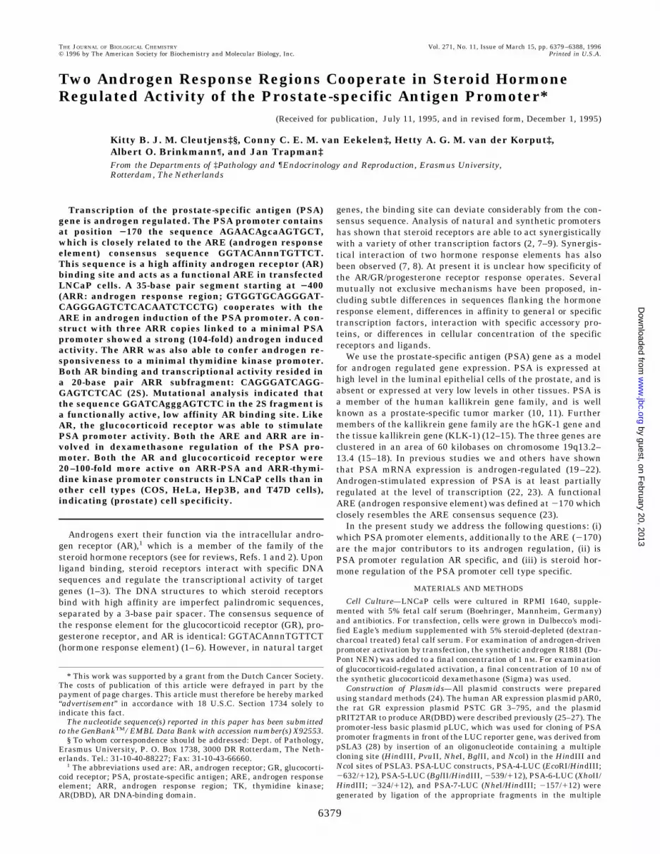
Two Androgen Response Regions Cooperate in Steroid Hormone Regulated Activity of the Prostate-specific Antigen Promoter* (Received for publication, July 11, 1995, and in revised form, December 1, 1995) Kitty B. J. M. Cleutjens‡§, Conny C. E. M. van Eekelen‡, Hetty A. G. M. van der Korput‡, Albert O. Brinkmann¶, and Jan Trapman‡ From the Departments of ‡Pathology and ¶Endocrinology and Reproduction, Erasmus University, Rotterdam, The Netherlands Transcription of the prostate-specific antigen (PSA) gene is androgen regulated. The PSA promoter contains at position 2170 the sequence AGAACAgcaAGTGCT, which is closely related to the ARE (androgen response element) consensus sequence GGTACAnnnTGTTCT. This sequence is a high affinity androgen receptor (AR) binding site and acts as a functional ARE in transfected LNCaP cells. A 35-base pair segment starting at 2400 (ARR: androgen response region; GTGGTGCAGGGAT- CAGGGAGTCTCACAATCTCCTG) cooperates with the ARE in androgen induction of the PSA promoter. A con- struct with three ARR copies linked to a minimal PSA promoter showed a strong (104-fold) androgen induced activity. The ARR was also able to confer androgen re- sponsiveness to a minimal thymidine kinase promoter. Both AR binding and transcriptional activity resided in a 20-base pair ARR subfragment: CAGGGATCAGG- GAGTCTCAC (2S). Mutational analysis indicated that the sequence GGATCAgggAGTCTC in the 2S fragment is a functionally active, low affinity AR binding site. Like AR, the glucocorticoid receptor was able to stimulate PSA promoter activity. Both the ARE and ARR are in- volved in dexamethasone regulation of the PSA pro- moter. Both the AR and glucocorticoid receptor were 20 –100-fold more active on ARR-PSA and ARR-thymi- dine kinase promoter constructs in LNCaP cells than in other cell types (COS, HeLa, Hep3B, and T47D cells), indicating (prostate) cell specificity. Androgens exert their function via the intracellular andro- gen receptor (AR), 1 which is a member of the family of the steroid hormone receptors (see for reviews, Refs. 1 and 2). Upon ligand binding, steroid receptors interact with specific DNA sequences and regulate the transcriptional activity of target genes (1–3). The DNA structures to which steroid receptors bind with high affinity are imperfect palindromic sequences, separated by a 3-base pair spacer. The consensus sequence of the response element for the glucocorticoid receptor (GR), pro- gesterone receptor, and AR is identical: GGTACAnnnTGTTCT (hormone response element) (1– 6). However, in natural target genes, the binding site can deviate considerably from the con- sensus sequence. Analysis of natural and synthetic promoters has shown that steroid receptors are able to act synergistically with a variety of other transcription factors (2, 7–9). Synergis- tical interaction of two hormone response elements has also been observed (7, 8). At present it is unclear how specificity of the AR/GR/progesterone receptor response operates. Several mutually not exclusive mechanisms have been proposed, in- cluding subtle differences in sequences flanking the hormone response element, differences in affinity to general or specific transcription factors, interaction with specific accessory pro- teins, or differences in cellular concentration of the specific receptors and ligands. We use the prostate-specific antigen (PSA) gene as a model for androgen regulated gene expression. PSA is expressed at high level in the luminal epithelial cells of the prostate, and is absent or expressed at very low levels in other tissues. PSA is a member of the human kallikrein gene family, and is well known as a prostate-specific tumor marker (10, 11). Further members of the kallikrein gene family are the hGK-1 gene and the tissue kallikrein gene (KLK-1) (12–15). The three genes are clustered in an area of 60 kilobases on chromosome 19q13.2– 13.4 (15–18). In previous studies we and others have shown that PSA mRNA expression is androgen-regulated (19 –22). Androgen-stimulated expression of PSA is at least partially regulated at the level of transcription (22, 23). A functional ARE (androgen responsive element) was defined at 2170 which closely resembles the ARE consensus sequence (23). In the present study we address the following questions: (i) which PSA promoter elements, additionally to the ARE (2170) are the major contributors to its androgen regulation, (ii) is PSA promoter regulation AR specific, and (iii) is steroid hor- mone regulation of the PSA promoter cell type specific. MATERIALS AND METHODS Cell Culture—LNCaP cells were cultured in RPMI 1640, supple- mented with 5% fetal calf serum (Boehringer, Mannheim, Germany) and antibiotics. For transfection, cells were grown in Dulbecco’s modi- fied Eagle’s medium supplemented with 5% steroid-depleted (dextran- charcoal treated) fetal calf serum. For examination of androgen-driven promoter activation by transfection, the synthetic androgen R1881 (Du- Pont NEN) was added to a final concentration of 1 nM. For examination of glucocorticoid-regulated activation, a final concentration of 10 nM of the synthetic glucocorticoid dexamethasone (Sigma) was used. Construction of Plasmids—All plasmid constructs were prepared using standard methods (24). The human AR expression plasmid pAR0, the rat GR expression plasmid PSTC GR 3–795, and the plasmid pRIT2TAR to produce AR(DBD) were described previously (25–27). The promoter-less basic plasmid pLUC, which was used for cloning of PSA promoter fragments in front of the LUC reporter gene, was derived from pSLA3 (28) by insertion of an oligonucleotide containing a multiple cloning site (HindIII, PvuII, NheI, BglII, and NcoI) in the HindIII and NcoI sites of PSLA3. PSA-LUC constructs, PSA-4-LUC (EcoRI/HindIII; 2632/112), PSA-5-LUC (BglII/HindIII, 2539/112), PSA-6-LUC (XhoII/ HindIII; 2324/112), and PSA-7-LUC (NheI/HindIII; 2157/112) were generated by ligation of the appropriate fragments in the multiple * This work was supported by a grant from the Dutch Cancer Society. The costs of publication of this article were defrayed in part by the payment of page charges. This article must therefore be hereby marked “advertisement” in accordance with 18 U.S.C. Section 1734 solely to indicate this fact. The nucleotide sequence(s) reported in this paper has been submitted to the GenBank TM /EMBL Data Bank with accession number(s) X92553. § To whom correspondence should be addressed: Dept. of Pathology, Erasmus University, P. O. Box 1738, 3000 DR Rotterdam, The Neth- erlands. Tel.: 31-10-40-88227; Fax: 31-10-43-66660. 1 The abbreviations used are: AR, androgen receptor; GR, glucocorti- coid receptor; PSA, prostate-specific antigen; ARE, androgen response element; ARR, androgen response region; TK, thymidine kinase; AR(DBD), AR DNA-binding domain. THE JOURNAL OF BIOLOGICAL CHEMISTRY Vol. 271, No. 11, Issue of March 15, pp. 6379 –6388, 1996 © 1996 by The American Society for Biochemistry and Molecular Biology, Inc. Printed in U.S.A. 6379 by guest, on February 20, 2013 www.jbc.org Downloaded from
-
Upload
independent -
Category
Documents
-
view
5 -
download
0
Transcript of Two androgen response regions cooperate in steroid hormone regulated activity of the...
Two Androgen Response Regions Cooperate in Steroid HormoneRegulated Activity of the Prostate-specific Antigen Promoter*
(Received for publication, July 11, 1995, and in revised form, December 1, 1995)
Kitty B. J. M. Cleutjens‡§, Conny C. E. M. van Eekelen‡, Hetty A. G. M. van der Korput‡,Albert O. Brinkmann¶, and Jan Trapman‡From the Departments of ‡Pathology and ¶Endocrinology and Reproduction, Erasmus University,Rotterdam, The Netherlands
Transcription of the prostate-specific antigen (PSA)gene is androgen regulated. The PSA promoter containsat position 2170 the sequence AGAACAgcaAGTGCT,which is closely related to the ARE (androgen responseelement) consensus sequence GGTACAnnnTGTTCT.This sequence is a high affinity androgen receptor (AR)binding site and acts as a functional ARE in transfectedLNCaP cells. A 35-base pair segment starting at 2400(ARR: androgen response region; GTGGTGCAGGGAT-CAGGGAGTCTCACAATCTCCTG) cooperates with theARE in androgen induction of the PSA promoter. A con-struct with three ARR copies linked to a minimal PSApromoter showed a strong (104-fold) androgen inducedactivity. The ARR was also able to confer androgen re-sponsiveness to a minimal thymidine kinase promoter.Both AR binding and transcriptional activity resided ina 20-base pair ARR subfragment: CAGGGATCAGG-GAGTCTCAC (2S). Mutational analysis indicated thatthe sequence GGATCAgggAGTCTC in the 2S fragment isa functionally active, low affinity AR binding site. LikeAR, the glucocorticoid receptor was able to stimulatePSA promoter activity. Both the ARE and ARR are in-volved in dexamethasone regulation of the PSA pro-moter. Both the AR and glucocorticoid receptor were20–100-fold more active on ARR-PSA and ARR-thymi-dine kinase promoter constructs in LNCaP cells than inother cell types (COS, HeLa, Hep3B, and T47D cells),indicating (prostate) cell specificity.
Androgens exert their function via the intracellular andro-gen receptor (AR),1 which is a member of the family of thesteroid hormone receptors (see for reviews, Refs. 1 and 2). Uponligand binding, steroid receptors interact with specific DNAsequences and regulate the transcriptional activity of targetgenes (1–3). The DNA structures to which steroid receptorsbind with high affinity are imperfect palindromic sequences,separated by a 3-base pair spacer. The consensus sequence ofthe response element for the glucocorticoid receptor (GR), pro-gesterone receptor, and AR is identical: GGTACAnnnTGTTCT(hormone response element) (1–6). However, in natural target
genes, the binding site can deviate considerably from the con-sensus sequence. Analysis of natural and synthetic promotershas shown that steroid receptors are able to act synergisticallywith a variety of other transcription factors (2, 7–9). Synergis-tical interaction of two hormone response elements has alsobeen observed (7, 8). At present it is unclear how specificity ofthe AR/GR/progesterone receptor response operates. Severalmutually not exclusive mechanisms have been proposed, in-cluding subtle differences in sequences flanking the hormoneresponse element, differences in affinity to general or specifictranscription factors, interaction with specific accessory pro-teins, or differences in cellular concentration of the specificreceptors and ligands.We use the prostate-specific antigen (PSA) gene as a model
for androgen regulated gene expression. PSA is expressed athigh level in the luminal epithelial cells of the prostate, and isabsent or expressed at very low levels in other tissues. PSA isa member of the human kallikrein gene family, and is wellknown as a prostate-specific tumor marker (10, 11). Furthermembers of the kallikrein gene family are the hGK-1 gene andthe tissue kallikrein gene (KLK-1) (12–15). The three genes areclustered in an area of 60 kilobases on chromosome 19q13.2–13.4 (15–18). In previous studies we and others have shownthat PSA mRNA expression is androgen-regulated (19–22).Androgen-stimulated expression of PSA is at least partiallyregulated at the level of transcription (22, 23). A functionalARE (androgen responsive element) was defined at 2170 whichclosely resembles the ARE consensus sequence (23).In the present study we address the following questions: (i)
which PSA promoter elements, additionally to the ARE (2170)are the major contributors to its androgen regulation, (ii) isPSA promoter regulation AR specific, and (iii) is steroid hor-mone regulation of the PSA promoter cell type specific.
MATERIALS AND METHODS
Cell Culture—LNCaP cells were cultured in RPMI 1640, supple-mented with 5% fetal calf serum (Boehringer, Mannheim, Germany)and antibiotics. For transfection, cells were grown in Dulbecco’s modi-fied Eagle’s medium supplemented with 5% steroid-depleted (dextran-charcoal treated) fetal calf serum. For examination of androgen-drivenpromoter activation by transfection, the synthetic androgen R1881 (Du-Pont NEN) was added to a final concentration of 1 nM. For examinationof glucocorticoid-regulated activation, a final concentration of 10 nM ofthe synthetic glucocorticoid dexamethasone (Sigma) was used.Construction of Plasmids—All plasmid constructs were prepared
using standard methods (24). The human AR expression plasmid pAR0,the rat GR expression plasmid PSTC GR 3–795, and the plasmidpRIT2TAR to produce AR(DBD) were described previously (25–27). Thepromoter-less basic plasmid pLUC, which was used for cloning of PSApromoter fragments in front of the LUC reporter gene, was derived frompSLA3 (28) by insertion of an oligonucleotide containing a multiplecloning site (HindIII, PvuII, NheI, BglII, and NcoI) in the HindIII andNcoI sites of PSLA3. PSA-LUC constructs, PSA-4-LUC (EcoRI/HindIII;2632/112), PSA-5-LUC (BglII/HindIII, 2539/112), PSA-6-LUC (XhoII/HindIII; 2324/112), and PSA-7-LUC (NheI/HindIII; 2157/112) weregenerated by ligation of the appropriate fragments in the multiple
* This work was supported by a grant from the Dutch Cancer Society.The costs of publication of this article were defrayed in part by thepayment of page charges. This article must therefore be hereby marked“advertisement” in accordance with 18 U.S.C. Section 1734 solely toindicate this fact.The nucleotide sequence(s) reported in this paper has been submitted
to the GenBankTM/EMBL Data Bank with accession number(s) X92553.§ To whom correspondence should be addressed: Dept. of Pathology,
Erasmus University, P. O. Box 1738, 3000 DR Rotterdam, The Neth-erlands. Tel.: 31-10-40-88227; Fax: 31-10-43-66660.
1 The abbreviations used are: AR, androgen receptor; GR, glucocorti-coid receptor; PSA, prostate-specific antigen; ARE, androgen responseelement; ARR, androgen response region; TK, thymidine kinase;AR(DBD), AR DNA-binding domain.
THE JOURNAL OF BIOLOGICAL CHEMISTRY Vol. 271, No. 11, Issue of March 15, pp. 6379–6388, 1996© 1996 by The American Society for Biochemistry and Molecular Biology, Inc. Printed in U.S.A.
6379
by guest, on February 20, 2013
ww
w.jbc.org
Dow
nloaded from

cloning site of pLUC.Constructs PSA-8 to PSA-11-LUC were obtained by exonuclease III
digestion of PSA-2-CAT (2632/112)(23) from the SalI site. After exo-nuclease III incubation according to the “Erase a base” protocol (Pro-mega, Madison, WI), the plasmid was digested with HindIII and thederived promoter fragments were ligated in the multiple cloning site ofpLUC. This resulted in the constructs PSA-8-LUC, starting at 2488,PSA-9-LUC, starting at 2456, PSA-10-LUC starting at 2395 and PSA-11-LUC, starting at 2376. Construct PSA-12-LUC was prepared byintroduction of a PstI site at position 2174 by polymerase chain reac-tion. The polymerase chain reaction product was digested with PstI andHindIII (112) and isolated from a 1.5% agarose gel. The isolated frag-ment was ligated in the PstI and HindIII sites of the pLUC multiplecloning site. One and three copies of the 2400 to 2366 oligomer werecloned in front of the PSA-12-LUC and the TK-LUC construct. Double-stranded oligonucleotides spanning ARR (2400 to 2366) 59-GATCCGT-GGTGCAGGGATCAGGGAGTCTCACAATCTCCTG-39 were insertedin the BamHI site of PSA-12-LUC and TK-LUC. Double strandedoligonucleotides spanning three copies of the ARR-1S, GTGGTGCAGG-GATCAGGGAG; ARR-2S, CAGGGATCAGGGAGTCTCAC; ARR-3S re-gion, GAGTCTACAATCTCCTG and the ARR-2S mutants (mutationsare underlined); ARR-2S-1, CAGGGGATGAGGGAGTCTCAC; ARR-2S-2, CAGGGATCAGGGACTCTCAC; and ARR-2S-3, CAGGGAT-CAGCGAGTCTCAC, containing SalI compatible ends were insertedin the SalI site of PSA-12-LUC. All constructs were verified bysequencing.Transfections—LNCaP cells were transfected according to the calcium
phosphate precipitation method essentially as described (29), using 1 3106 cells per 25-cm2 flask, 5 mg of the appropriate PSA-LUC construct,and where indicated 2.5 mg of pAR0 or PSTC GR 3–795 (GR expressionvector). After overnight incubation with the precipitate, the culture me-dium was removed and replaced by phosphate-buffered saline, containing15% glycerol (incubation for 90 s at room temperature). Subsequently,transfected cells were incubated in culture medium in the absence orpresence of the appropriate hormone (R1881 or dexamethasone) for atleast 24 h. Transfections were performed at least three times in duplicate,using at least two independent plasmid isolates.Luciferase activities were corrected for variations in protein concen-
trations within the 100 ml of cell extracts. Luciferase activities andrelative induction factors are expressed as mean and standard error(S.E.).Luciferase Assay—Cells were washed once with phosphate-buffered
saline and lysed in 300 ml of lysis buffer (25 mM Tris phosphate, pH 7.8,8 mM MgCl2, 1 mM dithiothreitol, 1% Triton X-100, 15% glycerol). Next,100 ml of Luciferin (0.25 mM) (Sigma), 0.25 mM ATP was added to 100 mlof each extract, and luciferase activity was measured in a LUMAC 2500M Biocounter (LUMAC, Landgraaf, The Netherlands). After a delay of2 s (according to supplier), the light emission during 5 s was recorded.Gel Retardation Analysis—Nuclear extracts were prepared as de-
scribed by Dignam et al. (30). Coupled transcription/translation of hu-man AR cDNA cloned in BluescriptII- KS (31) was carried out accordingto the protocol of the manufacturer (Promega). AR synthesis was in thepresence of 10 mM ZnCl2. Production in Escherichia coli, and purifica-tion of AR(DBD) was done as described previously (27).Double stranded oligonucleotide probes used in gel retardations are
as follows.
ARE: 59 GATCCTTGCAGAACAGCAAGTGCTAGCTG 39GAACGTCTTGTCGTTCACGATCGACCTAG
ARR: 59 GATCCGTGGTGCAGGGATCAGGGAGTCTCACAATCTCCTG 39GCACCACGTCCCTAGTCCCTCAGAGTGTTAGAGGACCTAG
ARR-1S: 59 GATCCGTGGTGCAGGGATCAGGGAG 39GCACCACGTCCCTAGTCCCTCCTAG
ARR-2S: 59 GATCCAGGGATCAGGGAGTCTCACG 39GTCCCTAGTCCCTCAGAGTGCCTAG
ARR-3S: 59 GATCCGAGTCTCACAATCTCCTGAG 39GCTCAGAGTGTTAGAGGACTCCTAG
ARR-2S-1: 59 GATCCAGGGATGAGGGAGTCTCACG 39GTCCCTACTCCCTCAGAGTGCCTAG
ARR-2S-2: 59 GATCCAGGGATCAGGGACTCTCACG 39GTCCCTAGTCCCTGAGAGTGCCTAG
ARR-2S-3: 59 GATCCAGGGATCAGCGAGTCTCACG 39GTCCCTAGTCGCTCAGAGTGCCTAG
ARR-2S-4: 59 GATCCAGGGATCAGGGAGTTCCACG 39GTCCCTAGTCCCTCAAGGTGCCTAG
ARR-2S-5: 59 GATCCAGGGAACAGGGTGTTCCACG 39GTCCCTTGTCCCACAAGGTGCCTAG
Oligonucleotides 1–10
Probes were filled in with Moloney murine leukemia virus-reversetranscriptase in the presence of [a-32P]dATP, and subsequently isolatedfrom nondenaturing polyacrylamide gel. For gel retardation assays,20–50 3 103 cpm of each probe was added to 20 ml of reaction mixture,containing 2 mg of poly(dI-dC), 2 mg of bovine serum albumin, 10 mM
ZnCl2, 1 mM dithiothreitol, and 2 ml of 10 3 binding buffer (100 mM
Hepes, pH 7.6, 300 mM KCl, 62.5 mM MgCl2, and 30% glycerol), and inindicated cases 10 mg of LNCaP nuclear protein, in vitro translated AR(7–10 fmol) or AR(DBD) (5 pmol). In experiments using the AR antibodySp197 (epitope amino acid residues 1–20), 0.1-ml portions of antiserumwere added to the reaction mixture (32). Incubation was for 30 min atroom temperature. In addition to oligonucleotides described above (100-fold excess of) double-stranded oligonucleotides containing a C/EBPbinding site, 59-GACCTTACCACTTTCACAATCTGCTAG-39 (33), and aglucocorticoid response element, 59-TCGACTGTACAGGATGTTCTAG-CTACT-39 (Promega), were used in competition experiments. Sampleswere loaded on a 4% polyacrylamide (19:1) gel and electrophoresed in a50 mM Tris-HCl, 41.5 mM boric acid, 0.5 mM EDTA buffer for 2 h at 150V and room temperature. Subsequently, gels were fixed, dried, andexposed to x-ray film.
RESULTS
Deletion Mapping of the PSA Promoter: Effect of AndrogenReceptor Overexpression—In a previous study we analyzed an-drogen regulation of the PSA promoter in COS cells which wereco-transfected with several different PSA promoter-chloram-phenicol acetyltransferase reporter gene constructs and the ARexpression vector pAR0 (23). This resulted in the functionalcharacterization of an ARE (AGAACAgcaAGTGCT), which isclosely related to the consensus sequence, at position 2170,and the identification of a second region, from 2539 to 2324,important for PSA promoter activity.Essentially the same data were obtained in LNCaP cells
which endogenously express the AR and PSA gene (Fig. 1A).Transfection of PSA-4-LUC (2632/112) or PSA-5-LUC (2539/112) resulted in an approximately 7-fold higher LUC activityin the presence of R1881 than in its absence. Similar resultswere found with constructs containing longer promoter frag-ments (up to approximately 4 kilobases (data not shown)).Removal of the 2539 to 2324 fragment (PSA-6-LUC) caused a3.5-fold drop in relative induction. Subsequent removal of the2324 to 2157 region (containing the ARE sequence at position2170) resulted in the complete abolishment of androgen induc-tion (construct PSA-7-LUC).To investigate in more detail sequences in the 2539 to 2324
region important for PSA promoter activity, a series of exonu-clease III deletions was generated (see Fig. 1B for PSA pro-moter fragments in different constructs). Transfection ofLNCaP cells with constructs PSA-8-LUC, PSA-9-LUC, andPSA-10-LUC resulted in a high (6–7-fold) androgen-regulatedPSA promoter activity, which is comparable to that of thePSA-5 construct. Importantly, construct PSA-11-LUC, startingat 2376, showed a low (2.2-fold), androgen-induced activity,which is identical to that of PSA-6 (starting at 2324). Theseresults indicated sequences in the region 2395 to 2376 to beessential for high androgen-induced PSA promoter activity.Co-transfection with the AR expression plasmid pAR0 re-
sulted in considerably higher PSA promoter activity, both inabsolute values as well as in relative induction levels withoutaffecting the significance of the 2324 to 2157 region contain-ing the ARE at position 2170, and the 2395 to 2376 region(Fig. 1A). The co-transfection experiments showed again thattruncation of the promoter from 2395 to 2376 gives rise to amarkedly lower androgen induced activity (compare PSA-10and PSA-11); deletion of the 2324 to 2157 region again re-sulted in a complete loss of androgen inducibility of the PSApromoter (compare PSA-6 and PSA-7).Mutations in the ARE at 2170 in construct PSA-4-LUC
resulted in an almost complete inhibition of androgen activa-tion of the PSA promoter (Ref. 23, and data not shown), indi-
Androgen Regulation of the PSA Promoter6380
by guest, on February 20, 2013
ww
w.jbc.org
Dow
nloaded from
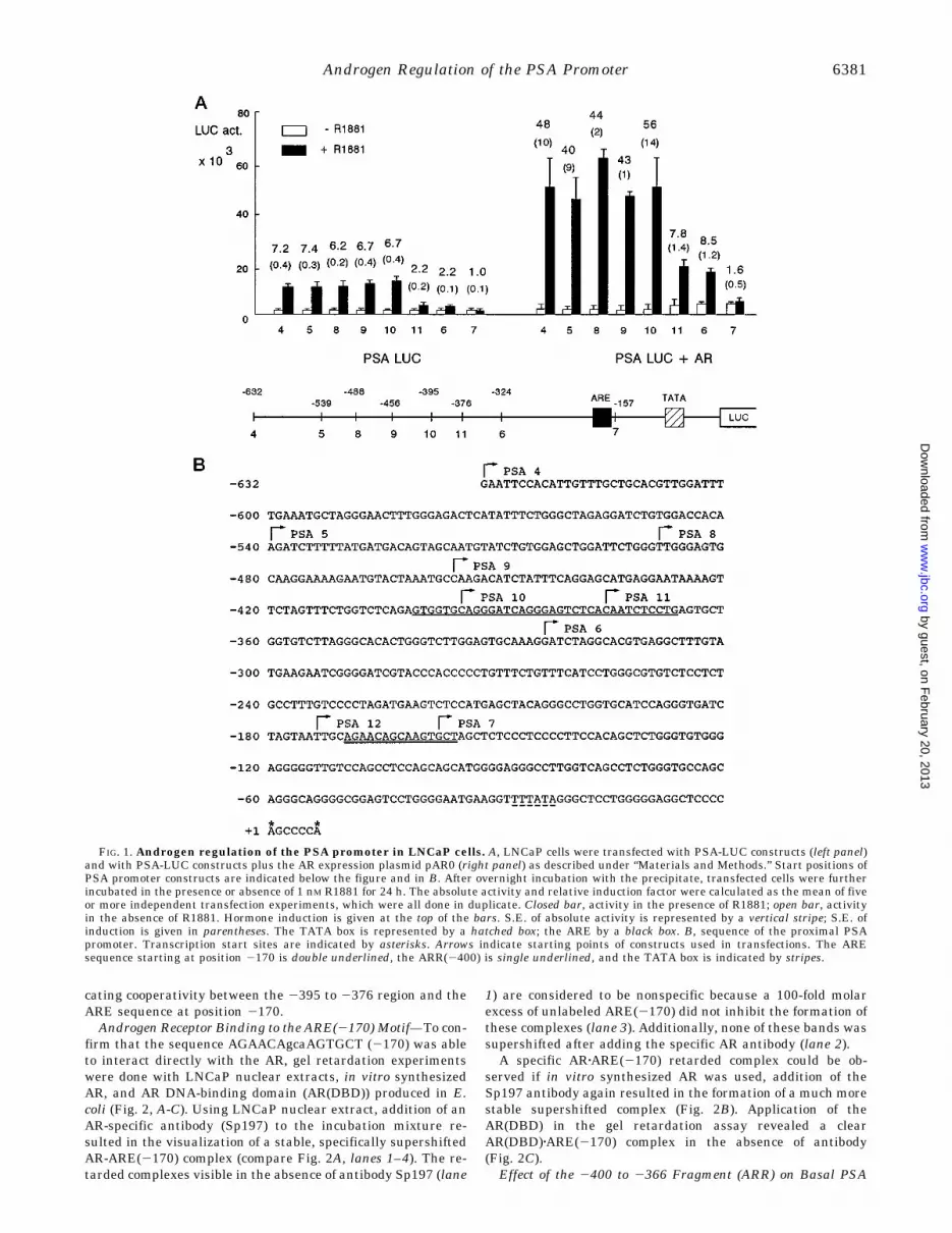
cating cooperativity between the 2395 to 2376 region and theARE sequence at position 2170.Androgen Receptor Binding to the ARE(2170) Motif—To con-
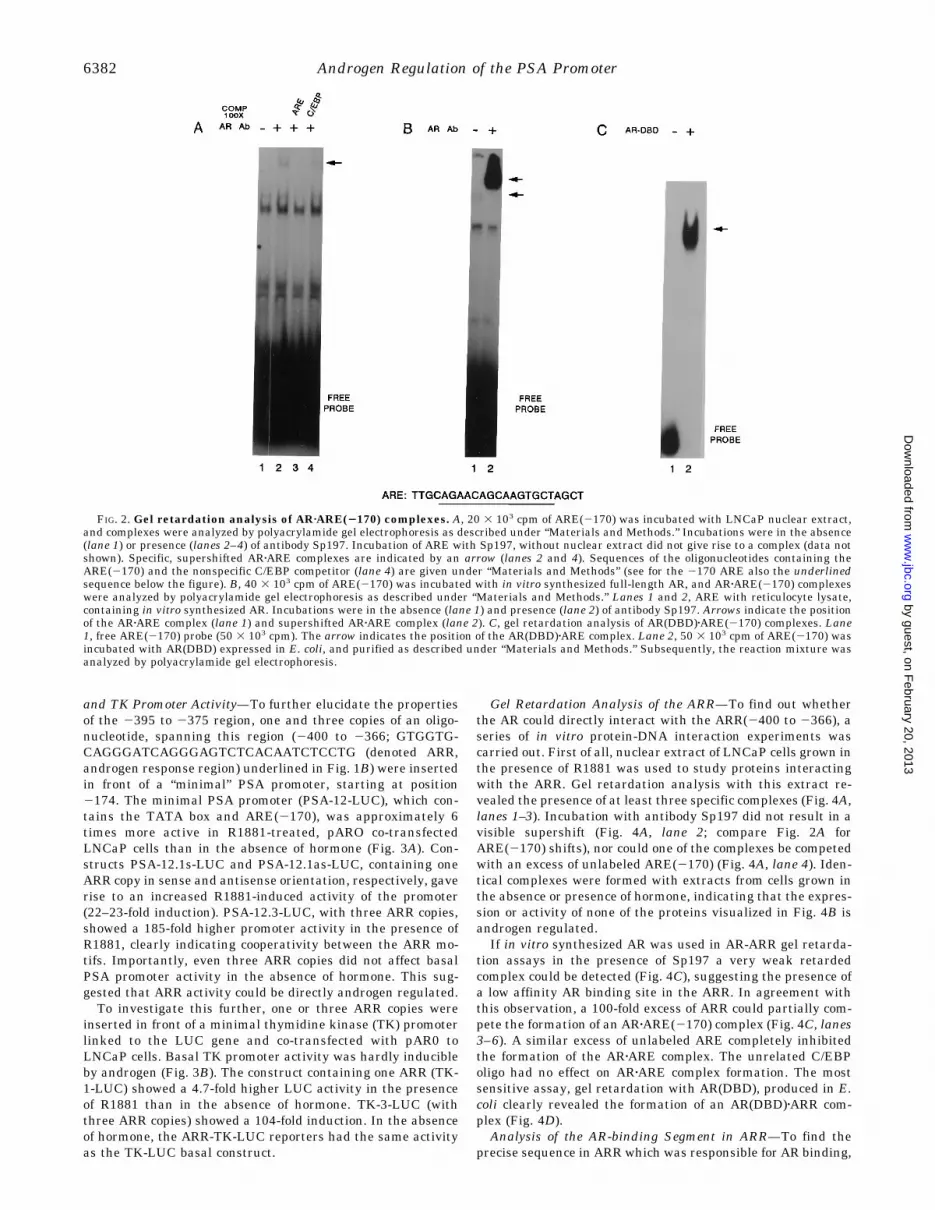
firm that the sequence AGAACAgcaAGTGCT (2170) was ableto interact directly with the AR, gel retardation experimentswere done with LNCaP nuclear extracts, in vitro synthesizedAR, and AR DNA-binding domain (AR(DBD)) produced in E.coli (Fig. 2, A-C). Using LNCaP nuclear extract, addition of anAR-specific antibody (Sp197) to the incubation mixture re-sulted in the visualization of a stable, specifically supershiftedAR-ARE(2170) complex (compare Fig. 2A, lanes 1–4). The re-tarded complexes visible in the absence of antibody Sp197 (lane
1) are considered to be nonspecific because a 100-fold molarexcess of unlabeled ARE(2170) did not inhibit the formation ofthese complexes (lane 3). Additionally, none of these bands wassupershifted after adding the specific AR antibody (lane 2).A specific ARzARE(2170) retarded complex could be ob-
served if in vitro synthesized AR was used, addition of theSp197 antibody again resulted in the formation of a much morestable supershifted complex (Fig. 2B). Application of theAR(DBD) in the gel retardation assay revealed a clearAR(DBD)zARE(2170) complex in the absence of antibody(Fig. 2C).Effect of the 2400 to 2366 Fragment (ARR) on Basal PSA
FIG. 1. Androgen regulation of the PSA promoter in LNCaP cells. A, LNCaP cells were transfected with PSA-LUC constructs (left panel)and with PSA-LUC constructs plus the AR expression plasmid pAR0 (right panel) as described under “Materials and Methods.” Start positions ofPSA promoter constructs are indicated below the figure and in B. After overnight incubation with the precipitate, transfected cells were furtherincubated in the presence or absence of 1 nM R1881 for 24 h. The absolute activity and relative induction factor were calculated as the mean of fiveor more independent transfection experiments, which were all done in duplicate. Closed bar, activity in the presence of R1881; open bar, activityin the absence of R1881. Hormone induction is given at the top of the bars. S.E. of absolute activity is represented by a vertical stripe; S.E. ofinduction is given in parentheses. The TATA box is represented by a hatched box; the ARE by a black box. B, sequence of the proximal PSApromoter. Transcription start sites are indicated by asterisks. Arrows indicate starting points of constructs used in transfections. The AREsequence starting at position 2170 is double underlined, the ARR(2400) is single underlined, and the TATA box is indicated by stripes.
Androgen Regulation of the PSA Promoter 6381
by guest, on February 20, 2013
ww
w.jbc.org
Dow
nloaded from
and TK Promoter Activity—To further elucidate the propertiesof the 2395 to 2375 region, one and three copies of an oligo-nucleotide, spanning this region (2400 to 2366; GTGGTG-CAGGGATCAGGGAGTCTCACAATCTCCTG (denoted ARR,androgen response region) underlined in Fig. 1B) were insertedin front of a “minimal” PSA promoter, starting at position2174. The minimal PSA promoter (PSA-12-LUC), which con-tains the TATA box and ARE(2170), was approximately 6times more active in R1881-treated, pARO co-transfectedLNCaP cells than in the absence of hormone (Fig. 3A). Con-structs PSA-12.1s-LUC and PSA-12.1as-LUC, containing oneARR copy in sense and antisense orientation, respectively, gaverise to an increased R1881-induced activity of the promoter(22–23-fold induction). PSA-12.3-LUC, with three ARR copies,showed a 185-fold higher promoter activity in the presence ofR1881, clearly indicating cooperativity between the ARR mo-tifs. Importantly, even three ARR copies did not affect basalPSA promoter activity in the absence of hormone. This sug-gested that ARR activity could be directly androgen regulated.To investigate this further, one or three ARR copies were
inserted in front of a minimal thymidine kinase (TK) promoterlinked to the LUC gene and co-transfected with pAR0 toLNCaP cells. Basal TK promoter activity was hardly inducibleby androgen (Fig. 3B). The construct containing one ARR (TK-1-LUC) showed a 4.7-fold higher LUC activity in the presenceof R1881 than in the absence of hormone. TK-3-LUC (withthree ARR copies) showed a 104-fold induction. In the absenceof hormone, the ARR-TK-LUC reporters had the same activityas the TK-LUC basal construct.
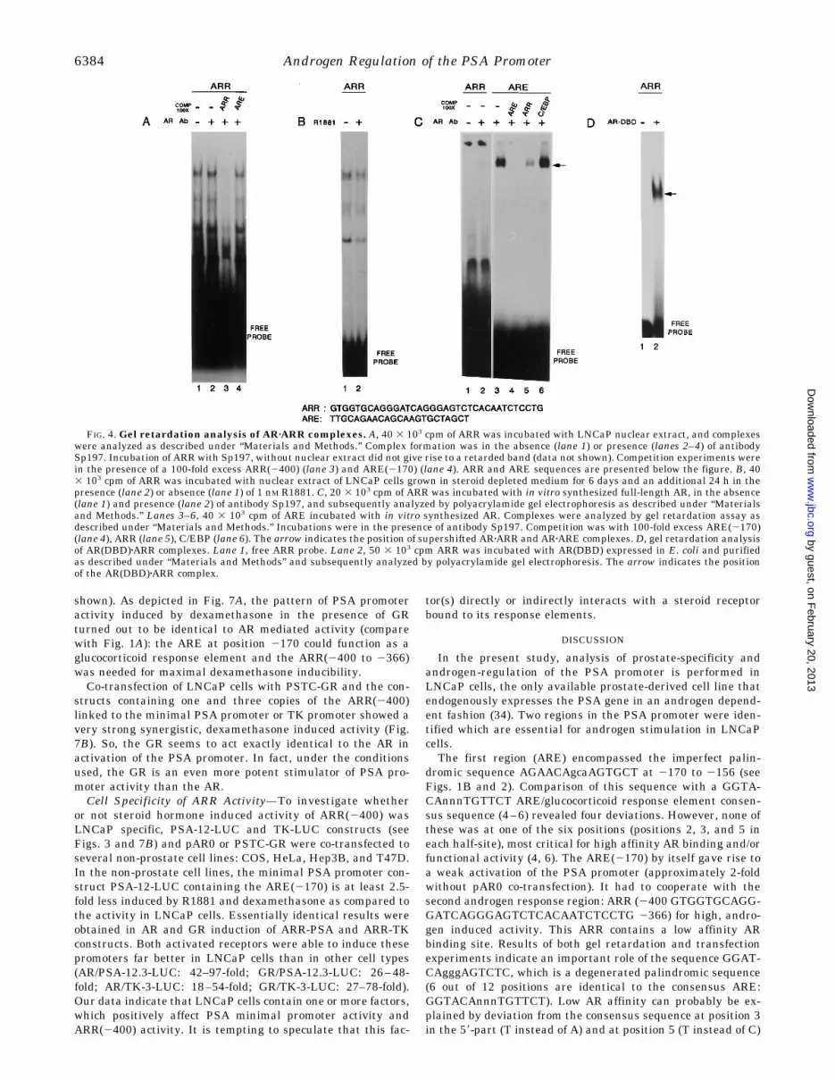
Gel Retardation Analysis of the ARR—To find out whetherthe AR could directly interact with the ARR(2400 to 2366), aseries of in vitro protein-DNA interaction experiments wascarried out. First of all, nuclear extract of LNCaP cells grown inthe presence of R1881 was used to study proteins interactingwith the ARR. Gel retardation analysis with this extract re-vealed the presence of at least three specific complexes (Fig. 4A,lanes 1–3). Incubation with antibody Sp197 did not result in avisible supershift (Fig. 4A, lane 2; compare Fig. 2A forARE(2170) shifts), nor could one of the complexes be competedwith an excess of unlabeled ARE(2170) (Fig. 4A, lane 4). Iden-tical complexes were formed with extracts from cells grown inthe absence or presence of hormone, indicating that the expres-sion or activity of none of the proteins visualized in Fig. 4B isandrogen regulated.If in vitro synthesized AR was used in AR-ARR gel retarda-
tion assays in the presence of Sp197 a very weak retardedcomplex could be detected (Fig. 4C), suggesting the presence ofa low affinity AR binding site in the ARR. In agreement withthis observation, a 100-fold excess of ARR could partially com-pete the formation of an ARzARE(2170) complex (Fig. 4C, lanes3–6). A similar excess of unlabeled ARE completely inhibitedthe formation of the ARzARE complex. The unrelated C/EBPoligo had no effect on ARzARE complex formation. The mostsensitive assay, gel retardation with AR(DBD), produced in E.coli clearly revealed the formation of an AR(DBD)zARR com-plex (Fig. 4D).Analysis of the AR-binding Segment in ARR—To find the
precise sequence in ARR which was responsible for AR binding,
FIG. 2. Gel retardation analysis of ARzARE(2170) complexes. A, 20 3 103 cpm of ARE(2170) was incubated with LNCaP nuclear extract,and complexes were analyzed by polyacrylamide gel electrophoresis as described under “Materials and Methods.” Incubations were in the absence(lane 1) or presence (lanes 2–4) of antibody Sp197. Incubation of ARE with Sp197, without nuclear extract did not give rise to a complex (data notshown). Specific, supershifted ARzARE complexes are indicated by an arrow (lanes 2 and 4). Sequences of the oligonucleotides containing theARE(2170) and the nonspecific C/EBP competitor (lane 4) are given under “Materials and Methods” (see for the 2170 ARE also the underlinedsequence below the figure). B, 40 3 103 cpm of ARE(2170) was incubated with in vitro synthesized full-length AR, and ARzARE(2170) complexeswere analyzed by polyacrylamide gel electrophoresis as described under “Materials and Methods.” Lanes 1 and 2, ARE with reticulocyte lysate,containing in vitro synthesized AR. Incubations were in the absence (lane 1) and presence (lane 2) of antibody Sp197. Arrows indicate the positionof the ARzARE complex (lane 1) and supershifted ARzARE complex (lane 2). C, gel retardation analysis of AR(DBD)zARE(2170) complexes. Lane1, free ARE(2170) probe (50 3 103 cpm). The arrow indicates the position of the AR(DBD)zARE complex. Lane 2, 50 3 103 cpm of ARE(2170) wasincubated with AR(DBD) expressed in E. coli, and purified as described under “Materials and Methods.” Subsequently, the reaction mixture wasanalyzed by polyacrylamide gel electrophoresis.
Androgen Regulation of the PSA Promoter6382
by guest, on February 20, 2013
ww
w.jbc.org
Dow
nloaded from

the ARR was subdivided into three partially overlapping frag-ments. Gel retardation assay with AR(DBD) and the three ARRsubfragments 1S (GTGGTGCAGGGATCAGGGAG (2400 to2381)), 2S (CAGGGATCAGGGAGTCTCAC (2394 to 2375)),and 3S (GAGTCTCACAATCTCCTG (2383 to 2366)) showedthat 2S contained the AR binding sequence, no or extremelyweak binding was detected with 1S and 3S (Fig. 5A).Next, three copies of 1S, 2S, and 3S were inserted in front of
the minimal PSA promoter construct PSA-12-LUC and co-transfected with pAR0 to LNCaP cells. The construct contain-ing three copies of the 1S region (PSA-1S-LUC) gave rise to a9.6-fold higher activity in the presence of R1881 than in theabsence (Fig. 5B). Construct PSA-2S-LUC, containing threecopies of 2S gave rise to a 128-fold higher activity upon R1881treatment. The construct with three copies of 3S (PSA-3S-LUC)produced a 7.1-fold higher activity in the presence of R1881. So,there is complete concordance between the presence of ARbinding and functional, hormone-dependent enhancer activityin 2S, and the absence of these activities in 1S and 3S.Analysis of the AR Binding Site in ARR-2S—Sequence align-
ment showed that in 2S the highest percentage of homology tothe ARE consensus sequence is in the sequence GGATCAgg-gAGTCTC. This sequence deviates in 2 out of 6 most essentialpositions, (positions 2, 3, and 5 in each half-site, underlined)and overall in 6 out of 12 positions from the ARE consensusGGTACAnnnTGTTCT. To test whether this sequence couldindeed be responsible for low affinity AR binding, gel retarda-tion analyses were performed with five ARR-2S mutants (mu-tations are underlined). Two mutants expected to decrease ARaffinity to the putative AR binding site, ARR-2S-1 (GGAT-
GAgggAGTCTC) and ARR-2S-2 (GGATCAgggACTCTC), andone presumed silent mutant, ARR-2S-3 (GGATCAgcgAG-TCTC), were tested for their AR binding capacity (Fig. 6A). Gelretardation experiments confirmed our hypothesis: AR bindingto ARR-2S-1 and ARR-2S-2 was almost completely abolished;ARR-2S-3 did not show a marked difference in AR affinity ascompared to ARR-2S. AR(DBD) gel retardation with mutantswith a higher homology to the consensus ARE sequence (ARR-2S-4 and ARR-2S-5: GGATCAgggAGTTCC and GGAACAgggT-GTTCC, respectively) substantiated these findings.Next, mutated 2S elements were tested in LNCaP cells for
enhancer activity. To this end, three of the mutant oligonucleo-tides, 2S-1, 2S-2 (both abolishing AR-binding), and 2S-3 (spacermutation), were cloned in front of the minimal PSA promoterconstruct PSA-12-LUC. PSA-2S-1-LUC and PSA-2S-2-LUCwere hardly more active than PSA-12-LUC upon R1881 induc-tion in AR co-transfected LNCaP cells (Fig. 6B). ConstructPSA-2S-3-LUC showed a 100-fold higher activity in the pres-ence than in the absence of R1881. These data strongly suggestthe importance of the GGATCAgggAGTCTCmotif in androgen-regulated activity of the ARR in the PSA promoter.Effect of Glucocorticoid Receptor Overexpression on PSA Pro-
moter Activity—Because on the one hand the DNA motif forhigh affinity AR and GR binding is identical and on the otherhand AR and GR might show specificity on individual promot-ers, we investigated whether GR was able to mediate PSApromoter activity. LNCaP cells were co-transfected with theGR expression plasmid PSTC-GR and selected PSA-LUC con-structs. Without GR co-transfection, no response of the differ-ent PSA promoters to dexamethasone was observed (data not
FIG. 3. A, effect of the 2400 to 2366region (ARR) on basal PSA promoter ac-tivity in LNCaP cells overexpressing theAR. The ARR is represented by a hatchedtriangle, the ARE by a black box, and theTATA element by a hatched box. B, effectof the 2400 to 2366 region of the PSApromoter (ARR) on TK promoter activityin LNCaP cells overexpressing AR. TheARR is represented by a hatched triangle.The mean of luciferase activity and rela-tive induction levels are from four inde-pendent, duplicate experiments. Experi-mental details are identical to thosedescribed in Fig. 1A. The ARR sequence issingle underlined in Fig. 1B.
Androgen Regulation of the PSA Promoter 6383
by guest, on February 20, 2013
ww
w.jbc.org
Dow
nloaded from
shown). As depicted in Fig. 7A, the pattern of PSA promoteractivity induced by dexamethasone in the presence of GRturned out to be identical to AR mediated activity (comparewith Fig. 1A): the ARE at position 2170 could function as aglucocorticoid response element and the ARR(2400 to 2366)was needed for maximal dexamethasone inducibility.Co-transfection of LNCaP cells with PSTC-GR and the con-
structs containing one and three copies of the ARR(2400)linked to the minimal PSA promoter or TK promoter showed avery strong synergistic, dexamethasone induced activity (Fig.7B). So, the GR seems to act exactly identical to the AR inactivation of the PSA promoter. In fact, under the conditionsused, the GR is an even more potent stimulator of PSA pro-moter activity than the AR.Cell Specificity of ARR Activity—To investigate whether
or not steroid hormone induced activity of ARR(2400) wasLNCaP specific, PSA-12-LUC and TK-LUC constructs (seeFigs. 3 and 7B) and pAR0 or PSTC-GR were co-transfected toseveral non-prostate cell lines: COS, HeLa, Hep3B, and T47D.In the non-prostate cell lines, the minimal PSA promoter con-struct PSA-12-LUC containing the ARE(2170) is at least 2.5-fold less induced by R1881 and dexamethasone as compared tothe activity in LNCaP cells. Essentially identical results wereobtained in AR and GR induction of ARR-PSA and ARR-TKconstructs. Both activated receptors were able to induce thesepromoters far better in LNCaP cells than in other cell types(AR/PSA-12.3-LUC: 42–97-fold; GR/PSA-12.3-LUC: 26–48-fold; AR/TK-3-LUC: 18–54-fold; GR/TK-3-LUC: 27–78-fold).Our data indicate that LNCaP cells contain one or more factors,which positively affect PSA minimal promoter activity andARR(2400) activity. It is tempting to speculate that this fac-
tor(s) directly or indirectly interacts with a steroid receptorbound to its response elements.
DISCUSSION
In the present study, analysis of prostate-specificity andandrogen-regulation of the PSA promoter is performed inLNCaP cells, the only available prostate-derived cell line thatendogenously expresses the PSA gene in an androgen depend-ent fashion (34). Two regions in the PSA promoter were iden-tified which are essential for androgen stimulation in LNCaPcells.The first region (ARE) encompassed the imperfect palin-
dromic sequence AGAACAgcaAGTGCT at 2170 to 2156 (seeFigs. 1B and 2). Comparison of this sequence with a GGTA-CAnnnTGTTCT ARE/glucocorticoid response element consen-sus sequence (4–6) revealed four deviations. However, none ofthese was at one of the six positions (positions 2, 3, and 5 ineach half-site), most critical for high affinity AR binding and/orfunctional activity (4, 6). The ARE(2170) by itself gave rise toa weak activation of the PSA promoter (approximately 2-foldwithout pAR0 co-transfection). It had to cooperate with thesecond androgen response region: ARR (2400 GTGGTGCAGG-GATCAGGGAGTCTCACAATCTCCTG 2366) for high, andro-gen induced activity. This ARR contains a low affinity ARbinding site. Results of both gel retardation and transfectionexperiments indicate an important role of the sequence GGAT-CAgggAGTCTC, which is a degenerated palindromic sequence(6 out of 12 positions are identical to the consensus ARE:GGTACAnnnTGTTCT). Low AR affinity can probably be ex-plained by deviation from the consensus sequence at position 3in the 59-part (T instead of A) and at position 5 (T instead of C)
FIG. 4. Gel retardation analysis of ARzARR complexes. A, 40 3 103 cpm of ARR was incubated with LNCaP nuclear extract, and complexeswere analyzed as described under “Materials and Methods.” Complex formation was in the absence (lane 1) or presence (lanes 2–4) of antibodySp197. Incubation of ARR with Sp197, without nuclear extract did not give rise to a retarded band (data not shown). Competition experiments werein the presence of a 100-fold excess ARR(2400) (lane 3) and ARE(2170) (lane 4). ARR and ARE sequences are presented below the figure. B, 403 103 cpm of ARR was incubated with nuclear extract of LNCaP cells grown in steroid depleted medium for 6 days and an additional 24 h in thepresence (lane 2) or absence (lane 1) of 1 nM R1881. C, 20 3 103 cpm of ARR was incubated with in vitro synthesized full-length AR, in the absence(lane 1) and presence (lane 2) of antibody Sp197, and subsequently analyzed by polyacrylamide gel electrophoresis as described under “Materialsand Methods.” Lanes 3–6, 40 3 103 cpm of ARE incubated with in vitro synthesized AR. Complexes were analyzed by gel retardation assay asdescribed under “Materials and Methods.” Incubations were in the presence of antibody Sp197. Competition was with 100-fold excess ARE(2170)(lane 4), ARR (lane 5), C/EBP (lane 6). The arrow indicates the position of supershifted ARzARR and ARzARE complexes. D, gel retardation analysisof AR(DBD)zARR complexes. Lane 1, free ARR probe. Lane 2, 50 3 103 cpm ARR was incubated with AR(DBD) expressed in E. coli and purifiedas described under “Materials and Methods” and subsequently analyzed by polyacrylamide gel electrophoresis. The arrow indicates the positionof the AR(DBD)zARR complex.
Androgen Regulation of the PSA Promoter6384
by guest, on February 20, 2013
ww
w.jbc.org
Dow
nloaded from

in the 39-part of the palindromic sequence.Although several androgen-regulated genes have been de-
scribed, only few functional AREs have been studied in detail.The mechanism of PSA promoter regulation by androgensseems different from other genes studied in this respect. In themouse vas deferens protein gene promoter, two ARE sequencesare present (35). Only the proximal ARE is functionally active,and no synergism between the two AREs was detected. In the
probasin promoter, also two fragments, which are importantfor androgen-regulation of the promoter and which bind AR,can be found (36, 37). Although both sequences are able tointeract with AR outside the probasin promoter context, bothAR binding sites are individually functionally inactive, eventhree copies of the two separated AR binding sites fail to giverise to androgen-induced reporter gene activity. This is in con-trast to the AR binding sites in the PSA promoter, ARE(2170)and ARR-2S, which are clearly independently active, and whenmultimerized act synergistically (Ref. 23, and this study).Three candidate AREs have been found in the C3 gene; only
one of them, Core II, C, is functionally active in transfectionexperiments (38–40). Activity of Core II is strongly enhancedby surrounding sequences including candidate OCT-1 and NF-Ibinding sites (41).Functional synergism between multiple ARE-like sequences
and binding sites for other transcription factors has been foundin the complex enhancer elements of the 20-kDa protein andSlp genes. In the androgen responsive enhancer in the pro-moter of the Slp gene, three tandemly repeated hormone re-sponse element-like sequences are present (42). Additionally,several non-receptor binding elements contribute to the char-acteristic androgen response of this complex enhancer (43). Thefirst intron of the 20-kDa protein gene contains a cluster ofthree ARE-like half-sites spanning a 39-base pair fragment(N39) which shows AR binding and confers weak androgenresponsiveness to a heterologous promoter. Additional se-quences surrounding this cluster of ARE-like sequences areneeded for full activity of this enhancer (44). These additionalsequences include a region (D2) that shows high AR bindingaffinity and androgen induced transcriptional activity, but nocandidate ARE sequences were identified. Taking into consid-eration the results obtained in our present study, which showthat multiple weak AR binding sites can give rise to strongandrogen inducibility, weak AREs might be postulated to bepresent in the D2 region. Like the Slp and C3 promoter, non-steroid receptor factors, including OCT-1 are supposed to beinvolved in establishment of the full AR specific response of the20-kDa protein promoter.The complex promoters of the Slp and 20-kDa protein genes
show an AR, but no GR response in transfection assays, al-though both AR and GR are able to bind and induce activity ofsmaller enhancer fragments (42, 44). Both AR and GR are ableto stimulate probasin promoter activity, however, AR is mark-edly more potent than GR in this respect (36). This in contrastto results on the PSA promoter presented in this study. Co-transfection of LNCaP cells with PSA-LUC constructs and aGR expression plasmid showed that GR can replace AR in high,steroid hormone-regulated PSA promoter activation. This isnot only related to the ARE(2170), but is also true for the ARRat 2400. Preliminary evidence indicates that GR is also able toactivate the promoter of the endogenous PSA gene in LNCaPcells.2 From these findings we conclude that the apparent ARspecificity of the PSA promoter in LNCaP cells is due to theabsence of other members of the steroid receptor family in thisparticular cell line. The absence of PSA expression in GR or ARpositive, non-prostate cells must be explained by additionalinhibitory mechanisms in these cells or absence of other essen-tial regulatory proteins involved in PSA expression.Transfection experiments further indicated that ARR(2400)
and minimal PSA promoter activity (including ARE(2170)) arecell dependent. We studied this aspect in more detail forARR(2400). Even in the presence of high levels of AR and GR,ARR(2400) activity in LNCaP cells is much higher than in the
2 K. B. J. M. Cleutjens, unpublished data.
FIG. 5. Analysis of the AR binding segment in ARR. A, gelretardation analysis of AR(DBD)zARR subfragment complexes. 50 3103 cpm of ARR-1S (lanes 1 and 2), -2S (lanes 3 and 4), and -3S (lanes5 and 6) were incubated with AR(DBD) and analyzed by gel retardationassay as described under “Materials and Methods.” 1S, 2S, and 3Ssequences are shown below the figure. The arrow indicates the positionof the AR(DBD)zARR-2S complex. B, effect of the ARR-1S (2400 to2381), -2S (2394 to 2375), and -3S (2383 to 2366) on PSA basalpromoter activity in LNCaP cells overexpressing the AR. The ARRsubfragments are represented by triangles, the ARE(2170) by a blackbox, and the TATA box by a hatched box. Mean values and S.E. are fromthree independent, duplicate experiments. Further experimental de-tails are identical to those described in the legend to Fig. 1A.
Androgen Regulation of the PSA Promoter 6385
by guest, on February 20, 2013
ww
w.jbc.org
Dow
nloaded from

FIG. 6.Mutation analysis of the ARR-2S segment. A, gel retardation analysis of ARR-2S and ARR-2S mutants with AR(DBD). 50 3 103 cpmof ARR-2S (lanes 1 and 2), ARR-2S-1 (lanes 3 and 4), -2S-2 (lanes 5 and 6), -2S-3 (lanes 7 and 8), -2S-4 (lanes 9 and 10), and -2S-5 (lanes 11 and12) were incubated with AR(DBD) and analyzed by gel retardation assay as described under “Materials and Methods.” Sequences of ARR-2S andARR-2S mutants are shown below the figure. The arrow indicates the position of the AR(DBD)-probe complex. B, effect of the ARR-2S-1, -2S-2, and-2S-3 sequences on PSA basal promoter activity in LNCaP cells overexpressing the AR. ARR-2S and mutant ARR-2S oligo’s are represented bytriangles, the ARE(2170) by a black box, and the TATA box by a hatched box. Mean values of luciferase activity and induction levels and theirrespective S.E. are from three independent, duplicate experiments. Experimental details are as described in the legend to Fig. 1A. Closed bar,activity in the presence of R1881 (1 nM); open bar, activity in the absence of hormone.
Androgen Regulation of the PSA Promoter6386
by guest, on February 20, 2013
ww
w.jbc.org
Dow
nloaded from

non-prostate cell lines tested (Table I). This indicates that inaddition to AR or GR, other factors are involved in steroidreceptor regulated PSA promoter activation. These factorsmight be present in a higher concentration in LNCaP cells thanin the non-prostate cell lines tested. Candidates regardingARR(2400) could be the proteins detected in gel retardationexperiments with the ARR(2400) and LNCaP nuclear extract
(Fig. 4A). However, these factors are not androgen regulated(Fig. 4B) and in gel retardation experiments using nuclearextracts from the non-prostate cell lines COS, T47D, HeLa,and Hep3B essentially the same protein complexes could befound (data not shown). The absence of LNCaP specificARR(2400)zprotein complexes may be due to a relative weak orunstable interaction of these factors with the ARR(2400). Al-
TABLE IEffect of R1881 (A) and dexamethasone (B) on ARR(2400) PSA promoter and ARR(2400) TK promoter activity in
COS, HeLa, Hep3B, T47D, and LNCaP cells co-transfected with (A) the androgen receptor expressionplasmid pARO and (B) glucocorticoid receptor expression plasmid PSTC-GR
ConstructRelative induction (6 hormone)a
LNCaP COS HeLa Hep3B T47D
A PSA-12-LUCb 4.4 6 1.7 1.1 6 0.1 1.1 6 0.4 1.8 6 0.4 1.1 6 0.2PSA-12.3-LUC 127 6 28 2.0 6 0.3 3.1 6 0.8 3.1 6 0.8 1.3 6 0.2TK-LUC 1.5 6 0.2 1.1 6 0.1 1.1 6 0.3 1.1 6 0.3 0.8 6 0.1TK-3-LUC 81 6 22 2.1 6 0.4 2.0 6 0.9 4.4 6 2.0 1.5 6 0.3
B PSA-12-LUC 8.9 6 2.3 1.3 6 0.3 3.5 6 0.8 3.6 6 1.2 1.8 6 0.4PSA-12.3-LUC 361 6 83 9.7 6 1.0 7.5 6 1.3 14 6 3.0 13 6 4.7TK-LUC 1.7 6 0.4 0.8 6 0.2 1.7 6 0.7 1.0 6 0.1 1.1 6 0.1TK-3-LUC 468 6 101 13 6 1.7 6.3 6 1.9 17 6 3.4 8.2 6 2.6
a Induction factor is the mean of three to seven independent, duplicate experiments 6 S.E.b Constructs are as described under “Materials and Methods.”
FIG. 7. Effect of glucocorticoid re-ceptor overexpression on PSA pro-moter activity in LNCaP cells. A,LNCaP cells were transfected with PSApromoter constructs in the presence of theGR expression plasmid PSTC-GR (see“Materials and Methods” for experimen-tal details). After transfection, cells werecultured in the presence or absence of 10nM dexamethasone. Closed bar, activity inthe presence of dexamethasone; open bar,activity in the absence of hormone. Induc-tion value of the various promoter con-structs is indicated to the right of eachblack bar. Absolute luciferase activity andinduction values are the means of at leastfour independent experiments performedin duplicate. S.E. of luciferase activity isgiven by the horizontal bar; S.E. of induc-tion factor is given in parentheses. B, ef-fect of the 2400 to 2366 region (ARR) onminimal PSA and TK promoter activity inLNCaP cells overexpressing the GR. TheARR is represented by a hatched triangle,the ARE by a black box, and the TATA boxby a hatched box. Further experimentaldetails are as described in A.
Androgen Regulation of the PSA Promoter 6387
by guest, on February 20, 2013
ww
w.jbc.org
Dow
nloaded from
ternatively, specific factors affect AR and GR activity by pro-tein-protein interaction.
Acknowledgments—We thank Dr. M. van Dijk for kindly providing uswith pSLA3, Dr. S. Rusconi for providing us with PSTC GR 3–795, andDr. P. De Vos for pRIT2TAR. We are indebted to F. van der Panne forphotography.
REFERENCES
1. Carson-Jurica, M. A., Schrader, W. T., and O’Malley, B. W. (1990) Endocr. Rev.11, 201–220
2. Truss, M., and Beato, M. (1993) Endocr. Rev. 14, 459–4793. Yamamoto, K. R. (1985) Annu. Rev. Genet. 19, 209–2524. Nordeen, S. K., Suh, B., Kuhnel, B., and Hutchinson, C. A., III (1990) Mol.
Endocrinol. 4, 1866–18735. Lieberman, B. A., Bona, B. J., Edwards, D. P., and Nordeen, S. K. (1993) Mol.
Endocrinol. 7, 515–5276. Roche, P. J., Hoare, S. A., and Parker, M. G. (1992) Mol. Endocrinol. 6,
2229–22357. Schule, R., Muller, M., Kaltschmidt, C., and Renkawitz, R. (1988) Science 242,
1418–14208. Strahle, U., Schmid, W., and Schultz, G. (1988) EMBO J. 7, 3389–33959. Bruggemeier, U., Kalff, M., Franke, S., Scheidereit, C., and Beato, M. (1991)
Cell 64, 565–57210. Catalona, W. J., Smith, D. S., Ratcliff, T. L., Dodds, K. M., Coplen, D. E., Yuan,
J. J., Petros, J. A., and Andriole, G. L. (1991) N. Engl. J. Med. 324,1156–1161
11. Oesterling, J. E. (1991) J. Urol. 145, 907–92312. Riegman, P. H. J., Vlietstra, R. J., van der Korput, J. A. G. M., Romijn, J. C.,
and Trapman, J. (1989) Biochem. Biophys. Res. Commun. 159, 95–10213. Lundwall, A. (1989) Biochem. Biophys. Res. Commun. 161, 1151–115914. Schedlich, L. J., Bennets, B. H., and Morris, B. J. (1987) DNA 6, 429–43715. Evans, B. A., Yun, Z. X., Close, J. A., Tregear, G. W., Kitamura, N., Nakanishi,
S., Callen, D. F., Baker, E., Hyland, V. J., Sutherland, G. R., and Richards,R. I. (1988) Biochemistry 27, 3124–3129
16. Riegman, P. H. J., Vlietstra, R. J., Klaassen, P., van der Korput, J. A. G. M.,Romijn, J. C., and Trapman, J. (1989) FEBS Lett. 247, 123–126
17. Schonk, D., van Dijk, P., Riegman, P. H. J., Trapman, J., Holm, C., Willcocks,T. C., Sillekens, P., van Venrooij, W., Wimmer, E. M., Geurts van Kessel, A.,Ropers, H. H., and Wieringa, B. (1990) Cytogenet. Cell. Genet. 54, 15–19
18. Riegman, P. H. J., Vlietstra, R. J., Suurmeijer, L., Cleutjens, C. B. J. M., andTrapman, J. (1992) Genomics 14, 6–11
19. Riegman, P. H. J., Vlietstra, R. J., van der Korput, J. A. G. M., Romijn, J. C.,and Trapman, J. (1991) Mol. Cell. Endocrinol. 76, 181–190
20. Young, C. Y. F., Andrews, P. E., Montgomery, B. T., and Tindall, D. J. (1992)Biochemistry 31, 818–824
21. Henntu, P., Liao, S., and Vikho, P. (1992) Endocrinology 130, 766–77222. Wolf, D. A., Schulz, P., and Fittler, F. (1992) Mol. Endocrinol. 6, 753–762
23. Riegman, P. H. J., Vlietstra, R. J., van der Korput, J. A. G. M., Brinkmann,A. O., and Trapman, J. (1991) Mol. Endocrinol. 5, 1921–1930
24. Sambrook, J., Fritsch, E. F., and Maniatis, T. (1989) Molecular Cloning: ALaboratory Manual, Cold Spring Harbor Laboratory Press, Cold SpringHarbor, NY
25. Brinkmann, A. O., Faber, P. W., van Rooij, H. C. J., Kuiper, G. G. J. M., Ris,C., Klaassen, P., van der Korput, J. A. G. M., Voorhorst, M. M., van Laar,J. H., Mulder, E., and Trapman, J. (1989) J. Steroid Biochem. 34, 307–310
26. Severne, Y., Wieland, S., Schaffner, W., and Rusconi, S. (1988) EMBO J. 7,2503–2508
27. De Vos, P., Claessens, F., Winderickx, J., Van Dijck, P., Celis, L., Peeters, B.,Rombauts, W., Heyns, W., and Verhoeven, G. (1991) J. Biol. Chem. 266,3439–3443
28. van Dijk, M. A., van Schaik, F. M. A., Bootsma, H. J., Holthuizen, P., andSussenbach, J. S. (1991) Mol. Cell. Endocrinol. 81, 81–94
29. Chen, C., and Okyama, H. (1987) Mol. Cell. Biol. 7, 2745–275230. Dignam, J. D., Lebovitz, R. M., and Roeder, R. G. (1993) Nucleic Acids Res. 11,
1475–148931. Kuiper, G. G. J. M., de Ruiter, P. E., Trapman, J., Jenster, G., and Brinkmann,
A. O. (1993) Biochem. J. 296, 161–16732. Kuiper, G. G. J. M., de Ruiter, P. E., Trapman, J., Boersma, W. J. A.,
Grootegoed, J. A., and Brinkmann, A. O. (1993) Biochem. J. 291, 95–10133. Crossly, M., and Brownlee, G. G. (1990) Nature 345, 444–44634. Horoszewicz, J. S., Leong, S. S., Ming Chu, T., Wajsman, Z. L., Friedman, M.,
Papsidero, L., Kim, U., Chai, L. S., Kakati, S., Arya, S. K., and Sandberg,A. A. (1980) Prog. Clin. Biol. Res. 37, 115–132
35. Fabre, S., Manin, M., Pailhoux, E., Veyssiere, G., and Jean, C.(1994) J. Biol.Chem. 269, 5857–5864
36. Rennie, P. S., Bruchovsky, N., Leco, K. J., Sheppard, P. C., McQueen, S. A.,Cheng, H., Snoek, R., Hamel, A., Bock, M. E., MacDonald, B. S., Nickel, B.E., Chang, C., Liao, S., Cattini, P. A., and Matusik, R. J. (1993) Mol.Endocrinol. 7, 23–36
37. Kasper, S., Rennie, P. S., Bruchovsky, N., Sheppard, P. C., Cheng, H., Lin, L.,Shiu, R. P. C., Snoek, R., and Matusik, R. J. (1994) J. Biol. Chem. 269,31763–31769
38. Claessens, F., Celis, L., Peeters, B., Heyns, W., Verhoeven, G., and Rombauts,W. (1989) Biochem. Biophys. Res. Commun. 164, 833–840
39. Claessens, F., Celis, L., De Vos, P., Peeters, B., Heyns, W., Verhoeven, G., andRombauts, W. (1993) Biochem. Biophys. Res. Commun. 191, 688–694
40. Tan, J., Marschke, K. B., Ho, K.-C., Perry, S. T., Wilson, E. M., and French,F. S. (1992) J. Biol. Chem. 267, 4456–4466
41. Celis, L., Claessens, F., Peeters, B., Verhoeven, G., and Rombauts, W. (1993)Mol. Cell. Endocrinol. 94, 165–172
42. Adler, A. J., Danielsen, M., and Robins, D. M. (1992) Proc. Natl. Acad. Sci.U. S. A. 89, 11660–11663
43. Adler, A. J., Schuller, A., and Robins, D. M. (1993) Mol. Cell. Biol. 10,6326–6335
44. Ho, K.-C., Marschke, K. B., Tan, J., Power, S. G. A., Wilson, E. M., and French,F. S. (1993) J. Biol. Chem. 268, 27226–27235
Androgen Regulation of the PSA Promoter6388
by guest, on February 20, 2013
ww
w.jbc.org
Dow
nloaded from