Functional Estrogen Receptors in the Mitochondria of Breast Cancer Cells
13
Molecular Biology of the Cell Vol. 17, 2125–2137, May 2006 Functional Estrogen Receptors in the Mitochondria of Breast Cancer Cells Ali Pedram,* † Mahnaz Razandi,* † Douglas C. Wallace, ‡§ and Ellis R. Levin* ‡ *Division of Endocrinology, Veterans Affairs Medical Center, Long Beach, Long Beach, CA 90822; and Departments of † Medicine, ‡ Biochemistry, and Pharmacology, and the § Center for Molecular and Mitochondrial Medicine and Genetics, University of California, Irvine, Irvine CA 92717 Submitted November 4, 2005; Revised January 26, 2006; Accepted February 15, 2006 Monitoring Editor: M. Bishr Omary Steroid hormones have been reported to indirectly impact mitochondrial functions, attributed to nuclear receptor-induced production of proteins that localize in this cytoplasmic organelle. Here we show high-affinity estrogen receptors in the mitochondria of MCF-7 breast cancer cells and endothelial cells, compatible with classical estrogen receptors ER and ER. We report that in MCF-7, estrogen inhibits UV radiation-induced cytochrome C release, the decrease of the mitochondrial membrane potential, and apoptotic cell death. UV stimulated the formation of mitochondrial reactive oxygen species (mROS), and mROS were essential to inducing mitochondrial events of cell death. mROS mediated the UV activation of c-jun N-terminal kinase (JNK), and protein kinase C (PKC) , underlying the subsequent translocation of Bax to the mitochondria where oligomerization was promoted. E2 (estradiol) inhibited all these events, directly acting in mitochondria to inhibit mROS by rapidly up-regulating manganese superoxide dismutase activity. We implicate novel functions of ER in the mitochondria of breast cancer that lead to the survival of the tumor cells. INTRODUCTION Estrogen promotes the development, proliferation, migra- tion, and survival of target cells including breast cancer. The actions of estradiol (E2) have traditionally been thought to occur through binding nuclear steroid receptors (Jensen and Jacobson, 1962). Nuclear estrogen receptors (ER) transcribe genes whose protein products lead to the biological actions of the sex steroid. Most mitochondrial proteins are tran- scribed in the nucleus and E2 and nuclear ER regulate some relevant genes in this respect. However, recent work has supported the idea that a second pool of ER, localized to the plasma membrane, also importantly contributes to the ac- tions of E2 (Levin, 2001). Additionally, a large pool of ER have been described in the cytoplasm of various target cells, but the precise local- ization and functions are unclear. Recent data support the idea that some ER localize to the mitochondria of MCF-7 cells (Chen et al., 2004). However, it is largely unknown whether these receptors participate in any actions of the steroid that impact the function of this organelle or the whole cell. In breast cancer, abnormal proliferation and the increased survival of transformed cells are essential to the pathogen- esis of the disease (Wang, 2001; Chen et al., 2004). Tumor therapies for this malignancy are particularly effective when cell survival mechanisms are disrupted, inducing apoptotic cell death (Yu et al., 1998; Wang, 2001). Regarding apoptosis, a lynchpin event for many stimuli to enact this form of cell death is the release of cytochrome C from mitochondria into the cytoplasm, after the development of the mitochondrial transition pore. Cytochrome C in the cytoplasm complexes to and oligomerizes apoptosis activating factor-1 (Apaf-1), leading to the activation of caspase 9 and the effector caspase cascade. Effector caspases (such as caspases 3, 7, and 10) cleave and activate many substrates that commit the cell irrevocably to death (Wang, 2001). Translocation of cyto- chrome C to cytoplasm often precedes the decrease of the mitochondrial membrane potential (Danial and Korsmeyer, 2004), another marker of subsequent cell death. Modulation of these events determines cell fate but whether and how E2 regulates the intrinsic mitochondrial pathway of apoptosis in breast cancer is unknown. Relevant to these considerations, we identify high-affinity mitochondrial ER compatible with classical ER and ER in several cell types. In breast cancer, E2/ER promotes the proliferation and survival of these malignant cells. We spec- ulated that because mitochondria are an essential part of both the extrinsic and intrinsic pathways of programmed cell death (Wang, 2001), the mitochondrial ER pool might contribute through several mechanisms to cell survival. We therefore determined how radiation induces the key mito- chondrial events of apoptosis and the role of mitochondrial ER to prevent many of these steps. MATERIALS AND METHODS Cell Culture and Plasmid Preparation MCF-7 and bovine aortic or mouse brain capillary EC were isolated and cultured as previously described (Razandi et al., 2000, 2004a). The targeting of the E domain of ER to the mitochondria was accomplished as follows. The mouse ER-E domain was subcloned by RT-PCR using the primers 5- TTGGATCCGAACAGCCTGGCCTTGTC-3 and 5-AAACCGGTGTGGGCG- CATGTAGGC-3. The PCR product was cloned using the TOPO TA 2.1 This article was published online ahead of print in MBC in Press (http://www.molbiolcell.org/cgi/doi/10.1091/mbc.E05–11–1013) on February 22, 2006. Address correspondence to: Ellis R. Levin ([email protected]). Abbreviations used: ER, estrogen receptor; ERK, extracellular-reg- ulated protein kinase; JNK, c-Jun N-terminal kinase; MnSOD, man- ganese superoxide dismutase; PKC, protein kinase C; ROS, reactive oxygen species. © 2006 by The American Society for Cell Biology 2125
-
Upload
independent -
Category
Documents
-
view
5 -
download
0
Transcript of Functional Estrogen Receptors in the Mitochondria of Breast Cancer Cells
Molecular Biology of the CellVol. 17, 2125–2137, May 2006
Functional Estrogen Receptors in the Mitochondria ofBreast Cancer CellsAli Pedram,*† Mahnaz Razandi,*† Douglas C. Wallace,‡§ and Ellis R. Levin*‡�
*Division of Endocrinology, Veterans Affairs Medical Center, Long Beach, Long Beach, CA 90822;and Departments of †Medicine, ‡Biochemistry, and �Pharmacology, and the §Center for Molecular andMitochondrial Medicine and Genetics, University of California, Irvine, Irvine CA 92717
Submitted November 4, 2005; Revised January 26, 2006; Accepted February 15, 2006Monitoring Editor: M. Bishr Omary
Steroid hormones have been reported to indirectly impact mitochondrial functions, attributed to nuclear receptor-inducedproduction of proteins that localize in this cytoplasmic organelle. Here we show high-affinity estrogen receptors in themitochondria of MCF-7 breast cancer cells and endothelial cells, compatible with classical estrogen receptors ER� andER�. We report that in MCF-7, estrogen inhibits UV radiation-induced cytochrome C release, the decrease of themitochondrial membrane potential, and apoptotic cell death. UV stimulated the formation of mitochondrial reactiveoxygen species (mROS), and mROS were essential to inducing mitochondrial events of cell death. mROS mediated theUV activation of c-jun N-terminal kinase (JNK), and protein kinase C (PKC) �, underlying the subsequent translocationof Bax to the mitochondria where oligomerization was promoted. E2 (estradiol) inhibited all these events, directly actingin mitochondria to inhibit mROS by rapidly up-regulating manganese superoxide dismutase activity. We implicate novelfunctions of ER in the mitochondria of breast cancer that lead to the survival of the tumor cells.
INTRODUCTION
Estrogen promotes the development, proliferation, migra-tion, and survival of target cells including breast cancer. Theactions of estradiol (E2) have traditionally been thought tooccur through binding nuclear steroid receptors (Jensen andJacobson, 1962). Nuclear estrogen receptors (ER) transcribegenes whose protein products lead to the biological actionsof the sex steroid. Most mitochondrial proteins are tran-scribed in the nucleus and E2 and nuclear ER regulate somerelevant genes in this respect. However, recent work hassupported the idea that a second pool of ER, localized to theplasma membrane, also importantly contributes to the ac-tions of E2 (Levin, 2001).
Additionally, a large pool of ER have been described inthe cytoplasm of various target cells, but the precise local-ization and functions are unclear. Recent data support theidea that some ER localize to the mitochondria of MCF-7cells (Chen et al., 2004). However, it is largely unknownwhether these receptors participate in any actions of thesteroid that impact the function of this organelle or thewhole cell.
In breast cancer, abnormal proliferation and the increasedsurvival of transformed cells are essential to the pathogen-esis of the disease (Wang, 2001; Chen et al., 2004). Tumortherapies for this malignancy are particularly effective whencell survival mechanisms are disrupted, inducing apoptotic
cell death (Yu et al., 1998; Wang, 2001). Regarding apoptosis,a lynchpin event for many stimuli to enact this form of celldeath is the release of cytochrome C from mitochondria intothe cytoplasm, after the development of the mitochondrialtransition pore. Cytochrome C in the cytoplasm complexesto and oligomerizes apoptosis activating factor-1 (Apaf-1),leading to the activation of caspase 9 and the effector caspasecascade. Effector caspases (such as caspases 3, 7, and 10)cleave and activate many substrates that commit the cellirrevocably to death (Wang, 2001). Translocation of cyto-chrome C to cytoplasm often precedes the decrease of themitochondrial membrane potential (Danial and Korsmeyer,2004), another marker of subsequent cell death. Modulationof these events determines cell fate but whether and how E2regulates the intrinsic mitochondrial pathway of apoptosisin breast cancer is unknown.
Relevant to these considerations, we identify high-affinitymitochondrial ER compatible with classical ER� and ER� inseveral cell types. In breast cancer, E2/ER promotes theproliferation and survival of these malignant cells. We spec-ulated that because mitochondria are an essential part ofboth the extrinsic and intrinsic pathways of programmedcell death (Wang, 2001), the mitochondrial ER pool mightcontribute through several mechanisms to cell survival. Wetherefore determined how radiation induces the key mito-chondrial events of apoptosis and the role of mitochondrialER to prevent many of these steps.
MATERIALS AND METHODS
Cell Culture and Plasmid PreparationMCF-7 and bovine aortic or mouse brain capillary EC were isolated andcultured as previously described (Razandi et al., 2000, 2004a). The targeting ofthe E domain of ER� to the mitochondria was accomplished as follows. Themouse ER�-E domain was subcloned by RT-PCR using the primers 5�-TTGGATCCGAACAGCCTGGCCTTGTC-3� and 5�-AAACCGGTGTGGGCG-CATGTAGGC-3�. The PCR product was cloned using the TOPO TA 2.1
This article was published online ahead of print in MBC in Press(http://www.molbiolcell.org/cgi/doi/10.1091/mbc.E05–11–1013)on February 22, 2006.
Address correspondence to: Ellis R. Levin ([email protected]).
Abbreviations used: ER, estrogen receptor; ERK, extracellular-reg-ulated protein kinase; JNK, c-Jun N-terminal kinase; MnSOD, man-ganese superoxide dismutase; PKC, protein kinase C; ROS, reactiveoxygen species.
© 2006 by The American Society for Cell Biology 2125
cloning kit (Invitrogen, Carlsbad, CA), into BamHI and AgeI restriction siteson the pECFP-Mito vector (Clontech, Palo Alto, CA). The vector was con-firmed by sequencing. Nuclear and membrane-targeted E domain construc-tion and validation was previously described (Razandi et al., 2003, 2004b).
Mitochondrial IsolationMitochondria were isolated by the method adapted from Trounce et al. (1996).Subconfluent MCF7 cells or BAEC cells were collected by gentle scraping andcentrifuged at 1000 rpm for 5 min to pellet the cells. Cell pellets were washedonce with phosphate-buffered saline (PBS) and twice with buffer A (0.25 Msucrose, 210 mM d-mannitol, pH 7.8). Final cell pellets were resuspended inbuffer A containing 1 mM EDTA and DNase (prevent aggregation by releasedDNA) and protease inhibitor cocktail (Sigma, St. Louis, MO). Cells werehomogenized using 15–20 strokes of the pestle of a tight-fitting Douncehomogenizer until �90% of the cells were broken. Homogenates were cen-trifuged at 1000 � g for 10 min to pellet the nuclear fraction. The supernatantswere layered onto a discontinues sucrose gradient (1.0–2.5 M) made up inbuffer containing 10 mM Tris, pH 7.6, 2 mM EDTA, 2 mM DTT, proteaseinhibitor cocktail, and 1 mM phenylmethylsulfonyl fluoride. The mixtureswere centrifuged at 2000 rpm for 30 min at 4°C producing a top layer (cytosol)and a mitochondrial layer at the 1.0–1.5 M sucrose gradient interface. Themitochondrial fractions were removed from the centrifuge tube with a 22-gauge needle and 1-ml syringe, washed in four volumes of mannitol/sucrosebuffer, and centrifuged at 1000 rpm for 15 min at 4°C. The pellets containingmitochondria were suspended in PBS, pH 7.4, sampled for protein concen-tration (BCA method), and stored at �80°C until further analysis. Mitochon-drial protein was determined by the method of Lowry.
Saturation Binding StudiesSaturation and competitive binding of 3H-estradiol were carried out on wholecells as described (Razandi et al., 2003), and the cells then were fractionatedinto plasma membrane, nuclear (Razandi et al., 2004b), and mitochondrialisolates by sucrose gradient centrifugation. Unlabeled E2 from 0.01 to 1000nM was used for competition, and the studies were carried out at 60 min at37°C. Free [3H[17-�-E2 was separated by resin adsorption of the ligand–receptor complex using the hydroxylapatite (HAP) technique (Wecksler andNorman, 1999). Binding was analyzed by Scatchard analysis, using the LI-GAND binding computer program. Purity of the subcellular isolates wasestablished by Western blot of fractional proteins after SDS-PAGE separationand transfer to nitrocellulose. Antibodies used were from Santa Cruz Biotech-nology (Santa Cruz, CA) and detection was accomplished using the ECL kit(Amersham, Indianapolis, IN).
ER Detection in MitochondriaMCF-7 cells were cultured on coverslips overnight and then incubated for 20min with Mitotracker dye (red; Molecular Probes, Eugene, OR) that localizesto the mitochondria. After washing with PBS, the cells were fixed withparaformaldehyde and permeabilized with 0.2% Triton-X and then incubatedwith antibody to either the C-terminus of ER� (Santa Cruz Biotechnology) orthe N-terminus of ER� (Zymed, South San Francisco, CA; Razandi et al.,2004a). This was followed by second antibody conjugated to FITC (greencolor). ER antibodies preabsorbed with purified ER� or ER� protein, ornonspecific IgG were used as additional negative controls. Western blot ofspecific proteins in isolated mitochondria, plasma membrane, and nuclear cellfractions was also performed, and samples from each condition were normal-ized by measuring the total protein (Bradford assay). Antibodies were fromSanta Cruz Biotechnology.
Manganese Superoxide Dismutase AssaySuperoxide dismutase (SOD) activities were determined from protein-nor-malized aliquots from each condition, using a Superoxide Dismutase AssayKit (Cayman Chemical, Ann Arbor, MI). Tetrazolium salt was used fordetection of superoxide radicals, and the radicals generated by providedxanthine oxidase were quenched by known amounts of exogenous SOD. Thisgenerated a standard curve for comparison to cell samples containing endog-enous SOD activity. MCF-7 cells were collected by scraping and centrifuged,and the pellets were sonicated in cold buffer containing sucrose. In someexperiments, a mitochondrial extract was prepared. The cell or mitochondrialextract supernatant was centrifuged at 1500 � g for 5 min at 4°C and thencentrifuged at 10,000 � g for 15 min at 4°C. The resulting supernatantcontained cytosolic SOD (CuZnSOD), and the pellet contained mitochondrialSOD (MnSOD). Also, in whole cell preparations, we used 2 mM potassiumcyanide to inhibit both Cu/Zn-SOD and extracellular SOD, resulting in thedetection of only MnSOD activity. The samples were analyzed using a platereader with a 450-nm filter. Each data point was performed in triplicate, andthe results from multiple experiments were combined and reported as meanabsorption � SE.
Mitochondrial Functions in ApoptosisMCF-7 cells were exposed to 50 Joules of UV irradiation in 1 s to induceapoptosis (Razandi et al., 2000, 2004a). The cells were processed for Western
blotting of cytochrome C, both released into cytosol and remaining in mito-chondria over a 4-h period. Some cells were exposed to 10 nM E2 just beforeirradiation and for the duration of the experiment. In additional experiments,HCC-1569 were transiently transfected to express the E domain of ER�,incorporated into plasmids that included cell membrane, mitochondrial, ornuclear targeting sequences (Clontech), the cells were recovered overnight,and similar studies were carried out.
Decreases in the mitochondrial membrane potential were evaluated using theApo Alert Mitochondrial Membrane Sensor Kit (Clontech Laboratories). MCF-7cells cultured on coverslip were exposed to UV � 10 nM E2 with or withoutICI182780 and then left for 4 h. Cells were also exposed to ICI alone or E2 aloneas controls. When using HCC-1569 cells, the cells were grown on coated glassbottom culture dishes (MatTek, Ashland, MA). After transient transfection withthe E domain of ER� targeted to different compartments, cells were synchronizedand treated as described. At the end of the treatment, all cells were washed in PBSand incubated with Mitosensor dye (5 �g/ml) for 20 min at 37°C. The cells wereexamined at 20� by fluorescent confocal microscopy using a band-pass filter.The Mitosensor dye aggregates in the mitochondria of healthy cells and showsred fluorescence. In apoptotic cells, the mitochondrial membrane potential isaltered; Mitosensor cannot accumulate in the mitochondria and remains as agreen fluorescent monomer in the cytoplasm.
ROS FormationCultured cells were loaded with 10 �M 2�7�-dichlorodihydrofluorescin diac-etate (CM-H2DCFDA, Molecular Probes) for 1 h before the apoptotic stress. Incells, esterases cleave the acetate esters to release free CM-H2DCF, which isnonfluorescent. After oxidation by ROS, CM-H2DCF is converted to green-fluorescing dichlorofluorescein (DCF; Curtin et al., 2002). After brief UVexposure, the cells were left for 4 h, then washed, fixed, and permeabilizedwith PBS containing 20% ethanol and 0.1% Tween 20. In some experiments, 10nM E2 was added for 4 h. Cell extracts were centrifuged, and supernatantscollected. DCF fluorescence was quantified with a fluorescence plate readerusing 450-nm excitation and 530-nm emission filters. Confocal microscopyprovided a qualitative assessment. The study was carried out three times, andthe mean and SD were analyzed by ANOVA plus Schefe’s test at a p � 0.05level of significance.
Bax Dimerization and TranslocationMCF7 cells at near confluency were synchronized and then subjected to UVirradiation � E2 for 6 h except control cultures (sham irradiated). Isolatedmitochondria were washed, and the cross-linker, dithiobis (DSP; Pierce, Rock-ford, IL), was added in dimethyl sulfoxide. Cross-linking for 30 min at roomtemperature preceded centrifugation at 10,000 � g for 15 min, and the mitochon-dria pellet was suspended in lysis buffer. Anti-Bax monoclonal antibody (6A7,Sigma) was preincubated with protein A Sepharose. The cell lysates were nor-malized for protein content, and 500 �g of total protein in Chaps-containing lysisbuffer was added to the tube containing Bax antibody-loaded beads and incu-bated at 4°C overnight. After rinsing, beads were collected and Bax protein waseluted with sample buffer for Western blotting. The separated proteins weretransferred to nitrocellulose followed by blocking with 5% (wt/vol) nonfat milkpowder in buffer. Membranes were probed with primary antibody followed byperoxidase-conjugated secondary antibody, visualized using an ECL detectionkit.
Interfering RNA to PKC and MnSOD StudiesDouble-stranded RNA for PKC�, PKC�, PKC� (control), or MnSOD or GFP(control) was obtained from Santa Cruz Biotechnology (SC-36521,36252,29450) orDharmacon (Boulder, CO; M-009784). We transfected 2.5 �g each of the siRNAsin MCF-7 using OLIGOfectamine as described (Razandi et al., 2004b), recoveredand synchronized the cells over 48 h, and then carried out the various experi-ments. Immunoblots for the proteins were carried out 48 h after transfection tovalidate the protein knockdown and specificity of the constructs.
PKC and JNK ActivityPKC� and PKC� activities were determined as phosphorylation of the proteinisoform at the active site. Cells were exposed to various conditions and thecells were lysed, then separated by SDS-PAGE, transferred to nitrocellulose,and immunoblotted with antibodies to tyrosine 311 (PKC�) or serine 729(PKC�; Santa Cruz) or with antibodies to determine total PKC protein. JNKactivity was determined as previously described (Razandi et al., 2000).SP600125 (JNK1 inhibitor) and Rottlerin (PKC� inhibitor) were from Calbio-chem (San Diego, CA). PKC isoform activity against peptide substrate wasalso determined. Cells were incubated with 10 nM E2 in the presence ofabsence of UV exposure for 4 h. PKC� or PKC� was immunoprecipitated fromMCF-7 cells lysate using specific antibodies (Santa Cruz), and the proteinswere normalized for an in vitro activity assay. The tube activity assay in-cluded 32P-ATP, substrate peptide (glycogen synthase kinase; Calbiochem),and normalized PKC isoform protein from each experimental condition. PKCactivity was determined after a 30-min incubation at 30°C, and the phosphor-ylated substrate was identified after SDS-PAGE. Total PKC protein wasdetermined by Western blot for sample normalization.
A. Pedram et al.
Molecular Biology of the Cell2126

RESULTS
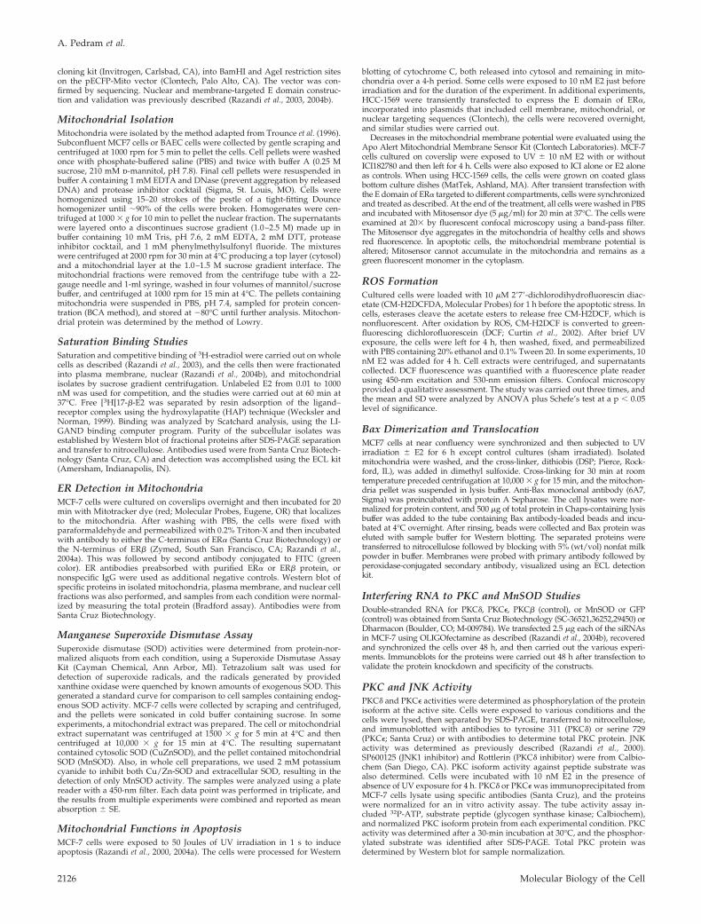
Identification of Mitochondrial ERWe first established high-affinity, saturable ER in plasmamembrane, nucleus, and mitochondrial cell fractions ofMCF-7 cells (Kd � 0.2–0.3 nM; Figure 1A, left). Approxi-mately 85% of receptors were present in the nucleus, 5% inthe plasma membrane, and 10% in the mitochondria. Thelack of contamination of cell fractions was demonstrated byWestern blot for integral membrane (5�nucleotidase [NT]),nuclear (transportin and NTF2), and mitochondrial proteins(cytochrome C; Figure 1A, right). High-affinity mitochon-drial ER were also found in endothelial cells (EC; unpub-lished data).
Using antibodies to traditional ER isoforms, we determinedin MCF-7 cells that endogenous ER� and ER� are present inplasma membrane, nucleus, and mitochondrial cell fractions.The overall cellular ER� protein is considerably less abundantthan ER� (Figure 1B, left), consistent in that ER� is produced inlow abundance in breast cancer (Fuqua and Cui, 2004). Inter-estingly, the small amount of ER� is concentrated in mitochon-dria. This is different from EC, where ER� (and ER�) arepredominantly expressed in the nucleus, and the relativeamount of ER� exceeds ER� in mitochondria (Figure 1B, right).Confocal microscopy confirmed ER� in membrane, nuclear,and mitochondrial cell compartments of the MCF-7 cell (Figure1C, left). ER� was also detected (Figure 1C, right), and both ERisoforms colocalized with the mitochondria specific dye, Mito-tracker. Again, ER� was predominantly found in the mito-chondria of MCF-7 (Figure 1C, right). Antibodies preabsorbedwith purified ER� or ER� protein or nonspecific IgG showedno staining of proteins in the cells (unpublished data).
To further support the idea that mitochondrial ER� andER� derive from the same genes coding for classical nuclearER isoforms, we examined EC from combined ER�/ER�knockout mice (DERKO) (11). Wild-type EC demonstratedendogenous ER� in several cell locations, most prominentlyin the nucleus, and ER� was found substantially in mito-chondria. In contrast, DERKO EC produced no detectableER in any region of the cells (Figure 1D). Thus, in EC,mitochondrial ER derive from the same genes (ER� or ER�)that also produce nuclear and membrane ER (Razandi et al.,2004b).
ER Functions in the Cell Survival of Breast CancerBreast cancer cells respond to irradiation by undergoingapoptotic cell death (Xia and Powell, 2002). We previouslyreported that E2 inhibits radiation-induced cell death inMCF-7 cells (Razandi et al., 2000, 2004a). In part, this resultedfrom membrane-initiated steroid signaling to activation ofERK and PI3 kinases, perhaps impacting the mitochondria.E2/ER inhibition of JNK activity through undetermined ERpools or mechanisms also contributed (Razandi et al., 2000,2004a). Nuclear and membrane ER also signal to the up-regulation of genes such as Bcl2 (Dong et al., 1999), theprotein products of which promote long-term cell survival.Whether E2 acts directly at mitochondrial ER to impact cellfate is unknown.
To understand whether E2 influences apoptotic events inthe mitochondria, we determined the release of cytochromeC from this organelle in UV-irradiated MCF-7 cells. UVinduced the maximal translocation of cytochrome C frommitochondria into the cytoplasm at 60 min, significantlyprevented by E2 (Figures 2A, left and right). Cytochrome Credistribution was also visualized in the intact cell, andICI182780, an estrogen receptor antagonist, substantially al-though incompletely blocked the action of E2 (Figure 2B).
Irradiation of MCF-7 cells also decreased the mitochondrialmembrane potential, reflecting the commitment to apoptosis(Figure 2C). The shift from mainly red to green color oc-curred as the dye failed to be taken up in the mitochondriaof dying cells. E2 significantly decreased the numbers ofcells showing loss of membrane potential by �60%, substan-tially prevented by ICI182780.
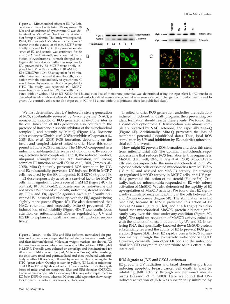
Role of Mitochondrial ERTo determine whether E2 acted at mitochondrial ER, wetargeted the ligand-binding domain (E domain) of ER� tothe plasma membrane or nucleus (exclusive targeting tothese pools shown in Razandi et al. 2004b), or to the mito-chondria of ER negative, HCC-1569 breast cancer or CHOcells. Expression of the E domain in mitochondria was dem-onstrated by substantial overlap with Mitotracker dye andby Western blot of cell fractions (Figure 3A). We then exam-ined cytochrome C translocation.
In HCC-1569 cells containing the mitochondrial-targetedE domain, E2 prevented the cytochrome C effects of UV(Figure 3B). UV also reduced the mitochondrial membranepotential in all cells, but this was significantly reversed bysex steroid in mitochondrial ER-targeted cells (Figure 3C). InER null breast cancer cells expressing the pcDNA3 plasmid(strict control), there were no E2 effects (unpublished data),supporting the need for ER to mediate E2 actions. Interest-ingly, plasma membrane targeting of the ER� E domain alsosupported E2 reversal of both UV effects (Figure 3, B and C).Perhaps this resulted from ERK or PI3 kinase signaling fromthe membrane to cell survival (Razandi et al., 2000, 2004a).
By contrast, targeted expression of the E domain in thenucleus did not significantly impact cytochrome C translo-cation or mitochondrial membrane potential, determined 1and 4 h, respectively, after UV exposure. This nuclear onlymodel fully supports E2-induced proliferation of breast can-cer cells (Razandi et al., 2004b), and a comparably truncatedER� supports nuclear transcription (Lopez et al., 1999). Wealso targeted a full-length ER� to the nucleus and found asimilar failure of E2 to reverse these events of apoptosis(unpublished data).
As a second model, we isolated mitochondria from MCF-7cells, the purity established as in Figure 1A. UV stress in-duced the release of cytochrome C from mitochondria intothe incubation medium (Figure 3D). E2 prevented this (lane3), indicating direct action at this cytoplasmic organelle. Todetermine which ER isoform mediates this action, mitochon-dria were exposed to UV in the presence of 10 nM E2, PPT(an ER� agonist), or DPN (an ER� agonist; Harrington et al.,2003). Each inhibited cytochrome C release, but the ER�agonist was more potent than the ER� agonist. Taken to-gether, the combined effects of ER� and ER� agonists wereroughly equivalent to the inhibition by E2, likely reflectingcontributions from each mitochondrial ER isoform to steroidaction.
Mechanisms of ER Action in MitochondriaHow does mitochondrial ER protect the viability of irradi-ated cells? In cancer, reactive oxygen species (ROS) areimportant to apoptosis induced by radiation, chemothera-peutic agents, and many cell stressors (Benhar et al., 2002). Inmost stress circumstances, mitochondria generate the greatmajority of ROS (Finkel and Holbrook, 2000). ROS genera-tion here is normally a result of oxidative phosphorylation,and several of the respiratory complexes produce ROS as aresult of electron transport between complexes (Curtin et al.,2002). Complexes I and III (especially the latter) have beenparticularly implicated during stress-related ROS formation.
ER in Mitochondria
Vol. 17, May 2006 2127
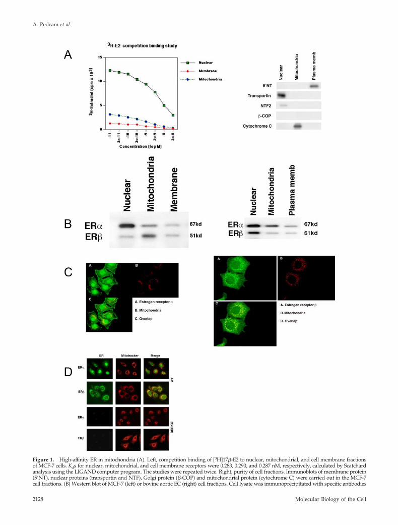
Figure 1. High-affinity ER in mitochondria (A). Left, competition binding of [3H]17�-E2 to nuclear, mitochondrial, and cell membrane fractionsof MCF-7 cells. Kds for nuclear, mitochondrial, and cell membrane receptors were 0.283, 0.290, and 0.287 nM, respectively, calculated by Scatchardanalysis using the LIGAND computer program. The studies were repeated twice. Right, purity of cell fractions. Immunoblots of membrane protein(5�NT), nuclear proteins (transportin and NTF), Golgi protein (�-COP) and mitochondrial protein (cytochrome C) were carried out in the MCF-7cell fractions. (B) Western blot of MCF-7 (left) or bovine aortic EC (right) cell fractions. Cell lysate was immunoprecipitated with specific antibodies
A. Pedram et al.
Molecular Biology of the Cell2128
We first determined that UV induced a strong generationof ROS, substantially reversed by N-acetlycystine (NAC), anonspecific inhibitor of ROS generated at multiple sites inthe cell. Inhibition of ROS generation also occurred in thepresence of Rotenone, a specific inhibitor of the mitochondrialcomplex I, and potently by Mito-Q (Figure 4A). Rotenoneeither enhances (Ohnishi et al., 2005) or inhibits (Chapman et al.,2005; Sato et al., 2005) ROS formation, depending on theinsult and coupled state of mitochondria. Here, this com-pound inhibits ROS formation. The Mito-Q compound is amitochondrial-targeted derivative of ubiquinone. By accept-ing electrons from complexes I and II, the reduced product,ubiquinol, strongly reduces ROS formation, influencingcomplex III function as well (Kelso et al., 2001; James et al.,2005). Mito-Q potently prevented ROS formation by UV,and E2 substantially prevented UV-induced ROS in MCF-7cells, reversed by the ER antagonist, ICI182780 (Figure 4B).
E2 dose-responsively acted as a survival factor for MCF-7cells, with significant effects seen at 1 nM E2 (Figure 4C). Incontrast, 10 nM 17-�-E2, progesterone, or testosterone didnot block UV-induced cell death, indicating steroid specific-ity. ER�- and ER�-specific agonists each significantly pre-vented UV-induced cell viability, with the ER� agonist beingslightly more potent (Figure 4C). We also determined thatNAC, rotenone, and especially Mito-Q prevented UV-induced loss of cell viability (Figure 4D). These results focusattention on mitochondrial ROS as regulated by UV andE2/ER to explain cell death and survival functions, respec-tively.
If mitochondrial ROS generation underlies the radiation-induced mitochondrial death program, then preventing ox-idant formation should rescue these events. We found thatUV-induced cytochrome C translocation was almost com-pletely reversed by NAC, rotenone, and especially Mito-Q(Figure 4E). Additionally, Mito-Q prevented the loss ofmembrane potential (unpublished data). Thus, local ROSstimulation by UV and inhibition by E2 underlies mitochon-drial cell fate events.
How might E2 prevent ROS formation and does this stemfrom mitochondrial ER? The dominant mitochondria-spe-cific enzyme that reduces ROS formation in this organelle isMnSOD (Halliwell, 1999; Huang et al., 2000). MnSOD rap-idly reduces superoxide, the main mitochondrial ROS. Weexposed whole cells or isolated mitochondria from MCF-7 toUV � E2 and assayed for MnSOD activity. E2 stronglyup-regulated MnSOD activity in MCF-7 cells, and UV par-tially prevented this action of the steroid (Figure 5A). Crit-ically, isolated mitochondria responded to E2 with robustactivation of MnSOD. We also determined the rapidity of E2up-regulation of MnSOD activity; We found that E2 signif-icantly stimulated enzymatic activity in the cells between 10-and 20-min exposure (Figure 5B). The stimulation was ERmediated, because ICI182780 prevented this action of E2both at 20 min (Figure 5C, left) and at 4 h (right). We alsofound that mitochondrial MnSOD protein did not signifi-cantly vary over this time under any condition (Figure 5C,right). The rapid up-regulation of MnSOD activity coincideswith the kinetics of kinase modulation by UV and E2. Inter-fering RNA that specifically knocked down MnSOD proteinsubstantially reversed the ability of E2 to prevent ROS gen-eration (Figure 5D). Thus, E2 rapidly prevents ROS forma-tion mainly through the exclusively mitochondrial SOD.However, cross-talk from other ER pools to the mitochon-drial MnSOD enzyme might contribute to this effect in theintact cell.
ROS Signals to JNK and PKC� ActivationE2 prevents UV radiation and taxol chemotherapy frominducing apoptotic breast cancer cell death in part byinhibiting JNK activity through undetermined mecha-nisms (Razandi et al., 2000). Here we found that UV-induced activation of JNK was substantially inhibited by
Figure 1 (cont). to the ER� and ER� isoforms, normalized for pro-tein, and proteins were separated by gel electrophoresis, transferred,and then immunoblotted. Molecular weight markers are shown. (C)Immunofluorescence confocal microscopy of ER� (left) and ER� (right)in MCF-7. The cells were cultured on coverslips and then incubated for20 min with Mitotracker dye (red; Molecular Probes). After washing,the cells were fixed and permeabilized and then incubated with anti-body to either ER isoform, followed by second antibody conjugated toFITC (green color). Overlap is seen in yellow. (D) Lack of mitochon-drial ER in ER�/ER� deleted cells. EC were isolated from the capil-laries of mice bred for combined ER� and ER� deletion (DERKO).Confocal microscopy fails to show any ER in any cell compartment inEC from DERKO mice, whereas EC from wild-type mice show recep-tors for each ER isoform in various cell locations.
Figure 2. Mitochondrial effects of E2. (A) Left,cells were treated with brief UV exposure (50J/s) and abundance of cytochrome C was de-termined in MCF-7 cell fractions by Westernblot for up to 240 min. The study was repeated.Right, E2 prevents UV-induced cytochrome Crelease into the cytosol at 60 min. MCF-7 werebriefly exposed to UV in the presence or ab-sence of E2, and steroid was continued for 60min. (B) A predominantly mitochondrial distri-bution of cytochrome c (control) changed to alargely diffuse cytosolic pattern in response toUV, prevented by E2. MCF-7 were briefly ex-posed to UV, with or without 10 nM E2, orE2�ICI182780 (1 �M; ER antagonist) for 60 min.After fixing and permeabilizing the cells, incu-bation with the first antibody to cytochrome Cwas followed by second antibody conjugated toFITC. The study was repeated. (C) MCF-7were briefly exposed to UV, the cells incu-bated with or without E2 or ICI182780 for 4 h, and then loss of membrane potential was determined using the Apo-Alert kit (Clontech) asdescribed in Materials and Methods. Decreased mitochondrial membrane potential was seen as a color change from predominantly red togreen. As controls, cells were also exposed to ICI or E2 alone without significant effect (unpublished data).
ER in Mitochondria
Vol. 17, May 2006 2129
Figure 3. E domain of ER� mediates E2 effects at the mitochondria. (A) Left, expression of the mitochondrial-targeted E domain of ER�.CHO cells were transfected to express the E domain of ER� (green) targeted to the mitochondria, using a targeting vector containing amitochondrial localization sequence (Clontech). Mitotracker dye indicates the mitochondria (red). The merged panel shows virtuallycomplete colocalization of ER with Mitotracker dye (orange-yellow). Right, Western blot of ER� E domain targeted to the mitochondria andexpressed in HCC-1569 cells. Western blot of cell fractions was accomplished with an antibody to the ER� ligand-binding domain (H222).(B) The E domain of ER� was targeted to the plasma membrane, mitochondria, or nucleus in HCC-1569 cells. Western blot of cytochromeC in cytoplasm at 60 min post-UV in the presence or absence of 10 nM E2 treatment is shown. The study is representative of two experiments.(C) Mitochondrial membrane potential and apoptosis in E domain-targeted HCC-1569 cells. Cells were briefly exposed to UV, then �E2, orE2 alone, and membrane potential/apoptosis was determined at 4 h. Control cells were transfected but sham exposed to UV. The bar graphrepresents the mean plus SEM from three combined studies, 200 cells counted per condition in each. *p � 0.05 for control or E2 alone versusUV, �p � 0.05 for UV�E2 versus UV by ANOVA plus Schefe’s test. (D) ER� and ER� mediate E2 action at the mitochondria. Isolatedmitochondria from MCF-7 cells were exposed to UV � 10 nM of E2, PPT, or DPN, and cytochrome C release into the incubation mediumor in the mitochondrial pellet was determined by Western blot at 60 min. The representative study was repeated twice additionally.
A. Pedram et al.
Molecular Biology of the Cell2130

Figure 4. Reactive oxygen species (ROS) underlies cell fate decisions. (A) MCF-7 cells were exposed to UV � antioxidants NAC(N-acetylcystine) 5 mM, rotenone 2.5 �M, or Mito-Q 10 �M. ROS production was determined 4 h after UV exposure by confocal microscopyusing the fluorescent indicator, CM-H2DCFDA, and quantified by spectroflourometry. The bar graph represents three studies combined. *p �0.05 for control versus UV, �p � 0.05 for UV versus UV plus antioxidant. None of the antioxidant compounds had significant effects on ROSformation by themselves (unpublished data). (B) Estradiol/ER prevents UV-induced ROS formation. The cells were exposed to brief UV, withor without E2 or 1 �M ICI182780, and incubated for 4 h. The bar graph is data from three combined experiments. *p � 0.05 for control versusUV, �p � 0.05 for UV versus UV�E2, ��p � 0.05 for UV�E2 versus same plus ICI182780. (C) MCF-7 were exposed to UV � 0.1–10 nM17-�-E2, 10 nM 17-�-E2, 10 nM progesterone (P) or testosterone (T), or 10 nM PPT or DPN. Steroid compounds were continued for the lengthof the experiment. Cell viability was determined by the MTT assay at 4 h. Decreased viability is shown by a loss of spectral absorbance tothe dye. Data are from three experiments; *p � 0.05 for control versus UV, �p � 0.05 for UV versus UV plus steroid. (D) MCF-7 were exposedto UV � antioxidants and cell viability was determined by the MTT assay at 4 h. Data are from three experiments; *p � 0.05 for control versusUV, �p � 0.05 for UV versus UV plus antioxidant. (E) Inhibitors of ROS generation reverse UV-induced cytochrome C release into thecytoplasm. A representative study shown here was repeated. Actin serves as loading control.
ER in Mitochondria
Vol. 17, May 2006 2131
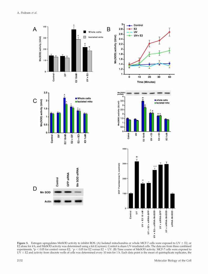
Figure 5. Estrogen upregulates MnSOD activity to inhibit ROS. (A) Isolated mitochondria or whole MCF-7 cells were exposed to UV � E2, orE2 alone for 4 h, and MnSOD activity was determined using a kit (Cayman). Control is sham UV-irradiated cells. The data are from three combinedexperiments, *p � 0.05 for control versus E2, �p � 0.05 for E2 versus E2 � UV. (B) Time course of MnSOD activity. MCF-7 cells were exposed toUV � E2 and activity from discrete wells of cells was determined every 10 min for 1 h. Each data point is the mean of quintuplicate replicates, the
A. Pedram et al.
Molecular Biology of the Cell2132

all the antioxidants, especially Mito-Q (Figure 6A). Theresults implicate mitochondrial ROS as the critical regulatorof UV-induced JNK activation and suggest that E2 inhibitionof ROS shown here underlies the inhibition of kinase activity(Razandi et al., 2000). We then asked whether JNK activation
contributes to mitochondrial apoptosis events. A specificinhibitor of JNK1 activity, SP 600125, partially preventedUV-induced cytochrome C release into cytoplasm (Figure6B) and also significantly blocked UV-induced loss of mito-chondrial membrane potential (unpublished data).
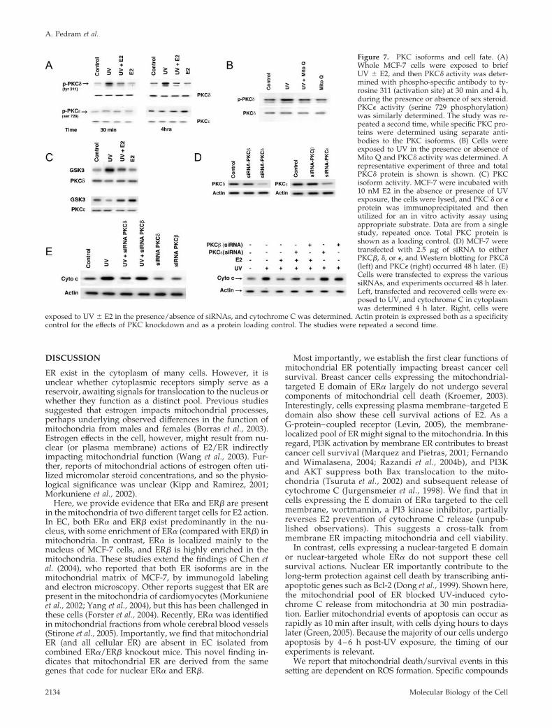
Another important mediator of apoptosis in many cellinsult models is the novel PKC isoform, PKC� (Steinberg,2004). This isoform translocates to mitochondria in responseto H2O2, where it participates in the mitochondrial programof apoptosis (Majumder et al., 2001). By comparison, PKC� insome cell types contributes to cell survival (McJilton et al.,2003; Murriel and Mochly-Rosen, 2003). We determinedwhether these PKC isoforms contributed to cell fate inMCF-7. After 30 min and persisting for 4 h, UV stronglyactivated PKC�, noted as tyrosine 311 phosphorylation (Fig-ure 7A). E2 strongly inhibited this activation. Furthermore,UV-activated PKC� was significantly dependent on ROS(Figure 7B). We also determined PKC� activity, using a PKCsubstrate peptide (from glycogen synythase kinase; Figure7C). We found that UV stimulated, whereas E2 preventedPKC� activation by UV. In contrast, PKC� phosphorylationand activity were stimulated at 4 h by E2 but not by UV(Figure 7, A and C). Therefore, the two PKC isoforms couldbe differential mediators of cell fate in our system.
To support this, we investigated PKC functions in the mod-ulation of cytochrome C release. To do this, we first validatedthat specific siRNAs for PKC� and PKC� knock down theintended target proteins (Figure 7D). Importantly, the PKCcontrol siRNA to PKC� had no effects on either the � or �isoforms. Silencing PKC� substantially prevented UV-stimu-lated cytochrome C concentration in cytosol, limited by ourtransfection efficiency (�65%; Figure 7E, left). The siRNA forPKC� had no effect in this regard. In contrast, silencing PKC�partially reversed the inhibition of cytochrome C localizationby E2 (Figure 7E, right). Again, the siRNA for PKC� had noeffect, and PKC� knockdown showed no influence on UV-induced cytochrome C release (in the absence of E2). Col-lectively, PKC� contributes to UV-induced cell death via themitochondria, whereas PKC� specifically enables a cell sur-vival-related action of the sex steroid.
Bax Translocation and Oligomerization at the MembraneIn response to many death stimuli, the proapoptotic proteinBax translocates to the mitochondria, where it undergoesoligomerization/activation and insertion into the outer mi-tochondrial membrane (Gross et al., 1998; Dejean et al., 2005).Active Bax then promotes the release of cytochrome C fromthe mitochondria into the cytoplasm, stimulating the forma-tion of the apoptosome (Jurgensmeier et al., 1998). We de-termined whether UV and E2 oppose each other in theseregards. Under control conditions, Bax exists as a monomerin the cytosol of the MCF-7 cells (Figure 8A, lanes 1 and 5).UV caused both the dimerization and translocation of Bax tothe mitochondrial fraction (lanes 2 and 6), significantly pre-vented by E2. Antioxidants prevented UV-induced translo-cation of Bax to the mitochondria (Figure 8B) and the PKC�inhibitor rottlerin, substantially inhibited UV-induced Baxtranslocation (Figure 8C). JNK activation by UV also con-tributed, because the specific inhibitor of JNK1 activity, SP600125, partially blocked Bax translocation to the mitochon-dria. Thus, the signaling that results from mitochondrialROS formation leads to Bax participation in mitochondrialapoptosis, the inhibition of which provides a mechanism forE2 survival effects (Figure 8D).
Figure 5 (cont). study repeated a second time. (C) ER mediates E2stimulation of MnSOD activity. MCF-7 cells or isolated mitochondriawere exposed to brief UV and then incubated with 10 nM E2 in thepresence or absence of 1 �M ICI182780, for 20 min (left) or 4 h (right).The bar graphs represent three experiments combined. *p � 0.05 forcontrol versus E2, �p � 0.05 for E2 versus same � either UV orICI182780. Also shown is an immunoblot of MnSOD protein over 4 hin isolated mitochondria. (D) Left, validation of siRNA knockdown ofMnSOD protein. Western blot for MnSOD or actin occurred 48 h aftertransfection of MCF-7 cells with either siRNA to MnSOD or GFP(control). Right, MCF-7 were transfected and recovered and then ex-posed to UV � 10 nM E2. ROS were measured 4 h later and the dataare from three combined experiments. *p � 0.05 for control versus UV,�p � 0.05 for UV versus UV � E2, ��p � 0.05 for UV � E2 versussame plus siRNA to MnSOD.
Figure 6. Role of JNK in UV-induced cell death. (A) MCF-7 cellswere exposed to UV in the presence of absence of Mito-Q, and JNKactivity was determined sequentially over a 4-h period. The toppanel shows the 30 min time point. A representative study is shown,repeated once. (B) Cells were briefly exposed to UV � SP 600125, aJNK-1 inhibitor, the latter continued for 4 h. Cytochrome C in thecytoplasm was determined by Western blot, and the study wasrepeated.
ER in Mitochondria
Vol. 17, May 2006 2133
DISCUSSION
ER exist in the cytoplasm of many cells. However, it isunclear whether cytoplasmic receptors simply serve as areservoir, awaiting signals for translocation to the nucleus orwhether they function as a distinct pool. Previous studiessuggested that estrogen impacts mitochondrial processes,perhaps underlying observed differences in the function ofmitochondria from males and females (Borras et al., 2003).Estrogen effects in the cell, however, might result from nu-clear (or plasma membrane) actions of E2/ER indirectlyimpacting mitochondrial function (Wang et al., 2003). Fur-ther, reports of mitochondrial actions of estrogen often uti-lized micromolar steroid concentrations, and so the physio-logical significance was unclear (Kipp and Ramirez, 2001;Morkuniene et al., 2002).
Here, we provide evidence that ER� and ER� are presentin the mitochondria of two different target cells for E2 action.In EC, both ER� and ER� exist predominantly in the nu-cleus, with some enrichment of ER� (compared with ER�) inmitochondria. In contrast, ER� is localized mainly to thenucleus of MCF-7 cells, and ER� is highly enriched in themitochondria. These studies extend the findings of Chen etal. (2004), who reported that both ER isoforms are in themitochondrial matrix of MCF-7, by immunogold labelingand electron microscopy. Other reports suggest that ER arepresent in the mitochondria of cardiomyocytes (Morkunieneet al., 2002; Yang et al., 2004), but this has been challenged inthese cells (Forster et al., 2004). Recently, ER� was identifiedin mitochondrial fractions from whole cerebral blood vessels(Stirone et al., 2005). Importantly, we find that mitochondrialER (and all cellular ER) are absent in EC isolated fromcombined ER�/ER� knockout mice. This novel finding in-dicates that mitochondrial ER are derived from the samegenes that code for nuclear ER� and ER�.
Most importantly, we establish the first clear functions ofmitochondrial ER potentially impacting breast cancer cellsurvival. Breast cancer cells expressing the mitochondrial-targeted E domain of ER� largely do not undergo severalcomponents of mitochondrial cell death (Kroemer, 2003).Interestingly, cells expressing plasma membrane–targeted Edomain also show these cell survival actions of E2. As aG-protein–coupled receptor (Levin, 2005), the membrane-localized pool of ER might signal to the mitochondria. In thisregard, PI3K activation by membrane ER contributes to breastcancer cell survival (Marquez and Pietras, 2001; Fernandoand Wimalasena, 2004; Razandi et al., 2004b), and PI3Kand AKT suppress both Bax translocation to the mito-chondria (Tsuruta et al., 2002) and subsequent release ofcytochrome C (Jurgensmeier et al., 1998). We find that incells expressing the E domain of ER� targeted to the cellmembrane, wortmannin, a PI3 kinase inhibitor, partiallyreverses E2 prevention of cytochrome C release (unpub-lished observations). This suggests a cross-talk frommembrane ER impacting mitochondria and cell viability.
In contrast, cells expressing a nuclear-targeted E domainor nuclear-targeted whole ER� do not support these cellsurvival actions. Nuclear ER importantly contribute to thelong-term protection against cell death by transcribing anti-apoptotic genes such as Bcl-2 (Dong et al., 1999). Shown here,the mitochondrial pool of ER blocked UV-induced cyto-chrome C release from mitochondria at 30 min postradia-tion. Earlier mitochondrial events of apoptosis can occur asrapidly as 10 min after insult, with cells dying hours to dayslater (Green, 2005). Because the majority of our cells undergoapoptosis by 4–6 h post-UV exposure, the timing of ourexperiments is relevant.
We report that mitochondrial death/survival events in thissetting are dependent on ROS formation. Specific compounds
Figure 7. PKC isoforms and cell fate. (A)Whole MCF-7 cells were exposed to briefUV � E2, and then PKC� activity was deter-mined with phospho-specific antibody to ty-rosine 311 (activation site) at 30 min and 4 h,during the presence or absence of sex steroid.PKC� activity (serine 729 phosphorylation)was similarly determined. The study was re-peated a second time, while specific PKC pro-teins were determined using separate anti-bodies to the PKC isoforms. (B) Cells wereexposed to UV in the presence or absence ofMito Q and PKC� activity was determined. Arepresentative experiment of three and totalPKC� protein is shown is shown. (C) PKCisoform activity. MCF-7 were incubated with10 nM E2 in the absence or presence of UVexposure, the cells were lysed, and PKC � or �protein was immunoprecipitated and thenutilized for an in vitro activity assay usingappropriate substrate. Data are from a singlestudy, repeated once. Total PKC protein isshown as a loading control. (D) MCF-7 weretransfected with 2.5 �g of siRNA to eitherPKC�, �, or �, and Western blotting for PKC�(left) and PKC� (right) occurred 48 h later. (E)Cells were transfected to express the varioussiRNAs, and experiments occurred 48 h later.Left, transfected and recovered cells were ex-posed to UV, and cytochrome C in cytoplasmwas determined 4 h later. Right, cells were
exposed to UV � E2 in the presence/absence of siRNAs, and cytochrome C was determined. Actin protein is expressed both as a specificitycontrol for the effects of PKC knockdown and as a protein loading control. The studies were repeated a second time.
A. Pedram et al.
Molecular Biology of the Cell2134
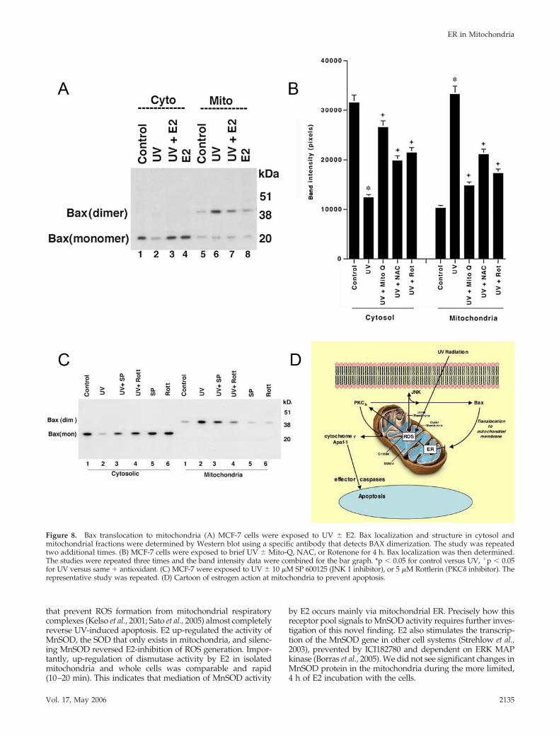
that prevent ROS formation from mitochondrial respiratorycomplexes (Kelso et al., 2001; Sato et al., 2005) almost completelyreverse UV-induced apoptosis. E2 up-regulated the activity ofMnSOD, the SOD that only exists in mitochondria, and silenc-ing MnSOD reversed E2-inhibition of ROS generation. Impor-tantly, up-regulation of dismutase activity by E2 in isolatedmitochondria and whole cells was comparable and rapid(10–20 min). This indicates that mediation of MnSOD activity
by E2 occurs mainly via mitochondrial ER. Precisely how thisreceptor pool signals to MnSOD activity requires further inves-tigation of this novel finding. E2 also stimulates the transcrip-tion of the MnSOD gene in other cell systems (Strehlow et al.,2003), prevented by ICI182780 and dependent on ERK MAPkinase (Borras et al., 2005). We did not see significant changes inMnSOD protein in the mitochondria during the more limited,4 h of E2 incubation with the cells.
Figure 8. Bax translocation to mitochondria (A) MCF-7 cells were exposed to UV � E2. Bax localization and structure in cytosol andmitochondrial fractions were determined by Western blot using a specific antibody that detects BAX dimerization. The study was repeatedtwo additional times. (B) MCF-7 cells were exposed to brief UV � Mito-Q, NAC, or Rotenone for 4 h. Bax localization was then determined.The studies were repeated three times and the band intensity data were combined for the bar graph. *p � 0.05 for control versus UV, �p � 0.05for UV versus same � antioxidant. (C) MCF-7 were exposed to UV � 10 �M SP 600125 (JNK 1 inhibitor), or 5 �M Rottlerin (PKC� inhibitor). Therepresentative study was repeated. (D) Cartoon of estrogen action at mitochondria to prevent apoptosis.
ER in Mitochondria
Vol. 17, May 2006 2135
What signals downstream of ROS mediate mitochondrialcell death? We found that ROS generated by UV inducedPKC� and JNK activities and E2 prevented this. PKC� pro-tein knockdown 1) partially prevented Bax translocation andoligomerization at the mitochondria and 2) prevented cyto-chrome C release and MCF-7 cell death. PKC� overexpres-sion contributes to apoptosis through association of the ki-nase with and modification of proapoptotic BCL-2 familymembers (Brodie and Blumberg, 2003; Murriel and Mochly-Rosen, 2003). In contrast, PKC� has been implicated in thesurvival of cardiomyocytes (Gray et al., 2004), prostate(McJilton et al., 2003), and lung cancer (Ding et al., 2002).Here we report the involvement of PKC� in breast cancer cellsurvival induced by E2, limiting cytochrome C release. Fur-ther studies will be needed to delineate the precise functionsof this kinase.
We also found that UV-induced ROS lead to JNK activa-tion, JNK promoted Bax translocation and dimerization atthe mitochondria, and E2 prevented each of these linkedevents. Radiation, chemotherapy, and tamoxifen activateJNK, contributing to breast cancer apoptosis (Mandlekarand Kong, 2001; Mingo-Sion et al., 2004). ROS up-regulatesJNK kinase activity by blocking JNK phosphatase function(Kamata et al., 2005). Others (Tsuruta et al., 2004) and wecould not show that Bax is directly phosphorylated by thismitogen-activated protein kinase. Tsuruta et al. (2004) re-cently determined that JNK phosphorylates the 14-3-3 pro-teins that sequester BAX in the cytoplasm, releasing Bax totranslocate to the mitochondria where it undergoes oli-gomerization and inserts into the mitochondrial membrane.This promotes pore formation and the release of cytochromeC into the cytoplasm (Jurgensmeier et al., 1998). Inhibition ofROS-induced JNK provides a second mechanism by whichE2 prevents Bax involvement in mitochondrial death.
Which mitochondrial ER isoform mediates E2 effects? Spe-cific agonists for either ER� or ER� inhibited UV-inducedcytochrome C release from isolated mitochondria; the ER�agonist was more potent. In addition, targeting of the ER� Edomain only to the mitochondria supported several cellsurvival functions of E2. Thus, both receptor isoforms ap-pear to contribute to sex steroid action in this organelle.Consistent with this, ER� or ER� agonists prevented UV-induced cell death, and steroid specificity was shown in thatneither progesterone nor testosterone affected cell fatethrough mitochondria. Our results also suggest that theligand binding/AF-2 region (E) is a functional domain forthis pool of ER�.
Mitochondrial-generated ROS production may contributeto oncogenesis because cancer-causing mutations in mito-chondrial DNA up-regulate ROS formation (Petros et al.,2005). When produced in small amounts, ROS can serve as aproliferation-related signaling mechanism (Preston et al.,2001). As recently reported, E2-induced G1/S progression inbreast cancer may in part be related to this mechanism (Feltyet al., 2005), a mechanism well described for signaling bymany growth factor tyrosine kinase receptors (Aslan andOzben, 2003). In contrast, adjuvant therapies for breast can-cer generate large amounts of ROS, essential to the inductionof cell death (Benhar and Levitzki, 2002). Direct preventionby E2/ER of excessive mitochondrial ROS formation is anovel mechanism to prevent apoptotic cell death. Thus, it isthe balance of ROS that mediates important aspects of car-cinogenesis and provides a therapeutic target to alter tumorbiology.
ACKNOWLEDGMENTS
This work was supported by grants from the Research Service of the Depart-ment of Veteran’s Affairs and the National Institutes of Health (CA-100366) toE.R.L.
REFERENCES
Aslan, M., and Ozben, T. (2003). Oxidants in receptor tyrosine kinase signaltransduction pathways. Antioxid. Redox. Signal 5, 781–788.
Benhar, M., Engelberg, D., and Levitzki, A. (2002). ROS, stress-activatedkinases and stress signaling in cancer. EMBO Rep. 3, 420–425.
Borras, C., Sastre, J., Garcia-Sala, D., Lloret, A., Pallardo, F. V., and Vina, J.(2003). Mitochondria from females exhibit higher antioxidant gene expressionand lower oxidative damage than males. Free Radic. Biol. Med. 34, 546–552.
Borras, C., Gambini, J., Gomez-Cabrera, M. C., Sastre, J., Pallardo, F. V., Mann,G. E., and Vina, J. (2005).. 17beta-oestradiol up-regulates longevity-related,antioxidant enzyme expression via the ERK1 and ERK2[MAPK]/NFkappaBcascade. Aging Cell 4, 113–118.
Brodie, C., and Blumberg, P. M. (2003). Regulation of cell apoptosis by proteinkinase c delta. Apoptosis 8, 19–27.
Chapman, K. E., Sinclair, S. E., Zhuang, D., Hassid, A., Desai, L., and Waters,C. M. (2005). Cyclic mechanical strain increases reactive oxygen species pro-duction in pulmonary epithelial cells. Am. J. Physiol. Lung Cell Mol. Physiol.289, L834–L841.
Chen, J. Q., Delannoy, M., Cooke, C., and Yager, J. D. (2004). Mitochondriallocalization of ER� and ER� in human MCF-7 cells. Am. J. Physiol. Endocri-nol. Metab. 28, E1011–E1022.
Curtin, J. F., Donovan, M., and Cotter, T. H. (2002). Regulation and measure-ment of oxidative stress response in apoptosis. J. Immunol. Methods 265,49–72.
Danial, N. K., and Korsmeyer, S. J. (2004). Cell death: critical control points.Cell 116, 205–219.
Dejean, L. M. et al. (2005). Oligomeric Bax is a component of the putativecytochrome c release channel MAC, mitochondrial apoptosis-induced chan-nel. Mol. Biol. Cell 16, 2424–2432.
Ding, L., Wang, H., Lang, W., and Xiao, L. (2002). PKC-epsilon promotessurvival of lung cancer cells by suppressing apoptosis through dysregulationof the mitochondrial caspase pathway. J. Biol. Chem. 277, 35305–35313.
Dong, L., Wang, W., Wang, F., Stoner, M., Reed, J. C., Harigai, M., Samudio,I., Kladde, M. P., Vyhlidal, C., and Safe, S. (1999). Mechanisms of transcrip-tional activation of bcl-2 gene expression by 17beta-estradiol in breast cancercells. J. Biol. Chem. 274, 32099–32107.
Felty, Q., Singh, K. P., and Roy, D. (2005). Estrogen-induced G(1)/S transitionof G(0)-arrested estrogen-dependent breast cancer cells is regulated by mito-chondrial oxidant signaling. Oncogene 24, 4883–4893.
Fernando, R. I., and Wimalasena, J. (2004). Estradiol abrogates apoptosis inbreast cancer cells through inactivation of BAD: Ras-dependent nongenomicpathways requiring signaling through ERK and Akt. Mol. Biol. Cell 15,3266–3284.
Finkel, T., and Holbrook, N. J. (2000). Oxidants, oxidative stress and thebiology of ageing. Nature 408, 239–247.
Forster, C., Kietz, S., Hultenby, K., Warner, M., and Gustafsson, J. A. (2004).Characterization of the ERbeta-/-mouse heart. Proc. Natl. Acad. Sci. USA 101,14234–14239.
Fuqua, S. A., and Cui, Y. (2004). Estrogen and progesterone receptor isoforms:clinical significance in breast cancer. Breast Cancer Res. Treat. 87, S3–10.
Gray, M. O., Zhou, H. Z., Schafhalter-Zoppoth, I., Zhu, P., Mochly-Rosen, D.,and Messing, R. O. (2004). Preservation of base-line hemodynamic functionand loss of inducible cardioprotection in adult mice lacking PKC epsilon.J. Biol. Chem. 279, 3596–3604.
Green, D. G. (2005). Apoptotic pathways: ten minutes to dead. Cell 121,671–674.
Gross, A., Jockel, J., Wei, M. C., and Korsmeyer, S. J. (1998). Enforced dimer-ization of BAX results in its translocation, mitochondrial dysfunction andapoptosis. EMBO J. 17, 3878–3885.
Halliwell, B. (1999). Antioxidant defence mechanisms: from the beginning tothe end (of the beginning). Free Radic. Res. 31, 261–272.
Harrington, W. R., Sheng, S., Barnett, D. H., Petz, L. N., Katzenellenbogen,J. A., and Katzenellenbogen, B. S. (2003). Activities of estrogen receptor alpha-and beta-selective ligands at diverse estrogen responsive gene sites mediatingtransactivation or transrepression. Mol. Cell Endocrinol. 206, 13–22.
A. Pedram et al.
Molecular Biology of the Cell2136
Huang, P., Feng, L., Oldham, E. A., Keating, M. J., and Plunkett, W. (2000).Superoxide dismutase as a target for the selective killing of cancer cells.Nature 407, 390–395.
James, A. M., Cocheme, H. M., Smith, R. A., and Murphy, M. P. (2005).Interactions of mitochondria-targeted and untargeted ubiquinones with themitochondrial respiratory chain and reactive oxygen species. Implications forthe use of exogenous ubiquinones as therapies and experimental tools. J. Biol.Chem. 280, 21295–21312.
Jensen, E. V., and Jacobson, H. I. (1962). Basic guides to the mechanism ofestrogen action. Rec. Prog. Horm. Res. 18, 387–414.
Jurgensmeier, J. M., Xie, Z., Deveraux, Q., Ellerby, L., Bredesen, D., and Reed,J. C. (1998). Bax directly induces release of cytochrome c from isolatedmitochondria. Proc. Natl. Acad. Sci. USA 95, 4997–5002.
Kamata, H., Honda, S., Maeda, S., Chang, L., Hirata, H., and Karin, M. (2005).Reactive oxygen species promote TNFalpha-induced death and sustainedJNK activation by inhibiting MAP kinase phosphatases. Cell 120, 649–661.
Kelso, G. F., Porteous, C. M., Coulter, C. V., Hughes, G., Porteous, W. K.,Ledgerwood, E. C., Smith, R. A., and Murphy, M. P. (2001). Selective targetingof a redox-active ubiquinone to mitochondria within cells: antioxidant andantiapoptotic properties. J. Biol. Chem. 276, 4588–4596.
Kipp, J. L., and Ramirez, V. D. (2001). Effect of estradiol, diethylstilbestrol, andresveratrol on F0F1-ATPase activity from mitochondrial preparations of ratheart, liver, and brain. Endocrine 15, 165–175.
Kroemer, G. (2003). Mitochondrial control of apoptosis: an introduction.Biochem. Biophys. Res. Commun. 304, 433–435.
Levin, E. R. (2001). Cell localization, physiology and nongenomic actions ofestrogen receptors. J. Appl. Physiol. 91, 1860–1867.
Levin, E. R. (2005). Integration of the extra-nuclear and nuclear actions ofestrogen. Mol. Endocrinol. 19, 1951–1959.
Lopez, G. N., Webb, P., Shinsako, J. H., Baxter, J. D., Greene, G., and Kushner,P. J. (1999). Titration by estrogen receptor activation function-2 of targets thatare downstream from coactivators. Mol. Endocrinol. 13, 897–909.
Majumder, P. K., Mishra, N. C., Sun, X., Bharti, A., Kharbanda, S., Saxena, S.,and Kufe, D. (2001). Targeting of PKC delta to mitochondria in the oxidativestress response. Cell Growth Differ. 12, 465–470.
Mandlekar, S., and Kong, A. N. (2001). Mechanisms of tamoxifen-inducedapoptosis. Apoptosis 6, 469–477.
Marquez, D. C., and Pietras, R. J. (2001). Membrane-associated binding sitesfor estrogen contribute to growth regulation of human breast cancer cells.Oncogene 20, 5420–5430.
McJilton, M. A. et al. (2003). Protein kinase Cepsilon interacts with Bax andpromotes survival of human prostate cancer cells. Oncogene 22, 7958–7968.
Mingo-Sion, A. M., Marietta, P. M., Koller, E., Wolf, D. M., and Van Den Berg,C. L. (2004). Inhibition of JNK reduces G2/M transit independent of p53,leading to endoreduplication, decreased proliferation, and apoptosis in breastcancer cells. Oncogene 23, 596–604.
Morkuniene, R., Akabsone, A., and Borutaite, V. (2002). Estrogens preventcalcium-induced release of cytochrome c from heart mitochondria. FEBS Lett.521, 53–56.
Murriel, C. L., and Mochly-Rosen, D. (2003). Opposing roles of delta andepsilon PKC in cardiac ischemia and reperfusion: targeting the apoptoticmachinery. Arch. Biochem. Biophys. 15, 246–254.
Ohnishi, S. T., Ohnishi, T., Muranaka, S., Fujita, H., Kimura, H., Uemura, K.,Yoshida, K., and Utsumi, K. (2005). A possible site of superoxide generationin the complex I segment of rat heart mitochondria. J. Bioenerg. Biomembr. 37,1–15.
Petros, J. A. et al. (2005). mtDNA mutations increase tumorigenicity in pros-tate cancer. Proc. Natl. Acad. Sci. USA 102, 719–724.
Preston, T. J., Muller, W. J., and Singh, G. (2001). Scavenging of extracellularH2O2 by catalase inhibits the proliferation of HER-2/Neu-transformed rat-1fibroblasts through the induction of a stress response. J. Biol. Chem. 276,9558–9564.
Razandi, M., Pedram, A., and Levin, E. R. (2000). Plasma membrane estrogenreceptors signal to anti-apoptosis in breast cancer. Mol. Endocrinol. 14, 1434–1447.
Razandi, M., Alton, G., Pedram, A., Ghonshani, S., Webb, D., and Levin, E. R.(2003). Identification of a structural determinant for the membrane localiza-tion of ER�. Mol. Cell Biol. 23, 1633–1646.
Razandi, M., Pedram, A., Merchenthaler, I., Greene, G. L., and Levin, E. R.(2004a). Plasma membrane estrogen receptors exist and function as dimers.Mol. Endocrinol. 18, 2854–2865.
Razandi, M., Pedram, A., Rosen, E., and Levin, E. R. (2004b). BRCA1 inhibitsmembrane estrogen and growth factor receptor signaling to cell proliferationin breast cancer. Mol. Cell Biol. 24, 5900–5913.
Sato, H., Sato, M., Kanai, H., Uchiyama, T., Iso, T., Ohyama, Y., Sakamoto, H.,Tamura, J., Nagai, R., and Kurabayashi, M. (2005). Mitochondrial reactiveoxygen species and c-Src play a critical role in hypoxic response in vascularsmooth muscle cells. Cardiovasc. Res. 67, 714–722.
Steinberg, S. F. (2004). Distinctive activation mechanisms and functions forprotein kinase Cdelta. Biochem. J. 384, 449–459.
Stirone, C., Duckles, S. P., Krause, D. N., and Procaccio, V. (2005). Estrogenincreases mitochondrial efficiency and reduces oxidative stress in cerebralblood vessels. Mol. Pharmacol. 68, 959–965.
Strehlow, K., Rotter, S., Wassmann, S., Adam, O., Grohe, C., Laufs, K., Bohm,M., and Nickenig, G. (2003). Modulation of antioxidant enzyme expressionand function by estrogen. Circ. Res. 93, 170–177.
Trounce, I., Kim, Y. L., Jun, A. S., and Wallace, D. C. (1996). Assessment ofmitochondrial oxidative phosphorylation in patient muscle biopsies, lympho-blasts, and transmitochondrial cell lines. Methods Enzymol. 264, 484–509.
Tsuruta, F., Masuyama, N., and Gotoh, Y. (2002). The phosphatidylinositol3-kinase (PI3K)-Akt pathway suppresses Bax translocation to mitochondria.J. Biol. Chem. 277, 14040–14047.
Tsuruta, F., Sunayama, J., Mori, Y., Hattori, S., Shimizu, S., Tsujimoto, Y.,Yoshioka, K., Masuyama, N., and Gotoh, Y. (2004). JNK promotes Bax trans-location to mitochondria through phosphorylation of 14–3-3 proteins. EMBOJ. 23, 1889–1899.
Wang, X. (2001). The expanding role of mitochondria in apoptosis. Genes Dev.15, 2922–2933.
Wang, X., Simpkins, J. W., Dykens, J. A., and Cammarata, P. R. (2003).Oxidative damage to human lens epithelial cells in culture: estrogen protec-tion of mitochondrial potential, ATP, and cell viability. Invest. Ophthalmol.Vis. Sci. 44, 2067–2075.
Wecksler, W. R., and Norman, A. W. (1999). An hydroxylapatite batch assayfor the quantitation of 1alpha,25-dihydroxyvitamin D3-receptor complexes.Annal. Biochem. 92, 314–323.
Xia, F., and Powell, S. N. (2002). The molecular basis of radiosensitivity andchemosensitivity in the treatment of breast cancer. Semin. Radiat. Oncol. 12,296–304.
Yang, S.-H. et al. (2004). Mitochondrial localization of estrogen receptor beta.Proc. Natl. Acad. Sci. USA 101, 4130–4135.
Yu, D., Jing, T., Liu, B., Yao, J., Tan, M., McDonnell, T. J., and Hung, M. C.(1998). Overexpression of ErbB2 blocks taxol-induced apoptosis by upregu-lation of p21Cip1, which inhibits p34Cdc2 kinase. Mol. Cell 2, 581–591.
ER in Mitochondria
Vol. 17, May 2006 2137