Intratumoral estrogen sulfotransferase induction contributes to ...
Upload
independentCategory
view
2download
0
The Immunophilin-Like Protein XAP2 Is a NegativeRegulator of Estrogen Signaling through Interaction withEstrogen Receptor aWen Cai1., Tatiana V. Kramarova1., Petra Berg1, Marta Korbonits2, Ingemar Pongratz1*
1 Department of Biosciences and Nutrition, Karolinska Institutet, Huddinge, Sweden, 2 Endocrinology, Barts and the London School of Medicine, Queen Mary University of
London, London, United Kingdom
Abstract
XAP2 (also known as aryl hydrocarbon receptor interacting protein, AIP) is originally identified as a negative regulator of thehepatitis B virus X-associated protein. Recent studies have expanded the range of XAP2 client proteins to include thenuclear receptor family of transcription factors. In this study, we show that XAP2 is recruited to the promoter of ERaregulated genes like the breast cancer marker gene pS2 or GREB1 and negatively regulate the expression of these genes inMCF-7 cells. Interestingly, we show that XAP2 downregulates the E2-dependent transcriptional activation in an estrogenreceptor (ER) isoform-specific manner: XAP2 inhibits ERa but not ERb-mediated transcription. Thus, knockdown ofintracellular XAP2 levels leads to increased ERa activity. XAP2 proteins, carrying mutations in their primary structures, loosethe ability of interacting with ERa and can no longer regulate ER target gene transcription. Taken together, this study showsthat XAP2 exerts a negative effect on ERa transcriptional activity and may thus prevent ERa-dependent events.
Citation: Cai W, Kramarova TV, Berg P, Korbonits M, Pongratz I (2011) The Immunophilin-Like Protein XAP2 Is a Negative Regulator of Estrogen Signaling throughInteraction with Estrogen Receptor a. PLoS ONE 6(10): e25201. doi:10.1371/journal.pone.0025201
Editor: Susan Kovats, Oklahoma Medical Research Foundation, United States of America
Received July 2, 2011; Accepted August 29, 2011; Published October 3, 2011
Copyright: � 2011 Cai et al. This is an open-access article distributed under the terms of the Creative Commons Attribution License, which permits unrestricteduse, distribution, and reproduction in any medium, provided the original author and source are credited.
Funding: This work is supported by the Consortorium for Research into Nuclear Receptors in Development and Aging Integrated Project (LSHM-CT-2005-018652)and the CASCADE Network of Excellence (FOOD-CT-2004-506319 CASCADE). URL: www.cordis.lu. The funders had no role in study design, data collection andanalysis, decision to publish, or preparation of the manuscript.
Competing Interests: The authors have declared that no competing interests exist.
* E-mail: [email protected]
. These authors contributed equally to this work.
Introduction
The Hepatitis B virus X protein associated protein 2 (XAP2) is a
37 kD immunophilin-like factor also known as aryl hydrocarbon
receptor-associated protein 9 (ARA9) or aryl hydrocarbon
receptor-interacting protein (AIP) [1,2,3]. XAP2 is an ubiquitously
expressed protein, however, the intracellular levels of XAP2 vary
considerably between different tissues, with high levels of
expression observed in the spleen thymus and pituitary and low
expression levels in the liver, kidney and lung [1] [4,5,6].
XAP2 is originally identified as a negative regulator of the
hepatitis B virus X-associated protein [5]. Later, XAP2 was
identified as an Hsp90-associated protein that specifically interacts
with the aryl hydrocarbon receptor (AhR) and regulates both AhR
intracellular localization [7] and protein stability by inhibiting
AhR ubiquitination [8,9,10]. Additional studies, however, have
expanded the range of XAP2 client proteins to include also signal
transduction proteins like Ga13 [11] and nuclear receptor (NR)
superfamily of transcription factors like GR [12], TRb1 [13] and
PPARa [14].
Estrogen receptor a (ERa) and b (ERb) belong to the NR family
and mediate the biological effects of estrogens [15]. In the absence
of ligands the ERs are present in an inactive form [16]. Ligand-
binding induces the recruitment of ER to estrogen response
element (ERE) located within regulatory sequences of estrogen-
responsive genes, resulting in the transcription activation of
estrogen target genes. Estrogen signaling is involved in variety of
physiological processes, both in females and males, in both
reproductive and non-reproductive tissues [17,18]. Although both
ERa and ERb are the mediators of the effects of estrogen, they
have distinct, or even opposing effects in certain tissues where the
biological action of estrogen ligands depends on a balance between
ERa and ERb [19,20]. Several studies have demonstrated that the
tumorigenic effects of estrogens are primarily mediated by ERa.
Lifetime exposure and high estrogen levels and thus high ER
transcriptional activity represent a risk factor for developing
tumors in breast [21], endometrial [22], ovarian [23] pituitary [24]
and thyroid tissues [25]. In contrast, ERb has been shown to
possess a tumor suppressive effect in tissues such as the prostate
[26] and colon [27].
Recent studies suggest the involvement of XAP2 in a wide range
of biological processes with tumorigenic outcome [28]. For
example, disruption of XAP2 is observed in patients with family
history of pituitary tumors [6,29]. However, the mechanisms
behind the tumor suppressive-activity of XAP2 have not been
clarified yet. One possibility is that the XAP2 interacts with
regulatory factors and thus modulates pathways involved in tumor
development as well as other pathological processes. Previous
studies have also demonstrated a physical and functional role of
XAP2 in regulation of NR superfamily members PPARa and
TRb1, providing the possibility that XAP2 could act as a regulator
in NR activities [13,14]. Interestingly, several studies have showed
that estrogen could induce the formation and development of
pituitary tumor [30,31], suggesting the possible involvement of
PLoS ONE | www.plosone.org 1 October 2011 | Volume 6 | Issue 10 | e25201
ER-regulated signaling pathways in pituitary tumor pathogenesis.
In addition, precautious puberty in a one-year-old female XAP2
mutation carrier has been reported [32], possibly implying a
modified ER signaling in XAP2 mutated individuals.
In this study we have analyzed the impact of XAP2 on E2-
dependent transcriptional activation. We show that XAP2
negatively regulates the transcriptional activity of ER in an
isoform-specific manner, by inhibiting ERa-mediated but not
ERb-mediated transcription. Our studies demonstrate that XAP2
action is dependent on the protein-protein interaction of XAP2
with ERa on the promoter of ER-target gene.
Taken together, our experiments demonstrate that XAP2 is a
negative regulator of ERa transcriptional activity and thus expand
the list of XAP2 client proteins to include ERa.
Materials and Methods
Recombinant plasmidsThe vectors pSG5-ERa, pSG5-ERb, pCMV5-bGal (b-gal) and
the 36ERE-luciferase and pS2-luciferase reporter construct have
been described elsewhere [33,34]. Human pSG5-hXAP2 [9] and
XAP2 mutation constructs [6] have been described elsewhere.
Details regarding construction of the different plasmid constructs
are available from the authors upon request.
Cell cultures and Reporter assaysHeLa cells [7] and MCF-7 cells [35] were maintained in
Dulbecco’s modified Eagle’s medium (DMEM, Invitrogen,
Carlsbad, CA, USA) containing 10% fetal calf serum (FCS,
Invitogen), 2 mM L-glutamine and 1% antibiotic (penicillin/
streptomycin (100 units/ml)). HC11 and HC11 36ERE cells [36]
were propagated in RPMI 1640 medium (Invitrogen) supple-
mented with 10% FCS, 2 mM L-glutamine, 1% gentamycin
(Invitrogen), 5 mg/ml insulin (Sigma-Aldrich, St Louis, MO, USA)
and 10 ng/ml EGF (Sigma-Aldrich).
For reporter assays, cells were plated into 12-well or 24-well
plates 24 h before transfection and maintained at 37uC. When
80% confluent the cells were transfected with Lipofectamin or
Lipofectamin Plus according to the manufacturer’s instructions
(Invitrogen). A b-gal plasmid was used as an internal transfection
control in the reporter assays. After 4 h of transfection, the
medium was exchanged with phenol red-free medium supple-
mented with 5% dextran-coated, charcoal-treated FCS (DCC-
FCS), cells were treated with DMSO or 10 nM E2 for another
48 h before luciferase activities were determined using luciferase
assay kit (BioThema, Dalaro, Sweden).
Cell extracts and immunoblot assayFor immunoprecipitation and Whole Cell Extract (WCE)
experiments, HC11 cells were seeded out on 15 cm dishes. 24 h
before treatment and/or transfection, the HC11 culture medium
was changed to phenol red-free medium supplemented with DCC-
FCS. The cells were then treated with DMSO or 10 nM E2 for
1 h. Cells were washed twice with cold PBS, collected by
centrifugation, and suspended in WCE buffer (0.4 M KCl,
20 mM Hepes pH 7.4, 1 mM DTT and 20% glycerol) supple-
mented with a protease inhibitor (Complete-Mini; Roche
Diagnostics, Mannheim, Germany), 10 mM Na2MoO4 and
25 mM Mg132 (Sigma-Aldrich). The freeze-thaw cycle with the
cell suspensions were repeated 4 times. Lysates were cleared by
centrifugation. 150 mg of cellular proteins were used for analysing
the WCE experiments. For immunoprecipitation experiments
200 mg of cellular protein was incubated with 10 mM Na2MoO4
and anti-ARA9 (XAP2) antibody (Novus Biologicals, Littleton,
CO) at 4uC for 1.5 h. Immunocomplexes were precipitated by
adding 30 ml of 50% slurry of protein-G-Sepharose (Amersham-
Pharmacia Biotech, Buckinghamshire, UK) plus 0.05% BSA
followed by incubation at 4uC under slow rotation for 1.5 h. After
centrifugation the resulting pellet were washed four times with
500 ml PBS.
For immunoprecipitaion of XAP2 mutations, HeLa cells were
seeded out on 6-well-plate. 24 h after transfection, cells were then
treated with DMSO or 10 nM E2 for 1 h. Immunoprecipitaion
assays were performed using Pierce classic IP kit (Thermo
Scientific) according to the manufacturer’s instructions.
Precipitated proteins and whole cell extracts were analyzed by
7.5 or 10% SDS PAGE and transferred to nitrocellulose
membranes. The membranes were treated with 5% nonfat milk
in PBS at 4uC over night followed by incubation with primary
antibodies. The primary antibodies used are ERa (Santa-Cruz;
dilution 1:1000), mouse c-myc (Santa Cruz; dilution 1:500) and b-
actin (Sigma-Aldrich; dilution 1:10 000) in blocking solution.
Horseradish peroxidase-conjugated anti-mouse or anti-rabbit
immunoglobulins (DakoCytomation, Glostrup, Denmark) were
used as secondary antibody. Immunocomplexes were visualized
after extensive washing in PBS-0.1% Tween-20 using enhanced
chemiluminescence reagents (ECL plus) (Amersham Pharmacia
Biotech) according to the manufacturer’s recommendations.
RNA interference (RNAi)siRNA against mouse XAP2 (msiXAP2) has been described
previously [13]. siRNA against human XAP2 (hsiXAP2) was
designed using the HiPerformance Design Algorithm licensed
from Novartis AG (Qiagen) targeted hXAP2 nucleotides 113–133
(59 AAGGAGGATGGCGGATATCAT39). An AllStar scrambled
sequence (Scr) from Novartis AG (Qiagen) where used as control.
The cells were transfected with a final concentration of 20 nM of
siRNA against XAP2 or Scr using Lipofectamine or Lipofecta-
mine LTX with Plus Reagent according to the manufacturer’s
instructions (Invitrogen). After 48 h, cells were treated with
DMSO or 10 nM E2 for 1 h and harvested for Western blot
and immunoprecipitation experiments; Real-time RT-PCR ex-
periments were performed essentially as described previously [37]
for reporter assay in HC11 36ERE cells.
In vitro translation assayIn vitro translation was performed using rabbit reticulocyte
lysate system (TNTH T7/SP6 Coupled Reticulocyte Lysate
System, Cat#L5020, Promega), according to manufacturer’s
insctructions. In brief, purified XAP2-pSG5, ERa-pSG5 vector
constructs (1 mg) were added to translation mix containing 35S-
Methionine (500 mCi, 10 mCi/ml, Perkin Elmer), T7 polymerase,
rabbit reticulocyte lysate, amino acid mixture (minus methyonine),
RNase inhibitor and incubated for 90 min at 30uC. Equal
amounts of translated products were then mixed and incubated
on ice for 60 min, with gentle shaking. After incubation, XAP2
antibodies (NB100–127, Novus Biologicals) or rabbit IgG (Santa-
Cruz Biotech) were added to each mix and incubated on ice for
60 min with gentle shaking, then Sepharose A/G was added and
immunoprecipitated complexes were washed 36with PBS, eluted
with SDS-loading buffer and analysed by SDS protein gel
electrophoresis.
Re-ChIP assaysSequential chromatin immunoprecipitation (Re-ChIP) assays
were performed essentially as described previously [34]. MCF-7
cells were grown on 15 cm plates to 90% confluence in phenol
red-free DMEM supplemented with 5% DCC-FCS. After treating
XAP2 Modulates ERa Transcriptional Activity
PLoS ONE | www.plosone.org 2 October 2011 | Volume 6 | Issue 10 | e25201

Figure 1. Suppressive effects of XAP2 on ERa-mediated gene expression. (A) The expression of endogenous pS2 gene in MCF-7 cells wasmonitored following the transfection with XAP2 siRNA (hsiXAP2) or scramble siRNA (Scr). After 48 h of transfection, cells were treated with DMSO(veh) or 10 nM E2 (E2) for 6 h before harvest. The mRNA levels of pS2 were determined by real-time RT-PCR and activity of scramble siRNA transfectedE2 treated samples were arbitrarily set to 1. (B) The expression of endogenous GREB1 gene in MCF-7 cells was monitored following the transfectionwith XAP2 siRNA (hsiXAP2) or scramble siRNA (Scr). After 48 h of transfection, cells were treated with DMSO or 10 nM E2 for 6 h before harvest. ThemRNA levels of GREB1 were determined by real-time RT-PCR and activity of scramble siRNA transfected E2 treated samples were arbitrarily set to 1. (C)ERa-independent (2ERa) or dependent (+ERa) activation of 36ERE-TATA-Luc reporter gene transcription in HeLa cells was monitored following co-transfection with XAP2 siRNA (hsiXAP2) or scramble siRNA (Scr). After 4 h of transfection, cells were treated with DMSO or 10 nM E2 for 48 h beforereporter gene activity was determined. Activity of scramble siRNA transfected DMSO treated cell samples were arbitrarily set to 1. (D) XAP2 and b-actin protein levels in MCF-7 and HeLa cells transfected with XAP2 siRNA (hsiXAP2) or Scramble siRNA (Scr) were determined by Western blot. (E) Theprotein expressions on Western blot in (D) were quantified by density of specific bands and normalized to b-actin. The XAP2/b-actin ratio in Scrsequence-transfected cells was arbitrarily set to 1. Data were expressed as means 6 SE of three independent experiments performed in duplicate.*, P,0.05 (Student’s t test).doi:10.1371/journal.pone.0025201.g001
XAP2 Modulates ERa Transcriptional Activity
PLoS ONE | www.plosone.org 3 October 2011 | Volume 6 | Issue 10 | e25201
with DMSO or 10 nM E2 for 45 min, cells were cross-linked for
15 min with 1.5% formaldehyde. Chromatin was then sonicated
and an aliquot of the sonicated soluble chromatin was taken and
later used as input control. Antibodies used were XAP2 (Novus
Biologicals, Littleton, CO) and ERa (Santa-Cruz, H-184). After
the first precipitation, specific antibody-protein complexes were
bound to Sepharose G Fast Flow (Amersham Biosciences), washed
and eluted with 10 mM dithiothreitol, precipitated with second
antibodies, washed and eluted in 0.1 M NaHCO3, 1% SDS.
Cross-linking was reversed at 65uC overnight. Eluted DNA
fragments were purified using QIAquick Spin Kit (Qiagen,
Valencia, CA). PCR experiments were performed at standard
conditions and the PCR-amplified fragments were analyzed on
2% agarose gels. Real-time RT-PCR was performed for
quantification. Primers: pS2: (F-59-CCGGCCATCTCTCACTA-
TGAA-39) (R-59-CCTCCCGCCAGGGTAAATAC-39); GREB1:
(F-59-AGCAGTGAAAAAAAGTGTGGCAACTGGG-39) (R-59-
CGACCCACAGAAATGAAAAGGCAGCAAACT-39);
Results
XAP2 has suppressive effects on ERa target geneexpression
Previous studies have shown a casual role for XAP2 as a tumor
suppressor and possibly as a modulator of ER activity; however,
the direct involvement between XAP2 and the ERs has not been
assessed. Therefore, we decided to test whether XAP2 has any
regulatory effect on ER-dependent gene expression. For this
purpose, we performed siRNA experiments to knock down the
intracellular levels of XAP2 in MCF-7 cells, a human breast
adenocarcinoma cell line [35], extensively used to characterize E2
signaling pathways, and which expresses XAP2 and ERa (data not
shown). We introduced siRNA constructs that targeted XAP2
mRNA or in control experiments, a scrambled sequence and
assessed the effect of XAP2 knockdown on E2 target gene
expression. Interestingly, when we knocked down XAP2 in MCF-
7 cells (Fig. 1D–E, MCF-7), we observed a statistically significant
up-regulation of endogenous expression of the breast cancer
marker gene pS2 in the presence of E2 (Fig. 1A, compare Scr E2
and hsiXAP2 E2). In addition, the expression of another ERa-
regulate gene GREB1 (growth regulation by estrogen in breast
cancer 1) [38] is also increased upon XAP2 depletion in MCF-7
cells (Fig. 1B, compare Scr E2 and hsiXAP2 E2), suggesting a
suppressive effect of XAP2 on the expression of ERa target genes.
To confirm this observation, and to validate the role of ERa, we
performed the similar RNAi assay in HeLa cells, where ERs are
not expressed. We transfected HeLa cells with siRNA against
XAP2 (hsiXAP2) or a scrambled sequence as negative control
together with ERa expression construct and a synthetic 36ERE
reporter construct derived from the vitellogenin xenopus promot-
er. Knockdown of XAP2 (Fig. 1D–E, HeLa) resulted in increased
ERa-mediated 36ERE activation (Fig. 1C, compare 3 E2 and 4
E2), in accordance with the results we obtained in MCF-7 cells. In
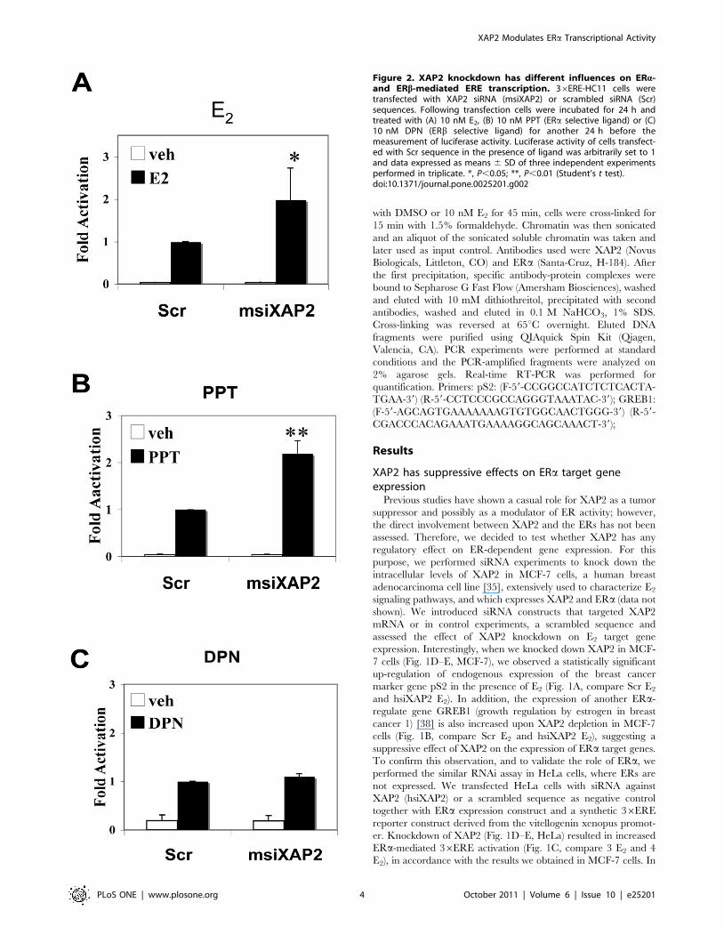
Figure 2. XAP2 knockdown has different influences on ERa-and ERb-mediated ERE transcription. 36ERE-HC11 cells weretransfected with XAP2 siRNA (msiXAP2) or scrambled siRNA (Scr)sequences. Following transfection cells were incubated for 24 h andtreated with (A) 10 nM E2, (B) 10 nM PPT (ERa selective ligand) or (C)10 nM DPN (ERb selective ligand) for another 24 h before themeasurement of luciferase activity. Luciferase activity of cells transfect-ed with Scr sequence in the presence of ligand was arbitrarily set to 1and data expressed as means 6 SD of three independent experimentsperformed in triplicate. *, P,0.05; **, P,0.01 (Student’s t test).doi:10.1371/journal.pone.0025201.g002
XAP2 Modulates ERa Transcriptional Activity
PLoS ONE | www.plosone.org 4 October 2011 | Volume 6 | Issue 10 | e25201

addition, no effect of XAP2 depletion was observed on 36ERE
dependent transcription in the absence of transfected ERa(Fig. 1C, compare 1 E2 and 2 E2), suggesting that the effects of
XAP2 are mediated by ERa. Taken together, these results suggest
that XAP2 has an inhibitory effect on E2-induced transcription of
ERa target genes.
XAP2 represses E2 transcriptional activity in anisoform-specific manner
Although both ERa and ERb mediate estrogen signaling,
biological functions of these two ER isoforms are distinct,
especially in tumorogenesis [19,20]. Therefore, we decided to
investigate whether XAP2 has effects on both ERa and ERb-
dependent transcriptional regulation. For this purpose, we
performed siRNA assay in stable HC11-36ERE cells, a mouse
mammary epithelial cell line, which expresses XAP2 (data not
shown) and both estrogen receptor isoforms, ERa and ERb [36].
HC11 cells were transfected with XAP2 siRNA (msiXAP2) or a
scrambled sequence (Scr), and treated with different ER isoform-
specific ligands, i.e. the ERa agonist propyl pyrazole triol (PPT)
[39], the ERb agonist diarylpropionitrile (DPN) [40] or the
panagonist E2. Remarkably, after XAP2 depletion, no significant
difference in 36ERE luciferase activity was observed in DPN
treated cells (Fig. 2C). However, luciferase expression was
increased by 2 -fold in E2 treated cells (Fig. 2A) and 2.2 -fold in
PPT treated cells (Fig. 2B), following XAP2 depletion. These
experiments suggest that the influence XAP2 on ER transcrip-
tional activity is ER isoform-specific.
To further verify that XAP2 modulates ERa-dependent but not
ERb-dependent E2 signaling, we performed transient transfections
in HeLa cells, since this cell line expresses neither ERa nor ERb,
thus allowing us to assess the effects of XAP2 on the individual
estrogen receptor isoforms. HeLa cells were transiently co-
transfected with fixed amounts of ERa or ERb expression vectors
together with increasing amounts of XAP2. The effects of XAP2
on ERa and ERb transcriptional activation was tested on two
different estrogen-regulated luciferase reporter gene constructs, the
36ERE reporter (Fig. 3A–B) and the promoter of pS2 gene
(Fig. 3C–D). Following transfection, the cells were treated with
10 nM E2 or vehicle for 48 hours before the cells were harvested,
and luciferase activity was determined as described previously [7].
Co-transfection with increasing amount of XAP2 expression
vector resulted in significant dose-dependent reductions of ERa-
mediated 36ERE activity (Fig. 3A) as well as the pS2 promoter
activity (Fig. 3C). However, co-transfection of ERb and XAP2 did
not result in any significant change of transcription induction of
the reporter constructs (Fig. 3B and D).
Taken together, these results indicate that XAP2 negatively
regulates E2-dependent transcriptional activity in an ER isoform-
specific manner, by inhibiting ERa but not ERb-mediated
transcriptional activity.
XAP2 interacts with ERa but has no effect on theintracellular level of ERa
Previous studies have shown that XAP2 stabilizes AhR protein
levels by inhibiting protein degradation of the latent receptor [9].
Figure 3. XAP2 represses ERa but not ERb mediated transcription. (A–B) HeLa cells were transiently co-transfected with 1 ng of ERa (A) orERb (B) expression vectors together with increasing amounts of XAP2 (1–10 ng) together with 100 ng of a 36ERE-TATA-Luc reporter. (C–D) HeLa cellswere transiently transfected with 1 ng of ERa (C) or ERb (D) expression vectors upon increasing amounts of XAP2 (1–10 ng) together with 100 ng of apS2 promoter luciferase reporter construct. 3 h after transfection, cells were treated with DMSO or 10 nM E2 for 48 h. Whole cell extracts (WCE) wereprepared and luciferase activity was measured. Reporter gene activity was determined and normalized to b-galactosidase. Results were compared tobasic luciferase activity of the reporter constructs, which were arbitrarily set to 1. Data were expressed as means 6 SD of three independentexperiments performed in triplicate. *, P,0.05 (Student’s t test).doi:10.1371/journal.pone.0025201.g003
XAP2 Modulates ERa Transcriptional Activity
PLoS ONE | www.plosone.org 5 October 2011 | Volume 6 | Issue 10 | e25201
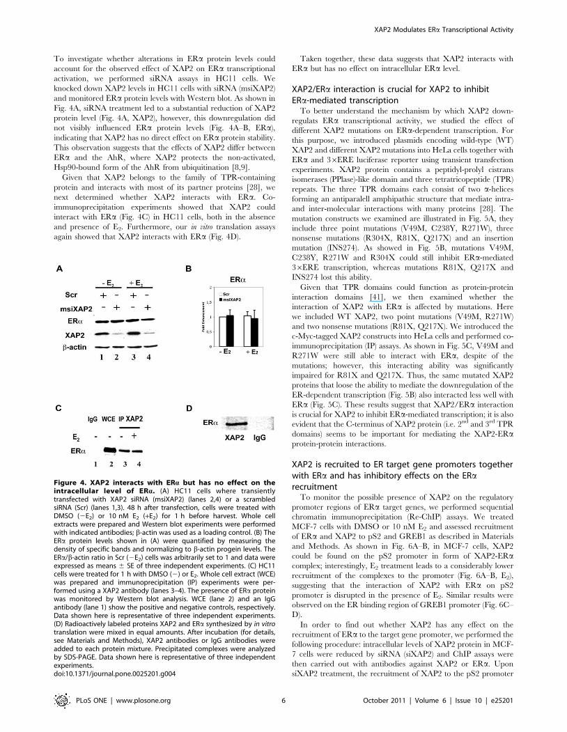
To investigate whether alterations in ERa protein levels could
account for the observed effect of XAP2 on ERa transcriptional
activation, we performed siRNA assays in HC11 cells. We
knocked down XAP2 levels in HC11 cells with siRNA (msiXAP2)
and monitored ERa protein levels with Western blot. As shown in
Fig. 4A, siRNA treatment led to a substantial reduction of XAP2
protein level (Fig. 4A, XAP2), however, this downregulation did
not visibly influenced ERa protein levels (Fig. 4A–B, ERa),
indicating that XAP2 has no direct effect on ERa protein stability.
This observation suggests that the effects of XAP2 differ between
ERa and the AhR, where XAP2 protects the non-activated,
Hsp90-bound form of the AhR from ubiquitination [8,9].
Given that XAP2 belongs to the family of TPR-containing
protein and interacts with most of its partner proteins [28], we
next determined whether XAP2 interacts with ERa. Co-
immunoprecipitation experiments showed that XAP2 could
interact with ERa (Fig. 4C) in HC11 cells, both in the absence
and presence of E2. Furthermore, our in vitro translation assays
again showed that XAP2 interacts with ERa (Fig. 4D).
Taken together, these data suggests that XAP2 interacts with
ERa but has no effect on intracellular ERa level.
XAP2/ERa interaction is crucial for XAP2 to inhibitERa-mediated transcription
To better understand the mechanism by which XAP2 down-
regulats ERa transcriptional activity, we studied the effect of
different XAP2 mutations on ERa-dependent transcription. For
this purpose, we introduced plasmids encoding wild-type (WT)
XAP2 and different XAP2 mutations into HeLa cells together with
ERa and 36ERE luciferase reporter using transient transfection
experiments. XAP2 protein contains a peptidyl-prolyl cistrans
isomerases (PPlase)-like domain and three tetratricopeptide (TPR)
repeats. The three TPR domains each consist of two a-helices
forming an antiparalell amphipathic structure that mediate intra-
and inter-molecular interactions with many proteins [28]. The
mutation constructs we examined are illustrated in Fig. 5A, they
include three point mutations (V49M, C238Y, R271W), three
nonsense mutations (R304X, R81X, Q217X) and an insertion
mutation (INS274). As showed in Fig. 5B, mutations V49M,
C238Y, R271W and R304X could still inhibit ERa-mediated
36ERE transcription, whereas mutations R81X, Q217X and
INS274 lost this ability.
Given that TPR domains could function as protein-protein
interaction domains [41], we then examined whether the
interaction of XAP2 with ERa is affected by mutations. Here
we included WT XAP2, two point mutations (V49M, R271W)
and two nonsense mutations (R81X, Q217X). We introduced the
c-Myc-tagged XAP2 constructs into HeLa cells and performed co-
immunoprecipitation (IP) assays. As shown in Fig. 5C, V49M and
R271W were still able to interact with ERa, despite of the
mutations; however, this interacting ability was significantly
impaired for R81X and Q217X. Thus, the same mutated XAP2
proteins that loose the ability to mediate the downregulation of the
ER-dependent transcription (Fig. 5B) also interacted less well with
ERa (Fig. 5C). These results suggest that XAP2/ERa interaction
is crucial for XAP2 to inhibit ERa-mediated transcription; it is also
evident that the C-terminus of XAP2 protein (i.e. 2nd and 3rd TPR
domains) seems to be important for mediating the XAP2-ERaprotein-protein interactions.
XAP2 is recruited to ER target gene promoters togetherwith ERa and has inhibitory effects on the ERarecruitment
To monitor the possible presence of XAP2 on the regulatory
promoter regions of ERa target genes, we performed sequential
chromatin immunoprecipitation (Re-ChIP) assays. We treated
MCF-7 cells with DMSO or 10 nM E2 and assessed recruitment
of ERa and XAP2 to pS2 and GREB1 as described in Materials
and Methods. As shown in Fig. 6A–B, in MCF-7 cells, XAP2
could be found on the pS2 promoter in form of XAP2-ERacomplex; interestingly, E2 treatment leads to a considerably lower
recruitment of the complexes to the promoter (Fig. 6A–B, E2),
suggesting that the interaction of XAP2 with ERa on pS2
promoter is disrupted in the presence of E2. Similar results were
observed on the ER binding region of GREB1 promoter (Fig. 6C–
D).
In order to find out whether XAP2 has any effect on the
recruitment of ERa to the target gene promoter, we performed the
following procedure: intracellular levels of XAP2 protein in MCF-
7 cells were reduced by siRNA (siXAP2) and ChIP assays were
then carried out with antibodies against XAP2 or ERa. Upon
siXAP2 treatment, the recruitment of XAP2 to the pS2 promoter
Figure 4. XAP2 interacts with ERa but has no effect on theintracellular level of ERa. (A) HC11 cells where transientlytransfected with XAP2 siRNA (msiXAP2) (lanes 2,4) or a scrambledsiRNA (Scr) (lanes 1,3). 48 h after transfection, cells were treated withDMSO (2E2) or 10 nM E2 (+E2) for 1 h before harvest. Whole cellextracts were prepared and Western blot experiments were performedwith indicated antibodies; b-actin was used as a loading control. (B) TheERa protein levels shown in (A) were quantified by measuring thedensity of specific bands and normalizing to b-actin progein levels. TheERa/b-actin ratio in Scr (2E2) cells was arbitrarily set to 1 and data wereexpressed as means 6 SE of three independent experiments. (C) HC11cells were treated for 1 h with DMSO (2) or E2. Whole cell extract (WCE)was prepared and immunoprecipitation (IP) experiments were per-formed using a XAP2 antibody (lanes 3–4). The presence of ERa proteinwas monitored by Western blot analysis. WCE (lane 2) and an IgGantibody (lane 1) show the positive and negative controls, respectively.Data shown here is representative of three independent experiments.(D) Radioactively labeled proteins XAP2 and ERa synthesized by in vitrotranslation were mixed in equal amounts. After incubation (for details,see Materials and Methods), XAP2 antibodies or IgG antibodies wereadded to each protein mixture. Precipitated complexes were analyzedby SDS-PAGE. Data shown here is representative of three independentexperiments.doi:10.1371/journal.pone.0025201.g004
XAP2 Modulates ERa Transcriptional Activity
PLoS ONE | www.plosone.org 6 October 2011 | Volume 6 | Issue 10 | e25201
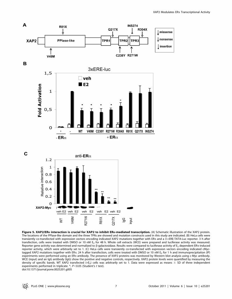
Figure 5. XAP2/ERa interaction is crucial for XAP2 to inhibit ERa-mediated transcription. (A) Schematic illustration of the XAP2 protein.The locations of the PPlase-like domain and the three TPRs are showed and mutation constructs used in this study are indicated. (B) HeLa cells weretransiently co-transfected with expression vectors encoding indicated XAP2 mutations together with ERa and a 36ERE-TATA-Luc reporter. 3 h aftertransfection, cells were treated with DMSO or 10 nM E2 for 48 h. Whole cell extracts (WCE) were prepared and luciferase activity was measured.Reporter gene activity was determined and normalized to b-galactosidase. Results were compared to luciferase activity of E2 dependent ERa-inducedreporter activity, which were arbitrarily set to 1. (C) HeLa cells were transiently co-transfected with expression vectors encoding indicated cMyc-tagged XAP2 mutations together with ERa. 24 h after transfection, cells were treated with DMSO or 10 nM E2 for 1 h and immunoprecipitation (IP)experiments were performed using an ERa antibody. The presence of XAP2 proteins was monitored by Western blot analysis using c-Myc antibody.WCE (input) and an IgG antibody (IgG) show the positive and negative controls, respectively. XAP2 protein levels were quantified by measuring thedensity of specific bands. WT XAP2 transfected (+E2) cells was arbitrarily set to 1. Data were expressed as means 6 SD of three independentexperiments performed in triplicate. *, P,0.05 (Student’s t test).doi:10.1371/journal.pone.0025201.g005
XAP2 Modulates ERa Transcriptional Activity
PLoS ONE | www.plosone.org 7 October 2011 | Volume 6 | Issue 10 | e25201
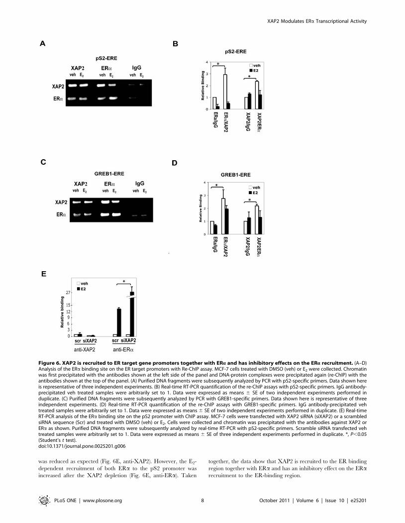
was reduced as expected (Fig. 6E, anti-XAP2). However, the E2-
dependent recruitment of both ERa to the pS2 promoter was
increased after the XAP2 depletion (Fig. 6E, anti-ERa). Taken
together, the data show that XAP2 is recruited to the ER binding
region together with ERa and has an inhibitory effect on the ERarecruitment to the ER-binding region.
Figure 6. XAP2 is recruited to ER target gene promoters together with ERa and has inhibitory effects on the ERa recruitment. (A–D)Analysis of the ERa binding site on the ER target promoters with Re-ChIP assay. MCF-7 cells treated with DMSO (veh) or E2 were collected. Chromatinwas first precipitated with the antibodies shown at the left side of the panel and DNA-protein complexes were precipitated again (re-ChIP) with theantibodies shown at the top of the panel. (A) Purified DNA fragments were subsequently analyzed by PCR with pS2-specific primers. Data shown hereis representative of three independent experiments. (B) Real-time RT-PCR quantification of the re-ChIP assays with pS2-specific primers. IgG antibody-precipitated veh treated samples were arbitrarily set to 1. Data were expressed as means 6 SE of two independent experiments performed induplicate. (C) Purified DNA fragments were subsequently analyzed by PCR with GREB1-specific primers. Data shown here is representative of threeindependent experiments. (D) Real-time RT-PCR quantification of the re-ChIP assays with GREB1-specific primers. IgG antibody-precipitated vehtreated samples were arbitrarily set to 1. Data were expressed as means 6 SE of two independent experiments performed in duplicate. (E) Real-timeRT-PCR analysis of the ERa binding site on the pS2 promoter with ChIP assay. MCF-7 cells were transfected with XAP2 siRNA (siXAP2) or a scrambledsiRNA sequence (Scr) and treated with DMSO (veh) or E2. Cells were collected and chromatin was precipitated with the antibodies against XAP2 orERa as shown. Purified DNA fragments were subsequently analyzed by real-time RT-PCR with pS2-specific primers. Scramble siRNA transfected vehtreated samples were arbitrarily set to 1. Data were expressed as means 6 SE of three independent experiments performed in duplicate. *, P,0.05(Student’s t test).doi:10.1371/journal.pone.0025201.g006
XAP2 Modulates ERa Transcriptional Activity
PLoS ONE | www.plosone.org 8 October 2011 | Volume 6 | Issue 10 | e25201
Discussion
In the present study, we have investigated the role of XAP2 in
regulation of E2-dependent transcriptional activation. XAP2 was
originally identified as a negative regulator of the hepatitis B virus
X-associated protein [5], and it has been shown to protect AhR
from protein degradation by inhibiting AhR ubiquitination [9].
XAP2 is also known to be associated with a number of cellular
factors, such as PPARa, TRb1 and Ga13 protein [11,13,14]. Our
current results demonstrate that XAP2 is involved in E2-mediated
signaling pathway, interacting with ERa and reveal, for the first
time, a mechanistic role of XAP2 affecting the transcription by
regulating transcription factors on the target gene promoter.
In MCF-7 cells, we observed a negative regulatory effect of
XAP2 on the breast cancer marker gene pS2 as well as GREB1,
another ER target gene (Fig. 1). Remarkably, our experiments
show that XAP2 downregulates the E2-dependent transcriptional
activation in an ER isoform-specific manner, by regulating ERabut not ERb-mediated transcription (Fig. 2–3). Although XAP2
has previously been shown to protect AhR from protein
degradation by inhibiting AhR ubiquitination [9], reduction of
XAP2 protein does not affect the intracellular protein levels of
ERa (Fig. 4A–B). Our results show that XAP2 could interact with
ERa (Fig. 4C–D); using mutated forms of XAP2 protein we
demonstrate that mutations that disrupt this interaction could no
longer regulate ERa-mediated gene transcription (Fig. 5). We also
show that XAP2 is recruited, or already present on ER-regulated
promoters, together with ERa; addition of the ligand leads to a
lower recruitment of the ERa/XAP2 complex (Fig. 6A–D).
Knocking down XAP2 expression leads in its turn to increased
recruitment of ERa to ER-target gene promoters (Fig. 6E). Based
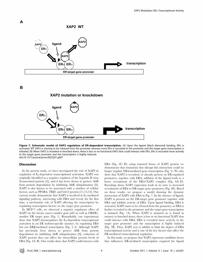
on these results, we propose a model showing the dynamic
interaction of XAP2 with ERa in Fig. 7. In the absence of ligand,
XAP2 is present on the ER-target gene promoter together with
ERa and inhibits activity of ERa. Upon ligand binding, ERa is
activated, XAP2 starts to be released from the promoter, as ERa is
further recruited to the promoter and the target gene transcription
is initiated (Fig. 7A). When XAP2 is mutated (as is found in
tumors) or knocked down, there is less or no functional XAP2 that
could interact with ERa, ERa is recruited more actively to the
target gene promoter and the transcription is highly induced
(Fig. 7B). Thus, XAP2 acts to inhibit or limit the degree of ERatranscriptional activity and is one of the key factors that affect the
ER-mediated transcriptional regulation.
In this study, we propose that XAP2 represents candidate factor
that influences ER-mediated transcription required for ligand
Figure 7. Schematic model of XAP2 regulation of ER-dependent transcription. (A) Upon the ligand (black diamond) binding, ERa isactivated, WT XAP2 is starting to be released from the promoter whereas more ERa is recruited to the promoter and the target gene transcription isinitiated. (B) When XAP2 is mutated or knocked down, there is less or no functional XAP2 that could interact with ERa, ERa is recruited more activelyto the target gene promoter and the transcription is highly induced.doi:10.1371/journal.pone.0025201.g007
XAP2 Modulates ERa Transcriptional Activity
PLoS ONE | www.plosone.org 9 October 2011 | Volume 6 | Issue 10 | e25201
inducibility of ERa, which may contribute to a cell-type or tissue-
specific regulation of E2 responsive genes. In addition, XAP2 may
serve as a negative regulator of numerous cellular regulatory
pathways. Tissue distribution and intracellular levels of XAP2 may
thereby affect the gene expression. Furthermore, the nature of
ligands may also play an important role. E2 is the main estrogenic
compound in pre-menopausal females, while the fetus and post-
menopausal females rely on compounds like estriol and estrone
respectively. These ligands are considerably weaker compared to
E2 and this could partly be due to their ability to displace XAP2.
In addition, xenoestrogens like bisphenol A or PCBs may also
display different abilities to displace XAP2 and this may reflect on
their endocrine disruptive effects. Clearly, additional studies need
to be performed to better understand the full impact of XAP2 on
nuclear receptor transcriptional regulation.
Germline mutations that disrupt the XAP2 protein have been
reported in both familial and sporadic pituitary tumor patients and
are possibly associated with other tumors [42]. In addition,
targeted disruption of XAP2 in mice leads to cardiac malformation
and embryonic lethality [43,44]. These findings suggest XAP2
could be a key player in various physiological and pathological
processes. In this study, we have demonstrated that XAP2
regulates the transcriptional response to E2 in an ER isoform-
specific manner, as XAP2 inhibits ERa, but not ERb-mediated
transcription (Fig. 2–3). Thus, the disturbance in XAP2 expres-
sion, for instance, in individuals carrying XAP2 gene mutants,
could over-activate ERa-mediated estrogen-signaling pathway.
This will presumably break the normal balance between ERa and
ERb actions and may lead to a higher risk of developing estrogen-
related disorders.
In conclusion, our study shows that XAP2 influences E2
signaling mediated by ERa by interacting with ERa. Further
investigations, such as studies using clinical material from pituitary
tumor patients with reported XAP2 mutations will allow to firmly
establishing the physiological meaning of this regulation.
Author Contributions
Conceived and designed the experiments: IP. Performed the experiments:
WC TK PB. Analyzed the data: WC TK. Contributed reagents/materials/
analysis tools: MK. Wrote the paper: WC MK. Interpreted data: IP.
Corrected the manuscript: IP MK.
References
1. Meyer BK, Pray-Grant MG, Vanden Heuvel JP, Perdew GH (1998) Hepatitis B
virus X-associated protein 2 is a subunit of the unliganded aryl hydrocarbon
receptor core complex and exhibits transcriptional enhancer activity. Mol Cell
Biol 18: 978–988.
2. Carver LA, Bradfield CA (1997) Ligand-dependent interaction of the aryl
hydrocarbon receptor with a novel immunophilin homolog in vivo. J Biol Chem
272: 11452–11456.
3. Ma Q, Whitlock JP (1997) A novel cytoplasmic protein that interacts with the Ah
receptor, contains tetratricopeptide repeat motifs, and augments the transcrip-
tional response to 2,3,7,8-tetrachlorodibenzo-p-dioxin. J Biol Chem 272:
8878–8884.
4. Carver LA, LaPres JJ, Jain S, Dunham EE, Bradfield CA (1998) Character-
ization of the Ah receptor-associated protein, ARA9. J Biol Chem 273:
33580–33587.
5. Kuzhandaivelu N, Cong YS, Inouye C, Yang WM, Seto E (1996) XAP2, a novel
hepatitis B virus X-associated protein that inhibits X transactivation. Nucleic
Acids Res 24: 4741–4750.
6. Leontiou CA, Gueorguiev M, van der Spuy J, Quinton R, Lolli F, et al. (2008)
The role of the aryl hydrocarbon receptor-interacting protein gene in familial
and sporadic pituitary adenomas. J Clin Endocrinol Metab 93: 2390–2401.
7. Berg P, Pongratz I (2002) Two parallel pathways mediate cytoplasmic
localization of the dioxin (aryl hydrocarbon) receptor. J Biol Chem 277:
32310–32319.
8. Meyer BK, Perdew GH (1999) Characterization of the AhR-hsp90-XAP2 core
complex and the role of the immunophilin-related protein XAP2 in AhR
stabilization. Biochemistry 38: 8907–8917.
9. Kazlauskas A, Poellinger L, Pongratz I (2000) The immunophilin-like protein
XAP2 regulates ubiquitination and subcellular localization of the dioxin
receptor. J Biol Chem 275: 41317–41324.
10. Swedenborg E, Pongratz I (2010) AhR and ARNT modulate ER signaling.
Toxicology 268: 132–138.
11. Nakata A, Urano D, Fujii-Kuriyama Y, Mizuno N, Tago K, et al. (2009) G-
protein signalling negatively regulates the stability of aryl hydrocarbon receptor.
EMBO Rep 10: 622–628.
12. Laenger A, Lang-Rollin I, Kozany C, Zschocke J, Zimmermann N, et al. (2009)
XAP2 inhibits glucocorticoid receptor activity in mammalian cells. FEBS Lett
583: 1493–1498.
13. Froidevaux MS, Berg P, Seugnet I, Decherf S, Becker N, et al. (2006) The co-
chaperone XAP2 is required for activation of hypothalamic thyrotropin-
releasing hormone transcription in vivo. EMBO Rep 7: 1035–1039.
14. Sumanasekera WK, Tien ES, Turpey R, Vanden Heuvel JP, Perdew GH (2003)
Evidence that peroxisome proliferator-activated receptor alpha is complexed
with the 90-kDa heat shock protein and the hepatitis virus B X-associated
protein 2. J Biol Chem 278: 4467–4473.
15. Swedenborg E, Ruegg J, Hillenweck A, Rehnmark S, Faulds MH, et al. (2008) 3-
Methylcholanthrene displays dual effects on estrogen receptor (ER) alpha and
ER beta signaling in a cell-type specific fashion. Mol Pharmacol 73: 575–586.
16. Pratt WB, Toft DO (1997) Steroid receptor interactions with heat shock protein
and immunophilin chaperones. Endocr Rev 18: 306–360.
17. Swedenborg E, Ruegg J, Makela S, Pongratz I (2009) Endocrine disruptive
chemicals: mechanisms of action and involvement in metabolic disorders. J Mol
Endocrinol 43: 1–10.
18. Meltser I, Tahera Y, Simpson E, Hultcrantz M, Charitidi K, et al. (2008)
Estrogen receptor beta protects against acoustic trauma in mice. J Clin Invest
118: 1563–1570.
19. Bardin A, Boulle N, Lazennec G, Vignon F, Pujol P (2004) Loss of ERbeta
expression as a common step in estrogen-dependent tumor progression. Endocr
Relat Cancer 11: 537–551.
20. Heldring N, Pike A, Andersson S, Matthews J, Cheng G, et al. (2007) Estrogen
receptors: how do they signal and what are their targets. Physiol Rev 87:
905–931.
21. Feigelson HS, Henderson BE (1996) Estrogens and breast cancer. Carcinogen-
esis 17: 2279–2284.
22. Persson I, Weiderpass E, Bergkvist L, Bergstrom R, Schairer C (1999) Risks of
breast and endometrial cancer after estrogen and estrogen-progestin replace-
ment. Cancer Causes Control 10: 253–260.
23. Greiser CM, Greiser EM, Doren M (2007) Menopausal hormone therapy and
risk of ovarian cancer: systematic review and meta-analysis. Hum Reprod
Update 13: 453–463.
24. Heaney AP (2007) Targeting pituitary tumors. Horm Res 68 Suppl 5: 132–136.
25. Zeng Q, Chen G, Vlantis A, Tse G, van Hasselt C (2008) The contributions of
oestrogen receptor isoforms to the development of papillary and anaplastic
thyroid carcinomas. J Pathol 214: 425–433.
26. Pravettoni A, Mornati O, Martini PG, Marino M, Colciago A, et al. (2007)
Estrogen receptor beta (ERbeta) and inhibition of prostate cancer cell
proliferation: studies on the possible mechanism of action in DU145 cells. Mol
Cell Endocrinol 263: 46–54.
27. Hartman J, Edvardsson K, Lindberg K, Zhao C, Williams C, et al. (2009)
Tumor repressive functions of estrogen receptor beta in SW480 colon cancer
cells. Cancer Res 69: 6100–6106.
28. Trivellin G, Korbonits M (2011) AIP and its interacting partners. J Endocrinol
210: 137–155.
29. Igreja S, Chahal HS, King P, Bolger GB, Srirangalingam U, et al. (2010)
Characterization of aryl hydrocarbon receptor interacting protein (AIP)
mutations in familial isolated pituitary adenoma families. Hum Mutat 31:
950–960.
30. Fujimoto M, Yoshino E, Hirakawa K, Chihara K, Ibata Y (1987) Studies on
estrogen induced pituitary tumor in the rat with special reference to the
relationship of the tuberoinfundibular dopamine neuron system. J Neurooncol 5:
151–159.
31. Heaney AP, Horwitz GA, Wang Z, Singson R, Melmed S (1999) Early
involvement of estrogen-induced pituitary tumor transforming gene and
fibroblast growth factor expression in prolactinoma pathogenesis. Nat Med 5:
1317–1321.
32. Naves LA, Daly AF, Vanbellinghen JF, Casulari LA, Spilioti C, et al. (2007)
Variable pathological and clinical features of a large Brazilian family harboring a
mutation in the aryl hydrocarbon receptor-interacting protein gene.
Eur J Endocrinol 157: 383–391.
33. Brunnberg S, Pettersson K, Rydin E, Matthews J, Hanberg A, et al. (2003) The
basic helix-loop-helix-PAS protein ARNT functions as a potent coactivator of
estrogen receptor-dependent transcription. Proc Natl Acad Sci U S A 100:
6517–6522.
34. Ruegg J, Swedenborg E, Wahlstrom D, Escande A, Balaguer P, et al. (2008) The
transcription factor aryl hydrocarbon receptor nuclear translocator functions as
XAP2 Modulates ERa Transcriptional Activity
PLoS ONE | www.plosone.org 10 October 2011 | Volume 6 | Issue 10 | e25201

an estrogen receptor beta-selective coactivator, and its recruitment to alternative
pathways mediates antiestrogenic effects of dioxin. Mol Endocrinol 22: 304–316.35. Levenson AS, Jordan VC (1997) MCF-7: the first hormone-responsive breast
cancer cell line. Cancer Res 57: 3071–3078.
36. Faulds MH, Olsen H, Helguero LA, Gustafsson JA, Haldosen LA (2004)Estrogen receptor functional activity changes during differentiation of mammary
epithelial cells. Mol Endocrinol 18: 412–421.37. Cai W, Rambaud J, Teboul M, Masse I, Benoit G, et al. (2008) Expression levels
of estrogen receptor beta are modulated by components of the molecular clock.
Mol Cell Biol 28: 784–793.38. Ghosh MG, Thompson DA, Weigel RJ (2000) PDZK1 and GREB1 are
estrogen-regulated genes expressed in hormone-responsive breast cancer.Cancer Res 60: 6367–6375.
39. Stauffer SR, Coletta CJ, Tedesco R, Nishiguchi G, Carlson K, et al. (2000)Pyrazole ligands: structure-affinity/activity relationships and estrogen receptor-
alpha-selective agonists. J Med Chem 43: 4934–4947.
40. Meyers MJ, Sun J, Carlson KE, Marriner GA, Katzenellenbogen BS, et al.
(2001) Estrogen receptor-beta potency-selective ligands: structure-activityrelationship studies of diarylpropionitriles and their acetylene and polar
analogues. J Med Chem 44: 4230–4251.
41. Goebl M, Yanagida M (1991) The TPR snap helix: a novel protein repeat motiffrom mitosis to transcription. Trends Biochem Sci 16: 173–177.
42. Chahal HS, Chapple JP, Frohman LA, Grossman AB, Korbonits M (2010)Clinical, genetic and molecular characterization of patients with familial isolated
pituitary adenomas (FIPA). Trends Endocrinol Metab 21: 419–427.
43. Lin BC, Sullivan R, Lee Y, Moran S, Glover E, et al. (2007) Deletion of the arylhydrocarbon receptor-associated protein 9 leads to cardiac malformation and
embryonic lethality. J Biol Chem 282: 35924–35932.44. Kang BH, Xia F, Pop R, Dohi T, Socolovsky M, et al. (2011) Developmental
Control of Apoptosis by the Immunophilin Aryl Hydrocarbon Receptor-interacting Protein (AIP) Involves Mitochondrial Import of the Survivin Protein.
J Biol Chem 286: 16758–16767.
XAP2 Modulates ERa Transcriptional Activity
PLoS ONE | www.plosone.org 11 October 2011 | Volume 6 | Issue 10 | e25201
Copyright © 2022 FDOKUMEN