CTIP2 is a negative regulator of P-TEFb
6
CTIP2 is a negative regulator of P-TEFb Thomas Cherrier a,b , Valentin Le Douce a,1 , Sebastian Eilebrecht c,d,1 , Raphael Riclet a , Céline Marban a,e , Franck Dequiedt b , Yannick Goumon f , Jean-Christophe Paillart g , Mathias Mericskay h , Ara Parlakian h , Pedro Bausero h , Wasim Abbas i , Georges Herbein i , Siavash K. Kurdistani e , Xavier Grana j , Benoit Van Driessche k , Christian Schwartz a , Ermanno Candolfi a , Arndt G. Benecke c,d , Carine Van Lint k,2,3 , and Olivier Rohr a,l,2,3 a Institut de Parasitologie et de Pathologie Tropicale, Fédération de Médecine Translationnelle, University of Strasbourg, 67000 Strasbourg, France; b Laboratory of Protein Signaling and Interactions, University of Liège, Liège, Belgium, c Vaccine Research Institute, Institut National de la Santé et de la Recherche Médicale, Unité 955, 94010 Créteil, France; d Institut des Hautes Études Scientifiques, Centre National de la Recherche Scientifique, 91440 Bures sur Yvette, France; e Department of Biological Chemistry, University of California, Los Angeles, CA 92093; f Institut des Neurosciences Cellulaires et Intégratives, University of Strasbourg, Centre National de la Recherche Scientifique, 67000 Strasbourg, France; g Architecture et Réactivité de l’ARN, Centre National de la Recherche Scientifique Unité Propre de Recherche 9002, Institut de Biologie Moléculaire et Cellulaire, Université de Strasbourg, 67000 Strasbourg, France; h Unité de Recherche 4, Aging, Stress, Inflammation Department, Université Pierre et Marie Curie Université Paris 6, 75005 Paris, France; i Department of Virology, Institut Fédératif de Recherche 133, Institut National de la Santé et de la Recherche Médicale, University of Franche-Comté, 25000 Besançon, France; j Fels Institute for Cancer Research and Molecular Biology and Department of Biochemistry, Temple University School of Medicine, Philadelphia, PA 19140; k Institut de Biologie et de Médecine Moléculaires, Université Libre de Bruxelles, 6041 Gosselies, Belgium; and l Institut Universitaire de France, 75005 Paris, France Edited by Stephen P. Goff, Columbia University College of Physicians and Surgeons, New York, NY, and approved June 5, 2013 (received for review November 25, 2012) The positive transcription elongation factor b (P-TEFb) is involved in physiological and pathological events including inflammation, cancer, AIDS, and cardiac hypertrophy. The balance between its active and inactive form is tightly controlled to ensure cellular in- tegrity. We report that the transcriptional repressor CTIP2 is a major modulator of P-TEFb activity. CTIP2 copurifies and interacts with an inactive P-TEFb complex containing the 7SK snRNA and HEXIM1. CTIP2 associates directly with HEXIM1 and, via the loop 2 of the 7SK snRNA, with P-TEFb. In this nucleoprotein complex, CTIP2 sig- nificantly represses the Cdk9 kinase activity of P-TEFb. Accord- ingly, we show that CTIP2 inhibits large sets of P-TEFb- and 7SK snRNA-sensitive genes. In hearts of hypertrophic cardiomyopathic mice, CTIP2 controls P-TEFb-sensitive pathways involved in the establishment of this pathology. Overexpression of the β-myosin heavy chain protein contributes to the pathological cardiac wall thickening. The inactive P-TEFb complex associates with CTIP2 at the MYH7 gene promoter to repress its activity. Taken together, our results strongly suggest that CTIP2 controls P-TEFb function in physiological and pathological conditions. D iscovered in 1995 (1), P-TEFb (CyclinT1/Cdk9) is involved in physiological and pathological transcriptionally regulated events such as cell growth, differentiation, cancer, cardiac hy- pertrophy, and AIDS (for review, see refs. 2 and 3). It has been suggested to be required for transcription of most RNA poly- merase II-dependent genes. However, a recent study suggests that a subset of cellular genes are distinctively sensitive to Cdk9 inhibition (4). P-TEFb is dynamically regulated by both positive and negative regulators. In contrast to Brd4, which is associated with the active form of P-TEFb (5, 6), the 7SK small nuclear RNA (7SK snRNA) and HEXIM1 inhibit Cdk9 activity in the inactive P-TEFb complex (7–10). P-TEFb elongation complexes are crucial for HIV-1 gene transactivation and viral replication. Recently, new P-TEFb complexes containing the HIV-1 Tat protein have been characterized (11, 12), providing evidence for the recruitment of an inactive Tat/P-TEFb complex to the HIV-1 promoter (13). However, defining the diverse nature and func- tions of the different P-TEFb complexes will require further investigations. The cellular protein CTIP2 (Bcl11b) has been highlighted as a key transcription factor for thymocyte (14, 15) and neuron development (16), odontogenesis (17), cancer evo- lution (18), and HIV-1 gene silencing (19). Besides AIDS, hy- pertrophic cardiomyopathy is a well-described P-TEFb–dependent pathology (for review, see refs. 20 and 21). Here, we report that CTIP2 represses P-TEFb function as part of an inactive P-TEFb complex. In hearts of hypertrophic car- diomyopathic mice, CTIP2 controls P-TEFb-sensitive pathways involved in the establishment of this pathology. Together with the inactive P-TEFb complex, CTIP2 associates with the β-myosin heavy chain promoter to repress its activity. Thereby, CTIP2 might contribute to the regulation of the size of heart sarcomeres in physiological or pathological conditions. Results CTIP2 Is Associated with the Inactive P-TEFb Complex. First, we in- vestigated, whether or not CTIP2 associates with one of the previously described P-TEFb complexes. We performed immu- noprecipitation experiments, revealing that CTIP2 coimmuno- precipitates with the CyclinT1 and Cdk9 components of the P- TEFb complex (Fig. 1A). To further define the CTIP2-contain- ing complexes, we separated the previously described P-TEFb complexes by gel filtration chromatography (Fig. 1B). As shown in Fig. 1B, Cdk9 was detected in the low molecular weight (LMW) complex (”free” P-TEFb complex) and in the high molecular weight (HMW) complex that coeluted with HEXIM1 and the 7SK snRNA. Interestingly, we found CTIP2 in those latter fractions containing the HMW P-TEFb complex (Fig. 1B). These obser- vations suggested that CTIP2 may be part of an inactive HEXIM1/ 7SK-including P-TEFb complex. To confirm this hypothesis, we performed additional coimmunoprecipitation experiments target- ing the active and the inactive form of P-TEFb. We found that CTIP2 copurified with CyclinT1, Cdk9, HEXIM1, and 7SK but not with Brd4, making CTIP2 a previously undescribed compo- nent of the inactive P-TEFb complex. Confocal observations further confirmed the colocalization of CTIP2, P-TEFb, and HEXIM1 in the previously described CTIP2-induced nuclear structures (22) (Fig. S1). As suggested by the gel filtration elution profile, CTIP2 was not found in the active Brd4/P–TEFb complexes. The 7SK snRNA functions as a scaffold RNA facilitating the interaction between HEXIM1 and P-TEFb. To investigate how CTIP2 associates with the inactive P-TEFb complex, we performed coimmunoprecipitation experiments following RNase treatment Author contributions: T.C., F.D., A.G.B., C.V.L., and O.R. designed research; T.C., V.L.D., S.E., R.R., C.M., Y.G., J.-C.P., and M.M. performed research; A.P., X.G., and A.G.B. contrib- uted new reagents/analytic tools; T.C., V.L.D., S.E., R.R., C.M., F.D., Y.G., J.-C.P., M.M., A.P., P.B., W.A., G.H., S.K.K., X.G., B.V.D., C.S., E.C., A.G.B., C.V.L., and O.R. analyzed data; and O.R. wrote the paper. The authors declare no conflict of interest. This article is a PNAS Direct Submission. 1 V.L.D. and S.E. contributed equally to this work. 2 C.V.L. and O.R. contributed equally to this work. 3 To whom correspondence may be addressed. E-mail: [email protected] or cvlint@ ulb.ac.be. This article contains supporting information online at www.pnas.org/lookup/suppl/doi:10. 1073/pnas.1220136110/-/DCSupplemental. www.pnas.org/cgi/doi/10.1073/pnas.1220136110 PNAS | July 30, 2013 | vol. 110 | no. 31 | 12655–12660 CELL BIOLOGY
-
Upload
independent -
Category
Documents
-
view
2 -
download
0
Transcript of CTIP2 is a negative regulator of P-TEFb
CTIP2 is a negative regulator of P-TEFbThomas Cherriera,b, Valentin Le Doucea,1, Sebastian Eilebrechtc,d,1, Raphael Ricleta, Céline Marbana,e, Franck Dequiedtb,Yannick Goumonf, Jean-Christophe Paillartg, Mathias Mericskayh, Ara Parlakianh, Pedro Bauseroh, Wasim Abbasi,Georges Herbeini, Siavash K. Kurdistanie, Xavier Granaj, Benoit Van Driesschek, Christian Schwartza,Ermanno Candolfia, Arndt G. Beneckec,d, Carine Van Lintk,2,3, and Olivier Rohra,l,2,3
aInstitut de Parasitologie et de Pathologie Tropicale, Fédération de Médecine Translationnelle, University of Strasbourg, 67000 Strasbourg, France;bLaboratory of Protein Signaling and Interactions, University of Liège, Liège, Belgium, cVaccine Research Institute, Institut National de la Santé et de laRecherche Médicale, Unité 955, 94010 Créteil, France; dInstitut des Hautes Études Scientifiques, Centre National de la Recherche Scientifique, 91440 Bures surYvette, France; eDepartment of Biological Chemistry, University of California, Los Angeles, CA 92093; fInstitut des Neurosciences Cellulaires et Intégratives,University of Strasbourg, Centre National de la Recherche Scientifique, 67000 Strasbourg, France; gArchitecture et Réactivité de l’ARN, Centre National de laRecherche Scientifique Unité Propre de Recherche 9002, Institut de Biologie Moléculaire et Cellulaire, Université de Strasbourg, 67000 Strasbourg, France;hUnité de Recherche 4, Aging, Stress, Inflammation Department, Université Pierre et Marie Curie Université Paris 6, 75005 Paris, France; iDepartment ofVirology, Institut Fédératif de Recherche 133, Institut National de la Santé et de la Recherche Médicale, University of Franche-Comté, 25000 Besançon, France;jFels Institute for Cancer Research and Molecular Biology and Department of Biochemistry, Temple University School of Medicine, Philadelphia, PA 19140;kInstitut de Biologie et de Médecine Moléculaires, Université Libre de Bruxelles, 6041 Gosselies, Belgium; and lInstitut Universitaire de France, 75005 Paris, France
Edited by Stephen P. Goff, Columbia University College of Physicians and Surgeons, New York, NY, and approved June 5, 2013 (received for reviewNovember 25, 2012)
The positive transcription elongation factor b (P-TEFb) is involvedin physiological and pathological events including inflammation,cancer, AIDS, and cardiac hypertrophy. The balance between itsactive and inactive form is tightly controlled to ensure cellular in-tegrity. We report that the transcriptional repressor CTIP2 is a majormodulator of P-TEFb activity. CTIP2 copurifies and interacts withan inactive P-TEFb complex containing the 7SK snRNA and HEXIM1.CTIP2 associates directly with HEXIM1 and, via the loop 2 of the7SK snRNA, with P-TEFb. In this nucleoprotein complex, CTIP2 sig-nificantly represses the Cdk9 kinase activity of P-TEFb. Accord-ingly, we show that CTIP2 inhibits large sets of P-TEFb- and 7SKsnRNA-sensitive genes. In hearts of hypertrophic cardiomyopathicmice, CTIP2 controls P-TEFb-sensitive pathways involved in theestablishment of this pathology. Overexpression of the β-myosinheavy chain protein contributes to the pathological cardiac wallthickening. The inactive P-TEFb complex associates with CTIP2 atthe MYH7 gene promoter to repress its activity. Taken together,our results strongly suggest that CTIP2 controls P-TEFb function inphysiological and pathological conditions.
Discovered in 1995 (1), P-TEFb (CyclinT1/Cdk9) is involvedin physiological and pathological transcriptionally regulated
events such as cell growth, differentiation, cancer, cardiac hy-pertrophy, and AIDS (for review, see refs. 2 and 3). It has beensuggested to be required for transcription of most RNA poly-merase II-dependent genes. However, a recent study suggeststhat a subset of cellular genes are distinctively sensitive to Cdk9inhibition (4). P-TEFb is dynamically regulated by both positiveand negative regulators. In contrast to Brd4, which is associatedwith the active form of P-TEFb (5, 6), the 7SK small nuclearRNA (7SK snRNA) and HEXIM1 inhibit Cdk9 activity in theinactive P-TEFb complex (7–10). P-TEFb elongation complexesare crucial for HIV-1 gene transactivation and viral replication.Recently, new P-TEFb complexes containing the HIV-1 Tatprotein have been characterized (11, 12), providing evidence forthe recruitment of an inactive Tat/P-TEFb complex to the HIV-1promoter (13). However, defining the diverse nature and func-tions of the different P-TEFb complexes will require furtherinvestigations. The cellular protein CTIP2 (Bcl11b) has beenhighlighted as a key transcription factor for thymocyte (14, 15)and neuron development (16), odontogenesis (17), cancer evo-lution (18), and HIV-1 gene silencing (19). Besides AIDS, hy-pertrophic cardiomyopathy is a well-described P-TEFb–dependentpathology (for review, see refs. 20 and 21).Here, we report that CTIP2 represses P-TEFb function as part
of an inactive P-TEFb complex. In hearts of hypertrophic car-diomyopathic mice, CTIP2 controls P-TEFb-sensitive pathwaysinvolved in the establishment of this pathology. Together with
the inactive P-TEFb complex, CTIP2 associates with the β-myosinheavy chain promoter to repress its activity. Thereby, CTIP2 mightcontribute to the regulation of the size of heart sarcomeres inphysiological or pathological conditions.
ResultsCTIP2 Is Associated with the Inactive P-TEFb Complex. First, we in-vestigated, whether or not CTIP2 associates with one of thepreviously described P-TEFb complexes. We performed immu-noprecipitation experiments, revealing that CTIP2 coimmuno-precipitates with the CyclinT1 and Cdk9 components of the P-TEFb complex (Fig. 1A). To further define the CTIP2-contain-ing complexes, we separated the previously described P-TEFbcomplexes by gel filtration chromatography (Fig. 1B). As shown inFig. 1B, Cdk9 was detected in the low molecular weight (LMW)complex (”free” P-TEFb complex) and in the high molecularweight (HMW) complex that coeluted with HEXIM1 and the 7SKsnRNA. Interestingly, we found CTIP2 in those latter fractionscontaining the HMW P-TEFb complex (Fig. 1B). These obser-vations suggested that CTIP2 may be part of an inactive HEXIM1/7SK-including P-TEFb complex. To confirm this hypothesis, weperformed additional coimmunoprecipitation experiments target-ing the active and the inactive form of P-TEFb. We found thatCTIP2 copurified with CyclinT1, Cdk9, HEXIM1, and 7SK butnot with Brd4, making CTIP2 a previously undescribed compo-nent of the inactive P-TEFb complex. Confocal observations furtherconfirmed the colocalization of CTIP2, P-TEFb, and HEXIM1 inthe previously described CTIP2-induced nuclear structures (22)(Fig. S1). As suggested by the gel filtration elution profile, CTIP2was not found in the active Brd4/P–TEFb complexes. The 7SKsnRNA functions as a scaffold RNA facilitating the interactionbetween HEXIM1 and P-TEFb. To investigate how CTIP2associates with the inactive P-TEFb complex, we performedcoimmunoprecipitation experiments following RNase treatment
Author contributions: T.C., F.D., A.G.B., C.V.L., and O.R. designed research; T.C., V.L.D.,S.E., R.R., C.M., Y.G., J.-C.P., and M.M. performed research; A.P., X.G., and A.G.B. contrib-uted new reagents/analytic tools; T.C., V.L.D., S.E., R.R., C.M., F.D., Y.G., J.-C.P., M.M., A.P.,P.B., W.A., G.H., S.K.K., X.G., B.V.D., C.S., E.C., A.G.B., C.V.L., and O.R. analyzed data; andO.R. wrote the paper.
The authors declare no conflict of interest.
This article is a PNAS Direct Submission.1V.L.D. and S.E. contributed equally to this work.2C.V.L. and O.R. contributed equally to this work.3To whom correspondence may be addressed. E-mail: [email protected] or [email protected].
This article contains supporting information online at www.pnas.org/lookup/suppl/doi:10.1073/pnas.1220136110/-/DCSupplemental.
www.pnas.org/cgi/doi/10.1073/pnas.1220136110 PNAS | July 30, 2013 | vol. 110 | no. 31 | 12655–12660
CELL
BIOLO
GY
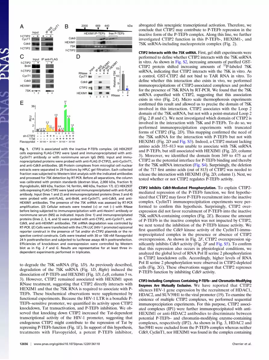
to degrade the 7SK snRNA (Fig. 1D). As previously described,degradation of the 7SK snRNA (Fig. 1D, Right) induced thedissociation of P-TEFb and HEXIM1 (Fig. 1D, Left, column 5 vs.3). However, CTIP2 remained associated with HEXIM1 uponRNase treatment, suggesting that CTIP2 directly interacts withHEXIM1 and that the 7SK RNA is required to associate with P-TEFb. These biochemical observations were supplemented byfunctional experiments. Because the HIV-1 LTR is a bonafide P-TEFb–sensitive promoter, we quantified its activity upon CTIP2knockdown, Tat transactivation, and P-TEFb inhibition. We ob-served that knocking down CTIP2 increased the Tat-dependenttranscriptional activity of the HIV-1 promoter, suggesting thatendogenous CTIP2 might contribute to the repression of Tat byrepressing P-TEFb function (Fig. 1E). In support of this hypothesis,treatments with Flavopyridol, a potent P-TEFb inhibitor,
abrogated this synergistic transcriptional activation. Therefore, weconclude that CTIP2 may contribute to P-TEFb repression in theinactive form of the P-TEFb complex. Along this line, we furtherinvestigated CTIP2 function in this P-TEFb-, HEXIM1-, and7SK snRNA-including nucleoprotein complex (Fig. 2).
CTIP2 Interacts with the 7SK snRNA. First, gel shift experiments wereperformed to define whether CTIP2 interacts with the 7SK snRNAin vitro. As shown in Fig. S2, increasing amounts of purified GST-CTIP2 protein shifted increasing amounts of 32P-labeled 7SKsnRNA, indicating that CTIP2 interacts with the 7SK in vitro. Asa control, GST-CTIP2 did not bind to TAR RNA in vitro. Todefine whether this interaction also exists in vivo, we performedimmunoprecipitations of CTIP2-associated complexes and probedfor the presence of 7SK RNA by RT-PCR. We found that the 7SKsnRNA copurified with CTIP2, suggesting that this associationexists in vivo (Fig. 2A). Micro scale thermophoresis experimentsconfirmed this result and allowed us to precise the domain of 7SKinvolved in this interaction. CTIP2 associates with the Loop 2domain of the 7SK snRNA, but not with a point-mutated Loop 2(Fig. 2 B and C). We next investigated which domain of CTIP2 isinvolved in the interaction with 7SK and P-TEFb. To do so, weperformed immunoprecipitation experiments with truncatedforms of CTIP2 (Fig. 2D). This mapping confirmed the need ofthe 7SK snRNA for the interaction with P-TEFb but not withHEXIM1 (Fig. 2D and Fig. S3). Indeed, a CTIP2 mutant lackingamino acids 355–813 was unable to associate with 7SK snRNAand P-TEFb, but still associated with HEXIM1 (Fig. 2D, column3). Moreover, we identified the domain from 349 to 475 aa ofCTIP2 as the potential interface for P-TEFb binding and therebyfor the 7SK snRNA interaction (Fig. S4). Surprisingly, the deletionof the 717 first amino acids (out of 813) of CTIP2 was needed torelease the interaction with HEXIM1 (Fig. 2D, column 1). Next, wetested whether or not CTIP2 regulates P-TEFb activity.
CTIP2 Inhibits Cdk9-Mediated Phosphorylation. To explain CTIP2-mediated repression of the P-TEFb function, we first hypothe-sized that CTIP2 may favor P-TEFb recruitment into the inactivecomplex. CyclinT1 immunoprecipitation experiments were per-formed to confirm this hypothesis. Surprisingly, CTIP2 over-expression did not favor recruitment of P-TEFb into the inactive7SK snRNA-containing complex (Fig. 2E). Because the amountof P-TEFb in the inactive complex was not impacted by CTIP2,we next tested the inhibition of the P-TEFb kinase activity. Wefirst quantified the Cdk9 kinase activity of the CyclinT1-immu-noprecipitated complex in the presence or absence of CTIP2overexpression. As shown in Fig. 2F, CTIP2 overexpression sig-nificantly inhibits Cdk9 activity (Fig. 2F and Fig. S5). To confirmthat this repression also occurs in physiological conditions, weanalyzed the global level of RNA Pol II serine 2 phosphorylationin CTIP2 knockdown cells. Accordingly, higher levels of RNAPol II serine 2 phosphorylation were observed in CTIP2-depletedcells (Fig. 2G). These observations suggest that CTIP2 repressesP-TEFb function by inhibiting Cdk9 activity.
CTIP2-Including Complexes Containing P-TEFb– and Chromatin-ModifyingEnzymes Are Mutually Exclusive. We have reported that CTIP2silences HIV-1 gene expression by the recruitment of HDAC1,HDAC2, and SUV39H1 to the viral promoter (19). To examine theexistence of multiple CTIP2 complexes, we performed sequentialimmunoprecipiation experiments. For this purpose, CTIP2 associ-ated complexes (IP1) were further immunoprecipitated with anti-HEXIM1 or anti-HDAC2 antibodies to discriminate betweenpotential P-TEFb– and chromatin-modifying enzyme-containingcomplexes, respectively (IP2). As shown in Fig. 3, HDAC2 andSuv39H1 were excluded from the P-TEFb complex whereas neitherCdk9, CyclinT1, nor HEXIM1 was found in the complex containing
f-CTIP2 +- +- + Input IP N
ISIP C
ycT1
CycT1Cdk9
f-CTIP2
A 2000 669 440 kDa
Cdk9HEXIM1
7SKf-CTIP2
HEXIM1/P-TEFb free P-TEFb
B
Inputf-CTIP2 +- +-
IP anti-FLAG
Brd4
f-CTIP2
CycT1
Cdk9
HEXIM1
7SK1 2 3 4
C
RNase
0
5
10
15
1 2- +
Rel
ativ
e 7S
KInput
IP NISIP H
EXIM1
IP NISIP H
EXIM1
RNase + + +- -
CTIP2
HEXIM1
CycT1
Cdk9
1 2 3 4 5
D
0
2
4RLU
1 2 3 4 5 6 7 8
36
2.2
4.34.8
0.8 1.6
0.3
37
9 10
0.4 0.4
f-Tat - - - - + + + + + +- -Flavopyridol - 30100 - - 30100 - 3010030100
sh-CTIP2 - - - + - - - + + ++ +
1.20.7
E
Fig. 1. CTIP2 is associated with the inactive P-TEFb complex. (A) HEK293Tcells expressing FLAG-CTIP2 were lysed and immunoprecipitated with anti-CyclinT1 antibody or with nonimmune serum IgG (NIS). Input and immu-noprecipitated proteins were probed with anti-FLAG (f-CTIP2), anti-CyclinT1,and anti-Cdk9 antibodies. (B) Protein complexes from microglial cell nuclearextracts were separated in 29 fractions by HPLC gel filtration. Each collectedfraction was subjected to Western blot analysis with the indicated antibodiesand processed for 7SK detection by RT-PCR. Before all separations, the columnwas calibrated with protein standards (dextran blue, 2,000 kDa, fraction 9;thyroglobulin, 669 kDa, fraction 14; ferritin, 440 kDa, fraction 17). (C) HEK293Tcells expressing FLAG-CTIP2 were lysed and immunoprecipitated with anti-FLAGantibody. Input (lines 1 and 2) and immunoprecipitated proteins (lines 3 and 4)were probed with anti-FLAG, anti-Brd4, anti-CyclinT1, anti-Cdk9, and anti-HEXIM1 antibodies. The presence of the 7SK snRNA was assessed by RT-PCRamplification. (D) Cellular extracts were treated (+) or not (−) with RNasebefore being subjected to immunoprecipitation with anti-Hexim1 antibody ornonimmune serum (NIS) as indicated. Inputs (line 1) and immunoprecipitatedproteins (lines 2, 3, 4, and 5) were probed with anti-CTIP2, anti-CyclinT1, anti-Cdk9, and anti-HEXIM1 antibodies. 7SK snRNA degradation was controled byRT-PCR. (E) Cells were transfected with the LTR-LUC (HIV-1 promoter) epizomalreporter construct in the presence of Tat and/or sh-CTIP2 plasmids or the re-spective control constructs. Cells were treated with 30 or 100 nM flavopyridol24 h posttransfection and subjected to luciferase assays 48 h posttransfection.Efficiencies of knockdown and overexpression were controlled by Westernblot as in Fig. 2 F and G. Results are representative for at least three in-dependent experiments performed in triplicates.
12656 | www.pnas.org/cgi/doi/10.1073/pnas.1220136110 Cherrier et al.

the chromatin-modifying enzymes. These data demonstrate thatCTIP2 associates at least with two distinct nuclear complexes.
CTIP2 Regulates P-TEFb–Sensitive Genes. To validate the modelemerging from our observations, establishing CTIP2 as a negativeregulatory component of P-TEFb, we examined the genome-wide
transcriptional consequences of a CTIP2 knockdown in microglialand HEK293 cells. Using DNA microarray analysis, genes whoseexpression was modulated by siRNA-mediated deletion of CTIP2(Dataset S1) were compared with those regulated by the ex-pression of a dominant negative mutant of Cdk9 (dnCdk9) (4).Hierarchical clustering of these genes revealed a significantanticorrelation between the two gene expression profiles (Fig.4A). Clustering of highly coregulated genes (−1>Log2 > 1 inboth gene profiles) revealed that 86% of the Cdk9 sensitivegenes are inversely regulated by the CTIP2 knockdown (Fig. 4Band Fig. S6). The comparison of the genes significantly ðP< 0:05Þregulated by CTIP2 overexpression, knockdown, and dnCdk9expression in HEK293 cells confirmed the observations made inmicroglial cells (Figs. 4 C–E). The 25% of the genes whose ex-pression was significantly ðP< 0:05Þ altered following CTIP2 knock-down or overexpression were also sensitive to dnCdk9, and, amongthem, 76% were regulated consistently with a Cdk9-inhibitory ac-tivity of CTIP2. Note that the vast majority of these consistentlyregulated genes are repressed upon dnCdk9 expression, in con-cordance with the elongation-activating role of P-TEFb. Besidesthat, we identified 14% (microglial cells) and 24% (HEK293 cells)of the CTIP2 target genes to be divergently regulated upon dnCdk9expression, pointing at a different mechanism, where CTIP2 canpotentially also contribute to Cdk9 activation (Fig. 4D). Whetherthis mode of action is direct or indirect needs to be furtherinvestigated.
CTIP2 Regulates 7SK snRNA-Sensitive Genes. Because 7SK snRNAhas been described as a key inhibitor of P-TEFb, we next com-pared the 7SK- and the CTIP2-dependent transcriptome (Fig. 4F and G and Datasets S2–S5). About 48% of the genes were in-versely affected by CTIP2 overexpression or 7SK knockdown. Thisobservation is consistent with a P-TEFb–repressive role of CTIP2and coincides with our model, in which both 7SK snRNA andCTIP2 contribute to the inactivation of Cdk9. Surprisingly, 52% ofthe genes were found to be similarly regulated following CTIP2overexpression or 7SK knockdown, suggesting that CTIP2 regu-lates a subset of 7SK-sensitive genes by a still unknown, P-TEFb–independent mechanism (Fig. 4F).
CTIP2 Regulates Cdk9-Sensitive Genes Modulated During CardiacHypertrophy in Mouse. As a compound of the inactive P-TEFbcomplex, CTIP2 should contribute to the regulation of P-TEFb–related physiopathological events. To investigate the physiolog-ical relevance of our observations, we focused on one of the
WT Sh-C
TIP2
PS2-RNApol II RNApol II CTIP2
G
0
20
40
1 2
NIS
ant
i-CTI
P2
Rel
ativ
e 7S
K b
indi
ng
60
A
f-CTIP2total
protein
10 100 1000
0.0
0.5
1.0
fract
ion
boun
d
relative CTIP2 amount
7SK L2 wt7SK L2 m137
C1-
813
1-35
414
5-43
435
0-81
3
717-
813
Flag-CTIP2
1 145 350 434 610 716
Zincfinger
Zincfinger
Zincfingers
Zincfingers
0
20
40
60
Rel
ativ
e 7S
K b
indi
ng
1 2 3 4 5
80
IP
100
α-Cdk9α-HEXIM1
D
0
40
80
+1-
2+
Rel
ativ
e 7S
K b
indi
ng
IPCycT1
IPNIS
E IPanti-CycT1
100
18+/
-6
100
17+/
-3
f-CTIP2 - + CyclinT1
CDK9
f-CTIP2
GST-CTD 32P-CTD
32P-CDK9
F
B
130
150
7SK L2wt
120
130
150
GC
AU
7SK L2m137
120
f-CTIP2
Fig. 2. Associated with the 7SK snRNA, CTIP2 inhibits Cdk9-mediated phos-phorylation. (A) HEK293T cells were lysed and subjected to immunoprecipi-tations with indicated antibodies. The presence of 7SK snRNA was quantifiedby RT-Q-PCR after complex elution and RNA extraction. (B) Secondary struc-tures of 7SK L2 and the mutant 7SK L2 m137 were calculated using Assemblesoftware (version 2.1); nucleotides are indicated by color code and numberedwith respect to their position in full-length 7SK RNA and arrows indicatemutations in m137. (C) MST analyses were performed with cellular extractscontaining increasing amounts of FLAG-CTIP2 and Fam6-labeled 7SK L2 and7SK L2 m137. The FLAG-CTIP2 amount was monitored by Western blot usinganti-FLAG antibody, and the total protein amount was visualized by Coo-massie staining. The highest CTIP2 concentration was abitrarily set to 1,000. (D)Mock, FLAG-CTIP2 wild-type, or FLAG-CTIP2 constructs transfected HEK293Tcells were lysed and subjected to immunoprecipitation with anti-FLAG anti-body. The presence of 7SK snRNA was quantified by RT-Q-PCR after complexelution and RNA extraction. Detection of immunoprecipitated proteins wasdetermined by Western blot analysis with the indicated antibodies. (E) Mockor FLAG-CTIP2 transfected cells were lysed and subjected to immunoprecipi-tations with the indicated antibodies. The presence of 7SK snRNA was quan-tified by RT-Q-PCR after complex elution and RNA extraction. (F) FLAG-CTIP2or mock transfected cells were lysed and subjected to immunoprecipitationwith anti-CyclinT1 antibody. Immunoprecipitated proteins were incubated for1 h at 30 °C with GST-CTD and 1 μCi of 32P-labeled ATP. After SDS/PAGE, la-beled GST-CTD and endogenous Cdk9 were detected by autoradiography.Quantification of the signals was performed using ImageJ software. Thepresence of the indicated proteins was determined by Western blot analysiswith the indicated antibodies. The amount of GST-CTD was assessed by Coo-massie staining. Specificity for the GST-CTD substrate was controlled by usingmutated GST-CTD (Fig. S5). (G) Extracts fromWT or CTIP2 knockdown cell lineswere subjected to Western blot analysis with the indicated antibodies.
IP1:α-Flag
1 2 3
α-HEX
IM1
α-HDAC
2
IP2
Contr
ol
P-TEFb
Chromatin modifying enzymes
4 5 6
f-CTIP2 - + + - ++CTIP2CycT1Cdk9
HEXIM1HDAC2
SUV39H1
Fig. 3. CTIP2-including complexes containing P-TEFb and chromatin modi-fying enzymes are mutually exclusive. Mock or FLAG-CTIP2 transfectedHEK293T cells were lysed and subjected to immunoprecipitation with anti-FLAG antibody. After washing, antibody-bound complexes were eluted withFLAG peptide and immunoprecipitated with the indicated antibodies. Thepresence of the indicated proteins was determined by Western blot analysis.
Cherrier et al. PNAS | July 30, 2013 | vol. 110 | no. 31 | 12657
CELL
BIOLO
GY
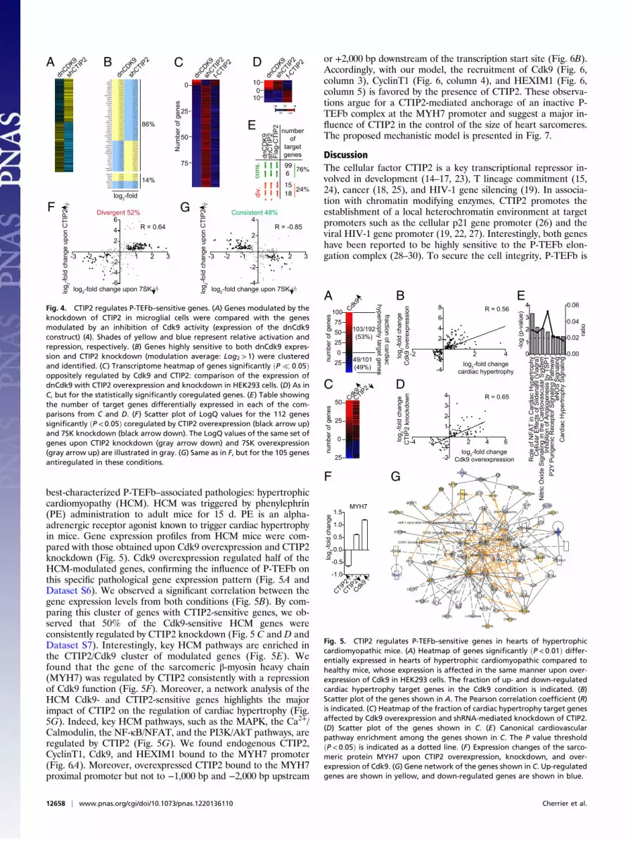
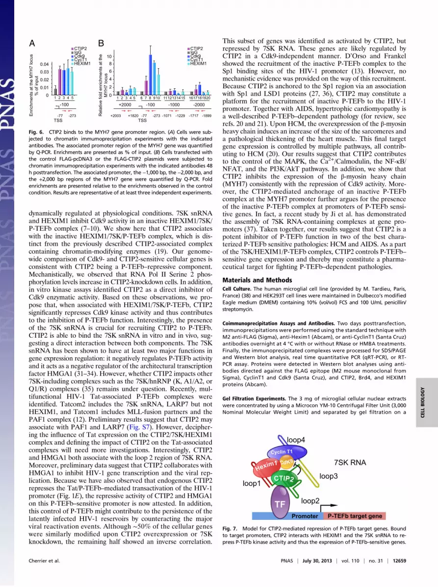
best-characterized P-TEFb–associated pathologies: hypertrophiccardiomyopathy (HCM). HCM was triggered by phenylephrin(PE) administration to adult mice for 15 d. PE is an alpha-adrenergic receptor agonist known to trigger cardiac hypertrophyin mice. Gene expression profiles from HCM mice were com-pared with those obtained upon Cdk9 overexpression and CTIP2knockdown (Fig. 5). Cdk9 overexpression regulated half of theHCM-modulated genes, confirming the influence of P-TEFb onthis specific pathological gene expression pattern (Fig. 5A andDataset S6). We observed a significant correlation between thegene expression levels from both conditions (Fig. 5B). By com-paring this cluster of genes with CTIP2-sensitive genes, we ob-served that 50% of the Cdk9-sensitive HCM genes wereconsistently regulated by CTIP2 knockdown (Fig. 5 C and D andDataset S7). Interestingly, key HCM pathways are enriched inthe CTIP2/Cdk9 cluster of modulated genes (Fig. 5E). Wefound that the gene of the sarcomeric β-myosin heavy chain(MYH7) was regulated by CTIP2 consistently with a repressionof Cdk9 function (Fig. 5F). Moreover, a network analysis of theHCM Cdk9- and CTIP2-sensitive genes highlights the majorimpact of CTIP2 on the regulation of cardiac hypertrophy (Fig.5G). Indeed, key HCM pathways, such as the MAPK, the Ca2+/Calmodulin, the NF-κB/NFAT, and the PI3K/AkT pathways, areregulated by CTIP2 (Fig. 5G). We found endogenous CTIP2,CyclinT1, Cdk9, and HEXIM1 bound to the MYH7 promoter(Fig. 6A). Moreover, overexpressed CTIP2 bound to the MYH7proximal promoter but not to −1,000 bp and −2,000 bp upstream
or +2,000 bp downstream of the transcription start site (Fig. 6B).Accordingly, with our model, the recruitment of Cdk9 (Fig. 6,column 3), CyclinT1 (Fig. 6, column 4), and HEXIM1 (Fig. 6,column 5) is favored by the presence of CTIP2. These observa-tions argue for a CTIP2-mediated anchorage of an inactive P-TEFb complex at the MYH7 promoter and suggest a major in-fluence of CTIP2 in the control of the size of heart sarcomeres.The proposed mechanistic model is presented in Fig. 7.
DiscussionThe cellular factor CTIP2 is a key transcriptional repressor in-volved in development (14–17, 23), T lineage commitment (15,24), cancer (18, 25), and HIV-1 gene silencing (19). In associa-tion with chromatin modifying enzymes, CTIP2 promotes theestablishment of a local heterochromatin environment at targetpromoters such as the cellular p21 gene promoter (26) and theviral HIV-1 gene promoter (19, 22, 27). Interestingly, both geneshave been reported to be highly sensitive to the P-TEFb elon-gation complex (28–30). To secure the cell integrity, P-TEFb is
Adn
CDK9
shCTIP2 B
dnCDK9
shCTIP2
log2-fold
86%
14%
IL8 4,9840 -3,2898DUSP1 2,4801 -1,7650HMGA2 2,1859 -1,7555G0S2 2,1124 -3,5520HBEGF 2,0573 -1,9356CYR61 2,0194 -4,0603CCND1 1,8583 -3,6303EHD1 1,7854 -1,6533
SERPINE1 1,6580 -2,9374DUSP4 1,5337 -1,0242ARPC4 1,5206 -1,6454ACTN4 1,5134 -1,3832SLC6A6 1,4707 -1,6831SFRP1 1,4366 -3,8496FJX1 1,4357 -1,0375FGF2 1,3974 -2,4767RND3 1,3748 -2,5728ETV5 1,3742 -4,7256CD59 1,3276 -1,8403
STARD13 1,3169 -2,3871TRIO 1,3033 -2,0963SF3B4 1,2752 -1,0457PKNOX1 1,2395 -1,3132IL12A 1,2216 -1,2442RUNX1 1,2155 -1,2131RAI14 1,1897 -1,6661SPHK1 1,1339 -2,0208MAP4K4 1,1014 -1,2199PLAUR 1,0993 -2,9720
GADD45A 1,0901 -1,2035BCAR3 1,0775 -2,4539NMT2 1,0691 -1,3115
SEPHS2 -1,0839 1,9197PLS1 -1,0985 1,6723
CHMP2B -1,1428 1,3789HOXD11 -1,1494 3,6122SSBP2 -1,1669 2,3579MPPED2 -1,2030 2,3546PRKAR2B -1,2363 1,7662P2RY5 -1,2588 4,5076LGR4 -1,3680 1,0595CDS1 -1,4375 3,0040CABC1 -1,4738 1,2014EYA1 -1,4978 4,5732FLRT3 -1,5133 6,2711AGL -1,5837 1,3625
ARMCX5 -1,7104 1,1657BAMBI -1,7799 2,2250
HSPA1A 1,3314 2,0354RORA 1,1396 1,0053MDC1 1,0665 1,1114AMFR 1,0124 1,9111
C5orf13 -1,0453 -2,6202TCF12 -1,0554 -1,1590IFIH1 -1,0947 -1,8616NAGK -1,2527 -1,4907
D
shCTIP
2f-C
TIP2
dnCDK9
010
10
C
shCTIP
2f-C
TIP2
dnCDK9
Num
ber o
f gen
es
0
25
50
75
-3 -2 -1 1 2 3
-6-4-2
246
log2-fold change upon 7SK log 2-f
old
chan
ge u
pon
CTI
P2
R = 0.64
Divergent 52%F
-3 -2 -1 1 2 3
-4
-2
2
4R = -0.85
log2-fold change upon 7SK log 2-f
old
chan
ge u
pon
CTI
P2 Consistent 48%G
E
dnC
DK
9sh
CTI
P2
Flag
-CTI
P2
996
1518
cons
.di
v.
76%
24%
numberof
targetgenes
Fig. 4. CTIP2 regulates P-TEFb–sensitive genes. (A) Genes modulated by theknockdown of CTIP2 in microglial cells were compared with the genesmodulated by an inhibition of Cdk9 activity (expression of the dnCdk9construct) (4). Shades of yellow and blue represent relative activation andrepression, respectively. (B) Genes highly sensitive to both dnCdk9 expres-sion and CTIP2 knockdown (modulation average: Log2 > 1) were clusteredand identified. (C) Transcriptome heatmap of genes significantly ðP � 0:05Þoppositely regulated by Cdk9 and CTIP2: comparison of the expression ofdnCdk9 with CTIP2 overexpression and knockdown in HEK293 cells. (D) As inC, but for the statistically significantly coregulated genes. (E) Table showingthe number of target genes differentially expressed in each of the com-parisons from C and D. (F) Scatter plot of LogQ values for the 112 genessignificantly ðP < 0:05Þ coregulated by CTIP2 overexpression (black arrow up)and 7SK knockdown (black arrow down). The LogQ values of the same set ofgenes upon CTIP2 knockdown (gray arrow down) and 7SK overexpression(gray arrow up) are illustrated in gray. (G) Same as in F, but for the 105 genesantiregulated in these conditions.
-2 2 4
-4-2
2468
log2-fold changecardiac hypertrophy
log 2-f
old
chan
geC
dk9
over
expr
essi
on R = 0.56
B
-4 -2 2 4 6
-2-1
1234
log2-fold changeCdk9 overexpression
log 2-f
old
chan
geC
TIP
2 kn
ockd
own R = 0.65
D
Rol
e of
NFA
T in
Car
diac
Hyp
ertro
phy
Cel
lula
r Effe
cts
of S
ilden
afil
(Via
gra)
Nitr
ic O
xide
Sig
nalin
g in
the
Car
diov
ascu
lar S
yste
mIn
hibi
tion
of A
ngio
gene
sis
by T
SP1
P2Y
Purig
enic
Rec
epto
r Sig
nalin
g Pa
thw
ayeN
OS
Sign
alin
gC
ardi
ac H
yper
troph
y Si
gnal
ing
0
1
2
3
4
0.00
0.02
0.04
0.06
-log
(p-v
alue
)
ratio
E
G
A
255075
100
250
fraction of cardiachypertrophy target genes
num
ber o
f gen
es103/192(53%)
49/101(49%)
Cdk9
C50
25
0
25
num
ber o
f gen
es
Cdk9CTIP
2
F
-1.0
-0.5
0.0
0.5
1.0
1.5
log 2-f
old
chan
ge
MYH7
CTIP2CTIP
2Cdk
9
Fig. 5. CTIP2 regulates P-TEFb–sensitive genes in hearts of hypertrophiccardiomyopathic mice. (A) Heatmap of genes significantly ðP < 0:01Þ differ-entially expressed in hearts of hypertrophic cardiomyopathic compared tohealthy mice, whose expression is affected in the same manner upon over-expression of Cdk9 in HEK293 cells. The fraction of up- and down-regulatedcardiac hypertrophy target genes in the Cdk9 condition is indicated. (B)Scatter plot of the genes shown in A. The Pearson correlation coefficient (R)is indicated. (C) Heatmap of the fraction of cardiac hypertrophy target genesaffected by Cdk9 overexpression and shRNA-mediated knockdown of CTIP2.(D) Scatter plot of the genes shown in C. (E) Canonical cardiovascularpathway enrichment among the genes shown in C. The P value thresholdðP < 0:05Þ is indicated as a dotted line. (F) Expression changes of the sarco-meric protein MYH7 upon CTIP2 overexpression, knockdown, and over-expression of Cdk9. (G) Gene network of the genes shown in C. Up-regulatedgenes are shown in yellow, and down-regulated genes are shown in blue.
12658 | www.pnas.org/cgi/doi/10.1073/pnas.1220136110 Cherrier et al.
dynamically regulated at physiological conditions. 7SK snRNAand HEXIM1 inhibit Cdk9 activity in an inactive HEXIM1/7SK/P-TEFb complex (7–10). We show here that CTIP2 associateswith the inactive HEXIM1/7SK/P-TEFb complex, which is dis-tinct from the previously described CTIP2-associated complexcontaining chromatin-modifying enzymes (19). Our genome-wide comparison of Cdk9- and CTIP2-sensitive cellular genes isconsistent with CTIP2 being a P-TEFb–repressive component.Mechanistically, we observed that RNA Pol II Serine 2 phos-phorylation levels increase in CTIP2-knockdown cells. In addition,in vitro kinase assays identified CTIP2 as a direct inhibitor ofCdk9 enzymatic activity. Based on these observations, we pro-pose that, when associated with HEXIM1/7SK/P-TEFb, CTIP2significantly represses Cdk9 kinase activity and thus contributesto the inhibition of P-TEFb function. Interestingly, the presenceof the 7SK snRNA is crucial for recruiting CTIP2 to P-TEFb.CTIP2 is able to bind the 7SK snRNA in vitro and in vivo, sug-gesting a direct interaction between both components. The 7SKsnRNA has been shown to have at least two major functions ingene expression regulation: it negatively regulates P-TEFb activityand it acts as a negative regulator of the architectural transcriptionfactor HMGA1 (31–34). However, whether CTIP2 impacts other7SK-including complexes such as the 7SK/hnRNP (K, A1/A2, orQ1/R) complexes (35) remains under question. Recently, mul-tifunctional HIV-1 Tat-associated P-TEFb complexes wereidentified. Tatcom2 includes the 7SK snRNA, LARP7 but notHEXIM1, and Tatcom1 includes MLL-fusion partners and thePAF1 complex (12). Preliminary results suggest that CTIP2 mayassociate with PAF1 and LARP7 (Fig. S7). However, decipher-ing the influence of Tat expression on the CTIP2/7SK/HEXIM1complex and defining the impact of CTIP2 on the Tat-associatedcomplexes will need more investigations. Interestingly, CTIP2and HMGA1 both associate with the loop 2 region of 7SK RNA.Moreover, preliminary data suggest that CTIP2 collaborates withHMGA1 to inhibit HIV-1 gene transcription and the viral rep-lication. Because we have also observed that endogenous CTIP2represses the Tat/P-TEFb–mediated transactivation of the HIV-1promoter (Fig. 1E), the repressive activity of CTIP2 and HMGA1on this P-TEFb–sensitive promoter is now attested. In addition,this control of P-TEFb might contribute to the persistence of thelatently infected HIV-1 reservoirs by counteracting the majorviral reactivation events. Although ∼50% of the cellular geneswere similarly modified upon CTIP2 overexpression or 7SKknockdown, the remaining half showed an inverse correlation.
This subset of genes was identified as activated by CTIP2, butrepressed by 7SK RNA. These genes are likely regulated byCTIP2 in a Cdk9-independent manner. D’Orso and Frankelshowed the recruitment of the inactive P-TEFb complex to theSp1 binding sites of the HIV-1 promoter (13). However, nomechanistic evidence was provided on the way of this recruitment.Because CTIP2 is anchored to the Sp1 region via an associationwith Sp1 and LSD1 proteins (27, 36), CTIP2 may constitute aplatform for the recruitment of inactive P-TEFb to the HIV-1promoter. Together with AIDS, hypertrophic cardiomyopathy isa well-described P-TEFb–dependent pathology (for review, seerefs. 20 and 21). Upon HCM, the overexpression of the β-myosinheavy chain induces an increase of the size of the sarcomeres anda pathological thickening of the heart muscle. This final targetgene expression is controlled by multiple pathways, all contrib-uting to HCM (20). Our results suggest that CTIP2 contributesto the control of the MAPK, the Ca2+/Calmodulin, the NF-κB/NFAT, and the PI3K/AkT pathways. In addition, we show thatCTIP2 inhibits the expression of the β-myosin heavy chain(MYH7) consistently with the repression of Cdk9 activity. More-over, the CTIP2-mediated anchorage of an inactive P-TEFbcomplex at the MYH7 promoter further argues for the presenceof the inactive P-TEFb complex at promoters of P-TEFb sensi-tive genes. In fact, a recent study by Ji et al. has demonstratedthe assembly of 7SK RNA-containing complexes at gene pro-moters (37). Taken together, our results suggest that CTIP2 is apotent inhibitor of P-TEFb function in two of the best chara-terized P-TEFb sensitive pathologies: HCM and AIDS. As a partof the 7SK/HEXIM1/P-TEFb complex, CTIP2 controls P-TEFb–sensitive gene expression and thereby may constitute a pharma-ceutical target for fighting P-TEFb–dependent pathologies.
Materials and MethodsCell Culture. The human microglial cell line (provided by M. Tardieu, Paris,France) (38) and HEK293T cell lines were maintained in Dulbecco’s modifiedEagle medium (DMEM) containing 10% (vol/vol) FCS and 100 U/mL penicillin/streptomycin.
Coimmunoprecipitation Assays and Antibodies. Two days posttransfection,immunoprecipitations were performed using the standard technique withM2 anti-FLAG (Sigma), anti-Hexim1 (Abcam), or anti-CyclinT1 (Santa Cruz)antibodies overnight at 4 °C with or without RNase or HMBA treatments.Finally, the immunoprecipitated complexes were processed for SDS/PAGEand Western blot analysis, real time quantitative PCR (qRT-PCR), or RT-PCR assay. Proteins were detected in Western blot analyses using anti-bodies directed against the FLAG epitope (M2 mouse monoclonal fromSigma), CyclinT1 and Cdk9 (Santa Cruz), and CTIP2, Brd4, and HEXIM1proteins (Abcam).
Gel Filtration Experiments. The 3 mg of microglial cellular nuclear extractswere concentrated by using a Microcon YM-10 Centrifugal Filter Unit (3,000Nominal Molecular Weight Limit) and separated by gel filtration on a
B
TSS -77 -273
0
2
4
6
Rel
ativ
e fo
ld e
nric
hmen
ts a
t the
MY
H7
locu
s
810
1112131415 1617181920
-1000 -2000
-1071 -1229 -1717 -1899
1 2
+2000
+2003 +1820
6 7 8 9
-100 103 4 5
Cdk9 CycT1 HEXIM1
CTIP2 IgG
ACdk9 CycT1 HEXIM1
CTIP2 IgG
0
0.01
0.02
0.03
Enr
ichm
ents
at t
he M
YH
7 lo
cus
% o
f inp
ut
0.04
1 2
TSS -77 -273
3 4
-100 5
Fig. 6. CTIP2 binds to the MYH7 gene promoter region. (A) Cells were sub-jected to chromatin immumoprecipitation experiments with the indicatedantibodies. The associated promoter region of the MYH7 gene was quantifiedby Q-PCR. Enrichments are presented as % of input. (B) Cells transfected withthe control FLAG-pcDNA3 or the FLAG-CTIP2 plasmids were subjected tochromatin immumoprecipitation experiments with the indicated antibodies 48h posttransfection. The associated promoter, the −1,000 bp, the −2,000 bp, andthe +2,000 bp regions of the MYH7 gene were quantified by Q-PCR. Foldenrichments are presented relative to the enrichments observed in the controlcondition. Results are representative of at least three independent experiments.
loop1
loop2
loop3
loop4
7SK RNA
Fig. 7. Model for CTIP2-mediated repression of P-TEFb target genes. Boundto target promoters, CTIP2 interacts with HEXIM1 and the 7SK snRNA to re-press P-TEFb kinase activity and thus the expression of P-TEFb–sensitive genes.
Cherrier et al. PNAS | July 30, 2013 | vol. 110 | no. 31 | 12659
CELL
BIOLO
GY
Superose 6 PC 3.2/30 column (Amersham Biosciences) as previously described(39). The 29 fractions collected were accessed by SDS/PAGE and Western blotanalysis. Upon RT-PCR amplification, the presence of the 7SK was visualizedby agarose gel electrophoresis.
Micro Scale Thermophoresis. Micro Scale Thermophoresis (MST) experimentswere conducted with Fam6-labeled 7SK L2 RNA or 7SK L2 m137 RNA andincreasing concentrations of FLAG-CTIP2 using a Monolith NT.115 (Nano-temper Technologies) as described previously (40) (Laser-power 20%, Laser-on time 60s, LED-power 30%).
Transcriptome Analyses. Transcriptome analyses were performed using eitherAgilent Human whole-genome array (G2534-60011), Applied Biosystemshuman arrays (Product nos. 4339628 and 4336875), or an Affymetrix Gen-eChip MOE 430 2.0 array according to the manufacturer’s instructions. Dataquality was determined using a QC procedure (41). Data were normalizedusing NeONORM with k = 0.02 (42–44). Subtraction profiling was performedas in refs. 31 and 32 using the CDS test (45).
Generation of Cardiac Hypertrophic Mice. Micropumps (Alzet 2002 OsmoticPumps) were set up to deliver (R)-(-) Phenylephrine hydrochloride (SigmaP6126-5G) at 80 mg·kg−1·day−1 or vehicle buffer only (PBS, 0.002% ascorbic
acid) for 15 d. The micropumps were implanted under the back skin offive C57BL/6N mice for each group under 2% Isofurane (Abbott), 2% inoxygen. Cardiac hypertrophy was validated by the increase of heart weightto body weight ratio in treated animals. Protocols are in accordance withthe ethic recommendations of the Université Pierre et Marie Curie, Paris 6University.
ACKNOWLEDGMENTS. We thank Dr. Olivier Bensaude for scientific discussionsand improvement of the manuscript. S.E. and R.R. are recipients of apostdoctoral fellowship from the Agence Nationale de Recherches sur leSIDA et les Hépatites Virales (ANRS). This work was supported by grantsfrom Centre National de la Recherche Scientifique (CNRS, France), theAgence Nationale de Recherches sur le SIDA et les hépatites virale (ANRS,France), Sidaction, the Institut Universitaire de France (IUF), the Belgian Fundfor Scientific Reseach (FRS-FNRS, Belgium), the Télévie Program of the FRS-FNRS, the Walloon Region (the Excellence Program ‘Cibles’ and WALEO 021/5110 Program), the European AIDS treatment network (NEAT) integrationgrant, the Internationale Brachet Stiftung, the Fondation Roi Baudouin, theUniversity of Franche-Comté, the National Institute of Mental Health (R21MH083585) and the Pennsylvania Department of Health. We thank the Insti-tut Universitaire de Technologie (IUT) Louis Pasteur de Schiltigheim (O.R., C.S.,and V.L.D.). B.V.D. and C.V.L. are Chargé de Recherches and Directeur deRecherches du FRS-FNRS, respectively.
1. Marshall NF, Price DH (1995) Purification of P-TEFb, a transcription factor required forthe transition into productive elongation. J Biol Chem 270(21):12335–12338.
2. Brasier AR (2008) Expanding role of cyclin dependent kinases in cytokine induciblegene expression. Cell Cycle 7(17):2661–2666.
3. Zhou Q, Yik JH (2006) The Yin and Yang of P-TEFb regulation: Implications for humanimmunodeficiency virus gene expression and global control of cell growth and dif-ferentiation. Microbiol Mol Biol Rev 70(3):646–659.
4. Garriga J, Xie H, Obradovic Z, Graña X (2010) Selective control of gene expression byCDK9 in human cells. J Cell Physiol 222(1):200–208.
5. Jang MK, et al. (2005) The bromodomain protein Brd4 is a positive regulatory com-ponent of P-TEFb and stimulates RNA polymerase II-dependent transcription.Mol Cell19(4):523–534.
6. Yang Z, et al. (2005) Recruitment of P-TEFb for stimulation of transcriptional elon-gation by the bromodomain protein Brd4. Mol Cell 19(4):535–545.
7. Michels AA, et al. (2004) Binding of the 7SK snRNA turns the HEXIM1 protein intoa P-TEFb (CDK9/cyclin T) inhibitor. EMBO J 23(13):2608–2619.
8. Nguyen VT, Kiss T, Michels AA, Bensaude O (2001) 7SK small nuclear RNA binds to andinhibits the activity of CDK9/cyclin T complexes. Nature 414(6861):322–325.
9. Yang Z, Zhu Q, Luo K, Zhou Q (2001) The 7SK small nuclear RNA inhibits the CDK9/cyclin T1 kinase to control transcription. Nature 414(6861):317–322.
10. Yik JH, et al. (2003) Inhibition of P-TEFb (CDK9/Cyclin T) kinase and RNA polymerase IItranscription by the coordinated actions of HEXIM1 and 7SK snRNA. Mol Cell 12(4):971–982.
11. He N, et al. (2010) HIV-1 Tat and host AFF4 recruit two transcription elongationfactors into a bifunctional complex for coordinated activation of HIV-1 transcription.Mol Cell 38(3):428–438.
12. Sobhian B, et al. (2010) HIV-1 Tat assembles a multifunctional transcription elonga-tion complex and stably associates with the 7SK snRNP. Mol Cell 38(3):439–451.
13. D’Orso I, Frankel AD (2010) RNA-mediated displacement of an inhibitory snRNPcomplex activates transcription elongation. Nat Struct Mol Biol 17(7):815–821.
14. Ikawa T, et al. (2010) An essential developmental checkpoint for production of theT cell lineage. Science 329(5987):93–96.
15. Li L, Leid M, Rothenberg EV (2010a) An early T cell lineage commitment checkpointdependent on the transcription factor Bcl11b. Science 329(5987):89–93.
16. Arlotta P, et al. (2005) Neuronal subtype-specific genes that control corticospinalmotor neuron development in vivo. Neuron 45(2):207–221.
17. Golonzhka O, et al. (2009) Ctip2/Bcl11b controls ameloblast formation during mam-malian odontogenesis. Proc Natl Acad Sci USA 106(11):4278–4283.
18. Ganguli-Indra G, et al. (2009) CTIP2 expression in human head and neck squamous cellcarcinoma is linked to poorly differentiated tumor status. PLoS ONE 4(4):e5367.
19. Marban C, et al. (2007) Recruitment of chromatin-modifying enzymes by CTIP2 pro-motes HIV-1 transcriptional silencing. EMBO J 26(2):412–423.
20. Harvey PA, Leinwand LA (2011) The cell biology of disease: Cellular mechanisms ofcardiomyopathy. J Cell Biol 194(3):355–365.
21. McKinsey TA, Kass DA (2007) Small-molecule therapies for cardiac hypertrophy:Moving beneath the cell surface. Nat Rev Drug Discov 6(8):617–635.
22. Rohr O, et al. (2003) Recruitment of Tat to heterochromatin protein HP1 via in-teraction with CTIP2 inhibits human immunodeficiency virus type 1 replication inmicroglial cells. J Virol 77(9):5415–5427.
23. Albu DI, et al. (2011) Transcription factor Bcl11b controls selection of invariant naturalkiller T-cells by regulating glycolipid presentation in double-positive thymocytes. ProcNatl Acad Sci USA 108(15):6211–6216.
24. Li P, et al. (2010) Reprogramming of T cells to natural killer-like cells upon Bcl11bdeletion. Science 329(5987):85–89.
25. De Keersmaecker K, et al. (2010) The TLX1 oncogene drives aneuploidy in T celltransformation. Nat Med 16(11):1321–1327.
26. Cherrier T, et al. (2009) p21(WAF1) gene promoter is epigenetically silenced by CTIP2and SUV39H1. Oncogene 28(38):3380–3389.
27. Marban C, et al. (2005) COUP-TF interacting protein 2 represses the initial phase ofHIV-1 gene transcription in human microglial cells. Nucleic Acids Res 33(7):2318–2331.
28. Giraud S, Hurlstone A, Avril S, Coqueret O (2004) Implication of BRG1 and cdk9 in theSTAT3-mediated activation of the p21waf1 gene. Oncogene 23(44):7391–7398.
29. Gomes NP, et al. (2006) Gene-specific requirement for P-TEFb activity and RNApolymerase II phosphorylation within the p53 transcriptional program. Genes Dev20(5):601–612.
30. Wei P, Garber ME, Fang SM, Fischer WH, Jones KA (1998) A novel CDK9-associatedC-type cyclin interacts directly with HIV-1 Tat and mediates its high-affinity, loop-specific binding to TAR RNA. Cell 92(4):451–462.
31. Eilebrecht S, et al. (2011) 7SK small nuclear RNA directly affects HMGA1 function intranscription regulation. Nucleic Acids Res 39(6):2057–2072.
32. Eilebrecht S, Bécavin C, Léger H, Benecke BJ, Benecke A (2011) HMGA1-dependentand independent 7SK RNA gene regulatory activity. RNA Biol 8(1):143–157.
33. Eilebrecht S, Benecke BJ, Benecke A (2011) 7SK snRNA-mediated, gene-specific co-operativity of HMGA1 and P-TEFb. RNA Biol 8(6):1084–1093.
34. Eilebrecht S, Schwartz C, Rohr O (2013) Non-coding RNAs: Novel players in chromatin-regulation during viral latency. Curr Opin Virol, 10.1016/j.coviro.2013.04.001.
35. Diribarne G, Bensaude O (2009) 7SK RNA, a non-coding RNA regulating P-TEFb,a general transcription factor. RNA Biol 6(2):122–128.
36. Le Douce V, et al. (2012) LSD1 cooperates with CTIP2 to promote HIV-1 transcriptionalsilencing. Nucleic Acids Res 40(5):1904–1915.
37. Ji X, et al. (2013) SR Proteins Collaborate with 7SK and Promoter-Associated NascentRNA to Release Paused Polymerase. Cell 153(4):855–868.
38. Janabi N, Peudenier S, Héron B, Ng KH, Tardieu M (1995) Establishment of humanmicroglial cell lines after transfection of primary cultures of embryonic microglial cellswith the SV40 large T antigen. Neurosci Lett 195(2):105–108.
39. Topark-Ngarm A, et al. (2006) CTIP2 associates with the NuRD complex on the pro-moter of p57KIP2, a newly identified CTIP2 target gene. J Biol Chem 281(43):32272–32283.
40. Eilebrecht S, Wilhelm E, Benecke BJ, Bell B, Benecke AG (2013) HMGA1 directly in-teracts with TAR to modulate basal and Tat-dependent HIV transcription. RNA Biol10(3):436–434.
41. Brysbaert G, Pellay FX, Noth S, Benecke A (2010) Quality assessment of transcriptomedata using intrinsic statistical properties. Genomics Proteomics Bioinformatics 8(1):57–71.
42. Noth S, Brysbaert G, Benecke A (2006) Normalization using weighted negative secondorder exponential error functions (NeONORM) provides robustness against asym-metries in comparative transcriptome profiles and avoids false calls. Genomics Pro-teomics Bioinformatics 4(2):90–109.
43. Noth S, Benecke A; Systems Epigenomics Group (2005) Avoiding inconsistencies overtime and tracking difficulties in Applied Biosystems AB1700/Panther probe-to-geneannotations. BMC Bioinformatics 6:307.
44. Noth S, Brysbaert G, Pellay FX, Benecke A (2006) High-sensitivity transcriptome datastructure and implications for analysis and biologic interpretation. Genomics Pro-teomics Bioinformatics 4(4):212–229.
45. Tchitchek N, et al. (2012) CDS: A fold-change based statistical test for concomitantidentification of distinctness and similarity in gene expression analysis. GenomicsProteomics Bioinformatics 10(3):127–135.
12660 | www.pnas.org/cgi/doi/10.1073/pnas.1220136110 Cherrier et al.