structural and functional macular inter-ocular asymmetry in ...
Upload
independentCategory
view
1download
0
Topical non-steroidal anti-inflammatory agents for diabetic
cystoid macular oedema (Review)
Sahoo S, Barua A, Myint KT, Haq A, Abas ABL, Nair NS
This is a reprint of a Cochrane review, prepared and maintained by The Cochrane Collaboration and published in The Cochrane Library
2015, Issue 2
http://www.thecochranelibrary.com
Topical non-steroidal anti-inflammatory agents for diabetic cystoid macular oedema (Review)
Copyright © 2015 The Cochrane Collaboration. Published by John Wiley & Sons, Ltd.

T A B L E O F C O N T E N T S
1HEADER . . . . . . . . . . . . . . . . . . . . . . . . . . . . . . . . . . . . . . .
1ABSTRACT . . . . . . . . . . . . . . . . . . . . . . . . . . . . . . . . . . . . . .
2PLAIN LANGUAGE SUMMARY . . . . . . . . . . . . . . . . . . . . . . . . . . . . . .
3BACKGROUND . . . . . . . . . . . . . . . . . . . . . . . . . . . . . . . . . . . .
4OBJECTIVES . . . . . . . . . . . . . . . . . . . . . . . . . . . . . . . . . . . . .
4METHODS . . . . . . . . . . . . . . . . . . . . . . . . . . . . . . . . . . . . . .
6RESULTS . . . . . . . . . . . . . . . . . . . . . . . . . . . . . . . . . . . . . . .
Figure 1. . . . . . . . . . . . . . . . . . . . . . . . . . . . . . . . . . . . . . 8
9DISCUSSION . . . . . . . . . . . . . . . . . . . . . . . . . . . . . . . . . . . . .
9AUTHORS’ CONCLUSIONS . . . . . . . . . . . . . . . . . . . . . . . . . . . . . . .
10ACKNOWLEDGEMENTS . . . . . . . . . . . . . . . . . . . . . . . . . . . . . . . .
10REFERENCES . . . . . . . . . . . . . . . . . . . . . . . . . . . . . . . . . . . . .
13DATA AND ANALYSES . . . . . . . . . . . . . . . . . . . . . . . . . . . . . . . . . .
13APPENDICES . . . . . . . . . . . . . . . . . . . . . . . . . . . . . . . . . . . . .
16CONTRIBUTIONS OF AUTHORS . . . . . . . . . . . . . . . . . . . . . . . . . . . . .
16DECLARATIONS OF INTEREST . . . . . . . . . . . . . . . . . . . . . . . . . . . . . .
17SOURCES OF SUPPORT . . . . . . . . . . . . . . . . . . . . . . . . . . . . . . . . .
iTopical non-steroidal anti-inflammatory agents for diabetic cystoid macular oedema (Review)
Copyright © 2015 The Cochrane Collaboration. Published by John Wiley & Sons, Ltd.

[Intervention Review]
Topical non-steroidal anti-inflammatory agents for diabeticcystoid macular oedema
Soumendra Sahoo1, Ankur Barua2 , Kay Thi Myint3, Adnaan Haq4, Adinegara BL Abas5, N S Nair6
1Ophthalmology, Melaka Manipal Medical College, Melaka, Malaysia. 2Department of Community Medicine, International Medical
University (IMU), Kuala Lumpur, Malaysia. 3Ophthalmology, Faculty of Medicine, SEGi University, Sibu, Malaysia. 4Medical School,
St. George’s University of London, London, UK. 5Department of Community Medicine, Melaka-Manipal Medical College, Melaka,
Malaysia. 6Department of Statistics, Manipal University, Manipal, India
Contact address: Soumendra Sahoo, Ophthalmology, Melaka Manipal Medical College, Bukit Baru, Melaka, 75150, Malaysia.
Editorial group: Cochrane Eyes and Vision Group.
Publication status and date: New, published in Issue 2, 2015.
Review content assessed as up-to-date: 12 January 2015.
Citation: Sahoo S, Barua A, Myint KT, Haq A, Abas ABL, Nair NS. Topical non-steroidal anti-inflammatory agents
for diabetic cystoid macular oedema. Cochrane Database of Systematic Reviews 2015, Issue 2. Art. No.: CD010009. DOI:
10.1002/14651858.CD010009.pub2.
Copyright © 2015 The Cochrane Collaboration. Published by John Wiley & Sons, Ltd.
A B S T R A C T
Background
Diabetic cystoid macular oedema (CMO) is a condition which involves fluid accumulation in the inner portion of the retina. It often
follows changes in retinal blood vessels which enhance the fluid to come out of vessels. Although it may be asymptomatic, symptoms
are primarily painless loss of central vision, often with the complaint of seeing black spots in front of the eye.
It is reported that CMO may resolve spontaneously, or fluctuate for months, before causing loss of vision. If left untreated or undiagnosed,
progression of CMO may lead to permanent visual loss.
It has been noted that patients with diabetic retinopathy have elevated inflammatory markers, and therefore it is likely that inflammation
aids in the progression of vascular disease in these patients. Several topical non-steroidal anti-inflammatory drugs (NSAIDs) such as
ketorolac 0.5%, bromfenac 0.09%, and nepafenac 0.1%, have therefore also been used topically to treat chronic diabetic CMO. Hence
this review was conducted to find out the effects of topical NSAIDs in diabetic CMO.
Objectives
To assess the effects of topical non-steroidal anti-inflammatory drugs (NSAIDs) for diabetic cystoid macular oedema (CMO).
Search methods
We searched CENTRAL (which contains the Cochrane Eyes and Vision Group Trials Register) (2014, Issue 12), Ovid MEDLINE, Ovid
MEDLINE In-Process and Other Non-Indexed Citations, Ovid MEDLINE Daily, Ovid OLDMEDLINE (January 1946 to January
2015), EMBASE (January 1980 to January 2015), Latin American and Caribbean Health Sciences Literature Database (LILACS) (Jan-
uary 1982 to January 2015), the ISRCTN registry (www.isrctn.com/editAdvancedSearch), ClinicalTrials.gov (www.clinicaltrials.gov)
and the WHO International Clinical Trials Registry Platform (ICTRP) (www.who.int/ictrp/search/en). We did not use any date or
language restrictions in the electronic searches for trials. We last searched the electronic databases on 12 January 2015.
1Topical non-steroidal anti-inflammatory agents for diabetic cystoid macular oedema (Review)
Copyright © 2015 The Cochrane Collaboration. Published by John Wiley & Sons, Ltd.
Selection criteria
Randomised controlled trials (RCTs) and quasi-RCTs investigating the effects of topically applied NSAIDs in the treatment of people
with diabetic CMO aged 18 years of age or over.
Data collection and analysis
Two review authors independently assessed trial eligibility and screened all available titles and abstracts for inclusion. There were no
discrepancies and we did not have to contact trial investigators for missing data.
Main results
We did not identify any RCTs matching the inclusion criteria for this review.
Authors’ conclusions
The review did not identify any RCTs investigating the effects of topical NSAIDs in the treatment of diabetic CMO. Most of the studies
identified through the electronic searches had been conducted to analyse the effect of topical NSAIDs for pseudophakic CMO.In the
absence of high quality evidence, clinicians need to use their clinical judgement and other low level evidence, such as observational
non-randomised trials, to decide whether to use topical NSAIDs in cases of diabetic CMO.
More research is needed to better understand the cause of this condition and its pathophysiology. This systematic review has identified
the need for well designed, adequately powered RCTs to assess possible beneficial and adverse effects of topical NSAIDs in people with
diabetic CMO. Future trials should aim to include a large sample size with an adequate follow-up period of up to one year.
P L A I N L A N G U A G E S U M M A R Y
Topical non-steroidal anti-inflammatory agents for diabetic cystoid macular oedema
Review question
We reviewed the evidence about the effect of non-steroidal anti-inflammatory drugs for diabetic cystoid macular oedema.
Background
Diabetic retinopathy is a frequent cause of blindness in adults aged between 20 and 74 years. The major cause of vision impairment in
those with diabetic retinopathy is the accumulation of fluid in the central part of the retina (macula) known as cystoid macular oedema
(CMO). CMO is the chronic and diffuse variety of diabetic macular oedema (DMO).The use of topical anti-inflammatory agents has
been suggested as a potential treatment for diabetic CMO.
We aimed to review randomised controlled trials (RCTs) and quasi-RCTs (these are clinical research studies, which give good quality
evidence on the effects of interventions) that investigated the effects of various topically applied non-steroidal anti-inflammatory drugs
(NSAIDs) in treating diabetic CMO, and evaluate whether significant benefits have occurred with topical NSAIDs.
We reviewed the evidence on the effect of locally applied NSAID eye preparations on restoring vision in people with diabetic CMO.
Although various topical NSAIDs have been used to treat diabetic CMO, namely bromfenac 0.09%, nepafenac 0.1% and ketorolac
0.5%, we did not find any RCTs or quasi-RCTs that were eligible for this review. We also found that most of the studies identified
through the electronic searches had been conducted to analyse the effect of topical NSAIDs for pseudophakic CMO.
Greater research is required to understand the effects of topical NSAIDs on diabetic CMO. We would recommend a RCT to assess the
effects of topical NSAIDs in patients with diabetic CMO. The trial would need to have a follow-up of at least one year, and include a
large sample size and a robust design in order to assess any potential long-term beneficial or adverse effects of locally applied NSAIDs.
Search date
The evidence is current to January 2015.
2Topical non-steroidal anti-inflammatory agents for diabetic cystoid macular oedema (Review)
Copyright © 2015 The Cochrane Collaboration. Published by John Wiley & Sons, Ltd.
B A C K G R O U N D
Description of the condition
Diabetes mellitus, especially Type 2, is escalating (Mokdad 2001)
and is estimated to reach epidemic proportions around the world
in the next 25 years (Bonow 2004). The prevalence of diabetes in
adults worldwide was estimated at 4.0% in 1995 and is expected
to rise to 5.4% by the year 2025 (Cockram 2000; King 1998).
Diabetic retinopathy is a known complication of diabetes mellitus
(Xiao-Ling 2006), and is increasingly becoming a major cause
of blindness throughout the world (Congdon 2003; Viswanath
2003).
Cystoid macular oedema (CMO) is a condition where accumula-
tion of fluid occurs in the central part of the retina, largely due to
capillary leakage. Although the most common cause of CMO is
cataract surgery and other intraocular surgeries, it has also been ob-
served in various other ocular conditions such as diabetic retinopa-
thy, age-related macular degeneration, uveitis, and eye injury etc.
In diabetic CMO, the cystoid changes usually occur in cases of dif-
fuse and chronic diabetic macular oedema (DMO) (Rotsos 2008).
Theories of the pathogenesis of CMO have looked at mechani-
cal factors, such as tractional forces on the macula and disruption
of the vitreoretinal interface (Rotsos 2008). However, the most
accepted theory to date is vascular leakage and retinal oedema,
initiated by the diffusion of mediators like prostaglandins being
released in the eye (Scholl 2010). This theory is supported by evi-
dence that cyclo-oxygenase inhibitors reduce the incidence of an-
giographic CMO. Although natural history studies of pseudopha-
kic CMO have shown that the majority of cases resolve sponta-
neously, one natural history study on diabetic CMO has shown
the persistence of cystoid spaces, resulting in a severe decrease in
visual acuity (Coscas 1984).
It is difficult to know the true incidence of CMO. Whilst subtle
CMO is difficult to identify clinically, there may also be other
factors that affect the accuracy of incidence estimates. New CMO
is normally reported through the surgeon’s findings or via fluores-
cein angiography and optical coherence tomography.
Although CMO is often symptomatic in terms of visual impair-
ment in either eye, it may be asymptomatic in some cases. It is
reported that CMO may resolve spontaneously, or fluctuate for
months before causing severe loss of vision, which often results in
diminution of visual acuity to 20/200 level (Massin-Korobelnik
1994). Fundoscopy usually shows an altered foveal reflex with a
honeycomb appearance of the macula. In cases where the diagnosis
of CMO is unclear, fundus fluorescein angiography may be used.
The classical angiography picture is a ’flower petal’ appearance at
the macula. The amount of macular oedema can also be detected
by a non-invasive procedure called optical coherence tomography.
Various treatment options are available for diabetic CMO. The
mainstays of treatment are grid photocoagulation (ETDRS 1987),
intravitreal steroids (Grover 2008), vitrectomy (Otani 2002), and
the most recent, intravitreal vascular endothelial growth factor
(Haritoglou 2006).
Description of the intervention
Medical therapies for diabetic CMO have included two broad
classes of agents: anti-inflammatory drugs and agents with molec-
ular targets (Boscia 2010). It has been found that patients with
diabetic retinopathy have elevated inflammatory markers. Thus
it is likely that inflammation aids in the progression of vascular
disease in these patients (Ke 2000; Meleth 2005). Several topical
non-steroidal anti-inflammatory drugs (NSAIDs) such as ketoro-
lac 0.5%, bromfenac 0.09%, and nepafenac 0.1%, have also been
used to treat chronic diabetic CMO. A Cochrane systematic re-
view found two trials with topical ketorolac 0.5% ophthalmic so-
lution that had a positive effect for treating chronic CMO follow-
ing cataract surgery, and two trials that revealed no significant dif-
ference between the intervention and control groups (Sivaprasad
2005).
How the intervention might work
Topical NSAIDs are commonly prescribed in ophthalmic practice
for their anti-inflammatory property. In diabetic CMO, there is
extracellular fluid accumulation and retinal oedema which is sec-
ondary to disruption of the blood retinal barrier (Gardner 2002).
Studies have also demonstrated an association between CMO and
inflammation mediated by prostaglandins (Bazan 1990; Miyake
2002; Scholl 2010). In the eye, prostaglandins are synthesised in
the ciliary body and iris, causing vasodilatation and increasing vas-
cular permeability with disruption of the blood-ocular barrier with
leukocyte migration, which results in oedema formation (Miyake
2002).
NSAIDs act as potent inhibitors for cyclo-oxygenase enzymes,
an active component of the inflammatory process involved in
prostaglandin synthesis. When administered topically, NSAIDs
achieve therapeutic levels in the aqueous humour, and are capable
of a reduction in the synthesis of prostaglandins in the ciliary body
and iris. More frequent administration of topical NSAIDs with
longer duration of treatment leads to higher aqueous levels (Bucci
2007). Three topical NSAIDs, ketorolac 0.4%, bromfenac 0.09%
and nepafenac 0.1% were proven to penetrate into the vitreous
cavity, and ketorolac lowers the vitreous level of Prostaglandin
E2 (PGE2) (Heier 2009) which is reportedly associated with va-
sodilatation and partial disruption of the blood-ocular barrier
(Quaranta 2013). It has been suggested that vasogenesis as well
3Topical non-steroidal anti-inflammatory agents for diabetic cystoid macular oedema (Review)
Copyright © 2015 The Cochrane Collaboration. Published by John Wiley & Sons, Ltd.

as vasogenic macular oedema can also be strategically controlled
by administration of anti-inflammatory drugs such as NSAIDs
(Boscia 2010).
Why it is important to do this review
Diabetic retinopathy is found to be the most frequent cause of
new cases of blindness in adults aged 20 to 74 years (Klein 1984).
Diabetic CMO is one of the factors for severe vision impairment
in patients with diabetic retinopathy. DMO is the leading cause of
visual impairment that occurs with diabetic retinopathy (Girach
2007). Diabetic CMO is more commonly found where there is
diffuse and chronic DMO.
As with most treatments mentioned earlier, however, there are lim-
itations and risks, most notably their invasive nature. The use of a
topical alternative would therefore be markedly safer and easier, if
proven to be effective. A few studies have reported the beneficial ef-
fect of topical NSAIDs in treating diabetic CMO as well as DMO;
however there is currently no available systematic appraisal of evi-
dence of the safety and effectiveness of topical NSAIDs for cases of
diabetic CMO. A systematic review would assist in analysing the
effects of topical NSAIDs in reducing or resolving diabetic CMO.
O B J E C T I V E S
To assess the effects of topical non-steroidal anti-inflammatory
drugs (NSAIDs) for diabetic cystoid macular oedema (CMO).
M E T H O D S
Criteria for considering studies for this review
Types of studies
We planned to include all randomised controlled trials (RCTs). We
also planned to include quasi-RCTs if evidence of effects (benefits
or harms) could not be adequately studied in RCTs and only if
there was sufficient evidence that intervention and control groups
were similar at baseline.
Types of participants
We did not take into consideration gender and race when selecting
trials, although participants had to be over the age of 18 years. We
included participants that had diabetic CMO diagnosed clinically.
We did not exclude from this review participants who were non-
responsive to previous treatment (i.e. photocoagulation).
Types of interventions
We planned to include trials where topical NSAIDs were compared
to placebo, no treatment, and other modalities of treatment.
Types of outcome measures
Primary outcomes
1. The primary outcome for this review was 2 or more lines
improvement of visual acuity from baseline (Early Treatment
Diabetic Retinopathy Study (ETDRS), Snellen or LogMAR
equivalent) at three months of treatment.
Secondary outcomes
1. Proportion of participants showing improvement in central
retinal thickness, measured with optical coherence tomography
after three months of treatment, as a continuous outcome.
2. Proportion of participants showing persistence of subretinal
fluid with optical coherence tomography after three months of
treatment as a dichotomous outcome.
3. Proportion of participants showing improvement in fundus
fluorescein angiography findings after three months of
treatment. (Improvement is defined by decreased leakage in
fundus fluorescein angiography).
4. Quality of life: we planned to summarise the data on
quality of life by any validated measure (such as National Eye
Institute 25-item Visual Function Questionnaire (NEI VFQ-25)
and Impact of Visual Impairment (IVI) Questionnaire) when
found to be reported in the included studies.
5. Adverse outcomes: we planned to tabulate all adverse effects
related to topical application of NSAIDs for the treatment of
diabetic CMO that are found to be reported in the included
studies.
Search methods for identification of studies
Electronic searches
We searched CENTRAL (which contains the Cochrane Eyes and
Vision Group Trials Register) (2014, Issue 12), Ovid MEDLINE,
Ovid MEDLINE In-Process and Other Non-Indexed Citations,
Ovid MEDLINE Daily, Ovid OLDMEDLINE (January 1946 to
January 2015), EMBASE (January 1980 to January 2015), Latin
American and Caribbean Health Sciences Literature Database
(LILACS) (January 1982 to January 2015), the ISRCTN reg-
istry (www.isrctn.com/editAdvancedSearch), ClinicalTrials.gov (
www.clinicaltrials.gov) and the WHO International Clinical Trials
Registry Platform (ICTRP) (www.who.int/ictrp/search/en). We
did not use any date or language restrictions in the electronic
4Topical non-steroidal anti-inflammatory agents for diabetic cystoid macular oedema (Review)
Copyright © 2015 The Cochrane Collaboration. Published by John Wiley & Sons, Ltd.

searches for trials. We last searched the electronic databases on 12
January 2015.
See: Appendices for details of search strategies for CENTRAL
(Appendix 1), MEDLINE (Appendix 2), EMBASE (Appendix 3),
LILACS (Appendix 4), ISRCTN (Appendix 5), ClinicalTrials.gov
(Appendix 6) and the ICTRP (Appendix 7).
Searching other resources
We handsearched the International Congress of Ophthalmology
from 1990 onwards until the last congress in 2012 to identify
unpublished studies. We contacted organisations and researchers
in the field of ophthalmology, and pharmaceutical companies for
information on current trials. We also checked the reference lists
of all trials identified by the above methods.
Data collection and analysis
Selection of studies
Two review authors (SS, KT) independently assessed trial eligi-
bility and screened all available titles and abstracts for inclusion.
If relevant data from the abstract were difficult to ascertain, the
full-text of the report was retrieved. Two review authors (SS, KT)
assessed the eligibility criteria independently by filling-in the eli-
gibility form that was designed in accordance with the inclusion
criteria. The review authors were unmasked to the trial authors,
institutions and trial results during their assessments. If a disagree-
ment occurred, they were solved by discussion, or if required, a
third review author (AH) was asked to express his view.
As no trials met our inclusion, we will follow the steps below for
future updates.
Data extraction and management
Two review authors (SS, KT) planned to independently extract
data for primary and secondary outcomes onto paper data collec-
tion forms developed by the Cochrane Eyes and Vision Group.
The same two authors then planned to share the responsibility of
entering the data into Review Manager 5 (RevMan 2014) and a
third author (AN) planned to check for errors and inconsisten-
cies. We planned to resolve any differences in data extraction by
discussion and consensus.
We planned to use a standard data extraction form which will
include at least the following items.
1. Method: duration, way of randomisation, allocation
concealment method, masking, country, and setting.
2. Participants: type of sampling, number in comparison
group, age, sex, similarity of group at base line, and losses to
follow-up with reason.
3. Interventions: placebo will be included, interventions (dose,
route and duration), comparison intervention (dose, route and
duration), and co-medication (dose, route and duration).
4. Outcomes: outcomes specified above, any other outcomes
assessed, times of assessment, and length of follow-up.
5. Notes: published or unpublished data, title, authors, source,
contact address, language of publication, year of publication, and
funding sources if any.
Assessment of risk of bias in included studies
Two review authors (AB, AN) planned to independently assess the
risk of bias of the included studies by using the criteria outlined in
Chapter 8 of theCochrane Handbook for Systematic Reviews of In-
terventions (Higgins 2011a). We planned to resolve any disagree-
ments by discussion or by the intervention of a third review author
(SN).
We planned to assess the following five components for each of
the trials: random sequence generation (selection bias); allocation
concealment (selection bias); masking (blinding) of participants
and personnel (performance bias), and masking of outcome as-
sessment (detection bias); incomplete outcome data (attrition bias
through withdrawals, drop outs and protocol deviations); and se-
lective reporting bias. We also planned to assess other sources of
bias as reported in the Cochrane Handbook for Systematic Reviews
of Interventions (Higgins 2011a), such as bias related to the specific
study design, early stoppage of trials, extreme baseline imbalance
or whether the study appeared to have been fraudulent. For each
of these components, we planned to assign one of the following
risk of bias judgements: ’low risk’ of bias, ’high risk’ of bias, or
‘unclear risk’ for uncertain risk of bias. We planned to record the
results in the standard table in Review Manager 5 (RevMan 2014),
and to summarise the findings in a ‘Risk of bias’ table or graph.
Measures of treatment effect
For data analysis, we planned to follow the guidelines set out in
Chapter 9 of the Cochrane Handbook for Systematic Review of In-
terventions (Deeks 2011).
For dichotomous data such as improvement of 2 or more Snellen
lines, persistence of subretinal fluid detected by optical coherence
tomography, improvement in fundus fluorescein angiography and
occurrence of adverse effects, we planned to present the results
using risk ratios (RRs) and their 95% confidence intervals (CIs).
For continuous outcomes such as central retinal thickness and
quality of life we planned to calculate mean difference (MD) if the
outcomes were measured by the same scales within the included
studies. If the same outcomes were measured by different scales,
we planned to use standard mean difference (SMD) with 95%
CIs.
5Topical non-steroidal anti-inflammatory agents for diabetic cystoid macular oedema (Review)
Copyright © 2015 The Cochrane Collaboration. Published by John Wiley & Sons, Ltd.

Unit of analysis issues
In ophthalmic RCTs, the unit of analysis can be either the partic-
ipant or the eye. If the unit of analysis is the eye, it can be one eye,
two eyes or mixed. In two-eyed studies, if both eyes received the
same treatment, we considered these studies as clustered, and if
both eyes received different treatments, they would be considered
as paired. For each trial included, we planned to document the
unit of analysis and study design. If included studies used differ-
ent methods, we planned to estimate the treatment effect at the
study level and perform meta-analysis by using the inverse vari-
ance method.
Dealing with missing data
Where data are missing due to participant drop out, we planned
to conduct a primary analysis based on participants with complete
data. We consider that missing outcomes will not be a problem
if loss to follow-up is documented and judged to be unrelated to
outcomes in both study arms, as per Chapter 16 of the Cochrane
Handbook for Systematic Review of Interventions (Higgins 2011b).
We planned to get full reports from authors where studies are
either published in abstract form or presented at meetings. We
planned to contact the primary investigator in case of missing data
or unclear information in the study reports. We also planned to
consider that missing outcome data are not a problem if the causes
are well documented. However, if the causes of missing data are
not available, we planned to document the possible effects of the
missing participants through a sensitivity analysis.
Assessment of heterogeneity
We planned to use the Chi2 test to assess statistical heterogeneity
and considered P < 0.1 as statistically significant. To quantify the
statistical heterogeneity, we planned to use forest plots and the I2 statistic. We planned to use the following guidelines for inter-
preting I2 values: 0% to 40% as insignificant heterogeneity, 30%
to 60% as moderate heterogeneity, 50% to 90% as substantial
heterogeneity, and 75% to 100% as considerable heterogeneity
(Deeks 2011). We also planned to assess clinical and methodolog-
ical heterogeneity by examining the characteristics and methodol-
ogy section of individual studies.
Assessment of reporting biases
Three review authors (SS, KT, AH) carried out comprehensive
searches to minimise publication and reporting biases, and they
planned to consider the likelihood of these biases. Within studies,
we planned to consider selective outcome reporting as part of the
risk of bias assessment. We planned to compare the ’Methods’ sec-
tion of the fully published paper to the ’Results’ section to ensure
that all of the outcomes which were measured, were reported. We
planned to assess possible publication bias by using funnel plots to
explore the relationship between effect size and study size. We also
planned to look at funnel plots only where we have sufficient trials
i.e. 10 trials or more. We would visually examine them for sym-
metry, with greater symmetry indicating a lower risk of reporting
bias.
Data synthesis
We planned to carry out statistical analysis using Review Manager
5 (RevMan 2014). If there are less than three studies, we would use
a fixed-effect model. If there is minimal statistical heterogeneity
and if there is minimal clinical heterogeneity between the trials, we
planned to combine the results in a meta-analysis using a random-
effects model. If there is considerable heterogeneity (I2 statistic of
50% or more), we will discuss the results in narrative and tabulated
form only. For identifying heterogeneity we would not only rely
on the statistical significance of a Chi2 test, but also examine the
results of the forest plot of the study. We planned to convert Early
Treatment Diabetic Retinopathy Study (ETDRS) letter scores to
logMar for calculations and then use them in the meta-analysis. If
we find studies in which Snellen (decimal) visual acuity is measured
by non-ETDRS or non-logarithmic charts, we would only extract
data if calculations are based on logMar transformed data and then
transformed back to decimals for reporting. If we find studies in
which means and standard deviations (SDs) are computed using
decimal visual acuity, we would not use them in the meta-analysis
but planned to summarise their results in the discussion.
Subgroup analysis and investigation of heterogeneity
We did not perform any subgroup analyses in this review.
Sensitivity analysis
We planned to carry out sensitivity analysis to investigate the ro-
bustness of the results regarding the various components of risk
of bias. We also planned to examine the effect on the primary
outcome of excluding any study judged to be at overall high risk
of bias.
Summary of findings
We planned to create a ‘Summary of findings” table using
GRADEpro software (version 3.6) (GRADEpro 2014) to assess
parameters such as limitations of design, inconsistency, indirect-
ness, imprecision and publication bias. In the table we planned to
include all the available outcomes reported in the included studies.
R E S U L T S
6Topical non-steroidal anti-inflammatory agents for diabetic cystoid macular oedema (Review)
Copyright © 2015 The Cochrane Collaboration. Published by John Wiley & Sons, Ltd.

Description of studies
Results of the search
The electronic searches yielded a total of 294 references (Figure
1). The Trials Search Co-ordinator scanned the search results,
removed 55 duplicates and then removed 180 references which
were not relevant to the scope of the review. We screened the
remaining 59 reports but did not identify any RCTs that met the
inclusion criteria for this review.
7Topical non-steroidal anti-inflammatory agents for diabetic cystoid macular oedema (Review)
Copyright © 2015 The Cochrane Collaboration. Published by John Wiley & Sons, Ltd.
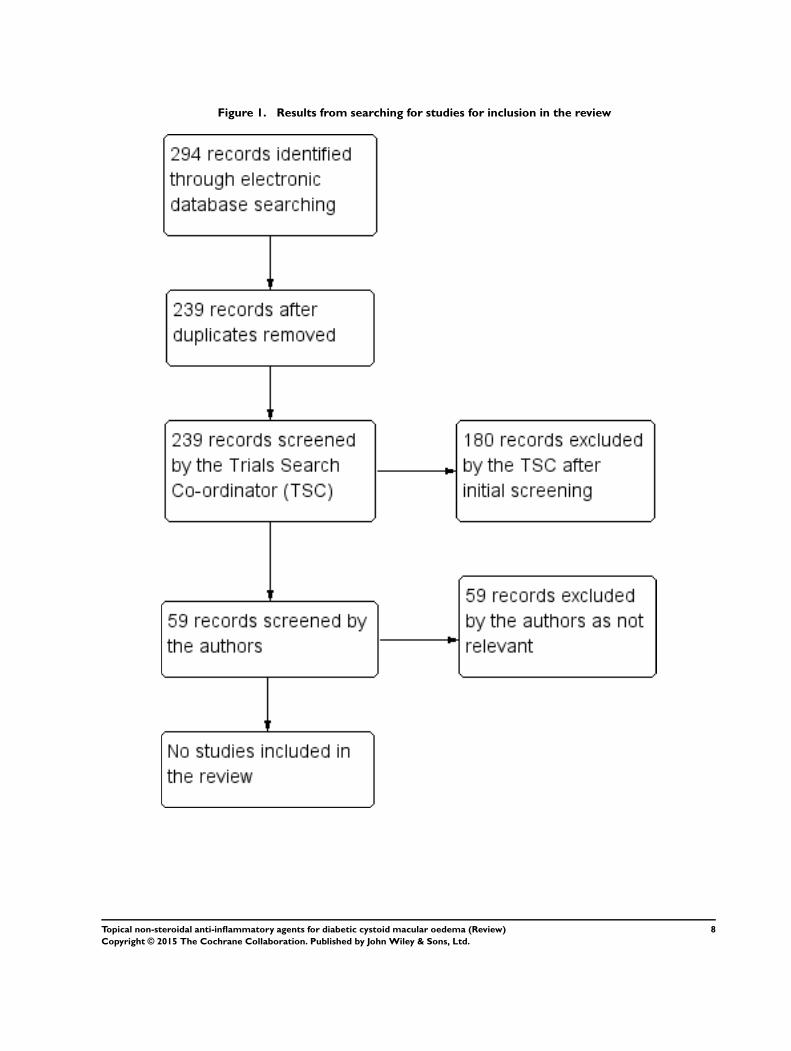
Figure 1. Results from searching for studies for inclusion in the review
8Topical non-steroidal anti-inflammatory agents for diabetic cystoid macular oedema (Review)
Copyright © 2015 The Cochrane Collaboration. Published by John Wiley & Sons, Ltd.

Included studies
We did not identify any RCTs that met the inclusion criteria.
Excluded studies
We did not exclude any RCTs.
Risk of bias in included studies
We did not identify any eligible trials for inclusion in the review.
Effects of interventions
The searches did not identify any RCTs, or any ongoing trials for
inclusion in this review.
D I S C U S S I O N
This review of topical non-steroidal anti-inflammatory drugs
(NSAIDs) for diabetic cystoid macular oedema (CMO) failed to
identify any randomised controlled trials (RCTs) or any ongoing
trials for inclusion in this review. Most of the studies identified
through the electronic searches had been conducted to analyse the
effect of topical NSAIDs for pseudophakic CMO.
A case series study evaluating the effects of topical nepafenac 0.1%
in six eyes with diabetic macular oedema (DMO) showed that
there was significant reduction in average foveal thickness from
417 µm to 267 µm, with statistically significant improvement in
mean visual acuity from 0.78 logMAR to 0.67 logMAR after a
mean follow-up period of 178 days (Callanan 2008).
Many studies have shown the benefits of single intravitreal in-
jection of NSAIDs in DMO. A single dose of intravitreal di-
clofenac (500 µg/0.1 mL) in eyes with clinically significant mac-
ular oedema reported a prominent improvement in visual acuity
(Soheilian 2010). Similar results were seen in two studies con-
ducted in eyes with DMO refractory to laser photocoagulation
(Maldonado 2011; Reis 2010) where intravitreal ketorolac (500
µg/0.1 mL and 3000 µg/0.1 mL) were given, respectively.
Summary of main results
This review failed to identify any published trials or ongoing stud-
ies from trial registers reporting the effects and safety of topical
NSAIDs for treating diabetic CMO. Although some case series
studies have suggested the benefit of topical NSAIDs in the treat-
ment of diabetic CMO, the absence of definitive RCTs suggest
that it is an area where more evidence is needed to inform the sci-
entific community as to the benefits and risks of treating diabetic
CMO with topical NSAIDs.
Agreements and disagreements with otherstudies or reviews
Diabetic CMO, a form of chronic CMO, is a challenge observed
in patients with diabetic maculopathy which results in a severe
impairment in visual acuity (Coscas 1984). Unfortunately, how-
ever, there are no RCTs; suggesting that evidence is needed for or
against the use of topical NSAIDs in the affected population.
A Cochrane systematic review evaluating the effects of NSAIDs
for treating pseudophakic CMO reported that topical ketoro-
lac tromethamine 0.5% had a positive effect for treating chronic
pseudophakic CMO (Sivaprasad 2012). Although diabetic CMO
has similar pathophysiology with that of chronic CMO following
cataract surgery, this evidence cannot be used as evidence for the
effects of topical NSAIDs in diabetic CMO.
A U T H O R S ’ C O N C L U S I O N S
Implications for practice
We did not identify any randomised controlled trials (RCTs) of
topical non-steroidal anti-inflammatory drugs (NSAIDs) in dia-
betic cystoid macular oedema (CMO) for inclusion in this review.
There is no evidence to inform that topical NSAIDs are of benefit
to people with diabetic CMO.
Implications for research
There is need for more research to better understand the cause of
this condition and its pathophysiology. This systematic review has
identified the need for well designed, adequately powered RCTs to
assess the effects and adverse effects of topical NSAIDs in people
with diabetic CMO suffering from impaired vision.
Although the literature shows that the incidence of angiographic
pseudophakic CMO may be as high as 9% to 19% (Mentes 2003;
Ursell 1999), and the estimated incidence of DMO is 7% of dia-
betic patients (Ding 2012), the exact incidence of diabetic CMO
is not reported in the literature. Hence, it is difficult to calcu-
late the sample size for future trials, but they should aim at a
large sample size with adequate follow-up. Since diabetic CMO
9Topical non-steroidal anti-inflammatory agents for diabetic cystoid macular oedema (Review)
Copyright © 2015 The Cochrane Collaboration. Published by John Wiley & Sons, Ltd.

is a chronic condition, and studies evaluating the effects of topi-
cal NSAIDs reported improvements in foveal thickness and visual
acuity at around four to six months (Callanan 2008; Hariprasad
2007; Warren 2010), this would suggest that a follow-up of ap-
proximately 12 months would be beneficial to prove the effects of
topical NSAIDs.
A C K N O W L E D G E M E N T S
We would like to acknowledge the Trials Search Co-ordinator for
the Cochrane Eyes and Vision Group (CEVG) for devising and
running the electronic searches. We thank Anupa Shah, Managing
Editor for CEVG for her assistance throughout the development
of the review We thank Maged Habib, Catey Bunce and Scott
Fraser for their comments on the protocol and Jennifer Evans,
Xuan Hui and Eli Pradhan for their comments on the review.
We also thank Prof Datuk, Dr Abdul Razzak Chief Executive
of Melaka Manipal Medical College, Malaysia and Prof Jaspal
Singh Sahota, Dean of Melaka Manipal Medical College, Malaysia
for their support, constructive comments and encouragement in
writing this protocol.
R E F E R E N C E S
Additional references
Bazan 1990
Bazan NG, de Abreu MT, Bazan HE, Belfort R Jr.
Arachidonic acid cascade and platelet-activating factor in
the network of eye inflammatory mediators: therapeutic
implications in uveitis. International Ophthalmology 1990;
14(5-6):335–44.
Bonow 2004
Bonow RO, Gheorghiade M. The diabetes epidemic: a
national and global crisis. American Journal of Medicine
2004;116(5):2–10.
Boscia 2010
Boscia F. Current approaches to the management of diabetic
retinopathy and diabetic macular oedema. Drugs 2010;70
(16):2171–200.
Bucci 2007
Bucci FA Jr, Waterbury LD, Amico LM. Prostaglandin E2
inhibition and aqueous concentration of ketorolac 0.4%
(acular LS) and nepafenac 0.1% (nevanac) in patients
undergoing phacoemulsification. American Journal of
Ophthalmology 2007;144(1):146–7.
Callanan 2008
Callanan D, Williams P. Topical nepafenac in the treatment
of diabetic macular oedema. Clinical Ophthalmology 2008;2
(4):689–92.
Cockram 2000
Cockram CS. The epidemiology of diabetes mellitus in
Asia-Pacific region. Hong Kong Medical Journal 2000;6(1):
43–52.
Congdon 2003
Congdon NG, Friedman DS, Lietman T. Important causes
of visual impairment in the world today. JAMA 2003;2909
(15):2057–60.
Coscas 1984
Coscas G, Gaudric A. Natural course of nonaphakic cystoid
macular edema. Survey of Ophthalmology 1984;28(Suppl):
471–84.
Deeks 2011
Deeks JJ, Higgins JPT, Altman DG (editors). Chapter 9:
Analysing data and undertaking meta-analyses. In: Higgins
JPT, Green S (editors). Cochrane Handbook for Systematic
Reviews of Interventions Version 5.1.0 [updated March
2011]. The Cochrane Collaboration, 2011. Available from
www.cochrane-handbook.org.
Ding 2012
Ding J, Wong TY. Current epidemiology of diabetic
retinopathy and diabetic macular edema. Current Diabetes
Report 2012;12(4):346–54.
ETDRS 1987
Early Treatment Diabetic Retinopathy Study Research
Group. Treatment techniques and clinical guidelines
for photocoagulation of diabetic macular edema: Early
Treatment Diabetic Retinopathy Study Report Number 2.
Ophthalmology 1987;94(7):761–74.
Gardner 2002
Gardner TW, Antonetti DA, Barber AJ, LaNoue KF,
Levison SW. Diabetic retinopathy: more than meets the
eye. Survey of Ophthalmology 2002;47(Suppl 2):S253–62.
Girach 2007
Girach A, Lund-Andersen H. Diabetic macular oedema: a
clinical overview. International Journal of Clinical Practice
2007;61(1):88–97.
Glanville 2006
Glanville JM, Lefebvre C, Miles JN, Camosso-Stefinovic J.
How to identify randomized controlled trials in MEDLINE:
ten years on. Journal of the Medical Library Association 2006;
94(2):130–6.
GRADEpro 2014
GRADE Working Group. GRADEpro. McMaster
University, 2014.
Grover 2008
Grover DA, Li T, Chong CCW. Intravitreal steroids
for macular edema in diabetes. Cochrane Database
of Systematic Reviews 2008, Issue 1. [DOI: 10.1002/
14651858.CD005656.pub2]
10Topical non-steroidal anti-inflammatory agents for diabetic cystoid macular oedema (Review)
Copyright © 2015 The Cochrane Collaboration. Published by John Wiley & Sons, Ltd.

Hariprasad 2007
Hariprasad SM, Callanan D, Gainey S, He YG, Warren
K. Cystoid and diabetic macular oedema treated with
nepafenac 0.1%. Journal of Ocular Pharmacology and
Therapeutics 2007;23(6):585–90.
Haritoglou 2006
Haritoglou C, Kook D, Neubauer A, Wolf A, Priglinger S,
Strauss R, et al. Intravitreal bevacizumab (Avastin) therapy
for persistent diffuse diabetic macular edema. Retina 2006;
26(9):999–1005.
Heier 2009
Heier JS, Awh CC, Busbee BG, Waterbury LD, Daniel P,
Stoller GL, et al. Vitreous nonsteroidal antiinflammatory
drug concentrations and prostaglandin E2 levels in
vitrectomy patients treated with ketorolac 0.4%, bromfenac
0.09%, and nepafenac 0.1%. Retina 2009;29(9):1310–3.
Higgins 2011a
Higgins JPT, Altman DG, Sterne JAC (editors). Chapter
8: Assessing risk of bias in included studies. In: Higgins
JPT, Green S (editors). Cochrane Handbook for Systematic
Reviews of Interventions Version 5.1.0 [updated March
2011]. The Cochrane Collaboration, 2011. Available from
www.cochrane-handbook.org.
Higgins 2011b
Higgins JPT, Deeks JJ, Altman DG (editors). Chapter
16: Special topics in statistics. In: Higgins JPT, Green S
(editors). Cochrane Handbook for Systematic Reviews
of Interventions Version 5.1.0 [updated March 2011].
The Cochrane Collaboration, 2011. Available from
www.cochrane-handbook.org.
Ke 2000
Ke TL, Graff G, Spellman JM, Yanni JM. Nepafenac, a
unique nonsteroidal prodrug with potential utility in the
treatment of trauma-induced ocular inflammation: II.
In vitro bioactivation and permeation of external ocular
barriers. Inflammation 2000;24(4):371–84.
King 1998
King H, Aubert RE, Herman WH. Burden of diabetes 1995-
2025: prevalence, numerical estimates and projections.
Diabetes Care 1998;21(9):1414–31.
Klein 1984
Klein R, Klein BE, Moss SE, Davis MD, DeMets DL. The
Wisconsin epidemiologic study of diabetic retinopathy.
Archives of Ophthalmology 1984;102(4):532–52.
Maldonado 2011
Maldonado RM, Vianna RN, Cardoso GP, de Magalhães
AV, Burnier MN Jr. Intravitreal injection of commercially
available ketorolac tromethamine in eyes with diabetic
macular edema refractory to laser photocoagulation.
Current Eye Research 2011;36(8):768–73.
Massin-Korobelnik 1994
Massin-Korobelnik P, Gaudric A, Coscas G. Spontaneous
evolution and photocoagulation of diabetic cystoid macular
edema. Graefe’s Archive for Clinical and Experimental
Ophthalmology 1994;232(5):279–89.
Meleth 2005
Meleth AD, Agrón E, Chan CC, Reed GF, Arora K,
Byrnes G, et al. Serum inflammatory markers in diabetic
retinopathy. Investigative Ophthalmology and Visual Science
2005;46(11):4295–301.
Mentes 2003
Mentes J, Erakgun T, Afrashi F, Kerci G. Incidence of cystoid
macular edema after uncomplicated phacoemulsification.
Ophthalmologica 2003;217(6):408-12.
Miyake 2002
Miyake K, Ibaraki N. Prostaglandins and cystoid macular
edema. Survey of Ophthalmology 2002;47(Suppl 1):S203–8.
Mokdad 2001
Mokdad AH, Bowman BA, Ford ES, Vinicor F, Marks JS,
Koplan JP. The continuing epidemics of obesity and diabetes
in the United States. JAMA 2001;286(10):1195–1200.
Otani 2002
Otani T, Kishi S. A controlled study of vitrectomy for
diabetic macular edema. American Journal of Ophthalmology
2002;134(2):214–29.
Quaranta 2013
Quaranta L, Katsanos A, Russo A, Riva I. 24-hour
intraocular pressure and ocular perfusion pressure in
glaucoma. Survey of Ophthalmology 2013;58(1):26–41.
Reis 2010
Reis Ado C, Vianna RN, Reis RS, Cardoso GP. Intravitreal
injection of ketorolac tromethamine in patients with diabetic
macular edema refractory to retinal photocoagulation.
Arquivos Brasileiros de Oftalmologica 2010;73(4):338–42.
RevMan 2014
The Nordic Cochrane Centre, The Cochrane Collaboration.
Review Manager (RevMan). 5.3. Copenhagen: The Nordic
Cochrane Centre, The Cochrane Collaboration, 2014.
Rotsos 2008
Rotsos TG, Moschos MM. Cystoid macular edema. Clinical
Ophthalmology 2008;2(4):919-30.
Scholl 2010
Scholl S, Augustin A, Loewenstein A, Rizzo S, Kupperman
B. General pathophysiology of macular edema. European
Journal of Ophthalmology 2010;21(Suppl 6):10–9.
Sivaprasad 2005
Sivaprasad S, Bunce C, Wormald R. Non-steroidal anti-
inflammatory agents for cystoid macular oedema following
cataract surgery: a systematic review. British Journal of
Ophthalmology 2005;89(11):1420–2.
Sivaprasad 2012
Sivaprasad S, Bunce C, Crosby-Nwaobi R. Non-steroidal
anti-inflammatory agents for treating cystoid macular
oedema following cataract surgery. Cochrane Database
of Systematic Reviews 2012, Issue 2. [DOI: 10.1002/
14651858.CD004239.pub3]
Soheilian 2010
Soheilian M, Karimi S, Ramezani A, Peyman GA. Pilot
study of intravitreal injection of diclofenac for treatment of
11Topical non-steroidal anti-inflammatory agents for diabetic cystoid macular oedema (Review)
Copyright © 2015 The Cochrane Collaboration. Published by John Wiley & Sons, Ltd.

macular edema of various etiologies. Retina 2010;30(3):
509–15.
Ursell 1999
Ursell PG, Spalton DJ, Whitcup SM, Nussenblatt
RB. Cystoid macular edema after phacoemulsification:
relationship to blood-aqueous barrier damage and visual
acuity. Journal of Cataract and Refractive Surgery 1999;25
(11):1492-7.
Viswanath 2003
Viswanath K, McGavin DD. Diabetic retinopathy: clinical
findings and management. Community Eye Health Journal
2003;16(46):21–4.
Warren 2010
Warren KA, Bahrani H, Fox JE. NSAIDs in combination
therapy for the treatment of chronic pseudophakic cystoid
macular oedema. Retina 2010;30(2):260–6.
Xiao-Ling 2006
Xiao-Ling C, Fang W, Li-Nong J. Risk factors of diabetic
retinopathy in type 2 diabetic patients. Chinese Medical
Journal 2006;119(10):822–6.
References to other published versions of this review
Sahoo 2012
Sahoo S, Barua A, Myint KT, Haq A, Abas ABL, Nair
NS. Topical non-steroidal anti-inflammatory agents for
diabetic cystoid macular oedema. Cochrane Database
of Systematic Reviews 2012, Issue 8. [DOI: 10.1002/
14651858.CD010009]∗ Indicates the major publication for the study
12Topical non-steroidal anti-inflammatory agents for diabetic cystoid macular oedema (Review)
Copyright © 2015 The Cochrane Collaboration. Published by John Wiley & Sons, Ltd.

D A T A A N D A N A L Y S E S
This review has no analyses.
A P P E N D I C E S
Appendix 1. CENTRAL search strategy
#1 MeSH descriptor Macular Edema
#2 macula* near/3 oedema
#3 macula* near/3 edema
#4 maculopath*
#5 CME or CSME or CMO or CSMO
#6 DMO or DME
#7 (#1 OR #2 OR #3 OR #4 OR #5 OR #6)
#8 MeSH descriptor Diabetes Mellitus
#9 MeSH descriptor Diabetic Retinopathy
#10 MeSH descriptor Diabetes Complications
#11 diabet*
#12 retinopath*
#13 (#8 OR #9 OR #10 OR #11 OR #12)
#14 MeSH descriptor Anti-Inflammatory Agents, Non-Steroidal
#15 nsaid*
#16 nonsteroidal anti-inflammator*
#17 non-steroidal anti-inflammator*
#18 MeSH descriptor Diclofenac
#19 diclofenac*
#20 fenoprofen*
#21 flurbiprofen*
#22 MeSH descriptor Indomethacin
#23 indometacin*
#24 MeSH descriptor Ketoprofen
#25 ketoprofen*
#26 ketorolac*
#27 piroxicam*
#28 bromfenac*
#29 oxyphenbutazone*
#30 suprofen*
#31 (#14 OR #15 OR #16 OR #17 OR #18 OR #19 OR #20 OR #21 OR #22 OR #23 OR #24 OR #25 OR #26 OR #27 OR #28
OR #29 OR #30)
#32 (#7 AND #13 AND #31)
13Topical non-steroidal anti-inflammatory agents for diabetic cystoid macular oedema (Review)
Copyright © 2015 The Cochrane Collaboration. Published by John Wiley & Sons, Ltd.
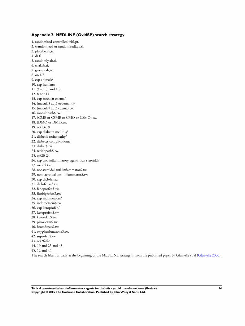
Appendix 2. MEDLINE (OvidSP) search strategy
1. randomized controlled trial.pt.
2. (randomized or randomised).ab,ti.
3. placebo.ab,ti.
4. dt.fs.
5. randomly.ab,ti.
6. trial.ab,ti.
7. groups.ab,ti.
8. or/1-7
9. exp animals/
10. exp humans/
11. 9 not (9 and 10)
12. 8 not 11
13. exp macular edema/
14. (macula$ adj3 oedema).tw.
15. (macula$ adj3 edema).tw.
16. maculopath$.tw.
17. (CME or CSME or CMO or CSMO).tw.
18. (DMO or DME).tw.
19. or/13-18
20. exp diabetes mellitus/
21. diabetic retinopathy/
22. diabetes complications/
23. diabet$.tw.
24. retinopath$.tw.
25. or/20-24
26. exp anti inflammatory agents non steroidal/
27. nsaid$.tw.
28. nonsteroidal anti-inflammator$.tw.
29. non-steroidal anti-inflammator$.tw.
30. exp diclofenac/
31. diclofenac$.tw.
32. fenoprofen$.tw.
33. flurbiprofen$.tw.
34. exp indometacin/
35. indometacin$.tw.
36. exp ketoprofen/
37. ketoprofen$.tw.
38. ketorolac$.tw.
39. piroxicam$.tw.
40. bromfenac$.tw.
41. oxyphenbutazone$.tw.
42. suprofen$.tw.
43. or/26-42
44. 19 and 25 and 43
45. 12 and 44
The search filter for trials at the beginning of the MEDLINE strategy is from the published paper by Glanville et al (Glanville 2006).
14Topical non-steroidal anti-inflammatory agents for diabetic cystoid macular oedema (Review)
Copyright © 2015 The Cochrane Collaboration. Published by John Wiley & Sons, Ltd.

Appendix 3. EMBASE (OvidSP) search strategy
1. exp randomized controlled trial/
2. exp randomization/
3. exp double blind procedure/
4. exp single blind procedure/
5. random$.tw.
6. or/1-5
7. (animal or animal experiment).sh.
8. human.sh.
9. 7 and 8
10. 7 not 9
11. 6 not 10
12. exp clinical trial/
13. (clin$ adj3 trial$).tw.
14. ((singl$ or doubl$ or trebl$ or tripl$) adj3 (blind$ or mask$)).tw.
15. exp placebo/
16. placebo$.tw.
17. random$.tw.
18. exp experimental design/
19. exp crossover procedure/
20. exp control group/
21. exp latin square design/
22. or/12-21
23. 22 not 10
24. 23 not 11
25. exp comparative study/
26. exp evaluation/
27. exp prospective study/
28. (control$ or prospectiv$ or volunteer$).tw.
29. or/25-28
30. 29 not 10
31. 30 not (11 or 23)
32. 11 or 24 or 31
33. exp retina macula edema/
34. (macula$ adj3 oedema).tw.
35. (macula$ adj3 edema).tw.
36. maculopath$.tw.
37. (CME or CSME or CMO or CSMO).tw.
38. (DMO or DME).tw.
39. or/33-38
40. exp diabetes mellitus/
41. diabetic retinopathy/
42. diabet$.tw.
43. retinopath$.tw.
44. or/40-43
45. exp nonsteroidal antiinflammatory agent/
46. nsaid$.tw.
47. nonsteroidal anti-inflammator$.tw.
48. non-steroidal anti-inflammator$.tw.
49. exp diclofenac/
50. diclofenac$.tw.
51. fenoprofen$.tw.
15Topical non-steroidal anti-inflammatory agents for diabetic cystoid macular oedema (Review)
Copyright © 2015 The Cochrane Collaboration. Published by John Wiley & Sons, Ltd.

52. flurbiprofen$.tw.
53. exp indometacin/
54. indometacin$.tw.
55. exp ketoprofen/
56. ketoprofen$.tw.
57. ketorolac$.tw.
58. exp piroxicam/
59. piroxicam$.tw.
60. bromfenac$.tw.
61. nepafenac$.tw.
62. oxyphenbutazone$.tw.
63. suprofen$.tw.
64. or/45-63
65. 39 and 44 and 64
66. 32 and 65
Appendix 4. LILACS search strategy
macula$ edema or macula$ oedema or CMO or CME and diabet$ and nonsteroidal antiinflammator$ or nonsteroidal anti inflammator$
or non steroidal anti inflammator$ or NSAID$ or diclofenac or fenoprofen or flurbiprofen or indometacin or ketoprofen or ketorolac
or piroxicam or bromfenac or nepafenac or oxyphenbutazone or suprofen
Appendix 5. ISRCTN search strategy
diabetic macular oedema
Appendix 6. ClinicalTrials.gov search strategy
Diabetic Macular Oedema AND (Non steroidal or NSAID)
Appendix 7. ICTRP search strategy
diabetic macular oedema and non steroidal
C O N T R I B U T I O N S O F A U T H O R S
Conceiving the review: Cochrane Eyes and Vision Group
Literature search for background: Soumendra Sahoo (SS), Kay Thi Myint (KT), Adnaan Haq (AH)
Writing the background, objectives and methods sections: SS
Writing data collection and analysis sections: Ankur Barua (AB), Adinegara BL Abas (AN), Sree Nair (SN), SS
Editing text to UK English: AH
16Topical non-steroidal anti-inflammatory agents for diabetic cystoid macular oedema (Review)
Copyright © 2015 The Cochrane Collaboration. Published by John Wiley & Sons, Ltd.
D E C L A R A T I O N S O F I N T E R E S T
Soumendra Sahoo: none known
Ankur Barua: none known
Kay Thi Myint: none known
Adnaan Haq : none known
Adinegara Abas: none known
Sree Nair: none known
S O U R C E S O F S U P P O R T
Internal sources
• Department of Statistics of Manipal University, India.
Advisory and logistics
External sources
• National Institute for Health Research, UK.
• Richard Wormald, Co-ordinating Editor for the Cochrane Eyes and Vision Group (CEVG) acknowledges financial support for
his CEVG research sessions from the Department of Health through the award made by the National Institute for Health Research to
Moorfields Eye Hospital NHS Foundation Trust and UCL Institute of Ophthalmology for a Specialist Biomedical Research Centre
for Ophthalmology.
• The NIHR also funds the CEVG Editorial Base in London.
The views expressed in this publication are those of the authors and not necessarily those of the NIHR, NHS, or the Department of
Health.
17Topical non-steroidal anti-inflammatory agents for diabetic cystoid macular oedema (Review)
Copyright © 2015 The Cochrane Collaboration. Published by John Wiley & Sons, Ltd.
Copyright © 2022 FDOKUMEN