Genetic Analysis of Seed-Soluble Oligosaccharides in Relation to Seed Storability of Arabidopsis
Upload
independentCategory
view
0download
0
Archives of Biochemistry and Biophysics 572 (2015) 58–65
Contents lists available at ScienceDirect
Archives of Biochemistry and Biophysics
journal homepage: www.elsevier .com/ locate /yabbi
Solubilization and stabilization of macular carotenoids by water solubleoligosaccharides and polysaccharides
http://dx.doi.org/10.1016/j.abb.2014.12.0100003-9861/� 2014 Elsevier Inc. All rights reserved.
⇑ Corresponding author at: Institutskaya Str., 3, Novosibirsk 630090, Russia.E-mail address: [email protected] (N.E. Polyakov).
Irina E. Apanasenko a,b, Olga Yu. Selyutina a,b, Nikolay E. Polyakov a,⇑, Lyubov P. Suntsova c,Elizaveta S. Meteleva c, Alexander V. Dushkin c, Preejith Vachali d, Paul S. Bernstein d
a Institute of Chemical Kinetics and Combustion, Novosibirsk, Russiab Novosibirsk State University, Novosibirsk, Russiac Institute of Solid State Chemistry and Mechanochemistry, Novosibirsk, Russiad Department of Ophthalmology and Visual Sciences, Moran Eye Center, University of Utah School of Medicine, Salt Lake City, UT, USA
a r t i c l e i n f o a b s t r a c t
Article history:Received 9 October 2014and in revised form 4 December 2014Available online 16 December 2014
Keywords:LuteinZeaxanthinWater-soluble complexesArabinogalactanGlycyrrhizic acidOzoneOxygen radicalsCarotenoid oxidationNMR relaxationSurface plasmon resonance
Xanthophyll carotenoids zeaxanthin and lutein play a special role in the prevention and treatment ofvisual diseases. These carotenoids are not produced by the human body and must be consumed in thediet. On the other hand, extremely low water solubility of these carotenoids and their instability restricttheir practical application as components of food or medicinal formulations. Preparation of supramolec-ular complexes of zeaxanthin and lutein with glycyrrhizic acid, its disodium salt and the natural polysac-charide arabinogalactan allows one to minimize the aforementioned disadvantages when carotenoids areused in food processing as well as for production of therapeutic formulations with enhanced solubilityand stability. In the present study, the formation of supramolecular complexes was investigated byNMR relaxation, surface plasmon resonance (SPR) and optical absorption techniques. The complexesincrease carotenoid solubility more than 1000-fold. The kinetics of carotenoid decay in reactions withozone molecules, hydroperoxyl radicals and metal ions were measured in water and organic solutions,and significant increases in oxidation stability of lutein and zeaxanthin in arabinogalactan and glycyrrhi-zin complexes were detected.
� 2014 Elsevier Inc. All rights reserved.
Introduction
Xanthophylls are the special group of carotenoids which con-tain oxygen functionalities at one or both ends of the molecule,providing them with unique biological, chemical and physicalproperties. In particular, hydroxyl groups are responsible for thexanthophylls’ abilities to orient within cell membranes in waysother carotenoids cannot [1–4]. The present study is devoted totwo representatives of xanthophyll carotenoids, lutein and zeaxan-thin (Fig. 1), which play a special role in the prevention and treat-ment of visual diseases. These carotenoids are not produced by thehuman body and must be consumed in the diet.
Lutein and zeaxanthin selectively accumulate at an extremelyhigh concentration in the macula of the primate eye retina throughthe action of specific high-affinity binding proteins [5], potentiallyslowing the onset of age-related macular degeneration [6,7], andthey have been recently added to the list of beneficial nutrientsprovided by leafy greens. It is suggested that lutein and zeaxanthin
play a role in blue light filtration and antioxidant function [8,9].The higher-energy, blue wavelengths of visible light are 100 timesmore effective at inducing free radical formation in the cells of theretina than the lower-energy, red wavelengths of visible light [10].Reacting as antioxidants with free radicals and reactive oxygenspecies, macular carotenoids protect the retina against peroxida-tion and photo-damage [11–15].
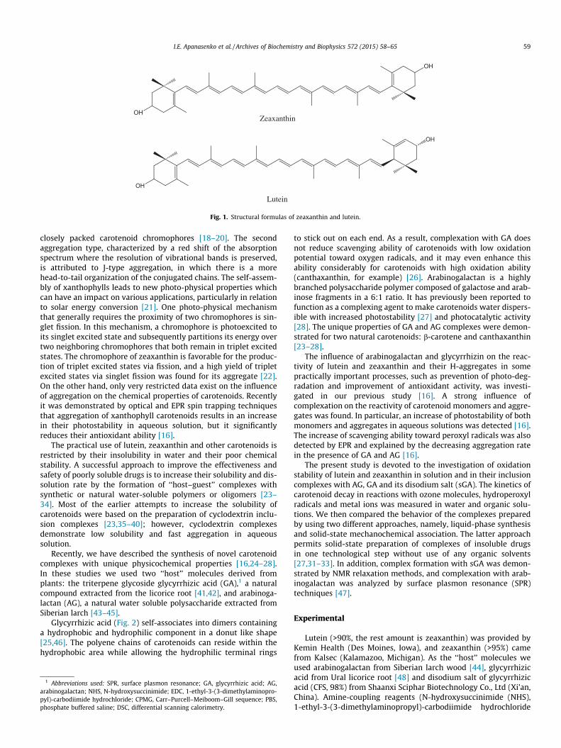
The important feature of xanthophyll carotenoids is their abilityto form J- and H-type of self-assembled complexes in aqueousmedia and even in lipid membranes [16–20]. Molecular self-assembly in biological systems attracts considerable attention,since it is important for the functioning of living organisms. Ithas been shown that lutein and zeaxanthin form aggregates whendissolved in hydrated polar solvents and that this aggregation ischaracterized by dramatic changes in their absorption spectraand photo-physical properties [16,18,20]. The H-aggregates, inwhich the molecules are tightly stacked with the conjugatedchains oriented more or less parallel to each other, show a largeblue shift of the absorption spectrum and loss of vibrational struc-ture of the S2 excited state. The blue shift of the absorption spec-trum is explained in terms of excitonic interaction between the
OH
OH
OH
OH
Zeaxanthin
Lutein
Fig. 1. Structural formulas of zeaxanthin and lutein.
I.E. Apanasenko et al. / Archives of Biochemistry and Biophysics 572 (2015) 58–65 59
closely packed carotenoid chromophores [18–20]. The secondaggregation type, characterized by a red shift of the absorptionspectrum where the resolution of vibrational bands is preserved,is attributed to J-type aggregation, in which there is a morehead-to-tail organization of the conjugated chains. The self-assem-bly of xanthophylls leads to new photo-physical properties whichcan have an impact on various applications, particularly in relationto solar energy conversion [21]. One photo-physical mechanismthat generally requires the proximity of two chromophores is sin-glet fission. In this mechanism, a chromophore is photoexcited toits singlet excited state and subsequently partitions its energy overtwo neighboring chromophores that both remain in triplet excitedstates. The chromophore of zeaxanthin is favorable for the produc-tion of triplet excited states via fission, and a high yield of tripletexcited states via singlet fission was found for its aggregate [22].On the other hand, only very restricted data exist on the influenceof aggregation on the chemical properties of carotenoids. Recentlyit was demonstrated by optical and EPR spin trapping techniquesthat aggregation of xanthophyll carotenoids results in an increasein their photostability in aqueous solution, but it significantlyreduces their antioxidant ability [16].
The practical use of lutein, zeaxanthin and other carotenoids isrestricted by their insolubility in water and their poor chemicalstability. A successful approach to improve the effectiveness andsafety of poorly soluble drugs is to increase their solubility and dis-solution rate by the formation of ‘‘host–guest’’ complexes withsynthetic or natural water-soluble polymers or oligomers [23–34]. Most of the earlier attempts to increase the solubility ofcarotenoids were based on the preparation of cyclodextrin inclu-sion complexes [23,35–40]; however, cyclodextrin complexesdemonstrate low solubility and fast aggregation in aqueoussolution.
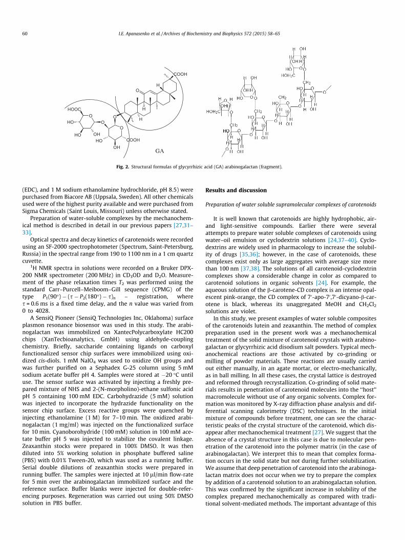
Recently, we have described the synthesis of novel carotenoidcomplexes with unique physicochemical properties [16,24–28].In these studies we used two ‘‘host’’ molecules derived fromplants: the triterpene glycoside glycyrrhizic acid (GA),1 a naturalcompound extracted from the licorice root [41,42], and arabinoga-lactan (AG), a natural water soluble polysaccharide extracted fromSiberian larch [43–45].
Glycyrrhizic acid (Fig. 2) self-associates into dimers containinga hydrophobic and hydrophilic component in a donut like shape[25,46]. The polyene chains of carotenoids can reside within thehydrophobic area while allowing the hydrophilic terminal rings
1 Abbreviations used: SPR, surface plasmon resonance; GA, glycyrrhizic acid; AGarabinogalactan; NHS, N-hydroxysuccinimide; EDC, 1-ethyl-3-(3-dimethylaminopro-pyl)-carbodiimide hydrochloride; CPMG, Carr–Purcell–Meiboom–Gill sequence; PBSphosphate buffered saline; DSC, differential scanning calorimetry.
,
,
to stick out on each end. As a result, complexation with GA doesnot reduce scavenging ability of carotenoids with low oxidationpotential toward oxygen radicals, and it may even enhance thisability considerably for carotenoids with high oxidation ability(canthaxanthin, for example) [26]. Arabinogalactan is a highlybranched polysaccharide polymer composed of galactose and arab-inose fragments in a 6:1 ratio. It has previously been reported tofunction as a complexing agent to make carotenoids water dispers-ible with increased photostability [27] and photocatalytic activity[28]. The unique properties of GA and AG complexes were demon-strated for two natural carotenoids: b-carotene and canthaxanthin[23–28].
The influence of arabinogalactan and glycyrrhizin on the reac-tivity of lutein and zeaxanthin and their H-aggregates in somepractically important processes, such as prevention of photo-deg-radation and improvement of antioxidant activity, was investi-gated in our previous study [16]. A strong influence ofcomplexation on the reactivity of carotenoid monomers and aggre-gates was found. In particular, an increase of photostability of bothmonomers and aggregates in aqueous solutions was detected [16].The increase of scavenging ability toward peroxyl radicals was alsodetected by EPR and explained by the decreasing aggregation ratein the presence of GA and AG [16].
The present study is devoted to the investigation of oxidationstability of lutein and zeaxanthin in solution and in their inclusioncomplexes with AG, GA and its disodium salt (sGA). The kinetics ofcarotenoid decay in reactions with ozone molecules, hydroperoxylradicals and metal ions was measured in water and organic solu-tions. We then compared the behavior of the complexes preparedby using two different approaches, namely, liquid-phase synthesisand solid-state mechanochemical association. The latter approachpermits solid-state preparation of complexes of insoluble drugsin one technological step without use of any organic solvents[27,31–33]. In addition, complex formation with sGA was demon-strated by NMR relaxation methods, and complexation with arab-inogalactan was analyzed by surface plasmon resonance (SPR)techniques [47].
Experimental
Lutein (>90%, the rest amount is zeaxanthin) was provided byKemin Health (Des Moines, Iowa), and zeaxanthin (>95%) camefrom Kalsec (Kalamazoo, Michigan). As the ‘‘host’’ molecules weused arabinogalactan from Siberian larch wood [44], glycyrrhizicacid from Ural licorice root [48] and disodium salt of glycyrrhizicacid (CFS, 98%) from Shaanxi Sciphar Biotechnology Co., Ltd (Xi’an,China). Amine-coupling reagents (N-hydroxysuccinimide (NHS),1-ethyl-3-(3-dimethylaminopropyl)-carbodiimide hydrochloride
HO
O
H
COOH
O
O
H
OH
O
OHOH
OH
OH
COOH
HOOC
GA
Fig. 2. Structural formulas of glycyrrhizic acid (GA) arabinogalactan (fragment).
60 I.E. Apanasenko et al. / Archives of Biochemistry and Biophysics 572 (2015) 58–65
(EDC), and 1 M sodium ethanolamine hydrochloride, pH 8.5) werepurchased from Biacore AB (Uppsala, Sweden). All other chemicalsused were of the highest purity available and were purchased fromSigma Chemicals (Saint Louis, Missouri) unless otherwise stated.
Preparation of water-soluble complexes by the mechanochem-ical method is described in detail in our previous papers [27,31–33].
Optical spectra and decay kinetics of carotenoids were recordedusing an SF-2000 spectrophotometer (Spectrum, Saint-Petersburg,Russia) in the spectral range from 190 to 1100 nm in a 1 cm quartzcuvette.
1H NMR spectra in solutions were recorded on a Bruker DPX-200 NMR spectrometer (200 MHz) in CD3OD and D2O. Measure-ment of the phase relaxation times T2 was performed using thestandard Carr–Purcell–Meiboom–Gill sequence (CPMG) of thetype P1(90�) � (s � P2(180�) � s)n – registration, wheres = 0.6 ms is a fixed time delay, and the n value was varied from0 to 4028.
A SensiQ Pioneer (SensiQ Technologies Inc, Oklahoma) surfaceplasmon resonance biosensor was used in this study. The arabi-nogalactan was immobilized on XantecPolycarboxylate HC200chips (XanTecbioanalytics, GmbH) using aldehyde-couplingchemistry. Briefly, saccharide containing ligands on carboxylfunctionalized sensor chip surfaces were immobilized using oxi-dized cis-diols. 1 mM NaIO4 was used to oxidize OH groups andwas further purified on a Sephadex G-25 column using 5 mMsodium acetate buffer pH 4. Samples were stored at �20 �C untiluse. The sensor surface was activated by injecting a freshly pre-pared mixture of NHS and 2-(N-morpholino)-ethane sulfonic acidpH 5 containing 100 mM EDC. Carbohydrazide (5 mM) solutionwas injected to incorporate the hydrazide functionality on thesensor chip surface. Excess reactive groups were quenched byinjecting ethanolamine (1 M) for 7–10 min. The oxidized arabi-nogalactan (1 mg/ml) was injected on the functionalized surfacefor 10 min. Cyanoborohydride (100 mM) solution in 100 mM ace-tate buffer pH 5 was injected to stabilize the covalent linkage.Zeaxanthin stocks were prepared in 100% DMSO. It was thendiluted into 5% working solution in phosphate buffered saline(PBS) with 0.01% Tween-20, which was used as a running buffer.Serial double dilutions of zeaxanthin stocks were prepared inrunning buffer. The samples were injected at 10 ll/min flow-ratefor 5 min over the arabinogalactan immobilized surface and thereference surface. Buffer blanks were injected for double-refer-encing purposes. Regeneration was carried out using 50% DMSOsolution in PBS buffer.
Results and discussion
Preparation of water soluble supramolecular complexes of carotenoids
It is well known that carotenoids are highly hydrophobic, air-and light-sensitive compounds. Earlier there were severalattempts to prepare water soluble complexes of carotenoids usingwater–oil emulsion or cyclodextrin solutions [24,37–40]. Cyclo-dextrins are widely used in pharmacology to increase the solubil-ity of drugs [35,36]; however, in the case of carotenoids, thesecomplexes exist only as large aggregates with average size morethan 100 nm [37,38]. The solutions of all carotenoid-cyclodextrincomplexes show a considerable change in color as compared tocarotenoid solutions in organic solvents [24]. For example, theaqueous solution of the b-carotene-CD complex is an intense opal-escent pink-orange, the CD complex of 70-apo-70,70-dicyano-b-car-otene is black, whereas its unaggregated MeOH and CH2Cl2
solutions are violet.In this study, we present examples of water soluble composites
of the carotenoids lutein and zeaxanthin. The method of complexpreparation used in the present work was a mechanochemicaltreatment of the solid mixture of carotenoid crystals with arabino-galactan or glycyrrhizic acid disodium salt powders. Typical mech-anochemical reactions are those activated by co-grinding ormilling of powder materials. These reactions are usually carriedout either manually, in an agate mortar, or electro-mechanically,as in ball milling. In all these cases, the crystal lattice is destroyedand reformed through recrystallization. Co-grinding of solid mate-rials results in penetration of carotenoid molecules into the ‘‘host’’macromolecule without use of any organic solvents. Complex for-mation was monitored by X-ray diffraction phase analysis and dif-ferential scanning calorimetry (DSC) techniques. In the initialmixture of compounds before treatment, one can see the charac-teristic peaks of the crystal structure of the carotenoid, which dis-appear after mechanochemical treatment [27]. We suggest that theabsence of a crystal structure in this case is due to molecular pen-etration of the carotenoid into the polymer matrix (in the case ofarabinogalactan). We interpret this to mean that complex forma-tion occurs in the solid state but not during further solubilization.We assume that deep penetration of carotenoid into the arabinoga-lactan matrix does not occur when we try to prepare the complexby addition of a carotenoid solution to an arabinogalactan solution.This was confirmed by the significant increase in solubility of thecomplex prepared mechanochemically as compared with tradi-tional solvent-mediated methods. The important advantage of this

0 300 600 900 1200
2
3
4
5
T2(Zea-GA, D2O) = 135 ± 10 msec
T2(Zea-GA, MeOD) = 285 ± 40 msec
T2(Zea, MeOD) = 526 ± 60 msec
T2 relaxation times of =CH- protons
time, msec
Ln(I)
Fig. 3. The echo decay kinetics of zeaxanthin CH protons in CPMG experiment inpure form in deuterated methanol and in the complex with sGA (5 mM) indeuterated methanol and D2O.
I.E. Apanasenko et al. / Archives of Biochemistry and Biophysics 572 (2015) 58–65 61
method over solvent-mediated methods is also the possibility ofcomplex preparation without using any toxic organic solvents.
For evidence of binding between ‘‘guest’’ and ‘‘host’’ moleculesin solution, various methods can be applied. For poorly solublemolecules like carotenoids, the increase in their solubility can beconsidered as a test for complex formation [49,50]. In the presentstudy, the solubility of mechanochemically prepared complexeswas measured by HPLC after paper filtration (Filters decalcified‘‘Blue Ribbon’’, ‘‘ECOHIM’’ Saint-Petersburg, Russia). The resultsare presented in Table 1. We suppose that the real water solubilityof non-complexed lutein and zeaxanthin may be even lower thanshown in Table 1 due to incomplete filtration of carotenoid micro-crystals. As one can see from the data of Table 1, the increase ofcarotenoid solubility in AG and GA complexes is approximately1000-fold.
Another approach to prove molecular complexation is thedetection of a change of the spectroscopic properties of the ‘‘guest’’molecule in the presence of the ‘‘host’’. In this study, we haveapplied NMR relaxation techniques to detect the binding of zea-xanthin with the disodium salt of GA in methanol and water solu-tions. The T2 relaxation time is very sensitive to intermolecularinteractions and diffusion mobility of molecules. Proton relaxationtime of the molecule inside the complex is significantly reducedbecause diffusive and rotational mobilities are slow [51]. A changeof T2 is usually considered as proof of complex formation[29,30,52]. Relaxation times T2 were measured for the CH protonsat the double bond. Fig. 3 shows the echo decay kinetics of zeaxan-thin CH protons in the CPMG experiment (see Experimental Sec-tion) in pure form in deuterated methanol and in the complexwith sGA (5 mM) in deuterated methanol and D2O.
Relaxation time of CH protons of free carotenoid in methanol is560 ± 60 ms. T2 is slowed down to 285 ± 40 ms after addition of5 mM of disodium salt of GA to this solution, due to complex for-mation. In the water environment, glycyrrhizic acid can formmicelles; therefore the further decrease of relaxation time to135 ± 10 ms can be explained by incorporation of carotenoid insidethe micelle.
SPR investigation of carotenoid binding to arabinogalactan
Arabinogalactan–zeaxanthin binding was investigated using SPRtechnique [47]. The SPR studies revealed that the arabinogalactancould bind zeaxanthin. We compared the binding affinities of Rus-sian arabinogalactan prepared from Siberian larch (Siberian-arabi-nogalactan) and American arabinogalactan obtained from SigmaChemicals (Sigma-arabinogalactan). Fig. 4 displays the sensorgramobtained from the interaction of the two arabinogalactan com-pounds and zeaxanthin. As shown in Table 2, the arabinogalactan–zeaxanthin interaction was fitted using a kinetic model with Qdat™analysis software (Biologic Software, Australia) to extract kineticparameters of binding. One can see from Fig. 4 that the Siberian-arabinogalactan has bimodal association and dissociation kinetics.We consider the presence at least two binding sites with differentbinding abilities (see Table 2). The Siberian-arabinogalactan had a
Table 1The solubility of mechanochemically prepared lutein and zeaxanthin complexes withAG and GA disodium and ammonium salts in water (in mg/l), measured by HPLC afterpaper filtration.
Sample Free AG GA–NH3 GA–Na2
Lutein 1:20 0.01a 18 15 10Lutein 1:40 10Zeaxanthin 1:20 0.01a 21 9 2.8Zeaxanthin 1:40 8
a This approximate value may be an upper limit due to penetration of smallcarotenoid crystals through paper filter.
strong affinity toward zeaxanthin with a KD(1) of 2.2 lM and KD(2)of 14.2 lM, whereas Sigma-arabinogalactan is 10 times weaker thanthe Siberian version. Unlike protein–carotenoid interactions, thisbinding interaction depends on the contact time, as it is driven bythe diffusion of zeaxanthin molecules into the arabinogalactanmatrix. We therefore used similar conditions for all comparisons.
The possible reason of the difference in the properties of Sibe-rian and Sigma arabinogalactan might be the different internalstructure of these branched polymers which results in differentpore size.
Investigations on the oxidation stability of carotenoids and theirinclusion complexes
The influence of complexation on the oxidation stability ofcarotenoids was investigated using model reactions with ozonemolecules, hydroperoxyl radicals and metal ions. Ozone moleculesare very reactive oxygen species which are able to break doublebonds of carotenoids with high efficiency [53]. The reaction occursvia addition of ozone to the double bond with formation of anozonide, followed by breakage of the double bond with formationof a number of oxygenated products with reduced conjugatedchains [54]:
In the present study, the decay kinetics of lutein and zeaxanthinin reaction with ozone were monitored by optical absorption tech-niques. The reaction was carried out in 75% water–ethanol solutionfor 5 min at 380 nm with continuous ozone bubbling. For free
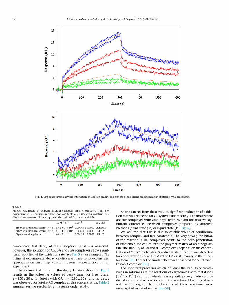
Fig. 4. SPR sensorgram showing interaction of Siberian arabinogalactan (top) and Sigma arabinogalactan (bottom) with zeaxanthin.
Table 2Kinetic parameters of zeaxanthin–arabinogalactan binding extracted from SPRexperiment. KD – equilibrium dissociation constant; ka – association constant; kd –dissociation constant. ⁄Errors represent the residual from the model fit.
ka, M�1 s�1 kd, s�1 KD, lM
Siberian arabinogalactan (site-1) 6.4 ± 0.3 � 102 0.00140 ± 0.0003 2.2 ± 0.1Siberian arabinogalactan (site-2) 4.9 ± 0.7 � 103 0.070 ± 0.003 14 ± 2Sigma arabinogalactan 48 ± 3 0.00118 ± 0.0002 25 ± 2
62 I.E. Apanasenko et al. / Archives of Biochemistry and Biophysics 572 (2015) 58–65
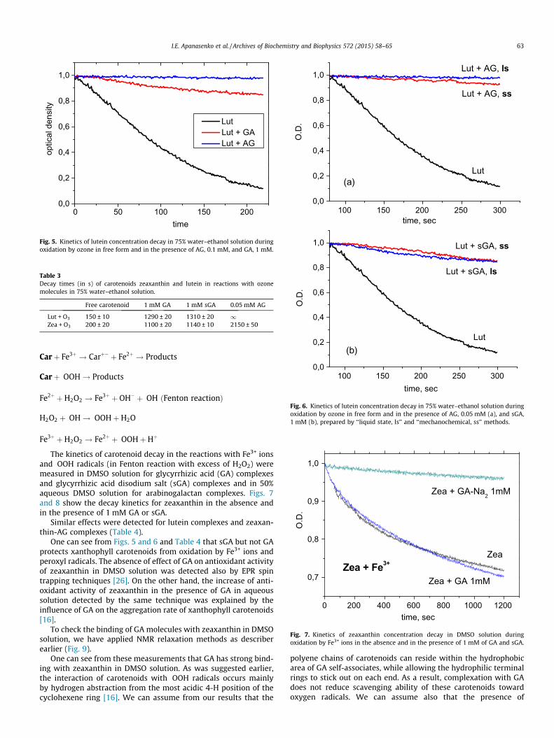
carotenoids, fast decay of the absorption signal was observed;however, the solutions of AG, GA and sGA complexes show signif-icant reduction of the oxidation rate (see Fig. 5 as an example). Thefitting of experimental decay kinetics was made using exponentialapproximation assuming constant ozone concentration duringexperiment.
The exponential fitting of the decay kinetics shown in Fig. 5results in the following values of decay time: for free lutein:s = 150 ± 20 s; for lutein with GA: s = 1290 ± 30 s; and no decaywas observed for lutein–AG complex at this concentration. Table 3summarizes the results for all systems under study.
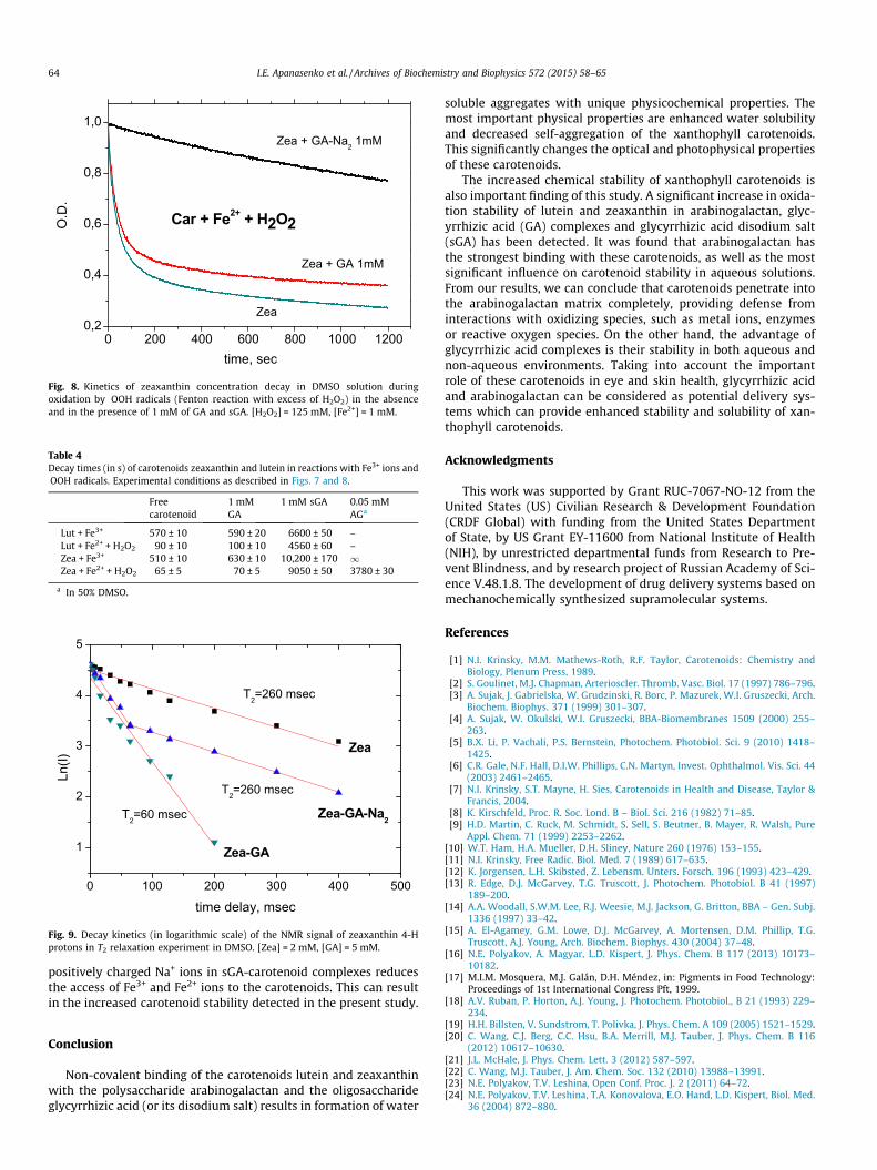
As one can see from these results, significant reduction of oxida-tion rate was detected for all systems under study. The most stableare the complexes with arabinogalactan. We did not observe sig-nificant differences between complexes prepared by differentmethods (solid state (ss) or liquid state (ls), Fig. 6).
We assume that this is due to establishment of equilibriumbetween complex and free carotenoid. The very strong inhibitionof the reaction in AG complexes points to the deep penetrationof carotenoid molecules into the polymer matrix of arabinogalac-tan. The stability of GA and sGA complexes depends on the concen-tration of ‘‘host’’ molecules. Significant stabilization was detectedfor concentrations near 1 mM when GA exists mainly in the micel-lar form [30]. Earlier the similar effect was observed for canthaxan-thin–GA complex [55].
The important processes which influence the stability of carote-noids in solutions are the reactions of carotenoids with metal ions(Cu2+ or Fe3+) and free radicals, mainly with peroxyl radicals pro-duced in Fenton-like reactions or in the reaction of C-centered rad-icals with oxygen. The mechanisms of these reactions wereinvestigated in detail earlier [56–59]:
0 50 100 150 2000,0
0,2
0,4
0,6
0,8
1,0
optic
al d
ensi
ty
time
Lut Lut + GA Lut + AG
Fig. 5. Kinetics of lutein concentration decay in 75% water–ethanol solution duringoxidation by ozone in free form and in the presence of AG, 0.1 mM, and GA, 1 mM.
Table 3Decay times (in s) of carotenoids zeaxanthin and lutein in reactions with ozonemolecules in 75% water–ethanol solution.
Free carotenoid 1 mM GA 1 mM sGA 0.05 mM AG
Lut + O3 150 ± 10 1290 ± 20 1310 ± 20 1Zea + O3 200 ± 20 1100 ± 20 1140 ± 10 2150 ± 50
0,0
0,2
0,4
0,6
0,8
1,0
O.D
.
time, sec
Lut
Lut + AG, ss
Lut + AG, ls
(a)
100 150 200 250 300
100 150 200 250 3000,0
0,2
0,4
0,6
0,8
1,0
O.D
.
time, sec
Lut
Lut + sGA, ss
Lut + sGA, ls
(b)
Fig. 6. Kinetics of lutein concentration decay in 75% water–ethanol solution duringoxidation by ozone in free form and in the presence of AG, 0.05 mM (a), and sGA,1 mM (b), prepared by ‘‘liquid state, ls’’ and ‘‘mechanochemical, ss’’ methods.
0 200 400 600 800 1000 1200
0,7
0,8
0,9
1,0
time, sec
O.D
.
Zea
Zea + GA 1mM
Zea + GA-Na2 1mM
Zea + Fe3+
Fig. 7. Kinetics of zeaxanthin concentration decay in DMSO solution duringoxidation by Fe3+ ions in the absence and in the presence of 1 mM of GA and sGA.
I.E. Apanasenko et al. / Archives of Biochemistry and Biophysics 572 (2015) 58–65 63
Carþ Fe3þ ! Carþ� þ Fe2þ ! Products
Carþ �OOH! Products
Fe2þ þH2O2 ! Fe3þ þ OH� þ �OH ðFenton reactionÞ
H2O2 þ �OH! �OOHþH2O
Fe3þ þH2O2 ! Fe2þ þ �OOHþHþ
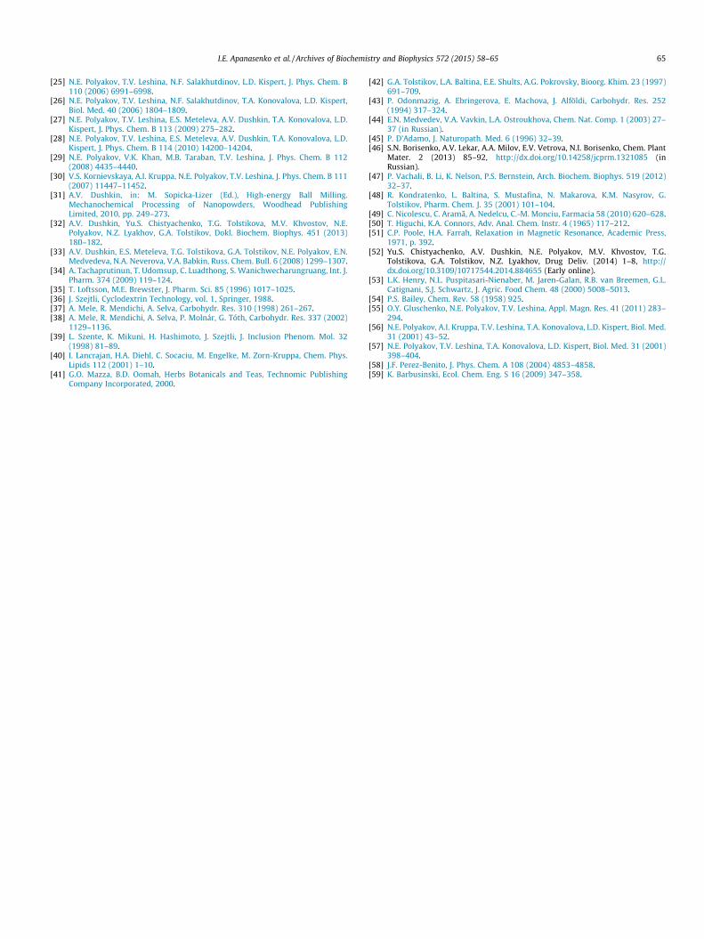
The kinetics of carotenoid decay in the reactions with Fe3+ ionsand �OOH radicals (in Fenton reaction with excess of H2O2) weremeasured in DMSO solution for glycyrrhizic acid (GA) complexesand glycyrrhizic acid disodium salt (sGA) complexes and in 50%aqueous DMSO solution for arabinogalactan complexes. Figs. 7and 8 show the decay kinetics for zeaxanthin in the absence andin the presence of 1 mM GA or sGA.
Similar effects were detected for lutein complexes and zeaxan-thin-AG complexes (Table 4).
One can see from Figs. 5 and 6 and Table 4 that sGA but not GAprotects xanthophyll carotenoids from oxidation by Fe3+ ions andperoxyl radicals. The absence of effect of GA on antioxidant activityof zeaxanthin in DMSO solution was detected also by EPR spintrapping techniques [26]. On the other hand, the increase of anti-oxidant activity of zeaxanthin in the presence of GA in aqueoussolution detected by the same technique was explained by theinfluence of GA on the aggregation rate of xanthophyll carotenoids[16].
To check the binding of GA molecules with zeaxanthin in DMSOsolution, we have applied NMR relaxation methods as describerearlier (Fig. 9).
One can see from these measurements that GA has strong bind-ing with zeaxanthin in DMSO solution. As was suggested earlier,the interaction of carotenoids with �OOH radicals occurs mainlyby hydrogen abstraction from the most acidic 4-H position of thecyclohexene ring [16]. We can assume from our results that the
polyene chains of carotenoids can reside within the hydrophobicarea of GA self-associates, while allowing the hydrophilic terminalrings to stick out on each end. As a result, complexation with GAdoes not reduce scavenging ability of these carotenoids towardoxygen radicals. We can assume also that the presence of
0 200 400 600 800 1000 12000,2
0,4
0,6
0,8
1,0
time, sec
O.D
.
Zea
Car + Fe2+ + H2O2
Zea + GA-Na2 1mM
Zea + GA 1mM
Fig. 8. Kinetics of zeaxanthin concentration decay in DMSO solution duringoxidation by �OOH radicals (Fenton reaction with excess of H2O2) in the absenceand in the presence of 1 mM of GA and sGA. [H2O2] = 125 mM, [Fe2+] = 1 mM.
Table 4Decay times (in s) of carotenoids zeaxanthin and lutein in reactions with Fe3+ ions and�OOH radicals. Experimental conditions as described in Figs. 7 and 8.
Freecarotenoid
1 mMGA
1 mM sGA 0.05 mMAGa
Lut + Fe3+ 570 ± 10 590 ± 20 6600 ± 50 –Lut + Fe2+ + H2O2 90 ± 10 100 ± 10 4560 ± 60 –Zea + Fe3+ 510 ± 10 630 ± 10 10,200 ± 170 1Zea + Fe2+ + H2O2 65 ± 5 70 ± 5 9050 ± 50 3780 ± 30
a In 50% DMSO.
0 100 200 300 400 500
1
2
3
4
5
Zea-GA
Zea-GA-Na2
T2=260 msec
T2=60 msec
T2=260 msec
Ln(I)
time delay, msec
Zea
Fig. 9. Decay kinetics (in logarithmic scale) of the NMR signal of zeaxanthin 4-Hprotons in T2 relaxation experiment in DMSO. [Zea] = 2 mM, [GA] = 5 mM.
64 I.E. Apanasenko et al. / Archives of Biochemistry and Biophysics 572 (2015) 58–65
positively charged Na+ ions in sGA-carotenoid complexes reducesthe access of Fe3+ and Fe2+ ions to the carotenoids. This can resultin the increased carotenoid stability detected in the present study.
Conclusion
Non-covalent binding of the carotenoids lutein and zeaxanthinwith the polysaccharide arabinogalactan and the oligosaccharideglycyrrhizic acid (or its disodium salt) results in formation of water
soluble aggregates with unique physicochemical properties. Themost important physical properties are enhanced water solubilityand decreased self-aggregation of the xanthophyll carotenoids.This significantly changes the optical and photophysical propertiesof these carotenoids.
The increased chemical stability of xanthophyll carotenoids isalso important finding of this study. A significant increase in oxida-tion stability of lutein and zeaxanthin in arabinogalactan, glyc-yrrhizic acid (GA) complexes and glycyrrhizic acid disodium salt(sGA) has been detected. It was found that arabinogalactan hasthe strongest binding with these carotenoids, as well as the mostsignificant influence on carotenoid stability in aqueous solutions.From our results, we can conclude that carotenoids penetrate intothe arabinogalactan matrix completely, providing defense frominteractions with oxidizing species, such as metal ions, enzymesor reactive oxygen species. On the other hand, the advantage ofglycyrrhizic acid complexes is their stability in both aqueous andnon-aqueous environments. Taking into account the importantrole of these carotenoids in eye and skin health, glycyrrhizic acidand arabinogalactan can be considered as potential delivery sys-tems which can provide enhanced stability and solubility of xan-thophyll carotenoids.
Acknowledgments
This work was supported by Grant RUC-7067-NO-12 from theUnited States (US) Civilian Research & Development Foundation(CRDF Global) with funding from the United States Departmentof State, by US Grant EY-11600 from National Institute of Health(NIH), by unrestricted departmental funds from Research to Pre-vent Blindness, and by research project of Russian Academy of Sci-ence V.48.1.8. The development of drug delivery systems based onmechanochemically synthesized supramolecular systems.
References
[1] N.I. Krinsky, M.M. Mathews-Roth, R.F. Taylor, Carotenoids: Chemistry andBiology, Plenum Press, 1989.
[2] S. Goulinet, M.J. Chapman, Arterioscler. Thromb. Vasc. Biol. 17 (1997) 786–796.[3] A. Sujak, J. Gabrielska, W. Grudzinski, R. Borc, P. Mazurek, W.I. Gruszecki, Arch.
Biochem. Biophys. 371 (1999) 301–307.[4] A. Sujak, W. Okulski, W.I. Gruszecki, BBA-Biomembranes 1509 (2000) 255–
263.[5] B.X. Li, P. Vachali, P.S. Bernstein, Photochem. Photobiol. Sci. 9 (2010) 1418–
1425.[6] C.R. Gale, N.F. Hall, D.I.W. Phillips, C.N. Martyn, Invest. Ophthalmol. Vis. Sci. 44
(2003) 2461–2465.[7] N.I. Krinsky, S.T. Mayne, H. Sies, Carotenoids in Health and Disease, Taylor &
Francis, 2004.[8] K. Kirschfeld, Proc. R. Soc. Lond. B – Biol. Sci. 216 (1982) 71–85.[9] H.D. Martin, C. Ruck, M. Schmidt, S. Sell, S. Beutner, B. Mayer, R. Walsh, Pure
Appl. Chem. 71 (1999) 2253–2262.[10] W.T. Ham, H.A. Mueller, D.H. Sliney, Nature 260 (1976) 153–155.[11] N.I. Krinsky, Free Radic. Biol. Med. 7 (1989) 617–635.[12] K. Jorgensen, L.H. Skibsted, Z. Lebensm. Unters. Forsch. 196 (1993) 423–429.[13] R. Edge, D.J. McGarvey, T.G. Truscott, J. Photochem. Photobiol. B 41 (1997)
189–200.[14] A.A. Woodall, S.W.M. Lee, R.J. Weesie, M.J. Jackson, G. Britton, BBA – Gen. Subj.
1336 (1997) 33–42.[15] A. El-Agamey, G.M. Lowe, D.J. McGarvey, A. Mortensen, D.M. Phillip, T.G.
Truscott, A.J. Young, Arch. Biochem. Biophys. 430 (2004) 37–48.[16] N.E. Polyakov, A. Magyar, L.D. Kispert, J. Phys. Chem. B 117 (2013) 10173–
10182.[17] M.I.M. Mosquera, M.J. Galán, D.H. Méndez, in: Pigments in Food Technology:
Proceedings of 1st International Congress Pft, 1999.[18] A.V. Ruban, P. Horton, A.J. Young, J. Photochem. Photobiol., B 21 (1993) 229–
234.[19] H.H. Billsten, V. Sundstrom, T. Polivka, J. Phys. Chem. A 109 (2005) 1521–1529.[20] C. Wang, C.J. Berg, C.C. Hsu, B.A. Merrill, M.J. Tauber, J. Phys. Chem. B 116
(2012) 10617–10630.[21] J.L. McHale, J. Phys. Chem. Lett. 3 (2012) 587–597.[22] C. Wang, M.J. Tauber, J. Am. Chem. Soc. 132 (2010) 13988–13991.[23] N.E. Polyakov, T.V. Leshina, Open Conf. Proc. J. 2 (2011) 64–72.[24] N.E. Polyakov, T.V. Leshina, T.A. Konovalova, E.O. Hand, L.D. Kispert, Biol. Med.
36 (2004) 872–880.
I.E. Apanasenko et al. / Archives of Biochemistry and Biophysics 572 (2015) 58–65 65
[25] N.E. Polyakov, T.V. Leshina, N.F. Salakhutdinov, L.D. Kispert, J. Phys. Chem. B110 (2006) 6991–6998.
[26] N.E. Polyakov, T.V. Leshina, N.F. Salakhutdinov, T.A. Konovalova, L.D. Kispert,Biol. Med. 40 (2006) 1804–1809.
[27] N.E. Polyakov, T.V. Leshina, E.S. Meteleva, A.V. Dushkin, T.A. Konovalova, L.D.Kispert, J. Phys. Chem. B 113 (2009) 275–282.
[28] N.E. Polyakov, T.V. Leshina, E.S. Meteleva, A.V. Dushkin, T.A. Konovalova, L.D.Kispert, J. Phys. Chem. B 114 (2010) 14200–14204.
[29] N.E. Polyakov, V.K. Khan, M.B. Taraban, T.V. Leshina, J. Phys. Chem. B 112(2008) 4435–4440.
[30] V.S. Kornievskaya, A.I. Kruppa, N.E. Polyakov, T.V. Leshina, J. Phys. Chem. B 111(2007) 11447–11452.
[31] A.V. Dushkin, in: M. Sopicka-Lizer (Ed.), High-energy Ball Milling.Mechanochemical Processing of Nanopowders, Woodhead PublishingLimited, 2010, pp. 249–273.
[32] A.V. Dushkin, Yu.S. Chistyachenko, T.G. Tolstikova, M.V. Khvostov, N.E.Polyakov, N.Z. Lyakhov, G.A. Tolstikov, Dokl. Biochem. Biophys. 451 (2013)180–182.
[33] A.V. Dushkin, E.S. Meteleva, T.G. Tolstikova, G.A. Tolstikov, N.E. Polyakov, E.N.Medvedeva, N.A. Neverova, V.A. Babkin, Russ. Chem. Bull. 6 (2008) 1299–1307.
[34] A. Tachaprutinun, T. Udomsup, C. Luadthong, S. Wanichwecharungruang, Int. J.Pharm. 374 (2009) 119–124.
[35] T. Loftsson, M.E. Brewster, J. Pharm. Sci. 85 (1996) 1017–1025.[36] J. Szejtli, Cyclodextrin Technology, vol. 1, Springer, 1988.[37] A. Mele, R. Mendichi, A. Selva, Carbohydr. Res. 310 (1998) 261–267.[38] A. Mele, R. Mendichi, A. Selva, P. Molnár, G. Tóth, Carbohydr. Res. 337 (2002)
1129–1136.[39] L. Szente, K. Mikuni, H. Hashimoto, J. Szejtli, J. Inclusion Phenom. Mol. 32
(1998) 81–89.[40] I. Lancrajan, H.A. Diehl, C. Socaciu, M. Engelke, M. Zorn-Kruppa, Chem. Phys.
Lipids 112 (2001) 1–10.[41] G.O. Mazza, B.D. Oomah, Herbs Botanicals and Teas, Technomic Publishing
Company Incorporated, 2000.
[42] G.A. Tolstikov, L.A. Baltina, E.E. Shults, A.G. Pokrovsky, Bioorg. Khim. 23 (1997)691–709.
[43] P. Odonmazig, A. Ebringerova, E. Machova, J. Alföldi, Carbohydr. Res. 252(1994) 317–324.
[44] E.N. Medvedev, V.A. Vavkin, L.A. Ostroukhova, Chem. Nat. Comp. 1 (2003) 27–37 (in Russian).
[45] P. D’Adamo, J. Naturopath. Med. 6 (1996) 32–39.[46] S.N. Borisenko, A.V. Lekar, A.A. Milov, E.V. Vetrova, N.I. Borisenko, Chem. Plant
Mater. 2 (2013) 85–92, http://dx.doi.org/10.14258/jcprm.1321085 (inRussian).
[47] P. Vachali, B. Li, K. Nelson, P.S. Bernstein, Arch. Biochem. Biophys. 519 (2012)32–37.
[48] R. Kondratenko, L. Baltina, S. Mustafina, N. Makarova, K.M. Nasyrov, G.Tolstikov, Pharm. Chem. J. 35 (2001) 101–104.
[49] C. Nicolescu, C. Arama, A. Nedelcu, C.-M. Monciu, Farmacia 58 (2010) 620–628.[50] T. Higuchi, K.A. Connors, Adv. Anal. Chem. Instr. 4 (1965) 117–212.[51] C.P. Poole, H.A. Farrah, Relaxation in Magnetic Resonance, Academic Press,
1971, p. 392.[52] Yu.S. Chistyachenko, A.V. Dushkin, N.E. Polyakov, M.V. Khvostov, T.G.
Tolstikova, G.A. Tolstikov, N.Z. Lyakhov, Drug Deliv. (2014) 1–8, http://dx.doi.org/10.3109/10717544.2014.884655 (Early online).
[53] L.K. Henry, N.L. Puspitasari-Nienaber, M. Jaren-Galan, R.B. van Breemen, G.L.Catignani, S.J. Schwartz, J. Agric. Food Chem. 48 (2000) 5008–5013.
[54] P.S. Bailey, Chem. Rev. 58 (1958) 925.[55] O.Y. Gluschenko, N.E. Polyakov, T.V. Leshina, Appl. Magn. Res. 41 (2011) 283–
294.[56] N.E. Polyakov, A.I. Kruppa, T.V. Leshina, T.A. Konovalova, L.D. Kispert, Biol. Med.
31 (2001) 43–52.[57] N.E. Polyakov, T.V. Leshina, T.A. Konovalova, L.D. Kispert, Biol. Med. 31 (2001)
398–404.[58] J.F. Perez-Benito, J. Phys. Chem. A 108 (2004) 4853–4858.[59] K. Barbusinski, Ecol. Chem. Eng. S 16 (2009) 347–358.
Copyright © 2022 FDOKUMEN