The Impact of Supplemental Macular Carotenoids in Alzheimer's Disease: A Randomized Clinical Trial.
13
Journal of Alzheimer’s Disease xx (20xx) x–xx DOI 10.3233/JAD-142265 IOS Press 1 The Impact of Supplemental Macular Carotenoids in Alzheimer’s Disease: A Randomized Clinical Trial 1 2 3 John M. Nolan a,∗ , Ekaterina Loskutova a , Alan Howard b,c , Riona Mulcahy d , Rachel Moran a , Jim Stack a , Maggie Bolger d , Robert F. Coen e , Jessica Dennison a , Kwadwo Owusu Akuffo a , Niamh Owens a , Rebecca Power a , David Thurnham e and Stephen Beatty a 4 5 6 a Macular Pigment Research Group, Department of Chemical and Life Sciences, Waterford Institute of Technology, Waterford, Ireland 7 8 b Howard Foundation, Cambridge, UK 9 c Downing College, University of Cambridge, Cambridge, UK 10 d University Hospital Waterford, Age-Related Care Unit, Waterford, Ireland 11 e Mercer’s Institute for Successful Ageing, St. James’s Hospital, Dublin, Ireland 12 f Northern Ireland, Centre for Food and Health (NICHE), University of Ulster, Coleraine, UK 13 Accepted 15 October 2014 Abstract. 14 Background: Patients with Alzheimer’s disease (AD) exhibit significantly less macular pigment (MP) and poorer vision when compared to control subjects. 15 16 Objective: To investigate supplementation with the macular carotenoids on MP, vision, and cognitive function in patients with AD versus controls. 17 18 Methods: A randomized, double-blind clinical trial with placebo and active arms. 31 AD patients and 31 age-similar control subjects were supplemented for six months with either Macushield (10 mg meso-zeaxanthin [MZ]; 10 mg lutein [L]; 2 mg zeaxanthin [Z]) or placebo (sunflower oil). MP was measured using dual-wavelength autofluorescence (Heidelberg Spectralis ® ). Serum L, Z, and MZ were quantified by high performance liquid chromatography. Visual function was assessed by best corrected visual acuity and contrast sensitivity (CS). Cognitive function was assessed using a battery of cognition tests, including the Cambridge Neuropsychological Test Automated Battery (CANTAB)). 19 20 21 22 23 24 Results: Subjects on the active supplement (for both AD and non-AD controls) exhibited statistically significant improvement in serum concentrations of L, Z, MZ, and MP (p < 0.001, for all) and also CS at 1.2 cpd (p < 0.039). Also, for subjects on the active supplement, paired samples t-tests exhibited four significant results (from five spatial frequencies tested) in the AD group, and two for the non-AD group, and all indicating improvements in CS. We found no significant changes in any of the cognitive function outcome variables measured (p > 0.05, for all). 25 26 27 28 29 Conclusion: Supplementation with the macular carotenoids (MZ, Z, and L) benefits patients with AD, in terms of clinically meaningful improvements in visual function and in terms of MP augmentation. 30 31 Keywords: Age-related macular degeneration, Alzheimer’s disease, cognitive function, contrast sensitivity, lutein, meso- zeaxanthin, randomized clinical trial, visual function, zeaxanthin 32 33 ∗ Correspondence to: Professor John Nolan, Macular Pigment Research Group, Vision Research Centre, Carriganore House, Waterford Institute of Technology, West Campus, Carriganore, Waterford, Ireland. Tel.: +353 51 834074; E-mail: [email protected]. INTRODUCTION 34 We have recently reported in the Carotenoids and 35 Age-Related Dementia Study (CARDS, report 1) that 36 patients with mild to moderate AD exhibit significantly 37 ISSN 1387-2877/15/$35.00 © 2015 – IOS Press and the authors. All rights reserved This article is published online with Open Access and distributed under the terms of the Creative Commons Attribution Non-Commercial License.
Transcript of The Impact of Supplemental Macular Carotenoids in Alzheimer's Disease: A Randomized Clinical Trial.
Journal of Alzheimer’s Disease xx (20xx) x–xxDOI 10.3233/JAD-142265IOS Press
1
The Impact of Supplemental MacularCarotenoids in Alzheimer’s Disease: ARandomized Clinical Trial
1
2
3
John M. Nolana,∗, Ekaterina Loskutovaa, Alan Howardb,c, Riona Mulcahyd, Rachel Morana, Jim Stacka,Maggie Bolgerd, Robert F. Coene, Jessica Dennisona, Kwadwo Owusu Akuffoa, Niamh Owensa,Rebecca Powera, David Thurnhame and Stephen Beattya
4
5
6
aMacular Pigment Research Group, Department of Chemical and Life Sciences, Waterford Institute of Technology,Waterford, Ireland
7
8
bHoward Foundation, Cambridge, UK9
cDowning College, University of Cambridge, Cambridge, UK10
dUniversity Hospital Waterford, Age-Related Care Unit, Waterford, Ireland11
eMercer’s Institute for Successful Ageing, St. James’s Hospital, Dublin, Ireland12
f Northern Ireland, Centre for Food and Health (NICHE), University of Ulster, Coleraine, UK13
Accepted 15 October 2014
Abstract.14
Background: Patients with Alzheimer’s disease (AD) exhibit significantly less macular pigment (MP) and poorer vision whencompared to control subjects.
15
16
Objective: To investigate supplementation with the macular carotenoids on MP, vision, and cognitive function in patients withAD versus controls.
17
18
Methods: A randomized, double-blind clinical trial with placebo and active arms. 31 AD patients and 31 age-similar controlsubjects were supplemented for six months with either Macushield (10 mg meso-zeaxanthin [MZ]; 10 mg lutein [L]; 2 mgzeaxanthin [Z]) or placebo (sunflower oil). MP was measured using dual-wavelength autofluorescence (Heidelberg Spectralis®).Serum L, Z, and MZ were quantified by high performance liquid chromatography. Visual function was assessed by best correctedvisual acuity and contrast sensitivity (CS). Cognitive function was assessed using a battery of cognition tests, including theCambridge Neuropsychological Test Automated Battery (CANTAB)).
19
20
21
22
23
24
Results: Subjects on the active supplement (for both AD and non-AD controls) exhibited statistically significant improvementin serum concentrations of L, Z, MZ, and MP (p < 0.001, for all) and also CS at 1.2 cpd (p < 0.039). Also, for subjects on theactive supplement, paired samples t-tests exhibited four significant results (from five spatial frequencies tested) in the AD group,and two for the non-AD group, and all indicating improvements in CS. We found no significant changes in any of the cognitivefunction outcome variables measured (p > 0.05, for all).
25
26
27
28
29
Conclusion: Supplementation with the macular carotenoids (MZ, Z, and L) benefits patients with AD, in terms of clinicallymeaningful improvements in visual function and in terms of MP augmentation.
30
31
Keywords: Age-related macular degeneration, Alzheimer’s disease, cognitive function, contrast sensitivity, lutein, meso-zeaxanthin, randomized clinical trial, visual function, zeaxanthin
32
33
∗Correspondence to: Professor John Nolan, Macular PigmentResearch Group, Vision Research Centre, Carriganore House,Waterford Institute of Technology, West Campus, Carriganore,Waterford, Ireland. Tel.: +353 51 834074; E-mail: [email protected].
INTRODUCTION 34
We have recently reported in the Carotenoids and 35
Age-Related Dementia Study (CARDS, report 1) that 36
patients with mild to moderate AD exhibit significantly 37
ISSN 1387-2877/15/$35.00 © 2015 – IOS Press and the authors. All rights reserved
This article is published online with Open Access and distributed under the terms of the Creative Commons Attribution Non-Commercial License.
2 J.M. Nolan et al. / Macular Pigment and Vision Improvements Alzheimer’s Disease
less macular pigment (MP), poorer vision, and a38
higher occurrence of age-related macular degeneration39
(AMD; another age-related disorder), when compared40
to control subjects [1].41
MP, which is made up of the dietary carotenoids42
lutein (L), zeaxanthin (Z), and meso-zeaxanthin (MZ)43
[2], is found exclusively at the central macula and can44
be measured in vivo [3, 4]. Of note, the macula (the45
central part of the retina) is part of the central ner-46
vous system, and it is this specialized part of the retina47
that is responsible for central and detailed vision [5].48
We know that the macular carotenoids, via their short-49
wavelength (blue) light filtering [6] and antioxidant50
properties [7, 8], play a protective role in AMD [9].51
We also know that MP is positively related to visual52
function [10], and that enrichment of MP with nutri-53
tional supplements containing the macular carotenoids54
improves visual function in normal subjects (i.e., sub-55
jects without retinal disease) [11] and in subjects with56
early stages of AMD [12–15]. Indeed, the optical prop-57
erties of MP, which include its preferential absorption58
of short-wavelength (blue) light, is likely to explain the59
visual benefits noted in previous clinical trials [16].60
Of interest, we know from previous work that L and61
Z are present in the brain, including in the cerebel-62
lum, pons, and frontal and occipital cortices [17–19],63
and that their concentrations in the brain are positively64
correlated with retinal concentrations of these nutrients65
in primates [18] including humans [19]. Furthermore,66
there is a growing body of evidence suggesting a67
positive relationship between MP levels and cogni-68
tive function in humans [20–22] and Johnson et al.69
have reported that supplementation with the macular70
carotenoids impacts positively on cognitive function in71
older women [23].72
Given the growing body of evidence showing that73
oxidative stress and inflammation contribute to cogni-74
tive impairment [24, 25] and AD pathogenesis [26, 27],75
it is plausible that carotenoids in the brain could protect76
against such stresses, given their proven antioxidant [7,77
8] and anti-inflammatory properties [28, 29]. It has also78
been suggested that the carotenoids may play a benefi-79
cial role by enhancing gap junctional communication80
in the brain [30–32].81
In summary, we have already reported that patients82
with AD have significantly less MP, lower serum con-83
centrations of L and Z, poorer vision, and a higher84
occurrence of AMD when compared to control sub-85
jects. Also, there is a biologically plausible rationale,86
supported by a growing body of scientific evidence,87
which suggests that enrichment of retinal and brain88
nutrition with the carotenoids L, Z, and MZ will protect89
and enhance cognitive function in humans. This study 90
was conducted to investigate the impact of supplemen- 91
tation with the macular carotenoids on MP (primary 92
outcome measure), and vision and cognitive function 93
(secondary outcome measures) in patients with AD 94
compared with controls of similar age, and is the first 95
study to do so. 96
MATERIALS AND METHODS 97
Study design and subject recruitment 98
This clinical trial began in January 2013 (i.e., the 99
first subject visit) and ended in September 2013 (i.e., 100
last subject six month visit). 101
31 patients with mild to moderate AD (pre- 102
dominantly moderate) were recruited (through the 103
Age-Related Care Unit at University Hospital Water- 104
ford [UHW]) into the study. Subjects recruited into 105
this group (the AD group) where eligible if they had 106
mild to moderate AD, which was defined as having 107
an average Mini-Mental State Examination (MMSE) 108
score of 14 to 24 with documented difficulties in other 109
domains, such as carrying out activities of daily liv- 110
ing, or behavioral changes. Subjects were excluded 111
if they were currently taking supplements contain- 112
ing the macular carotenoids, or if they had done so 113
over the previous 12 months. Other screening tests 114
to check for eligibility included the clock drawing 115
test and semantic fluency score. Co-morbid diagnoses 116
were documented, including vascular risk factors and 117
diabetes. Current medications were verified including 118
cholinesterase inhibitors and glutamate receptor antag- 119
onists. Social histories were documented and collateral 120
histories were collated from a family member or carer 121
for all patients. Non-contrast computed tomography 122
(CT) brain scan was performed to rule out radiological 123
evidence of stroke disease. 124
Of note, 10 (32%) had also participated in the 125
cross-sectional study previously reported by our group 126
(CARDS1) [1] and 21 (68%) were newly recruited. 127
Importantly, the subjects that had already participated 128
in CARDS1 were re-examined at baseline of this study 129
because of the time difference between the cross- 130
sectional examination and the start of this clinical 131
trial. Subjects with AD were randomly allocated, in 132
a double-blind fashion to a supplement consisting of 133
either Macushield™ (Macuvision Europe Ltd. Blythe 134
Valley Innovation Centre, Central Boulevard, Blythe 135
Valley Business Park, Solihull B90 8AJ, United King- 136
dom) (n = 16, active supplement containing 10 mg MZ; 137
10 mg L; 2 mg Z) or placebo (n = 15, sunflower oil). 138

J.M. Nolan et al. / Macular Pigment and Vision Improvements Alzheimer’s Disease 3
The intervention and placebo supplements were iden-139
tical in external appearance and therefore the two140
treatments were indistinguishable from each other.141
An equal number (n = 31) of age-similar controls142
free of AD (the none-AD Group) were similarly143
allocated to Macushield (n = 15) or Placebo (n = 16).144
Subjects were eligible for this group if they were145
aged (years) ≥65. Subjects were excluded if they were146
currently taking supplements containing the macular147
carotenoids, or if they had done so over the previous148
12 months.149
Main study visits were at baseline and after six150
months of supplementation. All measurements were151
performed at the Vision Research Centre, Carriganore152
House, Waterford, Ireland. This clinical trial facility153
offers a very efficient and calm environment to con-154
duct clinical trials. For consistency, all measurements155
were performed by the same researcher (EL) who was156
suitably trained on all aspects and technologies for this157
clinical trial.158
Significant efforts were made to ensure subject com-159
pliance to the study supplements. Compliance was160
assessed on an ongoing basis (house visits and phone161
calls to care givers) by the study nurse (MB) for the AD162
subjects, and by the researchers (NO and EL) for the163
control subjects (mainly phone calls directly to the sub-164
jects). In addition, compliance was assessed by exam-165
ining pill sleeves at the six month visit and by assessing166
serum carotenoid response using high performance liq-167
uid chromatography (HPLC, see below). The code was168
broken at 6 months, and the statistical analysis was169
performed by the Study Statistician (JS) and Princi-170
pal Investigator (JMN). The results of this analysis are171
presented below. The methodology used to measure172
MP, visual function and cognitive function has already173
been described in detail (see CARDS report 1), so only174
a brief account of each method is presented below.175
Ethics176
The project was conducted in accordance with full177
sensitivity to the ethical requirements of the subjects178
recruited. The study objectives and methodology com-179
plied fully with the widely recognized international180
text and codes of practice such as the Declaration of181
Helsinki. A protocol was developed specifically for this182
study by the Principal Investigator (JMN) and Consul-183
tant Geriatrician (RM) at UHW to ensure that informed184
consent was obtained appropriately, and in keeping185
with the ethical code germane to obtaining consent186
from vulnerable subjects (which includes patients with187
AD). Ethical approval was granted from the local188
Waterford South East (of Ireland) Region Ethics Com- 189
mittee prior to the study commencing. 190
Demographic, medical, ophthalmic, and lifestyle 191
assessment 192
A demographic, medical, ophthalmic, and lifestyle 193
case history was obtained for each subject at baseline. 194
Body mass index (BMI) was calculated (kg/m2) with 195
subject height (m) measured with the Leicester Height 196
Measure, and weight (kg) measured with the SECA 197
weighing scales (SECA, Birmingham, UK). Smok- 198
ing status was classed as either current smoker (i.e., 199
smoked ≥100 cigarettes in lifetime and at least one 200
cigarette within the last 12 months) or non-smoker 201
(everybody else). Exercise was assessed by calculating 202
the total exercise for any sporting activity measured as 203
minutes per week. Diabetes was assessed by self-report 204
and also by measuring HbA1c in blood (analysis con- 205
ducted offsite at Biomnis Ireland, Three Rock Road, 206
Sandyford Business Estate, Dublin 18, Ireland). 207
Cognitive function assessment 208
Cognition was assessed using a selection of vali- 209
dated measures. The MMSE was used to measure the 210
severity of cognitive impairment. A semantic fluency 211
score was obtained using ‘Animal” as the category (as 212
many exemplars as possible in one minute) and phone- 213
mic fluency was measured using the ‘FAS Test’ (as 214
many words as possible starting with each letter, one 215
minute per letter) [33]. Also, three tasks were chosen 216
from the Cambridge Neuropsychological Test Auto- 217
mated Battery (CANTAB) [34]. All were administered 218
using a finger-operated touch-screen tablet PC using 219
a set of scripted instructions. The Paired Associates 220
Learning task was selected to assess visual learning 221
and memory [35]. A modified version of the Verbal 222
Recognition Memory task was selected to assess ver- 223
bal learning and memory [33]. In the modified version 224
a free recall format was used instead of a recognition 225
format. The CANTAB Motor Screening Task was used 226
to assess motor speed and accuracy by instructing the 227
subject to touch the center of a series of crosses that 228
are presented on the screen [36]. 229
Best corrected visual acuity and contrast 230
sensitivity 231
The eye with best BCVA was selected as the study 232
eye for vision testing. If both eyes had the same BCVA, 233
the right one was selected. BCVA was measured with a 234

4 J.M. Nolan et al. / Macular Pigment and Vision Improvements Alzheimer’s Disease
computerized LogMAR ETDRS test chart (Test Chart235
2000 Xpert; Thomson Software Solutions) viewed at236
4 meters (m). The Sloan Early Treatment Diabetic237
Retinopathy Study (ETDRS) letterset was used for this238
test. Letter contrast sensitivity (CS) was assessed using239
the computerized LogMAR ETDRS test chart (Test240
Chart 2000 Pro; Thomson Software Solutions) at five241
different spatial frequencies (1.2, 2.4, 6.0, 9.6, 15.15242
cpd) [37]. Both these methods have been described in243
more detail elsewhere [10, 38, 39].244
Retinal photograph assessment245
45 degree monoscopic color photographs, centered246
on the macula, were taken in both eyes using a Zeiss247
Visucam 200 (Carl Zeiss Meditec AG, Jena, Germany).248
Retinal photographs were assessed for the presence249
or absence of early AMD, in accordance with the250
International Classification and Grading System for251
Age-Related Macular Degeneration by a consultant252
ophthalmologist (SB) with a special interest in retinal253
disease and with a published track record in grading254
this condition [40, 41]. In brief, the presence of soft255
drusen and/or hypo-/hyper-pigmentary changes at the256
macula were classed as early AMD.257
Macular pigment measurement258
MP was measured using the Heidelberg Spectralis®259
HRA+OCT Multicolor (Heidelberg Engineering260
GmbH, Heidelberg, Germany). This new technology261
utilizes confocal scanning laser ophthalmoscopy262
(cSLO) imaging with diode lasers and uses dual-263
wavelength autofluorescence (AF) for measuring MP264
[4, 42]. Dual-wavelength AF in this device uses two265
excitation wavelengths, one that is well-absorbed by266
MP (488 nm, blue), and one that is not well absorbed267
by the pigment (518 nm, green). Of note, the AF268
method utilized in this study has previously been269
compared with the customized heterochromatic flicker270
photometry (cHFP) technique for measuring MP, and271
the measurements recorded from these two devices272
exhibited excellent concordance [4]. However, the273
physical (objective) AF device was deemed more274
appropriate for this study, because patients with275
AD might not have been able to use the subjective276
(non-physical) cHFP device.277
The Heidelberg Spectralis® AF method provides an278
image of MP across its spatial profile, but here we279
report just central MP (at 0.23 degrees eccentricity)280
and MP volume (calculated as MP average times the281
area under the curve out to 8 degrees eccentricity).
Dietary intake of carotenoids 282
A subject’s weekly intake of carotenoid-rich foods 283
(eggs, broccoli, corn, dark leafy vegetables) was 284
inputted into the “L/Z screener” to give a carotenoid- 285
based diet score. The L and Z values used in the 286
screener were those reported by Perry et al. [43]. This 287
method of assessing and controlling for dietary intake 288
of carotenoids has been used with success elsewhere 289
[12]. Values are weighted for frequency of intake of 290
the food and for bioavailability of L and Z within these 291
foods. A ranking score reflecting the relative intakes 292
(representing arbitrary units) was generated and used 293
in analysis. For the AD subject, dietary habits were 294
confirmed by a family member or carer. 295
Serum carotenoid assessment 296
Non-fasting blood samples were collected in 9 ml 297
vacuette tubes containing a ‘Z Serum Sep Clot Acti- 298
vator’. The blood samples were allowed to clot at 299
room temperature for approximately 30 min and then 300
centrifuged at 2700 rpm for 10 min in a Gruppe GC 301
12 centrifuge (Desaga Sarstedt) to separate the serum 302
from the whole blood. The resulting serum samples 303
were stored at circa −80◦C until the time of batch 304
analysis using HPLC. 305
First, the serum samples were analysed for L and 306
total Z (co-eluted Z and MZ) using a reversed-phase 307
HPLC method (Assay 1, for details of method see pub- 308
lication by Nolan et al. [1]). The mixed Z fraction was 309
automatically collected from Assay 1 using an Agilent 310
1260 fraction collector. The eluent was dried under a 311
solvent concentrator (MiVac, GeneVac, Mason Tech- 312
nologies, Dublin, Ireland) and analyzed on Assay 2 313
for quantification of Z and MZ (Assay 2, for details of 314
method see publication by Thurnham et al. [44]). 315
Statistics 316
The statistical packages IBM SPSS version 21 was 317
used for statistical analysis. Random numbers (for the 318
allocation of subjects to active supplement or placebo) 319
were generated in Minitab version 16; block random- 320
ization was used. This study was very close in design 321
to a 22 factorial design (two factors each at two levels: 322
Macushield/Placebo and AD/Control) with 15 subjects 323
per cell. Such a study has statistical power of 81% to 324
detect a main effect of 0.75 standard deviations, and 325
power of 70% to detect an interaction effect of the 326
same magnitude, at the 5% level of significance [45]. 327
Outcome variables analyzed included serum 328
carotenoids, MP, visual function measures, and 329
J.M. Nolan et al. / Macular Pigment and Vision Improvements Alzheimer’s Disease 5
cognitive function measures. Between-group differ-330
ences in these outcome variables at baseline (e.g., AD331
versus controls) were analyzed using Independent332
Samples t-tests or chi-squared tests as appropriate.333
Differences at baseline in demographic and lifestyle334
variables were also investigated, and controlled for in335
subsequent analyses, as appropriate.336
The main focus of the present study was the inves-337
tigation of change in the outcome variables over time338
(i.e., from baseline to six months). In other words, did339
supplementing with Macushield lead to improvements340
in these outcome variables, relative to the Placebo, and341
did the supplement work differently for AD and con-342
trol subjects? Both of these research questions were343
addressed using Repeated Measures Analysis of Vari-344
ance, with supplement [Macushield versus Placebo]345
and Group [AD yes/no] as between-subjects factors,346
and age and diet score as covariates. These covariates347
were included in theanalysesbecauseageanddiet score348
were significantly different between AD and controls at349
baseline. For some cognitive scores, the assumptions350
required for Repeated Measures Analysis of Variance351
were violated, and in these cases we resorted to infor-352
mal comparisons of change in score between AD and353
control subjects and between supplements.354
In reporting findings in tables and figures, however,355
we considered that it would be more informative to356
report the results of paired t-tests, separately within357
each Supplement/Group patient category.358
The 5% level of significance was used through-359
out all analyses, without adjustment for multiple360
comparisons. On standard assumptions (5% level of361
significance, two-tailed tests), the paired t-test sub-362
group analyses reported here, with about 15 subjects363
in each subgroup, had adequate power (82%) to364
detect “large” effect sizes (0.8 standard deviations, on365
Cohen’s definition [46]). In general, however, it should366
be borne in mind that this small exploratory study was367
under-powered for the detection of smaller effect sizes368
and for the other analyses reported.369
RESULTS370
Baseline371
Table 1 below presents baseline statistics for the372
AD and control groups. Of note, the sample and data373
presented here is slightly different to our already pub-374
lished cross-sectional paper (CARDS1), given that the375
sample was not precisely the same for CARDS1 and376
CARDS2. However, the conclusions are the same. We377
confirm that, at baseline in CARDS2, AD subjects have378
significantly lower MP, poorer vision, poorer cogni- 379
tive function, and a significantly higher prevalence of 380
AMD, when compared to the control group. 381
Although we had attempted, when recruiting sub- 382
jects for this study, to match the AD and control groups 383
in terms of age, it can be seen in Table 1 that the 384
AD group is significantly older (on average), and we 385
therefore controlled for age in any analysis comparing 386
outcome variables in AD and control groups. We also 387
adjusted for diet score, the other variable which dif- 388
fered significantly at baseline between AD and control 389
groups. 390
Dropouts 391
Control group 392
All 16 subjects on placebo completed their six month 393
study visit, whereas there were 2 dropouts (n61 and 394
n68) in the active (Macushield) group, resulting in 13 395
subjects in this arm of the study. Reasons given for 396
dropout include: logistical difficulties (e.g., transport) 397
and did not want to continue (willingness to partici- 398
pate). 399
AD group 400
12 subjects on placebo completed their six month 401
study visit and there were 3 dropouts. Reasons for 402
dropout include: logistical difficulties and did not want 403
to continue (ADCD7 and ADN33); moved to nurs- 404
ing home and could not continue (ADN30). Also, 12 405
subjects in the active (Macushield) group completed 406
their six month visit and there were 4 dropouts. Rea- 407
sons for dropout include: logistical difficulties and did 408
not want to continue (ADCD13, ADN22, ADN35, and 409
ADN36). 410
Compliance 411
All subjects returned their capsule box and sleeves 412
at their six month assessment visit. Assessment of cap- 413
sule sleeves indicated that all subjects were consuming 414
the supplements over the six-month study period. Also, 415
serum carotenoid response confirmed that subjects in 416
the active group were consuming the carotenoid inter- 417
vention and that subjects in the placebo group exhibited 418
no change in their serum carotenoid concentrations. 419
Changes from baseline to six months 420
Serum concentrations of lutein, zeaxanthin, and 421
meso-zeaxanthin after six months of 422
supplementation 423
In the Repeated Measures Analysis of change 424
in serum L, the within-subjects Time*Supplement 425

6 J.M. Nolan et al. / Macular Pigment and Vision Improvements Alzheimer’s Disease
Table 1Demographic, lifestyle, vision, and cognition data of the AD and control subjects at baseline
Variables AD (n = 31) Control (n = 31) Sig.
Demographic and HealthAge (years) 80 ± 7.8 76 ± 6.6 0.031Body mass index (Kg/m2) 24.6 ± 5.8 26.4 ± 3.4 0.174Exercise (total minutes of exercise per week) 174 ± 218 226 ± 16 0.304Diet (estimated lutein and zeaxanthin intake) 16 ± 8 24 ± 14 0.008Serum lutein (�mol/L) 0.232 ± 0.113 0.297 ± 0.179 0.104Serum zeaxanthin (�mol/L) 0.051 ± 0.035 0.074 ± 0.042 0.03Education (total years in education) 11 ± 4 14 ± 4 0.003Smoking (% current) 8.60% 9.70% 0.88Gender (% female) 58% 42% 0.203VisionMP 0.23 0.41 ± 0.21 0.57 ± 0.17 0.002MP vol 4074 ± 2585 6326 ± 2258 0.001BCVA 88.9 ± 11.4 95.8 ± 8.4 0.009CS1.2 (cpd) 1.49 ± 0.23 1.75 ± .22 <0.001CS2.4 (cpd) 1.47 ± 0.25 1.79 ± 0.21 <0.001CS6.0 (cpd) 1.19 ± 0.31 1.42 ± 0.24 0.004CS9.6 (cpd) 0.94 ± 0.30 1.18 ± 0.26 0.005AMD (% with AMD) 48.00% 16.00% 0.007CognitionMMSE 19 ± 3.7 29 ± 1.7 <0.001Semantic fluency score 6.0 ± 3.2 15.4 ± 5.2 <0.001Phonemic fluency score 15.7 ± 10.3 32.5 ± 13.8 <0.001VRM (phase 1) 1.4 ± 1.2 5.1 ± 2.6 <0.001VRM (phase 2) 2.5 ± 1.6 7.3 ± 2.7 <0.001VRM (phase 3) 3.4 ± 2.1 8.1 ± 2.9 <0.001VRM Delayed Recall 0.4 ± 1.2 6.6 ± 3.3 <0.001VRM Savings Score 0.1 ± 0.2 0.8 ± 0.5 <0.001PAL (total errors adjusted) 136 ± 14.5 69 ± 39 <0.001PAL (total errors adjusted 6 shapes) 28.5 ± 5.8 17.7 ± 10.7 <0.001PAL Stages Complete 0.5 ± 0.9 4 ± 1.7 <0.001PAL Patterns Reached 2.5 ± 0.9 7.2 ± 4.9 <0.001PAL First Trial Memory Score 0.8 ± 2.3 9.7 ± 5.5 <0.001
Data displayed are mean ± standard deviation for interval data and percentages for categorical data. Variables, variables analyzed in the study;AD, subjects recruited into the study confirmed as having mild to moderate Alzheimer’s disease; Control, subjects free of mild to moderateAD and of similar age to the AD subjects; Sig., the statistical difference (p value) between AD and control subjects assessed using independentsamples t-tests or chi-squared depending on the variable of interest; Exercise, total exercise for any sporting activity measured as minutes perweek; Diet, estimate of dietary intake of L and Z; Serum lutein, serum concentrations of lutein in �mol/; Serum zeaxanthin, serum concentrationsof zeaxanthin in �mol/L; Smoking, current (smoked ≥100 cigarettes in lifetime and at least one cigarette within the last 12 months) or non-smoking (smoked ≤100 cigarettes in lifetime and none within the last 12 months); MP 0.23, central macular pigment measured at 0.23 degreeseccentricity measured using the Heidelberg Spectralis®. MP vol, a volume of MP calculated as MP average times the area under the curve outto 8 degrees eccentricity (measured using the Heidelberg Spectralis®); BCVA, best corrected visual acuity; CS 1.2, CS 2.4, CS 6.0, and CS9.6 = letter contrast sensitivity measured using the Thomson Software Solutions at 1.2, 2.4, 6.0, and 9.6 cycles per degree; AMD; age-relatedmacular degeneration; MMSE, Mini-Mental State Examination; Semantic fluency score, a semantic fluency (categorical verbal fluency) scoreobtained from the number of animals named by the subject in 1 minute; Phonemic fluency score, a phonemic fluency (word fluency) scoregenerated by the total number of words produced for the each of the letters F, A, and S, in 1 minute. MOT (mean latency), motor screeningtask measures the subject’s speed of response; MOT (mean error), motor screening task measures the accuracy of the subject’s pointing at crosstargets; VRM (phase 1), VRM (phase 2), VRM (phase 3), Verbal Free Recall Memory immediate, three consecutive trials; VRM Delayed Recall,Verbal Free Recall Memory of the previous words after a delay period; VRM Savings Score, Delayed verbal recall divided by phase 3 immediaterecall; PAL, Paired Associates Learning test which measure visual memory and new learning of the subjects; PAL (total errors adjusted), theadjusted score and includes an adjustment made for any stages not reached, allowing it to be comparable to all subjects even if the task was endedprematurely due to cognitive limitation; PAL (total errors adjusted 6 shapes), total errors made at the 6-pattern stage, adjusted for subjects whodid not reach this stage; PAL Stages Complete, The number of stages successfully completed; PAL Patterns Reached, The number of patternson the last problem in the task that the subject completed successfully; PAL First Trial Memory Score, The number of patterns correctly locatedafter the first trial, summed across the stages completed.
interaction effect was significant (p < 0.001). Nei-426
ther the Time*Group interaction (p = 0.65) nor the427
Time*Supplement*Group interaction (p = 0.97) was428
significant. Thus, there was a significant increase in 429
serum L concentrations after 6 months for subjects 430
on the active (Macushield) supplement compared with 431
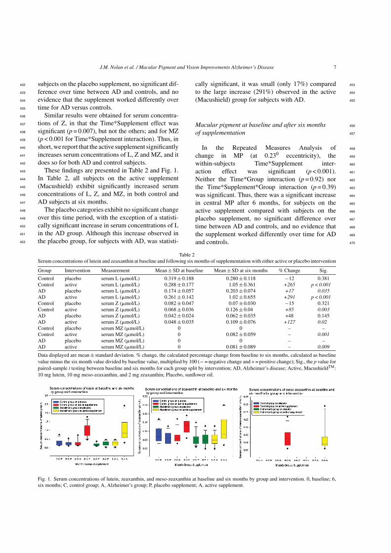
J.M. Nolan et al. / Macular Pigment and Vision Improvements Alzheimer’s Disease 7
subjects on the placebo supplement, no significant dif-432
ference over time between AD and controls, and no433
evidence that the supplement worked differently over434
time for AD versus controls.435
Similar results were obtained for serum concentra-436
tions of Z, in that the Time*Supplement effect was437
significant (p = 0.007), but not the others; and for MZ438
(p < 0.001 for Time*Supplement interaction). Thus, in439
short, we report that the active supplement significantly440
increases serum concentrations of L, Z and MZ, and it441
does so for both AD and control subjects.442
These findings are presented in Table 2 and Fig. 1.443
In Table 2, all subjects on the active supplement444
(Macushield) exhibit significantly increased serum445
concentrations of L, Z, and MZ, in both control and446
AD subjects at six months.447
The placebo categories exhibit no significant change448
over this time period, with the exception of a statisti-449
cally significant increase in serum concentrations of L450
in the AD group. Although this increase observed in451
the placebo group, for subjects with AD, was statisti-452
cally significant, it was small (only 17%) compared 453
to the large increase (291%) observed in the active 454
(Macushield) group for subjects with AD. 455
Macular pigment at baseline and after six months 456
of supplementation 457
In the Repeated Measures Analysis of 458
change in MP (at 0.230 eccentricity), the 459
within-subjects Time*Supplement inter- 460
action effect was significant (p < 0.001). 461
Neither the Time*Group interaction (p = 0.92) nor 462
the Time*Supplement*Group interaction (p = 0.39) 463
was significant. Thus, there was a significant increase 464
in central MP after 6 months, for subjects on the 465
active supplement compared with subjects on the 466
placebo supplement, no significant difference over 467
time between AD and controls, and no evidence that 468
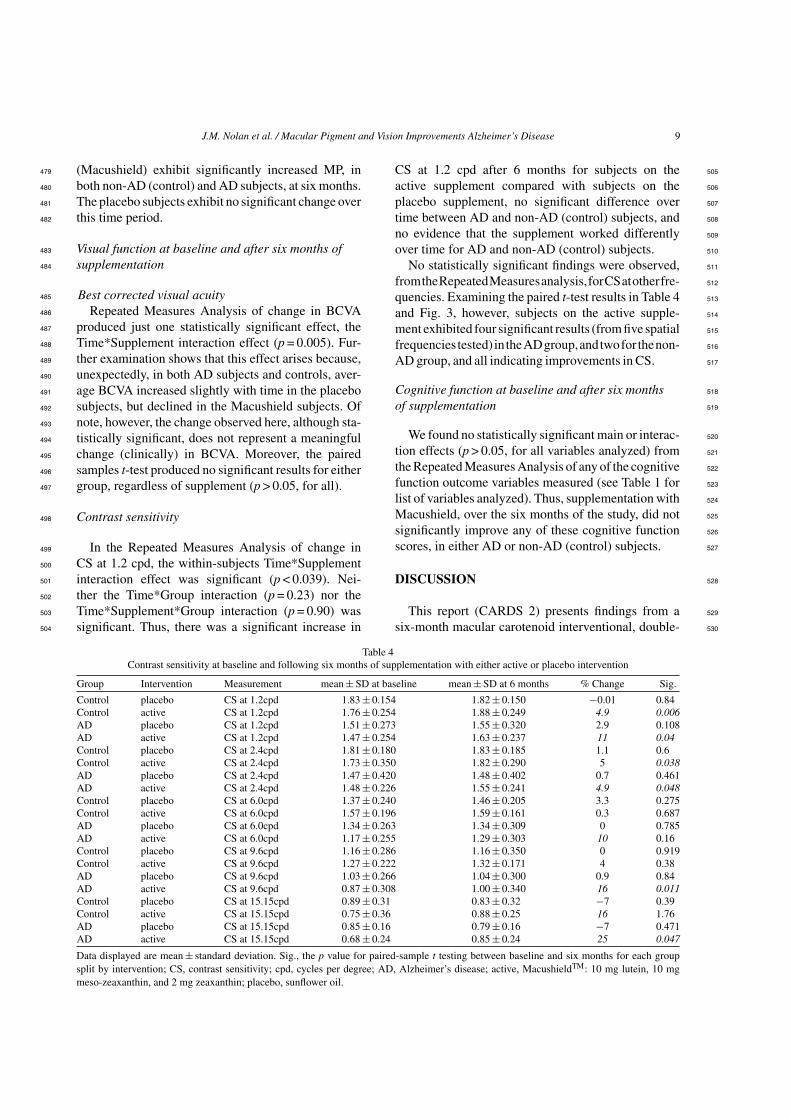
the supplement worked differently over time for AD 469
and controls. 470
Table 2Serum concentrations of lutein and zeaxanthin at baseline and following six months of supplementation with either active or placebo intervention
Group Intervention Measurement Mean ± SD at baseline Mean ± SD at six months % Change Sig.
Control placebo serum L (�mol/L) 0.319 ± 0.188 0.280 ± 0.118 −12 0.381Control active serum L (�mol/L) 0.288 ± 0.177 1.05 ± 0.361 +265 p < 0.001AD placebo serum L (�mol/L) 0.174 ± 0.057 0.203 ± 0.074 +17 0.035AD active serum L (�mol/L) 0.261 ± 0.142 1.02 ± 0.655 +291 p < 0.001Control placebo serum Z (�mol/L) 0.082 ± 0.047 0.07 ± 0.030 −15 0.321Control active serum Z (�mol/L) 0.068 ± 0.036 0.126 ± 0.04 +85 0.003AD placebo serum Z (�mol/L) 0.042 ± 0.024 0.062 ± 0.035 +48 0.145AD active serum Z (�mol/L) 0.048 ± 0.035 0.109 ± 0.076 +127 0.02Control placebo serum MZ (�mol/L) 0 0 – –Control active serum MZ (�mol/L) 0 0.082 ± 0.059 – 0.001AD placebo serum MZ (�mol/L) 0 0 – –AD active serum MZ (�mol/L) 0 0.081 ± 0.089 – 0.009
Data displayed are mean ± standard deviation. % change, the calculated percentage change from baseline to six months, calculated as baselinevalue minus the six month value divided by baseline value, multiplied by 100 (− = negative change and + = positive change); Sig., the p value forpaired-sample t testing between baseline and six months for each group split by intervention; AD, Alzheimer’s disease; Active, MacushieldTM:10 mg lutein, 10 mg meso-zeaxanthin, and 2 mg zeaxanthin; Placebo, sunflower oil.
Fig. 1. Serum concentrations of lutein, zeaxanthin, and meso-zeaxanthin at baseline and six months by group and intervention. 0, baseline; 6,six months; C, control group; A, Alzheimer’s group; P, placebo supplement; A, active supplement.
8 J.M. Nolan et al. / Macular Pigment and Vision Improvements Alzheimer’s Disease
Similar results were obtained for MP volume, in471
that only the Time*Supplement effect is significant472
(p < 0.001). Thus, in short, we report that the supple-473
ment works to increase MP, both centrally and across474
the spatial profile, and it does so for both AD and 475
non-AD (control) subjects. 476
These findings are presented in Table 3 and Fig. 2. 477
In Table 3, all subjects on the active supplement 478
Table 3Macular pigment at baseline and following six months of supplementation with either active or placebo intervention
Group Intervention Measurement Mean ± SD at baseline Mean ± SD at six months % Change Sig.
Control placebo MP at 0.23◦ 0.58 ± 0.18 0.54 ± 0.18 −7 0.300Control active MP at 0.23◦ 0.58 ± 0.18 0.68 ± 0.19 +17 0.002AD placebo MP at 0.23◦ 0.40 ± 0.17 0.38 ± 0.18 −5 0.86AD active MP at 0.23◦ 0.41 ± 0.26 0.48 ± 0.19 +17 0.009Control placebo MP volume 6543 ± 2150 6473 ± 2131 −1 0.394Control active MP volume 6593 ± 2116 8291 ± 2692 +26 p < 0.001AD placebo MP volume 4008 ± 2084 4327 ± 1948 +7 0.304AD active MP volume 3804 ± 2255 5408 ± 3130 +42 0.001
Data displayed are mean ± standard deviation. % change, the calculated percentage change from baseline to six months, calculated as baselinevalue minus the six month value divided by baseline value, multiplied by 100 (− = negative change and + = positive change); Sig., the pvalue for paired-sample t testing between baseline and six months for each group split by intervention; MP at 0.23◦, macular pigment at 0.23degrees eccentricity; MP volume, MP average times the area under the curve out to 8 degrees eccentricity; AD, Alzheimer’s disease; active,MacushieldTM: 10 mg lutein, 10 mg meso-zeaxanthin, and 2 mg zeaxanthin; placebo, sunflower oil.
Fig. 2. Mean macular pigment at baseline and after six months of supplementation with either active supplement (Macushield) or placebo insubjects with Alzheimer’s disease and control subjects.
J.M. Nolan et al. / Macular Pigment and Vision Improvements Alzheimer’s Disease 9
(Macushield) exhibit significantly increased MP, in479
both non-AD (control) and AD subjects, at six months.480
The placebo subjects exhibit no significant change over481
this time period.482
Visual function at baseline and after six months of483
supplementation484
Best corrected visual acuity485
Repeated Measures Analysis of change in BCVA486
produced just one statistically significant effect, the487
Time*Supplement interaction effect (p = 0.005). Fur-488
ther examination shows that this effect arises because,489
unexpectedly, in both AD subjects and controls, aver-490
age BCVA increased slightly with time in the placebo491
subjects, but declined in the Macushield subjects. Of492
note, however, the change observed here, although sta-493
tistically significant, does not represent a meaningful494
change (clinically) in BCVA. Moreover, the paired495
samples t-test produced no significant results for either496
group, regardless of supplement (p > 0.05, for all).497
Contrast sensitivity498
In the Repeated Measures Analysis of change in499
CS at 1.2 cpd, the within-subjects Time*Supplement500
interaction effect was significant (p < 0.039). Nei-501
ther the Time*Group interaction (p = 0.23) nor the502
Time*Supplement*Group interaction (p = 0.90) was503
significant. Thus, there was a significant increase in504
CS at 1.2 cpd after 6 months for subjects on the 505
active supplement compared with subjects on the 506
placebo supplement, no significant difference over 507
time between AD and non-AD (control) subjects, and 508
no evidence that the supplement worked differently 509
over time for AD and non-AD (control) subjects. 510
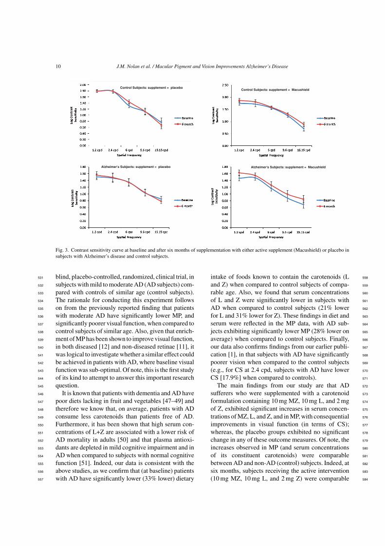
No statistically significant findings were observed, 511
fromtheRepeatedMeasuresanalysis,forCSatotherfre- 512
quencies. Examining the paired t-test results in Table 4 513
and Fig. 3, however, subjects on the active supple- 514
ment exhibited four significant results (from five spatial 515
frequencies tested) intheADgroup,andtwofor thenon- 516
AD group, and all indicating improvements in CS. 517
Cognitive function at baseline and after six months 518
of supplementation 519
We found no statistically significant main or interac- 520
tion effects (p > 0.05, for all variables analyzed) from 521
the Repeated Measures Analysis of any of the cognitive 522
function outcome variables measured (see Table 1 for 523
list of variables analyzed). Thus, supplementation with 524
Macushield, over the six months of the study, did not 525
significantly improve any of these cognitive function 526
scores, in either AD or non-AD (control) subjects. 527
DISCUSSION 528
This report (CARDS 2) presents findings from a 529
six-month macular carotenoid interventional, double- 530
Table 4Contrast sensitivity at baseline and following six months of supplementation with either active or placebo intervention
Group Intervention Measurement mean ± SD at baseline mean ± SD at 6 months % Change Sig.
Control placebo CS at 1.2cpd 1.83 ± 0.154 1.82 ± 0.150 −0.01 0.84Control active CS at 1.2cpd 1.76 ± 0.254 1.88 ± 0.249 4.9 0.006AD placebo CS at 1.2cpd 1.51 ± 0.273 1.55 ± 0.320 2.9 0.108AD active CS at 1.2cpd 1.47 ± 0.254 1.63 ± 0.237 11 0.04Control placebo CS at 2.4cpd 1.81 ± 0.180 1.83 ± 0.185 1.1 0.6Control active CS at 2.4cpd 1.73 ± 0.350 1.82 ± 0.290 5 0.038AD placebo CS at 2.4cpd 1.47 ± 0.420 1.48 ± 0.402 0.7 0.461AD active CS at 2.4cpd 1.48 ± 0.226 1.55 ± 0.241 4.9 0.048Control placebo CS at 6.0cpd 1.37 ± 0.240 1.46 ± 0.205 3.3 0.275Control active CS at 6.0cpd 1.57 ± 0.196 1.59 ± 0.161 0.3 0.687AD placebo CS at 6.0cpd 1.34 ± 0.263 1.34 ± 0.309 0 0.785AD active CS at 6.0cpd 1.17 ± 0.255 1.29 ± 0.303 10 0.16Control placebo CS at 9.6cpd 1.16 ± 0.286 1.16 ± 0.350 0 0.919Control active CS at 9.6cpd 1.27 ± 0.222 1.32 ± 0.171 4 0.38AD placebo CS at 9.6cpd 1.03 ± 0.266 1.04 ± 0.300 0.9 0.84AD active CS at 9.6cpd 0.87 ± 0.308 1.00 ± 0.340 16 0.011Control placebo CS at 15.15cpd 0.89 ± 0.31 0.83 ± 0.32 −7 0.39Control active CS at 15.15cpd 0.75 ± 0.36 0.88 ± 0.25 16 1.76AD placebo CS at 15.15cpd 0.85 ± 0.16 0.79 ± 0.16 −7 0.471AD active CS at 15.15cpd 0.68 ± 0.24 0.85 ± 0.24 25 0.047
Data displayed are mean ± standard deviation. Sig., the p value for paired-sample t testing between baseline and six months for each groupsplit by intervention; CS, contrast sensitivity; cpd, cycles per degree; AD, Alzheimer’s disease; active, MacushieldTM: 10 mg lutein, 10 mgmeso-zeaxanthin, and 2 mg zeaxanthin; placebo, sunflower oil.
10 J.M. Nolan et al. / Macular Pigment and Vision Improvements Alzheimer’s Disease
Alzheimer's Subjects: supplement = MacushieldAlzheimer's Subjects: supplement = placebo
Control Subjects: supplement = placeboControl Subjects: supplement = Macushield
Fig. 3. Contrast sensitivity curve at baseline and after six months of supplementation with either active supplement (Macushield) or placebo insubjects with Alzheimer’s disease and control subjects.
blind, placebo-controlled, randomized, clinical trial, in531
subjects with mild to moderate AD (AD subjects) com-532
pared with controls of similar age (control subjects).533
The rationale for conducting this experiment follows534
on from the previously reported finding that patients535
with moderate AD have significantly lower MP, and536
significantly poorer visual function, when compared to537
control subjects of similar age. Also, given that enrich-538
ment of MP has been shown to improve visual function,539
in both diseased [12] and non-diseased retinae [11], it540
was logical to investigate whether a similar effect could541
be achieved in patients with AD, where baseline visual542
function was sub-optimal. Of note, this is the first study543
of its kind to attempt to answer this important research544
question.545
It is known that patients with dementia and AD have546
poor diets lacking in fruit and vegetables [47–49] and547
therefore we know that, on average, patients with AD548
consume less carotenoids than patients free of AD.549
Furthermore, it has been shown that high serum con-550
centrations of L+Z are associated with a lower risk of551
AD mortality in adults [50] and that plasma antioxi-552
dants are depleted in mild cognitive impairment and in553
AD when compared to subjects with normal cognitive554
function [51]. Indeed, our data is consistent with the555
above studies, as we confirm that (at baseline) patients556
with AD have significantly lower (33% lower) dietary557
intake of foods known to contain the carotenoids (L 558
and Z) when compared to control subjects of compa- 559
rable age. Also, we found that serum concentrations 560
of L and Z were significantly lower in subjects with 561
AD when compared to control subjects (21% lower 562
for L and 31% lower for Z). These findings in diet and 563
serum were reflected in the MP data, with AD sub- 564
jects exhibiting significantly lower MP (28% lower on 565
average) when compared to control subjects. Finally, 566
our data also confirms findings from our earlier publi- 567
cation [1], in that subjects with AD have significantly 568
poorer vision when compared to the control subjects 569
(e.g., for CS at 2.4 cpd, subjects with AD have lower 570
CS [17.9%] when compared to controls). 571
The main findings from our study are that AD 572
sufferers who were supplemented with a carotenoid 573
formulation containing 10 mg MZ, 10 mg L, and 2 mg 574
of Z, exhibited significant increases in serum concen- 575
trations of MZ, L, and Z, and in MP, with consequential 576
improvements in visual function (in terms of CS); 577
whereas, the placebo groups exhibited no significant 578
change in any of these outcome measures. Of note, the 579
increases observed in MP (and serum concentrations 580
of its constituent carotenoids) were comparable 581
between AD and non-AD (control) subjects. Indeed, at 582
six months, subjects receiving the active intervention 583
(10 mg MZ, 10 mg L, and 2 mg Z) were comparable 584
J.M. Nolan et al. / Macular Pigment and Vision Improvements Alzheimer’s Disease 11
in terms of average circulating serum concentrations585
of L, Z, and MZ, irrespective of whether they were in586
the AD or non-AD (control) groups, with no signif-587
icant difference between these groups for any of the588
carotenoids at this point (p > 0.05 for all comparisons).589
The importance of this finding rests on the logical590
conclusion that the observed and relative lack of circu-591
lating serum carotenoid concentrations and MP in AD592
[1] is not attributable to an inability of these patients to593
respond to carotenoid intake (e.g., they are not compro-594
mised in terms of carotenoid absorption, transport, or595
uptake). In other words, the findings are consistent with596
the view that the reason why patients with AD have597
lower MP compared to control subjects is likely due to598
an associated poor dietary intake of foods containing599
carotenoids (fruits and vegetables).600
With respect to the serum and MP response exhib-601
ited in both AD and non-AD (control) groups, our data602
is consistent with previous studies where a supple-603
ment containing all three of the macular carotenoids604
(10 mgMZ, 10 mgL, and 2 mgZ) was used [11, 12,605
52, 53]. Indeed, it is noteworthy from previous studies606
that carotenoid supplements that do not contain MZ607
in their formulation did not augment MP significantly608
at six months [38, 54]. It appears, therefore, that best609
results in terms of increasing serum carotenoid concen-610
trations (for MZ, L, and Z), and MP augmentation, is611
achieved when all three of the macular carotenoids are612
included in the formulation, and this observation also613
holds true for patients with AD. Further, supplementa-614
tion with macular carotenoids, and consequential MP615
augmentation, is associated with risk reduction for616
AMD, a particularly important benefit as intervention617
with current treatment modalities (i.e., monthly injec-618
tions, under local anesthesia, into the eye) would be619
problematic in this patient group.620
We believe that it is important to draw attention to621
our findings pertaining to visual function. Firstly, we622
confirm that CS is significantly lower in AD subjects623
when compared to non-AD controls. Addressing this624
sensory defect in vulnerable AD patients should be625
a priority for those involved in the care of patients626
with this form of cognitive impairment, and routine627
and frequent assessment of visual function should be628
incorporated into the delivery of that care. For example,629
improvements seen among AD subjects supplemented630
with MZ, L, and Z were clinically meaningful at631
spatial frequencies of 1.2 cpd and 15.15 cpd, equat-632
ing to approximately one line of improvement on633
standard Pelli-Robson chart, and likely to enhance634
visual appreciation of small and large targets by these635
subjects. We suggest that further studies may con-636
sider other measures of visual function (e.g., glare 637
disability and photostress recovery), however, the fea- 638
sibility of including these measures will need to be 639
considered given the time required to perform the 640
tests and the ability of the subject to perform each 641
test. 642
Of note, no improvements in cognitive function were 643
demonstrated as a result of supplementation in either 644
AD or non-AD control subjects, a finding that is neither 645
surprising nor counter-intuitive. The rationale whereby 646
antioxidants are important for cognition rests on their 647
ability to prevent or attenuate oxidative damage, as 648
opposed to tissue repair. In other words, there is a 649
biologically plausible rationale, supported by emerg- 650
ing evidence, that antioxidant intake is protective for 651
cognition, but the notion that established cognitive 652
impairment could be reversed by supplementation with 653
antioxidants is less probable, especially in the context 654
of a short period of intervention (as reported herein). 655
Therefore, to investigate properly if supplementation 656
with the carotenoids L, Z, and MZ impact positively 657
on cognitive health/function, we suggest that subjects 658
with very early signs of cognitive decline, and subjects 659
of comparable age with no signs of cognitive decline 660
are selected, and are followed for at least 3 years. The 661
current study confirms that AD patients respond to 662
carotenoid supplements in the same way as normal 663
controls, and therefore it is possible that supplemen- 664
tation with these nutrients, if achieved early enough, 665
may support and protect cognitive health. 666
In conclusion, our data suggests that supplementa- 667
tion with the macular carotenoids (MZ, Z, and L) may 668
benefit patients with AD, in terms of clinically mean- 669
ingful improvements in visual function and in terms 670
of MP augmentation (and consequential risk reduction 671
for AMD). The impact of sustained supplementation 672
on cognition and visual function in AD subjects, and 673
on risk for AD, both warrant further study. 674
ACKNOWLEDGMENTS 675
We would like to thank the Howard Foundation, 676
Cambridge, CB22 5LA, United Kingdom for sup- 677
porting this research. We would like to acknowledge 678
Cambridge Cognition, UK for guidance with respect to 679
the assessment of cognitive function. Also, we would 680
like to thank all the staff at the UHW, Age-Related 681
Care Unit and at the Vision Research Centre, Waterford 682
Institute of Technology for assisting this study. 683
Authors’ disclosures available online (http://www.j- 684
alz.com/disclosures/view.php?id=2602).

12 J.M. Nolan et al. / Macular Pigment and Vision Improvements Alzheimer’s Disease
REFERENCES685
[1] Nolan JM, Loskutova E, Howard AN, Moran R, Mulcahy686
R, Stack J, Bolger M, Dennison J, Akuffo KO, Owens N,687
Thurnham DI, Beatty S (2014) Macular pigment, visual func-688
tion, and macular disease among subjects with Alzheimer’s689
disease: An exploratory study. J Alzheimers Dis 42,690
1191-1202.691
[2] Bone RA, Landrum JT, Fernandez L, Tarsis SL (1988) Anal-692
ysis of the macular pigment by HPLC: Retinal distribution693
and age study. Invest Ophthalmol Vis Sci 29, 843-849.694
[3] Wooten BR, Hammond BR, Land RI, Snodderly DM (1999)695
A practical method for measuring macular pigment optical696
density. Invest Ophthalmol Vis Sci 40, 2481-2489.697
[4] Dennison JL, Stack J, Beatty S, Nolan JM (2013)698
Concordance of macular pigment measurements obtained699
using customized heterochromatic flicker photometry,700
dual-wavelength autofluorescence, and single-wavelength701
reflectance. Exp Eye Res 116, 190-198.702
[5] Hirsch J, Curcio CA (1989) The spatial resolution capacity of703
human foveal retina. Vision Res 29, 1095-1101.704
[6] Snodderly DM, Brown PK, Delori FC, Auran JD (1984) The705
macular pigment. I. absorbance spectra, localization, and dis-706
crimination from other yellow pigments in primate retinas.707
Invest Ophthalmol Vis Sci 25, 660-673.708
[7] Khachik F, Bernstein PS, Garland DL (1997) Identifi-709
cation of lutein and zeaxanthin oxidation products in710
human and monkey retinas. Invest Ophthalmol Vis Sci 38,711
1802-1811.712
[8] Li B, Ahmed F, Bernstein PS (2010) Studies on the singlet713
oxygen scavenging mechanism of human macular pigment.714
Arch Biochem Biophys 504, 56-60.715
[9] Chew EY, Clemons TE, SanGiovanni JP, Danis RP, Ferris FL716
III, Elman MJ, Antoszyk AN, Ruby AJ, Orth D, Bressler SB,717
Fish GE, Hubbard GB, Klein ML, Chandra SR, Blodi BA,718
Domalpally A, Friberg T, Wong WT, Rosenfeld PJ, Agron719
E, Toth CA, Bernstein PS, Sperduto RD (2013) Secondary720
analyses of the effects of lutein/zeaxanthin on age-related721
macular degeneration progression: AREDS2 report No. 3.722
JAMA Ophthalmol 132, 142-149.723
[10] Loughman J, Akkali MC, Beatty S, Scanlon G, Davison PA,724
O’Dwyer V, Cantwell T, Major P, Stack J, Nolan JM (2010)725
The relationship between macular pigment and visual perfor-726
mance. Vision Res 50, 1249-1256.727
[11] Loughman J, Nolan JM, Howard AN, Connolly E,728
Meagher K, Beatty S (2012) The impact of macular pig-729
ment augmentation on visual performance using different730
carotenoid formulations. Invest Ophthalmol Vis Sci 53,731
7871-7880.732
[12] Sabour-Pickett S, Beatty S, Connolly E, Loughman J, Stack J,733
Howard A, Klein R, Klein BE, Meuer SM, Myers CE, Akuffo734
KO, Nolan JM (2014) Supplementation with three different735
macular carotenoid formulations in patients with early age-736
related macular degeneration. Retina 34, 1757-1766.737
[13] Huang YM, Yan SF, Ma L, Zou ZY, Xu XR, Dou HL, Lin738
XM (2013) Serum and macular responses to multiple xantho-739
phyll supplements in patients with early age-related macular740
degeneration. Nutrition 29, 387-392.741
[14] Murray IJ, Makridaki M, van der Veen RL, Carden D, Parry742
NR, Berendschot TT (2013) Lutein supplementation over a743
one-year period in early AMD might have a mild beneficial744
effect on visual acuity: The CLEAR study. Invest Ophthalmol745
Vis Sci 54, 1781-1788.746
[15] Weigert G, Kaya S, Pemp B, Sacu S, Lasta M, Werk-747
meister RM, Dragostinoff N, Simader C, Garhofer G,748
Schmidt-Erfurth U, Schmetterer L (2011) Effects of lutein 749
supplementation on macular pigment optical density and 750
visual acuity in patients with age-related macular degener- 751
ation. Invest Ophthalmol Vis Sci 52, 8174-8178. 752
[16] Wooten BR, Hammond BR (2002) Macular pigment: Influ- 753
ences on visual acuity and visibility. Prog Retin Eye Res 21, 754
225-240. 755
[17] Craft NE, Haitema TB, Garnett KM, Fitch KA, Dorey CK 756
(2004) Carotenoid, tocopherol, and retinol concentrations in 757
elderly human brain. J Nutr Health Aging 8, 156-162. 758
[18] Vishwanathan R, Neuringer M, Snodderly DM, Schalch W, 759
Johnson EJ (2013) Macular lutein and zeaxanthin are related 760
to brain lutein and zeaxanthin in primates. Nutr Neurosci 16, 761
21-29. 762
[19] Johnson EJ, Vishwanathan R, Johnson MA, Hausman DB, 763
Davey A, Scott TM, Green RC, Miller LS, Gearing M, 764
Woodard J, Nelson PT, Chung HY, Schalch W, Wittwer 765
J, Poon LW (2013) Relationship between serum and brain 766
carotenoids, alpha-tocopherol, and retinol concentrations and 767
cognitive performance in the oldest old from the Georgia 768
Centenarian Study. J Aging Res 2013, 951786. 769
[20] Vishwanathan R, Iannaccone A, Scott TM, Kritchevsky SB, 770
Jennings BJ, Carboni G, Forma G, Satterfield S, Harris T, 771
Johnson KC, Schalch W, Renzi LM, Rosano C, Johnson EJ 772
(2014) Macular pigment optical density is related to cognitive 773
function in older people. Age Ageing 43, 271-275. 774
[21] Renzi LM, Dengler MJ, Puente A, Miller LS, Hammond 775
BR Jr (2014) Relationships between macular pigment opti- 776
cal density and cognitive function in unimpaired and mildly 777
cognitively impaired older adults. Neurobiol Aging 35, 1695- 778
1699. 779
[22] Feeney J, Finucane C, Savva GM, Cronin H, Beatty S, Nolan 780
JM, Kenny RA (2013) Low macular pigment optical density 781
is associated with lower cognitive performance in a large, 782
population-based sample of older adults. Neurobiol Aging 34, 783
2449-2456. 784
[23] Johnson EJ, McDonald K, Caldarella SM, Chung HY, 785
Troen AM, Snodderly DM (2008) Cognitive findings of an 786
exploratory trial of docosahexaenoic acid and lutein supple- 787
mentation in older women. Nutr Neurosci 11, 75-83. 788
[24] Keller JN, Schmitt FA, Scheff SW, Ding Q, Chen Q, But- 789
terfield DA, Markesbery WR (2005) Evidence of increased 790
oxidative damage in subjects with mild cognitive impairment. 791
Neurology 64, 1152-1156. 792
[25] Teunissen CE, van Boxtel MP, Bosma H, Bosmans E, 793
Delanghe J, De BC, Wauters A, Maes M, Jolles J, Steinbusch 794
HW, de Vente J (2003) Inflammation markers in relation to 795
cognition in a healthy aging population. J Neuroimmunol 134, 796
142-150. 797
[26] Pappolla MA, Smith MA, Bryant-Thomas T, Bazan N, 798
Petanceska S, Perry G, Thal LJ, Sano M, Refolo LM 799
(2002) Cholesterol, oxidative stress, and Alzheimer’s disease: 800
Expanding the horizons of pathogenesis. Free Radic Biol Med 801
33, 173-181. 802
[27] Wyss-Coray T (2006) Inflammation in Alzheimer disease: 803
Driving force, bystander or beneficial response? Nat Med 12, 804
1005-1015. 805
[28] Ciccone MM, Cortese F, Gesualdo M, Carbonara S, Zito A, 806
Ricci G, De Pascalis F, Scicchitano P, Riccioni G (2013) 807
Dietary intake of carotenoids and their antioxidant and anti- 808
inflammatory effects in cardiovascular care. Mediators Inflam 809
2013, 782137. 810
[29] Kijlstra A, Tian Y, Kelly ER, Berendschot TT (2012) Lutein: 811
More than just a filter for blue light. Prog Retin Eye Res 31, 812
303-315. 813
J.M. Nolan et al. / Macular Pigment and Vision Improvements Alzheimer’s Disease 13
[30] Johnson EJ (2012) A possible role for lutein and zeaxanthin814
in cognitive function in the elderly. Am J Clin Nutr 96, 1161S-815
1165S.816
[31] Stahl W, Sies H (2001) Effects of carotenoids and retinoids817
on gap junctional communication. Biofactors 15, 95-98.818
[32] Stahl W, Nicolai S, Briviba K, Hanusch M, Broszeit G, Peters819
M, Martin HD, Sies H (1997) Biological activities of natural820
and synthetic carotenoids: Induction of gap junctional com-821
munication and singlet oxygen quenching. Carcinogenesis822
18, 89-92.823
[33] Strauss E, Scherman EMS, Spreen O (2006) A Compendium824
of Neuropsychological Tests. Administration, Norms and825
Commentary, Oxford University Press; New York.826
[34] Robbins TW, James M, Owen AM, Sahakian BJ, McInnes L,827
Rabbitt P (1994) Cambridge Neuropsychological Test Auto-828
mated Battery (CANTAB): A factor analytic study of a large829
sample of normal elderly volunteers. Dementia 5, 266-281.830
[35] Sahakian BJ, Morris RG, Evenden JL, Heald A, Levy R,831
Philpot M, Robbins TW (1988) A comparative study of visu-832
ospatial memory and learning in Alzheimer-type dementia833
and Parkinson’s disease. Brain 111(Pt 3), 695-718.834
[36] Owen AM, Downes JJ, Sahakian BJ, Polkey CE, Robbins835
TW (1990) Planning and spatial working memory following836
frontal lobe lesions in man. Neuropsychologia 28, 1021-1034.837
[37] Charalampidou S, Nolan J, Loughman J, Stack J, Higgins838
G, Cassidy L, Beatty S (2011) Psychophysical impact and839
optical and morphological characteristics of symptomatic840
non-advanced cataract. Eye (Lond) 25, 1147-1154.841
[38] Nolan JM, Loughman J, Akkali MC, Stack J, Scanlon G,842
Davison P, Beatty S (2011) The impact of macular pig-843
ment augmentation on visual performance in normal subjects:844
COMPASS. Vision Res 51, 459-469.845
[39] Akuffo KO, Beatty S, Stack J, Dennison J, O’Regan S,846
Meagher KA, Peto T, Nolan J (2014) Central Retinal847
Enrichment Supplementation Trials (CREST): Design and848
methodology of the CREST randomized controlled trials.849
Ophthalmic Epidemiol 21, 111-123.850
[40] Bird AC, Bressler NM, Bressler SB, Chisholm IH, Coscas G,851
Davis DM, de Jong PT, Klaver CC, Klein BE, Klein R, et al.852
(1995) An international classification and grading system for853
age-related maculopathy and age-related macular degenera-854
tion. The International ARM Epidemiological Study Group.855
Surv Ophthalmol 39, 367-374.856
[41] Neelam K, Muldrew A, Hogg R, Stack J, Chakravarthy U,857
Beatty S (2009) Grading of age-related maculopathy: Slit-858
lamp biomicroscopy versus an accredited grading center.859
Retina 29, 192-198.
[42] Delori FC (2004) Autofluorescence method to measure 860
macular pigment optical densities fluorometry and autoflu- 861
orescence imaging. Arch Biochem Biophys 430, 156-162. 862
[43] Perry A, Rasmussen H, Johnson EJ (2009) Xanthophyll 863
(lutein, zeaxanthin) content in fruits, vegetables and corn and 864
egg products. J Food Compost Anal 22, 9-15. 865
[44] Thurnham DI, Tremel A, Howard AN (2008) A supple- 866
mentation study in human subjects with a combination 867
of meso-zeaxanthin, (3R,3’R)-zeaxanthin and (3R,3’R,6’R)- 868
lutein. Br J Nutr 100, 1307-1314. 869
[45] Hintze J (2008) PASS 2008. NCSS LLC, http://www. 870
ncss.com 871
[46] Cohen J (1988) Statistical Power Analysis for the Behav- 872
ioral Sciences, Lawrence Erlbaum Associates, Hillsdale, New 873
Jersey. 874
[47] Morley JE (2010) Nutrition and the brain. Clin Geriatr Med 875
26, 89-98. 876
[48] Salerno-Kennedy R, Cashman KD (2007) The relationship 877
between nutrient intake and cognitive performance in people 878
at risk of dementia. Ir J Med Sci 176, 193-198. 879
[49] Mi W, van Wijk N, Cansev M, Sijben JW, Kamphuis PJ (2013) 880
Nutritional approaches in the risk reduction and management 881
of Alzheimer’s disease. Nutrition 29, 1080-1089. 882
[50] Min JY, Min KB (2014) Serum lycopene, lutein and zeaxan- 883
thin, and the risk of Alzheimer’s disease mortality in older 884
adults. Dement Geriatr Cogn Disord 37, 246-256. 885
[51] Rinaldi P, Polidori MC, Metastasio A, Mariani E, Mattioli 886
P, Cherubini A, Catani M, Cecchetti R, Senin U, Mecocci 887
P (2003) Plasma antioxidants are similarly depleted in mild 888
cognitive impairment and in Alzheimer’s disease. Neurobiol 889
Aging 24, 915-919. 890
[52] Meagher KA, Thurnham DI, Beatty S, Howard AN, Connolly 891
E, Cummins W, Nolan JM (2013) Serum response to sup- 892
plemental macular carotenoids in subjects with and without 893
age-related macular degeneration. Br J Nutr 110, 289-300. 894
[53] Nolan JM, Akkali MC, Loughman J, Howard AN, Beatty S 895
(2012) Macular carotenoid supplementation in subjects with 896
atypical spatial profiles of macular pigment. Exp Eye Res 101, 897
9-15. 898
[54] Beatty S, Chakravarthy U, Nolan JM, Muldrew KA, Wood- 899
side JV, Denny F, Stevenson MR (2013) Secondary outcomes 900
in a clinical trial of carotenoids with coantioxidants versus 901
placebo in early age-related macular degeneration. Ophthal- 902
mology 120, 600-606. 903