Solubilization of proteins from human lymph node tissue and two-dimensional gel storage
7
-
Upload
independent -
Category
Documents
-
view
7 -
download
0
Transcript of Solubilization of proteins from human lymph node tissue and two-dimensional gel storage
Journal of Biochemistry and Molecular Biology, Vol. 39, No. 2, March 2006, pp. 216-222
Solubilization of Proteins from Human Lymph Node Tissue andTwo-Dimensional Gel Storage
Alessandra Bernadete Trovó de Marqui1,#, Alessandra Vidotto2,#, Giovana Mussi Polachini2,#,Cláudia de Mattos Bellato3, Hamilton Cabral4, Andréia Machado Leopoldino2, José Francisco de Góis Filho5,
Érica Erina Fukuyama5, Flávio Aurélio Parente Settanni5, Patrícia Maluf Cury2,Gustavo Orlando Bonilla-Rodriguez4, Mario Sergio Palma6 and Eloiza Helena Tajara2,*
1UNESP-Universidade Estadual Paulista, Instituto de Biociências, Letras e Ciências Exatas-IBILCE,
Departamento de Biologia, São José do Rio Preto, SP, Brazil2FAMERP-Faculdade de Medicina de São José do Rio Preto, Departamento de Biologia Molecular, São José do Rio Preto, SP, Brazil
3USP- Universidade de São Paulo, Centro de Energia Nuclear para Agricultura/CENA, Piracicaba, SP, Brazil4UNESP-Universidade Estadual Paulista, Instituto de Biociências, Letras e Ciências Exatas-IBILCE, Departamento de Física,
São José do Rio Preto, SP, Brazil5Hospital do Câncer Arnaldo Vieira de Carvalho, SP, Brazil
6UNESP-Universidade Estadual Paulista, Instituto de Biociências de Rio Claro, Centro de Estudos em Insetos Sociais,
Departamento de Biologia, Rio Claro, SP, Brazil
Received 9 August 2005, Accepted 13 October 2005
In the present study, we compared six different solubilizationbuffers and optimized two-dimensional electrophoresis (2-DE) conditions for human lymph node proteins. In addition,we developed a simple protocol for 2-D gel storage.Efficient solubilization was obtained with lysis bufferscontaining (a) 8 M urea, 4% CHAPS (3-[(3-cholamidopropyl)dimethylammonio]-1-propanesulfonate), 40 mM Tris base,65 mM DTT(dithiothreitol) and 0.2% carrier ampholytes;(b) 5 M urea, 2 M thiourea, 2% CHAPS, 2% SB 3-10 (N-decyl-N, N-dimethyl-3-ammonio-1-propanesulfonate), 40 mMTris base, 65 mM DTT and 0.2% carrier ampholytes or (c)7 M urea, 2 M thiourea, 4% CHAPS, 65 mM DTT and0.2% carrier ampholytes. The optimal protocol for isoelectricfocusing (IEF) was accumulated voltage of 16,500 Vh and0.6% DTT in the rehydration solution. In the experimentsconducted for the sodium dodecyl sulfate-polyacrylamidegel electrophoresis (SDS-PAGE), best results were obtainedwith a doubled concentration (50 mM Tris, 384 mMglycine, 0.2% SDS) of the SDS electrophoresis buffer in thecathodic reservoir as compared to the concentration in theanodic reservoir (25 mM Tris, 192 mM glycine, 0.1%SDS). Among the five protocols tested for gel storing,
success was attained when the gels were stored in plasticbags with 50% glycerol. This is the first report describingthe successful solubilization and 2D-electrophoresis ofproteins from human lymph node tissue and a 2-D gelstorage protocol for easy gel handling before massspectrometry (MS) analysis.
Keywords: Gel storage, Human lymph node tissue, Protein
solubilization, Two-dimensional gel electrophoresis
Introduction
The analytical potential of 2-DE is dependent on good sample
preparation, in order to obtain reproducibility, good resolution
and a great spot number of proteomic maps. In many cases,
the proteins of the sample need to be solubilized, disaggregated,
denatured and reduced (Shaw and Riederer, 2003). For this
purpose, mixtures of chaotropic compounds, detergents or
surfactants, reducing agents and carrier ampholytes are
employed (Molloy, 2000; Garfin, 2003).
The role of chaotropes, such as urea and thiourea, is to
disrupt hydrogen bonding, leading to protein unfolding and
denaturation. Surfactants such as CHAPS, SB 3-10, ASB-14
(amidosulfobetaine-14) and SDS act synergistically with
chaotropes. Reducing agents, such as DTT and TBP (tributyl
phosphine), are used to break intramolecular and intermolecular
#The authors contributed equally to this work.
*To whom correspondence should be addressed.
Tel 55 17 3201 5737, Fax 55 17 3234 6407
E-mail: [email protected]
Short communication

Solubilization of Proteins and Gel Storage Protocols 217
disulfide bonds. Carrier ampholytes enhance protein solubility
by minimizing protein aggregation due to charge-charge
interactions (Herbert, 1999; Molloy, 2000; Berkelman and
Stenstedt, 2002; Shaw and Riederer, 2003).
Due to variable protein expression from one tissue to
another, conditions of protein solubilization that are optimal
for one particular tissue type may not hold for others. In
addition, several 2-DE steps must be optimized for each tissue
in order to obtain good results. In the present study, a
comparison was made of six previously described solubilization
methods for obtaining proteomic maps of human lymph node
tissue. We also describe a simple 2-D gel storage protocol for
easy gel handling prior to MS analysis.
Materials and Methods
Thirty-one lymph nodes were collected from head and neck
squamous cell carcinoma patients at the Cancer Hospital “Arnaldo
Vieira de Carvalho”, São Paulo, Brazil. Tissue samples were
obtained immediately after the removal of the surgical specimen,
snap-frozen and stored in liquid nitrogen. The Ethics Committee
approved the research, and written informed consent was obtained
from all patients.
One lymph node sample was cut into six pieces of about 5 mm3.
500 µL of one out of six different lysis buffers were added to each
piece (Table 1). The specimens were disrupted by sonication 12
times at intervals of 10 s at 10oC and vortexed for 2 min. The
lysates were centrifuged at 10,000 rpm for 3 min at 4oC. The
supernatants were transferred to other tubes, the insoluble pellets
were washed with 200 µL lysis buffers and the second supernatants
were collected. The protein concentration of the supernatants was
determined by the Bradford method (Bradford, 1976). The protein
samples were stored at −70oC.
2-DE was performed using IPGphor and SE 600 Ruby (GE
Healthcare). For IEF, 500 µg protein were diluted with rehydration
solution (8 M urea, 2% CHAPS, 0.6% DTT, 0.5% IPG buffer,
bromophenol blue trace) to a total volume of 250 µL. IPG strips
(pH 3-10 L, 13 cm) were rehydrated in this solution for 12 h under
mineral oil. IEF was performed at 20oC, with the following
parameters: 500 V (1 h), 1,000 V (1 h), 8,000 V (2 h or 3 : 30 h or
5 h), 500 V (0 h or 1 h), until 16,500 Vh, 26,500 Vh or 41,500 Vh
were attained. The current was limited to 50 µA/strip. After IEF, the
IPG strip was stored at −70oC until analysis by SDS-PAGE.
The individual strips were incubated, at room temperature, in the
equilibrium solution A (2% SDS, 50 mM Tris-HCl pH 8.8, 6 M
urea, 30% glycerol, bromophenol blue trace and 1% DTT),
followed by solution B (solution A except that DTT was replaced
by 2.5% iodoacetamide), for 15 min each. When the proteins were
solubilized using solutions containing TBP, the strips were only
incubated in solution C (solution A except that DTT was replaced
by 5 mM TBP) for 30 min. The IPG strips were washed in SDS
electrophoresis buffer (25 mM Tris base, 192 mM glycine, 0.1%
SDS), placed on top of 12.5% SDS-PAGE and sealed in place with
sealing solution (0.5% low-melting agarose in SDS electrophoresis
buffer). The electrophoresis conditions were 15 mA/gel for 30 min,
followed by 30 mA/gel for 5 h at room temperature.
Proteins were detected by Coomassie Blue staining. Briefly, gels
were incubated overnight in fixing solution (50% ethanol, 10%
acetic acid), followed by a destaining solution (50% ethanol, 5%
acetic acid) for 3 min, and incubated for 90 min with 0.05% Coomassie
Brilliant Blue R-250 solution (0.125 g Coomassie Brilliant Blue R-
250, 40% methanol, 10% acetic acid). Subsequently, the gels were
washed four times with destaining solution, for 15, 45, 120 and
120 min, respectively, and incubated in preserving solution (5%
acetic acid) for approximately 72 h.
For gel storing, five protocols were tested.
• Protocol 1. The gel was washed twice in solution D (30%
ethanol) for 30 min, followed by solution E (30% ethanol,
3.5% glycerol) for 60 min.
• Protocol 2. The gel was incubated in 4.3% glycerol for 5 min.
• Protocol 3. The gel was washed four times in water for 4 h.
• Protocol 4. The gel was incubated in 8.7% glycerol for 60 min.
Table 1. Lysis buffer composition. Composition of six lysis buffers tested for protein solubilization efficiency
Buffer 1 Buffer 2 Buffer 3 Buffer 4 Buffer 5 Buffer 6
Chaotropes 8 M Urea5 M Urea2 M Thiourea
5 M Urea2 M Thiourea
7 M Urea 2 M Thiourea
5 M Urea 2 M Thiourea
7 M Urea 2 M Thiourea
Detergents 4% CHAPS2% CHAPS 2% SB 3-10
2% CHAPS 2% SB 3-10
4% CHAPS 2% CHAPS 1% SB 3-10 1% ASB -14
2% CHAPS 0.5% ASB -14
Salts 40 mM Tris base 40 mM Tris base 40 mM Tris base
Reducing agents 65 mM DTT 65 mM DTT 2 mM TBP 65 mM DTT 2 mM TBP 65 mM DTT
Carrierampholytes
0.2%(pH 3-10)
0.2%(pH 3-10)
0.2% (pH 3-10)
0.2% (pH 3-10)
0.2% (pH 3-10)
0.2% (pH 3-10)
References(Herbert, 1999;Molloy et al., 1998)
(Rabilloud et al., 1997)
(Rabilloud et al., 1997; Molloy et
al., 1998; Tachibana et al., 2003)
(Molloy, 2000;Berkelman and Stenstedt, 2002; Rabilloud et al., 1997; Görg et al., 2000)
(Garfin, 2003)(Castellanos-Serra and Paz-Lago, 2002)
218 Alessandra Bernadete Trovó de Marqui et al.
• Protocol 5. The gel was incubated in 10% methanol for 48 h.
The gels was placed between two cellophane sheets PT (Coopercel)
previously embedded in solution E (protocol 1), in water (protocol
2), in 8.7% glycerol (protocol 3 and 4) or stored in a clear plastic
bag with 2 ml of 50% glycerol solution applied over both surfaces
of the gel. The bag was sealed.
No gel drying equipment was used. Stained and stored gels were
scanned with an ImageScanner (GE-Healthcare), and spot detection
was manually performed with the Melanie 3.0 software (GeneBio).
One protein spot from 2-DE gel was selected, excised, digested
with trypsin and submitted to MALDI-TOF-TOF (Matrix Assisted
Laser Desorption - Time of Flight - Time of Flight) 4700 Proteomics
Analyser (Applied Biosystems) operated in positive ion reflectron
mode to identify the peptides. Such spot was manually cut out from
the gel in a clean-air cabinet, to prevent contamination. The protein
spot was placed into 0.5 mL tube previously washed with 50%
methanol and deionized water. The gel pieces were destained in
250 µL of 50% acetonitrile (ACN)/50 mM ammonium bicarbonate
under constant agitation to complete colourlessness. The gel pieces
were then dehydrated with 200 µL of ACN for 15 min; acetonitrile
was discarded and the gel pieces dried in Speed Vac for 30 min. For
rehydration and digestion, each gel piece was rehydrated with
20 µL of a trypsin solution (0.4 µg modified trypsin in 50 mM
acetic acid and 50 mM ammonium bicarbonate). After 30 min
incubation at room temperature, a volume of 50 µL of 50 mM
ammonium bicarbonate or the sufficient amount to cover the gel
pieces was added, and the sample was incubated for 24 h at 37oC in
a water bath, for enzymatic cleavage. Peptides were extracted with
50 µL 1% trifluoroacetic acid/TFA (first extraction: overnight) and
50 µL 1% TFA/50% ACN (second extraction: 2 h). The resulting
supernatants were mixed and concentrated in a vacuum centrifuge
to 10 µL.
About 1 µL of this solution was eluted in 1 µL matrix solution
(10 mg/mL α-cyano-4-hydroxycinnamic acid, 0.1% TFA in 50%
ACN). Then, 0.5 µL of the mixture was spotted on a sample plate
and introduced into the mass spectrometer after drying. The
instrument was calibrated externally using 4700 standard kit
(Applied Biosystems).
Proteins were identified by MASCOT MS/MS Ions Search (http:
//www.matrixscience.com/cgi/search_form.pl?FORMVER=2&
SEARCH = MIS). The search parameters were set up as follows:
MSDB (Mass Spectrometry Protein Sequence Database); taxonomy
Homo sapiens; 1 missed cleavage; carbamidomethylation of cysteine
and oxidation of methionine as fixed and variable modification,
respectively; peptide mass and MS/MS tolerance of 1 and 0.8 Da,
respectively; the peptide ion MH+ and monoisotopic masses.
In the present study, all chemicals used were of highest quality
(Merck, Calbiochem, GE Healthcare, Sigma and Bio-Rad).
Results and Discussion
Initial extraction and solubilization is a key factor for proteomic
analysis. In the present study, we tested six buffers for
solubilization of protein from human lymph node samples,
which differed in one or more components, including chaotropes,
detergents and reducing agents (Table 1).
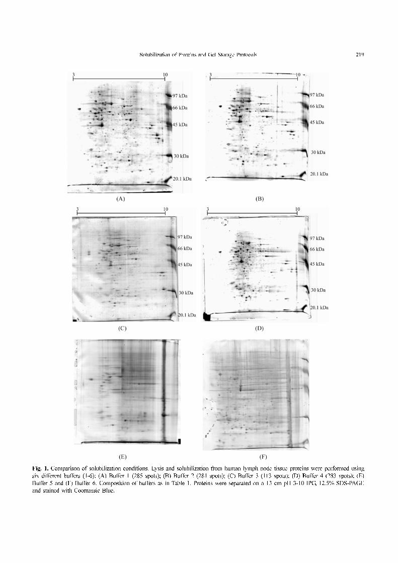
Extraction buffer 1 is a standard solution for protein
solubilization. About 285 spots were visualized in the gel
using this solution (Fig. 1A). Buffers 2 and 3 have similar
compositions, except for the reducing agent, and about 281
and 113 spots were visualized in the gels, respectively (Fig.
1B, C). In buffer 4, Tris base was not used for protein
solubilization. The results showed 283 spots (Fig. 1D). In
lysis buffers 5 and 6, ASB-14 was used as detergent and the
gels exhibited poor resolution and streaking (Figs. 1E, F). In
conclusion, buffers 1, 2 and 4 were the best solutions for
protein solubilization of human lymph node tissue with regard
to gel quality. Also, these solutions resulted in high protein
concentration (9.8µg/µL, 8.0µg/µL and 8.9µg/µL, respectively),
higher than buffer 3 (6.6 µg/µL), but much higher than buffers
5 and 6 (0.3 µg/µL and 1.2 µg/µL). Sharp differences are seen
comparing Fig. 1C with 1A, 1B and 1D and reflect the results
of the Bradford assay. Although this assay is sensitive to
various components of lysis buffers, the reagents of buffers 1-
6 probably had no effect on protein quantification, and the gel
quality showed be consequence of the buffer efficiency. Thirty
other human lymph node samples were also submitted to
buffer 4 and all gels showed excellent quality.
In the buffers tested, urea varied from 5 to 8 M, CHAPS
from 2 to 4%, SB 3-10 from 1 to 2%, and ASB-14 from 0.5 to
1%. The carrier ampholytes were used at a low concentration
(0.2%), in order to avoid extended running times, because
they contribute to the initial conductivity of the sample
solution (Garfin, 2003). Tris base was added to three buffers
(1, 2 and 3). This compound is used when basic conditions are
required for full solubilization or to minimize proteolysis
(Rabilloud, 1996). However, addition of ionic compounds in
buffers for protein solubilization can result in first-dimension
disturbances. Therefore, salts must be removed after the
solubilization step or maintained at as low a concentration
(lower than 10 mM) in the rehydration solution and IEF
(Berkelman and Stenstedt, 2002; Shaw and Riederer, 2003).
In our experiments, Tris base was added to the buffers at 40
mM, but the final salt concentration during rehydration was
maintained at approximately 10 mM.
Thiourea was introduced in combination with urea, to increase
the solubility of proteins, mainly of membrane proteins. The
use of this component inhibits the adsorption of protein to the
gel matrix, when IEF is conducted in IPG. This efficient
chaotrope is poorly soluble in water and requires high
concentrations of urea for solubility (optimal condition is 2 M
thiourea in 5-7 M urea). This reagent improved the solubility
of proteins as compared to urea alone, as already stated by
other authors (Rabilloud et al., 1997; Pasquali et al., 1997;
Musante et al., 1998; Rabilloud, 1998; Giavalisco et al., 2003;
Méchin et al., 2003; Taylor and Pfeiffer, 2003).
We also examined three zwitterionic detergents (CHAPS,
SB 3-10, ASB-14). The sulfobetaine CHAPS is the most
commonly used detergent for 2-DE, from 2% to 4% in high
concentrations of urea. Sulfobetaines with long linear tails
(i.e., SB 3-10, ASB-14) have been shown to possess a greater
Solubilization of Proteins and Gel Storage Protocols 219
Fig. 1. Comparison of solubilization conditions. Lysis and solubilization from human lymph node tissue proteins were performed using
six different buffers (1-6): (A) Buffer 1 (285 spots); (B) Buffer 2 (281 spots); (C) Buffer 3 (113 spots); (D) Buffer 4 (283 spots); (E)
Buffer 5 and (F) Buffer 6. Composition of buffers as in Table 1. Proteins were separated on a 13 cm pH 3-10 IPG, 12.5% SDS-PAGE
and stained with Coomassie Blue.
220 Alessandra Bernadete Trovó de Marqui et al.
ability to solubilize membrane proteins. However, SB 3-10
has poor solubility in high concentrations of urea (Herbert,
1999; Görg and Weiss, 1999). In contrast, ASB-14 is
compatible with 9 M urea, but results in large horizontal
streaking towards the basic end of the strip. This streaking
appears to be an interaction between ASB-14 and the cathode
rather than purely an issue with basic proteins, as the effect
was independent of the pH range of the IPG strip (pH 3-10 or
pH 4-7) (Stanley et al., 2003). Our gels also showed increased
streaking in all pH ranges when ASB-14 was used in the
extraction solution (Fig. 1E, 1F). Studies using three different
detergents (CHAPS, ASB-14 or NP-40) in the solubilization
solution also showed more streaking in the second dimension
with ASB-14 than with CHAPS (Carboni et al., 2002). The
ionic detergent SDS is very effective for protein solubilization,
however it is incompatible with IEF (Zuobi-Hasona et al.,
2005). For this reason, it was not selected for our optimization
experiments.
As for reducing agents, TBP and DTT are commonly used
in extraction solutions. When the proteins are solubilized using
reagents containing free thiol such as DTT, IPG equilibration
requires two steps: reduction (by DTT) and alkylation (by
iodoacetamide). DTT has been the standard reducing agent for
2-DE for many years (Molloy, 2000; Görg et al., 2000) and is
effective for reducing protein disulfide bonds prior to SDS-
PAGE, while iodoacetamide eliminates artifacts of disulfide
formation during electrophoresis, for less streaking and better
resolution. However, DTT is charged, especially at alkaline
pH, and thus migrates out of the pH gradient during IEF,
which results in loss of solubility for some proteins. In
contrast, TBP lacks a free thiol group, making the second
equilibration of the IPG strips unnecessary. In addition, TBP
is neutral and does not migrate during IEF, thus, the reducing
conditions are maintained over the entire focusing process. On
the other hand, TBP has a low solubility, is unstable, volatile
and toxic (Rabilloud, 1996; Herbert et al., 1998; Molloy et al.,
1998; Berkelman and Stenstedt, 2002). In the present study,
the buffers 2 and 3, used for protein solubilization, had similar
compositions, except for the reducing agent (DTT in buffer 2
and TBP in buffer 3). Nevertheless, the numbers of spots
visualized were very different (281 vs 113 spots), probably
due to the reducing agent. Therefore, DTT showed significant
improvements in the resolution of proteins by 2-DE. Similar
results were reported for myelin proteins (Taylor and Pfeiffer,
2003).
The solubilization of proteins was performed in the absence
of protease inhibitors, but using 2 M thiourea, and the sample
was processed at 4 to 10oC. According to literature, proteolysis
can be inhibited by preparing the sample at such a low
temperature, in the presence of Tris base, 2 M thiourea and in
strong denaturants such as 8 M urea (Rabilloud, 1996; Carboni
et al., 2002; Castellanos-Serra and Paz-Lago, 2002; Berkelman
and Stenstedt, 2002).
IEF and SDS-PAGE were also developed using duplicate
extracts from buffers 1, 2 and 4. Extracts from other buffers
were sufficient to perform only one experiment.
In the lysis buffer tests, IEF was carried out with total
focusing for 16,500 Vh. After lysis buffer optimization, other
IEF conditions were tested in 30 samples (16,500 to 41,500
Vh). The focusing settings are known to be critical for protein
separation. In particular, total voltage and slow sample entry
have a pronounced effect on the spot pattern quality (Görg et
al., 2000). The most remarkable results were the decreasing in
horizontal streaking and the well-rounded spots in 16,500 Vh
focused gels and by using double concentration of DTT
(0.6%) in the rehydration solution (Hoving et al., 2002). DTT
is negatively charged at alkaline pH, and migrates towards the
anode, causing depletion of DTT at the cathode. In this region
the formation of new disulfide bridges could occur, due to
oxidation of sulphydryl groups.
Experiments conducted in 30 samples for the SDS-PAGE
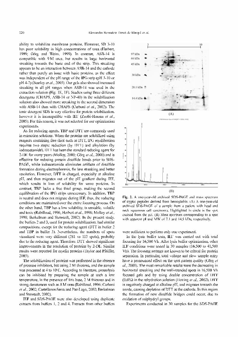
Fig. 2. A one-year-old archived SDS-PAGE and mass spectrum
of tryptic peptides derived from hemoglobin. (A) A one-year-old
archived SDS-PAGE of a sample from a patient with head and
neck squamous cell carcinoma. Highlighted in circle is the spot
excised from the gel. (B) Mass spectrum corresponding to a spot
with apparent pI and MW of 7.1 and 14.5 kDa, respectively.

Solubilization of Proteins and Gel Storage Protocols 221
step tested the SDS electrophoresis buffer at a doubled
concentration (50 mM Tris-base, 384 mM glycine, 0.2%
SDS) in the cathodic reservoir as compared to the anodic
reservoir. This condition resulted in decreased protein
smearing in the gel, which is usually caused by buffer
depletion during electrophoresis (data not shown). The most
important component of the typical second-dimension buffer
system must be SDS, which binds protein at a relatively
constant ratio, thereby allowing for the size-based separation
imparted by the sieving matrix. It was observed that buffer
depletion during electrophoresis causes dissociation of the
SDS from the protein and, consequently, elongated protein
patterns or smearing (Werner, 2003).
Five protocols for gel storing were tested and success was
attained when the gels were stored in plastic bags with 50%
glycerol solution (Protocol 5). This procedure permitted the
easy handling of the gel, without the risk of breakage and
without harming the image and MS analysis. The gels were
submitted to spot excision and MS analysis up to one year
after SDS-PAGE. Figure 2A shows a one-year-old archived
SDS-PAGE of a sample from a patient with head and neck
squamous cell carcinoma. The MS spectrum corresponding to
a spot with apparent pI and MW of 7.1 and 14.5 kDa,
respectively, is presented in Figure 2B. This protein spot was
identified as hemoglobin. The remaining protocols resulted in
cracking gels. Preservation of gels for a long time after
electrophoresis is frequently desirable, mainly if their transport
to another laboratory is necessary. Several methods for drying
polyacrylamide gels have been described. These protocols
have been routinely applied in thin gels (0.8 mm). However,
cracking gels were observed when these methods were
performed in thick 2-D gels (1.5 mm).
Progress in sample preparation methodology for 2-DE has
focused on improvements in sample buffer constituents to
achieve better representation of the proteome. However, it
must be taken into account that a variety of factors influence
the sample preparation steps, some of which have been
reported in this manuscript. We highlighted that this is the first
report describing the successful solubilization and 2-DE of
protein from human lymph node tissue and a simple protocol
for long time preservation of 2D gels.
Acknowledgments We thank Professor Carlos Roberto
Ceron, Keity Souza Santos, Lucilene Delazari dos Santos and
Ana Maria Galvan Custódio for useful suggestions. We also
thank Nicole S. L. Grosso for critically reading the English
manuscript and Celso Pereira Reis Filho for technical support.
This work has been supported by the Brazilian Synchrotron
Light Laboratory (LNLS) under proposal MAS 3159 and also
by grants from Fundação de Amparo à Pesquisa do Estado de
São Paulo/FAPESP (Grant 01/14473-0), Conselho Nacional de
Desenvolvimento Científico e Tecnológico/CNPq and Coordenação
de Aperfeiçoamento de Pessoal de Nível Superior/CAPES.
References
Berkelman, T. and Stenstedt, T. (2002) 2-D Electrophoresis using
immobilized pH gradients. Principles and Methods. Amersham
Biosciences, Uppsala, Sweden.
Bradford, M. M. (1976) A rapid and sensitive method for the
quantitation of microgram quantities of protein utilizing the
principle of protein-dye binding. Anal. Biochem. 72, 248-254.
Carboni, L., Piubelli, C., Righetti, P. G., Jansson, B. and
Domenici, E. (2002) Proteomic analysis of rat brain tissue:
Comparison of protocols for two-dimensional gel electrophoresis
analysis based on different solubilizing agents. Electrophoresis
23, 4132-4141.
Castellanos-Serra, L. and Paz-Lago, D. (2002) Inhibition of
unwanted proteolysis during sample preparation: Evaluation of
its efficiency in challenge experiments. Electrophoresis 23,
1745-1753.
Garfin, D. E. (2003) Two-dimensional gel electrophoresis: an
overview. Trends Analyt. Chem. 22, 263-272.
Giavalisco, P., Nordhoff, E., Lehrach, H., Gobom, J. and Klose, J.
(2003) Extraction of proteins from plant tissues for two-
dimensional electrophoresis analysis. Electrophoresis 24, 207-
216.
Görg, A. and Weiss, W. (1999) Analytical IPG-Dalt. Methods
Mol. Biol. 112, 189-195.
Görg, A., Obermaier, C., Boguth, G., Harder, A., Scheibe, B.,
Wildgruber, R. and Weiss, W. (2000) The current state of two-
dimensional electrophoresis with immobilized pH gradients.
Electrophoresis 21, 1037-1053.
Herbert, B. (1999) Advances in protein solubilisation for two-
dimensional electrophoresis. Electrophoresis 20, 660-663.
Herbert, B. R., Molloy, M. P., Gooley, A. A., Walsh, B. J.,
Bryson, W. G. and Williams, K. L. (1998) Improved protein
solubility in two-dimensional electrophoresis using tributyl
phosphine as reducing agent. Electrophoresis 19, 845-851.
Hoving, S., Gerrits, B., Voshol, H., Muler, D., Roberts, R. C. and
Oostrum, J. (2002) Preparative two-dimensional gel electrophoresis
at alkaline pH using narrow range immobilized pH gradients.
Proteomics 2, 127-134.
Méchin, V., Consoli, L., Guilloux, M. L. and Damerval, C. (2003)
An efficient solubilization buffer for plant proteins focused in
immobilized pH gradients. Proteomics 3, 1299-1302.
Molloy, M. P. (2000) Two-dimensional electrophoresis of
membrane proteins using immobilized pH gradients. Anal.
Biochem. 280, 1-10.
Molloy, M. P., Herbert, B. R., Walsh, B. J., Tyler, M. I., Traini,
M., Sanchez, J., Hochstrasser, D. F., Williams, K. L. and
Gooley, A. A. (1998) Extraction of membrane proteins by
differential solubilization for separation using two-dimensional
gel electrophoresis. Electrophoresis 19, 837-844.
Musante, L., Candiano, G. and Ghiggeri, G. M. (1998) Resolution
of fibronectin and other uncharacterized proteins by two-
dimensional polyacrilamide electrophoresis with thiourea. J.
Chromatogr. B. Biomed. Sci. Appl. 705, 351-356.
Pasquali, C., Fialka, I. and Huber, L.A. (1997) Preparative two-
dimensional gel electrophoresis of membrane proteins.
Electrophoresis 18, 2573-2581.
Rabilloud, T. (1996) Solubilization of proteins for electrophoretic
222 Alessandra Bernadete Trovó de Marqui et al.
analysis. Electrophoresis 17, 813-829.
Rabilloud, T. (1998) Use of thiourea to increase the solubility of
membrane proteins in two-dimensional electrophoresis. Electrophoresis
19, 758-760.
Rabilloud, T., Adessi, C., Giraudel, A. and Lunardi, J. (1997)
Improvement of the solubilization of proteins in two-
dimensional electrophoresis with immobilized pH gradients.
Electrophoresis 18, 307-316.
Shaw, M. M. and Riederer, B. M. (2003) Sample preparation for
two-dimensional gel electrophoresis. Proteomics 3, 1408-1417.
Stanley, B. A., Neverova, I., Brown, H. A. and Van Eyk, J. E.
(2003) Optimizing protein solubility for two-dimensional gel
electrophoresis analysis of human myocardium. Proteomics 3,
815-820.
Tachibana, M., Ohkura, Y., Kobayashi, Y., Sakamoto, H., Tanaka,
Y., Watanabe, J., Amikura, K., Nishimura, Y., Akagi, K. (2003)
Expression of apolipoprotein A1 in colonic adenocarcinoma.
Anticancer Res. 23, 4161-4167.
Taylor, C. M. and Pfeiffer, S. E. (2003) Enhanced resolution of
glycosylphosphatidylinositol-anchored and transmembrane
proteins from the lipid-rich myelin membrane by two-
dimensional gel electrophoresis. Proteomics 3, 1303-1312.
Werner, W. E. (2003) Run parameters affecting protein patterns
from second dimension electrophoresis gels. Anal. Biochem.
317, 280-283.
Zuobi-Hasona, K., Crowley, P. J., Hasona, A., Bleiweis, A. S.,
Brady, L. J. (2005) Solubilization of cellular membrane
proteins from Streptococcus mutans for two-dimensional gel
electrophoresis. Electrophoresis 26, 1200-1205.