a comparison of the costs of sentinel lymph node biopsy and of
Upload
khangminh22Category
view
4download
0
Gastric Cancer (2009) 12: 6–22DOI 10.1007/s10120-008-0492-5
Offprint requests to: R.M. KweeReceived: September 27, 2008 / Accepted: November 10, 2008
© 2009 byInternational andJapanese Gastric
Cancer Associations
Review article
Imaging in assessing lymph node status in gastric cancer
ROBERT MICHAEL KWEE1 and THOMAS CHRISTIAN KWEE
2
1 Department of Radiology, Maastricht University Medical Center, P. Debyelaan 25, 6202 AZ, Maastricht, The Netherlands2 Department of Radiology, University Medical Center Utrecht, Utrecht, The Netherlands
Key words Gastric cancer · Lymph node metastasis · Imaging · Staging · Systematic review
Introduction
Gastric cancer is the fourth most common cancer and the second leading cause of cancer-related death world-wide. In 2002, about 934 000 people were diagnosed with gastric cancer, and approximately 700 000 died of the disease [1].
Lymph node (LN) status is an important prognostic factor regarding long-term survival [2, 3]; in patients with N0 gastric cancer, the 5-year survival rate (after surgical treatment) is 86.1%, whereas in patients with N1, N2, and N3 gastric cancer, survival rates dramati-cally decrease to 58.1%, 23.3%, and 5.9% [3]. Patients with T1 tumors have a low risk of LN metastasis: 2.2% in T1a (mucosal) and 17.9% in T1b (submucosal) cancer [4]. In patients with T2 and those with T3 tumors, LN metastases rise to 44% and 64% [5]. The extent to which LN dissection should be performed is still a topic of debate [5–7]. Because extended lymphadenectomy is associated with high morbidity and mortality, patients without LN metastasis should be spared from undergo-ing such an aggressive procedure. Pretreatment knowl-edge of LN status would thus be extremely helpful for determining prognosis and planning the optimal extent of lymphadenectomy. In addition, pretreatment knowledge of LN status may help in selecting patients who might benefi t most from neoadjuvant chemother-apy [8].
As imaging technology continues to evolve [9], the purpose of this study was to systematically review the current role of imaging in assessing LN status in gastric cancer. This study reviews the role of imaging in dis-criminating node-negative from node-positive patients, rather than its role in assessing nodal stage according to
AbstractBackground. Accurate assessment of lymph node status is of crucial importance for appropriate treatment planning and determining prognosis in patients with gastric cancer. The aim of this study was to systematically review the current role of imaging in assessing lymph node (LN) status in gastric cancer.Methods. A systematic literature search was performed in the PubMed/MEDLINE and Embase databases. The method-ological quality and diagnostic performance of the included studies was assessed.Results. Six abdominal ultrasonography (AUS) studies, 30 endoscopic ultrasonography (EUS) studies, 10 multidetector-row computed tomography (MDCT) studies, 3 conventional magnetic resonance imaging (MRI) studies, 4 18F-fl uoro-2-deoxyglucose positron emission tomography (FDG-PET) studies, and 1 FDG-PET/CT fusion study were included. In general, the included studies had moderate methodological quality. The sensitivity and specifi city of AUS varied between 12.2% and 80.0% (median, 39.9%) and 56.3% and 100% (median, 81.8%). The sensitivity and specifi city of EUS varied between 16.7% and 95.3% (median, 70.8%) and 48.4% and 100% (median, 84.6%). The sensitivity and specifi city of MDCT varied between 62.5% and 91.9% (median, 80.0%) and 50.0% and 87.9% (median, 77.8%). The sensitivity and specifi city of MRI varied between 54.6% and 85.3% (median, 68.8%) and 50.0% and 100% (median, 75.0%). The sensitivity and specifi city of FDG-PET varied between 33.3% and 64.6% (median, 34.3%) and 85.7% and 97.0% (median, 93.2%). The sensitivity and specifi city of the FDG-PET/CT fusion study were 54.7% and 92.2%. For all the imaging modalities, there were no signifi cant differences between the mean sensitivities and specifi cities of high- and low-quality studies.Conclusion. AUS, EUS, MDCT, conventional MRI, and FDG-PET cannot reliably be used to confi rm or exclude the presence of LN metastasis. The performance of high-resolution PET/CT fusion and functional MRI techniques still has to be determined.
R.M. Kwee and T.C. Kwee: LN metastases in gastric cancer: imaging 7
the TNM or Japanese Gastric Cancer Association (JGCA) classifi cations.
Methods
Search strategy
A computer-aided search of the PubMed/MEDLINE and Embase databases was conducted to fi nd relevant publications on the diagnostic performance of abdomi-nal ultrasonography (AUS), endoscopic ultrasonogra-phy (EUS), multidetector-row computed tomography (MDCT), magnetic resonance imaging (MRI), 18F-fl uoro-2-deoxyglucose positron emission tomography (FDG-PET), and FDG-PET/CT fusion, in assessing LN status in gastric cancer. The following search terms were used: (“gastric cancer” or “stomach cancer” or “gastric carcinoma” or “stomach carcinoma”) and (“node metas-tasis” or “node metastases” or “nodal metastasis” or “nodal metastases” or “node involvement” or “nodal involvement” or “metastatic nodes” or “metastatic lymph nodes” or “lymphatic metastasis” or “lymphatic metastases” or “lymphatic involvement” or “lymph node involvement” or “lymph node metastatic disease” or “lymph node status” or “lymph node staging” or “N staging” or “TNM”) and (“ultrasound” or “sonogra-phy” or “ultrasonography” or “endoscopic ultrasound” or “endoscopic ultrasonography” or “EUS” or “com-puted tomography” or “CT” or “CAT” or “magnetic resonance” or “MR imaging” or “MRI” or “magnetic resonance tomography” or “nuclear magnetic reso-nance” or “NMR” or “fl uorodeoxyglucose” or “2-fl uoro-2-deoxy-D-glucose” or “FDG” or “positron emission tomography” or “positron-emission tomogra-phy” or “PET”). No beginning date limit was used. The search was updated until July 7, 2008. To expand our search, bibliographies of articles which fi nally remained after the selection process were screened for potentially suitable references.
Study selection
Studies investigating the diagnostic performance of AUS, EUS, MDCT (defi ned as CT with four or more detectors), MRI, FDG-PET, and/or FDG-PET/CT fusion in assessing LN status in patients with newly diagnosed, histologically proven gastric cancer were eli-gible for inclusion. Only studies dealing with adenocar-cinoma were included, because this is overwhelmingly the most important and most common malignant tumor that occurs in the stomach (range, 90% to 95%) [10]. Review articles, metaanalyses, abstracts, editorials or letters, case reports, studies involving ten or fewer patients with gastric cancer, tutorials, guidelines for
management, and non-English-language articles were excluded. Studies performed in animals and ex vivo studies were also excluded. Studies in which patients were presurgically treated with radiotherapy or chemo-therapy, which may cause downstaging, were excluded. Studies which investigated only patients with gastric cancer confi ned to a certain part of the stomach (e.g., the gastroesophageal junction) were excluded. Studies which provided insuffi cient data to construct a 2 × 2 contingency table to calculate sensitivity and specifi city for detecting LN metastasis on a per-patient basis were excluded. When data were presented in more than one article, the article with the largest number of patients was chosen.
Two researchers (R.M.K., T.C.K.) independently reviewed the titles and abstracts of the retrieved arti-cles, applying the inclusion and exclusion criteria men-tioned above. Articles were rejected if they were clearly ineligible. The same two researchers then independently reviewed the full-text version of the remaining articles to determine their eligibility for inclusion. Disagree-ments were resolved in a consensus meeting.
Data analysis
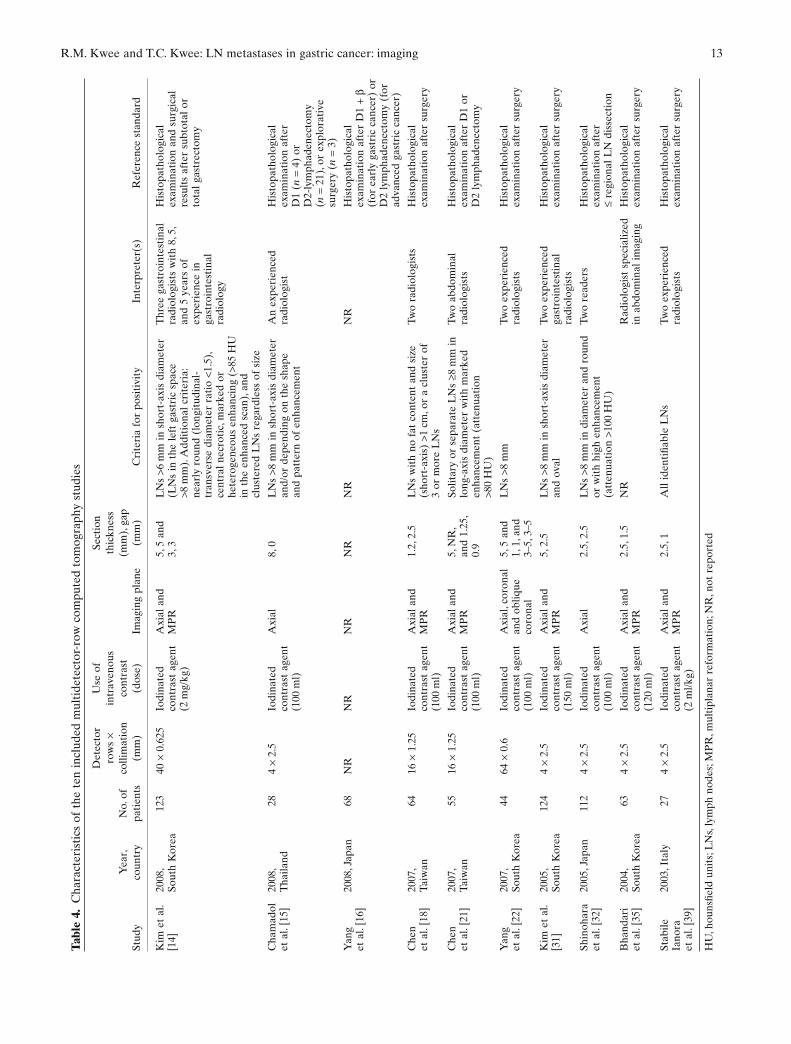
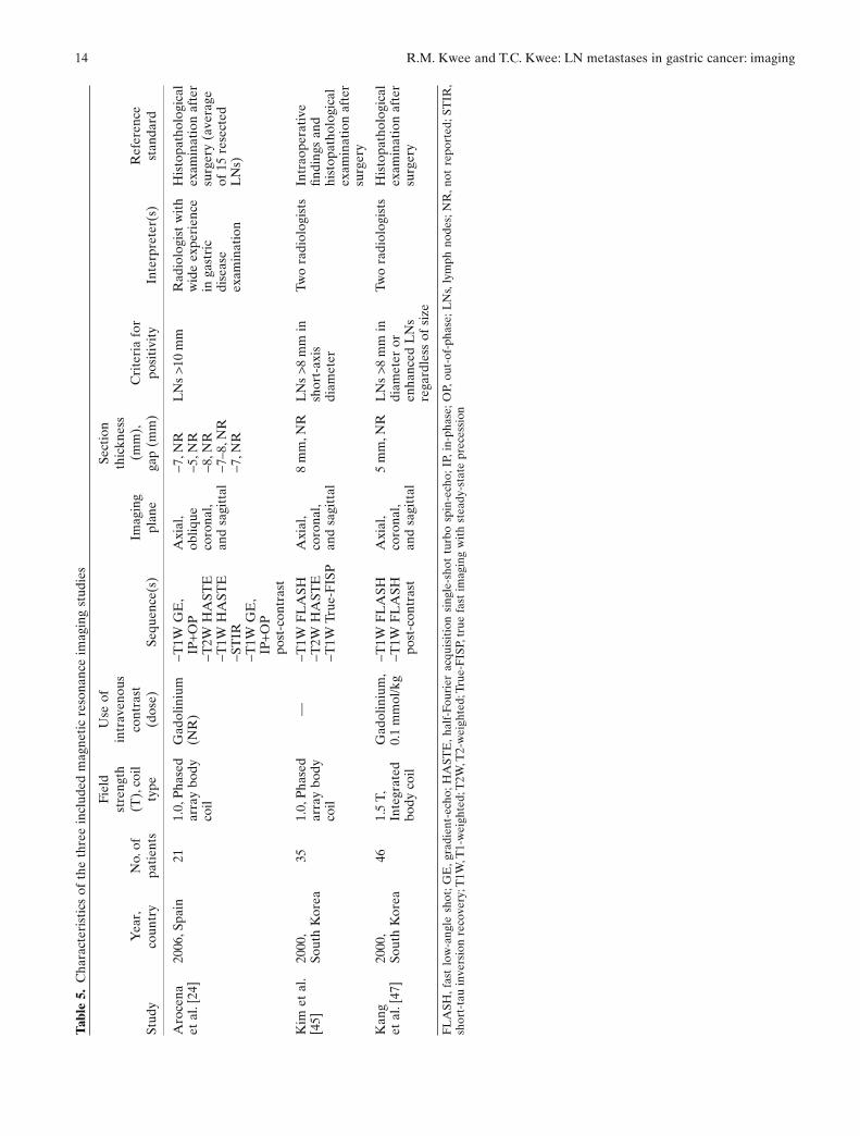
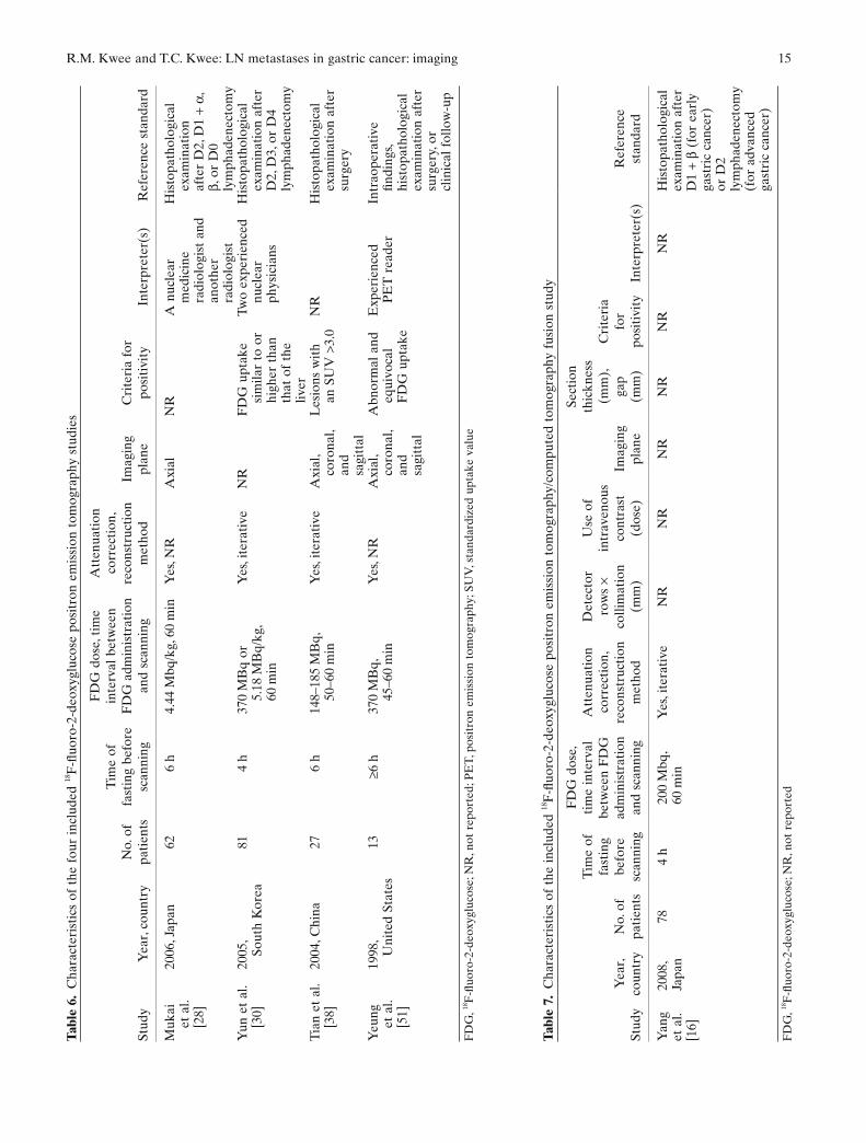
For each included study, information was collected con-cerning year of publication, country of origin, number of patients, technical details of the imaging modality under investigation, criteria for positivity, interpreter(s), and applied reference standard.
The methodological quality of the included studies was assessed in terms of the potential for bias (internal validity) and lack of generalizability (external validity). For this purpose, a checklist adapted from Kelly et al. [11] and Whiting et al. [12, 13] was used. The complete criteria list is presented in Table 1. Internal validity cri-teria and external validity scores were scored as positive (adequate methods) or negative (inadequate methods, potential bias). If insuffi cient information was provided on a specifi c item, a negative score was given. Two reviewers (R. M. K., T. C. K.) independently assigned the scores. Disagreements between the two researchers were discussed and resolved by consensus. Subtotals were calculated for internal (maximum eight) and exter-nal (maximum fi ve) validity separately. Total quality scores were expressed as a percentage of the maximum score. Studies which had a percentage of the maximum score of 60 or greater were considered to be of high methodological quality. Studies which had a percentage of the maximum score of less than 60 were considered to be of low methodological quality.
Sensitivities and specifi cities for the detection of LN metastasis (with corresponding 95% confi dence inter-vals [CIs]) were calculated from the original numbers given in the included studies, for each imaging modality.
8 R.M. Kwee and T.C. Kwee: LN metastases in gastric cancer: imaging
Forest plots for sensitivities and specifi cities were con-structed. The means of sensitivities and specifi cities between studies of high and low methodological quality were compared by using a paired samples t-test. The level of statistical signifi cance was set at 0.05. Statistical analyses were executed using Statistical Package for the Social Sciences version 12.0 software (SPSS, Chicago, IL, USA).
Results
Literature search
The computer-aided search revealed 1035 articles from PubMed/MEDLINE and 889 articles from Embase. Reviewing titles and abstracts from PubMed/MEDLINE revealed 87 studies potentially eligible for inclusion. Reviewing titles and abstracts from Embase revealed 66 articles potentially eligible for inclusion, of which 62 were already identifi ed by the PubMed/MEDLINE search. Thus, 91 articles remained for possible inclusion and were retrieved in full-text version. Screening refer-ences of these articles did not result in other potentially relevant articles. After reviewing the full article, 37 articles were excluded, the majority (n = 21) because they provided insuffi cient data to construct a 2 × 2 con-tingency table to calculate sensitivity and specifi city for the detection of LN metastasis. Other reasons for exclu-sion were: same data were used in a later study (n = 7),
patients examined with a single-slice CT scanner instead of with an MDCT scanner (n = 2), diagnostic perfor-mance for detecting LN metastasis was not investigated (n = 4), patients with esophageal and gastric cancer mixed (n = 1), patients with lymphomas and gastric cancer mixed (n = 1), and fewer than 10 patients with gastric cancer included (n = 1). Eventually, 6 AUS studies, 30 EUS studies, 10 MDCT studies, 3 (conven-tional) MRI studies, 4 FDG-PET studies, and 1 FDG-PET/CT fusion study [14–65] were included in this systematic review. The characteristics of the included studies are presented in Tables 2 to 7. The AUS studies were published between 1996 and 2006, the EUS studies between 1990 and 2008, the MDCT studies between 2003 and 2008, the MRI studies between 2000 and 2006, and the FDG-PET studies between 1998 and 2006. The included FDG-PET/CT fusion study was published in 2008. The number of patients in the AUS, EUS, MDCT, MRI, and FDG-PET studies varied from 22 to 198, 21 to 254, 27 to 124, 21 to 46, and 13 to 81, respectively. The included FDG-PET/CT fusion study comprised 78 patients.
Methodological quality assessment
For each of the included studies, 13 methodological quality items were assessed (Table 8).
• For the AUS studies, the total score for combined internal and external validity, expressed as a fraction
Table 1. Criteria list used to assess the methodological quality of the studies
Criteria of validity Positive score
Internal validity 1. Prospective study Mentioned in publication2. Adequate reference test D2 or more extensive lymphadenectomy and histopathological
analysis of resected lymph nodes performed in all patients3. Avoidance of disease progression
biasTime interval between index test and reference test <16 days
in all patients4. Avoidance of withdrawal bias <10% of patients who were examined by the index test did
not undergo the reference test5. Avoidance of study examination
bias<10% of indeterminate or uninterpretable results
6. Avoidance of diagnostic review bias
Blind interpretation of index test without knowledge of reference test
7. Avoidance of test review bias Blind interpretation of reference test without knowledge of index test
8. Avoidance of comparator review bias
Blinding index test to the other imaging modality, if more than one imaging modality was investigated
External validity 1. Avoidance of spectrum bias Only patients with newly diagnosed, histologically proven gastric cancer were included or a separate analysis was provided for these patients
2. Demographic information Study location (country), age and sex of patients reported3. Avoidance of selection bias Consecutive series of patients or random selection of patients4. Standard execution of index test Application of the same hardware and imaging protocol in all
patients5. Avoidance of observer variability
biasInterpreter(s) of index test described
R.M. Kwee and T.C. Kwee: LN metastases in gastric cancer: imaging 9
of the maximum score, ranged from 31% to 69% (median, 58%). Three AUS studies [23, 42, 53] were of high methodological quality (percentage of the maximum score of 60 or greater).
• For the EUS studies, the total methodological quality score ranged from 38% to 77% (median, 54%). Ten EUS studies [19, 29, 34, 37, 41, 43, 44, 48, 50, 61] were of high methodological quality.
• For the MDCT studies, the total methodological quality score ranged from 38% to 85% (median, 70%). Eight MDCT studies [14, 18, 21, 22, 31, 32, 35, 39] were of high methodological quality.
• For the MRI studies, the total methodological quality score ranged from 46% to 77% (median, 62%). Two MRI studies [45, 47] were of high methodological quality.
• For the FDG-PET studies, the total methodological quality score ranged from 46% to 62% (median, 58%). Two FDG-PET studies [28, 30] were of high methodological quality.
• For the FDG-PET/CT fusion study, the total meth-odological quality score was 54%.
Staging performance
The sensitivities and specifi cities of the included studies are displayed in Table 9 and Fig. 1.
• The sensitivity and specifi city of AUS for the detec-tion of LN metastasis varied between 12.2% and
80.0% (median, 39.9%) and 56.3% and 100% (median, 81.8%). There was no signifi cant difference between the mean sensitivity of AUS studies with high and low methodological quality (53.2% vs 36.6%; P = 0.697). There also was no signifi cant dif-ference between the mean specifi city of studies with high and low methodological quality (73.3% vs 86.4%; P = 0.166).
• The sensitivity and specifi city of EUS varied between 16.7% and 96.8% (median, 70.8%) and 48.4% and 100% (median, 84.6%). There was no signifi cant dif-ference between the mean sensitivity of EUS studies with high and low methodological quality (69.1% vs 64.1%; P = 0.551). There also was no signifi cant dif-ference between the mean specifi city of studies with high and low methodological quality (81.8% vs 82.8%; P = 0.827).
• The sensitivity and specifi city of MDCT varied between 62.5% and 91.9% (median, 80.0%) and 50.0% and 87.9% (median, 77.8%). There was no signifi cant difference between the mean sensitivity of MDCT studies with high and low methodological quality (80.1% vs 75.0%; P = 0.331). There also was no signifi cant difference between the mean specifi city of studies with high and low methodological quality (82.0% vs 75.5%; P = 0.473).
• The sensitivity and specifi city of MRI varied be-tween 54.6% and 85.3% (median, 68.8%) and 50.0% and 100% (median, 75.0%). The mean sen-sitivity and specifi city of the MRI studies with high
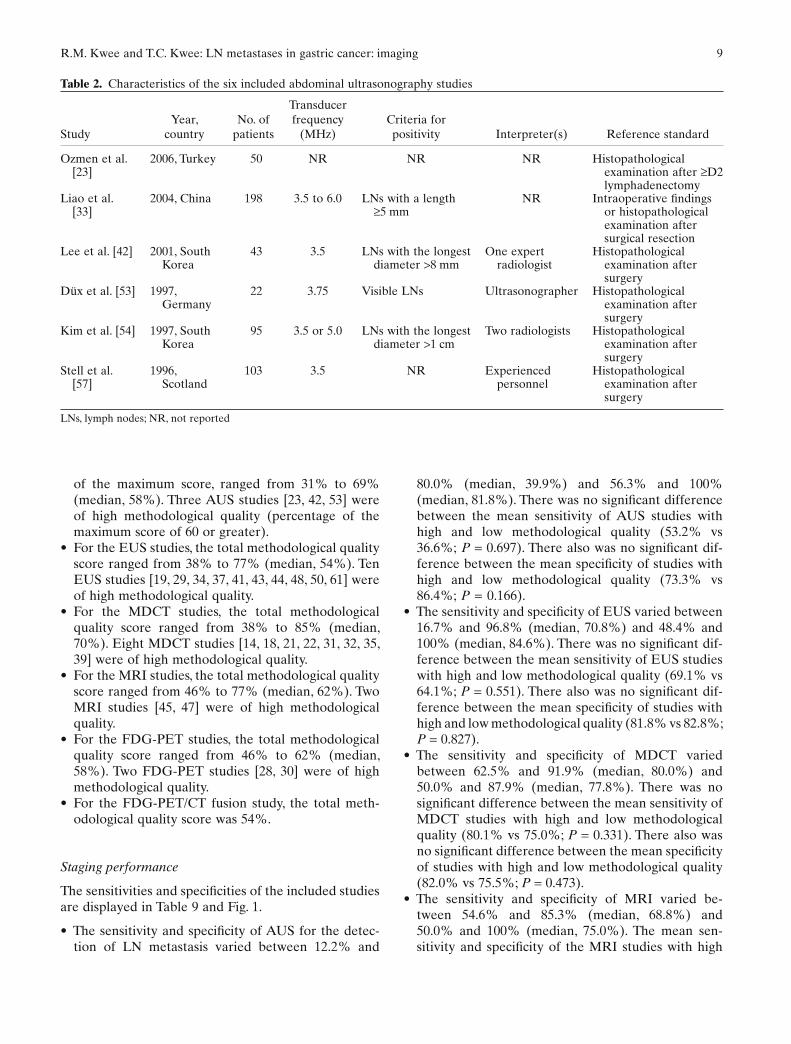
Table 2. Characteristics of the six included abdominal ultrasonography studies
StudyYear,
countryNo. of
patients
Transducer frequency
(MHz)Criteria for positivity Interpreter(s) Reference standard
Ozmen et al. [23]
2006, Turkey 50 NR NR NR Histopathological examination after ≥D2 lymphadenectomy
Liao et al. [33]
2004, China 198 3.5 to 6.0 LNs with a length ≥5 mm
NR Intraoperative fi ndings or histopathological examination after surgical resection
Lee et al. [42] 2001, South Korea
43 3.5 LNs with the longest diameter >8 mm
One expert radiologist
Histopathological examination after surgery
Düx et al. [53] 1997, Germany
22 3.75 Visible LNs Ultrasonographer Histopathological examination after surgery
Kim et al. [54] 1997, South Korea
95 3.5 or 5.0 LNs with the longest diameter >1 cm
Two radiologists Histopathological examination after surgery
Stell et al. [57]
1996, Scotland
103 3.5 NR Experienced personnel
Histopathological examination after surgery
LNs, lymph nodes; NR, not reported

10 R.M. Kwee and T.C. Kwee: LN metastases in gastric cancer: imaging
Tabl
e 3.
Cha
ract
eris
tics
of
the
30 in
clud
ed e
ndos
copi
c ul
tras
onog
raph
y st
udie
s
Stud
yY
ear,
coun
try
No.
of
pati
ents
Type
of
echo
endo
scop
e
Tran
sduc
er
freq
uenc
y (M
Hz)
Cri
teri
a fo
r po
siti
vity
Inte
rpre
ter(
s)R
efer
ence
sta
ndar
d
Lok
et
al. [
17]
2008
, H
ong
Kon
g12
3R
adia
l arr
ay12
or
7.5,
or
5 to
20,
or
12 a
nd 2
0
Hyp
oech
oic
LN
s, sh
arpl
y de
mar
cate
d, r
ound
ed
cont
our,
and
size
>1
0 m
m
One
of
four
exp
erie
nced
en
dosc
opis
ts w
ith
a sp
ecia
l int
eres
t in
E
US
His
topa
thol
ogic
al
exam
inat
ion
afte
r su
rger
y
Ben
trem
et
al.
[19]
2007
, U
nite
d St
ates
225
NR
7.5
or 1
2H
ypoe
choi
c, r
ound
, and
w
ell-
dem
arca
ted
LN
sA
gas
troe
nter
olog
ist
His
topa
thol
ogic
al
exam
inat
ion
afte
r R
0 re
sect
ion
Tan
et a
l. [2
0]20
07, C
hina
63R
adia
l arr
ay7.
5 or
20
NR
NR
His
topa
thol
ogic
al
exam
inat
ion
afte
r su
rger
yA
roce
na e
t al
. [2
4]20
06, S
pain
21L
inea
r ar
ray
12.5
LN
s >5
mm
, rou
nd,
hypo
echo
ic,
wel
l-de
mar
cate
d, a
nd
hom
ogen
eous
Exp
erie
nced
en
dosc
opis
tH
isto
path
olog
ical
ex
amin
atio
n af
ter
surg
ery
(ave
rage
of
15
rese
cted
LN
s)G
anpa
thi e
t al
. [2
5]20
06, S
inga
pore
126
Rad
ial a
rray
7.5
to 2
0E
cho-
poor
, rou
ndis
h,
wel
l-de
mar
cate
d L
Ns
and
LN
s >1
cm
One
of
thre
e ex
peri
ence
d op
erat
ors
Intr
aope
rati
ve fi
ndin
gs
or h
isto
path
olog
ical
ex
amin
atio
n af
ter
surg
ery
Tse
ndsu
ren
et a
l. [2
6]20
06, C
hina
41L
inea
r ar
ray
5.0
or 7
.5L
Ns
wit
h ro
und
bord
er
and
hypo
echo
ic
stru
ctur
es
NR
His
topa
thol
ogic
al
exam
inat
ion
afte
r su
rger
yA
ng e
t al
. [27
]20
06, S
inga
pore
77R
adia
l arr
ay7.
5L
Ns
>1 c
mN
RH
isto
path
olog
ical
ex
amin
atio
n af
ter
surg
ery
Potr
c et
al.
[29]
2006
, Slo
veni
a82
Rad
ial a
rray
7.5
or 1
2N
RTw
o ga
stro
ente
rolo
gist
sH
isto
path
olog
ical
ex
amin
atio
n af
ter
D2
lym
phad
enec
tom
yPo
lkow
ski e
t al
. [3
4]20
04, P
olan
d88
Rad
ial a
rray
7.5
or 1
2L
Ns
≥8 m
mO
ne e
xper
ienc
ed
inve
stig
ator
His
topa
thol
ogic
al
exam
inat
ion
afte
r ly
mph
aden
ecto
my
(≥15
res
ecte
d L
Ns)
Bha
ndar
i et
al.
[35]
2004
, So
uth
Kor
ea63
Rad
ial a
rray
7.5
or 2
0N
RE
ndos
copi
st w
ith
at
leas
t 5
year
s of
ex
peri
ence
His
topa
thol
ogic
al
exam
inat
ion
afte
r su
rger
yJa
vaid
et
al. [
36]
2004
, Ind
ia11
2R
adia
l arr
ay7.
5E
cho-
poor
, rou
ndis
h,
and
wel
l-de
mar
cate
d L
Ns
NR
His
topa
thol
ogic
al
exam
inat
ion
afte
r su
rger
yH
aber
man
n et
al
. [37
]20
04, G
erm
any
51R
adia
l arr
ay7.
5 or
12
LN
s ≥8
mm
in s
hort
-axi
s di
amet
erE
ndos
copi
stH
isto
path
olog
ical
ex
amin
atio
n af
ter
≥D1
lym
phad
enec
tom
yX
i et
al. [
40]
2003
, Chi
na35
Rad
ial a
rray
7.5,
12,
or
20N
RN
RH
isto
path
olog
ical
ex
amin
atio
n af
ter
surg
ery
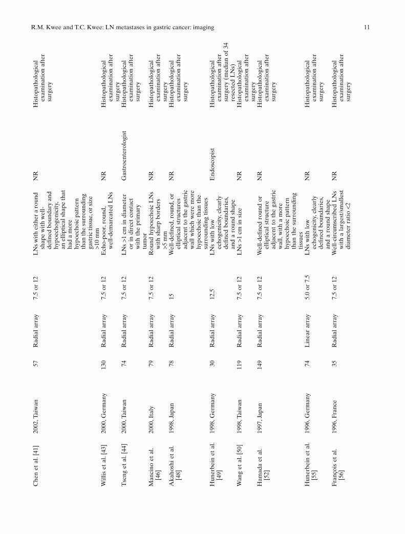
R.M. Kwee and T.C. Kwee: LN metastases in gastric cancer: imaging 11C
hen
et a
l. [4
1]20
02, T
aiw
an57
Rad
ial a
rray
7.5
or 1
2L
Ns
wit
h ei
ther
a r
ound
sh
ape
wit
h w
ell-
defi n
ed b
ound
ary
and
hypo
echo
geni
city
, an
elli
ptic
al s
hape
that
ha
d a
mor
e hy
poec
hoic
pat
tern
th
an th
e su
rrou
ndin
g ga
stri
c ti
ssue
, or
size
>1
0 m
m
NR
His
topa
thol
ogic
al
exam
inat
ion
afte
r su
rger
y
Will
is e
t al
. [43
]20
00, G
erm
any
130
Rad
ial a
rray
7.5
or 1
2E
cho-
poor
, rou
nd,
wel
l-de
mar
cate
d L
Ns
NR
His
topa
thol
ogic
al
exam
inat
ion
afte
r su
rger
yT
seng
et
al. [
44]
2000
, Tai
wan
74R
adia
l arr
ay7.
5 or
12
LN
s >1
cm
in d
iam
eter
or
in d
irec
t co
ntac
t w
ith
the
prim
ary
tum
or
Gas
troe
nter
olog
ist
His
topa
thol
ogic
al
exam
inat
ion
afte
r su
rger
y
Man
cino
et
al.
[46]
2000
, Ita
ly79
Rad
ial a
rray
7.5
or 1
2R
ound
hyp
oech
oic
LN
s w
ith
shar
p bo
rder
s >5
mm
NR
His
topa
thol
ogic
al
exam
inat
ion
afte
r su
rger
yA
kaho
shi e
t al
. [4
8]19
98, J
apan
78R
adia
l arr
ay15
Wel
l-de
fi ned
, rou
nd, o
r el
lipti
cal s
truc
ture
s ad
jace
nt t
o th
e ga
stri
c w
all w
hich
wer
e m
ore
hypo
echo
ic t
han
the
surr
ound
ing
tiss
ues
NR
His
topa
thol
ogic
al
exam
inat
ion
afte
r su
rger
y
Hun
erbe
in e
t al
. [4
9]19
98, G
erm
any
30R
adia
l arr
ay12
.5L
Ns
wit
h lo
w
echo
geni
city
, cle
arly
de
fi ned
bou
ndar
ies,
and
a ro
und
shap
e
End
osco
pist
His
topa
thol
ogic
al
exam
inat
ion
afte
r su
rger
y (m
edia
n of
34
rese
cted
LN
s)W
ang
et a
l. [5
0]19
98, T
aiw
an11
9R
adia
l arr
ay7.
5 or
12
LN
s >1
cm
in s
ize
NR
His
topa
thol
ogic
al
exam
inat
ion
afte
r su
rger
yH
amad
a et
al.
[52]
1997
, Jap
an14
9R
adia
l arr
ay7.
5 or
12
Wel
l-de
fi ned
rou
nd o
r el
lipti
cal s
truc
ture
ad
jace
nt t
o th
e ga
stri
c w
all,
wit
h a
mor
e hy
poec
hoic
pat
tern
th
an t
he s
urro
undi
ng
tiss
ues
NR
His
topa
thol
ogic
al
exam
inat
ion
afte
r su
rger
y
Hun
erbe
in e
t al
. [5
5]19
96, G
erm
any
74L
inea
r ar
ray
5.0
or 7
.5L
Ns
wit
h lo
w
echo
geni
city
, cle
arly
de
fi ned
bou
ndar
ies,
and
a ro
und
shap
e
NR
His
topa
thol
ogic
al
exam
inat
ion
afte
r su
rger
y
Fran
çois
et
al.
[56]
1996
, Fra
nce
35R
adia
l arr
ay7.
5 or
12
Wel
l-ci
rcum
scri
bed
LN
s w
ith
a la
rges
t/sm
alle
st
diam
eter
rat
io <
2
NR
His
topa
thol
ogic
al
exam
inat
ion
afte
r su
rger
y
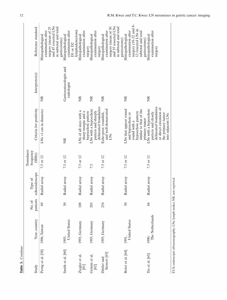
12 R.M. Kwee and T.C. Kwee: LN metastases in gastric cancer: imaging
Per
ng e
t al
. [58
]19
96, T
aiw
an69
Rad
ial a
rray
7.5
or 1
2L
Ns
>1 c
m in
dia
met
erN
RH
isto
path
olog
ical
ex
amin
atio
n af
ter
surg
ery
(mea
n of
25
and
45 r
esec
ted
LN
s in
sub
tota
l and
tot
al
gast
rect
omy)
Smit
h et
al.
[60]
1993
, U
nite
d St
ates
50R
adia
l arr
ay7.
5 or
12
NR
Gas
troe
nter
olog
ist
and
radi
olog
ist
His
topa
thol
ogic
al
exam
inat
ion
afte
r D
1 or
D2
lym
phad
enec
tom
yZ
iegl
er e
t al
. [6
1]19
93, G
erm
any
108
Rad
ial a
rray
7.5
or 1
2L
Ns
of a
ll si
zes
wit
h a
shar
p m
argi
n an
d a
hypo
echo
ic p
atte
rn
NR
His
topa
thol
ogic
al
exam
inat
ion
afte
r su
rger
yG
rim
m e
t al
. [6
2]19
93, G
erm
any
203
Rad
ial a
rray
7.5
LN
s w
ith
a hy
poec
hoic
pa
tter
n an
d sh
arpl
y de
linea
ted
boun
dari
es
NR
His
topa
thol
ogic
al
exam
inat
ion
afte
r su
rger
yD
ittl
er a
nd
Siew
ert
[63]
1993
, Ger
man
y25
4R
adia
l arr
ay7.
5 or
12
Ech
o-po
or, r
ound
ish,
an
d w
ell-
dem
arca
ted
LN
s
NR
His
topa
thol
ogic
al
exam
inat
ion
afte
r su
rger
y (m
ean
of 3
0 an
d 55
res
ecte
d L
Ns
in s
ubto
tal a
nd t
otal
ga
stre
ctom
y)B
otet
et
al. [
64]
1991
, U
nite
d St
ates
50R
adia
l arr
ay7.
5 or
12
LN
s th
at a
ppea
r ro
und
and
hypo
echo
ic o
r ro
und
wit
h a
hypo
echo
ic p
atte
rn
sim
ilar
to t
hat
of t
he
prim
ary
tum
or
NR
His
topa
thol
ogic
al
exam
inat
ion
afte
r su
rger
y (1
8–22
and
8–
12 r
esec
ted
LN
s in
su
btot
al a
nd t
otal
ga
stre
ctom
y)T
io e
t al
. [65
]19
90,
The
Net
herl
ands
84R
adia
l arr
ay7.
5 or
12
LN
s w
ith
a hy
poec
hoic
pa
tter
n an
d cl
earl
y de
linea
ted
boun
dari
es
or d
irec
t ex
tens
ion
of
the
prim
ary
tum
or
into
adj
acen
t L
Ns
NR
His
topa
thol
ogic
al
exam
inat
ion
afte
r su
rger
y
EU
S, e
ndos
copi
c ul
tras
onog
raph
y; L
Ns,
lym
ph n
odes
; NR
, not
rep
orte
d
Tabl
e 3.
Con
tinue
Stud
yY
ear,
coun
try
No.
of
pati
ents
Type
of
echo
endo
scop
e
Tran
sduc
er
freq
uenc
y (M
Hz)
Cri
teri
a fo
r po
siti
vity
Inte
rpre
ter(
s)R
efer
ence
sta
ndar
d
R.M. Kwee and T.C. Kwee: LN metastases in gastric cancer: imaging 13
Tabl
e 4.
Cha
ract
eris
tics
of
the
ten
incl
uded
mul
tide
tect
or-r
ow c
ompu
ted
tom
ogra
phy
stud
ies
Stud
yY
ear,
coun
try
No.
of
pati
ents
Det
ecto
r ro
ws
× co
llim
atio
n (m
m)
Use
of
intr
aven
ous
cont
rast
(d
ose)
Imag
ing
plan
e
Sect
ion
thic
knes
s (m
m),
gap
(mm
)C
rite
ria
for
posi
tivi
tyIn
terp
rete
r(s)
Ref
eren
ce s
tand
ard
Kim
et
al.
[14]
2008
, So
uth
Kor
ea12
340
× 0
.625
Iodi
nate
d co
ntra
st a
gent
(2
mg/
kg)
Axi
al a
nd
MP
R5,
5 a
nd
3, 3
LN
s >6
mm
in s
hort
-axi
s di
amet
er
(LN
s in
the
left
gas
tric
spa
ce
>8 m
m).
Add
itio
nal c
rite
ria:
ne
arly
rou
nd (
long
itud
inal
-tr
ansv
erse
dia
met
er r
atio
<1.
5),
cent
ral n
ecro
tic,
mar
ked
or
hete
roge
neou
s en
hanc
ing
(>85
HU
in
the
enh
ance
d sc
an),
and
clus
tere
d L
Ns
rega
rdle
ss o
f si
ze
Thr
ee g
astr
oint
esti
nal
radi
olog
ists
wit
h 8,
5,
and
5 ye
ars
of
expe
rien
ce in
ga
stro
inte
stin
al
radi
olog
y
His
topa
thol
ogic
al
exam
inat
ion
and
surg
ical
re
sult
s af
ter
subt
otal
or
tota
l gas
trec
tom
y
Cha
mad
ol
et a
l. [1
5]20
08,
Tha
iland
284
× 2.
5Io
dina
ted
cont
rast
age
nt
(100
ml)
Axi
al8,
0L
Ns
>8 m
m in
sho
rt-a
xis
diam
eter
an
d/or
dep
endi
ng o
n th
e sh
ape
and
patt
ern
of e
nhan
cem
ent
An
expe
rien
ced
radi
olog
ist
His
topa
thol
ogic
al
exam
inat
ion
afte
r D
1 (n
= 4
) or
D
2-ly
mph
aden
ecto
my
(n =
21)
, or
expl
orat
ive
surg
ery
(n =
3)
Yan
g et
al.
[16]
2008
, Jap
an68
NR
NR
NR
NR
NR
NR
His
topa
thol
ogic
al
exam
inat
ion
afte
r D
1 +
β (f
or e
arly
gas
tric
can
cer)
or
D2
lym
phad
enec
tom
y (f
or
adva
nced
gas
tric
can
cer)
Che
n et
al.
[18]
2007
, Ta
iwan
6416
× 1
.25
Iodi
nate
d co
ntra
st a
gent
(1
00 m
l)
Axi
al a
nd
MP
R
1.2,
2.5
LN
s w
ith
no f
at c
onte
nt a
nd s
ize
(sho
rt-a
xis)
>1
cm, o
r a
clus
ter
of
3 or
mor
e L
Ns
Two
radi
olog
ists
His
topa
thol
ogic
al
exam
inat
ion
afte
r su
rger
y
Che
n et
al.
[21]
2007
, Ta
iwan
5516
× 1
.25
Iodi
nate
d co
ntra
st a
gent
(1
00 m
l)
Axi
al a
nd
MP
R5,
NR
, an
d 1.
25,
0.9
Solit
ary
or s
epar
ate
LN
s ≥8
mm
in
long
-axi
s di
amet
er w
ith
mar
ked
enha
ncem
ent
(att
enua
tion
>8
0 H
U)
Two
abdo
min
al
radi
olog
ists
His
topa
thol
ogic
al
exam
inat
ion
afte
r D
1 or
D
2 ly
mph
aden
ecto
my
Yan
g et
al.
[22]
2007
, So
uth
Kor
ea44
64 ×
0.6
Iodi
nate
d co
ntra
st a
gent
(1
00 m
l)
Axi
al, c
oron
al
and
obliq
ue
coro
nal
5, 5
and
1,
1, a
nd
3–5,
3–5
LN
s >8
mm
Two
expe
rien
ced
radi
olog
ists
His
topa
thol
ogic
al
exam
inat
ion
afte
r su
rger
y
Kim
et
al.
[31]
2005
, So
uth
Kor
ea12
44
× 2.
5Io
dina
ted
cont
rast
age
nt
(150
ml)
Axi
al a
nd
MP
R5,
2.5
LN
s >8
mm
in s
hort
-axi
s di
amet
er
and
oval
Two
expe
rien
ced
gast
roin
test
inal
ra
diol
ogis
ts
His
topa
thol
ogic
al
exam
inat
ion
afte
r su
rger
y
Shin
ohar
a et
al.
[32]
2005
, Jap
an11
24
× 2.
5Io
dina
ted
cont
rast
age
nt
(100
ml)
Axi
al2.
5, 2
.5L
Ns
>8 m
m in
dia
met
er a
nd r
ound
or
wit
h hi
gh e
nhan
cem
ent
(att
enua
tion
>10
0 H
U)
Two
read
ers
His
topa
thol
ogic
al
exam
inat
ion
afte
r ≤
regi
onal
LN
dis
sect
ion
Bha
ndar
i et
al.
[35]
2004
, So
uth
Kor
ea63
4 ×
2.5
Iodi
nate
d co
ntra
st a
gent
(1
20 m
l)
Axi
al a
nd
MP
R2.
5, 1
.5N
RR
adio
logi
st s
peci
aliz
ed
in a
bdom
inal
imag
ing
His
topa
thol
ogic
al
exam
inat
ion
afte
r su
rger
y
Stab
ile
Iano
ra
et a
l. [3
9]
2003
, Ita
ly27
4 ×
2.5
Iodi
nate
d co
ntra
st a
gent
(2
ml/k
g)
Axi
al a
nd
MP
R2.
5, 1
All
iden
tifi a
ble
LN
sTw
o ex
peri
ence
d ra
diol
ogis
tsH
isto
path
olog
ical
ex
amin
atio
n af
ter
surg
ery
HU
, hou
nsfi e
ld u
nits
; LN
s, ly
mph
nod
es; M
PR
, mul
tipl
anar
ref
orm
atio
n; N
R, n
ot r
epor
ted
14 R.M. Kwee and T.C. Kwee: LN metastases in gastric cancer: imaging
Tabl
e 5.
Cha
ract
eris
tics
of
the
thre
e in
clud
ed m
agne
tic
reso
nanc
e im
agin
g st
udie
s
Stud
yY
ear,
coun
try
No.
of
pati
ents
Fiel
d st
reng
th
(T),
coil
type
Use
of
intr
aven
ous
cont
rast
(d
ose)
Sequ
ence
(s)
Imag
ing
plan
e
Sect
ion
thic
knes
s (m
m),
gap
(mm
)C
rite
ria
for
posi
tivi
tyIn
terp
rete
r(s)
Ref
eren
ce
stan
dard
Aro
cena
et
al.
[24]
2006
, Spa
in21
1.0,
Pha
sed
arra
y bo
dy
coil
Gad
olin
ium
(N
R)
− T1W
GE
, IP
+OP
−T2W
HA
STE
−T1W
HA
STE
−ST
IR− T
1W G
E,
IP+O
P
post
-con
tras
t
Axi
al,
obliq
ue
coro
nal,
and
sagi
ttal
−7, N
R−5
, NR
−8, N
R−7
–8, N
R−7
, NR
LN
s >1
0 m
mR
adio
logi
st w
ith
wid
e ex
peri
ence
in
gas
tric
di
seas
e ex
amin
atio
n
His
topa
thol
ogic
al
exam
inat
ion
afte
r su
rger
y (a
vera
ge
of 1
5 re
sect
ed
LN
s)
Kim
et
al.
[45]
2000
, So
uth
Kor
ea35
1.0,
Pha
sed
arra
y bo
dy
coil
—−T
1W F
LA
SH−T
2W H
AST
E−T
1W T
rue-
FIS
P
Axi
al,
coro
nal,
and
sagi
ttal
8 m
m, N
RL
Ns
>8 m
m in
sh
ort-
axis
di
amet
er
Two
radi
olog
ists
Intr
aope
rati
ve
fi ndi
ngs
and
hist
opat
holo
gica
l ex
amin
atio
n af
ter
surg
ery
Kan
g et
al.
[47]
2000
, So
uth
Kor
ea46
1.5
T,
Inte
grat
ed
body
coi
l
Gad
olin
ium
, 0.
1 m
mol
/kg
−T1W
FL
ASH
− T1W
FL
ASH
po
st-c
ontr
ast
Axi
al,
coro
nal,
and
sagi
ttal
5 m
m, N
RL
Ns
>8 m
m in
di
amet
er o
r en
hanc
ed L
Ns
rega
rdle
ss o
f si
ze
Two
radi
olog
ists
His
topa
thol
ogic
al
exam
inat
ion
afte
r su
rger
y
FL
ASH
, fas
t lo
w-a
ngle
sho
t; G
E, g
radi
ent-
echo
; HA
STE
, hal
f-Fo
urie
r ac
quis
itio
n si
ngle
-sho
t tu
rbo
spin
-ech
o; I
P, i
n-ph
ase;
OP,
out
-of-
phas
e; L
Ns,
lym
ph n
odes
; NR
, not
rep
orte
d; S
TIR
, sh
ort-
tau
inve
rsio
n re
cove
ry; T
1W, T
1-w
eigh
ted;
T2W
, T2-
wei
ghte
d; T
rue-
FIS
P, t
rue
fast
imag
ing
wit
h st
eady
-sta
te p
rece
ssio
n
R.M. Kwee and T.C. Kwee: LN metastases in gastric cancer: imaging 15Ta
ble
6. C
hara
cter
isti
cs o
f th
e fo
ur in
clud
ed 18
F-fl
uor
o-2-
deox
yglu
cose
pos
itro
n em
issi
on t
omog
raph
y st
udie
s
Stud
yY
ear,
coun
try
No.
of
pati
ents
Tim
e of
fa
stin
g be
fore
sc
anni
ng
FD
G d
ose,
tim
e in
terv
al b
etw
een
FD
G a
dmin
istr
atio
n an
d sc
anni
ng
Att
enua
tion
co
rrec
tion
, re
cons
truc
tion
m
etho
dIm
agin
g pl
ane
Cri
teri
a fo
r po
siti
vity
Inte
rpre
ter(
s)R
efer
ence
sta
ndar
d
Muk
ai
et a
l. [2
8]
2006
, Jap
an62
6 h
4.44
Mbq
/kg,
60
min
Yes
, NR
Axi
alN
RA
nuc
lear
m
edic
ine
radi
olog
ist
and
anot
her
radi
olog
ist
His
topa
thol
ogic
al
exam
inat
ion
afte
r D
2, D
1 +
α,
β, o
r D
0 ly
mph
aden
ecto
my
Yun
et
al.
[30]
2005
, So
uth
Kor
ea81
4 h
370
MB
q or
5.
18 M
Bq/
kg,
60 m
in
Yes
, ite
rati
veN
RF
DG
upt
ake
sim
ilar
to o
r hi
gher
tha
n th
at o
f th
e liv
er
Two
expe
rien
ced
nucl
ear
phys
icia
ns
His
topa
thol
ogic
al
exam
inat
ion
afte
r D
2, D
3, o
r D
4 ly
mph
aden
ecto
my
Tia
n et
al.
[38]
2004
, Chi
na27
6 h
148–
185
MB
q,
50–6
0 m
inY
es, i
tera
tive
Axi
al,
coro
nal,
and
sagi
ttal
Les
ions
wit
h an
SU
V >
3.0
NR
His
topa
thol
ogic
al
exam
inat
ion
afte
r su
rger
y
Yeu
ng
et a
l. [5
1]
1998
, U
nite
d St
ates
13≥6
h37
0 M
Bq,
45
–60
min
Yes
, NR
Axi
al,
coro
nal,
and
sagi
ttal
Abn
orm
al a
nd
equi
voca
l F
DG
upt
ake
Exp
erie
nced
P
ET
rea
der
Intr
aope
rati
ve
fi ndi
ngs,
hist
opat
holo
gica
l ex
amin
atio
n af
ter
surg
ery,
or
clin
ical
fol
low
-up
FD
G, 18
F-fl
uor
o-2-
deox
yglu
cose
; NR
, not
rep
orte
d; P
ET,
pos
itro
n em
issi
on t
omog
raph
y; S
UV
, sta
ndar
dize
d up
take
val
ue
Tabl
e 7.
Cha
ract
eris
tics
of
the
incl
uded
18F
-fl u
oro-
2-de
oxyg
luco
se p
osit
ron
emis
sion
tom
ogra
phy/
com
pute
d to
mog
raph
y fu
sion
stu
dy
Stud
yY
ear,
coun
try
No.
of
pati
ents
Tim
e of
fa
stin
g be
fore
sc
anni
ng
FD
G d
ose,
ti
me
inte
rval
be
twee
n F
DG
ad
min
istr
atio
n an
d sc
anni
ng
Att
enua
tion
co
rrec
tion
, re
cons
truc
tion
m
etho
d
Det
ecto
r ro
ws
× co
llim
atio
n (m
m)
Use
of
intr
aven
ous
cont
rast
(d
ose)
Imag
ing
plan
e
Sect
ion
thic
knes
s (m
m),
gap
(mm
)
Cri
teri
a fo
r po
siti
vity
Inte
rpre
ter(
s)R
efer
ence
st
anda
rd
Yan
g et
al.
[16]
2008
, Ja
pan
784
h20
0 M
bq,
60 m
inY
es, i
tera
tive
NR
NR
NR
NR
NR
NR
His
topa
thol
ogic
al
exam
inat
ion
afte
r D
1 +
β (f
or e
arly
ga
stri
c ca
ncer
) or
D2
lym
phad
enec
tom
y (f
or a
dvan
ced
gast
ric
canc
er)
FD
G, 18
F-fl
uor
o-2-
deox
yglu
cose
; NR
, not
rep
orte
d

16 R.M. Kwee and T.C. Kwee: LN metastases in gastric cancer: imaging
Table 8. Quality assessment of the included abdominal ultrasonography (AUS), endoscopic ultrasonography (EUS), multidetector-row computed tomography (MDCT), magnetic resonance imaging (MRI), 18F-fl uoro-2-deoxyglucose positron emission tomography (FDG-PET), and 18F-fl uoro-2-deoxyglucose positron emission tomography/computed tomography (FDG-PET/CT) fusion studies
Imaging modality Study Year
CriteriaTotal scores Percentage
of maximum score
IV EV
1 2 3 4 5 6 7 8 1 2 3 4 5 IV EV
AUS Ozmen et al. [23] 2006 − + − + + + − + + + − + − 5 3 62Liao et al. [33] 2004 − − − − − + − + + + − − − 2 2 31Lee et al. [42] 2001 − − − + + + − + + + + + + 4 5 69Düx et al. [53] 1997 + − + + − + − + + − + + + 5 4 69Kim et al. [54] 1997 − − + − + + − − + + − + + 3 4 54Stell et al. [57] 1996 − − − − + − − + + + + + + 2 5 54
EUS Lok et al. [17] 2008 − − − − + − − − + + + − + 1 4 38Bentrem et al. [19] 2007 + − − + + + − + + − + + + 5 4 69Tan et al. [20] 2007 − − + + + + − + + + − − − 5 2 54Arocena et al. [24] 2006 + − − − + + − − + + − + + 3 4 54Ganpathi et al. [25] 2006 − − − − + + − + + + + − + 3 4 54Tsendsuren et al. [26] 2006 − − − + + + − + + + − − − 4 2 46Ang et al. [27] 2006 + − − − + + − − + − − + − 3 2 38Potrc et al. [29] 2006 + + − + + + − + + − − + + 6 3 69Polkowski et al. [34] 2004 + − − − + + − + + + + + + 4 5 69Bhandari et al. [35] 2004 + − − − + + − + + + − − + 4 3 54Javaid et al. [36] 2004 − − − + + + − + + + − + − 4 3 54Habermann et al. [37] 2004 + − − + + + − + + + + − + 5 4 69Xi et al. [40] 2003 − − − + + + − + + + − + − 4 3 54Chen et al. [41] 2002 − − + + + + − + + + + + + 5 5 77Willis et al. [43] 2000 + − − + + + − + + + + + − 5 4 69Tseng et al. [44] 2000 − − − + + + − + + + − + + 4 4 62Mancino et al. [46] 2000 − − − + + + − + + + − − − 4 2 46Akahoshi et al. [48] 1998 + − − − + + − + + + + + − 4 4 62Hunerbein et al. [49] 1998 + − − − + + − + + − − − + 4 2 46Wang et al. [50] 1998 + − − + + + − + + + − + − 5 3 62Hamada et al. [52] 1997 − − − + + + − + + + − − − 4 2 46Hunerbein et al. [55] 1996 + − − − − + − + + − + + − 3 3 46François et al. [56] 1996 + − − − + + − + + + − + − 4 3 54Perng et al. [58] 1996 + − − + + − − − + + − + − 3 3 46Smith et al. [60] 1993 − − − + + + − + + − − − + 4 2 46Ziegler et al. [61] 1993 + − − + + + − − + + + + − 4 4 62Grimm et al. [62] 1993 + − − − − + − + + + − − − 3 2 38Dittler and Siewert [63] 1993 − − − + + + − + + + + − − 4 3 54Botet et al. [64] 1991 − − − + + + − − + + + − − 3 3 46Tio et al. [65] 1990 − − − + + + − + + + − − − 4 2 46
MDCT Kim et al. [14] 2008 − − − − + + − + + + + + + 3 5 62Chamadol et al. [15] 2008 − − − − + + − + + + − + + 3 4 54Yang et al. [16] 2008 − − − + + − − − + + − + − 2 3 38Chen et al. [18] 2007 + − − + + + − + + + + + + 5 5 77Chen et al. [21] 2007 + − + − + + − + + + + + + 5 5 77Yang et al. [22] 2007 + − − + + − − + + + − + + 4 4 62Kim et al. [31] 2005 + − − + + + − + + + + + + 5 5 77Shinohara et al. [32] 2005 + − + + + + − + + + + + + 6 5 85Bhandari et al. [35] 2004 + − − − + + − + + + − + + 4 4 62Stabile Ianora et al. [39] 2003 + − + + + + − + + + − + + 6 4 77
MRI Arocena et al. [24] 2006 + − − − − + − − + + − + + 2 4 46Kim et al. [45] 2000 + − + − + + − + + + + + + 5 5 77Kang et al. [47] 2000 − − + + + + − − + + − + + 4 4 62
FDG-PET Mukai et al. [28] 2006 − − − + + + − − + + + + + 3 5 62Yun et al. [30] 2005 − + − + + + − + + + − − + 5 3 62Tian et al. [38] 2004 + − − + + − − + + − − + − 4 2 46Yeung et al. [51] 1998 − − − + + − − + + + − + + 3 4 54
FDG-PET/CT Yang et al. [16] 2008 − − − + + + − + + + − + − 4 3 54
IV, internal validity; EV, external validity
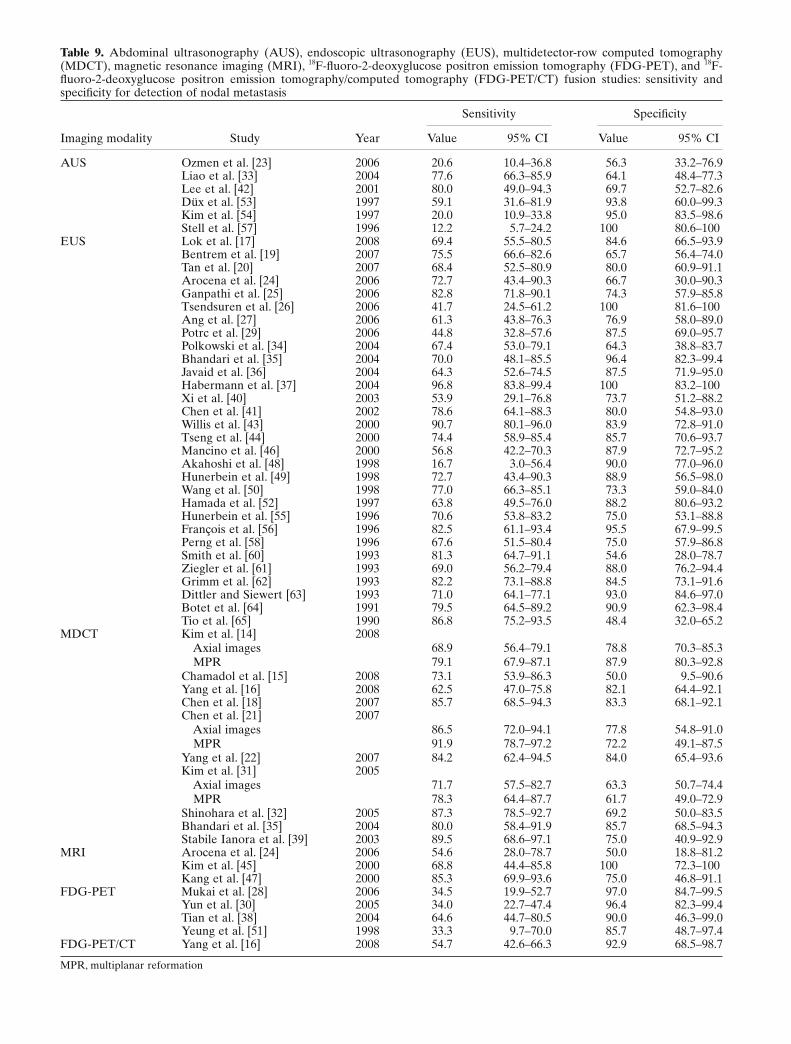
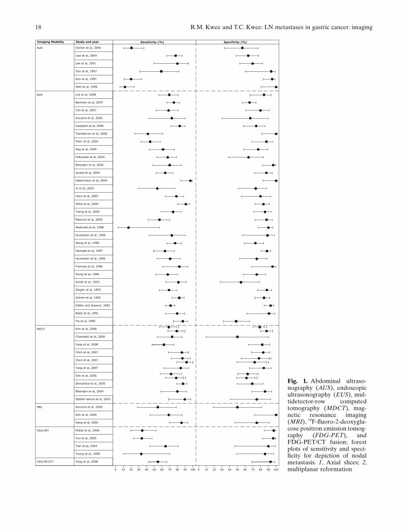
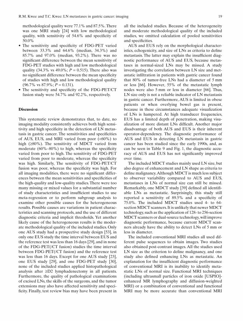
Table 9. Abdominal ultrasonography (AUS), endoscopic ultrasonography (EUS), multidetector-row computed tomography (MDCT), magnetic resonance imaging (MRI), 18F-fl uoro-2-deoxyglucose positron emission tomography (FDG-PET), and 18F-fl uoro-2-deoxyglucose positron emission tomography/computed tomography (FDG-PET/CT) fusion studies: sensitivity and specifi city for detection of nodal metastasis
Imaging modality Study Year
Sensitivity Specifi city
Value 95% CI Value 95% CI
AUS Ozmen et al. [23] 2006 20.6 10.4–36.8 56.3 33.2–76.9Liao et al. [33] 2004 77.6 66.3–85.9 64.1 48.4–77.3Lee et al. [42] 2001 80.0 49.0–94.3 69.7 52.7–82.6Düx et al. [53] 1997 59.1 31.6–81.9 93.8 60.0–99.3Kim et al. [54] 1997 20.0 10.9–33.8 95.0 83.5–98.6Stell et al. [57] 1996 12.2 5.7–24.2 100 80.6–100
EUS Lok et al. [17] 2008 69.4 55.5–80.5 84.6 66.5–93.9Bentrem et al. [19] 2007 75.5 66.6–82.6 65.7 56.4–74.0Tan et al. [20] 2007 68.4 52.5–80.9 80.0 60.9–91.1Arocena et al. [24] 2006 72.7 43.4–90.3 66.7 30.0–90.3Ganpathi et al. [25] 2006 82.8 71.8–90.1 74.3 57.9–85.8Tsendsuren et al. [26] 2006 41.7 24.5–61.2 100 81.6–100Ang et al. [27] 2006 61.3 43.8–76.3 76.9 58.0–89.0Potrc et al. [29] 2006 44.8 32.8–57.6 87.5 69.0–95.7Polkowski et al. [34] 2004 67.4 53.0–79.1 64.3 38.8–83.7Bhandari et al. [35] 2004 70.0 48.1–85.5 96.4 82.3–99.4Javaid et al. [36] 2004 64.3 52.6–74.5 87.5 71.9–95.0Habermann et al. [37] 2004 96.8 83.8–99.4 100 83.2–100Xi et al. [40] 2003 53.9 29.1–76.8 73.7 51.2–88.2Chen et al. [41] 2002 78.6 64.1–88.3 80.0 54.8–93.0Willis et al. [43] 2000 90.7 80.1–96.0 83.9 72.8–91.0Tseng et al. [44] 2000 74.4 58.9–85.4 85.7 70.6–93.7Mancino et al. [46] 2000 56.8 42.2–70.3 87.9 72.7–95.2Akahoshi et al. [48] 1998 16.7 3.0–56.4 90.0 77.0–96.0Hunerbein et al. [49] 1998 72.7 43.4–90.3 88.9 56.5–98.0Wang et al. [50] 1998 77.0 66.3–85.1 73.3 59.0–84.0Hamada et al. [52] 1997 63.8 49.5–76.0 88.2 80.6–93.2Hunerbein et al. [55] 1996 70.6 53.8–83.2 75.0 53.1–88.8François et al. [56] 1996 82.5 61.1–93.4 95.5 67.9–99.5Perng et al. [58] 1996 67.6 51.5–80.4 75.0 57.9–86.8Smith et al. [60] 1993 81.3 64.7–91.1 54.6 28.0–78.7Ziegler et al. [61] 1993 69.0 56.2–79.4 88.0 76.2–94.4Grimm et al. [62] 1993 82.2 73.1–88.8 84.5 73.1–91.6Dittler and Siewert [63] 1993 71.0 64.1–77.1 93.0 84.6–97.0Botet et al. [64] 1991 79.5 64.5–89.2 90.9 62.3–98.4Tio et al. [65] 1990 86.8 75.2–93.5 48.4 32.0–65.2
MDCT Kim et al. [14] 2008 Axial images 68.9 56.4–79.1 78.8 70.3–85.3 MPR 79.1 67.9–87.1 87.9 80.3–92.8Chamadol et al. [15] 2008 73.1 53.9–86.3 50.0 9.5–90.6Yang et al. [16] 2008 62.5 47.0–75.8 82.1 64.4–92.1Chen et al. [18] 2007 85.7 68.5–94.3 83.3 68.1–92.1Chen et al. [21] 2007 Axial images 86.5 72.0–94.1 77.8 54.8–91.0 MPR 91.9 78.7–97.2 72.2 49.1–87.5Yang et al. [22] 2007 84.2 62.4–94.5 84.0 65.4–93.6Kim et al. [31] 2005 Axial images 71.7 57.5–82.7 63.3 50.7–74.4 MPR 78.3 64.4–87.7 61.7 49.0–72.9Shinohara et al. [32] 2005 87.3 78.5–92.7 69.2 50.0–83.5Bhandari et al. [35] 2004 80.0 58.4–91.9 85.7 68.5–94.3Stabile Ianora et al. [39] 2003 89.5 68.6–97.1 75.0 40.9–92.9
MRI Arocena et al. [24] 2006 54.6 28.0–78.7 50.0 18.8–81.2Kim et al. [45] 2000 68.8 44.4–85.8 100 72.3–100Kang et al. [47] 2000 85.3 69.9–93.6 75.0 46.8–91.1
FDG-PET Mukai et al. [28] 2006 34.5 19.9–52.7 97.0 84.7–99.5Yun et al. [30] 2005 34.0 22.7–47.4 96.4 82.3–99.4Tian et al. [38] 2004 64.6 44.7–80.5 90.0 46.3–99.0Yeung et al. [51] 1998 33.3 9.7–70.0 85.7 48.7–97.4
FDG-PET/CT Yang et al. [16] 2008 54.7 42.6–66.3 92.9 68.5–98.7
MPR, multiplanar reformation
18 R.M. Kwee and T.C. Kwee: LN metastases in gastric cancer: imaging
Study and year
Ozmen et al, 2006
0 10 20 30 40
Sensitivity (%)
6050 70 80 10090 0 10 20 504030 60 70 10080 90
Specificity (%) Imaging Modality
AUS
Liao et al, 2004
Lee et al, 2001
Kim et al, 1997
Düx et al, 1997
Stell et al, 1996
EUS
Bentrem et al, 2007
Tan et al, 2007
Arocena et al, 2006
Ganpathi et al, 2006
Tsendsuren et al, 2006
Potrc et al, 2006
Ang et al, 2006
Xi et al, 2003
Habermann et al, 2004
Javaid et al, 2004
Bhandari et al, 2004
Polkowski et al, 2004
Chen et al, 2002
Willis et al, 2000
Tseng et al, 2000
Mancino et al, 2000
Akahoshi et al, 1998
Wang et al, 1998
Hamada et al, 1997
Hunerbein et al, 1996
Hunerbein et al, 1998
Francois et al, 1996
Perng et al, 1996
Smith et al, 1993
Ziegler et al, 1993
Grimm et al, 1993
Dittler and Siewert, 1993
Botet et al, 1991
Tio et al, 1990
MDCT
Chen et al, 2007
Yang et al, 2007
Kim et al, 2005
Bhandari et al, 2004
Stabile Ianora et al, 2003
MRI Arocena et al, 2006
Kim et al, 2000
Kang et al, 2000
FDG-PET Mukai et al, 2006
Yun et al, 2005
Tian et al, 2004
Yeung et al, 1998
11
22
1
22
1
Chen et al, 2007
Shinohara et al, 2005
Lok et al, 2008
Kim et al, 2008
Chamadol et al, 2008
Yang et al, 2008
1
2
1
2
FDG-PET/CT Yang et al, 2008
Fig. 1. Abdominal ultraso-nography (AUS), endoscopic ultrasonography (EUS), mul-tidetector-row computed tomography (MDCT), mag-netic resonance imaging (MRI), 18F-fl uoro-2-deoxyglu-cose positron emission tomog-raphy (FDG-PET), and FDG-PET/CT fusion; forest plots of sensitivity and speci-fi city for depiction of nodal metastasis. 1, Axial slices; 2, multiplanar reformation
R.M. Kwee and T.C. Kwee: LN metastases in gastric cancer: imaging 19
methodological quality were 77.1% and 87.5%. There was one MRI study [24] with low methodological quality, with sensitivity of 54.6% and specifi city of 50.0%
• The sensitivity and specifi city of FDG-PET varied between 33.3% and 64.6% (median, 34.3%) and 85.7% and 97.0% (median, 93.2%). There was no signifi cant difference between the mean sensitivity of FDG-PET studies with high and low methodological quality (34.3% vs 49.0%; P = 0.515). There also was no signifi cant difference between the mean specifi city of studies with high and low methodological quality (96.7% vs 87.9%; P = 0.131).
• The sensitivity and specifi city of the FDG-PET/CT fusion study were 54.7% and 92.2%, respectively.
Discussion
This systematic review demonstrates that, to date, no imaging modality consistently achieves both high sensi-tivity and high specifi city in the detection of LN metas-tasis in gastric cancer. The sensitivities and specifi cities of AUS, EUS, and MRI varied from poor (<60%) to high (≥80%). The sensitivity of MDCT varied from moderate (60%–80%) to high, whereas the specifi city varied from poor to high. The sensitivity of FDG-PET varied from poor to moderate, whereas the specifi city was high. Similarly, The sensitivity of FDG-PET/CT fusion was poor, whereas the specifi city was high. For all imaging modalities, there were no signifi cant differ-ences between the mean sensitivities and specifi cities of the high-quality and low-quality studies. There were too many missing or mixed values for a substantial number of study characteristics and insuffi cient studies to use meta-regression or to perform subgroup analysis to examine other possible causes for the heterogeneous results. Possible causes are variations in patient charac-teristics and scanning protocols, and the use of different diagnostic criteria and implicit thresholds. Yet another likely cause of the heterogeneous results is the moder-ate methodological quality of the included studies. Only one AUS study had a prospective study design [53], in only one EUS study the time interval between EUS and the reference test was less than 16 days [29], and in none of the FDG-PET(/CT fusion) studies the time interval between FDG-PET(/CT fusion) and the reference test was less than 16 days. Except for one AUS study [23], one EUS study [29], and one FDG-PET study [30], none of the included studies applied histopathological analysis after ≥D2 lymphadenectomy in all patients. Furthermore, the quality of pathological examinations of excised LNs, the skills of the surgeons, and the tumor extensions may also have affected sensitivity and speci-fi city. Finally, test review bias may have been present in
all the included studies. Because of the heterogeneity and moderate methodological quality of the included studies, we omitted calculation of pooled sensitivities and specifi cities.
AUS and EUS rely on the morphological character-istics, echogenicity, and size of LNs as criteria to defi ne metastasis. The latter may explain the insuffi cient diag-nostic performance of AUS and EUS, because metas-tases in normal-sized LNs may be missed. A study investigating the correlation between LN size and met-astatic infi ltration in patients with gastric cancer found that 80% of tumor-free LNs had a diameter of 5 mm or less [66]. However, 55% of the metastatic lymph nodes were also 5 mm or less in diameter [66]. Thus, LN size only is not a reliable indicator of LN metastasis in gastric cancer. Furthermore, AUS is limited in obese patients or when overlying bowel gas is present, because in these circumstances adequate visualization of LNs is hampered. At high transducer frequencies, EUS has a limited depth of penetration, making visu-alization of more distant LNs diffi cult. Another major disadvantage of both AUS and EUS is their inherent operator-dependency. The diagnostic performance of AUS and EUS in detecting LN metastasis in gastric cancer has been studied since the early 1990s, and, as can be seen in Table 9 and Fig. 1, the diagnostic accu-racy of AUS and EUS has not signifi cantly improved over time.
The included MDCT studies mainly used LN size, but also degree of enhancement and LN shape as criteria to defi ne malignancy. Although MDCT is much less subject to observer variability compared to AUS and EUS, metastases in LNs of normal size can still be missed. Remarkably, one MDCT study [39] defi ned all identifi -able LNs as metastatic. Surprisingly, this study still reported a sensitivity of 89.5% and a specifi city of 75.0%. The included MDCT studies used 4- to 64-section MDCT scanners. It is unlikely that newer MDCT technology, such as the application of 128- to 256-section MDCT scanners or dual-source technology, will improve diagnostic performance, because current MDCT scan-ners already have the ability to detect LNs of 5 mm or less in diameter.
The included conventional MRI studies all used dif-ferent pulse sequences to obtain images. Two studies also obtained post-contrast images. All the studies used LN size as the criterion to defi ne malignancy, and one study also defi ned enhancing LNs as metastatic. An explanation for the insuffi cient diagnostic performance of conventional MRI is its inability to identify meta-static LNs of normal size. Functional MRI techniques (including ultrasmall particles of iron oxide [USPIO]-enhanced MR lymphography and diffusion-weighted MRI) or a combination of conventional and functional MRI may be more accurate than conventional MRI
20 R.M. Kwee and T.C. Kwee: LN metastases in gastric cancer: imaging
alone. USPIO-enhanced lymphography allows the iden-tifi cation of malignant nodal infi ltration independent of LN size. After intravenous administration, USPIOs are taken up by macrophages in the reticuloendothelial system, predominantly within the LNs. Normal homo-geneous uptake of USPIOs in nonmetastatic LNs short-ens the T2 and T2*, turning these nodes hypointense on T2- and T2*-weighted images, whereas malignant LNs lack uptake and remain hyperintense. USPIO-enhanced lymphography indeed has been shown to achieve higher diagnostic precision than does conventional, unen-hanced MRI for the detection of LN metastases of various tumors [67]. Although no USPIO-enhanced lymphography studies were identifi ed for inclusion in this systematic review, its usefulness in detecting meta-static LNs in gastric cancer has already been demon-strated by a recent pilot study in 17 patients [68]. MR lymphography using other contrast agents may also have high potential [69], but this remains to be investi-gated. Diffusion-weighted MRI is another functional imaging technique, based on water diffusivity. Cancer-ous lesions which have architectural malformations are highlighted by this technique, because they have a restricted diffusion [70]. However, no studies on diffusion-weighted MRI were identifi ed for inclusion in this systematic review.
A possible reason for the reported low to moderate sensitivity of FDG-PET is its limited resolution; current FDG-PET units have a 4- to 5-mm resolution [71], but it has been reported that 14.5% of metastatic LNs in gastric cancer have a largest diameter of less than 3 mm [66]. Consequently, these LN metastases can be missed by FDG-PET. Low FDG uptake of metastatic LNs may also explain the low sensitivity of FDG-PET; Stahl et al. [72] found that diffusely growing and mucus-containing gastric cancers may exhibit low FDG uptake. Another possible explanation for the low sensitivity of FDG-PET is the masking of perigastric LNs by FDG uptake of the adjacent primary tumor. On the other hand, FDG uptake of the primary tumor may mimic involvement of adjacent LNs, thereby decreasing speci-fi city. Similarly, physiological FDG uptake of the stomach [73] may also mask or mimic metastatic peri-gastric LNs. FDG-PET/CT fusion provides both ana-tomic and functional information, and allows more accurate localization of foci with increased FDG uptake than stand-alone PET; this may reduce the problems of missing metastatic LNs with low FDG uptake, physio-logical FDG uptake being misinterpreted as pathologi-cal, and false localization of disease [74]. Additional advantages of using a combined PET/CT scanner are decreased scanning time and improved quality of the FDG-PET images [74]. However, the results of the FDG-PET/CT fusion study [16] included in this system-atic review suggest that FDG-PET/CT fusion does not
improve sensitivity (or specifi city). Of note, however, the poor sensitivity may mainly be a result of the limited resolution of the PET/CT scanner used in that study [16], which is only 6.3 mm [75]. The performance of PET/CT scanners with a higher resolution still has to be determined, to our knowledge.
Laparoscopic sentinel node (SN) biopsy is another promising tool to more accurately determine nodal status in patients with gastric cancer. The SN concept is based on the premise that tumor cells will preferentially metastasize to the fi rst draining LN in the regional lym-phatics, the SN. After identifying the SN (by use of a radionucleotide tracer and/or dye), and laparoscopic biopsy, LN metastasis is confi rmed or ruled out by his-tological examination. A disadvantage of laparoscopic SN biopsy, however, is its invasiveness. Although studies on laparoscopic SN biopsy have shown its potential [76–79], various technical and material limitations still have to be overcome. Also, the reliability of laparo-scopic SN biopsy has yet to be determined by multi-center prospective clinical trials [80].
In conclusion, AUS, EUS, MDCT, conventional MRI, and FDG-PET do not achieve consistently high sensitivity and specifi city in detecting LN metastasis in patients with gastric cancer. The value of high-resolution PET/CT fusion and functional MRI tech-niques still has to be determined.
References
1. Parkin DM, Bray F, Ferlay J, Pisani P. Global cancer statistics, 2002. CA Cancer J Clin 2005;55:74–108.
2. Siewert JR, Böttcher K, Stein HJ, Roder JD. Relevant prognostic factors in gastric cancer: 10-year results of the German Gastric Cancer Study. Ann Surg 1998;228:449–61.
3. Zhang XF, Huang CM, Lu HS, Wu XY, Wang C, Guang GX, et al. Surgical treatment and prognosis of gastric cancer in 2613 patients. World J Gastroenterol 2004;10:3405–8.
4. Gotoda T, Yanagisawa A, Sasako M, Ono H, Nakanishi Y, Shimoda T, et al. Incidence of lymph node metastasis from early gastric cancer: estimation with a large number of cases at two large centers. Gastric Cancer 2000;3:219–25.
5. Hohenberger P, Gretschel S. Gastric cancer. Lancet 2003;362:305–15.
6. Swan R, Miner TJ. Current role of surgical therapy in gastric cancer. World J Gastroenterol 2006;12:372–9.
7. Brennan MF. Current status of surgery for gastric cancer: a review. Gastric Cancer 2005;8:64–70.
8. De Vita F, Giuliani F, Galizia G, Belli C, Aurilio G, Santabarbara G, et al. Neo-adjuvant and adjuvant chemotherapy of gastric cancer. Ann Oncol 2007;18(Suppl 6):20–3.
9. Barentsz J, Takahashi S, Oyen W, Mus R, De Mulder P, Reznek R, et al. Commonly used imaging techniques for diagnosis and staging. J Clin Oncol 2006;24:3234–44.
10. Rubin E, Palazzo JP. The gastrointestinal tract. In: Rubin E, Gorstein F, Rubin F, Schwarting R, Strayer D, editors. Rubin’s pathology. Clinicopathologic foundations of medicine. 4th ed. Philadelphia: Lippincott Williams & Wilkins; 2005. p. 660–739.
11. Kelly S, Berry E, Roderick P, Harris KM, Cullingworth J, Gathercole L, et al. The identifi cation of bias in studies of the
R.M. Kwee and T.C. Kwee: LN metastases in gastric cancer: imaging 21
diagnostic performance of imaging modalities. Br J Radiol 1997;70:1028–35.
12. Whiting P, Rutjes AW, Reitsma JB, Bossuyt PM, Kleijnen J. The development of QUADAS: a tool for the quality assessment of studies of diagnostic accuracy included in systematic reviews. BMC Med Res Methodol 2003;3:25.
13. Whiting PF, Weswood ME, Rutjes AW, Reitsma JB, Bossuyt PN, Kleijnen J. Evaluation of QUADAS, a tool for the quality assess-ment of diagnostic accuracy studies. BMC Med Res Methodol 2006;6:9.
14. Kim YN, Choi D, Kim SH, Kim MJ, Lee SJ, Lee WJ, et al. Gastric cancer staging at isotropic MDCT including coronal and sagittal MPR images: endoscopically diagnosed early vs advanced gastric cancer. Abdom Imaging 2009;34:26–34.
15. Chamadol N, Wongwiwatchai J, Bhudhisawasd V, Pairojkul C. Accuracy of spiral CT in preoperative staging of gastric carci-noma: correlation with surgical and pathological fi ndings. J Med Assoc Thai 2008;91:356–63.
16. Yang Q-M, Kawamura T, Itoh H, Bando E, Nemoto M, Akamoto S, et al. Is PET-CT suitable for predicting lymph node status for gastric cancer? Hepatogastroenterology 2008;55:782–5.
17. Lok K-H, Lee C-K, Yiu H-L, Lai L, Szeto M-L, Leung S-K. Current utilization and performance status of endoscopic ultra-sound in a community hospital. J Dig Dis 2008;9:41–7.
18. Chen BB, Liang PC, Liu KL, Hsiao JK, Huang JC, Wong JM, et al. Preoperative diagnosis of gastric tumors by three-dimensional multidetector row CT and double contrast barium meal study: correlation with surgical and histologic results. J Formos Med Assoc 2007;106:943–52.
19. Bentrem D, Gerdes H, Tang L, Brennan M, Coit D. Clinical cor-relation of endoscopic ultrasonography with pathologic stage and outcome in patients undergoing curative resection for gastric cancer. Ann Surg Oncol 2007;14:1853–9.
20. Tan SY, Wang JY, Shen L, Luo HS, Shen ZX. Relationship between preoperative staging by endoscopic ultrasonography and MMP-9 expression in gastric carcinoma. World J Gastroenterol 2007;13:2108–12.
21. Chen CY, Hsu JS, Wu DC, Kang WY, Hsieh JS, Jaw TS, et al. Gastric cancer: preoperative local staging with 3D multi-detector row CT–correlation with surgical and histopathologic results. Radiology 2007;242:472–82.
22. Yang DM, Kim HC, Jin W, Ryu CW, Kang JH, Park CH, et al. 64 Multidetector-row computed tomography for preoperative evaluation of gastric cancer: histological correlation. J Comput Assist Tomogr 2007;31:98–103.
23. Ozmen MM, Zulfi karoglu B, Kucuk NO, Ozalp N, Aras G, Koseoglu T, et al. Lymphoscintigraphy in detection of the regional lymph node involvement in gastric cancer. Ann R Coll Surg Engl 2006;88:632–8.
24. Arocena MG, Barturen A, Bujanda L, Casado O, Ramírez MM, Oleagoitia JM, et al. MRI and endoscopic ultrasonography in the staging of gastric cancer. Rev Esp Enferm Dig 2006;98:582–90.
25. Ganpathi IS, So JB, Ho KY. Endoscopic ultrasonography for gastric cancer: does it infl uence treatment? Surg Endosc 2006;20:559–62.
26. Tsendsuren T, Jun SM, Mian XH. Usefulness of endoscopic ultra-sonography in preoperative TNM staging of gastric cancer. World J Gastroenterol 2006;12:43–7.
27. Ang TL, Ng TM, Fock KM, Teo EK. Accuracy of endoscopic ultrasound staging of gastric cancer in routine clinical practice in Singapore. Chin J Dig Dis 2006;7:191–6.
28. Mukai K, Ishida Y, Okajima K, Isozaki H, Morimoto T, Nishiyama S. Usefulness of preoperative FDG-PET for detection of gastric cancer. Gastric Cancer 2006;9:192–6.
29. Potrc S, Skalicky M, Ivanecz A. Does endoscopic ultrasound staging already allow individual treatment regimens in gastric cancer? Wien Klin Wochenschr 2006;118 (Suppl 2):48–51.
30. Yun M, Lim JS, Noh SH, Hyung WJ, Cheong JH, Bong JK, et al. Lymph node staging of gastric cancer using (18)F-FDG PET: a comparison study with CT. J Nucl Med 2005;46:1582–8.
31. Kim HJ, Kim AY, Oh ST, Kim JS, Kim KW, Kim PN, et al. Gastric cancer staging at multi-detector row CT gastrography: comparison of transverse and volumetric CT scanning. Radiology 2005;236:879–85.
32. Shinohara T, Ohyama S, Yamaguchi T, Muto T, Kohno A, Ogura T, et al. Preoperative TNM staging of advanced gastric cancer with multi-detector row computed tomography. Japan Med Assoc J 2005;48:175–82.
33. Liao SR, Dai Y, Huo L, Yan K, Zhang L, Zhang H, et al. Trans-abdominal ultrasonography in preoperative staging of gastric cancer. World J Gastroenterol 2004;10:3399–404.
34. Polkowski M, Palucki J, Wronska E, Szawlowski A, Nasierowska-Guttmejer A, Butruk E. Endosonography versus helical computed tomography for locoregional staging of gastric cancer. Endoscopy 2004;36:617–23.
35. Bhandari S, Shim CS, Kim JH, Jung IS, Cho JY, Lee JS, et al. Usefulness of three-dimensional, multidetector row CT (virtual gastroscopy and multiplanar reconstruction) in the eva-luation of gastric cancer: a comparison with conventional endos-copy, EUS, and histopathology. Gastrointest Endosc 2004;59:619–26.
36. Javaid G, Shah OJ, Dar MA, Shah P, Wani NA, Zargar SA. Role of endoscopic ultrasonography in preoperative staging of gastric carcinoma. ANZ J Surg 2004;74:108–11.
37. Habermann CR, Weiss F, Riecken R, Honarpisheh H, Bohnacker S, Staedtler C, et al. Preoperative staging of gastric adenocarci-noma: comparison of helical CT and endoscopic US. Radiology 2004;230:465–71.
38. Tian J, Chen L, Wei B, Shao M, Ding Y, Yin D, et al. The value of vesicant 18F-fl uorodeoxyglucose positron emission tomogra-phy (18F-FDG PET) in gastric malignancies. Nucl Med Commun 2004;25:825–31.
39. Stabile Ianora AA, Pedote P, Scardapane A, Memeo M, Rotondo A, Angelelli G. Preoperative staging of gastric carcinoma with multidetector spiral CT. Radiol Med (Torino) 2003;106:467–80.
40. Xi WD, Zhao C, Ren GS. Endoscopic ultrasonography in preop-erative staging of gastric cancer: determination of tumor invasion depth, nodal involvement and surgical resectability. World J Gas-troenterol 2003;9:254–7.
41. Chen CH, Yang CC, Yeh YH. Preoperative staging of gastric cancer by endoscopic ultrasound: the prognostic usefulness of ascites detected by endoscopic ultrasound. J Clin Gastroenterol 2002;35:321–7.
42. Lee DH, Ko YT, Park SJ, Lim JW. Comparison of hydro-US and spiral CT in the staging of gastric cancer. Clin Imaging 2001;25:181–6.
43. Willis S, Truong S, Gribnitz S, Fass J, Schumpelick V. Endoscopic ultrasonography in the preoperative staging of gastric cancer: accuracy and impact on surgical therapy. Surg Endosc 2000;14:951–4.
44. Tseng LJ, Mo LR, Tio TL, Fresner YT, Jao N, Lin RC, et al. Video-endoscopic ultrasonography in staging gastric carcinoma. Hepatogastroenterology 2000;47:897–900.
45. Kim AY, Han JK, Seong CK, Kim TK, Choi BI. MRI in staging advanced gastric cancer: is it useful compared with spiral CT? J Comput Assist Tomogr 2000;24:389–94.
46. Mancino G, Bozzetti F, Schicchi A, Schiavo M, Spinelli P, Andre-ola S. Preoperative endoscopic ultrasonography in patients with gastric cancer. Tumori 2000;86:139–41.
47. Kang BC, Kim JH, Kim KW, Lee DY, Baek SY, Lee SW, et al. Value of the dynamic and delayed MR sequence with Gd-DTPA in the T-staging of stomach cancer: correlation with the histopa-thology. Abdom Imaging 2000;25:14–24.
48. Akahoshi K, Chijiwa Y, Hamada S, Sasaki I, Nawata H, Kabe-mura T, et al. Pretreatment staging of endoscopically early gastric
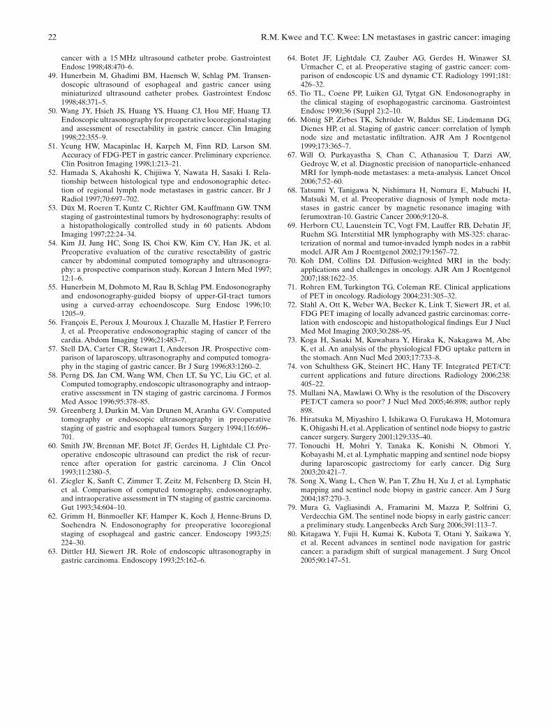
22 R.M. Kwee and T.C. Kwee: LN metastases in gastric cancer: imaging
cancer with a 15 MHz ultrasound catheter probe. Gastrointest Endosc 1998;48:470–6.
49. Hunerbein M, Ghadimi BM, Haensch W, Schlag PM. Transen-doscopic ultrasound of esophageal and gastric cancer using miniaturized ultrasound catheter probes. Gastrointest Endosc 1998;48:371–5.
50. Wang JY, Hsieh JS, Huang YS, Huang CJ, Hou MF, Huang TJ. Endoscopic ultrasonography for preoperative locoregional staging and assessment of resectability in gastric cancer. Clin Imaging 1998;22:355–9.
51. Yeung HW, Macapinlac H, Karpeh M, Finn RD, Larson SM. Accuracy of FDG-PET in gastric cancer. Preliminary experience. Clin Positron Imaging 1998;1:213–21.
52. Hamada S, Akahoshi K, Chijiiwa Y, Nawata H, Sasaki I. Rela-tionship between histological type and endosonographic detec-tion of regional lymph node metastases in gastric cancer. Br J Radiol 1997;70:697–702.
53. Düx M, Roeren T, Kuntz C, Richter GM, Kauffmann GW. TNM staging of gastrointestinal tumors by hydrosonography: results of a histopathologically controlled study in 60 patients. Abdom Imaging 1997;22:24–34.
54. Kim JJ, Jung HC, Song IS, Choi KW, Kim CY, Han JK, et al. Preoperative evaluation of the curative resectability of gastric cancer by abdominal computed tomography and ultrasonogra-phy: a prospective comparison study. Korean J Intern Med 1997;12:1–6.
55. Hunerbein M, Dohmoto M, Rau B, Schlag PM. Endosonography and endosonography-guided biopsy of upper-GI-tract tumors using a curved-array echoendoscope. Surg Endosc 1996;10:1205–9.
56. François E, Peroux J, Mouroux J, Chazalle M, Hastier P, Ferrero J, et al. Preoperative endosonographic staging of cancer of the cardia. Abdom Imaging 1996;21:483–7.
57. Stell DA, Carter CR, Stewart I, Anderson JR. Prospective com-parison of laparoscopy, ultrasonography and computed tomogra-phy in the staging of gastric cancer. Br J Surg 1996;83:1260–2.
58. Perng DS, Jan CM, Wang WM, Chen LT, Su YC, Liu GC, et al. Computed tomography, endoscopic ultrasonography and intraop-erative assessment in TN staging of gastric carcinoma. J Formos Med Assoc 1996;95:378–85.
59. Greenberg J, Durkin M, Van Drunen M, Aranha GV. Computed tomography or endoscopic ultrasonography in preoperative staging of gastric and esophageal tumors. Surgery 1994;116:696–701.
60. Smith JW, Brennan MF, Botet JF, Gerdes H, Lightdale CJ. Pre-operative endoscopic ultrasound can predict the risk of recur-rence after operation for gastric carcinoma. J Clin Oncol 1993;11:2380–5.
61. Ziegler K, Sanft C, Zimmer T, Zeitz M, Felsenberg D, Stein H, et al. Comparison of computed tomography, endosonography, and intraoperative assessment in TN staging of gastric carcinoma. Gut 1993;34:604–10.
62. Grimm H, Binmoeller KF, Hamper K, Koch J, Henne-Bruns D, Soehendra N. Endosonography for preoperative locoregional staging of esophageal and gastric cancer. Endoscopy 1993;25:224–30.
63. Dittler HJ, Siewert JR. Role of endoscopic ultrasonography in gastric carcinoma. Endoscopy 1993;25:162–6.
64. Botet JF, Lightdale CJ, Zauber AG, Gerdes H, Winawer SJ, Urmacher C, et al. Preoperative staging of gastric cancer: com-parison of endoscopic US and dynamic CT. Radiology 1991;181:426–32.
65. Tio TL, Coene PP, Luiken GJ, Tytgat GN. Endosonography in the clinical staging of esophagogastric carcinoma. Gastrointest Endosc 1990;36 (Suppl 2):2–10.
66. Mönig SP, Zirbes TK, Schröder W, Baldus SE, Lindemann DG, Dienes HP, et al. Staging of gastric cancer: correlation of lymph node size and metastatic infi ltration. AJR Am J Roentgenol 1999;173:365–7.
67. Will O, Purkayastha S, Chan C, Athanasiou T, Darzi AW, Gedroyc W, et al. Diagnostic precision of nanoparticle-enhanced MRI for lymph-node metastases: a meta-analysis. Lancet Oncol 2006;7:52–60.
68. Tatsumi Y, Tanigawa N, Nishimura H, Nomura E, Mabuchi H, Matsuki M, et al. Preoperative diagnosis of lymph node meta-stases in gastric cancer by magnetic resonance imaging with ferumoxtran-10. Gastric Cancer 2006;9:120–8.
69. Herborn CU, Lauenstein TC, Vogt FM, Lauffer RB, Debatin JF, Ruehm SG. Interstitial MR lymphography with MS-325: charac-terization of normal and tumor-invaded lymph nodes in a rabbit model. AJR Am J Roentgenol 2002;179:1567–72.
70. Koh DM, Collins DJ. Diffusion-weighted MRI in the body: applications and challenges in oncology. AJR Am J Roentgenol 2007;188:1622–35.
71. Rohren EM, Turkington TG, Coleman RE. Clinical applications of PET in oncology. Radiology 2004;231:305–32.
72. Stahl A, Ott K, Weber WA, Becker K, Link T, Siewert JR, et al. FDG PET imaging of locally advanced gastric carcinomas: corre-lation with endoscopic and histopathological fi ndings. Eur J Nucl Med Mol Imaging 2003;30:288–95.
73. Koga H, Sasaki M, Kuwabara Y, Hiraka K, Nakagawa M, Abe K, et al. An analysis of the physiological FDG uptake pattern in the stomach. Ann Nucl Med 2003;17:733–8.
74. von Schulthess GK, Steinert HC, Hany TF. Integrated PET/CT: current applications and future directions. Radiology 2006;238:405–22.
75. Mullani NA, Mawlawi O. Why is the resolution of the Discovery PET/CT camera so poor? J Nucl Med 2005;46:898; author reply 898.
76. Hiratsuka M, Miyashiro I, Ishikawa O, Furukawa H, Motomura K, Ohigashi H, et al. Application of sentinel node biopsy to gastric cancer surgery. Surgery 2001;129:335–40.
77. Tonouchi H, Mohri Y, Tanaka K, Konishi N, Ohmori Y, Kobayashi M, et al. Lymphatic mapping and sentinel node biopsy during laparoscopic gastrectomy for early cancer. Dig Surg 2003;20:421–7.
78. Song X, Wang L, Chen W, Pan T, Zhu H, Xu J, et al. Lymphatic mapping and sentinel node biopsy in gastric cancer. Am J Surg 2004;187:270–3.
79. Mura G, Vagliasindi A, Framarini M, Mazza P, Solfrini G, Verdecchia GM. The sentinel node biopsy in early gastric cancer: a preliminary study. Langenbecks Arch Surg 2006;391:113–7.
80. Kitagawa Y, Fujii H, Kumai K, Kubota T, Otani Y, Saikawa Y, et al. Recent advances in sentinel node navigation for gastric cancer: a paradigm shift of surgical management. J Surg Oncol 2005;90:147–51.
Copyright © 2022 FDOKUMEN