thee hi)tological lesion in lymph nodes in infectious ... - NCBI
Upload
independentCategory
view
3download
0
High Expression of Claudin-1 Protein in Papillary ThyroidTumor and its Regional Lymph Node Metastasis
Júlia Németh & Zsuzsanna Németh & Péter Tátrai &Ilona Péter & Áron Somorácz & Attila Marcell Szász &
András Kiss & Zsuzsa Schaff
Received: 15 April 2009 /Accepted: 25 June 2009 /Published online: 5 July 2009# Arányi Lajos Foundation 2009
Abstract Claudins, known as major contributors in theformation of the tight junction, are differentially expressedin malignant tumors as compared to the correspondinghealthy tissues. Therefore, they are thought to play a role incarcinogenesis and tumor progression. Altered expressionof claudin-1 has been reported in several tumor typesincluding endometrial, papillary renal cell and coloniccarcinoma, and increased claudin-1 mRNA levels havebeen observed in papillary thyroid carcinoma (PTC). In thisstudy, we aimed at determining the pattern of claudin-1expression in various types of thyroid lesions at the proteinlevel and investigating the immunolocalization of β-cateninreported to regulate claudin-1 expression. Samples included19 PTCs, ten cases of corresponding regional lymph nodemetastasis, eight papillary microcarcinomas (PMC), 17follicular thyroid carcinomas (FTC) and 19 follicularadenomas (FA). All cases were evaluated by quantitativeimmunohistochemistry. Conspicuous claudin-1 immuno-staining was detected in the majority of PTC/PMC primarytumors and lymph node metastases (19/27 and 9/10,respectively). On the other hand, we found weak or noclaudin-1 expression in any of the FA and FTC cases orperitumoral non-malignant thyroid tissues. Our data provethat high claudin-1 protein expression is specific for PTC
and its regional lymph node metastases, while we failed toverify that claudin-1 is regulated by β-catenin in thyroidtumors. Based on these results, claudin-1 may be a usefultumor marker for PTC.
Keywords Claudin-1 . Lymph node metastasis . Papillarythyroid carcinoma . Tumor marker
AbbreviationsPTC Papillary thyroid carcinomaPMC Papillary microcarcinomaFTC Follicular thyroid carcinomaFA Follicular adenomaHT Hashimoto’s thyroiditisTJ Tight junction
Introduction
Thyroid cancers, although they represent only 1% of allmalignant diseases, are among the most common endocrinemalignancies [1–3]. Most thyroid tumors derived from thefollicular epithelium [2, 3]. Follicular cell-derived carcino-mas are divided into well-differentiated, poorly differenti-ated, and undifferentiated types on the basis of histologicaland clinical features. Well-differentiated thyroid cancersinclude the papillary thyroid carcinomas and follicularthyroid carcinoma [3].
Papillary carcinoma is the most common type of thyroidcancer, comprising approximately 80% of thyroid epithelialmalignancies [1–4]. The more aggressive follicular carci-noma is far less prevalent (10–15% of thyroid malignan-cies) [2, 4, 5]. Neoplastic transformation is a multistep
J. Németh : Z. Németh : P. Tátrai :Á. Somorácz :A. M. Szász :A. Kiss : Z. Schaff (*)2nd Department of Pathology, Semmelweis University,Üllői út 93,1091 Budapest, Hungarye-mail: [email protected]
I. PéterNational Institute of Oncology,Ráth György u. 7-9,1122 Budapest, Hungary
Pathol. Oncol. Res. (2010) 16:19–27DOI 10.1007/s12253-009-9182-9
process that encompasses a broad spectrum of molecularand morphological changes, which transform the normalstate into a fully established neoplasm [3, 5]. Severalstudies have reported some connection between Hashimo-to’s thyroiditis (HT) and thyroid carcinoma, but their co-incidence has not exactly been determined (varies between0% to 30%) [6–8]. However, the increasing incidence ofHT associated carcinomas suggest that HT is likely to be aprecursor of thyroid carcinoma [8]. The majority of thesetumors are papillary carcinomas with a propensity tometastasize to the regional lymph nodes [2, 7]. Papillarymicrocarcinoma, measuring 10 mm or less in maximumdiameter, is found incidentally [2, 4]. Benign neoplasms inthe thyroid are adenomas, with the most common typebeing follicular adenoma [2, 4].
Claudins, a family of transmembrane proteins, are majorcomponents of the tight junction (TJ) [9, 10]. Claudin-1was the first member of the claudin family to be identifiedas a tight junction component [11]. So far 24 members ofthe claudin family have been described, and their role incarcinogenesis and cancer progression has been proposedrepeatedly [10, 12–14]. Altered expression of claudins wasfound in a wide variety of human malignancies includingendometrial [15], papillary renal cell [16], colon [17],pancreatic [18], breast [19] and cervical cancers [20].Claudins as membrane proteins showing differential ex-pression in the normal versus neoplastic tissues [12, 14, 21]may provide new opportunities for targeted cancer therapy[22, 23].
The expression of claudin-1,-4, and -7 protein inthyroid neoplastic samples has already been investigated[24], and previous studies have reported the increasedgene expression of claudin-1 in PTC [25, 26]. However,the expression of claudin-1 in different PTC samples(including PMC and HT) and in corresponding regionallymph node metastases has not yet been analyzed at theprotein level. It has been published recently that claudin- 1is expressed in the majority of papillary renal cellcarcinomas [16]. Previously, the high expression ofclaudin-1 protein was reported by our group in serouspapillary endometrial carcinoma [15]. Based on the ideathat high claudin-1 expression might be connected withpapillary morphology, we set out to evaluate the expres-sion of claudin-1 protein in PTC as well as non-papillarythyroid tumors. Furthermore, since the role of Wnt/ β-
catenin pathway in regulation of claudin-1 expression wasdemonstrated in colorectal tumorigenesis [27], we alsoaimed at investigating whether claudin-1 protein expres-sion correlates with β-catenin protein expression invarious types of thyroid lesions.
Materials and Methods
Tissue Samples
Between 1992 and 2007, 63 specimens of surgicallyremoved, formalin-fixed and paraffin-embedded thyroidlesions were collected from the archives of the 2ndDepartment of Pathology, Semmelweis University, Buda-pest, and the National Institute of Oncology, Budapest.Each case was classified according to the WHO histologicalclassification of thyroid tumors [4]. All available slideswere reviewed and the most representative blocks fromeach case were selected. The selected samples included 11males and 52 females ranging from 11 to 87 years old
Location Left lobe Right lobe Both lobes Isthmus Unknown
PMC (N=8) 3 5 0 0 0
PTC (N=19) 6 3 9 0 1
FTC ( N=17) 7 4 4 0 2
FA (N=19) 4 12 0 2 1
Table 1 Localization of thyroidlesions
Fig. 1 Digital morphometry analysis of claudin-1 immunoreactions.Abbreviations: FA, follicular adenoma; FTC, follicular thyroidcarcinoma; all PTC: all papillary thyroid carcinomas; PTC-LN: lymphnode metastases of PTCs; PMC, papillary microcarcinoma (asubgroup of PTC); PTC-HT: Hashimoto’s thyroiditis-associatedPTC; -N: non-malignant peritumoral thyroid tissue. *, 0.05 > p>0.01; ***, p<0.001, by Mann–Whitney U test. For the sake of clarity,significant differences relative to ‘all PTC’ are shown only; eachpairwise difference between both PTC subgroups (PTC-M and PTC-HT) and both follicular tumors (FA and FTC) were significant at p<0.001 level
20 J. Németh et al.
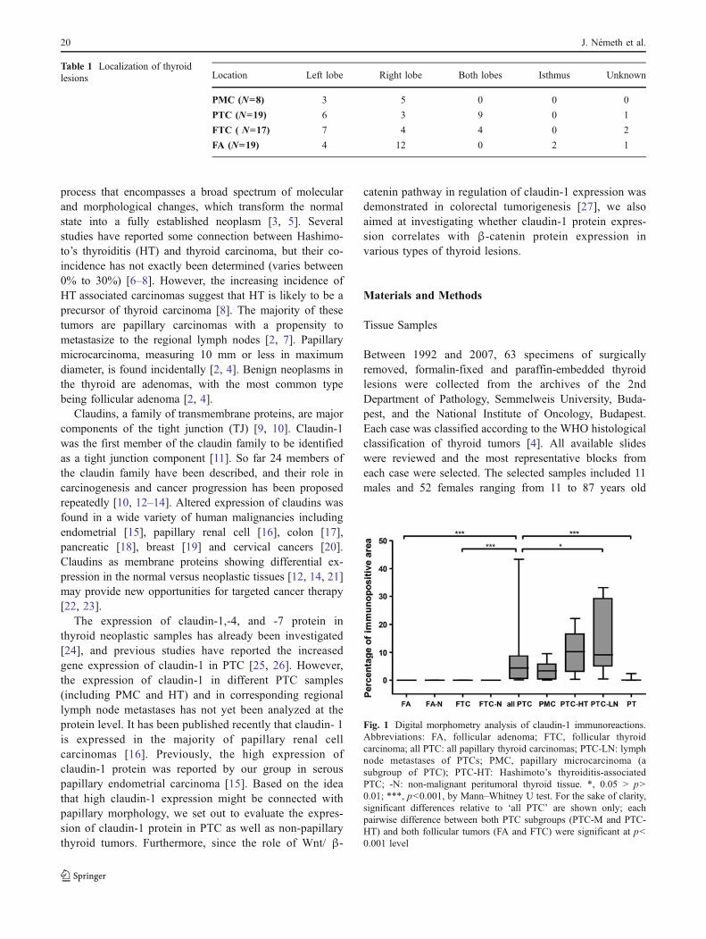
Fig. 2 Claudin-1 immunoreaction in papillary thyroid carcinoma andits regional lymph node metastases. Strong cell membrane claudin-1immunostaining in papillary thyroid carcinoma (a, b) and its regional
lymph node metastasis (c, d). Hashimoto’s thyroiditis-associatedpapillary thyroid carcinoma is shown in (e, f)
Claudin-1 in Thyroid Tumors 21

(mean: 55.3±8.6 years). Nineteen cases of papillarycarcinomas, ten corresponding regional (cervical) lymphnode metastases, eight papillary microcarcinomas, 17follicular carcinomas, and 19 follicular adenomas wereexamined. Of the 19 PTC cases, 5 were associated withHashimoto’s thyroiditis. Regarding histological subtype, 5PTCs were follicular variants (out of which 1 exhibitedoncocytic morphology); the rest comprised of conventionalPTC and a single tall cell variant. In the PTC samples thetumor localized to both lobes in the majority of cases. InPMC cases the localization of the tumor was dominantly inthe right lobe, while follicular carcinomas were mainlyfound in the left lobe. The majority of the follicularadenomas were localized to the right lobe (Table 1). Noneof the patients received chemotherapy or radiotherapy priorto surgery. The study was approved by the Regional Ethical
Committee of the Semmelweis Medical University (issuedunder #172/2003).
Immunohistochemistry
The immunohistochemical reactions were performed on 3–4 µm thick formalin-fixed paraffin-embedded sections.After deparaffination steps, slides were washed in PBS(pH 7.4), then were treated in Target Retrieval Solution(cat# S1699 from DAKO, Glostrup, Denmark) in amicrowave oven for 30 min. Immunoreactions forclaudin-1 were carried out in a Ventana ES automatedimmunostainer (Ventana Medical Systems Inc., Tucson,AZ, USA). Rabbit polyclonal claudin-1 antibody wasapplied in a dilution of 1:100 (cat# 18-7362 from ZymedInc., San Francisco, CA, USA). Reagents and the secondary
Fig. 3 Claudin-1 immunoreaction in follicular carcinoma and follicular adenoma of the thyroid. Weak or no claudin-1 expression was detected infollicular carcinoma (a, b) and in follicular adenoma (c, d)
22 J. Németh et al.

antibody from the iView DAB Detection Kit (cat# 760-091from Ventana Medical Systems Inc.) were used as providedby the manufacturer. Positive control recommended bymanufacturer (normal skin) was used to confirm correctimmunohistochemical staining for claudin-1.
In the case of β-catenin, after antigen retrieval stepstissues were blocked for endogenous peroxidase activitywith 3% H2O2. Mouse monoclonal antibody against β-catenin (cat# 610154, BD Transduction, San Diego, CA,USA) was applied in a dilution of 1:200. Negative controlsfor nonspecific binding, incubated with secondary anti-bodies only, were processed and revealed no signals. Forpositive control of β-catenin immunoreaction we usedhuman hepatoblastoma tissue previously reported to shownuclear positivity and analyzed for β-catenin mutation [28].
Evaluation of Immunoreactions
The results of claudin-1 immunohistochemical reactionswere photodocumented using Mirax MIDI Scanner (3DHis-tech Ltd., Budapest, Hungary). Ten non-overlapping repre-sentative fields were assessed. Digital images werequantified using Leica QWin V3.0 software (Leica Micro-systems Imaging Solutions Ltd., Cambridge, UK). Beforeperforming the measurements, a threshold level of colors tobe considered as positive was defined by selecting thestained areas on the digitized positive control tissues.Positive area was defined as percentage of pixels abovethe threshold within a defined area of interest. Statisticalanalysis for the comparison of immunopositive areas in thedifferent sample groups was performed using non-parametric Mann–Whitney test (SPSS 15.0, SPSS Inc.,Chicago, Ill, USA).
The intensity of β-catenin immunoreaction in the centerof lesions was graded semi-quantitatively as 0-absentstaining, 1-weakly positive, 2-moderately positive and 3-strongly positive.
Results
Immunohistochemical Analysis of Claudin-1 Expression
By digital morphometry, claudin-1 immunostaining wasobvious (immunopositive area was greater than 1%) in themajority (19/27) of PTCs, and it was strong (area% > 5) in12/27 cases. PTC tumor cells strongly expressed claudin-1along the cell membranes forming a honeycomb-likepattern. On the other hand, claudin-1 immunoreaction wasvirtually absent from FTC, FA, and the peritumoral non-malignant thyroid tissue (median immunopositive areas:4.4% for PTC, in contrast with 0.0% for FTC, FA, andperitumoral tissues) (Figs. 1, 2, 3). With other words, by
defining 1% immunostained area as a cut-off value forpositive claudin-1 immunoreaction, 70.3% of PTCs werepositive, while all FTCs, FAs, and non-tumorous thyroidtissues were negative. Thus, claudin-1 immunostainingalone differentiated PTC from both FTC and FA with aspecificity of 100% and a sensitivity of 70.3% (Pearson chi-square, P<0.001 for both). Claudin-1 expression waspreserved in the lymph node metastases of PTC as well(median immunopositive area: 9.1%). No significant differ-ences within the PTC group were found according tohistological subtype (follicular variant vs. conventional andtall/columnar cell variants), etiology (Hashimoto vs. non-Hashimoto), the presence of lymph node metastasis(Fig. 4), and the size of lesion (microcarcinoma vs.carcinoma > 1 cm in diameter).
Immunohistochemical Analysis of β-catenin Expression
Cell membrane and cytoplasmic β-catenin immunoreac-tions (Fig. 5) were evaluated semi-quantitatively (Fig. 6). Inperitumoral thyroid tissues of all tumor types, β-cateninwas seen primarily on the cell membranes of immaturefollicles. Such immature follicles were abundantly seen inthyroid tissues with increased regenerative activity, mainlyassociated with HT in the peritumoral areas of PTC. In thecarcinomas, cytoplasmic β-catenin immunostaining wasalso observed. PTCs and their lymph node metastasesshowed a weakly significant tendency of decreased (0−1+)membrane staining when compared with FTC (Pearson chi-square, P=0.045) or with all follicular tumors (FTC + FA,P=0.033). Cytoplasmic β-catenin staining, on the other
Fig. 4 Comparison of claudin-1 immunopositive area% between thelymph node metastases of papillary thyroid carcinomas and primarytumors with and without metastasis. Mann–Whitney U test failed todetect significant differences between either the metastatic (PTC-M)and non-metastatic (PTC-NM) subgroups of papillary thyroid carci-noma (PTC), or the lymph node metastases (PTC-LN) and theircorresponding primary cancers
Claudin-1 in Thyroid Tumors 23

Fig. 5 Immunoreaction of β-catenin in various thyroid samples. Inperitumoral thyroid tissues, β-catenin was primarily seen on the cellmembranes in immature follicles (a). Such immature follicles wereabundant in Hashimoto’s thyroiditis (b). Strong cell membrane β-catenin immunostaining was observed in follicular adenoma (c). InFTC, cytoplasmic β-catenin reaction appeared in addition to cell
surface immunostaining (d). In PTCs, a tendency of decreasedmembrane staining as compared with FTC and follicular adenomaswas often accompanied by marked cytoplasmic reaction (e). Intra-nuclear inclusions positive for β-catenin were predominantly seen inPTCs (f). PTC, papillary thyroid carcinoma; FTC, follicular thyroidcarcinoma
24 J. Németh et al.

hand, was significantly increased in the malignant tumors(PTC + FTC) when compared with adenomas (P=0.021).Comparisons within the PTC group according to histolog-ical subtype were not feasible due to small sample size.While true nuclear β-catenin positivity could not beverified in any of the tumors, immunostaining localized tothe nuclei, probably representing intranuclear inclusion(I.P., expert in thyroid pathology, oral communication)was observed in 7/16 PTC cases examined for β-cateninbut only 1/15 case of FTC.
Discussion
Papillary thyroid carcinoma is the most frequently occur-ring endocrine cancer and also the most common cancer ofthe thyroid, which most often metastasizes into the regional(cervical) lymph nodes [1–4]. In this present study, wereport a strong expression of claudin-1 protein in papillarythyroid carcinomas and their regional lymph node metas-tases. Contrarily, weak or no expression of claudin-1 wasdetected in follicular thyroid cancers, follicular adenomas,and in the peritumoral non-malignant thyroid tissue. Huczet al. demonstrated that claudin-1 gene may be used as amarker for PTC. They analyzed claudin-1 gene expressionand found large differences between papillary thyroidcancer and normal thyroid tissues [25]. Fluge et al.investigated the gene expression of claudin-1 and claudin-16 in fresh-frozen samples of papillary thyroid carcinomaspecimens, and found these genes to be upregulated inclassic PTC [26]. Our recent observations, along with theprevious data, encourage the inclusion of claudin-1 to theimmunohistochemical panel used for the differential diag-
nosis of thyroid nodules, based on the high selectivity ofclaudin-1 immunostaining for PTC against both benignthyroid tissue (non-tumorous thyroid, FA) and FTC. Other,formerly established members of this panel such asgalectin-3, cytokeratin-19, or Hector battifora mesothelial-1 (HBME-1) all possess sensitivity and specificity valuescomparable with, or even inferior to, those of claudin-1when applied alone for the discrimination of PTC and FTC[29].
In a marked contrast with our results, Tzelepi et al. havefound high claudin-1 protein expression in various thyroidtumors including FA, FTC, PTC, and PMC [24]. In ourstudy, high claudin-1 expression was observed in PTCsamples and their lymph node metastasis only, whereas thelevel of claudin-1 protein was virtually undetectable in FAand FTC samples. This discordance may be explained bythe different methods of evaluation applied (semiquantita-tive evaluation based on the percentage of positive cells vs.quantitative digital morphometry), as well as by technicaldifferences between specimens and their pretreatment (e.g.epitope retrieval protocol).
Numerous groups have investigated the expression ofclaudin-1 protein in various types of carcinomas [16, 20,30]. Increased expression of claudin-1 was observed e.g. inhuman primary colon carcinomas and their metastases [17,30], pancreatic ductal adenocarcinomas [18], gastric adeno-carcinomas [31], cervical intraepithelial neoplasias andcervical invasive carcinomas [20]. Previously, claudin-1protein overexpression was reported by our group in serouspapillary endometrial carcinoma [15]. It has been describedrecently that claudin-1 is expressed in the majority ofpapillary renal cell carcinomas, suggesting a diagnosticvalue of this marker [16]. These latter observations raise thepossibility of a relationship between papillary structure andhigh claudin-1 expression in malignant tumors of variousorgans.
Despite an increasing number of studies, the role ofclaudin-1 protein in cancer invasion and metastasis iscontroversial [31–33]. In serous papillary endometrialcarcinoma, which is the aggressive type of endometrialadenocarcinoma, we detected elevated claudin-1 proteinexpression in our earlier work [15], and now we havedemonstrated that strong claudin-1 protein expression ispreserved in the regional lymph node metastases of PTC.These results are consistent with the hypothesis that highexpression of claudin-1 is compatible with invasive andmetastatic phenotype. It is of note, however, that theclinical behavior of FTC, here shown not to expressclaudin-1, is typically more aggressive, with a propensityto form multiple distant metastases.
Former studies suggested different molecular origins forthe two main types of follicular-cell-derived thyroidcarcinomas: distinct molecular pathways are supposed to
Fig. 6 Semiquantitative evaluation of cell membrane and cytoplasmicβ-catenin immunoreaction in thyroid tumors. The distribution ofsamples among the semiquantitative score classes 0+–3+ is shown.Follicular adenoma (FA), n=15; follicular thyroid carcinoma (FTC),n=15; papillary thyroid carcinoma (PTC), n=16
Claudin-1 in Thyroid Tumors 25

lead to the development of follicular and papillary thyroidcarcinomas [2, 3, 34]. Differential expression of claudin-1in the two tumor types supports this notion; nevertheless,the upstream signaling pathways influencing claudin-1expression in thyroid tumors remain elusive. Claudin-1 isknown to be regulated by the Wnt/β-catenin signalingpathway in colorectal tumorigenesis [27]. Aberrant activa-tion of the canonical β-catenin/Wnt signaling pathwayoccurs in almost all colorectal cancers and contributes totheir growth and invasion [35, 36]. Claudin-1 protein levelis increased during colon carcinogenesis and particularly inmetastatic colorectal cancer [17, 30]. The role of Wnt/β-catenin signaling pathway has been explored in the contextof thyroid cancers as well [37]. Ishigaki et al. have foundthat β-catenin immunoreactivity was mainly localized inthe plasma membrane in FA. Membrane localization of β-catenin was occasionally diminished but still common inFTC, while it was decreased or lost in many cases of PTC.They suggest that aberrant activation of Wnt/β-cateninsignaling is strongly involved in thyroid tumorigenesis [37].
In our thyroid samples, no obvious correlation betweenclaudin-1 protein expression and either the quantity orlocalization of β-catenin was established. Cell membraneβ-catenin immunoreaction was seen in the immaturefollicles of non-malignant peritumoral thyroid tissue,especially in the actively regenerating cases of HT, but thisβ-catenin expression was not associated with the presenceof claudin-1. Cytoplasmic β-catenin immunostaining wasequally increased in both cancer types in comparison withthe adenomas. A minor difference regarding β-cateninbetween the claudin-1-positive PTC and the claudin-1-negative follicular tumors (FTC and FA) was a decreasedmembrane reaction in the former, which is in line withprevious observations [37]. Furthermore, β-catenin immu-nostaining in a localization probably corresponding tointranuclear inclusion was seen almost exclusively in PTC[38].
The reason for the differential expression of claudin-1within the PTC group also remains unclear. While 8/27PTCs were negative for claudin-1 immunostaining, claudin-1 expression of PTCs showed no obvious correlation withetiology, histological subtype, the presence of lymph nodemetastasis, or tumor size. However, PTCs are known to beheterogeneous regarding the molecular pathway involved incarcinogenesis: mutations of BRAF and RAS genes, as wellas RET/PTC rearrangement are distinct and mutuallyexclusive genetic alterations that may independently leadto the development of PTC [39]. Whether the observedheterogeneity in claudin-1 expression can be explained interms of the underlying genetic defect may be the subject offuture investigations.
In summary, previous studies have described thatclaudin-1 gene expression is increased in PTC. We have
found high expression of claudin-1 protein in PTC and itsregional lymph node metastases, in contrast with FTC andFA that expressed claudin-1 at nearly undetectable levels.Therefore, claudin-1 immunohistochemistry may proveuseful in the differential diagnosis of thyroid carcinomaswith ambiguous histology. Our data support that the twomain types of follicular-cell-derived thyroid carcinomas,namely, PTC and FTC, follow different pathways ofmolecular pathogenesis, and confirm our hypothesis thatpapillary morphology may be related to high claudin-1expression in various tumors (noting that strong claudin-1immunostaining was observed in the follicular variant ofPTC, too). Furthermore, the preservation of claudin-1 inlymph node metastases of PTC implicates that highexpression of claudin-1 protein is compatible with invasiveand metastatic behavior of thyroid tumors. The role of theWnt/β-catenin pathway as a key regulator of claudin-1expression could not be established in our study.
Acknowledgments We thank Mrs Magdolna Pekár and Mrs ErzsébetAzumah for preparing the immunohistochemical reactions andMrs ElviraRigó Kálé for careful reading and correction of the manuscript. This workwas supported by the grant no. 75468 from OTKA (Hungarian ScientificResearch Fund).
References
1. Pacini F, Schlumberger M, Dralle H et al (2006) Europeanconsensus for the management of patients with differentiatedthyroid carcinoma of the follicular epithelium. Eur J Endocrinol154:787–803
2. McNicol A (2007) Pathology of thyroid tumours. Surgery25:458–462
3. Kondo T, Ezzat S, Asa SL (2006) Pathogenetic mechanisms inthyroid follicular-cell neoplasia. Nat Rev Cancer 6:292–306
4. DeLellis RA, Lloyd RV, Heitz PU, Eng C (eds) (2004) WorldHealth Organization classification of tumours. Pathology andgenetics of tumours of endocrine organs. Thyroid and parathyroidtumours. IARC, Lyon, pp 51–123
5. Patricia SM (2008) Thyroid epithelial tumours. Diagn Histopathol14:236–246
6. Arif S, Blanes A, Diaz-Cano SJ (2002) Hashimoto’s thyroiditisshares features with early papillary thyroid carcinoma. Histopathol41:357–362
7. Di Pasquale M, Rothstein JL, Palazzo JP (2001) Pathologicfeatures of Hashimoto’s-associated papillary thyroid carcinomas.Hum Pathol 32:24–30
8. Repplinger D, Bargren A, Zhang YW et al (2008) Is Hashimoto’sthyroiditis a risk factor for papillary thyroid cancer? J Surg Res150:49–52
9. González-Mariscal L, Betanzos A, Nava P et al (2003) Tightjunction proteins. Prog Biophys Mol Biol 81:1–44
10. Chiba H, Osanai M, Murata M et al (2008) Transmembraneproteins of tight junctions. Biochim Biophys Acta 1778:588–600
11. Furuse M, Fujita K, Hiiragi T et al (1998) Claudin-1 and -2: novelintegral membrane proteins localizing at tight junctions with nosequence similarity to occludin. J Cell Biol 141:1539–1550
12. Hewitt KJ, Agarwal R, Morin PJ (2006) The claudin gene family:expression in normal and neoplastic tissues. BMC Cancer 6:186
26 J. Németh et al.

13. Soini Y (2005) Expression of claudins 1, 2, 3, 4, 5 and 7 invarious types of tumours. Histopathol 46:551–560
14 Swisshelm K, Macek R, Kubbies M (2005) Role of claudins intumorigenesis. Adv Drug Deliv Rev 57:919–928 Review
15. Sobel G, Németh J, Kiss A et al (2006) Claudin 1 differentiatesendometrioid and serous papillary endometrial adenocarcinoma.Gynecol Oncol 103:591–598
16. Fritzsche FR, Oelrich B, Johannsen M et al (2008) Claudin-1protein expression is a prognostic marker of patient survival inrenal cell carcinomas. Clin Cancer Res 14:7035–7042
17. Resnick MB, Konkin T, Routhier J et al (2005) Claudin-1 is astrong prognostic indicator in stage II colonic cancer: a tissuemicroarray study. Mod Pathol 18:511–518
18. Borka K, Kaliszky P, Szabó E et al (2007) Claudin expression inpancreatic endocrine tumors as compared with ductal adenocarci-nomas. Virchows Arch 450:549–557
19. Tőkés AM, Kulka J, Paku S et al (2005) Claudin-1, -3 and -4proteins and mRNA expression in benign and malignant breastlesions: a research study. Breast Cancer Res 7:R296–R305
20. Sobel G, Páska C, Szabó I et al (2005) Increased expression ofclaudins in cervical squamous intraepithelial neoplasia andinvasive carcinoma. Hum Pathol 36:162–169
21. Krause G, Winkler L, Mueller SL et al (2008) Structure andfunction of claudins. Biochim Biophys Acta 1778:631–645
22. Kominsky SL (2006) Claudins: emerging targets for cancertherapy. Expert Rev Mol Med 8:1–11
23. Morin PJ (2005) Claudin proteins in human cancer: promisingnew targets for diagnosis and therapy. Cancer Res 65:9603–9606
24. Tzelepi VN, Tsamandas AC, Vlotinou HD et al (2008) Tightjunctions in thyroid carcinogenesis: diverse expression of claudin-1, claudin-4, claudin-7 and occludin in thyroid neoplasms. ModPathol 21:22–30
25. Hucz J, Kowalska M, Jarząb M et al (2006) Gene expression ofmetalloproteinase 11, claudin 1 and selected adhesion relatedgenes in papillary thyroid cancer. Endokrynol Pol 57(SupplA):18–25
26. Fluge Ø, Bruland O, Akslen LA et al (2006) Gene expression inpoorly differentiated papillary thyroid carcinomas. Thyroid16:161–175
27. Miwa N, Furuse M, Tsukita S et al (2001) Involvement of claudin-1 in the β-catenin/Tcf signaling pathway and its frequentpregulation in human colorectal cancers. Oncol Res 12:469–476
28. Halász J, Holczbauer Á, Cs P, Kovács M, Benyó G, Verebély T,Zs S, Kiss A (2006) Claudin-1 and claudin-2 differentiate fetaland embryonal components in human hepatoblastoma. HumanPathol 37:555–561
29. Liu YY, Morreau H, Kievit J et al (2008) Combined immuno-staining with galectin-3, fibronectin-1, CITED-1, Hector Battiforamesothelial-1, cytokeratin-19, peroxisome proliferator-activatedreceptor-{gamma}, and sodium/iodide symporter antibodies forthe differential diagnosis of non-medullary thyroid carcinoma. EurJ Endocrinol 158:375–384
30. Dhawan P, Singh AB, Deane NG et al (2005) Claudin-1 regulatescellular transformation and metastatic behavior in colon cancer. JClin Invest 115:1765–1776
31. Wu YL, Zhang S, Wang GR et al (2008) Expression transforma-tion of claudin-1 in the process of gastric adenocarcinomainvasion. World J Gastroenterol 14:4943–4948
32. Kondo J, Sato F, Kusumi T et al (2008) Claudin-1 expression isinduced by tumor necrosis factor-alpha in human pancreaticcancer cells. Int J Mol Med 22:645–649
33. Chao YC, Pan SH, Yang SC et al (2009) Claudin-1 is a metastasissuppressor and correlates with clinical outcome in lung adenocar-cinoma. Am J Respir Crit Care Med 179:123–33
34. Aldred MA, Huang Y, Liyanarachchi S et al (2004) Papillary andfollicular thyroid carcinomas show distinctly different microarrayexpression profiles and can be distinguished by a minimum of fivegenes. J Clin Oncol 22:3531–3539
35. Firestein R, Bass AJ, Kim SY et al (2008) CDK8 is a colorectalcancer oncogene that regulates beta-catenin activity. Nature455:547–551
36. Elzagheid A, Buhmeida A, Korkeila E et al (2008) Nuclear beta-catenin expression as a prognostic factor in advanced colorectalcarcinoma. World J Gastroenterol 14:3866–3871
37. Ishigaki K, Namba H, Nakashima M et al (2002) Aberrantlocalization of beta-catenin correlates with overexpression of itstarget gene in human papillary thyroid cancer. J Clin EndocrinolMetab 87:3433–40
38. Wang Z, Qiu S, Eltorky MA et al (2007) Histopathologic andimmunohistochemical characterization of a primary papillarythyroid carcinoma in the lateral cervical lymph node. Exp MolPathol 82:91–4
39. Nikiforov YE (2008) Thyroid carcinoma: molecular pathways andtherapeutic targets. Mod Pathol 21(Suppl 2):S37–43
Claudin-1 in Thyroid Tumors 27
Copyright © 2022 FDOKUMEN