Funding Higher Education in Tight Fiscal Times: Is Performance ...

Double gene deletion reveals the lack of cooperation betweenclaudin 11 and claudin 14 tight junction proteins
Liron Elkouby-Naor1, Zaid Abassi2, Ayala Lagziel3, Alexander Gow4, and Tamar Ben-Yosef1,*1 Department of Genetics, The Rappaport Family Institute for Research in the Medical Sciences,Faculty of Medicine, Technion-Israel Institute of Technology, Haifa, Israel2 Department of Physiology and Biophysics, The Rappaport Family Institute for Research in theMedical Sciences, Faculty of Medicine, Technion-Israel Institute of Technology, Haifa, Israel3 Section on Human Genetics, Laboratory of Molecular Genetics, National Institute on Deafnessand Other Communication Disorders, National Institutes of Health, Rockville, Maryland, USA4 Center for Molecular Medicine and Genetics, Carman and Ann Adams Department of Pediatrics,Department of Neurology, Wayne State University, Detroit, Michigan, USA
SummaryMembers of the claudin family of proteins are the main components of tight junctions (TJs), themajor selective barrier of the paracellular pathway between epithelial cells. Selectivity and specificityof TJ strands are determined by the type of claudins present. It is thus important to understand thecooperation between different claudins in various tissues. To study the possible cooperation betweenclaudin 11 and claudin 14 we generated claudin11/claudin 14 double deficient mice. These miceexhibit a combination of the phenotypes found in each of the singly deficient mutants, includingdeafness, neurological deficits and male sterility. In the kidney we found that these two claudins havedistinct and partially overlapping expression patterns. Claudin 11 is located in both the proximal andthe distal convoluted tubules, while claudin 14 is located in both the thin descending and the thickascending limbs of the loop of Henle, as well as in the proximal convoluted tubules. Although dailyurinary excretion of Mg++, and to a lesser extent of Ca++, tended to be higher in claudin11/claudin14 double mutants, these changes did not reach statistical significance comparing to wt animals.These findings suggest that under normal conditions co-deletion of claudin11 and claudin 14 doesnot affect kidney function or ion balance. Our data demonstrate that despite the importance of eachof these claudins, there is probably no functional cooperation between them. Generation of additionalmouse models in which different claudins are abolished will provide further insight into the complexinteractions between claudin proteins in various physiological systems.
Keywordstight junction; claudin 11; claudin 14; knockout mouse
IntroductionThe transcellular and the paracellular routes are the pathways through which solutes and watermove across epithelial cells and between them, respectively. The major selective barrier of the
*Correspondence to: Tamar Ben-Yosef, Department of Genetics, Rappaport Faculty of Medicine, Technion-Israel Institute of Technology,P.O. Box 9649, Bat Galim, Haifa 31096, Israel. Phone: 972-4-829-5228. Fax: 972-4-829-5225. [email protected].
NIH Public AccessAuthor ManuscriptCell Tissue Res. Author manuscript; available in PMC 2010 September 22.
Published in final edited form as:Cell Tissue Res. 2008 September ; 333(3): 427–438. doi:10.1007/s00441-008-0621-9.
NIH
-PA Author Manuscript
NIH
-PA Author Manuscript
NIH
-PA Author Manuscript

paracellular pathway is created by tight junctions (TJs). TJs form regions of intimate contactbetween the plasma membranes of adjacent epithelial cells (Farquhar and Palade 1963), thuspreventing or reducing paracellular diffusion (Madara 1998). In addition to their barrierfunction TJs contribute to the maintenance of cellular polarity (Cereijido et al. 2000; Cereijidoet al. 1998). Discoveries made in recent years revealed that TJs play important roles in variousprocesses, including cell proliferation and differentiation, injury repair, immune response, drugdelivery and cancer (Cereijido et al. 2007).
TJ strands are composed of at least four types of membrane-spanning proteins: occludin(Furuse et al. 1993), members of the junction adhesion molecule (JAM) family (Mandell andParkos 2005), tricellulin (Ikenouchi et al. 2005; Riazuddin et al. 2006), and more than 20members of the claudin family (Tsukita and Furuse 2000; Van Itallie and Anderson 2006).Important insight into the role of claudins came from the phenotypes of humans and animalswith mutations in specific claudin genes. Mutations of human CLDN1 underlie neonatalichthyosis and sclerosing cholangitis (Hadj-Rabia et al. 2004). Cldn1-null mice and transgenicmice overexpressing Cldn6 die shortly after birth due to dysfunction of the epidermalpermeability barrier (Furuse et al. 2002; Turksen and Troy 2002). Cldn5-null mice die shortlyafter birth and show size-selective loosening of the blood-brain barrier (Nitta et al. 2003).Cldn15-null mice have megaintestine due to enhanced proliferation of normal cryptic cellsafter weaning (Tamura et al. 2008). CLDN16-deficiency leads to familial hypomagnesemiawith hypercalciuria and nephrocalcinosis (FHHNC) in humans and mice, and to chronicinterstitial nephritis in cattle (Hirano et al. 2000; Hou et al. 2007; Simon et al. 1999), due todysfunction of paracellular renal transport mechanisms. Mutations of CLDN19 in humans areassociated with the combination of FHHNC and severe ocular involvement (Konrad et al.2006). Cldn19-null mice present with peripheral nervous system deficits due to the absence ofTJs from Schwann cells (Miyamoto et al. 2005). Cldn11-null mice demonstrate neurologicaland reproductive deficits, due to the absence of TJs in the central nervous system (CNS) myelinand between Sertoli cells in the testis (Gow et al. 1999). These mice also have hearing loss dueto reduced endocochlear potentials (EP) (Gow et al. 2004; Kitajiri et al. 2004a). Mutations ofhuman CLDN14 cause profound, congenital deafness DFNB29 (Wilcox et al. 2001), andCldn14-null mice are also deaf, due to rapid degeneration of cochlear outer hair cells (OHCs)shortly after birth (Ben-Yosef et al. 2003).
Claudins have various tissue distribution patterns, and most tissues express several differentclaudins (Furuse et al. 1999; Morita et al. 1999). Selectivity and specificity of paired TJ strandsare determined by the type of claudins present and their stoichiometry (Furuse et al. 2001;Tsukita and Furuse 2000, 2002; Van Itallie and Anderson 2006). Within each organ differentclaudins may be part of the same heteropolymeric TJ strands or of separate TJ strands whichinteract with each other (Morita et al. 1999; Tsukita and Furuse 2000). In addition, differentclaudins may be located in separate segments within an organ. Segment-specific expressionpatterns of various claudins have been demonstrated in many organs, including the kidney(Kiuchi-Saishin et al. 2002; Reyes et al. 2002) and the inner ear (Kitajiri et al. 2004b).
A useful approach for studying the interactions between different claudins and their possiblesynergistic effects is to create mouse models in which more than one claudin is abolished. Sixclaudin mouse knockouts have been reported to date (Ben-Yosef et al. 2003; Furuse et al.2002; Gow et al. 1999; Miyamoto et al. 2005; Nitta et al. 2003; Tamura et al. 2008). We chosetwo of these knockout strains (Cldn11- and Cldn14-null mice) and generated claudin11/claudin14 double mutants. We carefully evaluated these mice to study the organ systems alreadyaffected in each of the strains, as well as other systems expressing both Cldn11 and Cldn14.
Elkouby-Naor et al. Page 2
Cell Tissue Res. Author manuscript; available in PMC 2010 September 22.
NIH
-PA Author Manuscript
NIH
-PA Author Manuscript
NIH
-PA Author Manuscript

Materials and methodsAll experiments were performed according to the guidelines of the committee for thesupervision of animal experiments, Technion- Israel Institute of Technology.
DNA and RNA analysisExtraction of genomic DNA from mouse tails was performed with the High Pure PCR TemplatePreparation Kit (Roche). Genotype of Cldn11 was determined by a PCR-based assay asdescribed previously (Gow et al. 1999) (Fig. 1b). Genotype of Cldn14 was determined by aPCR-based assay, as follows: The wild type (wt) allele was amplified with a common primer(GGC TGCATAACCAGGATACTC) and a wt primer(GTACAGGCTGAATGACTACGTG) (340bp product). The knockout allele was amplifiedwith the common primer and a mutant primer (CAGCTCATTCCTCCCACTCATGAT C)(275bp product) (Fig. 1b). Cycling conditions were 95°C for one minute, followed by 35 cyclesof 95°C for 30 seconds, 55°C for 30 seconds, and 72°C for 30 seconds, and a final step of 72°C for ten minutes.
Total RNA was isolated from mouse organs using Tri reagent (sigma), and treated with RQ1RNase-free DNase (promega). Reverse transcription was performed with 1 μg of total RNAin a 20-μl reaction volume using Superscript II reverse transcriptase (Invitrogen). RT-PCR wasperformed with 2 μl of cDNA in a 50-μl reaction volume in the presence of 1X PCR reactionbuffer, 0.02 U of thermostable DNA polymerase (gene choice), 20 pmol each of forward andreverse primers, 100 mM of each dNTP, and 1.5 mM MgCl2. Primer sequences and cyclingconditions for Cldn14 and Actb were reported previously (Ben-Yosef et al. 2003). Primersequences for Cldn11 were: TGAGTCGAGCTGCGTGGACGTC andCGTACAGCGAGTAGCCAAAGC. Cycling conditions were 95°C for one minute, followedby 35 cycles of 95°C for 30 seconds, 60°C for 30 seconds, and 72°C for 60 seconds, and a finalstep of 72°C for 10 minutes.
Relative expression levels of claudin family members in kidney and inner ear were analyzedby semi-quantitative RT-PCR. cDNA was subjected to PCR amplification with primer-pairsspecific to each claudin, under non-saturating conditions (21–32 cycles of amplification).Primer sequences were as follows: Cldn1: TTGCAGAGACCCCATCACCTTCGC andCTGTTTCCATACCATGCTGTGGC; Cldn19: CTGAGTCCTGGAACTCTCAGAC andCAGACGTACTCTCTGGCAGCAG; Cldn23: ACCTCGCAGATTCGAGGACAG andGCCAGTGACGTGATCATGAGTC; Cldn2 and Cldn7 (Yi et al. 2000); Cldn3, Cldn4, Cldn5,Cldn8, Cldn10, Cldn12, Cldn13, Cldn15, Cldn16 (Acharya et al. 2004); Cldn6 (Turksen andTroy 2002). PCR products were subjected to electrophoresis on a 10% acrylamide gel andrelative band intensities were quantified with ImageMaster (Bio-Rad) and TotalLab (PhoretixInternational) software. Results were normalized based on relative expression levels of β-actin(Actb) in each sample.
HistologyCochleae were obtained from mice of each genotype (wt, Cldn14, Cldn11−/− and Cldn11−/−/Cldn14−/−) at the age of two months. Cochleae were decalcified by incubation in a 4%paraformaldehyde/100 mM EDTA solution at 4°C for three days with continuous shaking,embedded in paraffin, sectioned at 8 μm, and stained with hematoxylin and eosin.
Serum and urine chemical analysisSerum and urine were collected from seven mice of each genotype (wt and Cldn11−/−/Cldn14−/−) at three to four months of age. Mice were fed standard rodent chow containing0.23% Na+, 0.2% Mg++, 1.01% Ca++, and 0.68% K+ (Harlan Teklad), and tap water ad
Elkouby-Naor et al. Page 3
Cell Tissue Res. Author manuscript; available in PMC 2010 September 22.
NIH
-PA Author Manuscript
NIH
-PA Author Manuscript
NIH
-PA Author Manuscript

libitum. The animals were kept in individual metabolic cages in a temperature-controlled room,for one week to obtain baseline values of urinary creatinine, protein, Ca++, Mg++, K+, andNa+ excretion. During this period, mean arterial blood pressure (MAP) was measured using atail-cuff method (IITC, model 31) after maintaining the animals in an incubator at 37°C for 15minutes to ensure vasodilatation. For each mouse, MAP was measured at least three times. Onday 8 the mice were sacrificed, and blood samples were collected for chemical analysis ofCa++, Mg++, K+, Na+, and creatinine (Biochemical Laboratories, Rambam Medical Center).
Auditory and neurologic testingNeurologic function of six mice of each genotype (wt, Cldn14−/−, Cldn11−/− and Cldn11−/−/Cldn14−/−) was evaluated at the age of three months by a beam-walking test, following standardprocedures (Carter et al. 1997). Hearing was evaluated by Auditory Brainstem Response (ABR)analysis using an auditory evoked potential diagnostic system (Intelligent Hearing Systems)with high-frequency transducers, as previously described (Ben-Yosef et al. 2003). Analysiswas performed on five to eleven mice of each genotype (wt, Cldn14−/−, Cldn11−/− andCldn11−/−/Cldn14−/−) at two months of age. The maximum sound intensities tested were 100dB-SPL for 8 and 32 kHz, and 90 dB-SPL for 16kHz.
ImmunohistochemistryInner ears of wt, Cldn14−/−, Cldn11−/− and Cldn11−/−/Cldn14−/− mice at postnatal day 16 (P16)were dissected from temporal bones and fixed in 4% paraformaldehyde in PBS for 2 hours.The organ of Corti was processed for immunohistochemistry as previously described(Belyantseva et al. 2003). Immunohistochemistry was performed with a specific antibodyraised against a peptide (CTASLPQEDMEPNATPTTPEA) located within the C-terminus ofmouse prestin. F-actin was visualized by rhodamine phalloidinstaining (Molecular Probes) aspreviously described (Beyer et al. 2000).
To determine the localization of claudin 11 and claudin 14 within the kidney, the followingmarkers for various nephron segments were used: aquaporin-1 (AQP1) (proximal convolutedtubules in the cortex and thin descending limb of the loop of Henle in the medulla); aquaporin-2(AQP2) (collecting ducts); Tamm-Horsfall glycoprotein (THP) (thick ascending limb of theloop of Henle); and cytokeratin K8 (CK8) (collecting ducts, distal convoluted tubules in thecortex, and thin ascending limb of the loop of Henle in the medulla) (Kiuchi-Saishin et al.2002; Piepenhagen et al. 1995). Kidneys were removed from wt male mice (three months old),frozen with liquid nitrogen, sectioned with a cryostat, mounted on single glass slides, and air-dried. Sections were then fixed with 95% ethanol at 4 °C for 30 min, followed by 100% acetoneat room temperature for 1 min. After incubating with 0.2% triton X-100 for 10 min and soakingin PBS containing 1% bovine serum albumin, sections were incubated for 60 min in a moistchamber with rabbit anti-claudin 14 (Wilcox et al. 2001), rabbit anti-claudin 11 (ZymedLaboratories), chicken anti-CK8 (Abcam), goat anti-THP, goat anti-AQP1, or goat anti-AQP2(Santa Cruz Biotechnology) as primary antibodies (pAb). Sections were then washed threetimes with PBS, followed by 60 min incubation with the appropriate secondary antibodies.Secondary antibodies used were FITC-conjugated donkey anti-rabbit IgG (AmershamBiosciences), Cy3-conjugated donkey anti-goat IgG, Cy3-conjugated goat anti-rabbit IgG, andCy2-conjugated goat anti-chicken IgG (Jackson ImmunoResearch Laboratories). After beingwashed with PBS, sections were embedded with Vectashield containing DAPI (VectorLaboratories). Slides were viewed with a BioRad confocal microscope.
Elkouby-Naor et al. Page 4
Cell Tissue Res. Author manuscript; available in PMC 2010 September 22.
NIH
-PA Author Manuscript
NIH
-PA Author Manuscript
NIH
-PA Author Manuscript

ResultsGeneration of claudin 11/claudin 14 double knockout mice
Claudin 11- and claudin 14-singly deficient mice were generated and characterized previously(Ben-Yosef et al. 2003; Gow et al. 1999). The mating scheme for generating claudin11/claudin14 double mutants is described in Figure 1a. Double mutants were obtained by threeconsecutive generations of matings. In the third generation, Cldn11−/−/Cldn14−/− miceconstituted 25% of the offspring, as expected (Fig. 1a). Genotypes were confirmed by PCR-based assays (Fig. 1b). No Cldn11 or Cldn14 transcripts were detected by RT-PCR from brainsof Cldn11−/−/Cldn14−/− mice (Fig. 1c). Claudin 11/claudin 14 double mutants are viable andexhibit normal growth, thus demonstrating that elimination of both these TJ proteins iscompatible with life.
Reproductive and neurological deficits in claudin 11/claudin 14 double mutantsCldn14-null mice are fertile and have normal neurological function (Ben-Yosef et al. 2003).Cldn11-null mice have neurological abnormalities, mainly presenting as hindlimb weakness,which is attributed to the lack of TJs in the CNS myelin. In addition, Cldn11-null males aresterile, due to the absence of TJs from between Sertoli cells in the testis (Gow et al. 1999). Asexpected, claudin 11/claudin 14 double mutant males are also sterile. In addition, both malesand females exhibit hindlimb weakness similar to claudin 11-deficient mice.
Hearing loss with normal vestibular function in claudin 11/claudin 14 double mutantsBoth Cldn11 and Cldn14 are expressed in the inner ear. However, within this organ theirexpression is confined to separate compartments. Cldn11 is mainly expressed in basal cells ofthe stria vascularis (St.V.), while Cldn14 is expressed in inner and outer hair cells (IHCs andOHCs) and in supporting cells in the organ of Corti. Low expression levels of both genes werereported in other sections of the inner ear, including the vestibular sensory epithelium (Ben-Yosef et al. 2003; Gow et al. 2004; Gow et al. 1999; Kitajiri et al. 2004a; Kitajiri et al.2004b). However, no vestibular abnormalities were found in claudin 14-deficient humans andmice, or in claudin 11-deficient mice (Ben-Yosef et al. 2003; Gow et al. 1999; Wilcox et al.2001). We made the same observation in claudin 11/claudin 14 double mutants, which displayno obvious abnormal behavior indicative of vestibular dysfunction in mice, such as circling,head tossing or hyperactivity.
Both Cld11- and Cldn14-null mice have severe hearing loss at all frequencies tested (Ben-Yosef et al. 2003; Gow et al. 2004; Kitajiri et al. 2004a). At the age of two months Cldn14-null mice have hearing loss of over 40 dB-SPL at the low frequencies (8 and 16 kHz), andapproximately 30 dB-SPL at the high frequencies (32kHz), in comparison to wt littermates.At the same age hearing loss in Cldn11-null mice appears to be milder (22–27 dB-SPL overall frequencies tested), although the difference is not statistically significant (Table 1). At twomonths of age claudin 11/claudin 14 double mutants were found to have ABR thresholdssimilar to those obtained from Cldn14-null mice at the same age (Table 1).
Cochlear pathology in claudin 11/claudin 14 double mutantsInner ears of Cldn14-nullmice develop normally and both IHCs and OHCs are present andundistinguishable from wt mice at P7. However, subsequently rapid loss of OHCs is observed,which is followed by loss of IHCs (Ben-Yosef et al. 2003) (Fig. 2). Cldn11-null mice havenormal and functional IHCs and OHCs. However, in these mice TJs between basal cells of theSt.V. are missing. No gross morphological malformations are observed in inner ears ofCldn11-nullmice, except for an edematous appearance of the St.V., which may reflect changesin ion composition of the intrastrial space, caused by infusion of perilymph from surrounding
Elkouby-Naor et al. Page 5
Cell Tissue Res. Author manuscript; available in PMC 2010 September 22.
NIH
-PA Author Manuscript
NIH
-PA Author Manuscript
NIH
-PA Author Manuscript

regions (Gow et al. 2004; Kitajiri et al. 2004a) (Fig. 2a). Histological analysis of inner earsections of claudin11/claudin 14 double mutants at the age of two months revealed acombination of hair cell loss at the organ of Corti, and an edematous appearance of the St.V.(Fig. 2a). No additional abnormalities were observed. The timing of OHC loss in claudin11/claudin 14 double mutants is similar to Cldn14-nullmice, as by P16 most OHCs are missingthroughout the cochlea in mice of both genotypes (Fig. 2b).
Claudin 11/Claudin 14 double knockout mice have normal kidney functionBoth Cldn11 and Cldn14 are highly expressed in kidney, yet no renal dysfunction was observedin humans and mice homozygous for CLDN14 mutations (Ben-Yosef et al. 2003; Wilcox etal. 2001), or in Cldn11- null mice (Gow et al. 1999). Within the kidney we found that claudin11 is mainly expressed in the cortex, where it partially co-localizes with AQP1 and fully co-localizes with CK8 (Fig. 3), but not with AQP2 (data not shown). These findings indicate thatclaudin 11 is located in both the proximal and the distal convoluted tubules, but not in thecollecting duct. Claudin 11 was also reported to co-localize with THP in the thick ascendinglimb of the loop of Henle (Kiuchi-Saishin et al. 2002). However, we did not find such co-localization. The reason for these different observations is unclear, but may result fromdifferences in antibody specificities.
We have previously found high expression levels of Cldn14 in the kidney medulla, based onX-gal staining (Ben-Yosef et al. 2003). Following immunostaining of mouse kidney sectionswith an antibody against claudin 14 we observed that claudin 14 was expressed in both medullaand cortex, although staining in the cortex was less intense. Co-immunostaining with antibodiesagainst claudin 14 and markers for various nephron segments revealed co-localization ofclaudin 14 with AQP1 in both medulla and cortex. In addition, claudin 14 co-localized withTHP in the medulla (Fig. 3). These findings indicate that claudin 14 is located in both the thindescending and the thick ascending limbs of the loop of Henle, as well as in the proximalconvoluted tubules.
Our data indicate that claudin 11 and claudin 14 have distinct and partially overlappingexpression patterns in the nephron. Both claudins are located in the proximal convolutedtubules, but only the later is expressed in Henles’ loop. In order to evaluate the physiologicalrelevance of Cldn11/Cldn14 double gene deletion, we performed a panel of tests includingplasma and urine chemical analysis and measurement of glomerular filtration rate (GFR) andblood pressure in claudin11/claudin 14 double mutants and wt animals. Selected renal andsystemic characteristics of the two groups of animals are depicted in Table 2. There were nosignificant differences in plasma concentrations of Ca++, Mg++, Na+, K+, and creatininebetween the two groups. In addition, kidney function, expressed as GFR, kidney weight, andblood pressure were comparable in claudin11/claudin 14 double mutants and their wtlittermates. While urinary Ca++ excretion increased by ~20% in claudin 11/claudin 14 doublemutants, urinary Mg++ was enhanced by ~71%. Although daily urinary excretion of Mg++, andto a lesser extent of Ca++, tended to be higher in claudin11/claudin 14 double mutant mice,these changes did not reach statistical significance when compared to wt animals. Thesefindings suggest that co-deletion of claudin11 and claudin 14 does not affect kidney functionor Mg++ and Ca++ balance, at least under normal conditions.
Expression levels of claudin genes in kidneys and inner ears of Cldn11/Cldn14 doublemutant mice
At least ten distinct claudins are expressed in various segments of the inner ear (Kitajiri et al.2004b) and at least 15 are expressed in the adult mouse kidney (Angelow et al. 2007; Ben-Yosef et al. 2003; Kiuchi-Saishin et al. 2002; Li et al. 2004). A possible mechanism formaintaining homeostasis within a TJ strand may involve tight co-regulation of different
Elkouby-Naor et al. Page 6
Cell Tissue Res. Author manuscript; available in PMC 2010 September 22.
NIH
-PA Author Manuscript
NIH
-PA Author Manuscript
NIH
-PA Author Manuscript

claudins. Therefore, alteration of one or more claudin may have regulatory repercussions onother claudins expressed in the same tissue. To test this hypothesis we used semi-quantitativeRT-PCR to analyze the expression levels of various claudin genes in kidneys and inner earsfrom wt and Cldn11−/−Cldn14−/− mice. Only a two-fold increase or higher was consideredsignificant. Our analysis revealed that in the ear the simultaneous elimination of bothCldn11 and Cldn14 did not lead to a compensatory up-regulation of other types of claudins(Fig. 4a). In the kidney, four claudin genes (Cldn3, Cldn5, Cldn7, and Cldn23) were slightlyup-regulated (2.2–2.5 fold increase in expression level) in the double mutants, in comparisonto wt mice (Fig. 4b).
DiscussionTJs play a central role in the regulation of paracellular permeability. Members of the claudinfamily contribute to the structure of TJs in a variety of tissue types. Particular combinationsand quantities of claudins modulate the charge selective permeability of the paracellularpathway and, hence, take part in the regulation of the ionic makeup of extracellular fluids (VanItallie and Anderson 2006). Claudins have various tissue distribution patterns. Some claudinsare restricted to specific cell types (e.g. claudin 16 in the thick ascending limb of the loop ofHenle) (Simon et al. 1999), and some tissues express a single claudin (e.g. claudin 11 in CNSoligodendrocytes and in Sertoli cells) (Gow et al. 1999). Yet, most tissues express severaldifferent claudins and most claudins are expressed in more than one tissue (Furuse et al.1999; Morita et al. 1999; Van Itallie and Anderson 2006).
Elimination of a certain claudin can have various effects on TJ structure and function. Forexample, the elimination of claudin 11 leads to complete absence of TJ strands in certainregions of the CNS, testes and inner ear (Gow et al. 2004; Gow et al. 1999; Kitajiri et al.2004a). On the other hand, in the absence of claudin 14 TJ strands between hair cells andsupporting cells in the organ of Corti are present and appear normal, but TJ function is severelyaltered (Ben-Yosef et al. 2003). Elimination of either claudin 11 or claudin 14 does not havean apparent effect on TJ function in the kidney (Gow et al. 2004; Gow et al. 1999; Kitajiri etal. 2004a). These findings further demonstrate that the combination and stoichiometry ofclaudins in a TJ are important, but that other factors (such as other TJ proteins) are also involvedin generation and maintenance of TJ structure and/or function in certain epithelial tissues.
To date six claudin mouse knockouts have been generated (Cldn1-, Cldn5-, Cldn15-, Cldn19-,Cldn11- and Cldn14-null mice), but a claudin double mutant has not yet been reported. Wegenerated claudin 11/claudin 14 double mutants and evaluated them to study the organ systemsalready affected in each of the singly deficient strains (mainly the inner ear), as well as otherorgans expressing both Cldn11 and Cldn14 (i.e., kidney). Our hypothesis was that if one ofthese two claudins can substitute for the other, then in the absence of both claudins, a newdefect might emerge, which is not elicited by the absence of each claudin separately. However,claudin 11/claudin 14 double mutants were found to exhibit a combination of the phenotypesfound in each of the singly deficient strains, and no additional phenotypes were detected.
At least ten distinct claudins are expressed in various segments of the inner ear (Kitajiri et al.2004b). Only two of them, claudin 11 and claudin 14, have been associated with hearing loss.However, the spatial expression patterns of Cldn11 and Cldn14 within the inner ear and theetiology of inner ear dysfunction caused by mutations of each of these claudins are different.Cldn11 is expressed in basal cells of the St.V., while Cldn14 is expressed in IHCs and OHCsand in supporting cells within the organ of Corti. . In Cldn14-null mice by the age of threeweeks nearly all OHCs are lost, and there is a partial loss of IHCs as well. In mammals, OHCsare electromechanical cochlear amplifiers which enhance hearing sensitivity by more than 40dB (Dallos and Harris 1978; Ryan and Dallos 1975). Indeed, a hearing loss of 40 dB or more
Elkouby-Naor et al. Page 7
Cell Tissue Res. Author manuscript; available in PMC 2010 September 22.
NIH
-PA Author Manuscript
NIH
-PA Author Manuscript
NIH
-PA Author Manuscript

is found in these mice as early as P15 (Ben-Yosef et al. 2003). We assumed that the processof OHC loss in Cldn14-null mice was due to an altered ionic composition within the space ofNuel, which surrounds the basolateral membranes of OHCs. Specifically, we have previouslydemonstrated that claudin 14 is highly selective against cations. These properties are preciselywhat would be required to maintain the high cation gradients between perilymph andendolymph. Presumably, in the absence of claudin 14 the ability to maintain the paracellularbarrier against cations at the reticular lamina is lost, and perhaps results in elevated K+
concentration in the space of Nuel. This environment is probably toxic to the basolateralmembrane of OHCs (Ben-Yosef et al. 2003).
The cause for hearing loss in Cldn11-null mice is reduced EP. The electrogenic machinery thatgenerates the EP is localized in the St.V. (Salt et al. 1987). In Cldn11-null mice the absence ofa paracellular barrier between basal cells of the St.V. renders the intrastrial space open toperilymph, and abolishes its electrical isolation (Gow et al. 2004; Gow et al. 1999; Kitajiri etal. 2004a).
Since Cldn11 and Cldn14 expression patterns within the inner ear do not overlap, physicalinteraction between them within the TJ strand is not possible. Yet, we hypothesized that theycould cooperate through their effects on inner ear ion balance and homeostasis. Hypothetically,reduction of endolymphatic K+ concentration due to the lack of claudin 11 could prevent theOHC degeneration that is caused by the lack of claudin 14. Nevertheless, such double-deficientmice would still be deaf, due to reduced EP. In fact, inner ear pathology in Cldn11/Cldn14double mutants involves both loosening of the intrastrial compartment and rapid degenerationof OHCs, which happens at the same timing as in Cldn14-null mice (Fig. 2). This findingindirectly indicates that in Cldn11/Cldn14 double mutants, like in Cldn11-singly deficientmice, despite the marked reduction in EP, the absence of basal cell TJs appears to cause minimaldisruption to ion homeostasis in the cochlea, as K+ levels in endolymph of Cldn11-null miceare normal (Gow et al. 2004;Kitajiri et al. 2004a).
Of the 24 claudins annotated in mammalian genomes, at least 15 are expressed in the adultmouse kidney (Angelow et al. 2007; Ben-Yosef et al. 2003; Kiuchi-Saishin et al. 2002; Li etal. 2004). In every segment of the kidney nephron several claudins are expressedsimultaneously (Kiuchi-Saishin et al. 2002). For example, at least five distinct claudins arelocated in the thick ascending limb of the loop of Henle. These include claudins 3, −10, −14,−16 and −19 (Angelow et al. 2007; Kiuchi-Saishin et al. 2002) (Fig. 3). Such expression patternmay lead to marked redundancy between claudins, and serve as a backup mechanism.Nevertheless, mutations of either claudin 16 or claudin 19 have been associated with a severerenal phenotype (FHHNC) (Hirano et al. 2000; Hou et al. 2007; Konrad et al. 2006; Simon etal. 1999). On the other hand, claudin 14-deficiency has no obvious effect on kidney function(Ben-Yosef et al. 2003; Wilcox et al. 2001). Taken together, these observations demonstratethat in certain tissues some claudins appear to be indispensable, while others appear to befunctionally redundant. However, it is possible that the combined loss of two or more“nonessential” claudins will elicit an abnormal phenotype.
Our data indicate that the segment-specific expression patterns of claudin 11 and claudin 14in kidney nephrons are partially overlapping, since both of them (as well as claudins 1, −2, −10and −12) are located in the adult mouse proximal convoluted tubules (Fig. 3) (Abuazza et al.2006;Kiuchi-Saishin et al. 2002). It has been shown that claudins determine epithelial ionpermeability through the electrostatic charges of specific amino acid residues on their firstextracellular loops (Colegio et al. 2003). For example, claudins 2 and −15 are cation-selective.In contrast, both claudin 11 and claudin 14 are anion-selective (discriminative against cations)(Ben-Yosef et al. 2003;Van Itallie et al. 2003). Based on these similar electrophysiologicalproperties, one may assume that claudin 11 and claudin 14 are compatible in the nephron, and
Elkouby-Naor et al. Page 8
Cell Tissue Res. Author manuscript; available in PMC 2010 September 22.
NIH
-PA Author Manuscript
NIH
-PA Author Manuscript
NIH
-PA Author Manuscript

that simultaneous elimination of both claudins would affect renal handling of certainelectrolytes. Yet, no significant kidney dysfunction or electrolyte disturbance were observedin Cldn11/Cldn14 double mutant mice. The co-expression of claudin 11 and claudin 14 in theproximal tubules supports our findings, since in this segment of the nephron the majority offiltered electrolytes are reabsorbed via the transcellular, rather than via the paracellular route(Dantzler 2003;Hebert 1999;Naderi and Reilly 2008). The elimination of claudin 14, whichwe found to be expressed in Henle’s loop, where two thirds of the filtered Mg++ and a quarterof the filtered Ca++ are paracellularly reabsorbed, may contribute to the observed mildenhancement in Mg++ and Ca++ excretion in Cldn11/Cldn14 double mutant mice. However,further studies are required to determine whether and to which extent claudin 14 is involvedin Mg++ and Ca++ homeostasis, as compared with the well-established role of claudin 16 andclaudin 19 in this process (Naderi and Reilly 2008).
While analyzing the phenotype of Cldn11/Cldn14 double mutants, one must consider thepossibility that simultaneous elimination of both claudins may have regulatory repercussionson other claudins, and affect the final outcome. Our analysis indicates that this is not the casein the inner ear, where no significant increase in expression levels of various claudin geneswas observed (Fig. 4a). In the kidney, a mild increase was observed in expression levels ofCldn3, Cldn5, Cldn7, and Cldn23 (Fig. 4b). Cldn5 is expressed only in endothelial cells ofblood vessels (Kiuchi-Saishin et al. 2002), and thus its relevance to kidney function in thedouble mutants is unclear. Cldn3 is expressed in Henle’s loop, in distal convoluted tubules,and in collecting ducts (Kiuchi-Saishin et al. 2002). The location of Cldn7 and Cldn23expression within the mouse nephron is unknown. It cannot be ruled out that increasedexpression levels of these genes in the double mutants represent a compensatory up-regulation.Nevertheless, their effect on the renal phenotype remains to be determined.
On one hand, our results further demonstrate the marked redundancy between claudins in thekidney. On the other hand, they raise questions regarding the biological basis for expressingsuch a wide repertoire of claudin family members in the kidney, given that at least some ofthem do not appear to be essential for normal renal function, and that even the combinedelimination of two claudins is well tolerated by this organ, as described here. It is possible thatsome claudins become important under certain types of physiological stress, but not undernormal conditions. From an evolutionary point of view, another possibility is that the varietyof claudins expressed in the kidney (and in other organs) is under a constant process of fine-tuning, and the fact that certain family members are expressed in the kidney at this point intime does not necessarily reflect an essential role in renal function.
In summary, Cldn11/Cldn14 double mutant mice exhibit a combination of the phenotypesfound in each of the singly deficient mutants, but no additional abnormalities. While it cannotbe said with certainty that claudin 11 and claudin 14 are not cooperative based on these findingsalone, our data do not support such hypothetical cooperation. Generation of additional mousemodels in which different claudin combinations are abolished will provide further insight intothe complex interactions between claudin family members in various physiological systems.
AcknowledgmentsWe thank Thomas Friedman for anti-prestin and anti-claudin 14 antibodies, Karen Avraham for the use of her ABRsystem, Hoda Awad for technical assistance, and Thomas Friedman and James Anderson for critical reading of thismanuscript.
Grant information: This work was supported by the Israel Science Foundation (grant number 24/05) and by NIDCD/NIH intramural research funds (Z01-DC-00039-11).
Elkouby-Naor et al. Page 9
Cell Tissue Res. Author manuscript; available in PMC 2010 September 22.
NIH
-PA Author Manuscript
NIH
-PA Author Manuscript
NIH
-PA Author Manuscript

ReferencesAbuazza G, Becker A, Williams SS, Chakravarty S, Truong HT, Lin F, Baum M. Claudins 6, 9, and 13
are developmentally expressed renal tight junction proteins. Am J Physiol Renal Physiol2006;291:F1132–1141. [PubMed: 16774906]
Acharya P, Beckel J, Ruiz WG, Wang E, Rojas R, Birder L, Apodaca G. Distribution of the tight junctionproteins ZO-1, occludin, and claudin-4, -8, and -12 in bladder epithelium. Am J Physiol Renal Physiol2004;287:F305–318. [PubMed: 15068973]
Angelow S, El-Husseini R, Kanzawa SA, Yu AS. Renal localization and function of the tight junctionprotein, claudin-19. Am J Physiol Renal Physiol 2007;293:F166–177. [PubMed: 17389678]
Belyantseva IA, Boger ET, Friedman TB. Myosin XVa localizes to the tips of inner ear sensory cellstereocilia and is essential for staircase formation of the hair bundle. Proc Natl Acad Sci U S A2003;100:13958–13963. [PubMed: 14610277]
Ben-Yosef T, Belyantseva IA, Saunders TL, Hughes ED, Kawamoto K, Van Itallie CM, Beyer LA, HalseyK, Gardner DJ, Wilcox ER, Rasmussen J, Anderson JM, Dolan DF, Forge A, Raphael Y, Camper SA,Friedman TB. Claudin 14 knockout mice, a model for autosomal recessive deafness DFNB29, are deafdue to cochlear hair cell degeneration. Hum Mol Genet 2003;12:2049–2061. [PubMed: 12913076]
Beyer LA, Odeh H, Probst FJ, Lambert EH, Dolan DF, Camper SA, Kohrman DC, Raphael Y. Hair cellsin the inner ear of the pirouette and shaker 2 mutant mice. J Neurocytol 2000;29:227–240. [PubMed:11276175]
Carter, J.; Morton, JA.; Dunnet, SB. Motor coordination and balance in rodents. In: Crawley, JC.;Rogawski, MA.; Sibley, DR.; Skolnik, P.; Wray, S., editors. Current protocols in neuroscience. Vol.2. John Wiley and Sons, Inc; New York: 1997. p. 8.12.11-18.12.14.
Cereijido M, Contreras RG, Flores-Benitez D, Flores-Maldonado C, Larre I, Ruiz A, Shoshani L. Newdiseases derived or associated with the tight junction. Arch Med Res 2007;38:465–478. [PubMed:17560451]
Cereijido M, Shoshani L, Contreras RG. Molecular physiology and pathophysiology of tight junctions.I. Biogenesis of tight junctions and epithelial polarity. Am J Physiol Gastrointest Liver Physiol2000;279:G477–482. [PubMed: 10960345]
Cereijido M, Valdes J, Shoshani L, Contreras RG. Role of tight junctions in establishing and maintainingcell polarity. Annu Rev Physiol 1998;60:161–177. [PubMed: 9558459]
Colegio OR, Van Itallie C, Rahner C, Anderson JM. Claudin extracellular domains determine paracellularcharge selectivity and resistance but not tight junction fibril architecture. Am J Physiol Cell Physiol2003;284:C1346–1354. [PubMed: 12700140]
Dallos P, Harris D. Properties of auditory nerve responses in absence of outer hair cells. J Neurophysiol1978;41:365–383. [PubMed: 650272]
Dantzler WH. Regulation of renal proximal and distal tubule transport: sodium, chloride and organicanions. Comp Biochem Physiol A Mol Integr Physiol 2003;136:453–478. [PubMed: 14613778]
Farquhar MG, Palade GE. Junctional complexes in various epithelia. J Cell Biol 1963;17:375–412.[PubMed: 13944428]
Furuse M, Furuse K, Sasaki H, Tsukita S. Conversion of zonulae occludentes from tight to leaky strandtype by introducing claudin-2 into Madin-Darby canine kidney I cells. J Cell Biol 2001;153:263–272. [PubMed: 11309408]
Furuse M, Hata M, Furuse K, Yoshida Y, Haratake A, Sugitani Y, Noda T, Kubo A, Tsukita S. Claudin-based tight junctions are crucial for the mammalian epidermal barrier: a lesson from claudin-1-deficient mice. J Cell Biol 2002;156:1099–1111. [PubMed: 11889141]
Furuse M, Hirase T, Itoh M, Nagafuchi A, Yonemura S, Tsukita S, Tsukita S. Occludin: a novel integralmembrane protein localizing at tight junctions. J Cell Biol 1993;123:1777–1788. [PubMed: 8276896]
Furuse M, Sasaki H, Tsukita S. Manner of interaction of heterogeneous claudin species within andbetween tight junction strands. J Cell Biol 1999;147:891–903. [PubMed: 10562289]
Gow A, Davies C, Southwood CM, Frolenkov G, Chrustowski M, Ng L, Yamauchi D, Marcus DC,Kachar B. Deafness in Claudin 11-null mice reveals the critical contribution of basal cell tightjunctions to stria vascularis function. J Neurosci 2004;24:7051–7062. [PubMed: 15306639]
Elkouby-Naor et al. Page 10
Cell Tissue Res. Author manuscript; available in PMC 2010 September 22.
NIH
-PA Author Manuscript
NIH
-PA Author Manuscript
NIH
-PA Author Manuscript

Gow A, Southwood CM, Li JS, Pariali M, Riordan GP, Brodie SE, Danias J, Bronstein JM, Kachar B,Lazzarini RA. CNS myelin and sertoli cell tight junction strands are absent in Osp/claudin-11 nullmice. Cell 1999;99:649–659. [PubMed: 10612400]
Hadj-Rabia S, Baala L, Vabres P, Hamel-Teillac D, Jacquemin E, Fabre M, Lyonnet S, De Prost Y,Munnich A, Hadchouel M, Smahi A. Claudin-1 gene mutations in neonatal sclerosing cholangitisassociated with ichthyosis: a tight junction disease. Gastroenterology 2004;127:1386–1390.[PubMed: 15521008]
Hebert SC. Molecular mechanisms. Semin Nephrol 1999;19:504–523. [PubMed: 10598539]Hirano T, Kobayashi N, Itoh T, Takasuga A, Nakamaru T, Hirotsune S, Sugimoto Y. Null mutation of
PCLN-1/Claudin-16 results in bovine chronic interstitial nephritis. Genome Res 2000;10:659–663.[PubMed: 10810088]
Hou J, Shan Q, Wang T, Gomes AS, Yan Q, Paul DL, Bleich M, Goodenough DA. Transgenic RNAidepletion of claudin-16 and the renal handling of magnesium. J Biol Chem. 2007
Ikenouchi J, Furuse M, Furuse K, Sasaki H, Tsukita S, Tsukita S. Tricellulin constitutes a novel barrierat tricellular contacts of epithelial cells. J Cell Biol 2005;171:939–945. [PubMed: 16365161]
Kitajiri S, Miyamoto T, Mineharu A, Sonoda N, Furuse K, Hata M, Sasaki H, Mori Y, Kubota T, Ito J,Furuse M, Tsukita S. Compartmentalization established by claudin-11-based tight junctions in striavascularis is required for hearing through generation of endocochlear potential. J Cell Sci 2004a;117:5087–5096. [PubMed: 15456848]
Kitajiri SI, Furuse M, Morita K, Saishin-Kiuchi Y, Kido H, Ito J, Tsukita S. Expression patterns ofclaudins, tight junction adhesion molecules, in the inner ear. Hear Res 2004b;187:25–34. [PubMed:14698084]
Kiuchi-Saishin Y, Gotoh S, Furuse M, Takasuga A, Tano Y, Tsukita S. Differential expression patternsof claudins, tight junction membrane proteins, in mouse nephron segments. J Am Soc Nephrol2002;13:875–886. [PubMed: 11912246]
Konrad M, Schaller A, Seelow D, Pandey AV, Waldegger S, Lesslauer A, Vitzthum H, Suzuki Y, LukJM, Becker C, Schlingmann KP, Schmid M, Rodriguez-Soriano J, Ariceta G, Cano F, Enriquez R,Juppner H, Bakkaloglu SA, Hediger MA, Gallati S, Neuhauss SC, Nurnberg P, Weber S. Mutationsin the tight-junction gene claudin 19 (CLDN19) are associated with renal magnesium wasting, renalfailure, and severe ocular involvement. Am J Hum Genet 2006;79:949–957. [PubMed: 17033971]
Li WY, Huey CL, Yu AS. Expression of claudin-7 and -8 along the mouse nephron. Am J Physiol RenalPhysiol 2004;286:F1063–1071. [PubMed: 14722018]
Madara JL. Regulation of the movement of solutes across tight junctions. Annu Rev Physiol 1998;60:143–159. [PubMed: 9558458]
Mandell KJ, Parkos CA. The JAM family of proteins. Adv Drug Deliv Rev 2005;57:857–867. [PubMed:15820556]
Miyamoto T, Morita K, Takemoto D, Takeuchi K, Kitano Y, Miyakawa T, Nakayama K, Okamura Y,Sasaki H, Miyachi Y, Furuse M, Tsukita S. Tight junctions in Schwann cells of peripheral myelinatedaxons: a lesson from claudin-19-deficient mice. J Cell Biol 2005;169:527–538. [PubMed: 15883201]
Morita K, Furuse M, Fujimoto K, Tsukita S. Claudin multigene family encoding four-transmembranedomain protein components of tight junction strands. Proc Natl Acad Sci U S A 1999;96:511–516.[PubMed: 9892664]
Naderi AS, Reilly RF Jr. Hereditary etiologies of hypomagnesemia. Nat Clin Pract Nephrol 2008;4:80–89. [PubMed: 18227801]
Nitta T, Hata M, Gotoh S, Seo Y, Sasaki H, Hashimoto N, Furuse M, Tsukita S. Size-selective looseningof the blood-brain barrier in claudin-5-deficient mice. J Cell Biol 2003;161:653–660. [PubMed:12743111]
Piepenhagen PA, Peters LL, Lux SE, Nelson WJ. Differential expression of Na(+)-K(+)-ATPase, ankyrin,fodrin, and E-cadherin along the kidney nephron. Am J Physiol 1995;269:C1417–1432. [PubMed:8572171]
Reyes JL, Lamas M, Martin D, del Carmen Namorado M, Islas S, Luna J, Tauc M, Gonzalez-MariscalL. The renal segmental distribution of claudins changes with development. Kidney Int 2002;62:476–487. [PubMed: 12110008]
Elkouby-Naor et al. Page 11
Cell Tissue Res. Author manuscript; available in PMC 2010 September 22.
NIH
-PA Author Manuscript
NIH
-PA Author Manuscript
NIH
-PA Author Manuscript

Riazuddin S, Ahmed ZM, Fanning AS, Lagziel A, Kitajiri S, Ramzan K, Khan SN, Chattaraj P, FriedmanPL, Anderson JM, Belyantseva IA, Forge A, Riazuddin S, Friedman TB. Tricellulin is a tight-junctionprotein necessary for hearing. Am J Hum Genet 2006;79:1040–1051. [PubMed: 17186462]
Ryan A, Dallos P. Effect of absence of cochlear outer hair cells on behavioural auditory threshold. Nature1975;253:44–46. [PubMed: 1110747]
Salt AN, Melichar I, Thalmann R. Mechanisms of endocochlear potential generation by stria vascularis.Laryngoscope 1987;97:984–991. [PubMed: 3613802]
Simon DB, Lu Y, Choate KA, Velazquez H, Al-Sabban E, Praga M, Casari G, Bettinelli A, Colussi G,Rodriguez-Soriano J, McCredie D, Milford D, Sanjad S, Lifton RP. Paracellin-1, a renal tight junctionprotein required for paracellular Mg2+ resorption. Science 1999;285:103–106. [PubMed: 10390358]
Tamura A, Kitano Y, Hata M, Katsuno T, Moriwaki K, Sasaki H, Hayashi H, Suzuki Y, Noda T, FuruseM, Tsukita S, Tsukita S. Megaintestine in claudin-15-deficient mice. Gastroenterology2008;134:523–534. [PubMed: 18242218]
Tsukita S, Furuse M. Pores in the wall: claudins constitute tight junction strands containing aqueouspores. J Cell Biol 2000;149:13–16. [PubMed: 10747082]
Tsukita S, Furuse M. Claudin-based barrier in simple and stratified cellular sheets. Curr Opin Cell Biol2002;14:531–536. [PubMed: 12231346]
Turksen K, Troy TC. Permeability barrier dysfunction in transgenic mice overexpressing claudin 6.Development 2002;129:1775–1784. [PubMed: 11923212]
Van Itallie CM, Anderson JM. Claudins and epithelial paracellular transport. Annu Rev Physiol2006;68:403–429. [PubMed: 16460278]
Van Itallie CM, Fanning AS, Anderson JM. Reversal of charge selectivity in cation or anion-selectiveepithelial lines by expression of different claudins. Am J Physiol Renal Physiol 2003;285:F1078–1084. [PubMed: 13129853]
Wilcox ER, Burton QL, Naz S, Riazuddin S, Smith TN, Ploplis B, Belyantseva I, Ben-Yosef T, LiburdNA, Morell RJ, Kachar B, Wu DK, Griffith AJ, Riazuddin S, Friedman TB. Mutations in the geneencoding tight junction claudin-14 cause autosomal recessive deafness DFNB29. Cell 2001;104:165–172. [PubMed: 11163249]
Yi X, Wang Y, Yu FS. Corneal epithelial tight junctions and their response to lipopolysaccharidechallenge. Invest Ophthalmol Vis Sci 2000;41:4093–4100. [PubMed: 11095601]
Elkouby-Naor et al. Page 12
Cell Tissue Res. Author manuscript; available in PMC 2010 September 22.
NIH
-PA Author Manuscript
NIH
-PA Author Manuscript
NIH
-PA Author Manuscript

Fig. 1.Generation of Cldn11/Cldn14 double knockout mice. a Shown are the Cldn14 and Cldn11genotypes of mice used in each set of matings and the genotypes of the offspring. The numbersabove the genotypes indicate the proportion of mice with this genotype among all offspring ina given mating. b For specific amplification of the wt and the knockout (ko) alleles, if present,from each DNA sample, three different PCR-primers were used in each case (a common primer,a wt primer, and a mutant primer). For Cldn11 a single multiplexed PCR reaction with all threedifferent PCR-primers is performed. PCR product sizes are 390 bp for the wt allele and 290bp for the ko allele. For Cldn14 two separate PCR reactions with two sets of primers (commonand wt primers or common and mutant primers) are performed. PCR product sizes are 340 bpfor the wt allele and 275 bp for the ko allele. c Brain cDNA was analyzed by RT-PCR forCldn11 and Cldn14 expression (512 and 664 bp products, respectively) in mice of variousgenotypes. As a control, the Actb gene (β-actin) (465 bp product) was equally amplified in allsamples.
Elkouby-Naor et al. Page 13
Cell Tissue Res. Author manuscript; available in PMC 2010 September 22.
NIH
-PA Author Manuscript
NIH
-PA Author Manuscript
NIH
-PA Author Manuscript
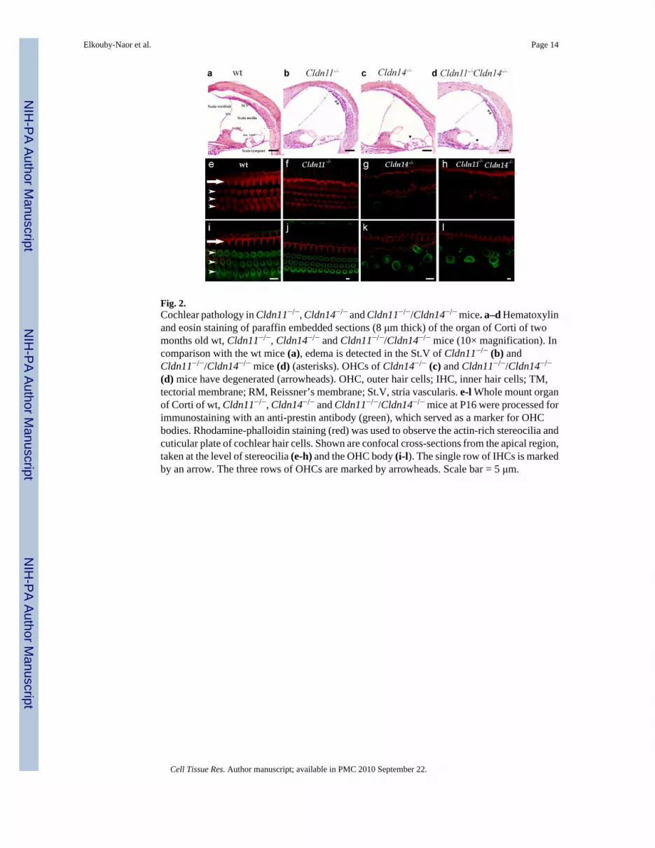
Fig. 2.Cochlear pathology in Cldn11−/−, Cldn14−/− and Cldn11−/−/Cldn14−/− mice. a–d Hematoxylinand eosin staining of paraffin embedded sections (8 μm thick) of the organ of Corti of twomonths old wt, Cldn11−/−, Cldn14−/− and Cldn11−/−/Cldn14−/− mice (10× magnification). Incomparison with the wt mice (a), edema is detected in the St.V of Cldn11−/− (b) andCldn11−/−/Cldn14−/− mice (d) (asterisks). OHCs of Cldn14−/− (c) and Cldn11−/−/Cldn14−/−
(d) mice have degenerated (arrowheads). OHC, outer hair cells; IHC, inner hair cells; TM,tectorial membrane; RM, Reissner’s membrane; St.V, stria vascularis. e-l Whole mount organof Corti of wt, Cldn11−/−, Cldn14−/− and Cldn11−/−/Cldn14−/− mice at P16 were processed forimmunostaining with an anti-prestin antibody (green), which served as a marker for OHCbodies. Rhodamine-phalloidin staining (red) was used to observe the actin-rich stereocilia andcuticular plate of cochlear hair cells. Shown are confocal cross-sections from the apical region,taken at the level of stereocilia (e-h) and the OHC body (i-l). The single row of IHCs is markedby an arrow. The three rows of OHCs are marked by arrowheads. Scale bar = 5 μm.
Elkouby-Naor et al. Page 14
Cell Tissue Res. Author manuscript; available in PMC 2010 September 22.
NIH
-PA Author Manuscript
NIH
-PA Author Manuscript
NIH
-PA Author Manuscript

Fig. 3.Segment-specific expression pattern of claudin 11 and claudin 14 in kidney nephrons. Kidneyfrozen sections of wt mice at the age of three months were stained with pAb for claudin 11 orclaudin 14 (green), and for nephron segment-specific markers (red). Claudin 11 is partially co-localized with AQP1 in the proximal convoluted tubule (a-c) and fully co-localized with CK8in the distal convoluted tubule (cortex) (j-l). Claudin 14 is co-localized with AQP1 in bothproximal convoluted tubule (cortex) (d-f) and thin descending limb of the loop of Henle(medulla) (g-i), and with THP in the thick ascending limb of the loop of Henle (m-o). Scalebar = 10 μm.
Elkouby-Naor et al. Page 15
Cell Tissue Res. Author manuscript; available in PMC 2010 September 22.
NIH
-PA Author Manuscript
NIH
-PA Author Manuscript
NIH
-PA Author Manuscript

Fig. 4.Relative expression levels of claudin genes in inner ears (a) and kidneys (b) of Cldn11/Cldn14 double knockout mice, as compared to wt mice.
Elkouby-Naor et al. Page 16
Cell Tissue Res. Author manuscript; available in PMC 2010 September 22.
NIH
-PA Author Manuscript
NIH
-PA Author Manuscript
NIH
-PA Author Manuscript

NIH
-PA Author Manuscript
NIH
-PA Author Manuscript
NIH
-PA Author Manuscript
Elkouby-Naor et al. Page 17
Table 1
Mean ABR thresholds (dB-SPL) of wt, Cldn11−/−, Cldn14−/− and Cldn11−/−/Cldn14−/− mice at the age of twomonths
Genotype No of ears analyzedFrequency (kHz)
8 16 32
Cldn11+/+Cld14+/+ 8–22 53±8 40±9 52±10
Cldn11−/−Cld14+/+ 9–10 80±12 63±13 74±10
Cldn11+/+Cld14−/− 8–10 >97±4 >81±7 81±3
Cldn11−/−Cld14−/− 10 95±5 80±5 75±7
Cell Tissue Res. Author manuscript; available in PMC 2010 September 22.

NIH
-PA Author Manuscript
NIH
-PA Author Manuscript
NIH
-PA Author Manuscript
Elkouby-Naor et al. Page 18
Table 2
Selected renal and systemic characteristics of WT and Cldn11−/−/Cldn14−/− mice
Parameter WT −/−Cldn11−/−Cld14 P value
PCa++ (mg/dl) 8.92±0.076 8.937±0.139 P=0.9508
UCa++.V (mg/24h) 0.142±0.017 0.1702±0.025 P=0.3788
PMg++ (mg/dl) 2.097±0.136 2.098±0.085 P=0.9931
U Mg++.V (mg/24h) 0.021±0.005 0.036±0.007 P=0.0887
PNa+ (mmol/L) 153.43±0.561 152.61±1.555 P=0.6083
UNa+.V (mEq/24h) 179.65±32.868 158.97±22.957 P=0.6246
PK+ (mmol/L) 8.58±2.276 7.6±0.717 P=0.7053
UK+.V (mEq/24h) 377.78±48.145 371.76±33.299 P=0.9219
P cr (mg/dl) 0.224±0.089 0.163±0.0115 P=0.5103
Daily urinary volume (ml/24h) 1.425±0.18003 1.2429±0.1601 P=0.4689
GFR (ml/min) 0.174±0.03 0.147±0.018 0.4528
Urinary protein excretion (mg/24h) 5.88±1.1902 4.6028±1.688 P=0.53
Body Weight-BW (g.) 25.625±0.73 25.85±0.553 P=0.8085
Kidney Weight-KW (g.) 0.175±0.0104 0.1627±0.0072 P=0.3672
KW/BW (%) 0.6799±0.028 0.63063±0.0295 P=0.2498
Mean Arterial Pressure (mmHg) 95.34±2.269 91.85±2.43 P=0.3536
Systolic Blood Pressure (mmHg) 114.69±2.416 108.71±2.66 P=0.1185
Diastolic Blood Pressure (mmHg) 85.695±2.75 83.76±3.18 P=0.6531
Heart Rate (beats/min) 390.39±6.81 405.8±6.19 P=0.1164
Values are mean±SEM. n=7 in each group of animals. PCa++, Plasma Ca++ concentration; UCa++. V, Urinary Ca++ excretion; PMg++, Plasma
Mg++ concentration; U Mg++. V, Urinary Mg++ excretion; PNa+, Plasma Na+ concentration; UNa+. V, Urinary Na+ excretion; PK+, Plasma
K+ concentration; UK+. V, Urinary K+ excretion; P cr, Plasma creatinine concentration; GFR, Glomerular filtration rate.
Cell Tissue Res. Author manuscript; available in PMC 2010 September 22.
Copyright © 2022 FDOKUMEN