VARIATIONS IN TIGHT AND GAP JUNCTIONS IN MAMMALIAN TISSUES
19
VARIATIONS IN TIGHT AND GAP JUNCTIONS IN MAMMALIAN TISSUES DANIEL S. FRIEND and NORTON B. GILULA From the Department of Pathology, University of California, San Francisco, California 941~, and the Depalq~mentof Physiology-Anatomy, University of California, Berkeley, California 947~0. Dr. Gilula's present address is the Department of Anatomy, Harvard Medmal School, Boston, Massachusetts 0~115. ABSTRACT The fine structure and distribution of fight (zonula occludens) and gap j unctions in epithelia of the rat pancreas, hver, adrenal cortex, epididymis, and duodenum, and in smooth muscle were examined in paraformaldehyde-glutaraldehyde-fixed, tracer-permeated (K-pyroanti- monate and lanthanum), and freeze-fractured tissue preparations. While many pentalaminar and septilaminar foci seen in thin-section and tracer preparauons can be recognized as corre- sponding to well-characterized freeze-fracture images of tight and gap junction membrane modifications, many others cannot be unequivocally categorized--nor can all freeze-etched aggregates of membrane particles. Generally, epithelia of exocrine glands (pancreas and liver) have moderate-sized tight junctions and large gap junctions, with many of their gap junctions basal to the junctional complex. In contrast, the adrenal cortex, a ductless gland, may not have a tight junction but does possess large gap junctions. Mucosal epithelia (epidid- ymis and intestine) have extensive tight junctions, but their gap junctions are not as well developed as those of glandular tissue. Smooth muscle contains numerous small gap junc- tions The incidence, size, and configuration of the junctions we observed correlate well with the known functions of the junctions and of the tissues where they are found. INTRODUCTION In a careful analysis of thin sections from various epithelia, Farquhar and Palade (10) first defined the junctional complex. They demonstrated that this epithelial complex is commonly composed of a zonula occludens (tight junction) which serves as a barrier to the diffusion of substances through the intercellular space, a zonula adherens (intermedi- ate junction), and a macula adherens (desmo- some). Subsequent technical innovations, particu- larly the introduction of a low molecular weight electron-opaque tracer (lanthanum) (8, 34) and the technique of freeze etching (3, 27, 28, 40), per- mitted a more thorough examination of the occlu- dens portion of the junctional complex. These techniques also helped unmask another junctional component, the gap junction (34) (nexus [6, 7J), now widely implicated in intercellular communica- tion-that is, in cell-to-cell transfer of ions (ionic coupling) (9, 15, 20, 30, 31, 38) and in cell-to-cell transfer of cellular metabolites (metabolic cou- pling) (15). With the use of lanthanum, this junction, hereto- fore considered to be of the occludens type, was conclusively demonstrated to have a 20-40 A inter- cellular gap (34). Revel and Karnovsky further found that in lanthanum preparations, the inter- cellular gap contained a hexagonal array of sub- units with a 90 A center-to-center spacing, reminis- cent of a similar subunit structure earlier observed by Robertson at the site of an electrical synapse 758 THE JOURNALOF CELL BIOLOGY- VOLUME 53, 1972 • pages 758-776 on November 10, 2014 jcb.rupress.org Downloaded from Published June 1, 1972
Transcript of VARIATIONS IN TIGHT AND GAP JUNCTIONS IN MAMMALIAN TISSUES
V A R I A T I O N S I N T I G H T A N D GAP J U N C T I O N S
I N M A M M A L I A N T I S S U E S
D A N I E L S. F R I E N D and N O R T O N B. G I L U L A
From the Department of Pathology, University of California, San Francisco, California 941~, and the Depalq~ment of Physiology-Anatomy, University of California, Berkeley, California 947~0. Dr. Gilula's present address is the Department of Anatomy, Harvard Medmal School, Boston, Massachusetts 0~115.
A B S T R A C T
The fine structure and distribution of fight (zonula occludens) and gap j unctions in epithelia of the rat pancreas, hver, adrenal cortex, epididymis, and duodenum, and in smooth muscle were examined in paraformaldehyde-glutaraldehyde-fixed, tracer-permeated (K-pyroanti- monate and lanthanum), and freeze-fractured tissue preparations. While many pentalaminar and septilaminar foci seen in thin-section and tracer preparauons can be recognized as corre- sponding to well-characterized freeze-fracture images of tight and gap junction membrane modifications, many others cannot be unequivocally categorized--nor can all freeze-etched aggregates of membrane particles. Generally, epithelia of exocrine glands (pancreas and liver) have moderate-sized tight junctions and large gap junctions, with many of their gap junctions basal to the junctional complex. In contrast, the adrenal cortex, a ductless gland, may not have a tight junction but does possess large gap junctions. Mucosal epithelia (epidid- ymis and intestine) have extensive tight junctions, but their gap junctions are not as well developed as those of glandular tissue. Smooth muscle contains numerous small gap junc- tions The incidence, size, and configuration of the junctions we observed correlate well with the known functions of the junctions and of the tissues where they are found.
I N T R O D U C T I O N
In a careful analysis of thin sections from various epithelia, Farquhar and Palade (10) first defined the junctional complex. They demonstrated that this epithelial complex is commonly composed of a zonula occludens (tight junction) which serves as a barrier to the diffusion of substances through the intercellular space, a zonula adherens (intermedi- ate junction), and a macula adherens (desmo- some). Subsequent technical innovations, particu- larly the introduction of a low molecular weight electron-opaque tracer (lanthanum) (8, 34) and the technique of freeze etching (3, 27, 28, 40), per- mitted a more thorough examination of the occlu- dens portion of the junctional complex. These techniques also helped unmask another junctional
component, the gap junction (34) (nexus [6, 7J), now widely implicated in intercellular communica- t i o n - t h a t is, in cell-to-cell transfer of ions (ionic coupling) (9, 15, 20, 30, 31, 38) and in cell-to-cell transfer of cellular metabolites (metabolic cou- pling) (15).
With the use of lanthanum, this junction, hereto- fore considered to be of the occludens type, was conclusively demonstrated to have a 20-40 A inter- cellular gap (34). Revel and Karnovsky further found that in lanthanum preparations, the inter- cellular gap contained a hexagonal array of sub- units with a 90 A center-to-center spacing, reminis- cent of a similar subunit structure earlier observed by Robertson at the site of an electrical synapse
758 THE JOURNAL OF CELL BIOLOGY - VOLUME 53, 1972 • pages 758-776
on Novem
ber 10, 2014jcb.rupress.org
Dow
nloaded from
Published June 1, 1972

(37). T he use of l a n t h a n u m and o ther tracers which permea te the gap junc t ion (u r an ium salts, ant imony, ru then ium) has demonst ra ted the wide dis t r ibut ion of the gap j unct ion in inver tebra te ( 19, 3I, 38) and ver tebra te (4, 16, 26, 34, 35) tissues and be tween ceils in tissue cul ture (20, 33)
Recent ly the freeze-etch technique has also be- come a valuable asset to the identif icat ion and study of t ight and gap junc t ions (5, 13, 16, 23, 26, 33, 39). T h e fracture process exposes the in terna l components of the cell m e m b r a n e (3, 32), reveal- ing specific m e m b r a n e modifications re la ted to these junct ions , and permits assessment of the pre- cise relat ionship of one cell j unc t ion to ano the r as well as to unmodif ied regions of the cell membrane . Besides reveal ing the extent and dis t r ibut ion of cell junct ions, the sigmficance of this t echnique is t ha t in some instances gap junc t ions canno t be posi- t ively identified in any other way.
In this s tudy we examine t ight and gap junct ions in a var ie ty of m a m m a l i a n g landu la r (pancreas, liver, adrena l cortex) and mucosal (epididymis, small intestine) epithelia and in smooth muscle (epididymis, small intestine), wi th par t icu lar em- phasis on their size, form, distr ibution, and inter- relationships. W e consider var ia t ions which could account for quant i t a t ive differences in ionic cou- pling between contiguous cells of various epi thel ia and raise several questions related to the inade- quacy of present terminology and technology in the definition and identif icat ion of cell junct ions
M A T E R I A L S A N D ~ I E T H O D S
Materials
The tissues used in these studies were the adrenal cortex, liver, pancreas, duodenum, and epidldymis (head and tail) of sexually mature male Sprague- Dawley rats. 15 rats and the following reagents and enzymes were employed. Hyaluronidase (Type I) (Sigma Chemical Co., St. Louis, 5{o.), collagenase (Type I) (Worthington Biochemical Corp., Freehold, N. J . ) ; l an thanum nitrate and K-pyroantimonate (Fisher Scientific Co., Pittsburgh, Pa.).
Methods
~:)RJEPA_R&TION FOR ]~LECTRON MICROSCOPY
Fixation of tissues was initiated by local injection of 1-2 ml Karnovsky's fixative (21), containing lC~ formaldehyde and 3°1~o distilled glutaraldehyde, and approximately 0.45 m~t CaC12 buffered to pH 7.4 with 0.1 M sodium cacodylate. After 2-3 min of local
perfusion, the tissue was removed, minced, and im- mersed in fixative for 4-6 hr at room temperature In some expemments, 1% I<_-pyroantimonate (22) was added and appeared to improve the staining of cell junctions. The tissues were washed overnight in 0 1 M sodium eacodylate buffer (pH 7.4) with 7% sucrose, followed by fixation for 2 hr at 4°C in acetate-Veronal-buffered 1,C/o OsO4 (pH 7.4) with 5% sucrose. The tissues were then treated for 1 hr at room temperature with buffered 0 5% uranyl acetate containing 4% sucrose, quickly dehydrated in graded ethanols, and embedded in Epon 812
For electron microscopy, silver-to-grey sections were cut with diamond knives on a Porter-Blum Sorvall MT-2 microtome (Ivan Sorvall, Inc., Nor- walk, Conn.). They were collected on carbon- and Formvar-coated grids, stained with alkaline lead alone or with 5% aqueous uranyl acetate followed by lead, and examined with a Siemens 1A electron microscope at 80 kv. For light microscopy, 1 sections were cut and stained with toluidine blue in borax.
OTHER PnOCEDUnES
LANTHANUM: Regional blood vessels or paren- chvma] tissues were injected with I-2 ml of a solution of 3% lanthanum nitrate in 0.05 M Tlus-hydrochloride buffer, pH 7.2, with 4% sucrose added After vascular rejection (I rain), the tissue was removed and placed in fixative as described above. After parenchymal tissue injection (3 min), the specimen was removed, finely minced, immersed in the lanthanum solution for a total of 5 min, and placed in fixative (see above).
PYROANTIMONATE PRECIPITATION: I IIii of
the saturated solution of pyroantlmonate was injected into the lumen of the epididymis. K-pvroant imonate was dissolved for 30 rain at 40°C in the buffer used to make up the fixative, before addition of the alde- hydes. In all other instances, parenchymal tissue was rejected with 1-2 ml of the formaldehyde-glutaralde- hyde fixative contaimng 5% K-pyroantimonate, pI-t 7.2, without calcium chloride. Fixation was con- tinued by immersing the minced tissue for 4-6 hr in the pyroantimonate-containing fixative. Isolated ceils were exposed to pyroantimonate just by im- mersion in this mixture. Subsequent processing was the same as that specified above.
C E L L ISOLATION: Isolated hepatic parenchy- real cells were prepared as described by Berry and Friend (2).
I~REEZI~ FRACTURING; For freeze fracturing, small pieces of tissue were placed in formaldehyde- glutaraldehyde fixative for 30 min and then im- mersed in 20% glycerol in 0.1 M cacodylate buffer for 3-4 hr. Pieces of tissue were mounted on card- board discs, frozen rapidly in liquid Freon 22 (chloro- difluoromethane), and fractured in a Balzers ap-
DANIEL S. FRIEND A~D NORTO~ B. GILIYLA T~g]tt and Gap Junctio~ 759
on Novem
ber 10, 2014jcb.rupress.org
Dow
nloaded from
Published June 1, 1972

paratus (Balzers AG, Balzers, Liechter~tein) with a stage temperature of --115°C (S, 27). Electron mlcrographs of freeze-fracture replicas are mounted with the shadow from bottom to top, and all shadow's are white.
Terminology Employed in this Paper
J U N C T I O N A L COMPLEX: The zonula occlu- dens (fight junction), zonula adherens (intermedi- ate junction), and macula adherens (desmosome), collectively. This term does not indicate whether or not a gap junction is present.
FR.EEZ~-FRACTUP~F. FACES : W h e n the frac- ture passes through the cell membranes in the re- gion of the junctional complex, two internal com- plementary fracture faces are produced. Fracture face A, associated with the cytoplasmic portion of the membrane, contains the larger number of intramembranous random particles, the ridges of the zonula occludens, and the closely packed parti- cles of the gap junction. The other fracture face, B, associated with the intercellular portion of the cell membrane, contains fewer particles and includes the furrows and depressions which complement the ridges and particles of face A. Chalcroft and BuI- livant have demonstrated that the fracture faces are indeed complemental~/(5).
T I G H T JUNCTION (ZONULA OCCLUDENB) :
As seen in thin section, the sum of the pentalaminar fusions between contiguous cell membranes. This junction is impermeable to lanthanum. As seen in freeze fractures, the sum of the ridges and furrows on faces A and B, respectively. (Although a solitary continuous ridge may actually constitute an imper- meable seal per se and therefore qualify as a tight junction, we do not refer to the single ridges ob- served among many as tight junctions. We refer to these as fasciae occludentes.)
GAP JUNCTION (NEXUS) : As seen in thin section, the pentalaminar or septilaminar mem- brane appositions with a 20-40 A gap, permeable to lanthanum, and possessing a periodic substruc- ture. The total width of the gap junction (cyto- plasm-to-cytoplasm) is greater than 150 A, but less than 210 A. As seen in freeze fractures, macular aggregates of polygonally arranged 80--90 A mem- brane particles (face A) and complementary de- pressions (face B).
O 1 3 S ~ R V 2 t T I O ~ C S
Cell Junctions in Glandular Epithelia
EXOCRINE PANOREAS: The junctions be- tween cells of the pancreatic acini will be consid-
ered first, since both tight and gap junctions are abundant in this tissue. Here gap junctions, espe- cially, are present in various locations, configura- tions, and sizes. Examination of the pancreatic junctions introduces problems of identification and characterization which will be further explored in other tissues.
In thin sections of acini of the exocrine pancreas, junctional complexes are found at the luminal sur- faces of all contiguous cells. Pentalamlnar fusions (Fig. 1, inset a) of adjacent membranes are com- mon along the interface between the luminal sur- face and the first macula adherens (desmosome). Morphologically, all points of membrane fusion appear alike, but lanthanum, introduced into the intercellular space from the basal, vascular pole of the cells, is excluded by some fusions (Fig. 1) but not by others (Fig. 1, inset b). The fusions which do not impede the flow of lanthanum are usually farthest from the luminal surface. The first fusion at the microvillar surface stops flow in either direc- tion. The differences in the capacity of pentalam- inar fusions to impede lanthanum indicate that some are continuous while others are discontinuous or focal, and that thin sections alone cannot distin- guish between the two in all instances (Figs. 1 and 4). (Nor can we be certain that the thin-section image does not represent a punctate gap junction.) Freeze etching confirms the variations in compo- nents of the fight junction by showing short, focal ridges and furrows as well as extensive continuous ones (Figs, 2 and 3). Compared with the tight junc- tions of mucosal epithelia which we will examine shortly, this mixture of continuous and discon- tinuous ridges and furrows is more evident as well as looser in array. Freeze etching does not clarify the nature (tight versus gap) of all the punctate points of apparent fusion seen in thin section, since the minimum size of a puncta (,-~80 A) is about the same as that of an ordinary membrane particle. Hence a junctional membrane comprised of a single membrane particle would be indistinguish- able from the rest of the membrane.
Another aspect of pancreatic junctions eluci- dated by freeze etching is the complete enclosure of some gap junctions within the confines of the zonulae occludentes (Fig. 6), as noted in the liver by Kreutziger (23). Tracers permeate and help to reveal some gap junctions near the luminal surface (Fig. 5) and the more extensive ones abluminal to the junctional complex (Fig. 7), wherever the gap junctions are accessible to permeation by low mo- lecular weight substances. Freeze etching readily
760 THE JOURNAL OF CELL BIOLOGY • VOLZrME 5S, 1972
on Novem
ber 10, 2014jcb.rupress.org
Dow
nloaded from
Published June 1, 1972

Fm~TREs 1-9 Figs. 1-9 are micrographs of pancreatic acinar cells.
F ia~r~ 1 The tissue in inset a was fixed in buffered paraformaldehyde-glutaraldehyde containing 1% K-pyroantimonate. The central linear density of the pentalammar fusion here does not stain well, due to its inaccessibihty to pyroantimonate at the point of fusion. The tissues in the principal figure and inset b were injected with lanthanum before fixation Lanthanum penetrated the intercellular space from the vascular pole. Focal fusions (arrows) between contiguous cell membranes of the exocrlne pancreas may correspond to one of several membrane modifications observed by freeze etching. Such thln-section pentalaminar profiles may appear on fractm'e face A as continuous, smoothly contom'ed ridges, dis- continuous ridges, beaded ridges~ or small focal aggregates of gap-junction-like membrane particles. Consistent with this structm'al variation in thin-section pentalamina~ images which look alike, such structures may serve different biological functions. Tile structure m the main figure halts the lumenward flow of lanthanum at the distal hmit of a zonula occludens, whereas one closer to the base of the cell (inset b) does not. Thus thin sections and tracer preparations alone do not permit positive classification of all junctional structures examined, and the impediment to the flow of tracers by tight junctions hinders visualizat[on of gap ~unctions in the apicaI region of thejunctionalcomplex.Z, zymogengranule. X 100,000; inset a, X ~00~000; inset b, X 150~000.
761
on Novem
ber 10, 2014jcb.rupress.org
Dow
nloaded from
Published June 1, 1972

lelGlrl~E 2 A low magnification view of a freeze-fracture replica through several pancreatic aciuar cells. This micrograph reveals the distribution of the series of ridges and complementary linear depressions which constitute the tight junction. Subsequent micrographs will depict these at higher magnifications. The inset illustrates a feature common to all tight junctions examined--the fusion of two ridges on the A face. X ~8,000; inset~ X 130,000.
I~IGVl~E 8 In this replica, both A and B fractmle faces (arTows) of the gap junction are visible within compa~ff, ments of the tight junction. This pattern of sequestration is also common in the liver. Note the relative amplitude of the enclosures formed by the ridges. X 155,000.
on Novem
ber 10, 2014jcb.rupress.org
Dow
nloaded from
Published June 1, 1972

confirms the configuration and size of gap junctions in these locations (Figs 6, 8, 9) Only- freeze etch- ing, however, reveals the gap junctions enclosed by continuous ridges of the zonulae occludentes Com- pletely surrounded, they are shielded from tracer permeation and consequently cannot be identified by the use of tracers in thin section As illustrated in Figs 3 and 6, the gap junction particle aggre- gates of face A and the depressions of face B are encompassed by the zonula occludens's network of ridges and furrows, respectively.
LIVER: The junctional complex between ad- jacent hepatic parenchymal ceils has been de- scribed in detail by other investigators (5, 10, 16, 18, 23, 25). In general, its tight and gap junctionai components resemble those of the pancreas in all major respects. Again, the zonula occludens is only moderately extensive (Fig 12) and occasionally contains gap junctions, large gap junctions are also present below the junctional complex region of the parenchymal cells (Fig. 11). One novel observa- tion is the fine delineation of the gap junction achieved with pyroantimonate as a tracer (Fig 10).
ADRENAL CORTEX Junctions of the adrenal cortex differ from those of other glandular epi- thelia such as the pancreas and liver As visuahzed in thin section, the classic junctional complex is not present in the gland's three major cortical zones. Desmosomes are rudimentary, the extensive zones of adhesion have a distinctive appearance (12, 14), and the pentalaminar fusions character- istic of tight junctions are rare Tracers readily pass between the ceils and encompass areas of apparent fusion, indicating that such images (Fig 13) corre- spond to fracture-face profiles of discontinuous or beaded ridges or small gap junctions In addition, tracers reveal that large gap junctions are common (Fig. 14), a fact which freeze etching confirms (Figs. 15 and 16). Actually, freeze etching demon- strates that gap junctions of various sizes and con- figurations are widely distributed throughout the cortex, but no definitive, smoothly contoured ridges or furrows are visible, implying that the pentalaminar profiles seen in thin sections of the adrenal cortex are probably gap junctions. Whether or not closely packed linear arrays of particles (inset, Fig. 16) represent portions of gap or tight junctions will be explored in the Discus- sion. In any case, pyroantimonate, lanthanum, and peroxidase all move freely around these struc- tures, and pyroantimonate and lanthanum may move through them
Cell Ju~tctions in Mucosal Epithelium
EPIDIDYMIS' Among the various epithelial cell contacts examined, the zonula occludens of the epididymis is the most highly developed. Epidid- ymal columnar cells are joined at their microvillar surfaces by the characteristic junctional complex comprised of a zonula occludens up to several microns long, a zonula adherens, and multiple desmosomes. Although the zonula occludens occa- sionally has the pentalammar image of membrane fusion along its entire length, such profiles are rare. Customarily the zonula occludens contains both regions of membrane fusion and regions where the intercellular space intervenes (Fig. 17, inset b), often varying in width from 20 A to more than 100 A. The occludens portion of the junctional complex excludes pyroantimonate from the intercellular space when the tracer is introduced via the lumen (Fig. 17, inset b) and excludes pyroantimonate and lanthanum Introduced via the bloodstream Fig. 17, inset a is a rare example where some lanthanum is seen near the microvillar surface Tracers do patchily permeate small segments of the intercel- iular space in the basal area of the complex, but they are always checked in the apical region
Below the junctional complex, lateral cell mem- branes generalN maintain an intercellular space of 150 A or more. Tins vicinity fills completely with lanthanum.
With the freeze-etching technique, extensive membrane fracture faces of the lateral cell mem- branes are easily obtained. At low magnification, the ramifying ridges and complementary furrows of the zonula occludens throughout the apical re- gion are particularly striking (Figs 17-19). BeIow the zonula occludens, in the region of the zonulae and maculae adherentes, no detectable modifica- tions of the fracture face are evident. If anything, the membranes here have fewer particles than do other areas of nonjunctional membrane (Fig. 18)
In the epididymis, the ridges of the zonula oc- cludens form a far-reaching horizontal network. Not always continuous individually, they may stretch abluminally for several microns In the basal part of the zonula occludens, a variety of ridge deployments are observed. One is a ramifying, compartmentalizing, continuous band, another is a continuous but open network, and still another is a completely open array of isolated ridges (Figs. 17-20).
In most cases, the ridges are perpendicular to the axis of the cell. At the juncture of three cells,
DANIEL S. FRIEND AND NORTON B. GILLmA Tight and Gap Junction.~ 763
on Novem
ber 10, 2014jcb.rupress.org
Dow
nloaded from
Published June 1, 1972

however, as also noted by Staehelin et al. in the intestine (39), they are vertically disposed, coin- cident with the cellular axis. Usually one major vertical ridge is present at the cell juncture, giving rise to sevcrat short horizontal ones which merge with the major portion of the zonula occludens network (Fig. 20). (Compare this image with Fig. 12, which is a similar view of the hepatic tight
junct ion ) All the ridges are about 80 A wide, most are smoothly contoured, and some apparently consist of fused particles 80-85 A in diameter (Fig. 18, inset a). Fused-particulate ridges frequently have missing portions, presumably representing the 80-85-A particles associated with the comple- mentary furrows of fracture face B (Fig 19).
Since mistakenly reporting that macular gap junctions were not present in the epididymis (13), we have discovered some basal to the tight junc- tions in the rat (Fig. 18, inset b). Revel (personal communicat ion and 36) has also found numerous ones intermingled with tight junctions, as well as basally, in the mouse.
SMALL INTESTINE (DUODENUM) : In the mucosal epithelium of the duodenum~ the freeze- fracture landscape of the tight junct ion is similar to that of the epididymis. But here the zonula occludens appears more compact (Fig. 21). The network of ridges and furrows is continuous at the microvillar surface, while some discontinuities are found in the basilar region. In the ablumina | pole of the complex, we often observe isolated ridges. We have not found gap junctions concealed be- tween the ridges of the small intestine's zonula
occludens, although small aggregates of particles, like those of the epididymis, are prevalent
Cell Junctions in Smooth Muscle
SMALL INTESTINE AND EPIDIDYMIS: The cells of the smooth muscle of the small intestine and epididymis are joined by gap junctions (nexus) with the same permeability to intercellular tracers as those in epithelia. In fracture faces, cell mem- branes contain plaques of aggregated particles varying from 0.1 # to I /z in diameter constituting the membrane differentiation associated with the gap junct ion (Fig. 22). While most of the particle arrays are macular, some are variegated in shape, including slngle-file distributions indistinguishable from the beaded ridges (inset, Fig. 22) associated with the zonulae occludentes in epithelia. Partic- ularly in the small intestine, one smooth muscle cell makes numerous gap junct ional contacts with either one or several neighboring muscle cells.
D I S C U S S I O N
Components of the Junctional Complex
The junctional complex is the most elaborate zone of contact between mammal ian epithelial cells. I t has long been established that the usual initial element of the complex, the zonula oc- chidens, forms a barrier to the passage of sub- stances from the lumen into the intercellular space l ining cavitary organs and between cells which form the acini of exocrine glands (10) The second
I~Gm~ES 4-6 Paraformaldehyde-glutaraldehyde (Fig. 4), lanthanum (Fig. 5), and freeze-fraetm-e (Fig. 6) preparations through portions of the junctional complex. In the routine preparation, the nature of the apparent points of fusion is difficult to assess. In regions where ridges of the tight junction do not exclude the tracer, permeation with lanthanum reveals the periodicity and spacing suggestive of a gap junction. t~'eeze-fracturing definitively illuminates small gap junctions confined by ridges of the zonula occludens, which would not be detected in routine and lanthanum preparations. Fig. 4, X 150,000; Fig. 5, X 190,000; Fig. 6, X 140,000.
I~G~rnES 7--9 Pyroantimonate (Fig. 7) and freeze-fracture (Figs. 8 and 9) preparations of extensive gap junctions below the junctional complex. Careful examination of Fig. 7 reveals the regular periodicity within this extensive gap junction permeated with pyroantimonate. ~ig. 8 is a cross-fracture shewing complementary A and B faces of a gap junction comparable to that in Fig. 7. Ill this instanee, the fracture process exposed the B face of one cell membrane and crosses the intercellular space (gap) (arrow), where it then exposes the A face of the adjacent cell membrane. Fig. 9" The magnitude of some pancreatic gap junctions is more apparent hel~. Gap junctions of the liver and adrenal cortex are comparable in size. A region of nonjmletional membrane (N) is often found within the gap junctional plaque in both liver and pancreas. A, fracture face A; B, fracture face B. Fig. 7, X 110,000; Fig. 8, X 105,000; Fig. 9, X 60,000.
764 T~E JOURNAL O~ C~LL BIO/~OOY • VOLUME 53, 197~
on Novem
ber 10, 2014jcb.rupress.org
Dow
nloaded from
Published June 1, 1972

DANIEL S. :FaIE~D AND NO~TON Bo GILVLA Tight and Gap Junctions 765
on Novem
ber 10, 2014jcb.rupress.org
Dow
nloaded from
Published June 1, 1972
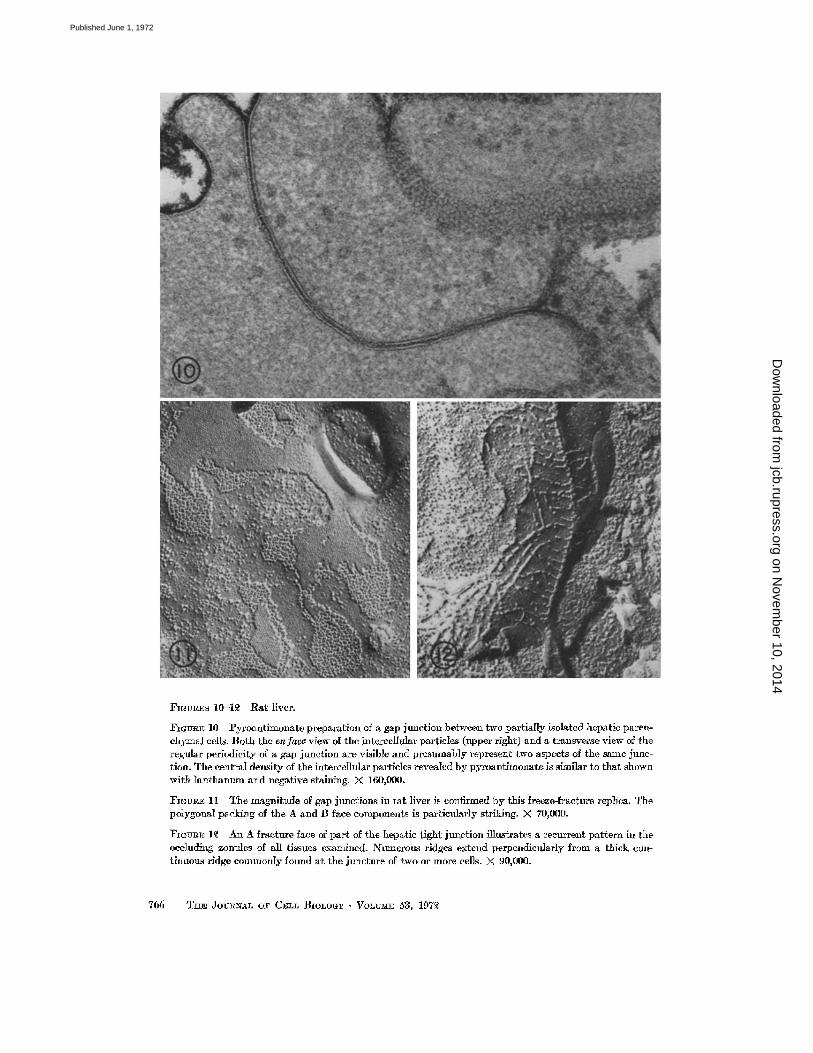
FIGURES 10-1~ Rat liver.
Fr~v~E 10 Pyroantimonate preparation of a gap junction between two partially isolated hepatic paren- chymal cells. Both the enfaea view of the intercellular particles (upper right) and a transverse view of the regular periodicity of a gap junction are visible and presumably represent two aspects of the same junc- tion. The central density of the intercellular particles revealed by pyroantimonate is similar to that shown with lanthanum and negative staining. X 160,000.
I~GUR~ 11 The magnitude of gap junctions in tat liver is confirmed by this freeze-fracture replica. The polygonal packing of the A and B face components is particularly striking. >( 70,000.
FmVRE 1~ An A fracture face of part of the hepatic tight junction illustrates a recurrent pattern in the occluding zonules of all tissues examined. Nmnerous ridges extend perpendietflarly from a thick con- tinuous ridge commonly found at the juncture of two or more cells. )< 90,000.
766 T ~ JOV~AL OF C~LL BIOLOOr • VOLtrME 53, 197~
on Novem
ber 10, 2014jcb.rupress.org
Dow
nloaded from
Published June 1, 1972

FtGVRES 13--16 Figs. 13--16 are routine (Fig. 18), lanthanum (Fig 1'~), and freeze-etched (Figs. 15 and 16) preparations of adrenal cortex Fig. 15 shows an A face to advantage; Fig. 16, a B face Gap junctions are as extensive here as in those glandular epithelia which possess full junctional complexes such as the pancleas and liver. But in the adrenal gland, no readily identifiable elements of the tight junction are present. Isolated rows of particles on the A face (inset~ Fig. 16) and depressions on the B face are com- monly found adjacent to the usual macular aggregates of the gap junction. Simdar rows of particles were observed in all other tissues, including those which have extensive tight junctions (epldidymis) and those without (smooth muscle). Compare this znset with those in Figs. 18 and o e. Fig. 13, X 130,000, Fig. 14~ X ~0,000 i Fig. 15~ X 150:000; Fig. 16, X 100,000; *nset~ X 150~000.
767
on Novem
ber 10, 2014jcb.rupress.org
Dow
nloaded from
Published June 1, 1972

FIGIZaES 17--~0 Epithelial cells lining the epididymis.
FIGVI~E 17 The lower half of this freeze-fracture image through the ridges and furrows of the zonula oceludens corresponds to the area shown in thin sechon in inset a. The inset demonstrates that small amounts of tracer can occasionally percolate from the basal pole of the cell up toward the mierovillar surface (arrow), although tracer flow is usually impeded several microns fl'om this surface. In inset b~ py- roantimonate is totally excluded from the intercellular space (arrow) after luminal introduction of the tracer. M V , mierovilli. X 80,000.
element, the zonula adherens, generally serves as the insertion site for microfilaments of the terminal web. And the final element, the macula adherens (desmosome), provides structural resistance to lateral shearing forces. The epithelial-type junc- tional complex is not found in muscle (smooth, skeletal, and cardiac), neural tissue, or most endo- crine glands (the thyroid gland is an exception). The junct ional complex as a whole, as well as the
zonula occludens, is confined to epithelia which permit no interchange of large molecules between the luminal contents and the intercellular space. Many epithelia have both tight and gap junctions, possibly serving as seals and means of intercellular communication and reflecting the diverse require- ments of the tissue in its normal functioning. Other epithelia apparently need primari ly one or the other type of junction. Careful identification of
768 THE JOURNAL OF CELL BIOLOGY • VOLUME 53, 197~
on Novem
ber 10, 2014jcb.rupress.org
Dow
nloaded from
Published June 1, 1972

FIGtmE 18 This fracture face is characteristic of the extensive, ramifying, compartmentalized tight junction of the epididymis. The basal, larger compartments (upper portion) are like those found in the pancreas and liver, while the apical, smaller ones (lower portion) resemble those of the intestine. Single, doublet, and triplet arrays of membrane particles are more common in the smaller compartments. All types of ridges--smoothly contoured continuous, discontinuous, fascial, and beaded--are apparent in this field, and the width of ridges and membrane particles is similar. Inset a depicts a beaded ridge which forms part of the epididymal zonula occludens. As illustrated in Figs. 16 and ~ , such beaded ridges are in- distinguishable from linear particle arrays associated with gap junctions. Their classification and function are undetermined. Inset b is a gap junction found near the base of an epididymal epltheliaI cell. X 90,000; inset a, X 175,000; inset b, X 105~000.
DArnEL S. F~IE~D AND NOI~O~ B. GILUI.A Tight and Gap Junctions 769
on Novem
ber 10, 2014jcb.rupress.org
Dow
nloaded from
Published June 1, 1972

FmVRE 19 Precise complementarism between ridges (face A) and furrows (face B) of the tight junction is customary in the epididymis. Some particles are associated with furrows of the B face; presumably these have been dislodged from the ridges, perhaps accounting for some of their apparent discontinmties. Other particles on both the A and B face (Fig. 18) are not directly associated with either the ridges or the fro'rows. Their relationship to junctional structm~ is not known. )< 115,000.
FIGURE 20 Like the horizontal continuous ridge of the tight junctional element which seals the lumen, the lateral vertical ridge where several cells abut is also continuous. Its multiple branches extend hori- zontally in opposite directions. Compare this figure with Fig. 12, a similar image of liver tissue. )< 115,000.
the junctions present enhances understanding of the tissue's function.
The observations in this study indicate that (a) the tight junct ion (zonula occludens) varies con- siderably in size and configuration, (b) the gap
junction is both a frequent component of the junc- tional complex and an independent structure distal to the complex, (c) the variations present in the constituents of cell junctions correlate well with the specialized functions of the cells with which
770 THE JOtmNAI~ OF CEI~L BIOLOGY • VOlmME 53, 1972
on Novem
ber 10, 2014jcb.rupress.org
Dow
nloaded from
Published June 1, 1972

Fm~n~ .Ol Duodenal mueosa. The tight junction of small intestinal epithelium ~s similar to that of the epididymis in all major respects, although it is less extensive. Some isolated furrows (faseiae oeeludentes), hke those frequently found m ihe epidldymis, are visible some distance from the mierovillar surface. X 5~,000.
the junct ional elements are associated, and (d) existing criteria for classifying junctional elements are inadequate for all the cell contacts seen in thin- sectioned and freeze-etched preparations.
Variations in Distribution and Configuration
of Tight Junctions
Tracers introduced via the bloodstream often permeate the intercellular space of the liver and exocrine pancreas from the basal pole alI the way
to the luminal surface before they are completely halted In the epididymis, however, tracers are usually stopped several microns from the luminal surface, at the far end of the zonula occludens. Freeze fracturing clarifies these observations, con- firming the general continuity of the barrier at the microvillar surface of all tissues examined, while revealing variations in tight junction stIhc- ture at the opposite end
Short, isolated ridges (fascia) often accompany the major, continuous components of the tight
DANIEL S FRIEND AND NOnTOZ~ B. GILVbA Tight and Gap Junctions 771
on Novem
ber 10, 2014jcb.rupress.org
Dow
nloaded from
Published June 1, 1972

F~G~-~tE ~ Smooth muscle, duodenum. Many gap junctioJls (arrows) are present in configurations vary- ing from regularly concent~'ic maeulae to geographic clusters and single strands (inset). The image in the inset demonstrates the difficulty in interpreting the true "junctional" nature of particle aggregates. Tight junctions have not been seen in this tissue. The replica also l~veals tile linear deployment of the annuli of numerous endocytic vesicles. X 20,000; inset, X 100,000.
junction. Commonly', the isolated segments are basal to the main occluding structure. Thei r close association suggests a developmental relationship- one, perhaps the progeny of the other~ caught in transition during membrane differentiation,
whether fusing or dissolving, we do not know. In the intestine and epididymis, the ridges basal to the continuous luminal one arc composed of short, straight segments which join at acute angles, form- ing an extensive, closed, ramifying network. In
772 T I ~ Joln~AI~ oF C~LL BIOLOGY • VOLI~ME 53, 197~
on Novem
ber 10, 2014jcb.rupress.org
Dow
nloaded from
Published June 1, 1972

the liver and pancreas, the ridges consist of longer, curved segments whmh join at obtuse angles to form a more open network The lengthy ridges in the hver and pancreas, more discontinuous than their counterparts in the intestine and epididymis, permit the tracing of potential passageways from the base to the apex of the zonula occludens. This is not the case in the epididvmis and intestine, where imperforate ridge configurations stretch up to several microns from the apical surface Variations in the tightness of the zonula occludens, therefore, have a structural basis which is revealed by freeze etching.
Correlatwn in Tight Junction ~Iorphology
and Tissue Function
The epididymis modulates the composition of its luminal content by selective secretion and ab- sorption along its span (24)--a process similar to the varying regional transport system within renal tubules. Fluctuations in fluid volume, electrolytes, and amphopbilic proteins in the head, body, and tail of the epididymis are presumably critical to the normal maturation of sperm. "~Vhatever the mechanisms for modulating the composition of epidldymal fluid, seals preventing the exchange of large molecules between the lumen and the extra- cellular space, and zonulae, perhaps functioning directly m transepithelial ion permeation as well (1), are important adjuncts in the over-all regula- tory function of the tissue In addition, shielding sperm from the host's immune system and prevent- ing the escape of sperm or its degradation products also obviate the production of auto-antibodies to sperm, a situation which could cause sterility. The extensive tight junction thus protects against steril- ity in at least two ways: by helping to maintain the epididymaI luminal environment and by pro- viding a barrier between sperm and the host's immunologically competent cells. The significance of tight junctions in other epithelia may be con- sidered in a similar light, funcUoning in the separa- tion of two disparate environments (10, 11, 25). In the epididymis, this function is actually a matter of species survival, and the tight junction is perhaps the most elaborate tight junction yet found.
What of epithelia without tight junctions? The zona fasciculata of the adrenal cortex seems to exemplify an epithelial tissue which requires the free flow of large molecules between cells and periendothelial space, presumably for the sake of
an unhindered flux of steroid hormones from pa- renchymal cells to the bloodstream. This tissue appears to lack occluding zonules--in fact, a dis- tinctive cell contact is present, perhaps to assist in keeping the intercellular spaces patent (12, 14). A very. high degree of synchrony is desirable in this epithelium, and gap junctions which are extensive probably provide for the ionic coupling observed (Jensen and Friend, unpublished observations).
Variations in Incidence and Location of
Gap Junctions
Whether or not tight junctions or the complete junctional complex are present, gap junctions have the same variations in location and configuration in all tissues where they appear. The macular gap iunction is a prominent element in tile junctional complexes of the pancreas and liver. In these glan- dular epithelia, they are seen bound by ridges of the zonula occludens, adjacent to open ridges at the base of the zonula, and as independent ele- ments toward the basal surfaces of ceils In tissues where they are part of the junctional complex, gap junctions are generally more extensive toward the basal lamina. In tissues which lack junctional com- plexes such as the adrenal cortex, independent gap junctions are found in the same variety of lo- cations with respect to the polarity of the cell and in the same variety of sizes and configurations. Obviously', tight junctions are not a requisite for the functioning of gap junctions or that of the tissue in general The intestine (duodenum) and epididymis, both of which have larger ught junc- tions than are found in glandular tissue, have fewer macular gap junctions in the apical regions of the cells. Diminutive gap junctional plaques have been recently identified in fracture faces of the basal cellular region of the small intestine (D Good- enough, personal communication).
Problems in Identifying and Classifying Junctional Elements
While the continuous ridge-type tight junction and the large macular-type gap junction are gen- erally identified with ease, certain configurations arc indistinguishable by currently- available tech- niques. For as well as observing components of the tight junction in the continuous ridge conforma- tion, we also see them as interrupted belts, beaded ridges, or mere puncta (short or single points of
DANIEL S. ~IEND AND NORTON B. GILULk Tight and Gap Junctions 773
on Novem
ber 10, 2014jcb.rupress.org
Dow
nloaded from
Published June 1, 1972

apparent fusion) which lanthanum can encircle, thereby affording the impression that they are gap junctions in thin sections. We do not know if beaded ridges, particularly, should be considered as portions of tight junctions or as linear gap junc- tions For gap junctions (in our opinion) may also exist in a variety of arrays besides the familiar hexogonal packing of uniform particles in a macu- lar configuration. Two or three strands of particles may extend from the macular array, eventually tapering to an isolated, beaded ridge, as is the case in the adrenal cortex and in smooth muscle And even more difficult to interpret are the triplets, doublets, and single isolated particles with comple- mentary depressions. Do any or all of these parti- culate configurations function as gap junctions? At present, morphological criteria and experi- mental approaches do not resolve the problem of distinguishing whether or not the varieties of "punc t a " function as gap junctions, focal tight junctions, or neither. Nor is it known whether large zonulae occludentes can act as electrotonic junctions.
Resolution of these problems awaits the dis- covery of a tissue truly devoid of macular gap junctions. Thus far, none has been found
T H E N E E D F O R A M O R E E X P L I C I T
D E F I N I T I O N O F A C E L L J U N C T I O N
The term ee/l .]unctzon generally refers to a variety of contacts between two similar cells or between a cell and a closely apposed extracellular structure, wherever modifications of the cell membrane or intercellular space are sufficient to impart to it a distinctive appearance in thin section. With the application of this concept, numerous "junctions" are commonly recognized-- t ight junctions, gap junctions, intermediate junctions, desmosomes, septate junctions, and others. With the advent of new techniques, we are findmg major differences among these contact areas--differences which per- haps should be exploited to narrow our definitions.
Several means for classifying cell contacts exist One is to separate those which have recognizable membrane modifications in freeze-fracture replicas from those which do not. Thus in the case of the junct ional complex, gap and ught junctions could be separated from intermediate junctions and desmosomes Further discrimination can be made on the basis of the susceptibility of a ceil contact to divalent cation-chelators and proteolytic en- zymes, to which tight and gap junctions are resist-
ant while zonulae and maculae adherentes are no t - they usually separate upon exposure to these agents (2, 9, 17, 29). In addition, proteases digest the plaques of desmosomes but do not affect the asso- ciated modifications of many other junctions. Discrimination may also be made on the basis of permeability, such as the exclusion of lanthanum by tight junctions but not by others. And more important than the differences in fine structure, freeze fracturing, dissociation, digestion, and per- meability are the differences in the unique func- tions of the specific junctions, such as gap junction mediation of ionic and metabolic coupling. Ultimately, any classification of cell junctions devised will have to take all these factors into ac- count. In anticipation of that development, we suggest that reports of new or incompletely de- scribed contacts include descriptions of all the foregoing parameters whenever possible. Naming the type of junction by fine structural impression alone is frequently insufficient.
Presently, the known cell contacts would seem to fall into two general categories which could suitably encompass new ones: those wherein ap- posing membranes seem to share a structural com- ponent as revealed by freeze fracturing (gap, tight, and septate) and those with no such mutual bond. Theoretically, those sharing a membrane component are most likely to participate in inter- cellular interactions such as ionic and metabolic coupling and absolute adhesion, while those with- out a mutual component probably fulfill another type of function. On this basis, we have elected to use the term cell junction for those which appar- ently share a structural component, and the term cell contact for all others. For us, the term cell junction then has a definite operational connota- tion in addition to its present general structural connotation.
We are grateful to Roz Bettencourt for critical editing and typing of the manuscript, to Irene Rudolf and Yvonne Jacques for excellent technical assistance, to Dr. D. Branton for the use of his freeze-fracture facilities, and to Dr. P. Satir for sup- port.
This work was supported by grants from The Population Council, Grant No. M71.0103c (New York) and The University of California School of Medicine (San Francisco) Research Evaluation and Allocation Committee to Dr. l~riend, Atomic Energy Commission Grant AT (04-3)-34 P.A. 142 to Dr. Branton, and United States Public Health Service Grant HE 13849 to Dr. Satir. Dr. Friend is the
774 THE JOURNAL OF CELL BIOLOGr • VOLUME 53. 1972
on Novem
ber 10, 2014jcb.rupress.org
Dow
nloaded from
Published June 1, 1972

recipient of United States Public Health Service Research Career Development Award 5 K03 GM 35313 from the National Institute of General lViedical Sciences. Dr. Gilula was a United States Public Health Service predoctoral trainee under Grant GNf 1021.
Received for publwat~on 26 October 1971, and ~n revised form 25 February 1972.
~ E F E R E N C E S
1. BARRY, P. H., J. M. DIAMOND, and E. 1~I. V']RIOHT. 197I. The mechanism of cation permeation in rabbit gall bladder. Dilution potentials and biionic potentials. J eliembrane Biol. 4,:358
2. BERRY, M. N., and D. S. FRIEND. 1969. High- yield preparation of isolated rat liver paren- chymal cells: a biochemical and fine structural study. 3". Cell Biol. 43:506.
3 BRANTON, D. 1966 Fracture faces of frozen membranes. Proo. :Vat. Acad. Sci. U S. A. 55: 1048.
4. BRIOHTMAN, ~X~. W., and T. S. REESE. 1969. Junctions between intimately apposed cell membranes in the vertebrate brain. J. Cell Biol. 40:648.
5. CHALCROFT, J. P., and S. BULLIVA~T. 1970 An interpretation of liver cell membrane and junction structure based on observation of freeze-fracture replicas of both sides of the fracture. J. Cell Biol. 4,7:49.
6. DEWEY, l~{. ~vi, and L. BARR 1964. A study of the structure and distribution of the nexus J. Cell Biol. 23:553
7. DEWEY, 1~i. Ni , and L. BARS 1962. Intercellular connection between smooth muscle cells the nexus. Science (Washington). 137:670.
8. DOOOENWEILER, C. F , and S. FREN~:. 1965. Staining properties of lanthanum on cell membranes Proc. Nat Acad. Sei. US.A. 53: 425.
9 Dm~iFuss, J. J., L. GIRARDIER, and W. G. FORSS- ~aANN. 1966. ]~tude de la propagation de l'excltation dans le ventricle de rat au moyen de solutions hypertoniques. Pflugers Arch. Gesamte Ph~swl. 2Vlenschen Tzere. 292:13.
10. FARQUHA~, M. G., and G E. PALADE 1963. Junctional complexes in various epithelia. J. Cell Bwl. 17:375.
11. FARQUHAR, ~ . G,, and G. E. PALADE. 1964, Functional organization of amphibian skin. Proc. Nat. Acad. Sc*. U S A . 51:569.
12. FRIEND. D. S. 1971 A unNue cell contact in the adrenal cortex. Proc. 29th Electron .¥lw~osv. Soe. Ame~. 504.
13. FRIEND, D. S., and N. B. GILULA. 1970. Cell
junctions of the rat epididymls. J. Cell Biol. ~7:66 a. (Abstr.)
14. FRIEND, D S., and N B. GILULA. 1972. A dis- tinctive ceil contact in the rat adrenal cortex. J. Cell Bwl 53:148.
15. GmULA, N B., O. R. REEVES, and A. STEIN- BACH. 1972. gIetabolic coupling, ionic cou- phng, and cell contacts Nature (London). 235: 262.
16. GOODENOUGH, D. A., and J. P. REVEL. 1970 A fine structural analysis of intercellular junctions in the mouse liver, o r. Cell Bzol. 45:272.
17. GOODENOUOH, D. A., and J. P. REVEL. 1971. The permeability of isolated and in s~tu mouse hepatic gap junctions studied with enzymatic tracers. 3" Cell Bzol. 50:81.
18. HEATH, T., and S. Wlssm. 1966. Fine structure of the surface of mouse hepatic cells. Ame~. J. Anat. 119:97.
19 t-IuDSPETH, A. J., and J. P. t~v~e . 1971. Co- existence of gap and septate junctions in an invertebrate epithelium. J. Cell Bwl. 50:92.
20. JOHNSON, R. G., and J. SHERIDAN. 1971. Junc- tions between cancer cells in culture: ultra- structure and permeability. Sczence (Washing- ton) 174:717.
21 KARNOVS~Y~ I~f. J. 1965. A formaldehyde- glutaraldehyde fixative of high osmolality for use in electron microscopy. J Cell Bwl. 27: 137A. (Abstr.)
22 KO~INICK, H., and U. KOMNm~:. 1963. Electron microscopische Untershungen zur funktionel- len morphologle des ion entransportes in der Salzdrusse yon La~us argentatus. Z. Zdlforsch. Mzkroskop. Anal 60:163.
23. KR~UTZmER, G. O. 1968 Freeze-etching of intercellular junctions of mouse liver. P~oc. 26th Electron Mzooso Soc Amer. 234.
24. LEVINE, N., and D J. MARSH. 1971. Micro- puncture studies of the electrochemical aspects of fluid and electrolyte transport in individual semmiferous tuhules, the epididymis, and the vas deferens in rats. J Physwl. (London). 213: 557.
25. MATTER, A., L. ORCI, and C. ROUILLER. 1969. A study on permeability barriers between Disse's space and the bile canaliculus. J . UItrastruct. Res. Suppl. II:5
26. McNUTT, N. S., and R. S. WEINSTmN. 1970. The ultrastructure of the nexus. A correlated thin-section and freeze-cleave study, d. Cell Biol. 47:666.
27. NIOOR, H., and K. hlrCHL~THALER 1963 Fine structure in frozen etched yeast cells. J. Cell
Bwl. 17:609. 28. NIOOR, H., K. ~IUHLETHALER, H. ~VALDNI~R,
and A. FREY-~VYssLINO. 1961. A new freezing
DANIEL S. FRIEND AND NOI~TON B, GILr:LA T*ght and Gap Junctions 77~
on Novem
ber 10, 2014jcb.rupress.org
Dow
nloaded from
Published June 1, 1972

ultramicrotome. J . Bzophys. Bwchem. C}tol. 10ft.
29. MUIR, A. R. 1967. The effect of divalent cations on the ultrastructure of the perfused rat heart, or. Anat. 101:239.
30. PApPAS, G. D., Y. ASADA, and ]VL V. L. BENNETT. 1971. Morphological correlates of increased coupling resistance at an electrotonic synapse. o r. Cell Bzol. 49:173.
31. PAYTON, B. W., M. V. g. BENNETT, and G. D. PAPPAS. 1969. Permeability and structure of junctional membranes at an electrotonic synapse. Saence (Washington). 166:1641.
32. PINTO DA SILVA, P., and D. BRANTON. 1970. Membrane splitting in freeze-etching. Co- valently bound ferritin as a membrane marker J . Cell Biol. 45:598.
33. PINTO DA SILVA, P., and N. B. GILULA. 1972. Gap junctions in normal and transformed fibroblasts in culture. Exp. Cell Res. In press.
34. REVEL, J. P., and 1V[. J. KARNOVSXY. 1967. Hexagonal array of subunits in intercellular junctions of the nlouse heart arid liver, d. Cell Biol. 33:C7.
35. REVEL, J. P., W. OLSON, and M. J. KARNOVSKY. 1967. A twenty-angstr6m gap junction with a hexagonal array of subunits in smooth muscle. J. Cell Biol. 35(2, Pt. 2):I12A. (Abstr.)
36. REVEL, J. P., A. G. YEE, and A. J. HUDSPET:~. 1971. Gap junctions between electrotonically coupled cells in tissue culture and in brown fat. Pro¢. Nat. Avad. Sd. U.S.A. 68:2924.
37. ROEERTSON, J. D. 1963. The occurrence of a subunit pattern in the unit membranes of club endings in Mauthner cell synapses in goldfish brains. J . Cell Biol. 19:201.
38. RosE, B. 1971. Intercellular communication and some structural aspects of membrane junctions in a simple cell system. J . ,~vlembrane Biol. 5:1.
39. STA~HELm, L A., T. M. MU~HE~JEE, and A. W. WILLIAMS. I969. Freeze-etch appearance of tight junctions in the epithelium of small and large intestine of mice. Protoplasma. 67:165.
40. STE~RE, I~. L. 1957. Electron microscopy of structural detail in frozen biological specimens. J. Biophys. Bioehem. Cytol. 3:45.
2"76 THE 3-OUtt.NAL O~' CELL BIOI~O~" • VOI~lY~I 5~ 1972
on Novem
ber 10, 2014jcb.rupress.org
Dow
nloaded from
Published June 1, 1972