Assembly and sealing of tight junctions: Possible participation of G-proteins, phospholipase C,...
10
J. Membrane Biol. 122, 193-202 (1991) 002226319100093 P The Journal of MembraneBiology Springer-Vet-lag New York Inc. 1991 Assembly and Sealing of Tight Junctions: Possible Participation of G-proteins, Phospholipase C, Protein Kinase C and Calmodulin M.S. Balda, L. Gonzfilez-Mariscal, R.G. Contreras, M. Macias-Silva, M.E. Torres-Marquez, J.A. Garcfa S~inz?, and M. Cereijido Center for Research and Advanced Studies, Department of Physiology and Biophysics, Mexico 14, D.F., 07000 Mexico, and +Instituto de Fisiologia Celular, UNAM, Mexico, D.F., 04510 Mexico Summary. The making and sealing of a tight junction (T J) requires cell-cell contacts and Ca 2= , and can be gauged through the devel- opment oftransepithelial electrical resistance (TER) and the accu- mulation of ZO-I peptide at the cell borders. We observe that pertussis toxin increases TER, while AIF 3 and carbamil choline (carbachol) inhibit it, and 5-guanylylimidodiphosphate (GTPFs) blocks the development of a cell border pattern of ZO-I, sug- gesting that G-proteins are involved. Phospholipase C (PLC) and protein kinase C (PKC) probably participate in these processes since (i) activation of PLC by thyrotropin-1 releasing hormone increases TER, and its inhibition by neomycin blocks the develop- ment of this resistance; (ii) 1,2-dioctanoylglycerol, an activator of PKC, stimulates TER development, while polymyxin B and l-(5-isoquinoline sulfonyl)-2-methyl-piperazine dihydrochloride (H7), which inhibit this enzyme, abolish TER. Addition of 3- isobutyl-l-methyl-xanthine, dB-cAMP or forskolin do not en- hance the value of TER, but have just the opposite effect. Triflu- operazine and catmidazoline inhibit TER development, sug- gesting that calmodulin (CAM) also plays a role in junction formation. These results indicate that junction formation may be controlled by a network of reactions where G-proteins, phospho- lipase C, adenylate cyclase, protein kinase C and CaM are in- volved. Key Words epithelia tight junctions - G-proteins , Ca z MDCK phospholipase C" protein kinase C. calmodulin exocytic fusion Introduction Cells from the Madin-Darby canine kidney (MDCK) epithelial line plated at confluence, form monolayers that establish tight junctions (TJs) through a process that requires cell-cell contacts and Ca 2§ This pro- cess can be followed through the development of a transepithelial electrical resistance (TER) that reaches a maximum in 12-15 hr (Cereijido et al., 1978a,b). Yet, if shortly after plating monolayers are transferred to media without Ca 2+, they do synthe- size junctional components, but these seem to be retained in an intracellular compartment placed be- tween the Golgi apparatus and the surface mem- brane, and TER remains negligible (Gonzfilez- Mariscal, Chavez de Ramirez & Cereijido, 1985). Addition of Ca 2+ to these monolayers (Ca switch) provokes (i) an exocytic fusion that increases the surface membrane by 22%; (fi) the appearance of junctional strands in freeze-fracture replicas; (iii) a blockade of the diffusion of ruthenium red through the intercellular space; and (iv) a faster development of TER (4-6 hr instead of 12-15) (Gonzgtlez-Mariscal et al., 1985, 1990). Ca triggering of junction formation requires the presence of this ion on the extracellular side, as the blockade of its penetration by La 3+ fails to suppress junction formation (Gonzgtlez-Mariscal et al., 1990). This indicates that the cell membrane must have a mechanism for transducing the Ca signal to intracel- lular effectors. Moreover, while all epithelial cells in a given organism are exposed to contacts with their neigh- bors as well as to the same Ca 2+ concentration in the interstitium, the degree of sealing of their TJs varies over several orders of magnitude, from the proximal tubule of the kidney (8-10 f~ cm 2) to the mucosa of the urinary bladder (several thousands f~/cm2). Therefore, besides cell contacts and Ca 2+, there must be some additional factors or mecha- nisms that affect the degree of sealing. Therefore, in the present work we use the Ca switch to investigate some mechanisms that may be intercalated between Ca 2+ triggering and junctional sealing. Materials and Methods CELL CULTURE Starter MDCK cultures were obtained from the American Type Culture Collection (MDCK, CCL-34). Cells were grown at 36.5~
Transcript of Assembly and sealing of tight junctions: Possible participation of G-proteins, phospholipase C,...
J. Membrane Biol. 122, 193-202 (1991)
002226319100093 P The Journal of
Membrane Biology �9 Springer-Vet-lag New York Inc. 1991
Assembly and Sealing of Tight Junctions: Possible Participation of G-proteins, Phospholipase C, Protein Kinase C and Calmodulin
M.S. Balda, L. Gonzfilez-Mariscal, R.G. Contreras, M. Macias-Silva, M.E. Torres-Marquez, J.A. Garcfa S~inz?, and M. Cereijido Center for Research and Advanced Studies, Department of Physiology and Biophysics, Mexico 14, D.F., 07000 Mexico, and +Instituto de Fisiologia Celular, UNAM, Mexico, D.F., 04510 Mexico
Summary. The making and sealing of a tight junction (T J) requires cell-cell contacts and Ca 2= , and can be gauged through the devel- opment oftransepithelial electrical resistance (TER) and the accu- mulation of ZO-I peptide at the cell borders. We observe that pertussis toxin increases TER, while AIF 3 and carbamil choline (carbachol) inhibit it, and 5-guanylylimidodiphosphate (GTPFs) blocks the development of a cell border pattern of ZO-I, sug- gesting that G-proteins are involved. Phospholipase C (PLC) and protein kinase C (PKC) probably participate in these processes since (i) activation of PLC by thyrotropin-1 releasing hormone increases TER, and its inhibition by neomycin blocks the develop- ment of this resistance; (ii) 1,2-dioctanoylglycerol, an activator of PKC, stimulates TER development, while polymyxin B and l-(5-isoquinoline sulfonyl)-2-methyl-piperazine dihydrochloride (H7), which inhibit this enzyme, abolish TER. Addition of 3- isobutyl-l-methyl-xanthine, dB-cAMP or forskolin do not en- hance the value of TER, but have just the opposite effect. Triflu- operazine and catmidazoline inhibit TER development, sug- gesting that calmodulin (CAM) also plays a role in junction formation. These results indicate that junction formation may be controlled by a network of reactions where G-proteins, phospho- lipase C, adenylate cyclase, protein kinase C and CaM are in- volved.
Key Words epithelia �9 tight junctions - G-proteins , Ca z �9 MDCK phospholipase C" protein kinase C. calmodulin �9 exocytic fusion
Introduction
Cells from the Madin-Darby canine kidney (MDCK) epithelial line plated at confluence, form monolayers that establish tight junctions (TJs) through a process that requires cell-cell contacts and Ca 2§ This pro- cess can be followed through the development of a transepithelial electrical resistance (TER) that reaches a maximum in 12-15 hr (Cereijido et al., 1978a,b). Yet, if shortly after plating monolayers are transferred to media without Ca 2+, they do synthe- size junctional components, but these seem to be retained in an intracellular compartment placed be- tween the Golgi apparatus and the surface mem-
brane, and TER remains negligible (Gonzfilez- Mariscal, Chavez de Ramirez & Cereijido, 1985). Addition of Ca 2+ to these monolayers (Ca switch) provokes (i) an exocytic fusion that increases the surface membrane by 22%; (fi) the appearance of junctional strands in freeze-fracture replicas; (iii) a blockade of the diffusion of ruthenium red through the intercellular space; and (iv) a faster development of TER (4-6 hr instead of 12-15) (Gonzgtlez-Mariscal et al., 1985, 1990).
Ca triggering of junction formation requires the presence of this ion on the extracellular side, as the blockade of its penetration by La 3+ fails to suppress junction formation (Gonzgtlez-Mariscal et al., 1990). This indicates that the cell membrane must have a mechanism for transducing the Ca signal to intracel- lular effectors.
Moreover, while all epithelial cells in a given organism are exposed to contacts with their neigh- bors as well as to the same Ca 2+ concentration in the interstitium, the degree of sealing of their TJs varies over several orders of magnitude, from the proximal tubule of the kidney (8-10 f~ �9 cm 2) to the mucosa of the urinary bladder (several thousands f~/cm2). Therefore, besides cell contacts and Ca 2+, there must be some additional factors or mecha- nisms that affect the degree of sealing.
Therefore, in the present work we use the Ca switch to investigate some mechanisms that may be intercalated between Ca 2+ triggering and junctional sealing.
Materials and Methods
CELL CULTURE
Starter MDCK cultures were obtained from the American Type Culture Collection (MDCK, CCL-34). Cells were grown at 36.5~
194 M.S. Balda et al.: Tight Junctions and lntracellular Signals
in disposable plastic bottles (Costar 3150, Cambridge, MA) with an air-5% CO 2 atmosphere (VIP CO 2 incubator 417, Lab. Line Instruments, New Brunswick, NY) and 20 ml of Dulbecco's modi- fied Eagle's basal medium (DMEM, Grand Island Biological -G1BCO- 430-1600, Grand Island, NY) with 100 U/ml of penicil- lin, 100/xg/ml of streptomycin (In vitro S.A., Mexico D.F.), 0.08 U/ml of insulin (Eli Lilly, Mexico D.F.), and 10% fetal bovine serum (Flow Laboratories, McLean, VA); this complete medium is referred to in the following text as normal calcium (NC) me- dium. Cells were harvested with trypsin-EDTA (In Vitro S.A., Mexico, D.F.) and plated on nitrocellulose filters (HAWP 293-25; pore diameter: 0.45/xm, Millipore, Bedford, MA). Upon allowing 1 hr for cell attachment, medium was discarded, and monolayers were switched to fresh media.
C a SWITCH
Once cells were [eft to attach for I hr as described above, the resulting monolayers were washed three times with PBS without Ca 2. (GIBCO 450-1300), and transferred to Minimal Essential Medium (GIBCO 410-1300) without Ca 2. , [determinations with a Ca"*-sensitive electrode detects 1-4 IxM Ca 2 + ; therefore, we refer to this medium as low calcium (LC)]. Twenty hours later the experimental protocol was started by changing the media to NC medium.
TRANSEPITHELIAL ELECTRICAL
RESISTANCE (TER)
The degree of sealing of the tight junctions was assessed by measuring the TER. After incubation under a given condition, the filter with the monolayer was mounted as a flat sheet between two Lucite chambers with an exposed area of 0.23 cm 2. Current was delivered via Ag/AgCI electrodes placed at 2.0 cm from the monolayer; the voltage deflection elicited was measured with a second set of electrodes placed at 1.0 mm from the membrane. Values of TER reported were obtained by subtracting the contri- bution of the filter ar/d the bathing solution. A given monolayer was used only for a single determination and discarded to avoid leaks due to edge damage.
ABBREVIATIONS AND SOURCE OF CHEMICALS
CTX: cholera toxin (from vibrio Cholera); GTPFs: 5-guanylylimi- dodiphosphate (sodium salt); IBMX: 3-isobutyl-l-methyl-xan- thine; dB-cAMP: Ne,O2'-dibutyryladenosine Y-5'-cyclic mono- phosphate; forskolin; PMB: polymyxin B snffate; TFPZ: trifluoperazine dihydrochloride; carbachol: carbami[ choline; TRH: thyrotropin-releasing hormone and neomycin sulfate were obtained from Sigma Chemical (St. Louis, MO). H7: 1-(5-isoqui- noline sulfonyl)-2-methyl-piperazine dihydrochloride; EGD: eth- ylene glycol dioctanoate; and d/C8: 1,2-dioctanoylglycerol were from Molecular Probes (Eugene, OR). CMZ: calmidazofine from Janssen Pharmaceutical (Beerzen, Belgium). Aluminum Fluoride was obtained from Aldrich (Milwaukee, WI). PTX: pertussis toxin (is/et activating protein) was purified from pertussis vaccine concentrates generously provided by the National Institute of Hygiene (Mexico) by the method of Sekura et al. (1983). Other abbreviations are: MDCK: Madin Darby Canine Kidney cells; TJ: tight junction; TER: transepithelial electrical resistance; Gi: inhibitory G-protein; G~ : stimuiatory G-protein; PIP2: phosphati-
dyl 4,5-diphosphoinositol: 1P~: inositol 1,4,5-trisphosphate; PLC: phospholipase C; PKC: protein kinase C; DAG: diacylglycerol, AC: adenylate cyclase; cAMP: cyclic adenosine monophosphate; CaM: calmodulin.
TREATMENT WITH DRUGS
To guarantee a proper access of the drugs to the cells during a Ca switch, all of them were first added during the last 30 min of incubation in LC. The monolayers were then switched to NC medium containing the same drug. Some chemicals were dis- solved and kept at -20~ in stocks prepared in DMSO: forskolin (24.6 mM); and diCg (25 mg/ml). CMZ was daily dissolved in DMSO. In a[l cases the final concentration of DMSO was -<0,5%; a concentration unharmful to these monolayers. Other drugs were prepared as stocks in H,O and kept frozen at -20~ PTX (50 /xg/ml), GTPFs (100 raM). EGD (100 mg/ml), and TRH (7 mM). The rest of the drugs used were prepared daily and directly dis- solved in low calcium DMEM. For each drug a trypan extrusion test was run to ensure that it would not affect cell viability.
Results are expressed as mean • experimental error (num- ber of observations).
DETERMINATION OF PERTUSSIS TOXIN SUBSTRATE
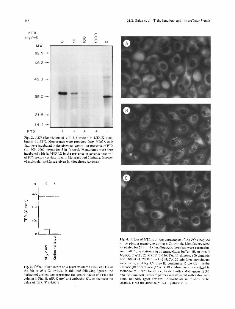
ADP-ribosylation of proteins in cell membranes was determined by measuring the 32p-ADP-ribose incorporation catalyzed by per- tussis toxin in vitro. MDCK cells incubated in the presence (10, 100, 1000 ng/ml) and absence of PTX for 5 hr were washed and suspended in ice-cold buffer consisting of 25 mM Tris HCI, pH 7,4; 1 mM EDTA; 1 mM dithiothreitol; and 10 p.g/ml aprotinin. Cellular homogenates were prepared with an homogenizer (10 strokes) and pelleted by centrifugation at 100,000 • g for 30 min (Portilla, Morrissey & Morrison, 1988).
The pellet was resuspended in the same buffer, and the protein concentration was measured by the method of Lowry et al. (1953). An aliquot of 200/xg of protein was incubated in a final volume of 100/xl at 30~ for 60 min in buffer containing 250 mM potassium phosphate, pH 7.5; 5 mM MgC]2; 2 mM ATP; 0.l mM GTP; 20 mya thymidine; 10 mM arginine; 0.75 mM NADP+; 10 /*Ci cz-32p - NAD and 10/xg of PTX. PTX was preactivated with 20 mM dithiothreitol at 37~ for 10 rain. A control in the absence of toxin was run in parallel. The reaction was terminated with 1 ml of cold potassium phosphate buffer, 100/zI of 50% TCA and 100 /*1 of 0.15% deoxycholate. The protein was precipitated, collected by centrifugation, resuspended in 100 /xl of urea/ Laemmli, and 5% of/3-mercaptoethanol was added before boiling for 5 rain at 100~ (Kawai, Whitset & Arinze, 1986; Hern~ndez- Sotomayor et al., 1990). The protein was separated by molecular mass on a 10% polyacrylamide SDS-containing gel by the method of Laemmli (1970). The gel was then stained, destained, dried and autoradiographed.
Z O - I INDIRECT IMMUNOFLUORESCENCE
Glass coversfips containing confluent monolayers of MDCK cells were rinsed twice with PBS, fixed and permeabilized with - 20~ methanol for 45 sec. Monolayers were washed with PBS, incu- bated with 3% fetal bovine serum in PBS for 30 min, and treated overnight at 4~ with ZO-I monoclonal antibody (R26.4C8C9C8, Stevenson et at., 1986, 1988) diluted l :50 in 1% fetal bovine
M.S. Balda et al.: Tight Junctions and lntracellular Signals 195
serum-PBS. Cells were washed twice with PBS, incubated 30 rain with 3% fetal bovine serum in PBS, stained with rhodamine conjugated rabbit anti-rat antibody (Sigma) for 60 min and washed twice with PBS. The glass coverslips were then mounted in p- phenyldiamine-glycerol (1:9) and examined with a Leitz Or- thoplan microscope (Leitz, Wetzlar, Germany).
ACTIN INDIRECT FLUORESCENCE
Confluent monolayers of MDCK cells on glass coverslips were rinsed twice with PBS, fixed with 1% formaldehyde, washed twice with PBS, treated with - 20~ acetone for 5 min and dried with air. Monolayers were incubated for 60 min with a rhodamine phalloidin solution (1:20 in PBS). Glass coverslips were washed twice with PBS and mounted in p - phenyldiamine-glycerol (I : 9) and viewed in a Leitz Orthoplan microscope (Leitz, Weitzlar, Germany).
Results are expressed as mean -+ sE (number of observa- tions).
Results
To facilitate comparisons between the effects of the different substances tested, the value of TER re- ported was normalized as follows:
TER = TER~ (X~II/X ~)
where TER~, is the value of TER measured in a given experimental monolayer, X~ is the mean value of the control group processed simultaneously, and X~n is the mean value of TERs measured in all control groups. Unless otherwise stated, TER was mea- sured before and 5 hr after switching the monolayers from l-4 /xM to 1.8 mg Ca 2".
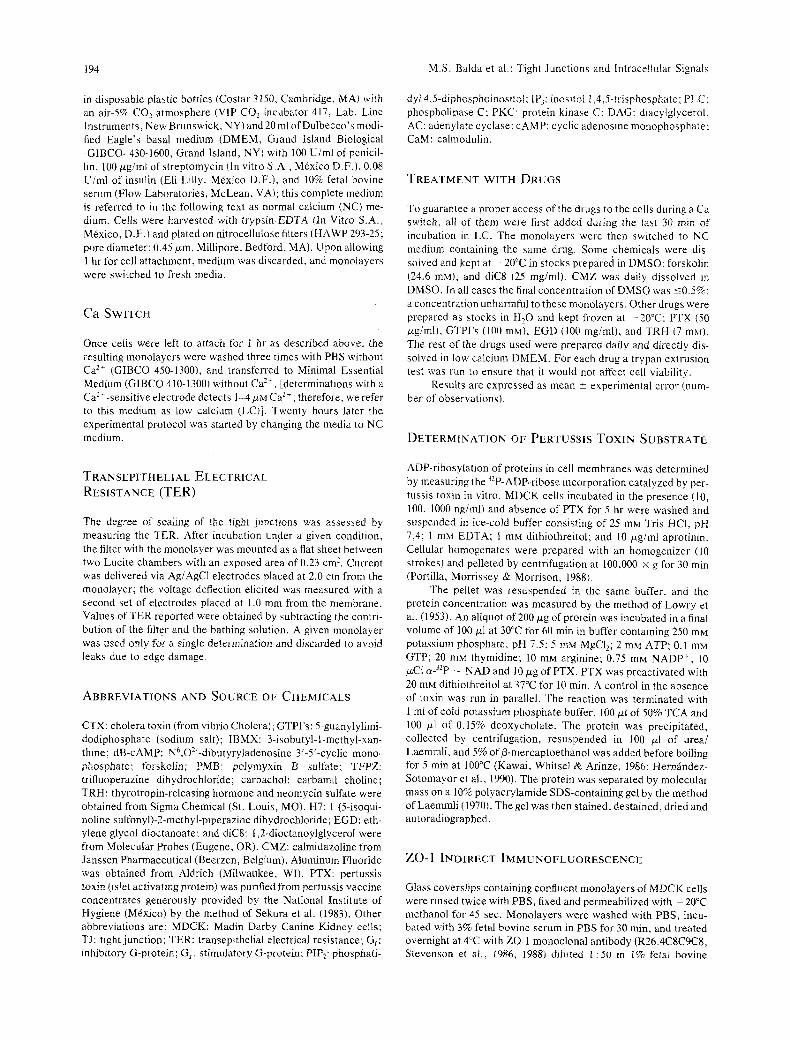
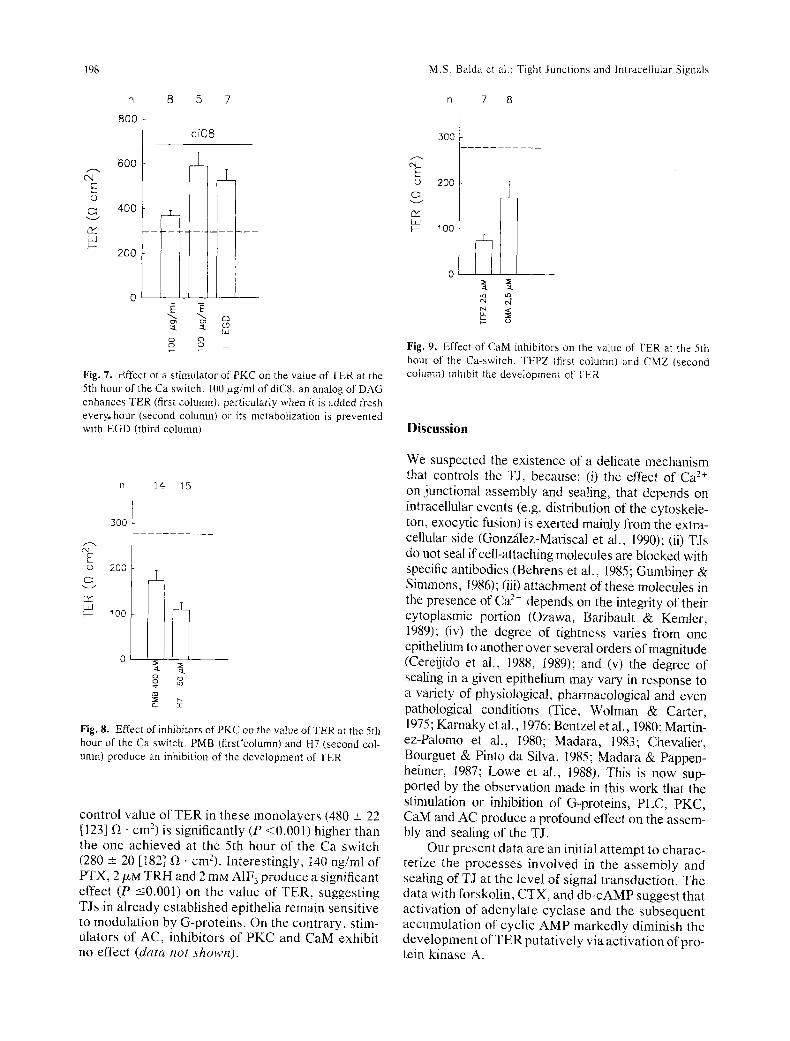
Figure 1 shows that before the Ca switch, mono- layers have a TER of 21 -+ 7 (21) f/ �9 cm 2 (first column); if they are not switched to Ca 2+ 5 hr later their TER remains low at 11 -+ 3 (15) (second col- umn), but if they are switched they develop a TER of 280 -+ 20 (182) (third column). PTX (14 ng" ml-~), which is known to inhibit the action of certain G- proteins (Murayama & Ui, 1984; Garcia-S~iinz, 1985), increases the level of TER recorded at the 5th hour by up to 511% (P -< 0.001; 4th column). However, the-action of pertussis toxin by itself can- not onset the sealing of TJs, because if the concen- tration of Ca 2+ is kept at 1-4/xM, TER remains low (last column).
To ensure that at the concentration used PTX was acting on G-proteins in MDCK cells, we mea- sured the degree of ADP-ribosylation of cell homog- enates pretreated with the toxin. This was performed in the presence of [c~-32p]-NAD. Figure 2 shows that, in fact, PTX reduces in a dose-dependent manner the degree of labeling of a 4l-kD protein. Densitometric
n 21 ~5 182 8 8
E �9
{32 Ld
1500
1000
500
_L
d
[ I r x " ~ + x ~ g 8 E ~ E c)
Fig. 1. Effect of PTX on the value of TER at the 5th hr of the Ca switch. MDCK monolayers were plated at confluence on Milli- poke filters in CDMEM and transferred to Ca-free medium 1 hr later. At the 20th hour of incubation in Ca-free medium mono- layers have a negligible TER (first column). Renewal of Ca-free medium for another 5 hr does not modify TER (second column), but switching to a medium with 1.8 mM Ca > triggers the assembly and sealing of TJs, that confer a TER of 280 +_ 20 fZ �9 cmz in 5 hr (third column). PTX added 30 rain before and during the Ca switch markedly enhances TER. PTX cannot increase TER by itself, as the addition of 14 ng/ml without Ca 2~, fails to seal the junctions (last column)
analysis reveals that, at the concentration used in Fig. 1 (14 ng/ml), pretreatment with PTX produces a 73% inhibition of ADP-ribosylation of the 41 kDa substrate(s) produced by the second PTX exposure.
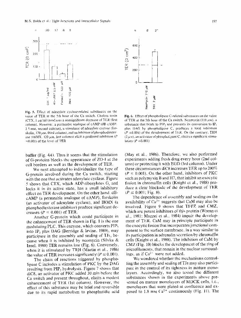
On the basis of results in Fig. 1, one would expect that stimulators of G-proteins would have an opposite action, i.e., an inhibitory influence on the development of TER. Accordingly, we tested A1F 3 (2 mM) which activates several G-proteins by mim- icking the F-phosphate group of GTP (Bigay et al., 1985; Blackmore et al., 1985), and carbachol (5/xM), which through muscarinic receptors activates some G-proteins (Nathanson, 1987). Both substances de- crease the development ofTER (P -< 0.001; Fig. 3). Finally, we used GTP-F-s, a non hydrolizable analog of GTP, to test the effect of a continuous activation of G protein(s) on the appearance of the ZO-1 pep- tide at the cell borders during a Ca-switch. Since GTPFs is nonpermeable, we treated the cells with digitonin (5/J,M, 20 min). Permeabilized monolayers switched from a LC condition to Ca 2+ containing buffer display clear ZO-I fluorescence at the cell borders (Fig. 4B), while monolayers transferred to Ca 2+ containing buffer in the presence of GTPFs do not (Fig. 4C). The latter resemble those left in LC
196 M.S. Balda et al.: Tight Junctions and Intracellular Signals
Fig. 2. ADP-ribosylation of a 41-kD protein in MDCK mem- branes by PTX. Membranes were prepared from MDCK cells that were incubated in the absence (control) or presence of PTX (10, 100, 1000 ng/ml) for 5 hr (above). Membranes were then incubated with [~J2P]NAD in the presence or absence (control) of PTX (below) as described in Materials and Methods. Markers of molecular weight are given in kilodaltons (arrows)
n 9 6
300
E ,o 200
c~ v
n~ D.J I.-- 100
T
0 I [ , . ,
{"4 0
Fig. 3. Effect of activators of G-proteins on the value of TER at the 5th hr of a Ca switch. In this and following figures, the horizontal dashed line represents the control value of TER (3rd column in Fig. 1). AIF3 (2 raM) and carbachol (5 p.M) decrease the value of TER (P -<0.001)
Fig. 4. Effect of GTPFs on theappearance of the ZO-I peptide at the plasma membrane during a Ca switch. Monolayers were incubated for 20 hr in LC medium (A), then they were permeabil- ized with 5 /xM digitonin in an intracellular buffer (IB, in raM: 2 MgSO4, 2 ATP, 20 PIPES, 0.4 EGTA, 10 glucose, t00 glutamic acid, 100KOH, 29 KCI and 16 NaCI). 20 min later monolayers were transferred for 3.5 br to IB containing 15 p.M Ca 2+ in the absence (B) or presence (C) of GTPFs. Monolayers were fixed in methanol at -20~ for 20 sec, treated with a Mab against ZO-1 and the immunofluorescent pattern was detected with a rhodami- hated antibody (goat anti-rat). Arrowheads in B show ZO-I strands. Note the absence of ZO-I pattern in C
M.S. Balda et aI.: Tight Junctions and lntracellular Signals
n 20 17 8 8 n 8 21
197
c'4 E r
Q:2 tad
300
2_ 200
2~
E
k = r na
Fig..5. Effect of adenylate cyclase-related substances on the value of TER at the 5th hour of the Ca switch. Cholera toxin (CTX, I/*g/ml) produces a nonsignificant decrease of TER (first column). However, a permeable analogue of cAMP (dB-cAMP, 2.5 raM. second column), a stimulator of adenylate cyclase (for- skolin, 120/*M, third column), and an inhibitor of phosphodiester- ase (1BMX, 120 NM, last column) elicit a profound inhibition (P <-0.001) of the level of TER
E O
02 Lid F-
6O0
400 ]
- - 4 - - 2~176 I' :at
o
._= :~
E
Z ~--
Fig. 6. Effect ofphospholipase C-related substances on the value of TER at the 5th hour of the Ca switch. Neomycin (110 p~M), a substance that binds to PIP 2 and prevents its conversion to IP3 plus DAG by phospholipase C, produces a total inhibition (P <0.001) of the development of TER. On the contrary, TRH (2/xM), an activator ofphospholipase C, elicits a significant stimu- lation (P ~0.001)
buffer (Fig. 4A). Thus it seems that the stimulation of G-proteins blocks the appearance of ZO-1 at the cell borders as well as the development of TER.
We next attempted to individualize the type of G-protein involved during the Ca switch, starting with the one that activates adenylate cyclase. Figure 5 shows that CTX, which ADP-ribosylates G, and locks it in its active state, has a small inhibitory effect on TER development. On the other hand, dB- cAMP (a permeable analogue of cAMP), forskolin (an activator of adenylate cyclase), and IBMX (a phosphodiesterase inhibitor) produce significant de- creases (P -< 0.001) of TER.
Another G-protein which could participate in the enhancement of TER shown in Fig. 1 is the one modulating PLC. This enzyme, which converts PIP 2 into IP 3 plus DAG (Berridge & Irvine, 1989), may participate in the assembly and sealing of TJs, be- cause when it is inhibited by neomycin (Silvka & Insel, 1988) TER remains low (Fig. 6). Conversely, when it is stimulated by TRH (Martin et al., 1986) the value of TER increases significantly (P -< 0.001).
The chain of reactions triggered by phospho- lipase C includes a stimulation of PKC by the DAG resulting from PIP2 hydrolysis. Figure 7 shows that diC8, an activator of PKC added 30 min before the Ca switch and present throughout, elicits a modest enhancement of TER (lst column). However, the effect of this substance may be brief and reversible due to its rapid metabolism to phosphatidic acid
(May et al., 1986). Therefore, we also performed experiments adding fresh drug every hour (2nd col- umn) or protecting it with EGD (3rd column). Under these circumstances diC8 increases TER up to 200% (P _< 0.001). On the other hand, inhibitors of PKC such as polymyxin B and H7, that inhibit an exocytic fusion in chromaffin cells (Knight et al., 1988) pro- duce a clear blockade of the development of TER (P _< 0.001; Fig. 8).
The dependence of assembly and sealing on the availability of Ca ?+ suggests that CaM may also be involved. Figure 9 shows that TFPZ and CMZ, which are potent inhibitors of this protein (Wrenn et al., 1981; Mazzei et al., 1984) impair the develop- ment of TER. CaM may in principle participate in the exocytic fusion that incorporates junctional com- ponent to the surface membrane, in a way similar to its participation in adrenalin secretion by chromaffin cells (Knight et al., 1988). The inhibition of CaM by CMZ (Fig. 10) blocks the development of the ring of microfilaments, that remain in the nuclear surround- ings, as if Ca 2+ were not added.
We wondered whether the mechanisms control- ling the assembly and sealing of TJs may also partici- pate in the control of its tightness in mature mono- layers. Accordingly, we also tested the different substances shown in the experiments above pre- sented on mature monolayers of MDCK cells, i.e., monolayers that were plated at confluence and ex- posed to 1.8 mM Ca 2§ continuously (Fig. 11). The
198
C'q E
LaJ I--
n 8 5 7
800 d i C 8
600 l 2_
400
200 - ~
0
g. g. c~ Ld
g ~ o +
Fig. 7. Effect of a s t imulator of PKC on the value of TER at the 5th hour of the Ca switch. 100/xg/ml of diC8, an analog of DAG enhances TER (first column), particularly when it is added fresh every, hour (second column) or its metabolization is prevented with EGD (third column)
n 14 15
300
L--.
(o 200
02 Lid }-- 100
::L :::L o o
=_ ?:
Fig. 8. Effect of inhibitors of PKC on the value o f T E R at the 5th hour of the Ca switch. PMB (first 'column) and H7 (second col- umn) produce an inhibition of the development of TER
control value of TER in these monolayers (480 _+ 22 [123] ~ �9 cm 2) is significantly (P <0.001) higher than the one achieved at the 5th hour of the Ca switch (280 -+- 20 [182] f~ �9 cruZ). Interestingly, t40 ng/ml of PTX, 2/*M TRH and 2 mM A1F 3 produce a significant effect (P -<0.001) on the value of TER, suggesting TJs in already established epithelia remain sensitive to modulation by G-proteins. On the contrary, stim- ulators of AC, inhibitors of PKC and CaM exhibit no effect (data not shown).
M.S. Balda et al.: Tight Junct ions and Intracellular Signals
n 7 8
300 a---. E o 200
rY L O ~- 100
at ~k
o
Fig. 9. Effect of CaM inhibitors on the value of TER at the 5th hour of the Ca-switch. TFPZ (first column) and CMZ (second column) inhibit the development of TER
Discussion
We suspected the existence of a delicate mechanism that controls the TJ, because: (i) the effect of Ca 2+ on junctional assembly and sealing, that depends on intracellular events (e.g. distribution of the cytoskele- ton, exocytic fusion) is exerted mainly from the extra- cellular side (Gonzalez-Mariscal et al., 1990); (ii) TJs do not seal if cell-attaching molecules are blocked with specific antibodies (Behrens et al., 1985; Gumbiner & Simmons, 1986); (iii) attachment of these molecules in the presence of Ca 2+ depends on the integrity of their cytoplasmic portion (Ozawa, Baribault & Kemler, 1989); (iv) the degree of tightness varies from one epithelium to another over several orders of magnitude (Cereijido et al., 1988, 1989); and (v) the degree of sealing in a given epithelium may vary in response to a variety of physiological, pharmacological and even pathological conditions (Tice, Wolman & Carter, 1975; Karnaky et al., 1976; Bentzel et al., 1980; Martin- ez-Palomo et at., t980; Madara, 1983; Chevalier, Bourguet & Pinto da Silva, 1985; Madara & Pappen- heimer, 1987; Lowe et al., 1988). This is now sup- ported by the observation made in this work that the stimulation or inhibition of G-proteins, PLC, PKC, CaM and AC produce a profound effect on the assem- bly and sealing of the TJ.
Our present data are an initial attempt to charac- terize the processes involved in the assembly and sealing of TJ at the level of signal transduction. The data with forskolin, CTX, and db-cAMP suggest that activation of adenylate cyclase and the subsequent accumulation of cyclic AMP markedly diminish the development of TER putatively via activation of pro- tein kinase A.
M.S. Balda et al.: Tight Junctions and lntracellular Signals 199
n 12,3
go0
2 5 9 6
E (D
Ld
600
3OO !
i
0
o~ 2g U_
�9 ~_ LL. r v
Fig. 11. Effect of different subs tances on the value of TER of mature monolayer s. M D C K cells were plated at confluence and incubated for 20 hr in C D M E M with 1.8 mM Ca'--. Drugs were added at this time and remained present throughout . T E R was recorded 5 hr later. PTX and TRH increase and A1Fs decreases the TER
Fig. 10. Effect of CaM inhibition on the pattern of actin fila- ments in monolayers of M DC K cells. M DCK cells were incu- bated for 20 hr in LC medium (A), transferred for 5 hr to NC medium in the absence (B) or presence of 2.5/xM CMZ (C). Indirect fluorescence staining of actin filament with phaloidinerhodamine was performed as described in Materials and Methods
On the contrary, the use of synthetic diacylglyc- erol, inhibitors of PKC and agonists, such as TRH thai stimulate PKC via receptor-mediated activation of phosphoinositide turnover, are consistent with the view that activation of PKC enhances the
development of TER. Thus, it seems that PKC and protein kinase A reciprocally regulate TER devel- opment.
It is well known that petussis toxin ADP-ribo- sylates G i and blocks receptor-mediated inhibition of adenylate cyclase (Garcia-S~tinz, 1985; Enjalbert et al., 1986; Linden & Delahunty, 1989; Bizzari et al., 1990). It seems unlikely that the increase in TER development induced by the toxin could be ex- plained by this action. However, other G-proteins are also substrates of PTX. It has been suggested that some receptors inhibit PLC via a PTX- sensitive G-protein, termed Gpi (Linden & Delahunty, 1989; Bizzarri et al., 1990). It is possible therefore, that PTX may exert its effect on TER development by blocking a constraint on PLC ex- erted by Gpi.
Although clearly speculative, we would like to suggest the following sequence of events in the func- tional assembly and sealing of TJs during a Ca switch (Fig. 12): (i) Ca 2+ activates uvomorulin (UV) and other Ca-dependent cell adhesion molecules, which are not located at the TJ itself, but at the zonula adherens (Boller, Vestweber & Kemler, 1985; Gum- biner, Stevenson & Grimaldi, 1988). G-proteins may in principle transduce signals from ceil-cell contacts to PLC. (ii) PLC produces DAG and IP 3, which in turn releases Ca 2+ from intracellular reservoirs (Pidikiti et al., 1985). Intracellular Ca 2+ and DAG
200 M.S. Balda et al.: Tight Junctions and Intracellular Signals
PiP~
PARACELLULAR ~ATHWAY
�9 , , , ~ i
A B C
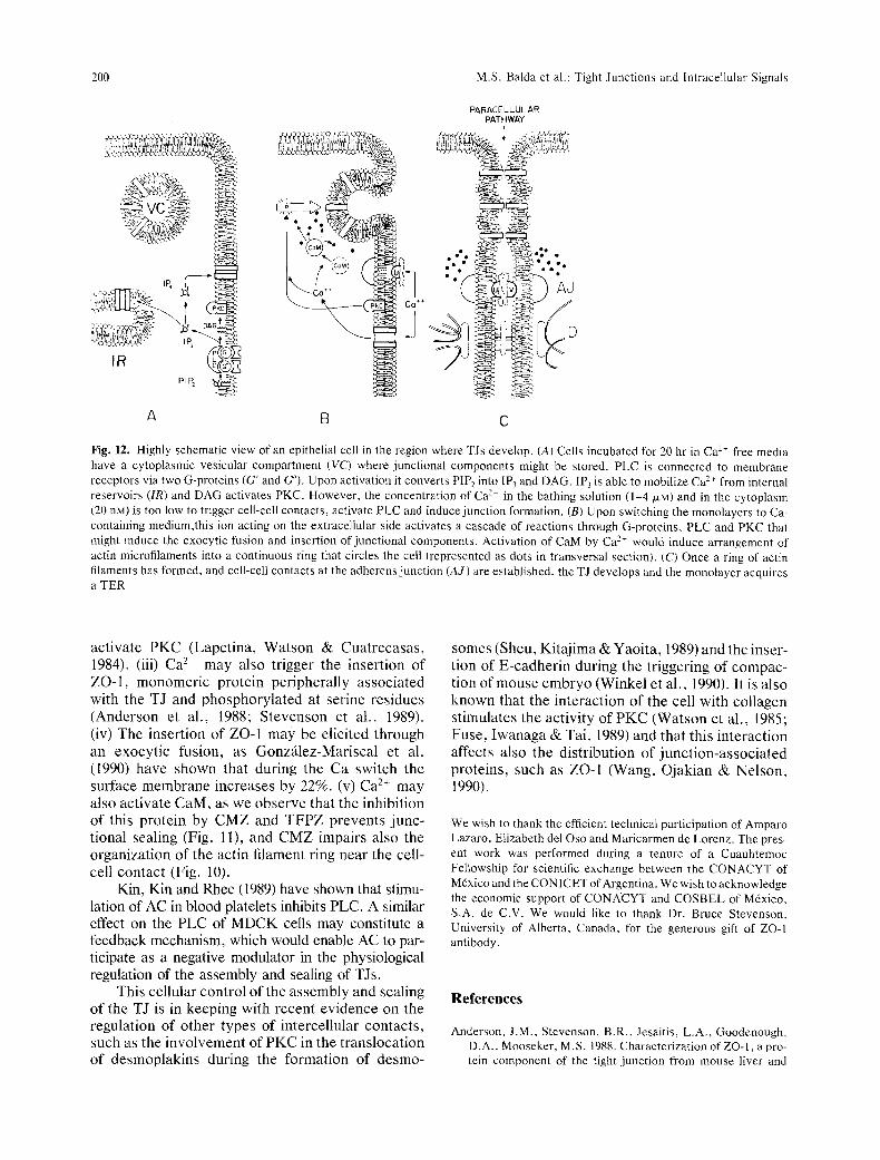
Fig. 12. Highly schematic view of an epithelial cell in the region where TJs develop. (A) Cells incubated for 20 hr in Ca 2~ free media have a cytoplasmic vesicular compartment (VC) where junctional components might be stored. PLC is connected to membrane receptors via two G-proteins (G' and G"). Upon activation it converts PIP 2 into IP3 and DAG. IP 3 is able to mobilize Ca 2" from internal reservoirs (IR) and DAG activates PKC. However, the concentration of Ca 2* in the bathing solution (1-4 ~xM) and in the cytoplasm (20 riM) is too low to trigger cell-cell contacts, activate PLC and induce junction formation. (B) Upon switching the monolayers to Ca- containing medium,this ion acting on the extracellular side activates a cascade of reactions through G-proteins, PLC and PKC that might induce the exocytic fusion and insertion of junctional components. Activation of CaM by Ca-'- would induce arrangement of actin microfilaments into a continuous ring that circles the cell (represented as dots in transversal section). (C) Once a ring of actin filaments has formed, and cell-cell contacts at the adherens junction (A J) are established, the TJ develops and the monolayer acquires a TER
activate PKC (Lapetina, Watson & Cuatrecasas, 1984). (iii) Ca 2. may also trigger the insertion of ZO-1, monomeric protein peripherally associated with the TJ and phosphorylated at serine residues (Anderson et al., 1988; Stevenson et al., 1989). (iv) The insertion of ZO-1 may be elicited through an exocytic fusion, as Gonz~_lez-Mariscal et al. (1990) have shown that during the Ca switch the surface membrane increases by 22%. (v) Ca 2+ may also activate CaM, as we observe that the inhibition of this protein by CMZ and TFPZ prevents junc- tional sealing (Fig. ll), and CMZ impairs also the organization of the actin filament ring near the cell- cell contact (Fig. 10).
Kin, Kin and Rhee (1989) have shown that stimu- lation of AC in blood platelets inhibits PLC. A similar effect on the PLC of MDCK cells may constitute a feedback mechanism, which would enable AC to par- ticipate as a negative modulator in the physiological regulation of the assembly and sealing of TJs.
This cellular control of the assembly and sealing of the TJ is in keeping with recent evidence on the regulation of other types of intercellular contacts, such as the involvement of PKC in the translocation of desmoplakins during the formation of desmo-
somes (Sheu, Kitajima & Yaoita, 1989) and the inser- tion of E-cadherin during the triggering of compac- tion of mouse embryo (Winkel et al., 1990). It is also known that the interaction of the cell with collagen stimulates the activity of PKC (Watson et al., 1985; Fuse, Iwanaga & Tai, 1989) and that this interaction affects also the distribution of junction-associated proteins, such as ZO-I (Wang, Ojakian & Nelson, 1990).
We wish to thank the efficient technical participation of Amparo Lazaro, Elizabeth del Oso and Maricarmen de Lorenz. The pres- ent work was performed during a tenure of a Cuauhtemoc Fellowship for scientific exchange between ~he CONACYT of M6xico and the CONICET of Argentina. We wish to acknowledge the economic support of CONACYT and COSBEL of M6xico, S.A. de C.V. We would like to thank Dr. Bruce Stevenson, University of Alberta, Canada, for the generous gift of ZO-I antibody.
References
Anderson, J.M., Stevenson, B.R., Jesaitis, L.A., Goodenough, D.A., Mooseker, M.S. 1988. Characterization of ZO-1, a pro- tein component of the tight junction from mouse liver and
M.S. Balda et aI.: Tight Junctions and lntracellular Signals 201
Madin-Darby Canine kidney cells. J. Cell Biol. 106:1141- 1149
Behrens, J., Birchmeier, W., Goodman. S.L., Imhof, B.A. 1985. Dissociation of Madin-Darby Canine Kidney epithelial Cells by the monoclonal antibody anti-arc-l: Mechanistic aspects and identification of the antigen as a component related to uvomorulin. J. Cell Biol. 10l:1307-1315
Bentzel, C.J.. Hainau, B., Ho, S., Huits, W., Edelman, A.. Anagnostopoulos, T., Benedetti, E.L. 1980. Cytoplasmic reg- ulation of tight-junction permeability: Effect of plant cytoki- nins. Am. J. Physiol. 239:C75-C82
Berridge, M.J., lrvine, R.F. 1989. lnositol phosphates and cell signalling. Nature 341:197-205
Bigay, J., Deterre, P., Pfister, C., Chabre, M. 1985. Fluoroalumi- nates activate transducin-GDP by mimicking the y-phosphate of GTP in its binding site. FEBS Lett. 191:181-185
Bizzari, C., Di Girolamo, M., D'Orazio, M.C., Corda, D. 1990. Evidence that guanine nucleotide-binding proteins linked to a muscarinic receptor inhibits directly phospholipase C. Proe. Natl. Acad. Sci. USA 87:4889-4893
Blackmore, P.F., Bocckino, S.B., Waynick, L.E., Exton, J.H. 1985. Role of guanine nucleotide binding regulatory protein in the hydrolysis of hepatocyte phosphatidyfinositol 4,5-biphos- phate by calcium-mobilizing hormones and the control of cell calcium: Studies utilizing atuminium fluoride. J. Biol. Chem. 260" 14477-14483
Boiler, K., Vestweber, D., Kemler, R. 1985. Cell-adhesion mole- cule uvomorulin is localized in the intramediate junctions of adult intestinal epithelial cells. J. Cell Biol. 100:327-332
Cereijido, M:, Gonzalez-Mariscal, L., Avila, G , Contreras, R.G. 1988. Tight junctions. Crit. Rev. Anat. Sci. 1:171-192
Cereijido, M., Gonzalez-Mariscal, L., Contreras, R.G. 1989. Tight junction: The barrier between higher organisms and environment. News.Physiol. Sci. 4:72-74
Cereijido, M., Robbins, E.S., Dolan, W.J., Rotunno, C.A., Sa- batini, D,D. 1978a. Polarized monolayers formed by epithelial cells on a permeable and translucent support. J. Cell Biol. 77:853-880
Cereijido, M., Rotunno, C.A., Robbins, E.S., Sabatini. D.D. 1978b. Polarized epithelial membranes produced in vitro. In: Membrane Transport Processes. J.F. Hoffman, editor. Ra- ven, New York
Chevalier, J., Bourguet, J., Pinto da Silva, P. 1985. Osmotic gradient reversal induces massive assembly of tight junction strands at the basal pole of toad bladder epithelial cells. 1NSERM (Paris) 67:28
Enjalbert, A., Sladeczek, F., Gillon, G., Bertrand, P., Shu, C., Epelbaum, J,, Garcfa-SS_inz, J.A., Jard, S., Lombard, C., Kordon, C., Bockaert, J. 1986. Angiotensin II and dopamine modulate both cAMP and inositol phosphate production in anterior pituitary cells. Involvement in prolactin secretion. J. Biol. Chem. 261:4071-4075
Fuse, I , lwanaga, T., Tai, H.-H. 1989. Phorbol ester, 1,2-diacyl- glycerol, and collagen induce inhibition of arachidonic acid incorporation into phospholipids in human platelets. J. Biol. Chem. 264:3890-3895
Garcfa-Sfiinz, J.A. 1985. Effect of pertussis toxin on the hormonal responsiveness of different tissues. In: Pertussis Toxin. R.D. Sekura, J. Moss and M. Vaugham, editors. Academic, New York
Gonz~dez-Mariscal, L., Chavez de Ramirez, B., Cereijido, M. 1985. Tight junction formation in cultured epithelial cells (MDCK). J. Membrane Biol. 86:113-125
GonzNez-Mariscal, L., Contreras, R.G., Bolivar, J.J., Ponce,
A., Chavez de Ramirez, B., Cereijido, M. 1990. The role of Ca -" in tight junction formation between epithelial cell (MDCK). Am. J. Physiol. 259:C978-C986
Gumbiner, B., Simmons, K. 1986. A functional assay for proteins involved in establishing of epithelial occluding barriers: Identi- fication of a uvomorulin-like peptide. J. Cell Biol. 102:457-468
Gumbiner, B., Stevenson, B., Grimaldi, A. t988. The role of the cell adhesion molecule uvomorulin in the formation and maintenance of the epithelial junctional complex. J. Cell Biol. 107:1575-1588
Hern~indez-Sotomayor, S.M.T., Macfas-Silva, M., Malbon, C.C., Garcfa Sfiinz, J.A. 1990. Modulation G, activity by phorbol myristate acetate in rat hepatocytes. Am. J. Physiol. (in press)
Karnaky, K.J., Kinter, L.B,, Kinter, W.B., Sterling, C.E. 1976. Teleost chloride cell. II. Autoradiographic localization of gill Na, K-ATPase in killfish Fundus heteroelitus adapted to low and high salinity environments. J. Cell Biol. 70:157-165
Kawai, Y., Whitsel, C., Arinze, I.J. 1986. NADP + enhanced cholera and pertussis toxin catalyzed ADP-ribosylation of membrane proteins. J. Cyclic Nucleotide Protein Phospho�9 Res. 11:265-274
Kin, U.H., Kin, J.W., Rhee, S.G. 1989. Phosphorylation ofphos- pholipase C by protein kinase A. J. Cell Biol. 109:50A
Knight, D.E., Sugden, D., Baker, P.F. 1988. Evidence implicat- ing protein kinase C in exocytosis from electropermeabilized bovine chromaffin cells. J. Membrane Biol. 104:21-34
Laemmli, U.K. 1970. Cleavage of structural proteins during the assembly of the head of bacteriophage T4. Nature 227:680-685
Lapetina, E.G., Watson, S.P., Cuatrecasas, P. 1984. Myo-lnosi- tol, 1,4,5-trisphosphate stimulates protein phosphorylation in saponin-permeabilized human platelets. Proc. Natl. Acad. Sci. USA 81:7431-7435
Linden, J., Delahunty, T.M. 1989. Receptors that inhibit phos- phoinositide breakdown. Trends Pharmacol. Sci. 10:114-120
Lowe, P.J., Miyai, K., Steiubach, J.H., Hardison, G.M. 1988. Hormonal regulation of hepatocyte tight junctional permeabil- ity. Am. J. Physiol. 255:G454-G461
Lowry, O.H., Rosebrough, N.J.. Farr, L.A., Randall, R.J. 1953. Protein measurement with Folin phenol reagent. J. Biol. Chem. 193:265-272
Madara, J.L. 1983. increases in guinea pig small intestinal trans- epithelial resistance induced by osmotic loads are accompa- nied by rapid alterations in absorptive-cell tight-junction struc- ture. J. Cell Biol. 97:125-135
Madara, J.L., Pappenheimer, J.R. 1987. Structural basis for phys- iological regulation of paracellular pathways in intestinal epi- thelia. J. Membrane Biol. 100:149-164
Martin, T.F.J., Lucas, D.O., Bajjaleih, S.M., Kowatchyk, J.A. 1986. Thyrotropin releasing hormone activates a Ca -" depen- dent polyphosphoinositide phosphodiesterase and pertussis toxin insensitive mechanism. J. Biol. Chem. 261:2918- 2927
Martinez-Palomo, A., Meza, I., Beaty, G., Cereijido, M. 1980. Experimental modulation of occluding junctions in a cultured transporting epithelium. J. Cell Biol. 87:736-745
May, W.S., Lapetina, E.G., Cuatrecasas, P. 1986. Intracellular activation of protein kinace C and regulation of surface trans- ferrin receptor by diacylglycerol is a spontaneous reversible process that is associated with rapid formation ofphosphatidic acid. Proc. Natl. Acad. Sci. USA 83:1281-1284
Mazzei, G.J., Schatzman, R.C., Scott-Turner, R., Vogler, W.R., Kuo, J.F. 1984. Phospholipid-sensitive CaZ+-dependent pro-

202
tein kinase inhibition by R-24571, a ca[modulin antagonist. Biochem. Pharmacol. 33:125-130
Murayama, T., Ui, M. 1984. [3H]GDP fi~lease from rat and ham- ster adipocyte membranes independen:tly linked to receptors involved in activation or inhibition ~of adenylate cyclase. Dif- ferential susceptibility to two toxins. J. Biol. Chem. 259:761-769
Nathanson, N.M. 1987. Molecular properties of the muscarinic acetylcholine receptor. Annu. Reu. Neurosci. 10:195-236
Ozawa, M., Baribault, H,, Kemler, R. 1989. The cytoplasmic domain of the cell adhesion molecule uvomorulin associated with three independent proteins structurally related in differ- ent species. EMBO J. 8:1711-1717
Pidikiti, N., Gamero, D., Gamero, J., Hassid, A. 1985. Bradyki- nin-evoked modulation of cytosolic Ca +- cormentration in cultured renal epithelial (MDCK)cells. Biochem. Biophys. Res. Commun. 130:807-813
Portilla, D., Morrissey, J., Morrison, A.R. ,1988. Bradykinin acti- vated membrane associated phospholipase C in Madin-Darby Canine Kidney cells. J. Clin. Invest. 81:1896-1902
Sekura, R.D., Fish, F., Manclark, C.R., Meade, B., Zhang, Y.-L. 1983. Pertussis toxin: Affinity purification of a new ADP-ryboxyltransferase. J. Biol. Chem. 258:14647-14651
Sheu, H., Kitajima, Y., Yaoita, H. 1989. Involvement of protein ki, nase C in translocation of desmoplakins from cytosol to plasma membrane during desmosome formation in human squamous cell carcinoma cells grown in low normal calcium concentration. Exp. Cell Res. 185:176-190
Slivka, S.R., Insel, P.A. 1988. Phorbol ester and neomycin disso- ciate bradykinin receptor-mediated arachidonic acid release and polyphosphoinositide hydrolysis in Madin-Darby canine kidney cells. J. Biol. Chem. 263:14640-14647
M.S. Balda et al.: Tight Junctions and lntracellular Signals
Stevenson, B.R., Anderson, J.M., Braun, I.D., Mooseker, M.S. 1989. Phosphorylation of the tight-junction protein ZO-I in two strains of Madin-Darby canine kidney cells which differ in transepithelial resistance. Biochem. J. 263:597-599
Stevenson, B.R., Anderson, J.M., Bullivant, S. 1988. The epithe- lial tight junction: Structure, Function and preliminary bio- chemical characterization. Mol. Cell. Biochem. 83:129-145
Stevenson, B.R., Sificiano, J.D., Mooseker, M.S., Goodenough, D.A. 1986. Identification of ZO-I: A high molecular weight polypeptide associated with the tight junction (zonula oc- cludens) in a variety of epithelia. J. Cell Biol. 103:755-766
Yice, L.W., Wolman, S.H., Carter, R.C. 1975. Changes in tight junctions of thyroid epithelium with changes in thyroid activ- ity. J. Cell Biol. 66:657-666
Wang, A.Z., Ojakian, G.K., Nelson, J. 1990. Steps inthe morpho- genesis of a polarized epithelium: 11. Disassembly and assem- bly of plasma membrane domains during reversal of epithelial cell polarity in multicellular epithelial (MDCK) cysts. J. Cell Sci. 95:153-165
Watson, S.P., Reep, B., McConnel, R.T.. Lapetina, E.G. 1985. Collagen stimulates [3H]inositol triphosphate formation in in- domethacin-treated human platetets. Biochem. J. 226:831-837
Winkel, G.K., Ferguson, J.E., Takeichi, M., Nuccitelli, S. 1990. Activation of protein kinase C triggers premature compaction in the four-cell stage mouse embryon. Dev. Biol. 138:1-15
Wrenn, B.W., Katoh, N,, Schatzman, C., Kuo, J.F. 1981. Inhibi- tion by phenothiazine antipsychotic drugs of calcium-depen- dent phosphorylation of cerebral cortex proteins regulated by phosphofipid or calmodulin. Life Sci. 29:725-733
Received 4 February 1991