Essential role of Ca2+/calmodulin in Early Endosome Antigen1 localization
11
Molecular Biology of the Cell Vol. 14, 2935–2945, July 2003 Essential role of Ca 2 /Calmodulin in Early Endosome Antigen-1 Localization □ V Deirdre C. Lawe,* Nachida Sitouah,* Susan Hayes, † Anil Chawla,* ‡ Joseph V. Virbasius,* ‡ Richard Tuft, § Kevin Fogarty, § Lawrence Lifshitz, § David Lambright,* †‡ and Silvia Corvera* † *Program in Molecular Medicine, † Interdisciplinary Graduate Program, ‡ Departments of Biochemistry and Molecular Pharmacology, and § Physiology, University of Massachusetts Medical School, Worcester, Massachusetts 10615 Submitted September 14, 2002; Revised February 14, 2003; Accepted February 15, 2003 Monitoring Editor: Keith Mostov Ca 2 is an essential requirement in membrane fusion, acting through binding proteins such as calmodulin (CaM). Ca 2 /CaM is required for early endosome fusion in vitro, however, the molecular basis for this requirement is unknown. An additional requirement for endosome fusion is the protein Early Endosome Antigen 1 (EEA1), and its recruitment to the endosome depends on phosphatidylinositol 3-phosphate [PI(3)P] and the Rab5 GTPase. Herein, we demonstrate that inhibition of Ca 2 /CaM, by using either chemical inhibitors or specific antibodies directed to CaM, results in a profound inhibition of EEA1 binding to endosomal membranes both in live cells and in vitro. The concentration of Ca 2 /CaM inhibitors required for a full dissociation of EEA1 from endosomal membranes had no effect on the activity of phosphatidylinositol 3-kinases or on endogenous levels of PI(3)P. However, the interaction of EEA1 with liposomes containing PI(3)P was decreased by Ca 2 /CaM inhibitors. Thus, Ca 2 /CaM seems to be required for the stable interaction of EEA1 with endosomal PI(3)P, perhaps by directly or indirectly stabilizing the quaternary organization of the C-terminal FYVE domain of EEA1. This requirement is likely to underlie at least in part the essential role of Ca 2 /CaM in endosome fusion. INTRODUCTION It has been widely recognized that membrane fusion in the exocytic and endocytic pathways in cells is a Ca 2 -depen- dent process (Hutton, 1986; Chamberlain et al., 1995; Em- manouilidou et al., 1999; Pryor et al., 2000; Chen et al., 2001), yet the specific calcium sensors that operate at each step have not been fully identified. In yeast, calmodulin (CaM) has been identified as a Ca 2 sensor required in the post- docking phase of vacuole fusion (Peters et al., 1998). Studies in mammalian cells have shown that endosome fusion in vitro requires calcium, and it has been hypothesized that calmodulin is a sensor for calcium in endosome fusion. (Colombo et al., 1997; Peters and Mayer, 1998; Huber et al., 2000; Mills et al., 2001) Evidence for a Ca 2 /CaM require- ment in endosome fusion is also supported by results in live cells. For example, CaM binding proteins have been detected in endosome-enriched fractions of liver (Enrich et al., 1988) and inhibitors of CaM profoundly inhibit transcytosis and transferrin recycling in polarized epithelial cells (Apodaca et al., 1994). Moreover, it has been hypothesized that patho- gens such as Mycobacterium tuberculosis may disrupt normal phagosome progression by inhibition of Ca 2 /CaM func- tion (Russell, 2001). The target protein(s) for Ca 2 /CaM in early endocytic trafficking, however, have not been identi- fied. The early endosomal protein EEA1 is a 170-kDa coiled- coil dimer that is crucial for endosome fusion in vitro (Mills et al., 1998; Simonsen et al., 1998; Christoforidis et al., 1999; Mills et al., 1999). The association of EEA1 with endosomes and subsequent endosome fusion events are regulated by the activities of phosphatidylinositol 3-kinase (PI 3-kinase) and the Rab5 GTPase. Both the PI 3-P binding FYVE domain and regions that interact with activated Rab5 are localized to the carboxy terminus of EEA1. A second Rab5 binding site as well as a C2H2 domain are present at the amino terminus of EEA1 (Simonsen et al., 1998). In addition, a potential CaM binding IQ-like motif is located just 5 to the Rab5 interac- tion domain in the carboxy terminus of EEA1 (Mu et al., Article published online ahead of print. Mol. Biol. Cell 10.1091/ mbc.E02– 09 – 0591. Article and publication date are available at www.molbiolcell.org/cgi/doi/10.1091/mbc.E02– 09 – 0591. □ V The online version of this article contains video material for some figures. Online version is available at [email protected]. Corresponding author. E-mail address: silvia.corvera@ umass- med.edu. © 2003 by The American Society for Cell Biology 2935 http://www.molbiolcell.org/molbiolcell/suppl/2003/03/21/E02-09-0591.DC1.html Supplemental Material can be found at:
-
Upload
independent -
Category
Documents
-
view
1 -
download
0
Transcript of Essential role of Ca2+/calmodulin in Early Endosome Antigen1 localization
Molecular Biology of the CellVol. 14, 2935–2945, July 2003
Essential role of Ca2�/Calmodulin in Early EndosomeAntigen-1 Localization□V
Deirdre C. Lawe,* Nachida Sitouah,* Susan Hayes,† Anil Chawla,*‡
Joseph V. Virbasius,*‡ Richard Tuft,§ Kevin Fogarty,§ Lawrence Lifshitz,§David Lambright,*†‡ and Silvia Corvera*†�
*Program in Molecular Medicine, †Interdisciplinary Graduate Program, ‡Departments of Biochemistryand Molecular Pharmacology, and §Physiology, University of Massachusetts Medical School,Worcester, Massachusetts 10615
Submitted September 14, 2002; Revised February 14, 2003; Accepted February 15, 2003Monitoring Editor: Keith Mostov
Ca2� is an essential requirement in membrane fusion, acting through binding proteins such ascalmodulin (CaM). Ca2�/CaM is required for early endosome fusion in vitro, however, themolecular basis for this requirement is unknown. An additional requirement for endosome fusionis the protein Early Endosome Antigen 1 (EEA1), and its recruitment to the endosome depends onphosphatidylinositol 3-phosphate [PI(3)P] and the Rab5 GTPase. Herein, we demonstrate thatinhibition of Ca2�/CaM, by using either chemical inhibitors or specific antibodies directed toCaM, results in a profound inhibition of EEA1 binding to endosomal membranes both in live cellsand in vitro. The concentration of Ca2�/CaM inhibitors required for a full dissociation of EEA1from endosomal membranes had no effect on the activity of phosphatidylinositol 3-kinases or onendogenous levels of PI(3)P. However, the interaction of EEA1 with liposomes containing PI(3)Pwas decreased by Ca2�/CaM inhibitors. Thus, Ca2�/CaM seems to be required for the stableinteraction of EEA1 with endosomal PI(3)P, perhaps by directly or indirectly stabilizing thequaternary organization of the C-terminal FYVE domain of EEA1. This requirement is likely tounderlie at least in part the essential role of Ca2�/CaM in endosome fusion.
INTRODUCTION
It has been widely recognized that membrane fusion in theexocytic and endocytic pathways in cells is a Ca2�-depen-dent process (Hutton, 1986; Chamberlain et al., 1995; Em-manouilidou et al., 1999; Pryor et al., 2000; Chen et al., 2001),yet the specific calcium sensors that operate at each stephave not been fully identified. In yeast, calmodulin (CaM)has been identified as a Ca2� sensor required in the post-docking phase of vacuole fusion (Peters et al., 1998). Studiesin mammalian cells have shown that endosome fusion invitro requires calcium, and it has been hypothesized thatcalmodulin is a sensor for calcium in endosome fusion.(Colombo et al., 1997; Peters and Mayer, 1998; Huber et al.,2000; Mills et al., 2001) Evidence for a Ca2�/CaM require-ment in endosome fusion is also supported by results in live
cells. For example, CaM binding proteins have been detectedin endosome-enriched fractions of liver (Enrich et al., 1988)and inhibitors of CaM profoundly inhibit transcytosis andtransferrin recycling in polarized epithelial cells (Apodaca etal., 1994). Moreover, it has been hypothesized that patho-gens such as Mycobacterium tuberculosis may disrupt normalphagosome progression by inhibition of Ca2�/CaM func-tion (Russell, 2001). The target protein(s) for Ca2�/CaM inearly endocytic trafficking, however, have not been identi-fied.
The early endosomal protein EEA1 is a 170-kDa coiled-coil dimer that is crucial for endosome fusion in vitro (Millset al., 1998; Simonsen et al., 1998; Christoforidis et al., 1999;Mills et al., 1999). The association of EEA1 with endosomesand subsequent endosome fusion events are regulated bythe activities of phosphatidylinositol 3-kinase (PI 3-kinase)and the Rab5 GTPase. Both the PI 3-P binding FYVE domainand regions that interact with activated Rab5 are localized tothe carboxy terminus of EEA1. A second Rab5 binding siteas well as a C2H2 domain are present at the amino terminusof EEA1 (Simonsen et al., 1998). In addition, a potential CaMbinding IQ-like motif is located just 5� to the Rab5 interac-tion domain in the carboxy terminus of EEA1 (Mu et al.,
Article published online ahead of print. Mol. Biol. Cell 10.1091/mbc.E02–09–0591. Article and publication date are available atwww.molbiolcell.org/cgi/doi/10.1091/mbc.E02–09–0591.
□V The online version of this article contains video material for somefigures. Online version is available at [email protected].
� Corresponding author. E-mail address: silvia.corvera@ umass-med.edu.
© 2003 by The American Society for Cell Biology 2935
http://www.molbiolcell.org/molbiolcell/suppl/2003/03/21/E02-09-0591.DC1.htmlSupplemental Material can be found at:
1995). Recently, this IQ-like motif has been shown to bindCaM in a protein overlay assay (Mills et al., 2001).
To further examine the role of CaM and Ca2�/CaM inEEA1 function, we made use of the chemical inhibitors[N-(6-aminohexyl)-5-chloro-1-naphthalenesulfonamides]that are known to inhibit the interaction of Ca2�/CaM withprotein targets, as well as of an inhibitory monoclonal anti-body (mAb) that binds CaM and Ca2�/CaM. The effects ofthese reagents on the localization of EEA1 to endosomalmembranes in live cells and in vitro was analyzed. Ourresults reveal a crucial requirement for Ca2�/CaM for theassociation of EEA1 with endosomal membranes. To deter-mine the molecular basis for this requirement, the in vitrointeraction of CaM with EEA1 and with diverse mutants ofEEA1 was studied. Our findings reveal two separate andindependent regions of EEA1 involved in CaM bindingthrough both Ca2�-dependent and Ca2�-independent mech-anisms. These observations suggest that the Ca2�/CaM re-quirement in early endosome fusion in mammalian cells is atleast in part dependent on its regulatory interactions withEEA1.
MATERIALS AND METHODS
Materials1,2-Bis(2-aminophenoxy)ethane-N,N,N�,N�-tetraacetic acid (BAPTA), N-(6-aminohexyl)-5-chloro-1-naphthalenesulfonamide (W7), N-(6-amino-hexyl)-l-napthalenesulfonamide, and calmidazolium were purchasedfrom Calbiochem (San Diego, CA). Wortmannin was purchased fromSigma-Aldrich (St. Louis, MO). Stock solution aliquots were prepared andstored in either dimethyl sulfoxide or water at �20°C; aliquots werethawed and used immediately before each experiment. Immobilized cal-modulin was from Stratagene (San Diego, CA).
Plasmids and Recombinant ProteinsGstRab5c, RFP-Rab5Q79L,eGFP-EEA1wt, and eGFP-EEA1-1277-1411 were generated as described previously (Lawe et al., 2002).eGFP-EEA1 Q1289L/R1293G was constructed by polymerase chainreaction (PCR)-based mutagenesis of eGFP-EEA1wt and confirmedby sequencing. GFP-EEA1wt was digested with XhoI and EcoR1 todelete the amino-terminal 2498 base pairs of EEA1 and form eGFP-EEA1-835-1411. Murine myc-tagged VPS34 was cloned by reversetranscription-PCR amplification of 3T3-L1 adipocyte RNA andcloned from a 3T3-F442A adipocyte cDNA library by using methodsdescribed previously (Virbasius et al., 1996). GST-SARA-FYVE, com-prised of residues 587 to 750 of the human SARA cDNA, wasconstructed by PCR from the full-length cDNA (gift from J. Wrana,Samuel Lunenfeld Research Institute, Mount Sinai Hospital, To-ronto, Canada). Purified recombinant protein production has beendescribed previously (Lawe et al., 2002).
Cell Culture and Preparation of Cellular ExtractsCOS-7 cells were maintained in Dulbeccco’s modified Eagle’s me-dium supplemented with 10% fetal calf serum. The cells weregrown in 100-mm dishes and transfected using FuGENE (RocheDiagnostics, Indianapolis, IN). To prepare cytosolic extracts, cellswere rinsed twice and swollen by incubation (10 min) with a 10-folddilution of cytosol buffer (25 mM HEPES, pH 7.0, 125 mM potas-sium acetate, 2.5 mM magnesium acetate, 0.2 M sucrose, 1 mMdithiothreitol, 1 mM ATP, 5 mM creatine phosphate, 0.01 mg/mlcreatine phosphokinase with 10 �g/ml leupeptin, 1 mM TAME, 1mM 1,10-phenantroline) in water. Cells were then washed by addi-tion of 10 ml of cytosol buffer, aspirated, and then scrapped into theremaining buffer (500 �l) and homogenized by repeated passage (30
times) through a 27-gauge needle. The cytosol was separated fromparticulate structures by centrifugation at 200,000 � g for 15 minand used in binding assays. 3T3-L1 cells were grown in 150-mmdishes and maintained and differentiated as described previously(Patki et al., 2001). Postnuclear supernatants were prepared as fol-lows. Cells were washed twice with ice-cold phosphate-bufferedsaline (PBS) and once with cytosol buffer. The cells were thenscrapped into 2.5 ml of cytosol buffer and homogenized by repeatedpassage (7–10 times) through a 27-gauge needle. Postnuclear ex-tracts were obtained by centrifugation at 1000 � g for 5 min toremove nuclei and unbroken cells and used in in vitro membraneassociation assays.
In Vitro Membrane Association AssayAliquots (100 �l) of postnuclear extract (containing �2.5 mg/mltotal protein) were incubated with the chemical inhibitors or anti-CaM antibody as indicated in each figure legend. Cytosol andmembranes were then separated by centrifugation at 200,000 � g for15 min. The cytosol was removed, and the membrane pellets resus-pended in 100 �l of cytosol buffer. Equal aliquots of cytosol andresuspended membrane were boiled in SDS sample buffer andanalyzed by SDS-PAGE and immunoblotting with an anti-EEA1polyclonal antiserum. In some experiments, the membrane pelletobtained from untreated postnuclear supernatant was resuspended,dispensed into 100-�l aliquots, and incubated with inhibitors oranti-CaM antibody. The release of EEA1 from the membrane intothe buffer was monitored by immunoblotting of the pellet andsupernatant obtained after a subsequent high-speed centrifugation.
Binding AssaysBinding of EEA1 from 3T3 cytosolic extracts to immobilized GST-Rab5c was performed as described previously (Lawe et al., 2002). Toassess the binding of EEA1 constructs to CaM, COS-7 cells weretransfected with the indicated enhanced green fluorescent protein(eGFP)-tagged EEA1 constructs described in each experiment andcytosolic extracts prepared as described above. Cytosolic extractswere supplemented with Ca2� chelators or additional CaCl2 asindicated in each experiment. Extracts were then mixed with 25 �lof Sepharose beads or CaM-Sepharose beads previously washed incytosol buffer containing 10 mg/ml bovine serum albumin. After 30min at room temperature, beads were washed three times withcytosol buffer containing 0.1% Tween 20. Proteins bound to thebeads were detected by immunoblotting with anti-GFP mAb(Zymed Laboratories, South San Francisco, CA).
Liposome Binding AssayTo measure the binding of EEA1 to phosphatidylinosi-tol-3-phosphate [PI(3)P], 3T3-L1 cytosolic extracts were divided intotwo aliquots; one aliquot was treated with 100 �M W7, and theother was incubated with the W7 solvent dimethyl sulfoxide for 1 hat room temperature. Liposomes containing PI(3)P were preparedas described previously (Lawe et al., 2002). Aliquots of cytosol (100�l containing approximately 3 mg/ml total protein) were incubatedwith 50-�l aliquots of suspended liposomes for 15 min at roomtemperature. The liposomes were collected by centrifugation at13,200 � g for 10 min, resuspended in SDS-sample buffer, andanalyzed by immunoblotting with anti-EEA1 antibody.
ImmunoblottingFilters were then incubated with anti-EEA1 polyclonal antibody orwith anti-glutathione S-transferase (GST) as indicated. Bound anti-body was detected using horseradish peroxidase-conjugated goatanti-mouse secondary antibody (Promega, Madison, WI), whichwas detected by Renaissance enhanced luminol reagent(PerkinElmer Life Sciences, Boston, MA). Films were scanned usingan Arcus II scanner and Photoshop 6.0 software. The intensity of
D. Lawe et al.
Molecular Biology of the Cell2936
each band in the digitized image was quantified using the histo-gram function on a rectangle of constant size placed within thecenter of each band. This method was found to be linear within 1order of magnitude, assessed using serial dilutions of control sam-ples (our unpublished data).
PI 3-Kinase Assays and Membrane PI(3)P LevelsCOS-7 cells transiently transfected with myc-Vps34 were lysed inbuffer A supplemented with 1% Triton X-100. Lysates were clarifiedby centrifugation at 100,000 � g for 15 min, and incubated with 10�l of protein A-Sepharose beads bound to mouse anti-myc anti-body. After 1 h, beads were washed three times and resuspended in40 �l of assay buffer (20 mM Tris-HCl, pH 7.5, 100 mM NaCl, 3.5mM MgCl2, 0.5 mM EGTA) containing the inhibitors indicated ineach experiment. Liposomes (0.2 mg/ml phosphatidylinosi-tol/phosphatidylserine 1:1; Avanti Polar Lipids, Birmingham, AL)and 10 �M ATP supplemented with 12.5 �Ci of [32P]ATP was thenadded, and after a further 10-min incubation lipids were extractedand separated by thin layer chromatography. PI 3-kinase activity incytosolic extracts was measured as follows: cytosolic extracts of3T3-L1 cells (100 �l) were incubated with the indicated inhibitors for10 min and then supplemented with 20 �g of liposomes as de-scribed above. After 30 min at room temperature, liposomes werepelleted by centrifugation at 15,000 � g, resuspended, and spottedonto Hybond nitrocellulose membranes by using a dot-blottingmanifold. To specifically detect PI(3)P, the nitrocellulose blot wasthen probed with 1.5 �g/ml GST-fusion protein of the FYVE do-main of SARA in Tris-buffered saline/Tween 20 supplemented with3% bovine serum albumin. Blots were then washed and probed withanti-GST antibody as described above. The PI(3)P content of mem-branes from postnuclear extracts of 3T3-L1 cells was measured asfollows: postnuclear supernatants were prepared as describedabove, and incubated in the absence or presence of 100 nM Wort-mannin or 100 �M W7 for 1 h at room temperature. Membraneswere pelleted by centrifugation at 200,000 � g for 15 min, cytosolwas removed, and membranes were resuspended in 50 �l of ice-cold cytosol buffer. Lipids were extracted by addition of 750 �l ofMeOH/1 M HCl (1:1) and 380 �l of CHCl3. After vortexing for 2min, the organic phase was separated, dried, resuspended in a smallvolume of Tris-buffered saline/Tween 20 and spotted onto Hybondnitrocellulose for probing with GST-SARA-FYVE protein as de-scribed above.
Fluorescence MicroscopyCOS-7 cells were grown to 40–50% confluence on coverslips and,where indicated, transfected using calcium phosphate precipitation.Twenty-four hours posttransfection, cells were treated with inhibi-tors as indicated. In some experiments cells were incubated withAlexa 594-labeled transferrin (Molecular Probes, Eugene, OR) at aconcentration of 25 �g/ml at 37°C for the indicated times. Cover-slips were then washed twice with cold PBS, fixed in 4% formalde-hyde for 10 min at 4°C, and permeabilized with 0.2% Triton X-100/PBS for 10 min at 4°C. Cells were then blocked with 1% fetal bovineserum in PBS for 30 min at 4°C and stained in the same buffer witha mAb directed to the NH2 terminus of EEA1 (Transduction Labo-ratories, Lexington, KY), or human antiserum to EEA1 (gift of Dr.Ban-Hoc Toh, Monash University Medical School, Victoria, Austra-lia). Secondary antibodies coupled to Alexa 488 (Molecular Probes)were used to detect the primary antibody. Coverslips were imagedusing a conventional wide-field microscope fitted with a 60- or100-Å Nikon plan-apo objective. For live cell imaging, cells weretransfected with eGFP-tagged constructs and imaged using high-speed microscopy (Patki et al., 2001). Stacks of 21 optical sections,spaced by 250 nm were acquired every 10 s for 20 continuousminutes. The haze originating from light sources outside the in-focus plane of the cell was reduced by image restoration.
RESULTS
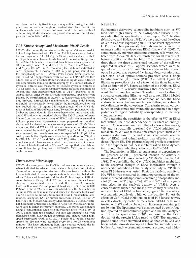
Sulfonamide-derivative calmodulin inhibitors such as W7bind with high affinity to the hydrophobic surface of cal-modulin that is specifically exposed upon Ca2� binding(Nishikawa and Hidaka, 1982). We have analyzed the effectsof W7 in live cells expressing an EEA1 construct tagged withGFP, which has previously been shown to behave in amanner similar to endogenous EEA1 (Lawe et al., 2002). Tosimultaneously monitor endosome content, cells were incu-bated with Alexa-594–labeled transferrin for 10 min at 37°Cbefore addition of the inhibitor. The fluorescence signalthroughout the three-dimensional volume of the cell wascaptured in stacks of 21 optical sections, which were ac-quired every 10 s for 15 min. The out-of-focus blur in eachoptical section was restored by image deconvolution, andeach stack of 21 optical sections projected onto a singletwo-dimensional (2D) image (Patki et al., 2001). Figure 1Aillustrates projections of stacks taken at the times indicatedafter addition of W7. Before addition of the inhibitor, EEA1was localized to vesicular structures that concentrated to-ward the juxtanuclear region. Transferrin was localized tostructures containing EEA1 as well as to non-EEA1 labeledendosomes. Within 5 min of addition of W7, the EEA1endosomal signal became much more diffuse, indicating itsrelocalization to the cytoplasm. Transferrin remained con-tained in endosomes and eventually accumulated in a tightjuxtanuclear region potentially corresponding to the recy-cling endosome.
To determine the specificity of the effect of W7 on EEA1localization, the dose dependency of its effect on endoge-nous EEA1 was compared with that of a less active analog,W5, and a structurally unrelated Ca2�/CaM inhibitor, cal-midazolium. W7 was at least 5 times more potent than W5 incausing a decrease in the endosomal steady-state localiza-tion of EEA1, and calmidazolium was effective at lowerdoses (Figure 1B). This pharmacological profile is consistentwith the hypothesis that these inhibitors affect EEA1 dynam-ics through their inhibitory actions on Ca2�/CaM.
The localization of EEA1 to endosomes is dependent onthe presence of PI(3)P generated through the activity ofmammalian PI-3 kinases, including VPS34 (Siddhanta et al.,1998). The possibility that Ca2�/CaM inhibition might leadto the observed changes in EEA1 localization through anonspecific inhibition of the catalytic activity of VPS34 orother PI 3-kinases was tested. First, the catalytic activity ofHA-VPS34 was measured in immunopreciptates of the en-zyme incubated with liposomes containing phosphatidylino-sitol (PI) and ATP (Figure 2A). W5 and W7 had no signifi-cant effect on the catalytic activity of VPS34, even atconcentrations higher than those at which they caused a fullredistribution of EEA1 in live cells (Figure 1B). In contrast,wortmannin completely inhibited this activity. To test theeffects of Ca2�/CaM inhibitors on total PI 3-kinase activityin cell extracts, cytosolic extracts from 3T3-L1 cells weretreated with W7 and incubated with liposomes containing PIfor 30 min. The liposomes were then pelleted by centrifuga-tion, spotted on nitrocellulose paper, and the blot analyzedwith a probe specific for PI(3)P, composed of the FYVEdomain of the protein SARA fused to GST. The amount ofprobe bound was determined using antibodies to GST andhorseradish peroxidase-coupled anti-rabbit secondary anti-bodies. Although wortmannin caused a pronounced inhibi-
Ca2�/Cam in EEA1 Localization
Vol. 14, July 2003 2937
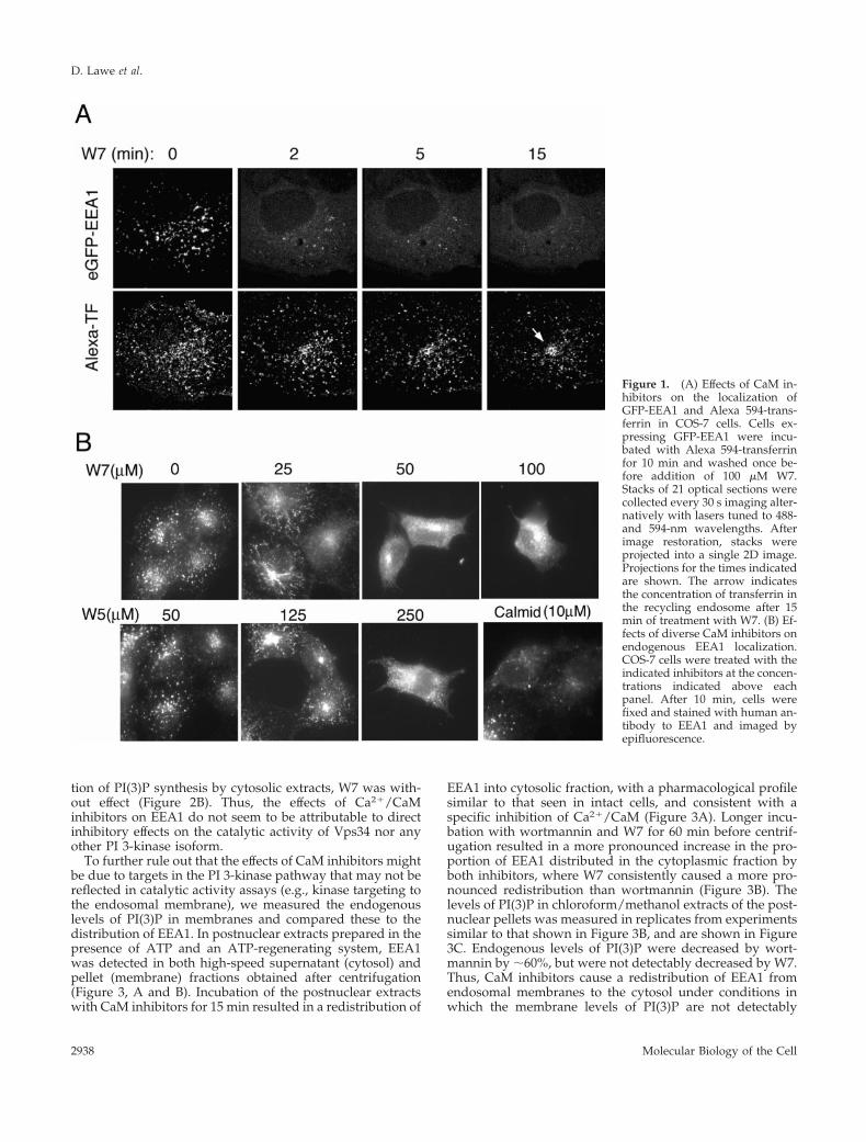
tion of PI(3)P synthesis by cytosolic extracts, W7 was with-out effect (Figure 2B). Thus, the effects of Ca2�/CaMinhibitors on EEA1 do not seem to be attributable to directinhibitory effects on the catalytic activity of Vps34 nor anyother PI 3-kinase isoform.
To further rule out that the effects of CaM inhibitors mightbe due to targets in the PI 3-kinase pathway that may not bereflected in catalytic activity assays (e.g., kinase targeting tothe endosomal membrane), we measured the endogenouslevels of PI(3)P in membranes and compared these to thedistribution of EEA1. In postnuclear extracts prepared in thepresence of ATP and an ATP-regenerating system, EEA1was detected in both high-speed supernatant (cytosol) andpellet (membrane) fractions obtained after centrifugation(Figure 3, A and B). Incubation of the postnuclear extractswith CaM inhibitors for 15 min resulted in a redistribution of
EEA1 into cytosolic fraction, with a pharmacological profilesimilar to that seen in intact cells, and consistent with aspecific inhibition of Ca2�/CaM (Figure 3A). Longer incu-bation with wortmannin and W7 for 60 min before centrif-ugation resulted in a more pronounced increase in the pro-portion of EEA1 distributed in the cytoplasmic fraction byboth inhibitors, where W7 consistently caused a more pro-nounced redistribution than wortmannin (Figure 3B). Thelevels of PI(3)P in chloroform/methanol extracts of the post-nuclear pellets was measured in replicates from experimentssimilar to that shown in Figure 3B, and are shown in Figure3C. Endogenous levels of PI(3)P were decreased by wort-mannin by �60%, but were not detectably decreased by W7.Thus, CaM inhibitors cause a redistribution of EEA1 fromendosomal membranes to the cytosol under conditions inwhich the membrane levels of PI(3)P are not detectably
Figure 1. (A) Effects of CaM in-hibitors on the localization ofGFP-EEA1 and Alexa 594-trans-ferrin in COS-7 cells. Cells ex-pressing GFP-EEA1 were incu-bated with Alexa 594-transferrinfor 10 min and washed once be-fore addition of 100 �M W7.Stacks of 21 optical sections werecollected every 30 s imaging alter-natively with lasers tuned to 488-and 594-nm wavelengths. Afterimage restoration, stacks wereprojected into a single 2D image.Projections for the times indicatedare shown. The arrow indicatesthe concentration of transferrin inthe recycling endosome after 15min of treatment with W7. (B) Ef-fects of diverse CaM inhibitors onendogenous EEA1 localization.COS-7 cells were treated with theindicated inhibitors at the concen-trations indicated above eachpanel. After 10 min, cells werefixed and stained with human an-tibody to EEA1 and imaged byepifluorescence.
D. Lawe et al.
Molecular Biology of the Cell2938
affected. These results indicate that the point at which CaMinhibitors disrupt the steady-state distribution of EEA1 isdownstream of the PI 3-kinase pathway.
Because PI 3-kinases remain active in the presence of W7it would be expected that the distribution of proteins otherthan EEA1 that bind to PI(3)P with high affinity wouldremain unchanged upon inhibition of CaM function. The PXdomains of p40phox and the protein kinase CISK have beenshown to interact with PI(3)P and to localize to endosomes(Cheever et al., 2001; Ellson et al., 2001; Kanai et al., 2001;Song et al., 2001; Virbasius et al., 2001; Xu et al., 2001; Yu andLemmon, 2001). We compared the effects of W7 on thelocalization of eGFP-tagged constructs of full-length EEA1,of the C-terminal domain of EEA1, and of the PX domains ofp40Phox and CISK. Although W7 caused a rapid and almostcomplete redistribution of full-length and C-terminal EEA1from endosomes to cytosol, it did not result in a comparableredistribution of the PX domain constructs (Figure 4), aspredicted from its lack of effect on PI(3)P levels. Interest-ingly, in several cells the eGFP-p40PX domain probes couldbe seen distributed on tubules that emanated from vesicularendosomes toward the cell periphery (Figure 4, lower left,arrow and Video 1). Tubular extensions occurred within the
time frame during which EEA1 dissociated from the endo-some. Tubulation of endosomes has also been observed inresponse to wortmannin in cells in which the association ofEEA1 with endosomes is sensitive to this inhibitor (Shpetneret al., 1996). Although the role of these tubular endosomalextensions in normal endosomal function is unclear, it ispossible that they may have an important role in trafficking.
Although it is widely accepted that W7 interacts specifi-cally with Ca2�/CaM, nonspecific interactions of this inhib-itor with other potential targets cannot be ruled out. Thus,we sought to complement these experiments with the use ofa potent mAb raised against CaM (Sacks, 1994). Increasingconcentrations of this antibody added to postnuclear ex-tracts of cells caused a progressive redistribution of EEA1into the cytoplasmic fraction (Figure 5A). Based on the re-ported total cellular concentration of CaM of 30–40 �M, theconcentration in these postnuclear extracts was estimated tobe 6–8 �M. Thus, EEA1 redistributed into the cytosol indirect proportion to concentrations of the antibody that ap-proached the concentration of CaM in the extract. To deter-mine the effects of anti-CaM antibody in the absence ofcytoplasmic CaM, postnuclear extracts were centrifuged, thecytosol removed, and pellets resuspended in cytosol buffercontaining anti-CaM antibody, equal amounts of controlIgG, W7, or wortmannin. After 1 h at room temperature,membranes were collected by centrifugation, and the releaseof EEA1 into the supernatant analyzed by SDS-PAGE andimmunoblotting with anti-EEA1 antibody (Figure 5B). Anti-CaM antibody, W7, and wortmannin, but not IgG control,significantly increased the release of EEA1 into the buffer.
The effect of microinjection of the anti-CaM mAb on EEA1localization in cells was also analyzed (Figure 5C). In cellsfixed within 5–10 min after microinjection with low antibodyconcentrations, the injected anti-CaM antibody localizedpredominantly at the cell periphery and to vesicular struc-tures that also contained endogenous EEA1. This result isconsistent with previous reports in which CaM has beenfound associated with endosomal fractions (Enrich et al.,1988). When cells were fixed longer after injection, EEA1displayed a more diffuse cytoplasmic localization and de-pletion from peripheral endosomes was observed in at least50% of the injected cells (Figure 5C, lower panels). Theseresults indicate that EEA1 containing endosomes are en-riched in CaM relative to other membrane structures andthat inhibition of CaM by titration with a specific mAbnegatively affects EEA1 binding to membranes.
In addition to the FYVE domain, which mediates multi-valent binding to PI(3)P (Dumas et al., 2001), the C termini ofEEA1 contains a region that interacts with the Rab5 GTPase.Cells expressing a persistently activated form of Rab5,dsRed-RabQ79L, display greatly enlarged endosomes, towhich the majority of transfected eGFP-EEA1 (Figure 6A) orendogenous cellular EEA1 (Figure 6B) is associated at steadystate. Exposure to W7 for �10 min resulted in a decrease inthe amount of transfected eGFP-EEA1 (Figure 6A) or endog-enous cellular EEA1 (Figure 6B) associated with Rab5Q79L-containing endosomes and a decrease in their size. Thedecrease in the amount of eGFP-EEA1 associated withdsRed-Rab5Q79-containing endosomes occurred with a timecourse that parallels the decrease in the size of such endo-somes (Figure 6A). The association of EEA1 with dsRed-Rab5Q79-containing endosomes is not greatly decreased by
Figure 2. (A) Effect of CaM inhibitors on PI 3-kinase activity.COS-7 cells were transfected with myc-tagged Vps34, lysed, andimmunoadsorbed with anti-myc antibody immobilized on Sepha-rose beads. Beads were washed and incubated with [��32P]ATPand liposomes in the presence of the concentrations of inhibitorindicated under each lane. Lipids were separated by TLC andanalyzed by autoradiography. The position of PI(3)P on the chro-matogram is indicated by the arrow. (B) Postnuclear extracts from3T3 cells were prepared as described in MATERIALS AND METH-ODS and incubated with wortmannin (Wort) or W7 at 100 nM or100 �M final concentration, respectively, for 1 h. Cytosol was sep-arated by high-speed centrifugation and incubated with PS/PI li-posomes for 30 min. Liposomes were pelleted and spotted on ni-trocellulose, which was probed with GST-SARA-FYVE andantibodies to GST. The specificity of the probe is depicted in thebottom row, in which 1 �g of the indicated lipids was spotted.
Ca2�/Cam in EEA1 Localization
Vol. 14, July 2003 2939

treatment for 15 min with wortmannin (Figure 6B), perhapsdue to a high level of PI(3)P generated by wortmannin-insensitive kinases or to a slow turnover of the pool of PI(3)Pbound to EEA1. The much larger effect of W7 comparedwith wortmannin suggests that Ca2�/CaM inhibition causeseither the dissociation of EEA1 from existing PI(3)P or in-hibits its association with persistently activated Rab5.
To distinguish between these possibilities, the binding ofEEA1 to Rab5 was measured directly using cytoplasmicextracts from 3T3 cells and recombinant GST-Rab5 immobi-lized on glutathione beads. Binding of endogenous EEA1 tobeads loaded with Rab5-GTP�S, but not to beads loadedwith Rab5GDP�S or GST alone was observed. Addition ofW7 to the cytosol 60 min before the incubation with beads
Figure 3. Effects of the Ca2�/CaM inhibitor W7 on the associa-tion of EEA1 with cell membranesand on the membrane levels ofPI(3)P. (A) Dose response ofEEA1 to Ca2�/CaM inhibitors: Apostnuclear supernatant from 3T3cells was obtained as described inMATERIALS AND METHODS,dispensed into aliquots, and incu-bated for 15 min with CaM inhib-itors at the concentrations indi-cated. Cytosol was separatedfrom membranes by high-speedcentrifugation. The membranepellet was resuspended in cytosolbuffer to the same volume as thecytosol and equal volume ali-quots analyzed by SDS-PAGEand immunoblotting with anti-bodies to EEA1 and chemilumi-nescence. The exposed films were
scanned and the intensity of each band quantified. The ratio of the cytosol and membrane intensities was plotted. Bars represent the mean,and lines the SE of three independent experiments. (B) The experiment was performed as described in A with no additions, 100 nMwortmannin (Wort), or 100 �M W7 for 60 min. The insets illustrate the immunoblots of the membrane and cytosolic extracts. Plotted are theintensities of each band. This experiment was repeated five times with similar results (C) The resuspended membrane pellets were extractedwith chlorophorm/methanol as described in MATERIALS AND METHODS. Extracted lipids were spotted on nitrocellulose and probed withGST-SARA-FYVE as described in MATERIALS AND METHODS and in Figure 2. Two experiments are illustrated, with their correspondingstandards. This experiment was repeated four times with similar results.
Figure 4. Effect of Ca2�/CaM inhi-bition on full-length EEA1, the C ter-mini of EEA1, and the p40Phox andCISK PX domains in live cells.COS-7 cells were transfected withthe construct indicated on the leftand exposed to 100 �M W7. Stacksof 21 optical sections were obtainedevery 20 s, restored, and projectedinto single 2D images. Projectionsfor the times indicated on top ofeach panel are shown. Arrow in bot-tom right panel indicates the forma-tion of tubules in response to W7.The complete set of images for thep40Phox construct can be seen as aQuickTime video (Video 1).
D. Lawe et al.
Molecular Biology of the Cell2940

did not impair the binding of EEA1 to Rab5GTP�S (Figure7A). Thus, the decreased interaction between EEA1 andRab5Q79L-enriched endosomes seems not to be due to adisruption in the interaction between Rab5 and EEA1. Totest the possibility that the failure of EEA1 to associate withthe endosomal membrane was due to a disruption of itsbinding to endosomal phosphoinositide, cytoplasmic ex-tracts were treated without or with W7 and then incubatedwith liposomes containing increasing concentrations ofPI(3)P. A significant decrease in the binding of cytosolicEEA1 to such endosomes was observed in response to W7.The inhibitory effect of W7 was greater at lower PI(3)Pconcentrations, suggesting that inhibition of Ca2�/CaM re-duces the affinity of EEA1 binding to the liposome surface(Figure 7B). This decreased affinity, in the context of theendosomal concentration of PI(3)P, could result in the dra-matic displacement of EEA1 from the endosomal membraneobserved in intact cells.
The pronounced effect of CaM inhibition to cause dis-placement of EEA1 from the lipid bilayer surface both invivo and in liposome binding assays suggest that CaMmight directly interact with EEA1. In fact, a direct associa-tion of Ca2�/CaM with the IQ-like motif at the C termini ofEEA1 has been proposed based on overlay assays of GST-fusion constructs of the EEA1 C termini in the presence ofhigh Ca2� concentrations (Mills et al., 2001). To determinewhether this interaction might be directly involved in theinteraction of EEA1 with the lipid bilayer in vivo, pointmutations were introduced in residues crucial to IQ domainstructure, and cells expressing wild-type GFP-tagged EEA1were compared with those expressing the IQ domain mu-tant (eGFPEEA1Q1289L/R1293G) at similar levels. As pre-viously reported by Stenmark et al. (1996), this mutation in
EEA1 did not detectably impair its association with endo-somes (our unpublished data) and suggests that the IQ-likemotif may not be the single target of CaM or Ca2�/CaM inEEA1.
To identify additional regions within EEA1 required forCa2�/CaM-dependent bilayer interactions, we analyzed thebinding of wild-type EEA1, of the Q1289L/R1293G muta-tion, and of deletion constructs of EEA1 to CaM immobi-lized on Sepharose beads. Cytosolic extracts prepared fromCOS-7 cells overexpressing each construct were incubatedwith CaM-Sepharose, or Sepharose beads alone. eGFP-tagged full-length EEA1 bound to CaM-Sepharose, but notto Sepharose control beads (Figure 8A). The Ca2� chelatorsBAPTA and EGTA markedly, but not completely, impairedthe binding of EEA1 to CaM-Sepharose. Further addition of2 �M Ca2� to the cytosolic extract did not augment thebinding of EEA1, and higher concentrations of Ca2� resultedin a pronounced increase in nonspecific binding, as assessedby the similarly increased binding of EEA1 to both CaM-Sepharose and Sepharose control beads (our unpublisheddata). The low calcium concentrations required for EEA1binding to CaM are similar to those reported to be requiredfor in vitro endosome fusion (Peters and Mayer, 1998; Hol-royd et al., 1999).
The binding of eGFP-EEA1Q1289L/R1293G to CaM-Sepharose was only slightly decreased compared withwild-type eGFP-EEA1 (Figure 8B), and the Ca2�-sensitiv-ity of binding was largely preserved. In contrast, deletionof the N terminus of EEA1 [eGFP-EEA1(835–1411)]greatly decreased Ca2�-dependent binding to CaM-Sepharose (Figure 8C, compare ratios of input to bound).Further deletion of the N terminus to amino acid 1277 hadno further effect, and neither of these truncations induced
Figure 5. Anti-CaM antibodycauses a release of EEA1 from en-dosomal membranes. (A) Post-nuclear supernatant from 3T3cells was obtained as described inMATERIALS AND METHODS,and aliquots were incubatedwithout or with 100 nM wort-mannin (Wort), 100 �M W7, orthe indicated concentration ofnonimmune mouse IgG (IgG) oranti-CaM IgG (anti-CaM) for 60min. Membranes and cytosolwere separated by high-speedcentrifugation and analyzed bySDS-PAGE and immunoblottingwith antibodies to EEA1 andchemiluminescence. The exposedfilms were scanned and the inten-sity of each band quantified andplotted. Inset illustrates the immu-noblot from the membrane fractions. Bars represent the mean and lines the SE of the mean from three independent experiments. (B) Themembrane fraction from 3T3 cells was isolated by high-speed centrifugation, resuspended in cytosol buffer, and incubated without or with100 nM wortmannin (Wort), 100 �M W7, or 5 �M nonimmune mouse IgG (IgG) or anti-CaM IgG (Anti-CaM) for 60 min at room temperature.The membrane pellet was separated from supernatant and equal quantities of the resulting fractions immunoblotted for EEA1. Immunoblotswere scanned, and the intensities of the bands quantified. Plotted are the ratios of supernatant and pellet. Bars represent the mean and linesthe SE of the mean from four independent experiments. Inset illustrates a Western blot of the supernatant from one experiment. (C) Anti-CaMantibody was microinjected into COS-7 cells, which were fixed within 5 min of injection (top) or after 45 min postinjection. Injected antibodywas detected with anti-mouse secondary antibodies and endogenous EEA1 with human anti-EEA1 antiserum and antihuman secondaryantibodies. Arrows in top panels point to sites of colocalization of the two probes and in lower panels point to the injected cell.
Ca2�/Cam in EEA1 Localization
Vol. 14, July 2003 2941

nonspecific binding to Sepharose beads alone (our unpub-lished data). These results suggest the presence of a Ca2�/CaM binding site within the N-terminal half of EEA1. Tofurther test this hypothesis, a GFP-construct containingthe first 314 amino acids of the EEA1 N terminus wasanalyzed. This construct bound avidly to CaM-Sepharose,but not Sepharose alone, in a Ca2�-dependent manner(Figure 8C). Thus, at least two separate domains in EEA1are capable of mediating Ca2�-dependent and Ca2�-inde-pendent CaM binding interactions.
DISCUSSION
A crucial requirement for Ca2� and CaM in endosome fu-sion in vitro and in vivo has been previously documented,but the precise mechanism for this requirement has not beenfully determined. In this manuscript, we identify EEA1 as amajor Ca2�/CaM-sensitive component of the endosome fu-sion machinery, which may at least in part explain theactions of CaM inhibition on this process.
EEA1 binding to endosomal membranes is known to de-pend on its C-terminal FYVE domain, which directly bindsto PI(3)P on the endosomal surface. In addition, EEA1 inter-acts with the activated small GTPase Rab5, and expression
Figure 6. (A) Effects of W7 on eGFP-EEA1 and endogenousEEA1 association to enlarged endosomes containing persistentlyactivated Rab5. COS-7 cells were cotransfected with dsRed-Rab5Q79L and eGFP-EEA1. Cells expressing both constructswere localized and imaged as described in Figure 1. Stacks of 21optical sections were obtained every 30 s, restored, and projectedinto single 2D images. Two time points are illustrated. The over-lap of the red and green signal is displayed as pure white inregions of equal pixel intensity. (B) COS-7 cells were transfectedwith dsRed-Rab5Q79L, and after 36 h treated for 10 min with 100nM wortmannin (Wort) or 100 �M W7. Cells were fixed andendogenous EEA1 was detected using a human antiserum, andAlexa 488 secondary antibodies. Cells shown express similarlevels of Rab5Q79L based on fluorescence intensity. A nontrans-fected cell (NT) is shown for comparison. (C) The fluorescenceintensity of EEA1 on the endosomes containing Rab5Q79L and onthe cytoplasmic region surrounding the endosomes was quanti-fied and the ratio plotted.
Figure 7. Effects of Ca2�/CaM inhibition on EEA1 associationwith activated Rab5 or PI(3)P. (A) Cytosolic extracts of 3T3 cellswere incubated with nothing or with 100 �M W7 for 60 min andthen incubated with GST alone or recombinant GST-tagged Rab5immobilized on glutathione-Sepharose. GST-Rab5 was loaded withGTP�S or GDP�S as indicated. Bound proteins were analyzed bySDS-PAGE and immunoblotting with antibodies to EEA1. The po-sition of EEA1 on the gel is indicated. Duplicates of each conditionwere done in each experiment, and three experiments with similarresults were obtained. (B) Cytosolic extracts of 3T3 cells were incu-bated with nothing or with 100 �M W7 for 60 min and incubatedwith PS/PI liposomes containing increasing concentrations ofPI(3)P as indicated above each lane. Liposomes were pelleted andanalyzed by immunoblotting with antibodies to EEA1. The exposedfilms were scanned and the intensity of each band quantified. Theinset represents the immunoblot from one experiment, and thegraph represents the results from a different experiment. Similarresults were obtained in three independent experiments.
D. Lawe et al.
Molecular Biology of the Cell2942

of persistently active Rab5 causes a marked increase in EEA1binding to endosomes and endosome enlargement. The ob-servations made herein indicate that Ca2�/CaM also plays acritical role in the binding of EEA1 to the endosome surface.This requirement may reflect additional, novel interactionsbetween EEA1, CaM, and yet unidentified targets on theendosomal surface. Alternatively, the effect of Ca2�/CaMmay be to regulate the conformation of EEA1 to effectivelyinteract with endosomal Rab5 or PI(3)P. The results pre-sented herein suggest the latter, based principally from theobserved inhibition of EEA1 binding to liposomes contain-ing PI(3)P upon inhibition of Ca2�/CaM.
Although the precise mode by which Ca2�/CaM mightcontrol the interaction between EEA1 and PI(3)P is not clear,the recently solved crystal structure of the C termini of EEA1complexed to PI(3)P head group (Dumas et al., 2001), as wellas by new examples of mechanisms of interaction of Ca2�/CaM with protein targets (Hoeflich and Ikura, 2002) suggestpossible mechanisms. The crystal structure of the C terminiof EEA1 reveals a parallel coiled-coil dimer that terminatesabruptly in a splayed FYVE domain (Figure 9). Notably, the
quaternary structural arrangement of the EEA1 FYVE do-main includes an extensive interface involving noncon-served residues of the FYVE domain structure. This interfaceis unusual in that, beyond displaying a high degree ofcomplementarity between the two interacting surfaces, thereare no amino acid residues that stabilize it through conven-tional electrostatic or hydrophobic interactions. Thus, arather complex interface seems to be held together solelythrough weak van der Waals forces (Dumas et al., 2001). Thisconformation suggests that additional energetic contribu-tions may be required for stabilization of the dimer interfaceand the FYVE domain quaternary structure. The heptadrepeats that immediately precede the FYVE domain mayprovide such additional stabilization, which may be furtherenhanced by external elements such as CaM. This stabiliza-tion may be crucial in enabling the bivalent interaction ofEEA1 with the PI(3)P head group, and thereby increasingthe affinity of EEA1 for PI(3)P on the endosomal membranesurface (Figure 9).
Elucidating the precise mechanism of CaM function re-quires information on the mechanism(s) of CaM interaction
Figure 8. Association of GFP-tagged wild-type and mutantEEA1 with immobilized CaM. (A)The cytoplasmic extract fromCOS-7 cells expressing GFP-EEA1wt was incubated in the ab-sence (C) or presence of 10 mMBAPTA, 10 mM EGTA or with anadditional 2 �M CaCl2 for 30 minat room temperature. The extractwas then incubated with CaM-Sepharose or Sepharose alone asdescribed in MATERIALS ANDMETHODS. Bound proteins wereanalyzed by immunoblottingwith antibody to GFP. (B) Cyto-plasmic extracts from COS-7 cellsexpressing either eGFP-EEA1 oreGFP-EEA1 containing mutationsin the IQ-like motif (eGFPEEA1Q1289L/R1293G) were incubated in the presence or absence of EGTA. Binding to immobilized CaM was thendetermined as described in A. The concentration of the eGFP constructs in 20% of the starting cell extract (input) is shown. (C) Cytosol fromcells expressing either GFP alone, GFP-EEA1wt, or EEA1 deletion constructs comprised of the amino acids indicated were incubated withor without EGTA and assessed for binding to CaM-Sepharose as described in A.
Figure 9. Model depicting hy-pothetical effect of CaM on thestructure of the C termini of EEA1and consequent binding to PI(3)Pon membranes. Solid bars repre-sent the coiled-coil region of eachmonomer, leading into thesplayed FYVE domains.
Ca2�/Cam in EEA1 Localization
Vol. 14, July 2003 2943
with EEA1. Previous results have shown that biotinylatedCaM can bind in an overlay assay to a GST-fusion proteincontaining the EEA1 C termini, but not to a similar constructcontaining a deletion of the IQ motif (Mills et al., 2001).Under these conditions, binding was enhanced by the pres-ence of large quantities (100 �M) of Ca2� or Zn2� in theoverlay buffer. These results suggested that the IQ motif inEEA1 directly binds Ca2�/CaM. In the present study, theinteraction of EEA1 from cytosolic extracts with CaM-Sepha-rose or Sepharose alone as a control was measured. Full-length EEA1 and a construct harboring point mutations inresidues critical for the conformation of the IQ motif boundcomparably to CaM-Sepharose (Figure 8B). These observa-tions suggested that Ca2�/CaM binding to EEA1 is notrestricted to the C-terminal IQ domain. Direct analysis oftruncation constructs of EEA1 reveals two independent re-gions capable of CaM binding. One site is located within thefirst 314 amino acids of the N terminus of EEA1, and theother within the 134 amino acids of the C termini. Althoughthe N-terminal binding site seems to bind exclusively toCa2�/CaM but not to CaM, both full-length EEA1 and con-structs containing the C-terminal region display significantCa2�-insensitive CaM binding (Figure 8). The detection ofCa2�-independent CaM binding by the C termini in ourstudies but not in those reported by Mills et al. (2001) may beexplained by the differences in the concentrations of Ca2�
used in each study, and inherent to the different techniquesused.
The extent of binding of EEA1 C-terminal constructs toCaM-Sepharose was similar to the binding of full-lengthEEA1 to CaM-Sepharose in the presence of chelators, sug-gesting a major contribution of the N-terminal binding sitein Ca2�/CaM binding, and the existence of a Ca2�-indepen-dent CaM binding contribution at the C termini. Notably,binding of the IQ domain point mutant of EEA1 to CaM-Sepharose was not fully inhibited by Ca2� chelators, sug-gesting CaM binding to the C termini on sites additional tothe IQ motif. Complex mechanisms for binding of bothapo-CaM and Ca2�/CaM to a homodimer have a precedentin the small conductance Ca2� activated potassium channelSK2 (Schumacher et al., 2001).
A previous study (Mills et al., 2001) has shown that largeconcentrations of CaM in the presence of 100 �M Ca2�
antagonize the binding of PI(3)P-containing liposomes andof Rab5-GTP to immobilized EEA1. This particular resultsuggests that Ca2�/CaM might be a negative regulator ofEEA1 association with the endosomal surface. This model isinconsistent with results presented in this manuscript, inwhich Ca2�/CaM seems to be a requirement for EEA1 bind-ing to endosomes in vitro and in intact cells. However, in thestudy byMills et al. (2001) addition of a large concentrationof Ca2�/CaM is reported not to impair the binding of EEA1to endosomal membranes in a broken cell system. This resultsuggests that under conditions that better represent the in-tracellular environment Ca2�/CaM is not a negative regu-lator of EEA1 association with the endosomal surface. More-over, biochemical studies of endosome fusion in vitro haveshown a profound inhibitory effect of CaM inhibitors andCa2� chelators on fusion (Gorvel et al., 1991; Colombo et al.,1997; Mills et al., 1998, 2001). Thus, most experimental resultsare consistent with a model in which Ca2�/CaM is a posi-
tive regulator of EEA1 binding to the endosomal surface andof endosome fusion.
A decrease in the endosomal binding of EEA1 is observedin response to wortmannin, to Ca2�/CaM inhibitors and todominant-negative Rab5 constructs, supporting a model inwhich EEA1 is a crucial element in early endosome fusionoperating downstream of PI 3-kinase and Rab5 in a CaM-dependent manner. Interestingly, however, at the cellularlevel these three manipulations lead to different phenotypes:cells expressing dominant-negative mutants of Rab5 containsmall endosomes and display a block in transferrin uptake,whereas cells treated with wortmannin or with CaM inhib-itors contain tubulated and enlarged endosomes, normaltransferrin uptake, but decreased transferrin recycling.These observations suggest that the results of in vitro earlyendosome fusion assays reflect the collective fusion eventstaking place in the uptake and recycling phases in the en-dosomal system. Because manipulations that impair the as-sociation of EEA1 with endosomes in intact cells resultmainly in defective recycling, this molecule may in factcontrol fusion events in the recycling portion more than inthe initial phases of the endosome fusion pathway. Moreextensive analysis of specific molecular elements that regu-late endosome fusion in vitro in intact cells will be requiredto fully understand the functional architecture of the endo-cytic pathway. Nevertheless, the results presented here ex-plain the molecular basis for the observed requirement forCa2�/CaM in endosome fusion in vitro and in intact cells.
ACKNOWLEDGMENTS
We acknowledge the helpful discussions with Dr. Akira Hayakawa,and the gift of anti-CaM antibody from Dr. David Sacks. This workwas funded by National Institutes of Health grants DK60564 andDK54479 (to S.C.).
REFERENCES
Apodaca, G., Enrich, C., and Mostov, K.E. (1994). The calmodulinantagonist, W-13, alters transcytosis, recycling, and the morphologyof the endocytic pathway in Madin-Darby canine kidney cells.J. Biol. Chem. 269, 19005–19013.
Chamberlain, L.H., Roth, D., Morgan, A., and Burgoyne, R.D.(1995). Distinct effects of alpha-SNAP, 14-3-3 proteins, and calmod-ulin on priming and triggering of regulated exocytosis. J. Cell Biol.130, 1063–1070.
Cheever, M.L., Sato, T.K., de Beer, T., Kutateladze, T.G., Emr, S.D.,and Overduin, M. (2001). Phox domain interaction with PtdIns(3)Ptargets the Vam7 t-SNARE to vacuole membranes. Nat. Cell Biol. 3,613–618.
Chen, Y.A., Scales, S.J., Duvvuri, V., Murthy, M., Patel, S.M., Schul-man, H., and Scheller, R.H. (2001). Calcium regulation of exocytosisin PC12 cells. J. Biol. Chem. 276, 26680–26687.
Colombo, M.I., Beron, W., and Stahl, P.D. (1997). Calmodulin reg-ulates endosome fusion. J. Biol. Chem. 272, 7707–7712.
Christoforidis, S., McBride, H.M., Burgoyne, R.D., and Zerial, M.(1999). The Rab5 effector EEA1 is a core component of endosomedocking. Nature 397, 621–625.
Dumas, J.J., Merithew, E., Sudharshan, E., Rajamani, D., Hayes, S.,Lawe, D., Corvera, S., and Lambright, D.G. (2001). Multivalentendosome targeting by homodimeric EEA1. Mol. Cell 8, 947–958.
D. Lawe et al.
Molecular Biology of the Cell2944

Ellson, C.D., et al. (2001). PtdIns(3)P regulates the neutrophil oxidasecomplex by binding to the PX domain of p40(phox). Nat. Cell Biol.3, 679–682.
Emmanouilidou, E., Teschemacher, A.G., Pouli, A.E., Nicholls, L.I.,Seward, E.P., and Rutter, G.A. (1999). Imaging Ca2� concentrationchanges at the secretory vesicle surface with a recombinant targetedcameleon. Curr. Biol. 9, 915–918.
Enrich, C., Bachs, O., and Evans, W.H. (1988). A 115 kDa calmod-ulin-binding protein is located in rat liver endosome fractions. Bio-chem. J. 255, 999–1005.
Gorvel, J.P., Chavrier, P., Zerial, M., and Gruenberg, J. (1991). rab5controls early endosome fusion in vitro. Cell 64, 915–925.
Hoeflich, K.P., and Ikura, M. (2002). Calmodulin in action: diversityin target recognition and activation mechanisms. Cell 108, 739–742.
Holroyd, C., Kistner, U., Annaert, W., and Jahn, R. (1999). Fusion ofendosomes involved in synaptic vesicle recycling. Mol. Biol. Cell.10, 3035–3044.
Huber, L.A., Fialka, I., Paiha, K., Hunziker, W., Sacks, D.B., Bahler,M., Way, M., Gagescu, R., and Gruenberg, J. (2000). Both calmodulinand the unconventional myosin Myr4 regulate membrane traffick-ing along the recycling pathway of MDCK cells. Traffic 1, 494–503.
Hutton, J.C. (1986). Calcium-binding proteins and secretion. CellCalcium 7, 339–352.
Kanai, F., Liu, H., Field, S.J., Akbary, H., Matsuo, T., Brown, G.E.,Cantley, L.C., and Yaffe, M.B. (2001). The PX domains of p47phoxand p40phox bind to lipid products of PI(3)K. Nat. Cell Biol. 3,675–678.
Lawe, D.C., A. Chawla, E. Merithew, J. Dumas, W. Carrington, K.Fogarty, L. Lifshitz, R. Tuft, D. Lambright, and S. Corvera. (2002).Sequential roles for phosphatidylinositol 3-phosphate and Rab5 intethering and fusion of early endosomes via their interaction withEEA1. J. Biol. Chem. 277, 8611–8617.
Mills, I.G., Jones, A.T., and Clague, M.J. (1998). Involvement of theendosomal autoantigen EEA1 in homotypic fusion of early endo-somes. Curr. Biol. 8, 881–884.
Mills, I.G., Jones, A.T., and Clague, M.J. (1999). Regulation of endo-some fusion. Mol. Membr. Biol. 16, 73–79.
Mills, I.G., Urbe, S., and Clague, M.J. (2001). Relationships betweenEEA1 binding partners and their role in endosome fusion. J. Cell Sci.114, 1959–1965.
Mu, F.T., Callaghan, J.M., Steele-Mortimer, O., Stenmark, H., Parton,R.G., Campbell, P.L., McCluskey, J., Yeo, J.P., Tock, E.P., and Toh,B.H. (1995). EEA1, an early endosome-associated protein. EEA1 is aconserved alpha-helical peripheral membrane protein flanked bycysteine “fingers” and contains a calmodulin-binding IQ motif.J. Biol. Chem. 270, 13503–13511.
Nishikawa, M., and Hidaka, H. (1982). Role of calmodulin in plateletaggregation. Structure-activity relationship of calmodulin antago-nists. J. Clin. Investig. 69, 1348–1355.
Patki, V., Buxton, J., Chawla, A., Lifshitz, L., Fogarty, K., Carrington,W., Tuft, R., and Corvera, S. (2001). Insulin action on GLUT4 trafficvisualized in single 3T3–l1 adipocytes by using ultra-fast micros-copy. Mol. Biol. Cell 12, 129–141.
Peters, C., and Mayer, A. (1998). Ca2�/calmodulin signals the com-pletion of docking and triggers a late step of vacuole fusion. Nature396, 575–580.
Pryor, P.R., Mullock, B.M., Bright, N.A., Gray, S.R., and Luzio, J.P.(2000). The role of intraorganellar Ca(2�) in late endosome-lyso-some heterotypic fusion and in the reformation of lysosomes fromhybrid organelles. J. Cell Biol. 149, 1053–1062.
Russell, D.G. (2001). Mycobacterium tuberculosis: here today, andhere tomorrow. Nat. Rev. Mol. Cell. Biol. 2, 569–577.
Sacks, D.B. (1994). Alteration of calmodulin-protein interactions bya monoclonal antibody to calmodulin. Biochim. Biophys. Acta 1206,120–128.
Schumacher, M.A., Rivard, A.F., Bachinger, H.P., and Adelman, J.P.(2001). Structure of the gating domain of a Ca2�-activated K� chan-nel complexed with Ca2�/calmodulin. Nature 410, 1120–1124.
Siddhanta, U., McIlroy, J., Shah, A., Zhang, Y., and Backer, J.M.(1998). Distinct roles for the p110alpha and hVPS34 phosphatidylino-sitol 3�-kinases in vesicular trafficking, regulation of the actin cy-toskeleton, and mitogenesis. J. Cell Biol. 143, 1647–1659.
Simonsen, A., Lippe, R., Christoforidis, S., Gaullier, J.M., Brech, A.,Callaghan, J., Toh, B.H., Murphy, C., Zerial, M., and Stenmark, H.(1998). EEA1 links PI(3)K function to Rab5 regulation of endosomefusion. Nature 394, 494–498.
Song, X., Xu, W., Zhang, A., Huang, G., Liang, X., Virbasius, J.V.,Czech, M.P., and Zhou, G.W. (2001). Phox homology domains spe-cifically bind phosphatidylinositol phosphates. Biochemistry 40,8940–8944.
Shpetner, H., Joly, M., Hartley, D., and Corvera, S. (1996). Potentialsites of PI-3 kinase function in the endocytic pathway revealed bythe PI-3 kinase inhibitor, wortmannin. J. Cell Biol. 132, 595–605.
Stenmark, H., Aasland, R., Toh, B.-H., and D’Arrigo, A. (1996).Endosomal localization of the autoantigen EEA1 is mediated by azinc-binding FYVE finger. J. Biol. Chem. 271, 24048–24054.
Virbasius, J.V., Guilherme, A., and Czech, M.P. (1996). Mouse p170is a novel phosphatidylinositol 3-kinase containing a C2 domain.J. Biol. Chem. 271, 13304–13307.
Virbasius, J.V., Song, X., Pomerleau, D.P., Zhan, Y., Zhou, G.W., andCzech, M.P. (2001). Activation of the Akt-related cytokine-indepen-dent survival kinase requires interaction of its phox domain withendosomal phosphatidylinositol 3-phosphate. Proc. Natl. Acad. Sci.USA 98, 12908–12913.
Xu, Y., Hortsman, H., Seet, L., Wong, S.H., and Hong, W. (2001).SNX3 regulates endosomal function through its PX-domain-medi-ated interaction with PtdIns(3)P. Nat. Cell Biol. 3, 658–666.
Yu, J.W., and Lemmon, M.A. (2001). All phox homology (PX) do-mains from Saccharomyces cerevisiae specifically recognize phospha-tidylinositol 3-phosphate. J. Biol. Chem. 276, 44179–44184.
Ca2�/Cam in EEA1 Localization
Vol. 14, July 2003 2945


![Near-Membrane [Ca2+] Transients Resolved Using the Ca2+ Indicator FFP18](https://static.fdokumen.com/doc/165x107/631286873ed465f0570a4533/near-membrane-ca2-transients-resolved-using-the-ca2-indicator-ffp18.jpg)