ESCRT-II, an Endosome-Associated Complex Required for Protein Sorting
11
Developmental Cell, Vol. 7, 559–569, October, 2004, Copyright 2004 by Cell Press ESCRT-II, an Endosome-Associated Complex Required for Protein Sorting: Crystal Structure and Interactions with ESCRT-III and Membranes Ten of these proteins are part of three multiprotein com- plexes (reviewed in Conibear, 2002; Katzmann et al., 2002; Morita and Sundquist, 2004). These are the 350 kDa ESCRT-I complex (consisting of Vps23, Vps28, and Vps37) (Katzmann et al., 2001), the 155 kDa ESCRT-II Hsiangling Teo, Olga Perisic, Beatriz Gonza ´ lez, and Roger L. Williams* MRC Laboratory of Molecular Biology Hills Road Cambridge CB2 2QH United Kingdom complex (Vps22, Vps25, and Vps36) (Babst et al., 2002b), and the ESCRT-III complex (Vps2, Vps24, Vps20, and Vps32) (Babst et al., 2002a). Each of these complexes has human orthologs that are required for lysosomal Summary traffic. The human ESCRT-I complex consists of Tsg101 (Vps23 homolog) (Babst et al., 2000; Garrus et al., 2001; ESCRT-I, -II, and -III protein complexes are sequen- Li and Cohen, 1996), hVps28 (Bishop and Woodman, tially recruited to endosomal membranes, where they 2001), and the recently identified Vps37 homologs orchestrate protein sorting and MVB biogenesis. In hVps37A and hVps37B (Bache et al., 2004; Stuchell et al., addition, they play a critical role in retrovirus budding. 2004), while the human ESCRT-II subunits correspond Structural understanding of ESCRT interaction net- to EAP20 (Vps25), EAP30 (Vps22), and EAP45 (Vps36) works is largely lacking. The 3.6 A ˚ structure of the (Kamura et al., 2001; Schmidt et al., 1999). The ESCRT- yeast ESCRT-II core presented here reveals a trilobal III components Vps2, Vps24, Vps32/Snf7, and Vps20 are complex containing two copies of Vps25, one copy of homologous with the human proteins CHMP2A, CHMP3, Vps22, and the C-terminal region of Vps36. Unexpect- CHMP4B, and CHMP6, respectively (Martin-Serrano et edly, the entire ESCRT-II core consists of eight repeats al., 2003; von Schwedler et al., 2003). of a common building block, a “winged helix” domain. Many of the cargo proteins destined for delivery to Two PPXY-motifs from Vps25 are involved in contacts vacuoles or lysosomes are monoubiquitinated (Katz- with Vps22 and Vps36, and their mutation leads to mann et al., 2001; Reggiori and Pelham, 2001; Urbanow- ESCRT-II disruption. We show that purified ESCRT-II ski and Piper, 2001). Several ubiquitin-recognizing mod- binds directly to the Vps20 component of ESCRT-III. ules are present in the ESCRT complexes, including the Surprisingly, this binding does not require the protrud- UEV domain of Vps23 (Katzmann et al., 2001; Pornillos ing N-terminal coiled-coil of Vps22. Vps25 is the chief et al., 2002; Sundquist et al., 2004; Teo et al., 2004) subunit responsible for Vps20 recruitment. This inter- and the NZF-finger domain of Vps36 (Alam et al., 2004). action dramatically increases binding of both compo- Deletion of Vps23 prevents formation of MVBs; however, nents to lipid vesicles in vitro. this defect can be partially overcome by overexpression of ESCRT-II, suggesting that the ESCRT-II functions Introduction downstream of ESCRT-I (Babst et al., 2002b). ESCRT- II forms a stable, soluble complex of defined stoichiome- Downregulation and subsequent degradation of mem- try. In contrast, the ESCRT-III subunits are cytosolic and brane receptors involves two ubiquitin-dependent mem- monomeric until they are recruited to membranes, a brane budding steps (Bonifacino and Traub, 2003; Hicke process that is dependent on ESCRT-II (Babst et al., and Dunn, 2003; Katzmann et al., 2002; Peschard and 2002a). Park, 2003; Raiborg et al., 2003). In the first step, the Interactions within the ESCRT complexes have been plasma membrane containing clustered ubiquitinated extensively investigated by yeast two-hybrid analysis receptors buds inward to form vesicles that are deliv- (Bowers et al., 2004; Martin-Serrano et al., 2003; von ered to endosomes. In the second step, the endosomal Schwedler et al., 2003). Within the ESCRT-II complex, membrane buds away from the cytosol to form vesicles all possible pairs of interactions among Vps22, Vps25, within the endosome. As the endosome accumulates and Vps36 were reported. In addition, these studies also internal vesicles, it takes on a morphology known as a show interaction between ESCRT-II and ESCRT-III. Al- multivesicular body (MVB). The limiting membrane of though the methodology does not unequivocally estab- the MVB eventually fuses with the lysosomal membrane lish direct interactions, each of these studies showed to expose the internal vesicles to the hydrolytic contents an interaction between the ESCRT-III Vps20 subunit (or of the lysosome (Gruenberg and Stenmark, 2004; Piper its human ortholog) and each of the components of and Luzio, 2001). In addition to this traffic from the the ESCRT-II complex. The ESCRT-III subunits share plasma membrane, biosynthetic cargo is transported several features including an N-terminal half rich in basic from the late Golgi via MVBs to the vacuolar lumen. residues, an acidic C-terminal half, and one or more Formation of inward-budded vesicles from the endo- regions predicted to have a coiled-coil architecture somal membrane is controlled by cytosolic factors that (Babst et al., 2002a). Although the ESCRT-III subunits are not related to the factors that control endocytosis. are monomeric in the cytosol, in the context of a Vps4 Genetic screens in yeast identified 18 class E vps mu- mutant that is defective in disassembly of the ESCRT tants that have defects in sorting proteins into MVBs. complexes, the ESCRT-III subunits colocalize on endo- somal membranes and can be coimmunoprecipitated. It has been proposed that initiation of the recruitment *Correspondence: [email protected]
-
Upload
independent -
Category
Documents
-
view
6 -
download
0
Transcript of ESCRT-II, an Endosome-Associated Complex Required for Protein Sorting

Developmental Cell, Vol. 7, 559–569, October, 2004, Copyright 2004 by Cell Press
ESCRT-II, an Endosome-Associated Complex Requiredfor Protein Sorting: Crystal Structure andInteractions with ESCRT-III and Membranes
Ten of these proteins are part of three multiprotein com-plexes (reviewed in Conibear, 2002; Katzmann et al.,2002; Morita and Sundquist, 2004). These are the 350kDa ESCRT-I complex (consisting of Vps23, Vps28, andVps37) (Katzmann et al., 2001), the 155 kDa ESCRT-II
Hsiangling Teo, Olga Perisic, Beatriz Gonzalez,and Roger L. Williams*MRC Laboratory of Molecular BiologyHills RoadCambridge CB2 2QHUnited Kingdom complex (Vps22, Vps25, and Vps36) (Babst et al., 2002b),
and the ESCRT-III complex (Vps2, Vps24, Vps20, andVps32) (Babst et al., 2002a). Each of these complexeshas human orthologs that are required for lysosomalSummarytraffic. The human ESCRT-I complex consists of Tsg101(Vps23 homolog) (Babst et al., 2000; Garrus et al., 2001;ESCRT-I, -II, and -III protein complexes are sequen-Li and Cohen, 1996), hVps28 (Bishop and Woodman,tially recruited to endosomal membranes, where they2001), and the recently identified Vps37 homologsorchestrate protein sorting and MVB biogenesis. InhVps37A and hVps37B (Bache et al., 2004; Stuchell et al.,addition, they play a critical role in retrovirus budding.2004), while the human ESCRT-II subunits correspondStructural understanding of ESCRT interaction net-to EAP20 (Vps25), EAP30 (Vps22), and EAP45 (Vps36)works is largely lacking. The 3.6 A structure of the(Kamura et al., 2001; Schmidt et al., 1999). The ESCRT-yeast ESCRT-II core presented here reveals a trilobalIII components Vps2, Vps24, Vps32/Snf7, and Vps20 arecomplex containing two copies of Vps25, one copy ofhomologous with the human proteins CHMP2A, CHMP3,Vps22, and the C-terminal region of Vps36. Unexpect-CHMP4B, and CHMP6, respectively (Martin-Serrano etedly, the entire ESCRT-II core consists of eight repeatsal., 2003; von Schwedler et al., 2003).of a common building block, a “winged helix” domain.
Many of the cargo proteins destined for delivery toTwo PPXY-motifs from Vps25 are involved in contactsvacuoles or lysosomes are monoubiquitinated (Katz-with Vps22 and Vps36, and their mutation leads tomann et al., 2001; Reggiori and Pelham, 2001; Urbanow-ESCRT-II disruption. We show that purified ESCRT-IIski and Piper, 2001). Several ubiquitin-recognizing mod-binds directly to the Vps20 component of ESCRT-III.ules are present in the ESCRT complexes, including theSurprisingly, this binding does not require the protrud-UEV domain of Vps23 (Katzmann et al., 2001; Pornillosing N-terminal coiled-coil of Vps22. Vps25 is the chiefet al., 2002; Sundquist et al., 2004; Teo et al., 2004)subunit responsible for Vps20 recruitment. This inter-and the NZF-finger domain of Vps36 (Alam et al., 2004).action dramatically increases binding of both compo-Deletion of Vps23 prevents formation of MVBs; however,nents to lipid vesicles in vitro.this defect can be partially overcome by overexpressionof ESCRT-II, suggesting that the ESCRT-II functions
Introduction downstream of ESCRT-I (Babst et al., 2002b). ESCRT-II forms a stable, soluble complex of defined stoichiome-
Downregulation and subsequent degradation of mem- try. In contrast, the ESCRT-III subunits are cytosolic andbrane receptors involves two ubiquitin-dependent mem- monomeric until they are recruited to membranes, abrane budding steps (Bonifacino and Traub, 2003; Hicke process that is dependent on ESCRT-II (Babst et al.,and Dunn, 2003; Katzmann et al., 2002; Peschard and 2002a).Park, 2003; Raiborg et al., 2003). In the first step, the Interactions within the ESCRT complexes have beenplasma membrane containing clustered ubiquitinated extensively investigated by yeast two-hybrid analysisreceptors buds inward to form vesicles that are deliv- (Bowers et al., 2004; Martin-Serrano et al., 2003; vonered to endosomes. In the second step, the endosomal Schwedler et al., 2003). Within the ESCRT-II complex,membrane buds away from the cytosol to form vesicles all possible pairs of interactions among Vps22, Vps25,within the endosome. As the endosome accumulates and Vps36 were reported. In addition, these studies alsointernal vesicles, it takes on a morphology known as a show interaction between ESCRT-II and ESCRT-III. Al-multivesicular body (MVB). The limiting membrane of though the methodology does not unequivocally estab-the MVB eventually fuses with the lysosomal membrane lish direct interactions, each of these studies showedto expose the internal vesicles to the hydrolytic contents an interaction between the ESCRT-III Vps20 subunit (orof the lysosome (Gruenberg and Stenmark, 2004; Piper its human ortholog) and each of the components ofand Luzio, 2001). In addition to this traffic from the the ESCRT-II complex. The ESCRT-III subunits shareplasma membrane, biosynthetic cargo is transported several features including an N-terminal half rich in basicfrom the late Golgi via MVBs to the vacuolar lumen. residues, an acidic C-terminal half, and one or more
Formation of inward-budded vesicles from the endo- regions predicted to have a coiled-coil architecturesomal membrane is controlled by cytosolic factors that (Babst et al., 2002a). Although the ESCRT-III subunitsare not related to the factors that control endocytosis. are monomeric in the cytosol, in the context of a �Vps4Genetic screens in yeast identified 18 class E vps mu- mutant that is defective in disassembly of the ESCRTtants that have defects in sorting proteins into MVBs. complexes, the ESCRT-III subunits colocalize on endo-
somal membranes and can be coimmunoprecipitated.It has been proposed that initiation of the recruitment*Correspondence: [email protected]
Developmental Cell560
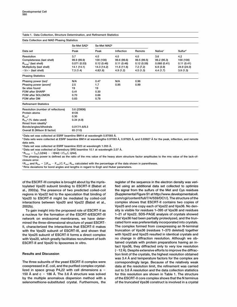
Table 1. Data Collection, Structure Determination, and Refinement Statistics
Data Collection and MAD Phasing Statistics
Se-Met SADa Se-Met MADb
Data set Peak Peak Inflection Remote Nativec Sulfurd
Resolution 3.7 4.0 4.0 4.0 3.6 4.2Completeness (last shell) 99.9 (99.9) 100 (100) 99.6 (99.6) 99.5 (99.5) 99.2 (95.3) 100 (100)Rmerge
e (last shell) 0.071 (0.53) 0.12 (0.46) 0.11 (0.46) 0.12 (0.59) 0.068 (0.41) 0.11 (0.41)Multiplicity (last shell) 14.1 (14.1) 14.3 (14.2) 11.6 (11.6) 7.2 (7.2) 6.9 (3.9) 24.9 (24.0)�I/�� (last shell) 7.3 (1.4) 4.8(1.6) 4.9 (1.2) 4.5 (1.3) 4.4 (1.7) 3.9 (1.3)
Phasing Statistics
Phasing power (iso)f N/A 0.47 N/A 0.96Phasing power (anom)f 2.5 1.7 0.95 0.99Se sites found 19 19FOM after SHARP 0.41 0.30FOM after SOLOMON 0.70 0.66FOM after DM 0.83 0.78
Refinement Statistics
Resolution (number of reflections) 3.6 (23806)Protein atoms 6135Rcryst
g 0.30Rfree
g (% data used) 0.34 (4.9)Rmsd from idealityh
Bonds/angles/dihedrals 0.017/1.6/8.3Overall B (Wilson B factor) 65 (110)
a Data set was collected at ESRF beamline BM14 at wavelength 0.97093 A.b Data sets were collected at ESRF beamline BM14 at wavelengths 0.97093 A, 0.97925 A, and 0.93927 A for the peak, inflection, and remotedata sets.c Data set was collected at ESRF beamline ID23 at wavelength 1.055 A.d Data set was collected at Daresbury SRS beamline 10.1 at wavelength 2.07 A.e Rmerge � �hkl�i|Ii(hkl) � �I(hkl)�|/�hkl�i Ii(hkl).f The phasing power is defined as the ratio of the rms value of the heavy atom structure factor amplitudes to the rms value of the lack-of-closure error.g Rcryst and Rfree � �|Fobs � Fcalc|/� Fobs; Rfree calculated with the percentage of the data shown in parentheses.h Rms deviations for bond angles and lengths in regard to Engh and Huber parameters.
of the ESCRT-III complex is brought about by the myris- register of the sequence in the electron density was veri-fied using an additional data set collected to optimizetoylated Vps20 subunit binding to ESCRT-II (Babst etthe signal from the sulfurs of the Met and Cys residuesal., 2002a). The presence of two predicted coiled-coil(Supplemental Figure S1 at http://www.developmentalcell.regions in Vps22 led to the speculation that binding ofcom/cgi/content/full/7/4/559/DC1/). The structure of theVps20 to ESCRT-II might be mediated by coiled-coilcomplex shows that ESCRT-II contains two copies ofinteractions between Vps20 and Vps22 (Babst et al.,Vps25 and one copy each of Vps22 and Vps36. No den-2002b).sity is visible for residues 1–395 of Vps36 and residuesTo gain insight into the proposed role of ESCRT-II as1–21 of Vps22. SDS-PAGE analysis of crystals showeda nucleus for the formation of the ESCRT-II/ESCRT-IIIthat Vps36 had been partially proteolyzed, and the trun-network on endosomal membranes, we have deter-cated form was preferentially incorporated into crystals.mined the three-dimensional structure of yeast ESCRT-The complex formed from coexpressing an N-terminalII, characterized the interactions that ESCRT-II makestruncation of Vps36 (residues 1–370 deleted) togetherwith the Vps20 subunit of ESCRT-III, and shown thatwith Vps22 and Vps25 resulted in identical crystals andthe Vps25 subunit of ESCRT-II forms a direct complexno change in diffraction resolution. Although we ob-with Vps20, which greatly facilitates recruitment of bothtained crystals with protein preparations having an in-ESCRT-II and Vps20 to liposomes in vitro.tact Vps36, they diffracted only to very low resolution(�12 A). Despite extensive efforts to improve the diffrac-
Results and Discussion tion limit of the crystals, the highest resolution obtainedwas 3.4 A and temperature factors for the complex are
The three subunits of the yeast ESCRT-II complex were correspondingly large. Because of the relatively weakcoexpressed in E. coli, and the purified complex crystal- data at the resolution limit, the refinement was carriedlized in space group P4322 with cell dimensions a � out to 3.6 A resolution and the data collection statistics150 A and c � 186 A. The 3.6 A structure was solved for this resolution are shown in Table 1. The structureby the multiple anomalous dispersion method with a of the ESCRT-II core complex shows that the N terminus
of the truncated Vps36 construct is involved in a crystalselenomethione-substituted crystal. Furthermore, the
The Crystal Structure of the ESCRT-II Complex561
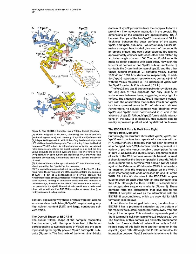
domain of Vps22 protrudes from the complex to form aprominent intermolecular interaction in the crystal. Thedimensions of the complex are approximately 120 Abetween the tips of the two Vps25 domains and 50 A inthickness between the outer surfaces of the pairedVps22 and Vps36 subunits. Two structurally similar do-mains arranged head-to-tail give each of the subunitsan oblong shape. The two Vps25 subunits are alignedapproximately colinear with each other and related bya rotation angle of about 156�. The two Vps25 subunitsmake no direct contacts with each other. However, theN-terminal domain of one Vps25 subunit (molecule B)contacts the C-terminal domain of Vps22, and the otherVps25 subunit (molecule C) contacts Vps36, burying1637 A2 and 1531 A2 surface area, respectively. In addi-tion, Vps36 makes much less extensive contacts (444 A2)with the Vps25 molecule B. The interface of Vps22 withthe Vps25 molecule C is minimal (155 A2).
The Vps22 and Vps36 subunits pair side-by-side alongthe long axis of their ellipsoids and bury 2669 A2 ofsurface area between them, suggesting a very tight in-terface. The extensive Vps22/Vps36 interface is consis-tent with the observation that neither Vps36 nor Vps22can be expressed alone in E. coli (data not shown).Furthermore, no soluble complex was obtained whenVps22 and Vps36 were coexpressed in E. coli in theabsence of Vps25. Although Vps25 forms stable interac-tions in the ESCRT-II complex, this subunit can bereadily expressed, purified, and crystallized on its own.
The ESCRT-II Core Is Built from EightFigure 1. The ESCRT-II Complex Has a Trilobal Overall Structure
Winged Helix Domains(A) Ribbon diagram of ESCRT-II, containing two Vps25 subunits
Surprisingly, the structure shows that Vps22, Vps25, and(each making one lobe), and one copy of Vps22 and Vps36 subunitVps36 each contain two repeats of a domain with an(tightly packed together into a third lobe). Only the C-terminal portionH1/1/H2/H3/2/3 topology that has been referred toof Vps36 is ordered in the crystals. The protruding N-terminal helical
domain of Vps22 subunit is colored orange, while its two winged as a “winged helix” (WH) domain, which is present in ahelix domains are yellow; the Vps36 subunit is pink and the two variety of proteins—most notably transcription factorsVps25 subunits are colored cyan and blue. The two winged helix (Figure 2; Gajiwala and Burley, 2000). The three helices(WH) domains in each subunit are labeled as WHA and WHB. The
of each of the WH domains assemble beneath a smallelements of secondary structure and the N and C termini are also in- sheet formed by the three antiparallel strands. Withindicated.each subunit, the N-terminal WH domain (WHA) packs(B) A view of the complex approximately 90� from the view in (A),
showing a rather flat “profile” of the complex. against the C-terminal WH domain (WHB) in a head-to-(C) The crystallographic coiled-coil interaction of the Vps22 N-ter- tail manner, with the exposed surface on the WHA minal helix. The asymmetric unit of the crystal contains one complex sheet interacting with ends of helices H1 and H3 of theof ESCRT-II, but as a consequence of a crystal contact, the
WHB. All of the WH domains in the ESCRT-II complexN-terminal helices of Vps22 molecules from two adjacent complexessuperimpose on each other with an rms deviation lesspack together, forming an antiparallel coiled-coil (one molecule isthan 2 A, although the three ESCRT-II subunits havecolored orange, the other white). ESCRT-II is a monomer in solution,
but potentially, the Vps22 N-terminal helix could form a coiled-coil no recognizable sequence similarity (Figure 3). Thesedimer, either with another ESCRT-II complex or some other (cur- domains form the interactions that give rise to therently unknown) binding partner. ESCRT-II complex, as well as the interactions with the
ESCRT-III subcomplexes, which are essential for MVBformation (see below).contact, explaining why these crystals were not able to
accommodate the full-length Vps36 despite having very In addition to the winged helix core, the structure ofESCRT-II has a prominent extension at the bottom ofhigh solvent content (72%) and large, continuous sol-
vent voids. the Vps22/Vps36 stem, which protrudes away from thebody of the complex. This extension represents part ofthe N-terminal 5-helix domain of Vps22 (residues 22–90).The Overall Shape of ESCRT-II
The overall trilobal shape of the complex resembles The first helix of this domain is a distinctly amphipathichelix that forms coiled-coil interactions with a 2-foldthe character , with the upper branches of the letter
corresponding to two molecules of Vps25 and the stem related copy of this helix from another complex in thecrystal (Figure 1C). Although this 2-fold intermolecularrepresenting the tightly packed Vps22 and Vps36 sub-
units (Figure 1). The first helix of the N-terminal 5-helix interaction between Vps22 subunits is extensive in the
Developmental Cell562
in protein-protein interactions that facilitate transcrip-tional activation. The analogous H3 and sheet surfacesof the Vps22 and Vps36 WHB domains expose a seriesof hydrophobic residues that interact with Vps25. Inaddition, Vps25 has prominent exposed hydrophobicpatches that could bind other proteins such as Vps20(see below).
Interactions between the Vps22and Vps36 SubunitsThe yeast Vps22/Vps36 interaction surface is largelyhydrophobic (Figure 4A), with several interspersed saltlinks. There are four salt links between the Vps22 WHAand Vps36 WHA domains; the residues forming thesesalt links are not conserved among Vps22 and Vps36subunits from other species (Figure 4A). The contactsbetween the Vps22 and Vps36 N-terminal WH domainsare more extensive than between the C-terminal WHdomains. These WHA contacts involve helix H1 and theH3/2 loop of Vps22. On the Vps36 side, the interactionsinvolve helices H2 and H3 and the loops preceding themand connecting them. The Vps22/36 contacts in theWHB domains are more restricted and involve H3 ofVps22 and the 2/3 loop (Wing 1) of Vps36.
Interactions of the Vps25 Subunitswith Vps22 and Vps36Whereas the Vps22/Vps36 subunits interact side-by-
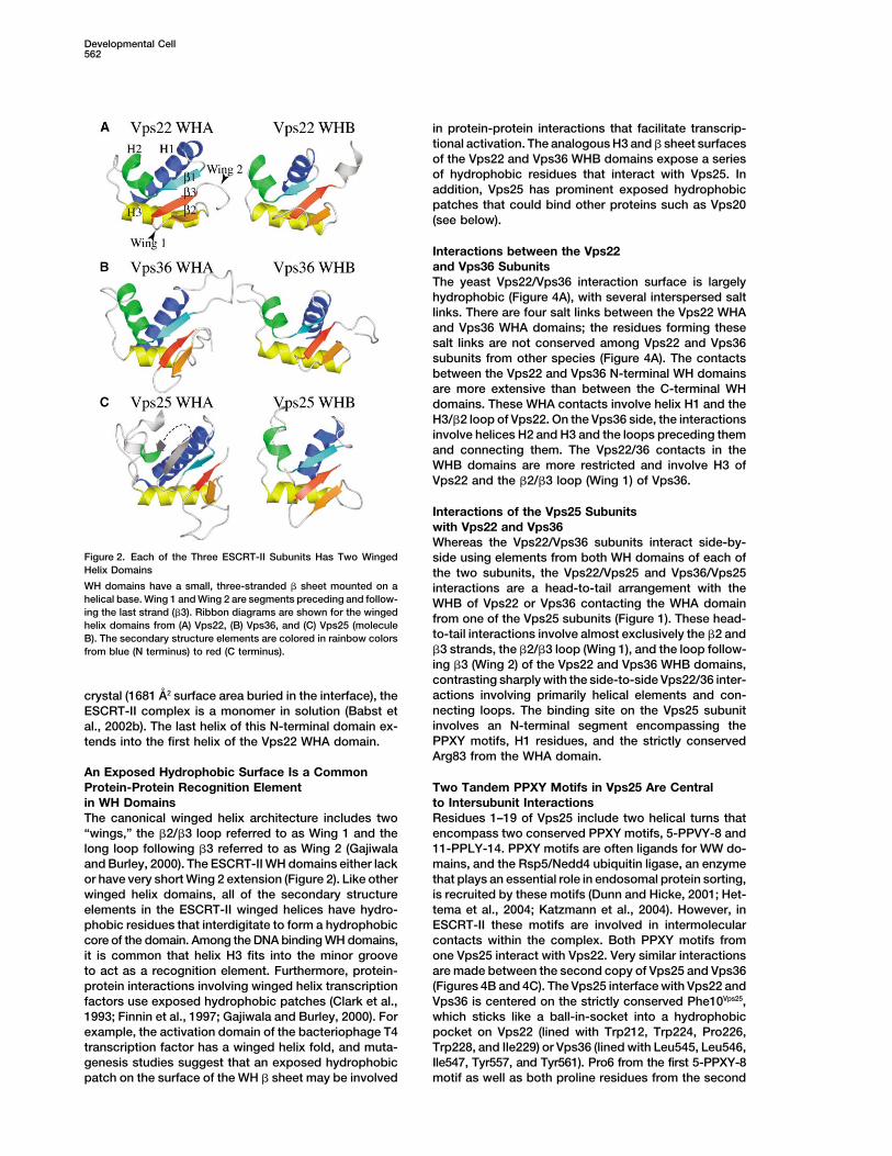
Figure 2. Each of the Three ESCRT-II Subunits Has Two Winged side using elements from both WH domains of each ofHelix Domains the two subunits, the Vps22/Vps25 and Vps36/Vps25WH domains have a small, three-stranded sheet mounted on a interactions are a head-to-tail arrangement with thehelical base. Wing 1 and Wing 2 are segments preceding and follow- WHB of Vps22 or Vps36 contacting the WHA domaining the last strand (3). Ribbon diagrams are shown for the winged from one of the Vps25 subunits (Figure 1). These head-helix domains from (A) Vps22, (B) Vps36, and (C) Vps25 (molecule
to-tail interactions involve almost exclusively the 2 andB). The secondary structure elements are colored in rainbow colors3 strands, the 2/3 loop (Wing 1), and the loop follow-from blue (N terminus) to red (C terminus).ing 3 (Wing 2) of the Vps22 and Vps36 WHB domains,contrasting sharply with the side-to-side Vps22/36 inter-actions involving primarily helical elements and con-crystal (1681 A2 surface area buried in the interface), thenecting loops. The binding site on the Vps25 subunitESCRT-II complex is a monomer in solution (Babst etinvolves an N-terminal segment encompassing theal., 2002b). The last helix of this N-terminal domain ex-PPXY motifs, H1 residues, and the strictly conservedtends into the first helix of the Vps22 WHA domain.Arg83 from the WHA domain.
An Exposed Hydrophobic Surface Is a CommonProtein-Protein Recognition Element Two Tandem PPXY Motifs in Vps25 Are Central
to Intersubunit Interactionsin WH DomainsThe canonical winged helix architecture includes two Residues 1–19 of Vps25 include two helical turns that
encompass two conserved PPXY motifs, 5-PPVY-8 and“wings,” the 2/3 loop referred to as Wing 1 and thelong loop following 3 referred to as Wing 2 (Gajiwala 11-PPLY-14. PPXY motifs are often ligands for WW do-
mains, and the Rsp5/Nedd4 ubiquitin ligase, an enzymeand Burley, 2000). The ESCRT-II WH domains either lackor have very short Wing 2 extension (Figure 2). Like other that plays an essential role in endosomal protein sorting,
is recruited by these motifs (Dunn and Hicke, 2001; Het-winged helix domains, all of the secondary structureelements in the ESCRT-II winged helices have hydro- tema et al., 2004; Katzmann et al., 2004). However, in
ESCRT-II these motifs are involved in intermolecularphobic residues that interdigitate to form a hydrophobiccore of the domain. Among the DNA binding WH domains, contacts within the complex. Both PPXY motifs from
one Vps25 interact with Vps22. Very similar interactionsit is common that helix H3 fits into the minor grooveto act as a recognition element. Furthermore, protein- are made between the second copy of Vps25 and Vps36
(Figures 4B and 4C). The Vps25 interface with Vps22 andprotein interactions involving winged helix transcriptionfactors use exposed hydrophobic patches (Clark et al., Vps36 is centered on the strictly conserved Phe10Vps25,
which sticks like a ball-in-socket into a hydrophobic1993; Finnin et al., 1997; Gajiwala and Burley, 2000). Forexample, the activation domain of the bacteriophage T4 pocket on Vps22 (lined with Trp212, Trp224, Pro226,
Trp228, and Ile229) or Vps36 (lined with Leu545, Leu546,transcription factor has a winged helix fold, and muta-genesis studies suggest that an exposed hydrophobic Ile547, Tyr557, and Tyr561). Pro6 from the first 5-PPXY-8
motif as well as both proline residues from the secondpatch on the surface of the WH sheet may be involved

The Crystal Structure of the ESCRT-II Complex563
Figure 3. A Schematic Illustration of the Domain Organization and Sequence Alignments of the ESCRT-II Subunits
(A) WHA denotes winged helix A and WHB denotes winged helix B. ND denotes N-terminal helical domain in Vps22, and NZF stands for Npl4zinc fingers in Vps36. Sc denotes Saccharomyces cerevisiae. Only the NZF-1 domain binds ubiquitin (Alam et al., 2004). The hatched regionin Vps36 represents an insertion in the yeast sequence relative to human. Consequently, NZF domains are absent in human Vps36.(B) Sequence alignments of yeast (Sc) and human (Hs) ESCRT-II sequences with secondary structure elements indicated above the yeastsubunits. Dashed lines indicate disordered regions.
PPXY motif (11-PP-12) make hydrophobic interactions protein), indicating the central importance of these inter-actions for ESCRT-II formation.with Vps22. Equivalent interactions are formed between
Vps36 and the second copy of Vps25. Although thetyrosines of the PPXY motifs make no direct contact Conserved Salt Links Stabilize the Interactionswith either the Vps22 or Vps36 subunits, they buttress of Vps25 with Vps22 and Vps36the Pro-Pro motifs against the 76–85 region of the Vps25 In addition to nonpolar interactions that appear to domi-WHA. A Vps25 double mutant, Y8A/Y14A, when coex- nate the Vps25/Vps22 interface, there is a salt link be-pressed with Vps22 and Vps36, yielded only free Vps25 tween Arg83 of Vps25 and Asp214 in the WHB domain ofand no intact complex (Vps22 and Vps36 coexpression Vps22 (Figure 4). Both of these residues are conserved
in Vps22 and Vps25 subunits from other species. Thein E. coli in the absence of Vps25 yields no soluble

Developmental Cell564
Figure 4. Hydrophobic Interactions Inter-sperse with a Few Salt Links to Form the Inter-subunit Contacts in ESCRT-II
In solid surface representation, hydrophobicresidues are colored white, residues involvedin intersubunit salt links are colored red(acidic) and blue (basic), and the rest of theresidues are colored bronze.(A) The interface between Vps22 (solid sur-face) and Vps36 (pink ribbon) involves bothWH domains from both subunits. Vps36 resi-dues making hydrophobic interactions areshown as pink sticks, and those forming saltlinks are blue or red sticks.(B) The interface between the WHB domainof Vps22 (solid surface) with the WHA domainof Vps25 (blue worm) is predominantly hy-drophobic and includes proline residues fromthe PPXY motifs in Vps25 (cyan). Phe10Vps25
(green) at the junction between the two PPXYmotifs fits into a hydrophobic pocket onVps22. Neither of the tyrosines (purple) fromthe PPXY motifs interacts with Vps22. Con-served residues forming a salt bridge be-tween the two molecules (Asp214Vps22 andArg83Vps25) are highlighted. The locations ofsome of the interacting residues from Vps22are also indicated.(C) The interface between the WHB domainof Vps36 (solid surface) with the second copy
of Vps25 (light blue worm) shows similar features as Vps22/25 interface, including the hydrophobic “ball-joint” formed by Phe10Vps25, interactionswith the PPXY motifs, and the salt bridge between the Asp548Vps36 and Arg83Vps25.
second molecule of Vps25 in the ESCRT-II complex In light of the ESCRT-II crystal structure, which showsthat the N-terminal helix of Vps22 protrudes from themakes equivalent interactions with the WHB domain ofstem of the complex and makes coiled-coil interactionsVps36, which is pseudo-symmetric with the WHB do-with and adjacent complex in the crystal, this was a verymain of Vps22. An analogous salt link is formed betweenattractive proposal. To test this, we made an N-terminalArg83 of Vps25 and Asp548 of Vps36. Similarly totruncation of Vps22 (residues 1–40 deleted) that re-Asp214 of Vps22, Asp548 of Vps36 is conserved inmoves most of the protruding helix H1 of the N-terminalother species.domain and coexpressed and purified the ESCRT-IIcomplex. This truncation had no noticeable affect onESCRT-II Interacts Directly with Vps20 Subunitcomplex formation, as expected based on the lack offrom ESCRT-IIIinteractions of this helix with the rest of the complex.Both genetic and biochemical data indicate that ESCRT-Surprisingly, the truncated complex bound as well asII interacts with Vps20 (Babst et al., 2002b; Bowers etfull-length ESCRT-II to GST-Vps20 (Figures 5A and 5B),al., 2004; Martin-Serrano et al., 2003; von Schwedler etshowing that the N-terminal helix of Vps22 is not respon-al., 2003), and it was shown that this interaction playssible for interaction with Vps20. In addition, the purifiedan important role in the formation of ESCRT-III complexN-terminal fragment of Vps22 (residues 1–90, which we(Babst et al., 2002a). However, because all the currentcould purify in large amounts [50 mg/l E. coli culture])evidence is based on yeast two-hybrid interactions ordid not show any binding to Vps20 (data not shown),pull-down assays using yeast extract, it is not clearconfirming that the N-terminal helical domain of Vps22
whether the interaction is direct or indirect, via an un-is neither necessary nor sufficient for Vps20 interaction.
known bridging molecule. We used purified ESCRT-IICurrently, we cannot exclude the possibility that this
and Vps20 to examine whether they can interact directly N-terminal helical domain of Vps22 could be involvedin vitro. Using GST-tagged Vps20 and pull-down assays, in coiled-coil interactions with some other ESCRT-IIIwe could demonstrate direct, specific interaction be- subunit. However, comprehensive yeast two-hybridtween full-length ESCRT-II and Vps20 that results in all analysis of human ESCRT-II/ESCRT-III interactions didthree ESCRT-II subunits being retained by GST-Vps20 not detect any interaction of ESCRT-II other than withon glutathione beads (Figure 5A). Vps20 subunit of ESCRT-III, suggesting that Vps22-medi-
ated coiled-coil interactions with ESCRT-III are not likelyThe Protruding N-Terminal Coiled-Coil Region (Bowers et al., 2004; Martin-Serrano et al., 2003; vonof Vps22 Does Not Bind Vps20 Schwedler et al., 2003).The next question is which ESCRT-II subunit(s) is re- In addition, we show that ESCRT-II complexes insponsible for interaction with Vps20. It was previously which the N terminus of Vps36 is truncated (residuesproposed that the predicted coiled-coil regions of Vps22 1–370 deleted) do not affect binding to Vps20 (Figure
5B). This region of Vps36 is, however, necessary forcould bind to coiled-coils of Vps20 (Babst et al., 2002b).
The Crystal Structure of the ESCRT-II Complex565
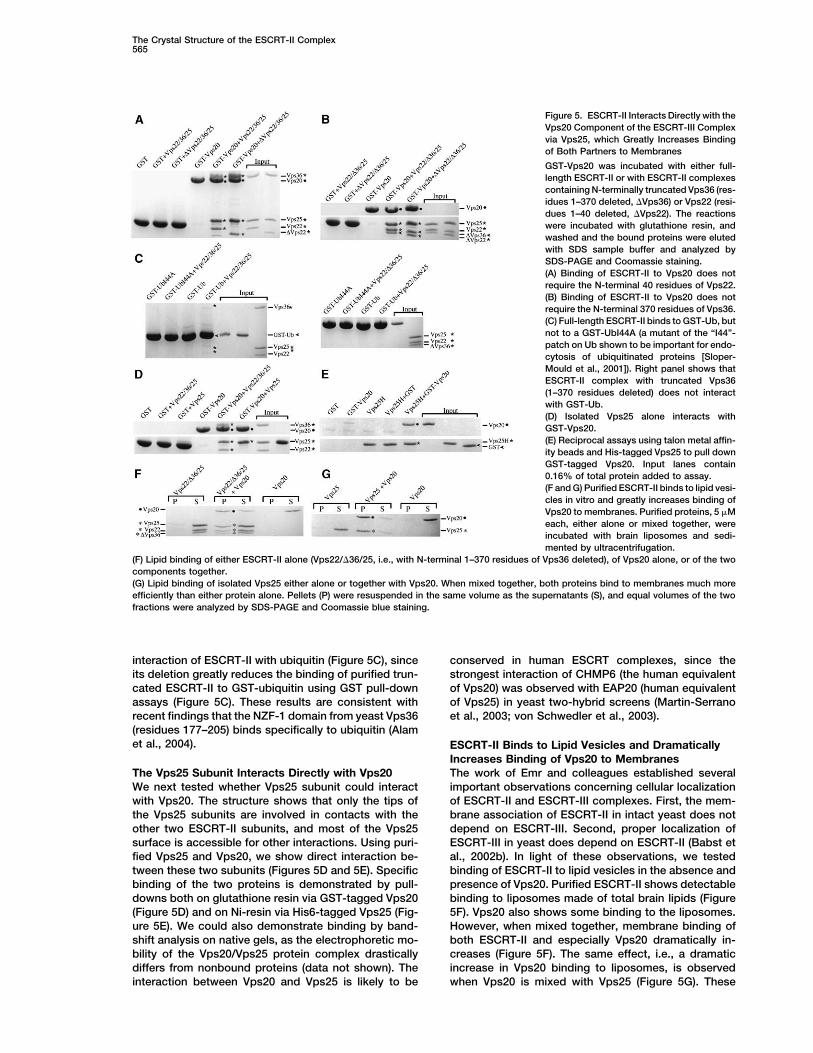
Figure 5. ESCRT-II Interacts Directly with theVps20 Component of the ESCRT-III Complexvia Vps25, which Greatly Increases Bindingof Both Partners to Membranes
GST-Vps20 was incubated with either full-length ESCRT-II or with ESCRT-II complexescontaining N-terminally truncated Vps36 (res-idues 1–370 deleted, �Vps36) or Vps22 (resi-dues 1–40 deleted, �Vps22). The reactionswere incubated with glutathione resin, andwashed and the bound proteins were elutedwith SDS sample buffer and analyzed bySDS-PAGE and Coomassie staining.(A) Binding of ESCRT-II to Vps20 does notrequire the N-terminal 40 residues of Vps22.(B) Binding of ESCRT-II to Vps20 does notrequire the N-terminal 370 residues of Vps36.(C) Full-length ESCRT-II binds to GST-Ub, butnot to a GST-UbI44A (a mutant of the “I44”-patch on Ub shown to be important for endo-cytosis of ubiquitinated proteins [Sloper-Mould et al., 2001]). Right panel shows thatESCRT-II complex with truncated Vps36(1–370 residues deleted) does not interactwith GST-Ub.(D) Isolated Vps25 alone interacts withGST-Vps20.(E) Reciprocal assays using talon metal affin-ity beads and His-tagged Vps25 to pull downGST-tagged Vps20. Input lanes contain0.16% of total protein added to assay.(F and G) Purified ESCRT-II binds to lipid vesi-cles in vitro and greatly increases binding ofVps20 to membranes. Purified proteins, 5 �Meach, either alone or mixed together, wereincubated with brain liposomes and sedi-mented by ultracentrifugation.
(F) Lipid binding of either ESCRT-II alone (Vps22/�36/25, i.e., with N-terminal 1–370 residues of Vps36 deleted), of Vps20 alone, or of the twocomponents together.(G) Lipid binding of isolated Vps25 either alone or together with Vps20. When mixed together, both proteins bind to membranes much moreefficiently than either protein alone. Pellets (P) were resuspended in the same volume as the supernatants (S), and equal volumes of the twofractions were analyzed by SDS-PAGE and Coomassie blue staining.
interaction of ESCRT-II with ubiquitin (Figure 5C), since conserved in human ESCRT complexes, since thestrongest interaction of CHMP6 (the human equivalentits deletion greatly reduces the binding of purified trun-
cated ESCRT-II to GST-ubiquitin using GST pull-down of Vps20) was observed with EAP20 (human equivalentof Vps25) in yeast two-hybrid screens (Martin-Serranoassays (Figure 5C). These results are consistent with
recent findings that the NZF-1 domain from yeast Vps36 et al., 2003; von Schwedler et al., 2003).(residues 177–205) binds specifically to ubiquitin (Alamet al., 2004). ESCRT-II Binds to Lipid Vesicles and Dramatically
Increases Binding of Vps20 to MembranesThe work of Emr and colleagues established severalThe Vps25 Subunit Interacts Directly with Vps20
We next tested whether Vps25 subunit could interact important observations concerning cellular localizationof ESCRT-II and ESCRT-III complexes. First, the mem-with Vps20. The structure shows that only the tips of
the Vps25 subunits are involved in contacts with the brane association of ESCRT-II in intact yeast does notdepend on ESCRT-III. Second, proper localization ofother two ESCRT-II subunits, and most of the Vps25
surface is accessible for other interactions. Using puri- ESCRT-III in yeast does depend on ESCRT-II (Babst etal., 2002b). In light of these observations, we testedfied Vps25 and Vps20, we show direct interaction be-
tween these two subunits (Figures 5D and 5E). Specific binding of ESCRT-II to lipid vesicles in the absence andpresence of Vps20. Purified ESCRT-II shows detectablebinding of the two proteins is demonstrated by pull-
downs both on glutathione resin via GST-tagged Vps20 binding to liposomes made of total brain lipids (Figure5F). Vps20 also shows some binding to the liposomes.(Figure 5D) and on Ni-resin via His6-tagged Vps25 (Fig-
ure 5E). We could also demonstrate binding by band- However, when mixed together, membrane binding ofboth ESCRT-II and especially Vps20 dramatically in-shift analysis on native gels, as the electrophoretic mo-
bility of the Vps20/Vps25 protein complex drastically creases (Figure 5F). The same effect, i.e., a dramaticincrease in Vps20 binding to liposomes, is observeddiffers from nonbound proteins (data not shown). The
interaction between Vps20 and Vps25 is likely to be when Vps20 is mixed with Vps25 (Figure 5G). These
Developmental Cell566
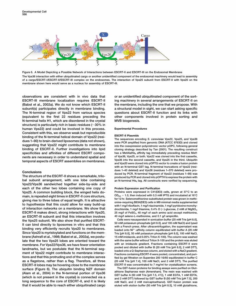
Figure 6. A Model Depicting a Possible Network of Interactions between ESCRT-II and ESCRT-III on the Endosomal Membrane
The Vps36 interaction with either ubiquitinated cargo or another unidentified component of the endosomal machinery would lead to assemblyof a cargo/ESCRT-I/ESCRT-II/ESCRT-III complex on the endosomes. The interaction of Vps25 subunit from ESCRT-II with Vps20 on themembrane shown here would serve as a nucleus for assembly of ESCRT-III.
observations are consistent with in vivo data that or an unidentified ubiquitinated component of the sort-ing machinery in several arrangements of ESCRT-II onESCRT-III membrane localization requires ESCRT-II
(Babst et al., 2002a). We do not know which ESCRT-II the membrane, including the one that we propose. Witha structural model in sight, we can start asking specificsubunit(s) participates directly in membrane binding.
The N-terminal region of Vps22 from various species questions about ESCRT-II function and its links withother components involved in protein sorting and(equivalent to the first 22 residues preceding the
N-terminal helix H1, which are disordered in the crystal MVB biogenesis.structure) is particularly rich in basic residues (�30% in
Experimental Procedureshuman Vps22) and could be involved in this process.Consistent with this, we observe weak but reproducible
ESCRT-II Plasmidsbinding of the N-terminal helical domain of Vps22 (resi-The sequences encoding S. cerevisiae Vps22, Vps25, and Vps36
dues 1–90) to brain-derived liposomes (data not shown), were PCR amplified from genomic DNA (ATCC 9763D) and clonedsuggesting that Vps22 might contribute to membrane into the coexpression polycistronic vector pOPC, following generalbinding of ESCRT-II. Further investigations into lipid cloning strategy described by Tan (2001). The resulting construct
has a MetAlaHis6 affinity tag immediately preceding residue Met1specificities and affinities of different ESCRT compo-of Vps36, Vps25, or both. Vps22 was cloned into the first cassette,nents are necessary in order to understand spatial andVps36 into the second cassette, and Vps25 in the third. Ubiquitintemporal aspects of ESCRT assemblies on membranes.and Vps20 were cloned into pOPTG vector to create a fusion proteinwith an N-terminal GST tag. N-terminal truncations of Vps22 (resi-dues 1–40 deleted) and Vps36 (residues 1–370 deleted) were pro-
Conclusions duced by PCR. N-terminal fragment of Vps22 (residues 1–90) wasThe structure of the ESCRT-II shows a remarkable, trilo- produced by PCR and cloned into pOPTH to express the protein withbal subunit arrangement, with one lobe containing an N-terminal His6 tag. All constructs were verified by sequencing.Vps22/Vps36 sandwiched together side-by-side andeach of the other two lobes containing one copy of Protein Expression and Purification
Proteins were expressed in C41(DE3) cells, grown at 37�C to anVps25. A common building block, the winged helix do-OD600 � 1.0, then induced with 0.3 mM IPTG and incubated at 16�Cmain, is repeated eight times in ESCRT-II core structure,for 12 hr. Selenomethionine-substituted protein was grown in methi-giving rise to three lobes of equal length. It is attractiveonine-requiring B834(DE3) cells in M9 minimal media supplemented
to hypothesize that this could allow for easy build-up with 1 mg/l riboflavin, 1 mg/l niacinamide, 1 mg/l pyridoxine monohy-of interaction networks on a membrane. We show that drochloride, 1 mg/l thiamine, 0.4% D (�)-glucose, 2 mM of MgSO4,
25 mg/l of FeSO4, 40 mg/l of each amino acid except methionine,ESCRT-II makes direct, strong interactions with Vps20,40 mg/l seleno-L-methionine, and 0.1 g/l ampicillin.an ESCRT-III subunit and that this interaction involves
Cells were resuspended in sonication buffer (20 mM Tris [pH 8.0],the Vps25 subunit. We also show that purified ESCRT-50 mM potassium phosphate [pH 8.0], and 100 mM NaCl) and lysedII can bind directly to liposomes in vitro and that thiswith a French press. After ultracentrifugation, the supernatant was
binding very efficiently recruits Vps20 to membranes. loaded onto Ni2�-affinity column equilibrated with buffer A (20 mMSince Vps20 is myristoylated and functions on the mem- Tris [pH 8.0], 50 mM potassium phosphate [pH 8.0], 100 mM NaCl,
15 mM imidazole, and 0.05% Triton X-100). The column was washedbrane (Ashrafi et al., 1998; Babst et al., 2002a), we postu-with the same buffer without Triton X-100 and the protein was elutedlate that the two Vps25 lobes are oriented toward thewith an imidazole gradient. Fractions containing ESCRT-II weremembrane. For Vps22/Vps36, we have fewer orientationpooled and diluted with buffer B (20 mM Tris [pH 8.0], 2 mM DTT),landmarks, but we postulate that the N-terminal seg-loaded onto a Q-Sepharose column, and eluted with a NaCl gradient.
ment of Vps22 might contribute to membrane interac- Fractions containing ESCRT-II were pooled, concentrated, and puri-tions and that this protruding end of the complex serves fied by gel filtration on Superdex 200 16/60 equilibrated in buffer C
(20 mM Tris [pH 7.4], 100 mM NaCl, and 2 mM DTT). The purifiedas a flagstone, rather than a flag. Therefore, all threeESCRT-II was concentrated to 7 mg/ml for crystallization screens.ESCRT-II lobes may be hovering close to the membrane
The GST-fusion proteins for binding assays were purified on Glut-surface (Figure 6). The ubiquitin binding NZF domainathione Sepharose resin (Amersham). The resin was washed with(Alam et al., 2004) in the N-terminal portion of Vps36GST buffer A (50 mM Tris [pH 7.5, 4�C], 1 mM EGTA, 1 mM EDTA,
(which is not present in the crystal) is connected via a and 2 mM DTT) followed by GST buffer B (50 mM Tris [pH 7.5], 200long sequence to the core of ESCRT-II, and it is likely mM NaCl, and 2 mM mercaptoethanol). GST-fusion protein was
eluted with elution buffer (50 mM Tris [pH 8.5], 10 mM glutathione,that it would be able to reach either ubiquitinated cargo
The Crystal Structure of the ESCRT-II Complex567
and 2 mM mercaptoethanol). The GST-fusion proteins were concen- complex and was more difficult to interpret. The density in the 1/H3loop was particularly difficult to interpret. Following strand 1, theretrated and desalted using PD-10 column from Amersham Biosci-
ences. is a -hairpin encompassing residues 50–74 with residues 55–71 notvisible. Given that there are no unambiguous experimental sequencemarkers in this region (no Met and no Cys residues), there is someCrystallizationuncertainty of the sequence register in the 50–79 region of Vps25Solutions for 1200 crystallization conditions were dispensed intoWHA. The region from 55 to 74 represents an insertion in the yeastreservoirs of 96-well crystallization plates (Corning, NY). Protein (100Vps25 relative to other species, so that the 50–74 -hairpin may benl) and reservoir (100 nl) solutions were added to the plates as sittingunique to S. cerevisiae. There are several regions in which the sidedrops using a Cartesian robot (Genomics Solutions, Huntingdon,chain density is not clear, but the main chain trace is unambiguous.UK) and incubated at 17�C. Optimal crystals were obtained in 3.1%This includes much of the N-domain of Vps22 and the WHB domainsPEG 35000, 0.1 M Tris acetate (pH 8.5), 1.36 M sodium formate,from Vps25. In general, the side chain density for larger residues,and 19% glycerol. Crystals used for data collection were grown byparticularly in the hydrophobic cores and at the intersubunit inter-adding 10 �l of protein to sitting wells containing 5 �l of silicafaces, can be discerned.hydrogel (Hampton Research) preincubated with 5 �l of reservoir
solution and 2 �l of additive, either 1 M ammonium sulfate or 0.1 MPull-Down AssaysATP. Silica hydrogel was made by mixing equal parts of sodiumFor GST pull-downs, glutathione resin in minispin columns (Amer-silicate solution (2.8% v/v) with diluted acetic acid. Crystals weresham Biosciences) was prewashed twice with binding buffer (PBScryoprotected by transferring them to cryoprotectant solution con-with 0.05% Triton X-100). A 200–250 �g aliquot of GST-tagged pro-taining 3.1% PEG 35000, 0.1 M Tris acetate (pH 8.5), 1.36 M sodiumtein and 600–700 �g of His-tagged protein were mixed with resinformate, and 22% glycerol before freezing in the cryostream atand E. coli cytosol (10 mg/ml) in binding buffer. Identical results100 K.were obtained when the E. coli cytosol was omitted from the assays.The assay mix was incubated for 2 hr at 4�C on a rotating wheelData Collection, Phasing, and Model Refinementand washed eight times with binding buffer. Bound proteins wereDiffraction data were collected at 100 K. A 3.7 A resolution SADeluted with SDS sample buffer and analyzed by SDS-PAGE. Fordata set was collected at the peak of the fluorescence spectrumHis-tag pull-downs, minispin columns (Perbio) were packed with 40for a dehydrated, selenomethionine-substituted crystal and used�l bed volume of talon metal affinity resin (Clontech) equilibrated infor initial phases and model building. Subsequently, a 4 A resolutionbinding buffer. Samples were washed with binding buffer containingSe-Met data set was collected for a crystal that was not dehydrated,20 mM imidazole (pH 8.0), and the bound proteins were eluted withand this was used to provide initial phases for a 3.6 A resolutionSDS sample buffer.data set from a native crystal that was not dehydrated. For the Se-
Met MAD phasing, data sets were collected at the fluorescenceLiposome Binding Assayspeak, inflection, and remote wavelengths. Intensities were inte-Purified proteins (5 �M) were incubated with 0.5 mg/ml sonicatedgrated with MOSFLM (Leslie, 1992) and scaled using SCALA (CCP4,liposomes in 100 �l reactions. Liposomes were prepared from bo-1994). A total of 19 selenium sites (there are 20 non-N-terminal Metvine brain lipid extract (Folch fraction type I, Sigma B1502) in bufferresidues) were located using the program SnB (Howell et al., 2000;containing 20 mM HEPES (pH 7.4), 150 mM NaCl, and 1 mM DTT.Smith et al., 1998; Weeks and Miller, 1999) and refined with auto-After 15 min incubation at 4�C, reactions were sedimented atSHARP (de La Fortelle and Bricogne, 1997). Solvent flattening was140,000 g in a Beckman TLA100 rotor for 15 min at 4�C. Thecarried out using a solvent content of 71.2% as optimized by SHARP.supernatants (S) and the pellets (P), resuspended in an equal volumeTable 1 lists statistics for data collection and phase refinement. Theof buffer, were analyzed by SDS-PAGE and Coomassie blue staining.phases from the Se-Met MAD data were extended to 3.6 A usingControl proteins, GST, and BSA (Sigma A7638) were used as con-the program DM. The assignment of the sequence to the densitytrols and showed no trace of liposome binding.was consistent with the Se-Met sites. In addition, a high multiplicity
data set was obtained using 2.07 A wavelength radiation in orderto detect sulfur positions by their anomalous scattering (Table 1). Acknowledgments
Anomalous difference electron density maps calculated with thelong wavelength (2.07 A) native data set and the Se peak wavelength We thank Joanne McCarthy, Ed Mitchell, Gordon Leonard, Andrew(0.9793 A) data set were used to establish unambiguous locations McCarthy, Martin Walsh, Didier Nurizzo, and David Hall for assis-of the Cys and Met residues. The long wavelength anomalous differ- tance with data collection at ESRF beamlines BM14, ID14-4, ID29,ence map showed sulfur peaks greater than 3� (average peak height and ID23. We are grateful to J. Helliwell, S. Hasnain, and M. Cianci for5�) for all Cys residues except Vps22 Cys68 and Vps25 Cys181 beamtime and help on Daresbury SRS NWSGC Station 10 (funded byand for all of the non-N-terminal Met residues. Moreover, each Met BBSRC grants 719/B15474 and 719/REI20571 to S. Hasnain and J.residue was associated with a peak greater than 3� in the Se peak Helliwell) and an NWDA project award (N0002170 to M. Cianci). Wewavelength anomalous difference map (average peak height is 11�). thank Michael Wilson for stimulating discussions and Brian Peter forNo Cys residue was associated with a peak in the Se peak wave- liposome preparations. H.T. is supported by the Agency for Science,length anomalous difference map. The use of these two anomalous Technology, and Research of Singapore and B.G. by a fellowshipdifference maps allowed us to identify all of the Cys and Met residues from Secretarıa de Estado de Educacion y Universidades and cofi-and to differentiate between them. This greatly facilitated building nanced by the European Social Fund. The work was supported bythe initial model. Although all three subunits have a similar domain the MRC (R.L.W.). The PDB ID number for the structure reported inorganization, the identities of the polypeptides were unambiguous this paper is 1W7P.even from the earliest stages of model building because of theunique patterns of Cys and Met residues in these subunits, which Received: August 20, 2004have no sequence conservation. The model was built with the pro- Revised: September 2, 2004gram O (Jones et al., 1991) then optimized with cycles of manual Accepted: September 7, 2004fitting and restrained/TLS refinement with REFMAC (Murshudov et Published online: September 16, 2004al., 1997). Nine TLS groups were defined corresponding to one groupfor each of the WH domains and the N-domain of Vps22. The TLS
Referencesrefinement resulted in a lower free R factor than a simple overall Bfactor refinement. Because of the low resolution, atomic B factors
Alam, S.L., Sun, J., Payne, M., Welch, B.D., Blake, B.K., Davis, D.R.,were not refined. Final statistics for all data sets are given in TableMeyer, H.H., Emr, S.D., and Sundquist, W.I. (2004). Ubiquitin interac-1. There are no residues in the disallowed regions of the Ramachan-tions of NZF zinc fingers. EMBO J. 23, 1411–1421.dran plot, and 81% are in the most favored regions as defined by
PROCHECK (Laskowski et al., 1993). Ashrafi, K., Farazi, T.A., and Gordon, J.I. (1998). A role for Saccharo-myces cerevisiae fatty acid activation protein 4 in regulating proteinThe Vps25 WHA domain has the poorest electron density in the

Developmental Cell568
N-myristoylation during entry into stationary phase. J. Biol. Chem. EAP45 and EAP20. A role for yeast EAP-like proteins in regulationof gene expression by glucose. J. Biol. Chem. 276, 16528–16533.273, 25864–25874.
Babst, M., Odorizzi, G., Estepa, E.J., and Emr, S.D. (2000). Mamma- Katzmann, D.J., Babst, M., and Emr, S.D. (2001). Ubiquitin-depen-lian tumor susceptibility gene 101 (TSG101) and the yeast homo- dent sorting into the multivesicular body pathway requires the func-logue, Vps23p, both function in late endosomal trafficking. Traffic tion of a conserved endosomal protein sorting complex, ESCRT-I.1, 248–258. Cell 106, 145–155.
Babst, M., Katzmann, D.J., Estepa-Sabal, E.J., Meerloo, T., and Emr, Katzmann, D.J., Odorizzi, G., and Emr, S.D. (2002). Receptor down-S.D. (2002a). ESCRT-III: an endosome-associated heterooligomeric regulation and multivesicular-body sorting. Nat. Rev. Mol. Cell Biol.protein complex required for MVB sorting. Dev. Cell 3, 271–282. 3, 893–905.Babst, M., Katzmann, D.J., Snyder, W.B., Wendland, B., and Emr, Katzmann, D.J., Sarkar, S., Chu, T., Audhya, A., and Emr, S.D. (2004).S.D. (2002b). Endosome-associated complex, ESCRT-II, recruits Multivesicular body sorting: ubiquitin ligase Rsp5 is required fortransport machinery for protein sorting at the multivesicular body. the modification and sorting of carboxypeptidase S. Mol. Biol. CellDev. Cell 3, 283–289. 15, 468–480.Bache, K.G., Slagsvold, T., Cabezas, A., Rosendal, K.R., Raiborg, Laskowski, R.A., MacArthur, M.W., Moss, D.S., and Thornton, J.M.C., and Stenmark, H. (2004). The growth-regulatory protein HCRP1/ (1993). PROCHECK: a program to check the stereochemical qualityhVps37A is a subunit of mammalian ESCRT-I and mediates receptor of protein structures. J. Appl. Crystallogr. 26, 283–291.downregulation. Mol. Biol. Cell 15, 4337–4346. Leslie, A.G.W. (1992). Recent changes to the MOSFLM package forBishop, N., and Woodman, P. (2001). TSG101/mammalian VPS23 processing film and image plate data. In Joint CCP4 and ESF-and mammalian VPS28 interact directly and are recruited to VPS4- EACMB Newsletter on Protein Crystallography (Warrington, UK:induced endosomes. J. Biol. Chem. 276, 11735–11742. Daresbury Laboratory).Bonifacino, J.S., and Traub, L.M. (2003). Signals for sorting of trans- Li, L., and Cohen, S.N. (1996). Tsg101: a novel tumor susceptibilitymembrane proteins to endosomes and lysosomes. Annu. Rev. Bio- gene isolated by controlled homozygous functional knockout ofchem. 72, 395–447. allelic loci in mammalian cells. Cell 85, 319–329.Bowers, K., Lottridge, J., Helliwell, S.B., Goldthwaite, L.M., Luzio, Martin-Serrano, J., Yarovoy, A., Perez-Caballero, D., and Bieniasz,J.P., and Stevens, T.H. (2004). Protein-protein interactions of ESCRT P.D. (2003). Divergent retroviral late-budding domains recruit vacuo-complexes in the yeast Saccharomyces cerevisiae. Traffic 5, lar protein sorting factors by using alternative adaptor proteins.194–210. Proc. Natl. Acad. Sci. USA 100, 12414–12419.CCP4 (Collaborative Computing Project 4) (1994). The CCP4 suite: Morita, E., and Sundquist, W.I. (2004). Retrovirus budding. Annu.programs for protein crystallography. Acta Crystallogr. D 50, Rev. Cell Dev. Biol. 20, 395–425.760–763.
Murshudov, G.N., Vagin, A.A., and Dodson, E.J. (1997). RefinementClark, K.L., Halay, E.D., Lai, E., and Burley, S.K. (1993). Co-crystal of macromolecular structures by the maximum-likelihood method.structure of the HNF-3/fork head DNA-recognition motif resembles Acta Crystallogr. D 53, 240–255.histone H5. Nature 364, 412–420.
Peschard, P., and Park, M. (2003). Escape from Cbl-mediated down-Conibear, E. (2002). An ESCRT into the endosome. Mol. Cell 10, regulation. A recurrent theme for oncogenic deregulation of receptor215–216. tyrosine kinases. Cancer Cell 3, 519–523.de La Fortelle, E., and Bricogne, G. (1997). Maximum-likelihood
Piper, R.C., and Luzio, J.P. (2001). Late endosomes: sorting andheavy-atom parameter refinement for multiple isomorphous re-
partitioning in multivesicular bodies. Traffic 2, 612–621.placement and multiwavelength anomalous diffraction methods.
Pornillos, O., Alam, S.L., Rich, R.L., Myszka, D.G., Davis, D.R., andMethods Enzymol. 276, 472–494.Sundquist, W.I. (2002). Structure and functional interactions of the
Dunn, R., and Hicke, L. (2001). Domains of the Rsp5 ubiquitin-proteinTsg101 UEV domain. EMBO J. 21, 2397–2406.
ligase required for receptor-mediated and fluid-phase endocytosis.Raiborg, C., Rusten, T.E., and Stenmark, H. (2003). Protein sortingMol. Biol. Cell 12, 421–435.into multivesicular endosomes. Curr. Opin. Cell Biol. 15, 446–455.Finnin, M.S., Cicero, M.P., Davies, C., Porter, S.J., White, S.W., andReggiori, F., and Pelham, H.R.B. (2001). Sorting of proteins intoKreuzer, K.N. (1997). The activation domain of the MotA transcriptionmultivesicular bodies: ubiquitin-dependent and -independent tar-factor from bacteriophage T4. EMBO J. 16, 1992–2003.geting. EMBO J. 20, 5176–5186.Gajiwala, K.S., and Burley, S.K. (2000). Winged helix proteins. Curr.Schmidt, A.E., Miller, T., Schmidt, S.L., Shiekhattar, R., and Shilati-Opin. Struct. Biol. 10, 110–116.fard, A. (1999). Cloning and characterization of the EAP30 subunitGarrus, J.E., von Schwedler, U.K., Pornillos, O.W., Morham, S.G.,of the ELL complex that confers derepression of transcription byZavitz, K.H., Wang, H.E., Wettstein, D.A., Stray, K.M., Cote, M., Rich,RNA polymerase II. J. Biol. Chem. 274, 21981–21985.R.L., et al. (2001). Tsg101 and the vacuolar protein sorting pathwaySloper-Mould, K.E., Jemc, J.C., Pickart, C.M., and Hicke, L. (2001).are essential for HIV-1 budding. Cell 107, 55–65.Distinct functional surface regions on ubiquitin. J. Biol. Chem.Gruenberg, J., and Stenmark, H. (2004). The biogenesis of multive-276, 30483–30489.sicular endosomes. Nat. Rev. Mol. Cell Biol. 5, 317–323.Smith, G.D., Nagar, B., Rini, J.M., Hauptman, H.A., and Blessing,Hettema, E.H., Valdez-Taubas, J., and Pelham, H.R. (2004). Bsd2R.H. (1998). The use of SnB to determine an anomalous scatteringbinds the ubiquitin ligase Rsp5 and mediates the ubiquitination ofsubstructure. Acta Crystallogr. D 54, 799–804.transmembrane proteins. EMBO J. 23, 1279–1288.Stuchell, M.D., Garrus, J.E., Muller, B., Stray, K.M., Ghaffarian, S.,Hicke, L., and Dunn, R. (2003). Regulation of membrane transportMcKinnon, R., Krausslich, H.G., Morham, S.G., and Sundquist, W.I.by ubiquitin and ubiquitin-binding proteins. Annu. Rev. Cell Dev.(2004). The human endosomal sorting complex required for trans-Biol. 19, 141–172.port (ESCRT-I) and its role in HIV-1 budding. J. Biol. Chem. 279,Howell, P.L., Blessing, R.H., Smith, G.D., and Weeks, C.M. (2000).36059–36071.Optimizing DREAR and SnB parameters for determining Se-atomSundquist, W.I., Schubert, H.L., Kelly, B.N., Hill, G.C., Holton, J.M.,substructures. Acta Crystallogr. D 56, 604–617.and Hill, C.P. (2004). Ubiquitin recognition by the human TSG101Jones, T.A., Zou, J.-Y., Cowan, S.W., and Kjeldgaard, M. (1991).protein. Mol. Cell 13, 783–789.Improved methods for building protein models in electron densityTan, S. (2001). A modular polycistronic expression system for over-maps and the location of errors in these models. Acta Crystallogr.expressing protein complexes in Escherichia coli. Protein Expr.A 47, 110–119.Purif. 21, 224–234.Kamura, T., Burian, D., Khalili, H., Schmidt, S.L., Sato, S., Liu, W.J.,
Conrad, M.N., Conaway, R.C., Conaway, J.W., and Shilatifard, A. Teo, H., Veprintsev, D.B., and Williams, R.L. (2004). Structural in-sights into endosomal sorting complex required for transport(2001). Cloning and characterization of ELL-associated proteins

The Crystal Structure of the ESCRT-II Complex569
(ESCRT-I) recognition of ubiquitinated proteins. J. Biol. Chem.279, 28689–28696.
Urbanowski, J.L., and Piper, R.C. (2001). Ubiquitin sorts proteins intothe intralumenal degradative compartment of the late-endosome/vacuole. Traffic 2, 622–630.
von Schwedler, U.K., Stuchell, M., Muller, B., Ward, D.M., Chung,H.Y., Morita, E., Wang, H.E., Davis, T., He, G.P., Cimbora, D.M., etal. (2003). The protein network of HIV budding. Cell 114, 701–713.
Weeks, C.M., and Miller, R. (1999). The design and implementationof SnB v2.0. J. Appl. Crystallogr. 32, 120–124.