Iodine123-IBZM-SPECT: Studies in 15 patients with pituitary tumors
Upload
independentCategory
view
7download
0
Molecular and Cellular Biochemistry 74:83-94 (1987) © Martinus Nijhoff Publishers, Boston - Printed in the Netherlands 83
Original Article
Distribution of calmodulin and calmodulin-binding proteins in bovine pi- tuitary: association of myosin light chain kinase with pituitary secretory granule membranes
Teresita Yap Nelson 1, Mary Y. Lorenson 2, Laurence S. Jacobs 3 and A. E. Boyd III 4 1,4Departments of Medicine and Cell Biology, Baylor College of Medicine, One Baylor Plaza, Houston, Texas 77030 USA (4address for offprints), 2,3the Endocrine-Metabolism Unit, Department of Medicine and the Clinical Research Center, University of Rochester School of Medicine and Dentistry, Rochester, New York 14642, USA
Received 25 August 1986
Keywords: calmodulin, adenohypophyseal secretory granules, MLCK
Abstract
Calcium is necessary for secretion of pituitary hormones. Many of the biological effects of Ca 2+ are medi- ated by the Ca 2+-binding protein calmodulin (CAM), which interacts specifically with proteins regulated by the Ca 2+-CAM complex. One of these proteins is myosin light chain kinase (MLCK), a Ca 2+-calmodulin de- pendent enzyme that phosphorylates the regulatory light chains of myosin, and has been implicated in motile processes in both muscle and non-muscle tissues. We determined the content and distribution of CaM and CaM-binding proteins in bovine pituitary homogenates, and subcellular fractions including secretory gran- ules and secretory granule membranes. CaM measured by radioimmunoassay was found in each fraction; although approximately one-half was in the cytosolic fraction, CaM was also associated with the plasma membrane and secretory granule fractions. CaM-binding proteins were identified by an lZSI-CaM gel overlay technique and quantitated by densitometric analysis of the autoradiograms. Pituitary homogenates con- tained nine major CaM-binding proteins of 146, 131, 90, 64, 58, 56, 52, 31 and 22 kilodaltons (kDa). Binding to all the bands was specific, Ca 2+-sensitive, and displaceable with excess unlabeled CaM. Severe heat treat- ment (100 °C, 15 min), which results in a 75% reduction in phosphodiesterase activation by CaM, markedly decreased ~25I-CaM binding to all protein bands. Secretory granule membranes showed enhancement for CaM-binding proteins with molecular weights of 184, 146, 131, 90, and 52000. A specific, affinity purified antibody to chicken gizzard MLCK bound to the 146 kDa band in homogenates, centrifugal subcellular frac- tions, and secretory granule membranes. No such binding was associated with the granule contents. The en- richment of MLCK and other CaM-binding proteins in pituitary secretory granule membranes suggests a possible role for CaM and/or CaM-binding proteins in granule membrane function and possibly exocytosis.
Introduction
Hypophysiotropic hormones bind to the plasma membrane of anterior pituitary cells and initiate a series of events which alter hormone secretion. The
concentration of free calcium in the cell is intimate- ly involved in coupling the signalling process to hormone release. Pituitary hormone secretion is de- pendent on an adequate extracellular calcium level [1-3], and ionophores [4] or depolarizing concen-

84
trations of potassium [5] increase the permeability of the plasma membrane to calcium and can trigger hormone release. Recently, several laboratories have shown that TRH stimulation of PRL secretion from cultured pituitary cells involves an increase in the free cytosolic calcium concentration [6 - 8]. The mechanisms by which calcium expresses the hypophysiotropic hormone signals are not clear, but may involve the calcium-binding protein, calmodulin (CAM). CaM has been found in virtual- ly every cell examined and it regulates cellular processes by specific interactions with other pro- teins. Studies using inhibitors of CaM in isolated anterior pituitary cells document the inhibition of hormone release by these drugs [9, 10]. Moreover, Corm et al. [11] have demonstrated concomitant lu- teinizing hormone secretion and CaM redistribu- tion to a plasma membrane fraction in pituitaries from rats treated with gonadotropin-releasing hor- mone. One CaZ+-CaM dependent protein possibly involved in secretion is myosin light chain kinase (MLCK), an enzyme implicated in the regulation of smooth muscle contraction and non-muscle motile processes [12]. In the present study, we examined the subcellular distribution of CaM and CaM- binding proteins with a gel overlay technique. We identified the 146 kDa CaM-binding protein as MLCK based on the immunologic cross-reactivity with anti-chicken gizzard MLCK; this band was en- riched in secretory granule membranes. The pres- ence of CaM and enrichment of MLCK and other CaM-binding proteins in the secretory granule frac- tion and secretory granule membranes is consistent with a possible role for CaM and these proteins in
exocytosis.
Experimental procedures
Frozen bovine adenohypophyses were obtained from Pel-Freez, Inc. (Rogers, AR). Electrophoresis standards (14.4-94 kDa) were purchased from Pharmacia (Piscataway, N J) and augmented with /3-galactosidase (116 kDa) from Worthington (Free- hold, N J). CaM and 125I-CaM were obtained from Dr. J. Chafouleas, chicken gizzard MLCK from Dr. J. Tash and anti-MLCK was a generous gift of Dr.
V. Guerriero. Purified bovine (b) GH (B-17) and bPRL (B-5) were from the National Pituitary
Agency. Horseradish peroxidase-coupled goat anti- rabbit IgG was purchased from Cappel Laborato- ries, Cochranville, PA. All other chemicals used were of the highest available purity.
Subcellular fractions including secretory gran- ules and secretory granule membranes were isolated from bovine anterior pituitaries as described previ- ously [13]. Briefly, anterior pituitaries were homogenized (33.3% wt/vol) in 0.05 M Tris HCI, pH 7.4-0.30 M sucrose and subjected to differen- tial and sucrose density gradient centrifugation. Marker enzyme studies previously had confirmed [13] that in the latter step, plasma membranes were primarily at the top of the gradient (Fraction I), and secretory granules at the bottom (Fraction V), with mitochondria and the soluble portion of the original sample in between (II and III, respective- ly). The granule fraction was well over 95% pure by electron microscopic morphology, and over 90% pure by marker enzyme studies [13]. For granule membrane isolation, isolated granules were Dounce homogenized, diluted 20-fold with 0.01 M Tris HC1, pH 8.4, and stirred at 4°C for 2.5 h. The lysed granules were then centrifuged at 96000 x g for 2 h in a sample/1.14 d/1.18 d sucrose gradient. Two distinct bands of membranes were observed, one at the sample/1.14 d interface and one in the 1.14 d/1.18 d area. These were combined to give the secretory granule membrane (SG membrane) frac- tion; this fraction has been characterized morpho-
logically and enzymatically, and is essentially free of mitochondrial and microsomal contamination [13]. The pelleted secretory granule fraction after hypotonic granule lysis is referred to as the 'core' fraction or SG core. Fractions were stored at - 7 0 °C until use at which time the protease inhibi- tor phenylmethyl sulfonyl fluoride was added to a final concentration of 0.1 mM. Protein content was determined by the method of Lowry et al. [14] using bovine serum albumin as standard.
Four preparations of pituitary homogenates and subcellular fractions were assayed for CaM and CaM-binding proteins. The CaM concentration of the fractions was measured using a radioim- munoassay developed by Chafouleas et al. [15].

Sample preparation for radioimmunoassay includ- ed a boiling step to ensure denaturation of CaM- binding proteins and full availability of CaM for assay. CaM-binding proteins were monitored using a gel overlay procedure [16] and binding of the lzsI-CaM quantitated as described [17]. Briefly, the proteins were electrophoresed, fixed, re-natured, and incubated at 4 °C for 8 - 1 6 h with 0.6 ~C 125I-
CaM/ml of overlay solution (1 mg bovine serum al- bumin per ml, 20 mM imidazole, 200 mM KC1, 0.02% NaN 3, pH 7.0). The solution also contained either 10 ixM CaC12, 1 mM CaCI2, 1 mM EGTA or 1 mM CaC12 with various concentrations of unla- beled CaM as detailed in the figure legends. Fol- lowing incubation, autoradiographic development and quantitation were carried out as previously described [17]; autoradiograms obtained after 125I-
CaM overlays were scanned on a Kontes Fiber Op- tic scanner interfaced with a Hewlett Packard in- tegrator, and exposure times were chosen to give band densities in the linear range of the scanner. Band intensities (integrator units/txg protein load- ed) were compared only when 1) the fractions were electrophoresed on the same gel, 2) taken through the same overlay procedure and 3) exposed to the same sheet of X-ray film.
Electrophoretic transfer of the proteins distribut- ed on SDS-polyacrylamide gels to nitrocellulose was performed essentially as described by Towbin et aL [18]. A transfer efficiency of 95°70 was esti- mated by comparison of duplicate gels in which
85
one was stained without transfer, and the other was transferred and the nitrocellulose stained.
Immunoblots of the protein bands were obtained using 1 ~g/ml affinity-purified rabbit anti-chicken gizzard MLCK [191 and were visualized with horse- radish peroxidase-coupled goat anti-rabbit IgG and 4-chloro-l-naphthol as chromogenic substrate [20].
All values are given as the mean + SE (n = 3); significant differences in the CaM content of the various subcellular fractions were determined by analysis of variance (one way) and Student- Newman-Keuls test [21].
Results
Concentration and distribution of CaM in pitui- tary subcellular fractions
Bovine pituitary homogenates were fractionated by differential and sucrose density gradient centrifu- gation and CaM in each fraction was determined as described in Experimental Procedures. Shown in Ta- ble 1 is the concentration of CaM relative to the protein content in each fraction obtained by differential centrifugation, and the recovery of CaM throughout the isolation procedures. CaM was present in all pituitary fractions and showed lit- tle variation in relative concentration through the first two low-speed differential centrifugation steps; 2.82 + 0.02 txg CaM/mg protein in
Table l. Differential centrifugation of pituitary homogenates: Distribution of CaM in isolated fractions.
Fraction [CAM] Total CaM Recovery
~g / mg protein tzg °70 of H
Homogenate (H)
Low speed pellet (PI+II) a
Low speed supernate (S 1 +ii) a High speed pellet (Pro) b High speed supernate (SIII) b
2.82 + 0.02 21 550 100.0
2.60-+ 0.06 6695 31.1
2.94 _+ 0.20 13 400 62.2 1.92-+0.07 2690 12.5
4.02 _+ 0.28 7 610 35.3
aLow speed pellets and supernates were obtained by centrifuging the homogenate for 10 min at 1500 x g, washing the pellet and recen-
trifuging, and resuspending the pellets in homogenizat ion buffer. Pellets and supernates f rom both centrifugation steps wee combined; bHigh speed pellet and supernate fractions were isolated after the supernatant fraction from the low speed centrifugation (S I+ ~I) was
spun for 20 min at 45000 x g. P III was suspended in buffer and the density adjusted to 1.18 with sucrose.
Further details regarding preparation of homogenates and differential centrifugation are given in Experimental Procedures and Ref. !3.

86
Table 2. CaM distribution in pituitary subcellular fractions obtained by sucrose density gradient centrifugation.
Fraction [CAM] Total CaM Recovery /zg/mg protein #g %a
A. Fractionation of 45000×g pellet (Pro) I 58 2.2 II 93 3.4 III 143 5.3 IV 148 5.5 V 746 27.7
B.
3.20 +_ 0.49 1.98 _+ 0.20 1.36+_0.48 1.28 _+ 0.45 1.15_+0.11
Fractionation of lysed secretory granules SG membrane 1.69 _+ 0.21 22 2.9 SG core 0.42 _+ 0.13 184 24.7 SG supernatant 5.41 + 0.05 380 50.9
Details of experimental procedures are as described in the text and Ref. 13. Briefly, centrifugation of the 45000 x g pellet (PIII, Table 1) was for 2 h at 96000 x g in a discontinuous sucrose gradient. This resulted in enrichment of plasma membranes in fraction I, mitochondria in II, soluble proteins in III, microsomes in IV, and secretory granules in V. CaM was determined after fractions were isolated, diluted, centrifuged for 20 min at 45000x g, and resuspended in buffer; thus, values in the table are for particulate CaM. Total CaM recovery in the gradient, including soluble as well as particulate protein, was 78.4% (3.2°70, I; 10.2%, II; 24.7%, III; 12.0%, IV; 28.3&, V). aRecovery is expressed as the % of the total CaM applied to the gradients.
homogena t e (H) c o m p a r e d to 2.6 +_ 0 .06 /xg /mg
pro te in and 2.94 _+ 0 .2 /~g /mg prote in in the com-
b ined pellet and superna tan t fractious, respectively.
Cen t r i fnga t ion o f the combined supe rna tan t frac-
t ions at 45000 × g for 20 min resulted in higher
specific act ivi ty in the supernate (4.02 + 0.28 Ixg
C a M / m g protein) t han in the pellet
(1.92 + 0.07 Izg). Recovery o f C a M was 35.3°7o in
the soluble f rac t ion (Siii) and 43.6070
(31.1°70 + 12.5°70) was associa ted with all pel le table
f ract ions (PI+II and PIII)- P m was subf rac t iona ted
by d i scon t inuous sucrose gradient cent r i fugat ion ,
and the d i s t r ibu t ion o f C a M is shown in Table 2.
The highest specific act ivi ty o f C a M was found in
Fi-action I, 3.2 _+ 0 .49 /xg /mg protein, the f rac t ion
enriched in p l a s m a membranes . However, the
m a j o r i t y of C a M was in f rac t ion V, the secretory
granule fract ion; in this fract ion, the pro te in con-
cen t ra t ion was high, and the C a M specific act ivi ty
was relat ively low. Dur ing sucrose gradient cen-
t r i fuga t ion and i so la t ion o f the pel le table f ract ions
I to V, much o f the C a M was solubil ized. Thus, as
ind ica ted in Table 2 overall recovery in the discon-
t inuous gradients was 78.4°7o when the to ta l soluble
and par t icu la te C a M in fract ions I t h rough V was
uti l ized, but only 44.1°70 when these fract ions were
cent r i fuged and the pel leted f ract ions assayed for
CaM. The C a M in F rac t ion V was 98°7o pelletable,
a subs tant ia l ly higher percentage than tha t in any
o f the o ther gradient fract ions.
To de te rmine i f the C a M in the secretory granule
f rac t ion was associa ted with the granule m e m b r a n e
or the granule contents , granules were rup tu red by
h o m o g e n i z a t i o n and hypo ton ic lysis and fract io-
na ted into a secretory granule m e m b r a n e (SG mem-
brane) , a secretory granule core (SG core) and a
secretory granule supe rna tan t f rac t ion (SG super-
na tan t ) on ano ther sucrose dens i ty gradient as
descr ibed in Ref. 13 and in Exper imen ta l Proce-
dures. As shown in Table 2, the C a M specific ac-
t ivi ty in the isola ted SG membrane f rac t ion was sig-
n i f icant ly greater (p < 0.05) t han tha t in SG core.
Overal l C a M recovery was app rox ima te ly 3% in
membranes c o m p a r e d to 25°7o in cores; much o f the
remainder was in the superna tan t . The concent ra-
t ion o f C a M in this la t ter f ract ion was
5.41 _+ 0 .05 /zg /mg protein, s igni f icant ly greater
(p <0.005) than tha t measured in ei ther secretory
granule membranes or secretory granule cores. In
cont ras t to the C a M dis t r ibu t ion , mos t o f the gran-
ule pro te in (68.5o7o) remained par t iculate .

87
Fig. 1. CaM-binding proteins in bovine pituitary fractions visualized on a 12.5% polyacrylamide-SDS gel. Twenty five/xg protein from the homogenate, and snbcellular fractions were electrophoresed on a 12.5% polyacrylamide gel. Mol wt markers (lane MW) are ce- galactosidase (116 kDa), phosphorylase B (94 kDa), bovine serum albumin (67 kDa), ovalbumin (43 kDa), carbonic anhydrase (30 kDa), trypsin inhibitor (20.I kDa), and cMactalbumin (14.4 kDa). The left panel shows the Coomassie-stained gel, the right presents the autoradiogram (-70 °C, 48 h exposure) after t25I-CaM (106 cpm/ml) exposure as described in Ref. 16 and Experimental Proce- dures. Phosphorylase B in lane MW binds CaM.
Binding of 125I-CaM to pituitary proteins
Fig. 1 depicts the protein stained 12.5% gel (left panel), and autoradiographic patterns after 125I-
CaM overlay (right panel), of bovine pituitary
homogenate and subcellular fractions. In examin-
ing the stained gels, one sees two major protein
bands of tool wts equal to 22500 and 24500 which correspond to GH and PRL, respectively. These
hormones are concentrated in the pellet (PIII) and secretory granule fraction (V).
The autoradiogram shows that the homogenate (H) contains major bands of CaM-binding activity
at > 100, 64/58, and 22.5 kDa. The plasma mem-
brane fraction (I) contained predominant CaM- binding activities at >100, 91, and 64/58 kDa
while the secretory granules were enriched in the > 100, 91, and 22. kDa bands. This autoradiograph
was developed to reveal even minor CaM-binding
bands; thus, the major CaM-binding proteins were
overexposed. Exposing the same gels for shorter
time periods resolved these latter CaM-binding
bands (data not shown). The higher tool wt CaM-
binding proteins were also visualized on 7.5% poly- acrylamide gels (see below).
The major low mol wt CaM-binding protein in
the pellet Pm and secretory granule fraction V is
the 22.5 kDa band which is GH. Purified GH stan- dards also bind a25I-CaM in a CaZ+-dependent
manner (data not shown). PRL, which is clearly
visible in the protein stained gel, did not bind CaM.
Specificity of 125I-CaM binding
In order to establish that CaM-binding to pituitary plasma membrane and secretory granule proteins required Ca 2+, these fractions were subjected to

81
o ~
0 ~ ° ~
i!-°=
~ = I j
c ) . =
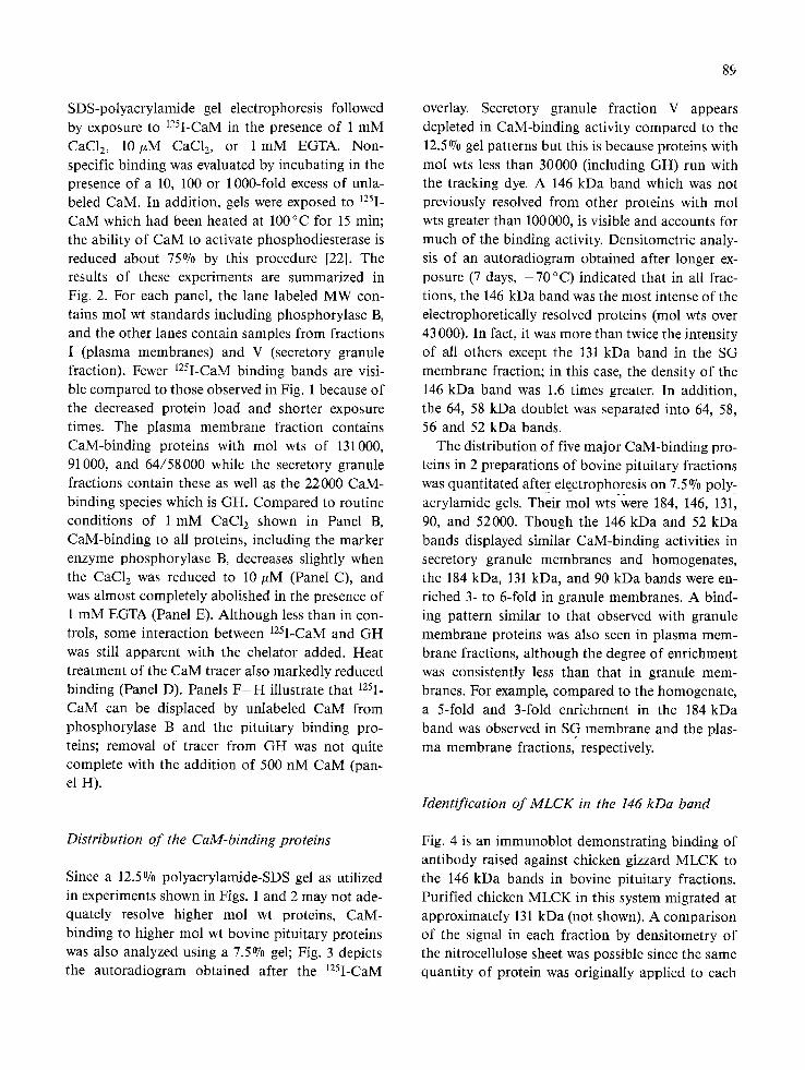
SDS-polyacrylamide gel electrophoresis followed by exposure to 125I-CaM in the presence of 1 mM CaC12, 10/xM CaC12, or 1 mM EGTA. Non- specific binding was evaluated by incubating in the presence of a 10, 100 or 1000-fold excess of unla- beled CaM. In addition, gels were exposed to 125I- CaM which had been heated at 100°C for 15 rain; the ability of CaM to activate phosphodiesterase is reduced about 75% by this procedure [22]. The results of these experiments are summarized in Fig. 2. For each panel, the lane labeled MW con- tains tool wt standards including phosphorylase B, and the other lanes contain samples from fractions I (plasma membranes) and V (secretory granule fraction). Fewer ~25I-CaM binding bands are visi- ble compared to those observed in Fig. 1 because of the decreased protein load and shorter exposure times. The plasma membrane fraction contains CaM-binding proteins with tool wts of 131000, 91000, and 64/58000 while the secretory granule fractions contain these as well as the 22000 CaM- binding species which is GH. Compared to routine conditions of 1 mM CaC12 shown in Panel B, CaM-binding to all proteins, including the marker enzyme phosphorylase B, decreases slightly when the CaC12 was reduced to 10/xM (Panel C), and was almost completely abolished in the presence of 1 mM EGTA (Panel E). Although less than in con- trols, some interaction between 125I-CaM and GH was still apparent with the chelator added. Heat treatment of the CaM tracer also markedly reduced binding (Panel D). Panels F - H illustrate that 125I- CaM can be displaced by unlabeled CaM from phosphorylase B and the pituitary binding pro- teins; removal of tracer from GH was not quite complete with the addition of 500 nM CaM (pan- el H).
Distribution of the CaM-binding proteins
Since a 12.5% polyacrylamide-SDS gel as utilized in experiments shown in Figs. 1 and 2 may not ade- quately resolve higher mol wt proteins, CaM- binding to higher mol wt bovine pituitary proteins was also analyzed using a 7.5% gel; Fig. 3 depicts the autoradiogram obtained after the 125I-CaM
89
overlay. Secretory granule fraction V appears depleted in CaM-binding activity compared to the 12.5% gel patterns but this is because proteins with mol wts less than 30000 (including GH) run with the tracking dye. A 146 kDa band which was not previously resolved from other proteins with tool wts greater than 100000, is visible and accounts for much of the binding activity. Densitometric analy- sis of an autoradiogram obtained after longer ex- posure (7 days, - 7 0 °C) indicated that in all frac- tions, the 146 kDa band was the most intense of the electrophoretically resolved proteins (mol wts over 43 000). In fact, it was more than twice the intensity of all others except the 131 kDa band in the SG membrane fraction; in this case, the density of the 146 kDa band was 1.6 times greater. In addition, the 64, 58 kDa doublet was separated into 64, 58, 56 and 52 kDa bands.
The distribution of five major CaM-binding pro- teins in 2 preparations of bovine pituitary fractions was quantitated after electrophoresis on 7.5% poly-
• - - .
acrylamide gels. Their tool wts were 184, 146, 131, 90, and 52000. Though the 146 kDa and 52 kDa bands displayed similar CaM-binding activities in secretory granule membranes and homogenates, the 184 kDa, 131 kDa, and 90 kDa bands were en- riched 3- to 6-fold in granule membranes. A bind- ing pattern similar to that observed with granule membrane proteins was also seen in plasma mem- brane fractions, although the degree of enrichment was consistently less than that in granule mem- branes. For example, compared to the homogenate, a 5-fold and 3-fold enrichment in the 184 kDa band was observed in SG membrane and the plas- ma membrane fractions, respectively.
Identification of MLCK in the 146 kDa band
Fig. 4 is an immunoblot demonstrating binding of antibody raised against chicken gizzard MLCK to the 146 kDa bands in bovine pituitary fractions. Purified chicken MLCK in this system migrated at approximately 131 kDa (not shown). A comparison of the signal in each fraction by densitometry of the nitrocellulose sheet was possible since the same quantity of protein was originally applied to each

90
Fig. 3. CaM-binding proteins in bovine pituitary fractions electrophoresed on a 7.5% polyacrylamide-SDS gel. Thirty/zg of each frac- tion was electrophoresed. An autoradiogram (-70 °C, 48 h exposure) after treatment with 125I-CaM (0.6 nM, 106 cpm/ml) in the pres- ence of 1 mM CaCI 2 is presented. The molecular masses, expressed in kDa, of the major CaM-binding proteins are indicated on the left, and of the mol wt markers on the right.
gel slot (175 txg). Anti-MLCK binding was enriched
as compared to the homogenate band in all cases:
14% in SI+II, 243% in PIII, 54070 in the secretory granule fraction V, and 249% in SG membrane. Binding of antibody in granules was clearly as-
sociated with the granule membrane fraction since
no detectable immunoactivity was observed after
the SG core fraction was electrophoresed (not
shown). In addition to the 146 kDa band, binding was also observed in two lower mol w t bands
(131000 and 128000) in Si+ii and Pill; these were very faint in V and undetectable in SG membrane
fractions.
Discussion
In this study, CaM was widely distributed in solu-
ble and particulate bovine pituitary subcellular
fractions. The highest concentration of CaM in pu-
rified particulate fractions relative to the protein
concentration was in plasma membranes whereas
the greatest total amount of CaM was in the secre- tory granule fraction. These locations for CaM are
consistent with a possible role in Ca2+-mediated
exocytosis. The concentration of CaM in particu-
late fractions other than granules is probably a minimum estimate since nearly 40% of the CaM
appeared loosely associated, being released during
dilution and resedimentation of the fractions.
Granule CaM required the harsher treatment of hypotonic exposure for release of roughly 50% (Ta- ble 2). CaM in non-pituitary tissues is primarily a
cytoplasmic protein; however, it has also been local- ized to coated vesicle [231 and post-synaptic densi- ties [24] from brain. In addition, 125I-CaM binds

91
Fig. 4. Immunoblot of pituitary homogenate and subfractions using anti-chicken gizzard MLCK. Pituitary fractions (175~g of each) were electrophoresed on 7.5 °70 polyacrylamide-SDS gels; immunoblotting with rabbit anti-chicken gizzard MLCK and visualization with horseradish peroxidase-coupled goat anti-rabbit IgG and 4-chloro-l-naphthol were as previously described [19, 20]. Color development was for 1 h resulting in a light purple background and visible negative staining of unreactive protein bands.
to adrenal chromaffin granules [25], platelet alpha granules [26], pancreatic islet secretion granules [27] and the plasma membrane of hormone respon- sive cells like adipocytes [28]. Hooper and Kelly characterized a high affinity, CaZ+-dependent CaM-binding site on the cytoplasmic surface of cholinergic synaptic vesicles [29]. In addition, they have identified a protein with immunologic and chemical characteristics indistinguishable from CaM which is tightly associated with the synaptic vesicle even in the absence of Ca 2+ [30]. They sug- gest that this CaM bound to vesicle membrane is in an ideal position to react quickly to changes in cytosolic Ca 2+ concentrations to trigger exocytosis.
We have not performed experiments to determine if any of the CaM associated with the purified pitui-
tary secretory granules is coupled to the secretory granule membrane in a Ca 2+-independent manner.
Direct evidence for the importance of CaM in the fusion of secretory granules with the plasma mem- brane comes from studies using sea urchin eggs. Fertilization leads to a rise in the ionized Ca 2÷ lev- el in the egg, which results in fusion of cortical granules with the plasma membrane. Using mem- brane surfaces isolated from the urchin eggs, Stein- hardt and Alderton [31] have blocked cortical gran- ule fusion by the addition of specific CaM antibodies. Furthermore, immunofluorescent localization of CaM following fertilization showed a heavy granular distribution of the CaM antigen in the plane of the plasma membrane. In control eggs or in other cells examined by indirect im-

92
munofluorescence, only occasional areas of CaM staining were seen along the plasma membrane. This redistribution of CaM during fertilization parallels the pituitary secretory studies of C o n n e t al. [11]. In these studies, CaM content and distribu- tion were monitored by radioimmunoassay in the pituitaries of ovariectomized rats before and during stimulation of gonadotropin secretion by gonadotropin-releasing hormone. There was a redistribution of CaM from the cytosol to the plas- ma membrane coincident with luteinizing hormone secretion, further suggesting a role for CaM in stimulus-secretion coupling.
It is unlikely that CaM acting alone is sufficient to catalyze the reactions involved in membrane fu- sion or granule movement. Thus, there has been great interest in identifying proteins which interact with CaM and may be involved in exocytosis. The gel overlay technique to monitor binding proteins was developed independently by Carlin et al. [32] and Glenney and Weber [33]. 125I-CaM binding to the proteins is Ca2+-dependent and is displaceable with unlabeled CaM. The affinities of most of the binding proteins for CaM appear to be in the low
nM range, since the concentrations of 125I-CaM in the overlay solutions are 0 .5-0 .6 nM, and major binding activities are easily detected even when the stained protein bands are barely visible. The gel overlay procedure we have used requires that the proteins in the polyacrylamide matrix refold into a configuration capable of binding the radioactive probe. It is, therefore, possible to miss CaM- binding proteins which do not renature easily or which require interaction among multiple subunits to form the binding site for CaM. The gel overlay method has been used to identify CaM-binding proteins in many cells and tissues [26, 27, 29, 34] and a wide variation in the mol wts of these CaM- binding proteins has been observed. This could be explained by the observations of Rubin et al. [35] who found no common proteins in membranes pu- rified from bovine chromaffin granules, zymogen granules, and platelet alpha-granules. Such heter- ogeneity might be related to multiple Ca2+-CaM functions. On the other hand, the discrepancies in mol wt could have been due to different handling of the tissues, especially in the use of protease-
inhibitors. With the gel overlay method utilized in this present study, Nelson et al. [16] identified CaM-binding proteins in an insulin-secreting cell line and demonstrated that glucose-stimulated in- sulin accumulation in the media was reversibly in- hibited by the anti-CaM drug, W13. This anti-CaM drug also blocked the association of CaM with 5 of the CaM-binding proteins in a dose-dependent manner suggesting the involvement of CaM in secretion through an interaction with CaM-binding proteins.
In addition to mol wt heterogeneity, the present data suggest some heterogeneity of interactions of CaM with its binding proteins. The data presented in Fig. 2 indicate that CaM-binding to the proteins in Fraction I, especially those of higher mol wt, is less labile to heat than CaM-binding to many of the proteins in Fraction V, including GH. In contrast, addition of excess unlabeled CaM was considerably more effective in displacing 125I-CaM from the proteins of Fraction I than from GH. It is possible that the details of the binding mechanisms may dif- fer from protein to protein.
Whatever the binding mechanism, CaM involve- ment in secretory processes has been further sug- gested by the studies of Watkins and collaborators on pancreatic islet granules, in which granule bind- ing to plasma membranes was increased by calcium and CaM, and inhibited by CaM inhibitors, and in which CaM-binding proteins were identified in iso- lated granules [27, 36]. These authors also suggest- ed that the granule CaM-binding proteins might play a role in exocytosis [27].
The definitive identification of the pituitary CaM-binding proteins has for the most part not been accomplished. Among the known CaM-
activated enzymes is phosphorylase kinase, which contains CaM as one of its subunits [37]; its alpha subunit has a molecular wt of 146000 and would therefore have been considered a prime candidate as the 146 kDa protein we have found to be en- riched in secretory granule membranes. However, the data presented in this paper clearly establish the 146 kDa CaM-binding protein to be closely related to MLCK, and may well be a mammalian form of MLCK. This enzyme, known to require CaM for activation, is about 131000 in mol wt from other

sources and is thought to be responsible for the mo- tive force of non-skeletal muscle cells [38]. The rea- son for the reactivity with anti-MLCK in 3 bands in the centrifugal fractions in the present studies is unknown, but could be related to proteolysis, since only the 146 kDa band was found in the SG mem- brane fraction. Other possibilities include precur- sor forms, or membrane-associated forms which might be larger due to glycosylation, lipidation, or phosphorylation. The high enrichment of MLCK and other CaM-binding proteins in the pituitary secretory granule membranes, however, is provoca- tive, and suggests that at least one function of CaM in pituitary is to activate granule membrane MLCK. However, these conclusions must still re- main tentative since, in the pituitary the mechan- isms regulating transport of the granules from the Golgi to the cell surface, fusion of the granule membrane and plasma membrane, and release of granule components are poorly defined. Neverthe- less, it seems possible that increased cytosolic free Ca2+-concentrations, CaM redistribution, and binding to CaM-activated proteins may all play a role in stimulus-secretion coupling.
Acknowledgements
We thank Drs. J. Putkey, J. Chafouleas, and A. Means for the generous gifts of 125I-CaM and CaM; the NIADDK for the bGH and bPRL stan- dards; and D. Turnquist and L. Anderson for typ- ing the manuscript.
This work was supported in part by the Diabetes and Endocrinology Research Center Grant AM 27685 (A.E.B), Grants AM 34447 (A.E.B), AM 31326 (M.Y.L.) and AM"21783 (L.S.J.) from the Na- tional Institute of Arthritis, Diabetes, Digestive, and Kidney Diseases, and General Clinical Re- search Center Grant RR-00044 (L.S.J., M.Y.L.) from the Division of Research Resources, NIH. A portion of this work was presented at the 7th Inter- national Congress of Endocrinology, Quebec City, Canada, July 1-7 , 1984.
93
References
1. Samli MH, Geschwind I: Some effects of energy-transfer inhibitors and of Ca 2+ free and K+-enhanced media on the release of luteinizing hormone from the rat pituitary gland in vitro. Endocrinology 82:225-231, 1968.
2. Parsons JA: Effects of cations on prolactin and growth hor- mone secretion by rat adenohypophyses in vitro. J Physiol 210:973-987, 1970.
3. Vale W, Burgus R, Guillemin R: Presence of calcium ions as a requisite for the in vitro stimulation of TSH release by hypothalamic TRF. Experientia 23:853-855, 1967.
4. Tam SW, Dannies PS: Dopaminergic inhibition of iono- phore A23187-stimulated release of prolactin from rat an- terior pituitary cells. J Biol Chem 255:6595-6599, 1980.
5. Ostlund Jr RE, Leung JT, Haiek SV, Winokur T, Melman M: Acute stimulated hormone release from cultured GH3 pituitary cells. Endocrinology 103:1245-1252, 1978. Gershengorn MC, Thaw C: Calcium influx is not required for TRH to elevate free cytoplasmic calcium in GH 3 cells. Endocrinology 113:1522-1524, 1983. Albert PR, Tashjian Jr AH: Thyrotropin-releasing hormone-induced spike and plateau in cytosolic free Ca 2+ concentrations in pituitary cells. J Biol Chem 259:5827-5832, 1984.
Schlegel W, Wollheim CB: Thyrotropin-releasing hormone increases cytosolic free Ca 2+ in clonal pituitary cells (GH 3 Cells): Direct evidence for the mobilization of cellular calci- um. J Cell Biol 99:83 87, 1984.
9. Merritt JE, Tomlinson S, Brown BL: Phenothiazines inhibit prolactin secretion in vitro. A possible role for calmodulin in stimulus-secretion coupling in the pituitary. FEBS Lett 135:107-110, 1981. Schettini G, Judd AM, MacLeod RM: In vitro studies on basal and stimulated prolactin release by rat anterior pitui- tary: A possible role for calmodulin. Endocrinology 112:64-70, 1983.
11. Conn PM, Chafouleas J, Rogers D, Means AR: Gonadotropin releasing hormone stimulates calmodulin redistribution in the rat pituitary. Nature 292:264-265, 1981.
Adelstein RS, Eisenberg E: Regulation and kinetics of the actin-myosin-ATP interaction. Ann Rev Biochem
49:921-956, 1980. Lorenson MY, Lee Y-C, Jacobs LS: Identification and characterization of an anion sensitive Mg2+-ATPase in pi- tuitary secretory granule membranes. J Biol Chem 256:12802-12810, 1981. Lowry OH, Rosebrough N J, Farr AL, Randall R J: Protein measurement with the Folin phenol reagent. J Biol Chem 193:265-275, 1951. Chafouleas JG, Dedman JR, Munjaal RP, Means AR: Calmodulin: Development and application of a sensitive radioimmunoassay. J Biol Chem 254:10262-10267, 1979.
6.
7.
8.
10.
12.
13.
14.
15.

94
16. Nelson TY, Oberwetter JM, Chafouleas JG, Boyd III AE: Calmodulin-binding proteins in a cloned rat insulinoma cell line. Diabetes 32:1126-1133, 1983.
17. Nelson TY, Snabes MC, Boyd III AE: Measurement of ac- tin and identification of actin- and calmodulin-binding pro- teins in insulin secreting cells. In: Methods in Diabetes Re- search. Lamer J, Pohl S (eds) J Wiley and Sons, New York, 1984, Vol 1, pp 277 285.
18. Towbin H, Staehlin T, Gordon J: Electrophoretic transfer of proteins from polyacrylamide gels to nitrocellulose sheets: Procedure and some applications. Proc Natl Acad Sci USA 76:4350-4354, 1979.
19. Guerriero V, Rowley DR, Means AR: Production and characterization of an antibody to myosin light chain kinase and intracellular localization of the enzyme. Cell 27:448-458, 1981.
20. Hawkes R, Niday E, Gordon J: A dot-immunobinding as- say for monoclonal and other antibodies. Anal Biochem 119:142-147, 1982.
21. Zar JH: Biostatistical analysis, 2nd ed, Prentice-Hall, Inc, Englewood Cliffs, NJ, 1984, p 162.
22. Beale EG, Dedman JR, Means AR: Isolation and regulation of the protein kinase inhibitor and the calcium-dependent cyclic nucleotide phosphodiesterase regulator in the sertoli cell-enriched testis. Endocrinology 101:1621-1634, 1977.
23. Linden CD, Dedman JR, Chafouleas JG, Means AR, Roth TF: Interactions of calmodulin with coated vesicles from brain. Proc Natl Acad Sci USA 78:308-312, 1981.
24. Grat~ DJ, Berzins K, Cohen RS, Siekevitz P: Presence of
calm°dulin in postsynaptic densities isolated from canine cerebral cortex. J Biol Chem 254:8690-8696, 1979.
25. Burgoyne RB, Giesow M J: Specific binding of 125I- calmodulin to and protein phosphorylation of adrenal chro- maffin granule membranes. FEBS Lett 131:127-131, 1981.
26. Grinstein S, Furuya W: Binding of 125I-calmodulin to plate- let alpha-granules. FEBS Lett 14,0:49-52, 1982.
27. Watkins D, White BA: Identification and characterization of calmodulin-binding proteins in islet secretory granules. J Biol Chem 260:5161-5165, 1985.
28. Goewert RR, Klaven NB, McDonald JM: Direct effect of in- sulin on the binding of calmodulin to rat adipocyte plasma membranes. J Biol Chem 258:9995-9999, 1983.
29. Hooper JE, Kelly RB: Calcium-dependent calmodulin binding to cholinergic synaptic vesicles. J Biol Chem 259:14,1-147, 1984.
30. Hooper JE, Kelly RB: Calmodulin is tightly associated with synaptic vesicles independent of calcium. J Biol Chem 259:148-153, 1984.
31. Steinhardt RA, Alderton JM: Calmodulin confers calcium sensitivity on secretory exocytosis. Nature 295:154-155, 1982.
32. Carlin RK, Grab DJ, Siekevitz P: Function of calmodulin in postsynaptic densities III calmodulin-binding proteins of the post synaptic density. J Cell Biol 89:449-455, 1981.
33. Glenney JR, Weber K: Calmodulin-binding proteins of the microfilaments present in isolated brush borders and microvilli of intestinal epithelial cells. J Biol Chem 255:10551-10554, 1980.
34. Giesow M J, Burgoyne RD: Recruitment of cytosolic pro- teins to a secretory granule membrane depends on Ca2+-calmodnlin. Nature 301:432-435, 1983.
35. Rubin RW, Lyubkin AK, Pressman BC: Comparison of the protein content of 3 different bovine secretory granule membrane types: A search for exocytosis-specific shared proteins. J Cell Biol 99:356-360, 1984.
36. Watkins DT, Cooperstein S J: Role of calcium and calmodu- lin in the interaction between islet cell secretion granules and plasma membranes. Endocrinology 112:766-768, 1983.
37. Cohen P, Burchell A, Foulkes JG, Cohen PTW, Vanaman TC, Nairn AC: Identification of the Ca2+-independent modulator protein as the fourth subunit of rabbit skeletal muscle phosphorylase kinase. FEBS Lett 92:287-293, 1978.
38. Means AR, Tash JS, Chafouleas JG: Physiological implica- tions of the presence, distribution, and regulation of calmodulin in eukaryotic cells. Physiological Reviews 62:1-39, 1982.
Copyright © 2022 FDOKUMEN