Neuronal survival induced by neurotrophins requires calmodulin
13
The Rockefeller University Press, 0021-9525/2001/08/585/13 $5.00 The Journal of Cell Biology, Volume 154, Number 3, August 6, 2001 585–597 http://www.jcb.org/cgi/doi/10.1083/jcb.200101023 JCB Article 585 Neuronal survival induced by neurotrophins requires calmodulin Joaquim Egea, 1 Carme Espinet, 1 Rosa M. Soler, 1 Xavier Dolcet, 1 Víctor J. Yuste, 1 Mario Encinas, 1 Montserrat Iglesias, 1 Nativitat Rocamora, 2 and Joan X. Comella 1 1 Grup de Neurobiologia Molecular, Departament de Ciències Mèdiques Bàsiques, Facultat de Medicina, Universitat de Lleida, 25198 Lleida, Catalonia, Spain 2 Laboratori de Biologia Molecular, Institut Català d’Oncologia, Barcelona, Catalonia, Spain t has been reported that phosphoinositide 3-kinase (PI 3-kinase) and its downstream target, protein kinase B (PKB), play a central role in the signaling of cell survival triggered by neurotrophins (NTs). In this report, we have analyzed the involvement of Ca 2 and calmodulin (CaM) in the activation of the PKB induced by NTs. We have found that reduction of intracellular Ca 2 concentration or functional blockade of CaM abolished NGF-induced acti- vation of PKB in PC12 cells. Similar results were obtained in cultures of chicken spinal cord motoneurons treated I with brain-derived neurotrophic factor (BDNF). Moreover, CaM inhibition prevented the cell survival triggered by NGF or BDNF. This effect was counteracted by the tran- sient expression of constitutive active forms of the PKB, indicating that CaM regulates NT-induced cell survival through the activation of the PKB. We have investigated the mechanisms whereby CaM regulates the activation of the PKB, and we have found that CaM was necessary for the proper generation and/or accumulation of the products of the PI 3-kinase in intact cells. Introduction Neurotrophins (NTs)* and their tyrosine kinase receptor, Trk, are involved in the development, maintenance, and re- pair of the nervous system (Lewin and Barde, 1996). NTs include NGF, brain-derived neurotrophic factor (BDNF), NT-3, and NT-4/5 (Davies, 1994). There is growing evidences indicating that phosphoinositide 3-kinase (PI 3-kinase), one of the intracellular effectors activated by these factors, plays a central role in the regulation of cell survival in a wide variety of neuronal cell populations (Kaplan and Miller, 2000). The pathway that leads to the activation of the PI 3-kinase after Trk activation begins with the autophosphorylation of the receptor at the residue Tyr 490 and the phosphorylation of Shc (Obermeier et al., 1993; Hallberg et al., 1998). This al- lows the recruitment to the membrane of several adaptor proteins, which upon tyrosine phosphorylation interact with and activate PI 3-kinase (Holgado-Madruga et al., 1997; Ya- mada et al., 1997). Furthermore, it has been reported that Ras cooperates in the activation of the PI 3-kinase, since dominant negative forms of this GTPase inhibit PI 3-kinase activity (Rodriguez-Viciana et al., 1994). PI 3-kinase cata- lyzes the phosphorylation of the D3 hydroxyl group of the inositol ring of phosphoinositides (PtdIns) (Czech, 2000). PtdIns-3,4-P 2 and PtdIns-3,4,5-P 3 are the main products generated and provide docking sites for pleckstrin homology (PH) domains of effector proteins such as the Ser/Thr protein kinase B (PKB), the cellular homologue of the transforming oncogene v-Akt (Bellacosa et al., 1991; Coffer and Woodgett, 1991; Jones et al., 1991). The interaction of PtdIns-3,4-P 2 /PtdIns-3,4,5-P 3 with PKB allows the transloca- tion of the protein to the plasma membrane where it becomes fully activated upon phosphorylation at two residues, Thr 308 and Ser 473 (Alessi et al., 1996). In a variety of cell systems, including neuronal cells, PKB mediates an important part of the trophic signal derived Address correspondence to Joan X. Comella, Grup de Neurobiologia Molecular, Departament de Ciències Mèdiques Bàsiques, Universitat de Lleida, Avda Rovira Roure, 44, 25198 Lleida, Spain. Tel.: 34-973-702414. Fax: 34-973-702438. E-mail: [email protected] *Abbreviations used in this paper: anti–P-Tyr, antiphosphotyrosine; BAPTA, 1,2 bis(2-aminophenoxy) ethene N,N,N,N-tetraacetic acid; BDNF, brain-derived neurotrophic factor; [Ca 2 ] i , intracellular Ca 2 concentration; CaM, calmodulin; CaMKK, CaM-dependent protein kinase kinase; EGFP, enhanced green fluorescence protein; ERK, extracellular signal–regulated kinase; GST, glutathione S-trans- ferase; MAP, mitogen-activated protein; MTN, motoneuron; NT, neurotrophin; PH, pleckstrin homology; PI 3-kinase, phosphoinosi- tide 3-kinase; PKB, protein kinase B; PtdIns, phosphoinositides; RBD, Ras-binding domain; TFP, trifluoperazine dimaleate; TUNEL, TdT-mediated dUTP nick end labeling. Key words: calmodulin; neurotrophin; cell survival; PKB; motoneuron on November 23, 2013 jcb.rupress.org Downloaded from Published August 6, 2001
Transcript of Neuronal survival induced by neurotrophins requires calmodulin

The Rockefeller University Press, 0021-9525/2001/08/585/13 $5.00The Journal of Cell Biology, Volume 154, Number 3, August 6, 2001 585–597http://www.jcb.org/cgi/doi/10.1083/jcb.200101023
JCB
Article
585
Neuronal survival induced by neurotrophins requires calmodulin
Joaquim Egea,
1
Carme Espinet,
1
Rosa M. Soler,
1
Xavier Dolcet,
1
Víctor J. Yuste,
1
Mario Encinas,
1
Montserrat Iglesias,
1
Nativitat Rocamora,
2
and Joan X. Comella
1
1
Grup de Neurobiologia Molecular, Departament de Ciències Mèdiques Bàsiques, Facultat de Medicina, Universitat de Lleida, 25198 Lleida, Catalonia, Spain
2
Laboratori de Biologia Molecular, Institut Català d’Oncologia, Barcelona, Catalonia, Spain
t has been reported that phosphoinositide 3-kinase (PI3-kinase) and its downstream target, protein kinase B(PKB), play a central role in the signaling of cell survival
triggered by neurotrophins (NTs). In this report, we have
analyzed the involvement of Ca
2
�
and calmodulin (CaM)in the activation of the PKB induced by NTs. We havefound that reduction of intracellular Ca
2
�
concentration orfunctional blockade of CaM abolished NGF-induced acti-vation of PKB in PC12 cells. Similar results were obtainedin cultures of chicken spinal cord motoneurons treated
I
with brain-derived neurotrophic factor (BDNF). Moreover,CaM inhibition prevented the cell survival triggered byNGF or BDNF. This effect was counteracted by the tran-
sient expression of constitutive active forms of the PKB,indicating that CaM regulates NT-induced cell survivalthrough the activation of the PKB. We have investigated themechanisms whereby CaM regulates the activation of
the PKB, and we have found that CaM was necessary for theproper generation and/or accumulation of the products ofthe PI 3-kinase in intact cells.
Introduction
Neurotrophins (NTs)* and their tyrosine kinase receptor,Trk, are involved in the development, maintenance, and re-pair of the nervous system (Lewin and Barde, 1996). NTsinclude NGF, brain-derived neurotrophic factor (BDNF),
NT-3, and NT-4/5 (Davies, 1994). There is growingevidences indicating that phosphoinositide 3-kinase (PI3-kinase), one of the intracellular effectors activated by thesefactors, plays a central role in the regulation of cell survivalin a wide variety of neuronal cell populations (Kaplan andMiller, 2000).
The pathway that leads to the activation of the PI 3-kinaseafter Trk activation begins with the autophosphorylation ofthe receptor at the residue Tyr
490
and the phosphorylation ofShc (Obermeier et al., 1993; Hallberg et al., 1998). This al-lows the recruitment to the membrane of several adaptorproteins, which upon tyrosine phosphorylation interact withand activate PI 3-kinase (Holgado-Madruga et al., 1997; Ya-mada et al., 1997). Furthermore, it has been reported thatRas cooperates in the activation of the PI 3-kinase, sincedominant negative forms of this GTPase inhibit PI 3-kinaseactivity (Rodriguez-Viciana et al., 1994). PI 3-kinase cata-lyzes the phosphorylation of the D3 hydroxyl group of theinositol ring of phosphoinositides (PtdIns) (Czech, 2000).PtdIns-3,4-P
2
and PtdIns-3,4,5-P
3
are the main productsgenerated and provide docking sites for pleckstrin homology(PH) domains of effector proteins such as the Ser/Thrprotein kinase B (PKB), the cellular homologue of the
transforming oncogene
v-Akt
(Bellacosa et al., 1991; Cofferand Woodgett, 1991; Jones et al., 1991). The interaction of
PtdIns-3,4-P
2
/PtdIns-3,4,5-P
3
with PKB allows the transloca-tion of the protein to the plasma membrane where it becomesfully activated upon phosphorylation at two residues, Thr
308
and Ser
473
(Alessi et al., 1996).In a variety of cell systems, including neuronal cells, PKB
mediates an important part of the trophic signal derived
Address correspondence to Joan X. Comella, Grup de NeurobiologiaMolecular, Departament de Ciències Mèdiques Bàsiques, Universitat de
Lleida, Avda Rovira Roure, 44, 25198 Lleida, Spain. Tel.: 34-973-702414.Fax: 34-973-702438. E-mail: [email protected]
*Abbreviations used in this paper: anti–P-Tyr, antiphosphotyrosine;
BAPTA, 1,2 bis(2-aminophenoxy) ethene N,N,N
�
,N
�
-tetraacetic
acid;
BDNF, brain-derived neurotrophic factor; [Ca
2
�
]
i
, intracellularCa
2
�
concentration; CaM, calmodulin; CaMKK, CaM-dependentprotein kinase kinase; EGFP, enhanced green fluorescence protein;
ERK, extracellular signal–regulated kinase; GST, glutathione
S
-trans-ferase; MAP, mitogen-activated protein; MTN, motoneuron; NT,neurotrophin; PH, pleckstrin homology; PI 3-kinase, phosphoinosi-tide 3-kinase; PKB, protein kinase B; PtdIns, phosphoinositides;RBD, Ras-binding domain; TFP, trifluoperazine dimaleate; TUNEL,TdT-mediated dUTP nick end labeling.Key words: calmodulin; neurotrophin; cell survival; PKB; motoneuron
on Novem
ber 23, 2013jcb.rupress.org
Dow
nloaded from
Published August 6, 2001
586 The Journal of Cell Biology
|
Volume 154, 2001
from PI 3-kinase activation (Dudek et al., 1997; Philpott etal., 1997; Crowder and Freeman, 1998). Several studieshave reported that PKB interferes with the cell death ma-chinery phosphorylating and inactivating proteins that aredirectly involved in the induction of apoptosis such asGSK3
�
, BAD (a member of the Bcl-2 family of proteins), ormembers of the Forkhead family of transcription factors in-volved in the transcription of Fas ligand (Datta et al., 1999).
Bioelectrical activity cooperates with NTs in promot-ing neuronal survival during development (Franklin andJohnson, 1992). Neuronal activity exerts its trophic effectsby moderately increasing the intracellular Ca
2
�
concentra-tion ([Ca
2
�
]
i
). Ca
2
�
triggers the activation of similar signal-ing pathways to those activated by NTs, mainly through the
Ca
2
�
receptor protein calmodulin (CaM) (Finkbeiner andGreenberg, 1996). Moreover, it has been reported that acti-vation of Trk leads to a small and rapid increase of [Ca
2
�
]
i
(Pandiella-Alonso et al., 1986; Jiang and Guroff, 1997).However, the involvement of Ca
2
�
in the response of thecells to the NTs has been poorly characterized. In thepresent work, we show that CaM is necessary for the promo-tion of cell survival triggered by NTs in PC12 cells and inchicken spinal cord motoneurons (MTNs). Our resultsdemonstrate that this effect is mainly due to the regulationof PKB activity. We provide evidence that CaM is necessaryto detect PtdIns-3,4-P
2
/PtdIns-3,4,5-P
3
in the plasma mem-brane of live cells thus providing a possible mechanism bywhich CaM regulates PKB activity and cell survival.
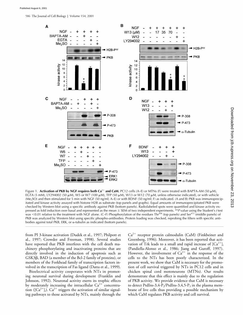
Figure 1. Activation of PKB by NGF requires both Ca2� and CaM. PC12 cells (A–E) or MTNs (F) were treated with BAPTA-AM (50 �M), EGTA (5 mM), LY294002 (50 �M), W5 or W7 (100 �M), TFP (50 �M), W13 or W12 (70 �M, unless otherwise indicated), or with vehicle (Me2SO) and then stimulated for 5 min with NGF (50 ng/ml; A–E) or with BDNF (50 ng/ml; F) as indicated. (A and B) PKB was immunoprecip-itated and kinase activity assayed with histone H2B as substrate (top panels and graphs). Equal amounts of immunoprecipitated PKB were checked by Western blot using a specific antibody against PKB (bottom panels). Radiolabeled spots were quantified and kinase activity ex-pressed as fold induction over basal and represented as the mean � SEM of two independent experiments. **P value using the Student’s t test was �0.01 relative to the treatment with NGF alone. (C–F) Phosphorylation of the residues Thr308 (top panels) and Ser473 (middle panels) of PKB was analyzed by Western blot using specific phospho-antibodies. Protein loading was checked, reprobing the filters with specific anti-bodies against total PKB, ERK, or �-tubulin as indicated (bottom panels).
on Novem
ber 23, 2013jcb.rupress.org
Dow
nloaded from
Published August 6, 2001

Calmodulin modulates neuronal survival |
Egea et al. 587
Results
NT-induced PKB activation requires Ca
2
�
and CaM
PKB is activated by NGF in PC12 cells through a mecha-nism involving PI 3-kinase (Park et al., 1996; Andjelkovic etal., 1998). We wanted to analyze the involvement of Ca
2
�
and CaM in this activation. For this, we chelated theintracellular Ca
2
�
using 1,2 bis(2-aminophenoxy) etheneN,N,N
�
,N
�
-tetraacetic acid (BAPTA) or the extracellularCa
2
�
using EGTA, and then we analyzed the activation ofPKB after NGF stimulation. NGF induced a strong increase
Figure 2. NGF requires CaM to promote cell survival in PC12 cells. PC12 cells were serum starved and treated with NGF (10 ng/ml), LY294002 (20 �M), W13 (30 �M), or left untreated as indicated. After 15 h, cells were fixed, stained with Hoechst 33258, or subjected to a TUNEL assay. (A) Percentage of cells displaying typical nuclear apoptotic morphology. The values represent the mean � SEM of three indepen-dent experiments. **P value using the Student’s t test was �0.01 relative to the treatment with NGF alone. (B) Representative photomicrographs showing the morphology of the nuclei of the cells in the different treatments. Arrowheads indicate the apoptotic nuclei. (C) Representative phase–contrast micrographs and TUNEL reaction of the same field of the cultures treated above. (D) PC12 cells were treated with W13 (30 �M) and then stimulated for the indicated times with NGF (10 ng/ml). Phosphorylation of the residues Thr308 (top panel) and Ser473 (middle panel) of PKB was analyzed by Western blot using specific phospho-antibodies. Protein loading was checked, reprobing the filters with a specific anti-body against �-tubulin (bottom panel). Bars: (B) 10 �m; (C) 20 �m.
on Novem
ber 23, 2013jcb.rupress.org
Dow
nloaded from
Published August 6, 2001

588 The Journal of Cell Biology
|
Volume 154, 2001
in PKB activity (
�
11-fold over basal) that was almost com-pletely prevented by BAPTA (Fig. 1 A). In contrast, concen-trations of EGTA that effectively block depolarization-
induced activation of extracellular signal–regulated kinase(ERK) mitogen-activated protein (MAP) kinases (Egea etal., 1999) did not significantly affect the activation of PKB
Figure 3. BDNF requires CaM to promote cell survival in MTNs. MTNs were starved of muscle extract and were treated with BDNF (10 ng/ml), W13 (30 mM), W12 (30 mM), or left untreated as indicated. (A) After 24 h, cells were counted, and survival was expressed as the percentage of cells remaining in each treatment. Graph shows the mean � SEM of three independent experiments. **P value using the Student’s t test was �0.01 relative to the treatment with BDNF alone. (B) After 15 h, cells were fixed and subjected to a TUNEL assay and stained with Hoechst 33258. The percentage of cells displaying typical nuclear apoptotic morphology and the percentage of TUNEL-positive cells were evaluated and repre-sented as the mean � SEM of two independent experiments. **P value using the Student’s t test was �0.01 relative to the treatment with BDNF alone. (C) Representative photomicrographs showing the morphology of the nuclei of the cells treated in B and stained with Hoechst 33258. Arrowheads indicate the apoptotic nuclei. (D) Representative photomicrographs showing TUNEL reaction and Hoechst staining of the same field of the cultures treated in B. (E) MTNs were treated with W13 (30 �M) and then stimulated for 5 min with BDNF (10 ng/ml) as indicated. Phosphorylation of the residues Thr308 (top panel) and Ser473 (middle panel) of PKB was analyzed by Western blot using specific phospho-anti-bodies. Protein loading was checked, reprobing the filters with a specific antibody against �-tubulin (bottom panel). Bars: (C) 10 �m; (D) 40 �m.
on Novem
ber 23, 2013jcb.rupress.org
Dow
nloaded from
Published August 6, 2001

Calmodulin modulates neuronal survival |
Egea et al. 589
(Fig. 1 A). In parallel experiments, we observed that theCaM antagonist W13 mimicked the effect of BAPTA onNGF-induced PKB activity. As shown in Fig. 1 B, increas-ing concentrations of W13 blocked the activation of PKB ina dose-dependent manner. At 70 mM, W13 reached an in-hibitory effect similar to that observed with the specific PI3-kinase inhibitor LY294002 (Vlahos et al., 1994) (Fig. 1B). At this concentration, the effect of W13 was specific,since the same concentration of W12, a less active structuralanalogue (W13
IC50
68
�
M versus W12
IC50
260
�
M;Hidaka and Tanaka, 1983), did not affect NGF-inducedPKB activity (Fig. 1 B). Moreover, 70
�
M of W13 effec-tively inhibits the autophosphorylation of CaMKII inducedby ionomycin in PC12 cells, a well-known Ca
2
�
/CaM-dependent process (unpublished data; Egea et al., 2000).
PKB activity is mainly induced by phosphorylation of theresidues Thr
308
and Ser
473
(Alessi et al., 1996). We used spe-cific phospho-antibodies against each of these two residues
to check the phosphorylation of PKB upon NGF stimula-tion in the presence of Ca
2
�
chelators or CaM antagonists.According to the experiments of kinase activity shownabove, BAPTA (Fig. 1 C) and W13 but not W12 (Fig. 1 D)blocked the phosphorylation of both residues. Moreover, theinhibition exerted by W13 was sustained over the time oftreatment (Fig. 2 D). Other CaM inhibitors, such as W7(100
�
M) or the W13 structurally unrelated trifluoperazinedimaleate (TFP; 50
�
M), displayed similar effects as thoseobserved with W13 (Fig. 1 E). In these experiments, we alsoincluded the CaM inhibitor W5 as a control of the unspe-cific effects of W7 (W7
IC50
28
�
M versus W5
IC50
240
�M; Hidaka and Tanaka, 1983). As shown in Fig. 1 E, W5did not significantly affect PKB phosphorylation, confirm-ing the specificity of W7 effects. Finally, as expected fromthe kinase activity assay shown in Fig. 1 A, EGTA did notmodify the phosphorylation of the PKB induced by NGF(unpublished data).
Figure 4. Constitutively active forms of PKB pre-vent the cell death induced by CaM antagonists in NT-maintained cultures. PC12 cells (A and B) or MTNs (C) were transiently cotransfected with pEGFP and pCMV5-HA-PKBT308D/S473D (PKBDD) or pSG5-Gag-PKB or the empty vector. (A and B) PC12 cells were serum starved and treated with NGF (10 ng/ml), W13 (30 �M), or left untreated as indicated. After 15 h, cells were fixed and stained with Hoechst 33258. The percentage of apoptotic cells scored into the EGFP-positive cell population was evaluated and represented as the mean � SEM of three independent experiments. (C) MTNs were treated with BDNF (10 ng/ml), W13 (30 �M), LY294002 (20 �M), or left untreated as indicated. After 24 h, EGFP-positive cells were counted, and survival was expressed as the percentage remain-ing of EGFP-positive cells in each treatment. The graph shows the mean � SEM of three independent experiments. **P value using the Student’s t test was �0.01 when comparing the cultures trans-fected with pCMV5-HA-PKBT308D/S473D or pSG5-Gag-PKB with those transfected with the empty vector in each treatment (A–C).
on Novem
ber 23, 2013jcb.rupress.org
Dow
nloaded from
Published August 6, 2001

590 The Journal of Cell Biology | Volume 154, 2001
We have reported previously that chicken spinal cordMTNs can be maintained in culture in the presence of BDNF(Becker et al., 1998). Like PC12 cells treated with NGF, thestimulation of cultured MTNs with BDNF phosphorylatesand activates PKB in a PI 3-kinase–dependent manner (Dol-cet et al., 1999). In this context, we analyzed whether MTNsalso required CaM to activate PKB. As shown in Fig. 1 F,W13 inhibited both Thr308 and Ser473 phosphorylations to asimilar extent to that observed with LY294002 (Fig. 1 F).
CaM antagonists block NT-induced cell survivalThe survival induced by NGF in PC12 cells can be stronglyattenuated by inhibitors of PI 3-kinase (Yao and Cooper,1995) or by treatments that block PKB activity (Weihl et al.,1999; Salinas et al., 2000). Therefore, we wanted to analyzewhether the inhibition of PKB induced by CaM antagonistshad any effect on cell survival. For this purpose, we main-tained PC12 cell cultures in the presence of NGF (10 ng/ml)plus W13 (30 mM) or LY294002 (20 mM) for 15 h. At theend of the treatments, cells were fixed and stained withHoechst 33258 or subjected to a TdT-mediated dUTP nickend labeling (TUNEL) assay. As shown in Fig. 2 A, the per-
centage of cells that display typical morphological apoptoticnuclei in the cultures treated with NGF plus W13 increasedsignificantly when compared with cultures treated with NGFalone. Values were similar to those obtained in culturesmaintained without any trophic support or in culturestreated with NGF plus LY294002. The values of apoptoticnuclei in W13 treatments were indistinguishable from thoseobserved in starved cultures or in cultures treated with NGFplus LY294002 (Fig. 2 B). Similarly, W13 increased thenumber of TUNEL-positive cells in cultures maintainedwith NGF (5.98 � 1.04%) when compared with cellstreated with NGF alone (3.26 � 0.37%), reaching similarvalues to those observed in cultures treated with NGF plusLY294002 (7.01 � 0.84%). (In these experiments, the per-centage of TUNEL-positive cells in cultures without trophicsupport was 9.96 � 1.17) [Fig. 2 C].) The concentration ofW13 used in these experiments did not increase the celldeath observed in trophic support-starved cultures, suggest-ing that W13 was specifically interfering with the intracellu-lar signaling pathway involved in NGF-induced cell survival(Fig. 2 A). As a control, we checked whether the concentra-tion of W13 used in the experiments of survival (30 �M) in-hibited the activation of PKB induced by 10 ng/ml NGF. As
Figure 5. Regulation by CaM of the NT-induced activation of PKB occurs downstream of PI 3-kinase. PC12 cells were treated with 70 mM of W12 or W13 and then stimulated for 5 (A–C) or 2 min (D) with NGF (50 ng/ml) as indi-cated. (A) TrkA was immunoprecipitated and tyrosine phosphorylation of the receptor analyzed by Western blot using an anti–P-Tyr antibody. (B) Shc proteins were immunoprecipitated and analyzed by Western blot with an anti–P-Tyr anti-body (top panels). Position of the 66-, 52-, and 48-kD isoforms of Shc is indi-cated. Protein loading was checked, re-probing the filters with a specific antibody against total Shc (bottom panels). (C) Ac-tive Ras (Ras-GTP) was precipitated with GST–RBD coupled to glutathione-Sepharose, and Ras was detected by Western blot using a pan-Ras antibody. (D) PI 3-kinase activity was assayed in P-Tyr immunoprecipitates using L-�-phos-phatidylinositol as substrate (top panel). Radiolabeled spots were quantified and kinase activity expressed as fold induc-tion over basal. Bottom graph represents the mean � SEM of two independent experiments. (E) MTNs were treated with 70 �M of W13, stimulated for 2 min with BDNF (50 ng/ml), and PI 3-kinase activ-ity assayed as in D (top panel). The same cell extracts were analyzed by Western blot using a specific phospho-antibody against the residue Ser473 of PKB (middle panel). Protein loading was checked rep-robing the filter with a specific antibody against �-tubulin (bottom panel).
on Novem
ber 23, 2013jcb.rupress.org
Dow
nloaded from
Published August 6, 2001

Calmodulin modulates neuronal survival | Egea et al. 591
shown in Fig. 2 D, this concentration of the drug induced aneffective and sustained inhibition of both Thr308 and Ser473
phosphorylations of PKB.We next analyzed the involvement of CaM in cell sur-
vival in a primary culture of neurons, and for this we usedchicken MTNs maintained with BDNF. This experimentalparadigm has several similarities with PC12 cells main-tained with NGF. For example, we have demonstrated pre-viously that BDNF-induced MTN survival is prevented byLY294002 (Dolcet et al., 1999), and here we have shownthat the activation of PKB triggered by BDNF requires Ca2�
and CaM (Fig. 1 F). Therefore, MTNs were treated withBDNF (10 ng/ml) plus W13 (30 �M), and cell survival wasanalyzed after 24 h. Our experiments showed that W13 butnot W12 prevented the BDNF-induced MTN survivalwithout increasing the cell death of parallel control culturesmaintained in basal media without BDNF (Fig. 3 A). Thiseffect correlated with a significative increase in the numberof TUNEL-positive cells (Fig. 3, B and D) and in the num-ber of the cells displaying typical morphological apoptoticnuclei (Fig. 3 B). As shown in Fig. 3 C, the morphology ofthe apoptotic nuclei in W13 treatments was indistinguish-able from that observed in starved cultures or in culturestreated with BDNF plus LY294002. Fig. 3 E shows that 30�M of W13 effectively blocked the phosphorylation of PKBinduced by 10 ng/ml BDNF.
Constitutive active forms of PKB prevent the cell death induced by CaM antagonistsThe results above show a good correlation between inhibitionof PKB and inhibition of cell survival induced by CaM antag-onists. To test whether the prevention of cell survival triggeredby these inhibitors was due to the inhibition of PKB, wetransfected PC12 cells and MTNs with one of the two typesof constitutive active forms: Gag-PKB, a constitutive plasmamembrane-bound protein, and HA-PKBT308D/S473D, a taggedform of the protein that carries a mutational acidic charge inaddition to its main regulatory phosphorylation sites (Burger-ing and Coffer, 1995; Alessi et al., 1996). The transfected cul-
tures were then treated with the corresponding NT plus W13,and at the end of the treatments cell survival was evaluatedand compared with control cultures transfected with theempty vector. Results showed that Gag-PKB protected bothPC12 cells and MTNs from the cell death induced by trophicfactor withdrawal (Fig. 4, A and C, untreated cultures). Simi-lar effects displayed HA-PKBT308D/S473D in serum-starvedPC12 cells (Fig. 4 B, untreated cultures). Interestingly, Gag-PKB and HA-PKBT308D/S473D were also able to prevent the celldeath triggered by W13 in cultures of PC12 maintained withNGF (Fig. 4, A and B, respectively). Accordingly, the celldeath induced by W13 in BDNF-maintained MTNs wasstrongly reduced in Gag-PKB– (Fig. 4 C) and in HA-PKBT308D/S473D-transfected cultures (unpublished data) whencompared with the empty vector–transfected cultures. Theseresults indicate that CaM antagonists exert their effect on NT-induced survival, mainly inhibiting the activation of PKB.
CaM does not modulate early signaling events involved in PKB activationThe results presented in Fig. 1 indicate that Ca2� and CaMregulate upstream event(s) involved in the phosphorylationand activation of PKB. We have shown previously that inhi-bition of CaM does not affect the phosphorylation of TrkAand Shc, the association of Shc to Grb2, or the activation ofRas (measured by glutathione S-transferase–Ras-binding do-main [GST–RBD] pull-down) induced by NGF in PC12cells (Fig. 5, A–C; Egea et al., 2000). In this work, we alsoanalyzed whether NGF-induced PI 3-kinase activity couldrequire CaM. PC12 cells were treated with W12 or W13,stimulated for 2 min with NGF, and the PI 3-kinase activitywas measured in antiphosphotyrosine (anti–P-Tyr) immu-noprecipitates. As shown in Fig. 5 D, PI 3-kinase displayedan �25-fold activation after NGF stimulation. However,W13 did not significantly inhibit PI 3-kinase activity whencompared with parallel cultures treated with similar concen-trations of W12 (Fig. 5 D, graph). Since W13 but not W12effectively blocked PKB phosphorylation in the same cell ly-sates (unpublished data; Fig. 1 B), we therefore concludethat W13 inhibits PKB phosphorylation at some step down-stream of PI 3-kinase. These experiments were also per-formed in cultures of MTNs, obtaining similar results. Asshown in Fig. 5 E, W13 did not inhibit the PI 3-kinase ac-tivity induced by BDNF in MTNs (Fig. 5 E, top panel), de-spite that in the same cell extracts, the BDNF-induced PKBphosphorylation was completely prevented by the inhibitor(Fig. 5 E, bottom panels). Finally, to rule out a direct effectof W13 on the PI 3-kinase activity of intact cells, we in-cluded W13 directly in the kinase buffer of PI 3-kinase as-says. In these experiments, we did not detect any significanteffect of CaM antagonists on the PI 3-kinase activity in-duced by NGF (unpublished data).
NGF does not require CaM-dependent protein kinase kinase to phosphorylate PKBA recent study reported that the Ca2�/CaM-dependent pro-tein kinase kinase (CaMKK) is able to mediate cell survivalby directly phosphorylating and activating PKB (Yano et al.,1998). To address the involvement of CaMKK in the activa-
Figure 6. CaMKK is not required for the activation of PKB in-duced by NGF. PC12 cells were transiently transfected with pcDNA3-CaMKKK157A (CaMKKK157A), empty vector, or left untrans-fected and then stimulated for 5 min with 5 or 50 ng/ml of NGF as indicated. Phosphorylation of Thr308 and Ser473 (top panels) of PKB was analyzed by Western blot using specific phospho-antibodies. Expression of CaMKKK157A in the same protein lysates was analyzed reprobing the filter with a specific antibody against CaMKK (middle panel). Protein loading was checked with a specific antibody against PKB (bottom panel).
on Novem
ber 23, 2013jcb.rupress.org
Dow
nloaded from
Published August 6, 2001

592 The Journal of Cell Biology | Volume 154, 2001
tion of PKB induced by NGF, PC12 cells were transfectedwith CaMKKK157A, a dominant negative form of CaMKKthat carries a point mutation in the ATP-binding site (Yanoet al., 1998). These cultures were stimulated for 5 min with5 or 50 ng/ml of NGF, and PKB activation was analyzed byWestern blot using specific phospho-antibodies. As shownin Fig. 6, phosphorylation of PKB at the residues Thr308 andSer473 in cultures transfected with the mutant CaMKKK157A
was not significantly lower than that observed in control cul-tures transfected with the empty vector or in nontransfectedcultures. Expression of CaMKKK157A in transfected cultureswas confirmed by Western blot using an antibody againstCaMKK (Fig. 6). PC12 cells were also transfected withCaMKK1–413, a constitutive active form of CaMKK thatlacks the regulatory CaM-binding domain (Enslen et al.,1996). CaMKK1–413 did not induced a significant increase inthe phosphorylation of residues Thr308 or Ser473 of Akt/PKB(unpublished data).
CaM antagonists block plasma membrane localization of EGFP-PH-PKBWe have shown that CaM antagonists block NT-inducedPKB phosphorylation without affecting the in vitro PI3-kinase activity. It has been suggested previously that CaMmay be required to detect the products generated by the PI3-kinase into the plasma membrane of intact cells (Yang et al.,2000). To analyze this possibility, we transiently transfectedPC12 cells with a construct encoding EGFP fused in framewith the PH domain of PKB� (EGFP–PKB–PH). After36 h, cells were treated with W12, W13, or LY294002, stimu-lated for 20 min with NGF, fixed, and the distribution ofthe fluorescence assessed with a confocal microscope. A typi-cal result is shown in Fig. 7. EGFP–PKB–PH showed a dif-fuse cytoplasmatic localization in nonstimulated PC12 cells(Fig. 7, N.S.). However, NGF induced a significant redistri-bution of the EGFP–PKB–PH fusion protein from the cyto-sol to the periphery of the cell (Fig. 7, NGF and graph).
Figure 7. CaM antagonists prevent the in vivo generation of PtdIns-3,4-P2/PtdIns-3,4,5-P3 in PC12 cells. PC12 cells were transiently transfected with EGFP–PKB–PH, treated with W12 (70 �M), W13 (70 �M), or LY294002 (50 �M), and then stimulated for 20 min with NGF (50 ng/ml) as indicated. N.S. indi-cates EGFP-PH-PKB transfected cells without any treatment. At the end of the treatments, cells were fixed, and EGFP distribution was visualized using a laser confocal microscope. Laser confocal sections through the middle of represen-tative cells in each treatment are shown. Arrows indicate the peripheral redistri-bution of the EGFP–PKB–PH. The lower graph represents the percentage of cells displaying plasma membrane fluores-cence (% positive cells) in each treat-ment. The values represent the mean � SEM of two independent experiments. *P value using the Student’s t test was �0.05 relative to the treatment with NGF alone or relative to the treatment with NGF plus W12. Bar, 10 mM.
on Novem
ber 23, 2013jcb.rupress.org
Dow
nloaded from
Published August 6, 2001

Calmodulin modulates neuronal survival | Egea et al. 593
This effect was due to the activation of the PI 3-kinase andthe subsequent generation of PtdIns-3,4-P2/PtdIns-3,4,5-P3
in the plasma membrane, since NGF did not induce any re-distribution of the fluorescence in PC12 cells transfectedwith EGFP alone (unpublished data) and treatment withLY294002 completely abolished the peripheral distributionof the EGFP–PKB–PH fusion protein induced by NGF(Fig. 7, NGF � LY and graph). Cells stimulated with NGFthat have been pretreated with W13 displayed a similar pat-tern of fluorescence to that observed in nonstimulated cellsor in cells treated with NGF plus LY294002 (Fig. 7, NGF �W13), reducing significantly the percentage of cells dis-playing peripheral fluorescence (Fig. 7, graph). This effectwas specific, since W12 did not significantly modify the pe-ripheral pattern of fluorescence induced by NGF (Fig. 7,NGF � W12 and graph). Similar experiments were alsoperformed in EGFP–PKB–PH–transfected MTNs. Con-focal sections of representative cells treated with BDNF,BDNF plus W13, BDNF plus LY294002, or left untreatedare shown in Fig. 8. As seen, W13 also abolished the redis-tribution of the fluorescence to the plasma membrane inMTNs stimulated with BDNF; this effect is similar to thatobserved using LY294002. Therefore, these results suggestthat CaM is involved in the generation and/or stabilizationof PtdIns-3,4-P2/PtdIns-3,4,5-P3 in the plasma membraneof intact cells.
DiscussionIn this study, we report that CaM is necessary for the induc-tion of cell survival triggered by NGF or BDNF in PC12cells or MTNs, respectively. Our results demonstrate thatthis effect on cell survival is mainly due to the regulation ofPKB activation. Moreover, we have shown that CaM regu-
lates the presence of detectable amounts of PtdIns-3,4-P2/PtdIns-3,4,5-P3 in the plasma membrane of intact cells thusproviding a possible mechanism whereby CaM regulates theactivation of PKB and the promotion of cell survival.
We have observed that extracellular Ca2� blockade didnot affect the activation of PKB induced by NGF in PC12cells thus confirming previous observations obtained inBalb/c-3T3 fibroblasts stimulated with EGF (Conus et al.,1998). In contrast, our results point out that intracellularCa2� is completely necessary for the activation of PKB. Thiseffect seems to be mediated by CaM, since CaM inhibitionmimicked the effects of intracellular Ca2� blockade on PKBactivation. It has been reported previously that Trk activa-tion induces a small and rapid increase of [Ca2�]i (Jiang andGuroff, 1997). This is probably achieved through the activa-tion of PLC and the subsequent release of Ca2� from intra-cellular stores through inositol 1,3,4-triphosphate receptors(Obermeier et al., 1996). This mechanism seems to partici-pate in the activation of the ERK MAP kinases, since TrkAreceptors that carry a point mutation in the tyrosine thatbinds PLC (TrkAY785F) fail to completely activate these ki-nases (Stephens et al., 1994). However, it is less clearwhether this IP3-dependent Ca2� release could participate inthe activation of PKB induced by Trk. For instance, NGFeffectively activates PKB in Rat-1 cells expressing TrkAY785F
(Ulrich et al., 1998). Furthermore, the inhibition of IP3 re-ceptors with the specific inhibitor 2-APB (Maruyama et al.,1997) does not block the activation of PKB induced byNGF in PC12 cells (unpublished data). On the basis ofthese observations, it is possible that an IP3-independentmechanism of intracellular Ca2� release induced by Trkcould be involved in the activation of PKB. Alternatively, itcould be also possible that basal levels of [Ca2�]i are requiredto activate PKB.
Figure 8. CaM antagonists prevent the in vivo generation of PtdIns-3,4-P2/PtdIns-3,4,5-P3 in MTNs. MTNs cells were transiently transfected with EGFP–PKB–PH, treated with W13 (70 �M) or LY294002 (50 �M), and then stimulated for 20 min with BDNF (50 ng/ml) as indicated. N.S. indicates EGFP-PH-PKB–transfected cells without any treatment. At the end of the treatments, cells were fixed, and EGFP distribution was visualized using a laser confocal microscope. Laser confocal sections through the middle of representative cells in each treatment are shown. Arrows indicate the peripheral redistribution of the EGFP–PKB–PH. Bar, 10 mM.
on Novem
ber 23, 2013jcb.rupress.org
Dow
nloaded from
Published August 6, 2001

594 The Journal of Cell Biology | Volume 154, 2001
It has been reported that membrane depolarization pro-motes cell survival in rat sympathetic neurons and granuleneurons through a mechanism involving CaMKII and PI3-kinase (Hack et al., 1993; Vaillant et al., 1999; Ikegami andKoike, 2000). However, we have not observed any effect ofKN-62, a specific inhibitor of CaMKII, on the activation ofPKB induced by NGF in PC12 cells (unpublished data). Ac-cording to this result, it has been reported that KN-62 doesnot block cell survival of sympathetic neurons maintainedwith NGF (Vaillant et al., 1999; Ikegami and Koike, 2000).Results obtained in depolarized cerebellar granule neuronshave provided contradictory conclusions regarding the par-ticipation of the PI 3-kinase in the depolarization-inducedcell survival (D’Mello et al., 1997; Dudek et al., 1997; Milleret al., 1997). However, it seems clear that depolarization-induced survival is a CaM-dependent PI 3-kinase-indepen-dent phenomenon in chicken MTNs (Soler et al., 1998). TheseCaM-dependent PI 3-kinase-independent mechanisms havebeen attributed recently to the CaMKK. It has been reportedthat N-methyl-D-aspartate–induced increase of [Ca2�]i inNG108 neuroblastoma cells activates CaMKK, which inturns directly phosphorylates and activates PKB (Yano et al.,1998). In this study, we have analyzed the participation ofCaMKK in PKB activation, and we have observed thatCaMKKK157A, a dominant negative form of the enzyme, didnot abolish the activation of PKB induced by NGF, even atlow nonsaturant concentrations of the NT (that is, 5 ng/ml).Moreover, CaMKKK157A did not have any effect on NGF-induced PC12 cell survival (unpublished data). According tothese results, the promotion of cell survival induced by insu-lin-like growth factor in NG108 neuroblastoma cells was notaffected by CaMKKK157A (Yano et al., 1998). Together, theseresults suggest that the CaM-dependent mechanisms in-volved in the promotion of cell survival induced by increasesof [Ca2�]i (for example, due to membrane depolarization orCa2�-mobilizing agonists) are distinct from those involved incell survival promoted by NTs.
To ascertain the mechanism(s) whereby CaM modulatesPKB activity in NT-treated cells, we explored some of theupstream steps involved in its activation. Neither the phos-phorylation of Trk or Shc, the interaction of Shc with Grb2,nor the activation of Ras induced by NGF in PC12 cells re-quired CaM (Egea et al., 2000). In the present work, wehave also analyzed the activation of PI 3-kinase in anti–P-Tyr immunoprecipitates in NGF-stimulated PC12 cells andin BDNF-stimulated MTNs, concluding that it was not af-fected specifically by the treatment with CaM antagonists.However, as judged by the lack of peripheral distribution ofthe EGFP–PKB–PH fusion protein CaM antagonists didnot allow to detect PtdIns-3,4-P2/PtdIns-3,4,5-P3 in theplasma membrane of intact cells after NGF and BDNFstimulation. These apparently contradictory results obtainedusing in vitro and in vivo assays have also been observed in3T3L1 adipocytes. In these cells, CaM antagonists preventthe presence of PtdIns-3,4-P2/PtdIns-3,4,5-P3 in the plasmamembrane induced by insulin or PDGF without affectingthe PI 3-kinase activity measured in anti–P-Tyr immuno-precipitates (Yang et al., 2000). Therefore, these results seemto indicate that CaM is necessary for the generation and/orstabilization of the PI 3-kinase products. Several mecha-
nisms can account for the role of CaM in this effect. For ex-ample, it can be possible that CaM exerts a regulation of PI3-kinase activity in intact cells that is undetectable in the invitro kinase assays. Such an explanation has been reported inthe regulation of the PI 3-kinase by Ras. Dominant negativeforms of this GTPase interact with and inhibit the activity ofthe PI 3-kinase, but this is only detectable in in vivo assays.In anti–P-Tyr immunoprecipitates, the interaction is lost,and therefore the inhibitory effect of the dominant negativeforms of Ras cannot be observed (Rodriguez-Viciana et al.,1994). Another explanation could be that CaM can be re-quired for the localization of the PI 3-kinase close to its sub-strates in intact cells. Indeed, CaM has been involved in theappropriate intracellular targeting of several proteins such asRad or p21(Cip1) (Moyers et al., 1997; Taules et al., 1999).These hypothesis are supported by recent findings, showingthat both the p85 and p110 subunits of the PI 3-kinase areable to interact with CaM in a Ca2�-dependent manner(Joyal et al., 1997; Fischer et al., 1998). Finally, the possibil-ity that CaM could negatively regulate the activity of thephosphatase(s) involved in the downregulation of the prod-ucts generated by the PI 3-kinase cannot be ruled out. Inthis case, the inhibition of CaM would accelerate the disap-pearance of PtdIns-3,4-P2/PtdIns-3,4,5-P3 from the plasmamembrane.
Our findings have relevant physiological implications inthe developing nervous system. It is well known that in-creases of [Ca2�]i underlying neuronal activity cooperatewith NTs to promote neuronal survival. This phenomenonensures that the correct and functional connections will beselected over the aberrant and nonfunctional ones (Franklinand Johnson, 1992). This phenomenon can be reproducedin vitro. For instance, levels of membrane depolarizationthat by themselves do not have any trophic effect synergizewith limited amounts of NGF to promote cell survival incultures of sympathetic neurons (Vaillant et al., 1999). Ourresults suggest a mechanism that could explain the synergis-tic effect of membrane depolarization on NT-induced cellsurvival in which Ca2� and CaM would sensitize the path-way involved in the activation of PKB and in the promotionof cell survival. Moreover, since Ca2� and CaM are also nec-essary for the activation of the ERK/MAP kinases inducedby NTs (Egea et al., 2000) we propose that Ca2� and CaMplay a central role in the regulation of the intracellular sig-naling pathways activated by NTs.
Materials and methodsCell culture and cell lysatesPC12 cells were cultured as described (Egea et al., 1999). Chicken spinalcord MTNs were purified from 5.5-d-old chick embryos (Comella et al.,1994) with minor modifications described previously (Soler et al., 1998)and cultured for 48 h in the presence of muscle extract before the treat-ment with BDNF. For acute stimulations, PC12 cells were serum starvedfor 12–15 h, and MTNs were starved of muscle extract for 3–5 h. Inhibitorswere added within the last hour of serum starvation.
Total cell lysates were obtained solubilizing the cells in 2% SDS and125 mM Tris, pH 6.8, and sonicated. For immunoprecipitations, kinase as-says, or Ras activity, cells were lysed at 4�C in the adequate lysis buffer(see below), and nuclei and cellular debris were removed by microfugecentrifugation. Protein concentration in cell lysates was quantified usingthe Bio-Rad Laboratories Dc protein assay.
on Novem
ber 23, 2013jcb.rupress.org
Dow
nloaded from
Published August 6, 2001

Calmodulin modulates neuronal survival | Egea et al. 595
Plasmids and cell transfectionCaMKK and PKB� cDNAs were cloned from PC12 total RNA using the Ro-busT reverse transcription-PCR kit (Finnzymes). The dominant negativeform (CaMKKK157A) and the constitutive active form (CaMKK1–413) ofCaMKK have been described previously (Enslen et al., 1996; Yano et al.,1998). CaMKKK157A point mutant was generated using the QuickChangeSite-directed mutagenesis kit (Stratagene) and sequenced to confirm themutation. CaMKK1–413 was generated by PCR, amplifying the sequence thatcodifies for the first 413 amino acids of the protein using the DyNAzymeEXT DNA polymerase (Finnzymes). NH2-terminally tagged green fluores-cent protein EGFP–PKB–PH was constructed incorporating a fragment of750 bp, encoding the first 250 amino acids of PKB� (containing the PH do-main) with the EGFP as described previously (Currie et al., 1999). All ofthe constructs were subcloned into the mammalian expression vectorpcDNA3 (Invitrogen). DNA purification for cell transfection was per-formed using the QIAGEN Plasmid Maxi kit.
PC12 cells were transfected by electroporation as described in Espinetet al. (2000). MTNs were transfected 3 h after purification using the Lipo-fectamine 2000 reagent as suggested by the manufacturer (Life Technolo-gies). When indicated, PC12 cells or MTNs were cotransfected withpEGFP (CLONTECH) and pSG5-Gag-PKB, pCMV5-HA-PKBT308D/S473D, orthe empty vector, using a one to four molar ratio.
Evaluation of cell survivalPC12 cells cultured in serum-containing medium were changed to a se-rum-free medium containing NGF (10 ng/ml) plus the indicated inhibitors.After 15 h, cells were fixed and stained with the DNA dye Hoechst 33258as described previously (Dolcet et al., 1999). When indicated, cells werealso subjected to a TUNEL assay using the in situ cell death detection kit,TMR red (Roche Diagnostics GmbH). Cell death was expressed as the per-centage of cells displaying typical nuclear apoptotic morphology or as thepercentage of TUNEL-positive cells. In the EGFP/Gag-PKB or EGFP/HA-PKBT308D/S473D cotransfection experiments, treatments started after 36 h oftransfection, and the percentage of apoptotic nuclei in each treatment wasscored in the EGFP-positive cell population.
MTNs maintained for 48 h in the presence of muscle extract werewashed and treated for an additional 24 h in basal medium containingBDNF (10 ng/ml) plus the indicated inhibitors. At the end of the treat-ments, cell survival was evaluated as described previously (Soler et al.,1998) (in the EGFP/Gag-PKB cotransfection experiments, only the EGFP-positive cells were counted). Alternatively, MTNs were fixed after 15 h oftreatment and subjected to a TUNEL assay and to a Hoechst staining as de-scribed above.
Western blot and immunoprecipitationWestern blot and immunoprecipitation were performed as described (Egeaet al., 2000). Anti–phospho-Akt-Thr308 and anti–phospho-Akt-Ser473 anti-bodies (New England Biolabs, Inc.), anti-pan ERK, anti-Shc, and anti-CaMKK(Transduction Laboratories), anti-PKB C-20 (Santa Cruz Biotechnology,Inc.), anti–P-Tyr (clone 4G10) (Upstate Biotechnology), and anti–�-tubulin(Sigma-Aldrich) were used as suggested by the manufacturer. TrkA immu-noprecipitation was performed using the antipan–Trk antibody (203) as de-scribed previously (Becker et al., 1998).
PI 3-kinase assayPI 3-kinase activity was measured in anti–P-Tyr immunoprecipitates usingL-�-phosphatidylinositol as substrate and [-32P] ATP (Amersham Pharma-cia Biotech) as described previously (Egea et al., 1999). Phosphorylatedlipids were resolved in a thin layer chromatography, detected by autora-diography, and quantified in a PhosphorImager (Boehringer).
Ras activity assayActivation of Ras was evaluated by a nonradioactive method using theGST–RBD (de Rooij and Bos, 1997) as described previously (Egea et al.,2000). Ras was detected using an antipan Ras antibody (Oncogene Re-search Products).
PKB kinase assayAt the end of treatments, cells were lysed in a buffer containing 1% TritonX-100, 50 mM Tris, pH 7.4, 1 mM EDTA, 1 mM EGTA, 150 mM NaCl,0.5% NP-40, 1 mM phenylmethylsulfonylfluoride, 10 mg/ml aprotinin, 2mM benzamidine, 20 mg/ml leupeptin, 1 mM Na3VO4, 40 mM �-glycero-phosphate, and 25 mM NaF. PKB was immunoprecipitated with the C-20anti-PKB antibody (Santa Cruz Biotechnology, Inc.) and protein G–Seph-arose. Immunocomplexes were sequentially washed three times with
lysis buffer, two times with 50 mM Tris, pH 7.5, 100 mM NaCl, 1% TritonX-100, 1 mM EDTA, two times with 0.5 M LiCl, 100 mM Tris, pH 8.0, 1mM EDTA, and once with 50 mM Tris, pH 7.4, 10 mM MgCl2. Kinase re-action was performed at room temperature for 30 min in a kinase buffercontaining 50 mM Tris, pH 7.5, 10 mM MgCl2, and 1 mM DTT supple-mented with 2.5 mg of histone H2B (Boehringer) and 3 mCi of [-32P] ATP(Amersham Pharmacia Biotech). The reaction was stopped by adding SDS-PAGE sample buffer. Histone H2B was resolved by SDS-PAGE. Radioac-tive spots were detected by autoradiography and quantified in a Phos-phorImager (Boehringer).
Fluorescence microscopyPC12 cells or MTNs were plated into poliornitine-coated coverslips or intopoliornitine/laminine-coated plates, respectively. Cells were transientlytransfected with the EGFP–PKB–PH construct, and after 36–48 h cells werestarved of trophic support and treated as indicated. At the end of stimula-tions, the medium was removed, and the cells were fixed in 4% (wt/vol)paraformaldehyde in PBS (pH 7.4) for 30 min. Cells were washed twicewith PBS and mounted using Vectashield (Vector Laboratories). Imageswere scanned on a Leica TCS 4D (PC12 cells) or on a ZEISS LSM 310(MTNs) laser confocal microscope using an oil immersion objective of63�. Percentage of cells displaying cell surface fluorescence was obtainedcounting 50–100 cells per coverslip using an epifluorescence microscopewith an objective of 40�.
MaterialsBAPTA-AM was from Molecular Probes. W5, W7, TFP, and LY294002were from Calbiochem-Novabiochem. BDNF was from Alomone Labora-tories. All other reagents were from Sigma-Aldrich. Antipan–Trk (203) andPC12 cells were a gift from D. Martin-Zanca (CSIC-Universidad de Sala-manca, Salamanca, Spain). The construct encoding the GST–RBD fusionprotein was a gift from F. McKenzie, (State University of New York, StonyBrook, NY). pSG5-Gag-PKB was a gift from J. Downward (Imperial CancerResearch Foundation, London, UK). pCMV5-HA-PKBT308D/S473D was a giftfrom D.R. Alessi (University of Dundee, Scotland, UK) and B.A. Hemmings(Friedrich Miescher Institute, Basel, Switzerland). 7S NGF was prepared inour laboratory as described previously (Mobley et al., 1976).
The authors dedicate this paper to Sara R. Comella. We thank colleaguesof our laboratory for criticisms and technical support. The comments of D.Martin-Zanca, M. Aldea, and C. Gallego, and the assistance of I. Sánchez,R. Pané, I. Montoliu, M.J. Pérez, and Y. de Pablo are specially acknowl-edged. The authors are thankful for the generous gift of the antipan–Trk an-tibody (203) and PC12 cells (D. Martin-Zanca), the expression vector GST–RBD (F. McKenzie), the plasmid pSG5-Gag-PKB (J. Downward), and theplasmid pCMV5-HA-PKBT308D/S473D (D.R. Alessi and B.A. Hemmings). Wealso thank A. Bosch and J.E. Esquerda for confocal assistance and A.Manonelles for help in the CaMKK sequencing.
This work was funded by the Comisión Interministerial de Ciencia yTecnología through the Plan Nacional de Salud y Farmacia (SAF 2000-0164-C02-01), Telemarató de TV3 (edició 1997: Malalties DegenerativesHereditàries), European Union Research, Transfer and Development pro-gram (QLG3-CT1999-00602). J. Egea is a postdoctoral fellow supported bythe European Union RTD grant.
Submitted: 9 January 2001Revised: 23 May 2001Accepted: 29 June 2001
ReferencesAlessi, D.R., M. Andjelkovic, B. Caudwell, P. Cron, N. Morrice, P. Cohen, and
B.A. Hemmings. 1996. Mechanism of activation of protein kinase B by in-sulin and IGF-1. EMBO J. 15:6541–6551.
Andjelkovic, M., H.S. Suidan, R. Meier, M. Frech, D.R. Alessi, and B.A. Hem-mings. 1998. Nerve growth factor promotes activation of the alpha, beta andgamma isoforms of protein kinase B in PC12 pheochromocytoma cells. Eur.J. Biochem. 251:195–200.
Becker, E., R.M. Soler, V.J. Yuste, E. Gine, C. Sanz-Rodriguez, J. Egea, D. Mar-tin-Zanca, and J.X. Comella. 1998. Development of survival responsivenessto brain-derived neurotrophic factor, neurotrophin 3 and neurotrophin 4/5,but not to nerve growth factor, in cultured motoneurons from chick embryospinal cord. J. Neurosci. 18:7903–7911.
Bellacosa, A., J.R. Testa, S.P. Staal, and P.N. Tsichlis. 1991. A retroviral oncogene,
on Novem
ber 23, 2013jcb.rupress.org
Dow
nloaded from
Published August 6, 2001

596 The Journal of Cell Biology | Volume 154, 2001
akt, encoding a serine-threonine kinase containing an SH2-like region. Sci-ence. 254:274–277.
Burgering, B.M., and P.J. Coffer. 1995. Protein kinase B (c-Akt) in phosphatidyl-inositol-3-OH kinase signal transduction. Nature. 376:599–602.
Coffer, P.J., and J.R. Woodgett. 1991. Molecular cloning and characterisation of anovel putative protein-serine kinase related to the cAMP-dependent andprotein kinase C families. Eur. J. Biochem. 201:475–481.
Comella, J.X., C. Sanz-Rodriguez, M. Aldea, and J.E. Esquerda. 1994. Skeletalmuscle-derived trophic factors prevent motoneurons from entering an activecell death program in vitro. J. Neurosci. 14:2674–2686.
Conus, N.M., B.A. Hemmings, and R.B. Pearson. 1998. Differential regulation bycalcium reveals distinct signaling requirements for the activation of Akt andp70S6k. J. Biol. Chem. 273:4776–4782.
Crowder, R.J., and R.S. Freeman. 1998. Phosphatidylinositol 3-kinase and Aktprotein kinase are necessary and sufficient for the survival of nerve growthfactor-dependent sympathetic neurons. J. Neurosci. 18:2933–2943.
Currie, R.A., K.S. Walker, A. Gray, M. Deak, A. Casamayor, C.P. Downes, P. Co-hen, D.R. Alessi, and J. Lucocq. 1999. Role of phosphatidylinositol 3,4,5-trisphosphate in regulating the activity and localization of 3-phosphoinosi-tide-dependent protein kinase-1. Biochem. J. 337:575–583.
Czech, M.P. 2000. PIP2 and PIP3: complex roles at the cell surface. Cell. 100:603–606.
Datta, S.R., A. Brunet, and M.E. Greenberg. 1999. Cellular survival: a play inthree Akts. Genes Dev. 13:2905–2927.
Davies, A.M. 1994. The role of neurotrophins in the developing nervous system. J.Neurobiol. 25:1334–1348.
de Rooij, J., and J.L. Bos. 1997. Minimal Ras-binding domain of Raf1 can be usedas an activation-specific probe for Ras. Oncogene. 14:623–625.
D’Mello, S.R., K. Borodezt, and S.P. Soltoff. 1997. Insulin-like growth factor and po-tassium depolarization maintain neuronal survival by distinct pathways: possi-ble involvement of PI 3-kinase in IGF-1 signaling. J. Neurosci. 17:1548–1560.
Dolcet, X., J. Egea, R.M. Soler, D. Martin-Zanca, and J.X. Comella. 1999. Activa-tion of phosphatidylinositol 3-kinase, but not extracellular-regulated kinases,is necessary to mediate brain-derived neurotrophic factor-induced motoneu-ron survival. J. Neurochem. 73:521–531.
Dudek, H., S.R. Datta, T.F. Franke, M.J. Birnbaum, R. Yao, G.M. Cooper, R.A.Segal, D.R. Kaplan, and M.E. Greenberg. 1997. Regulation of neuronal sur-vival by the serine-threonine protein kinase Akt. Science. 275:661–665.
Egea, J., C. Espinet, and J.X. Comella. 1999. Calcium influx activates extracellular-regulated kinase/mitogen-activated protein kinase pathway through a cal-modulin-sensitive mechanism in PC12 cells. J. Biol. Chem. 274:75–85.
Egea, J., C. Espinet, R.M. Soler, S. Peiro, N. Rocamora, and J.X. Comella. 2000. Nervegrowth factor activation of the extracellular signal-regulated kinase pathway ismodulated by Ca2� and calmodulin. Mol. Cell. Biol. 20:1931–1946.
Enslen, H., H. Tokumitsu, P.J. Stork, R.J. Davis, and T.R. Soderling. 1996. Regu-lation of mitogen-activated protein kinases by a calcium/calmodulin-depen-dent protein kinase cascade. Proc. Natl. Acad. Sci. USA. 93:10803–10808.
Espinet, C., X. Gomez-Arbones, J. Egea, and J.X. Comella. 2000. Combined useof the green and yellow fluorescent proteins and fluorescence-activated cellsorting to select populations of transiently transfected PC12 cells. J. Neuro-sci. Methods. 100:63–69.
Finkbeiner, S., and M.E. Greenberg. 1996. Ca2�-dependent routes to Ras: mechanismsfor neuronal survival, differentiation, and plasticity? Neuron. 16:233–236.
Fischer, R., J. Julsgart, and M.W. Berchtold. 1998. High affinity calmodulin targetsequence in the signalling molecule PI 3-kinase. FEBS Lett. 425:175–177.
Franklin, J.L., and E.M. Johnson. 1992. Suppression of programmed neuronal deathby sustained elevation of cytoplasmic calcium. Trends Neurosci. 15:501–508.
Hack, N., H. Hidaka, M.J. Wakefield, and R. Balazs. 1993. Promotion of granulecell survival by high K1 or excitatory amino acid treatment and Ca2�/cal-modulin-dependent protein kinase activity. Neuroscience. 57:9–20.
Hallberg, B., M. Ashcroft, D.M. Loeb, D.R. Kaplan, and J. Downward. 1998. Nervegrowth factor induced stimulation of Ras requires Trk interaction with Shcbut does not involve phosphoinositide 3-OH kinase. Oncogene. 17:691–697.
Hidaka, H., and T. Tanaka. 1983. Naphthalenesulfonamides as calmodulin antag-onists. Methods Enzymol. 102:185–194.
Holgado-Madruga, M., D.K. Moscatello, D.R. Emlet, R. Dieterich, and A.J.Wong. 1997. Grb2-associated binder-1 mediates phosphatidylinositol3-kinase activation and the promotion of cell survival by nerve growth fac-tor. Proc. Natl. Acad. Sci. USA. 94:12419–12424.
Ikegami, K., and T. Koike. 2000. Membrane depolarization-mediated survival ofsympathetic neurons occurs through both phosphatidylinositol 3-kinase-and CaM kinase II-dependent pathways. Brain Res. 866:218–226.
Jiang, H., and G. Guroff. 1997. Actions of the neurotrophins on calcium uptake. J.Neurosci. Res. 50:355–360.
Jones, P.F., T. Jakubowicz, F.J. Pitossi, F. Maurer, and B.A. Hemmings. 1991.Molecular cloning and identification of a serine/threonine protein kinase ofthe second-messenger subfamily. Proc. Natl. Acad. Sci. USA. 88:4171–4175.
Joyal, J.L., D.J. Burks, S. Pons, W.F. Matter, C.J. Vlahos, M.F. White, and D.B.Sacks. 1997. Calmodulin activates phosphatidylinositol 3-kinase. J. Biol.Chem. 272:28183–28186.
Kaplan, D.R., and F.D. Miller. 2000. Neurotrophin signal transduction in the ner-vous system. Curr. Opin. Neurobiol. 10:381–391.
Lewin, G.R., and Y.A. Barde. 1996. Physiology of the neurotrophins. Annu. Rev.Neurosci. 19:289–317.
Maruyama, T., T. Kanaji, S. Nakade, T. Kanno, and K. Mikoshiba. 1997. 2APB,2-aminoethoxydiphenyl borate, a membrane-penetrable modulator ofIns(1,4,5)P3-induced Ca2� release. J. Biochem. 122:498–505.
Miller, T.M., M.G. Tansey, E.M. Johnson, and D.J. Creedon. 1997. Inhibition ofphosphatidylinositol 3-kinase activity blocks depolarization- and insulin-likegrowth factor I-mediated survival of cerebellar granule cells. J. Biol. Chem.272:9847–9853.
Mobley, W.C., A. Schenker, and E.M. Shooter. 1976. Characterization and isolationof proteolytically modified nerve growth factor. Biochemistry. 15:5543–5552.
Moyers, J.S., P.J. Blan, J. Zhu, and C.R. Khan. 1997. Rad and Rad-related GTPasesinteract with calmodulin and calmodulin-dependent protein kinase II. J.Biol. Chem. 272:11832–11839.
Obermeier, A., R. Lammers, K. Weismuller, G. Lung, J. Schlessinger, and A. Ull-rich. 1993. Identification of Trk binding sites for SHC and phosphatidyl-inositol 3�-kinase and formation of a multimeric signaling complex. J. Biol.Chem. 268:22963–22966.
Obermeier, A., I. Tinhofer, H.H. Grunicke, and A. Ullrich. 1996. Transformingpotentials of epidermal growth factor and nerve growth factor receptors in-versely correlate with their phospholipase C gamma affinity and signal acti-vation. EMBO J. 15:73–82.
Pandiella-Alonso, A., A. Malgaroli, L.M. Vicentini, and J. Meldolesi. 1986. Earlyrise of cytosolic Ca2� induced by NGF in PC12 and chromaffin cells. FEBSLett. 208:48–51.
Park, E.K., S.I. Yang, and S.S. Kang. 1996. Activation of Akt by nerve growth fac-tor via phosphatidylinositol-3 kinase in PC12 pheochromocytoma cells.Mol. Cell. 6:494–498.
Philpott, K.L., M.J. McCarthy, A. Klippel, and L.L. Rubin. 1997. Activated phos-phatidylinositol 3-kinase and Akt kinase promote survival of superior cervi-cal neurons. J. Cell Biol. 139:809–815.
Rodriguez-Viciana, P., P.H. Warne, R. Dhand, B. Vanhaesebroeck, I. Gout, M.J.Fry, M.D. Waterfield, and J. Downward. 1994. Phosphatidylinositol-3-OHkinase as a direct target of Ras. Nature. 370:527–532.
Salinas, M., R. Lopez-Valdaliso, D. Martin, A. Alvarez, and A. Cuadrado. 2000.Inhibition of PKB/Akt1 by C2-ceramide involves activation of ceramide-activated protein phosphatase in PC12 cells. Mol. Cell. Neurosci. 15:156–169.
Soler, R.M., J. Egea, G.M. Mintenig, C. Sanz-Rodriguez, M. Iglesias, and J.X.Comella. 1998. Calmodulin is involved in membrane depolarization-medi-ated survival of motoneurons by phosphatidylinositol-3 kinase- and MAPK-independent pathways. J. Neurosci. 18:1230–1239.
Stephens, R.M., D.M. Loeb, T.D. Copeland, T. Pawson, L.A. Greene, and D.R.Kaplan. 1994. Trk receptors use redundant signal transduction pathways in-volving SHC and PLC-gamma 1 to mediate NGF responses. Neuron. 12:691–705.
Taules, M., A. Rodriguez-Vilarrupla, E. Rius, J.M. Estanyol, O. Casanovas, D.B.Sacks, E. Perez-Paya, O. Bachs, and N. Agell. 1999. Calmodulin binds top21(Cip1) and is involved in the regulation of its nuclear localization. J.Biol. Chem. 274:24445–24448.
Ulrich, E., A. Duwel, A. Kauffmann-Zeh, C. Gilbert, D. Lyon, B. Rudkin, G.Evan, and D. Martin-Zanca. 1998. Specific TrkA survival signals interferewith different apoptotic pathways. Oncogene. 16:825–832.
Vaillant, A.R., I. Mazzoni, C. Tudan, M. Boudreau, D.R. Kaplan, and F.D.Miller. 1999. Depolarization and neurotrophins converge on the phosphati-dylinositol 3-kinase–Akt pathway to synergistically regulate neuronal sur-vival. J. Cell Biol. 146:955–966.
Vlahos, C.J., W.F. Matter, K.Y. Hui, and R.F. Brown. 1994. A specific inhibitor ofphosphatidylinositol 3-kinase, 2-(4-morpholinyl)-8-phenyl-4H-1-benzopy-ran-4-one (LY294002). J. Biol. Chem. 269:5241–5248.
Weihl, C.C., G.D. Ghadge, S.G. Kennedy, N. Hay, R.J. Miller, and R.P. Roos.1999. Mutant presenilin-1 induces apoptosis and downregulates Akt/PKB.J. Neurosci. 19:5360–5369.
on Novem
ber 23, 2013jcb.rupress.org
Dow
nloaded from
Published August 6, 2001

Calmodulin modulates neuronal survival | Egea et al. 597
Yamada, M., H. Ohnishi, S. Sano, A. Nakatani, T. Ikeuchi, and H. Hatanaka.1997. Insulin receptor substrate (IRS)-1 and IRS-2 are tyrosine-phosphory-lated and associated with phosphatidylinositol 3-kinase in response to brain-derived neurotrophic factor in cultured cerebral cortical neurons. J. Biol.Chem. 272:30334–30339.
Yang, C., R.T. Watson, J.S. Elmendorf, D.B. Sacks, and J.E. Pessin. 2000. Cal-modulin antagonists inhibit insulin-stimulated GLUT4 (glucose transporter
4) translocation by preventing the formation of phosphatidylinositol 3,4,5-trisphosphate in 3T3L1 adipocytes. Mol. Endocrinol. 14:317–326.
Yano, S., H. Tokumitsu, and T.R. Soderling. 1998. Calcium promotes cell survivalthrough CaM-K kinase activation of the protein-kinase-B pathway. Nature.396:584–587.
Yao, R., and G.M. Cooper. 1995. Requirement for phosphatidylinositol-3 kinase inthe prevention of apoptosis by nerve growth factor. Science. 267:2003–2006.
on Novem
ber 23, 2013jcb.rupress.org
Dow
nloaded from
Published August 6, 2001

![Activation of HydA ΔEFG Requires a Preformed [4Fe4S] Cluster](https://static.fdokumen.com/doc/165x107/63164e0f0c69af6c1c0050c7/activation-of-hyda-defg-requires-a-preformed-4fe4s-cluster.jpg)