GABAergic neurons participate in the brain's response to birdsong auditory stimulation
Upload
independentCategory
view
0download
0
Nicotinic �7 Receptor Clusters on Hippocampal GABAergicNeurons: Regulation by Synaptic Activity and Neurotrophins
Hideki Kawai,* Wagner Zago,* and Darwin K. Berg
Neurobiology Section, Biology Division, University of California, San Diego, La Jolla, California 92093-0357
Nicotinic acetylcholine receptors containing the �7 gene prod-uct are expressed at substantial levels in the hippocampus.Because of their specific locations and their high relative cal-cium permeability, the receptors not only mediate cholinergictransmission in the hippocampus but also influence signaling atnoncholinergic synapses. We have used fluorescently labeled�-bungarotoxin to image �7-containing receptors on hip-pocampal neurons and to examine their regulation in culture.The highest levels of staining for such receptors were mostcommonly found on GABAergic interneurons identified immu-nohistochemically. The receptors were distributed in clusters onthe soma and dendrites and were localized in part at GABAer-gic synapses. A 3 d blockade of electrical activity with tetrodo-toxin or NMDA receptors with APV dramatically reduced theproportion of GABAergic neurons expressing high levels ofreceptor staining and reduced the mean number of distinguish-
able receptor clusters on individual neurons. Blockade of eitherGABAA receptors with bicuculline or nicotinic receptors withD-tubocurarine had no effect, although exposure to nicotinecould increase the level of receptor staining. Anti-BDNF andanti-NGF antibodies produced decrements equivalent to thoseof tetrodotoxin and APV, whereas addition of BDNF and NGFeach increased staining levels and increased the number ofdistinguishable receptor clusters on GABAergic neurons. Theexogenous neurotrophins could not, however, overcome theeffects of either tetrodotoxin or APV. The results indicate thatboth NMDA receptor activation and the neurotrophins BDNFand NGF are necessary to sustain the distribution patterns of�7-containing nicotinic receptors on GABAergic hippocampalneurons.
Key words: nicotinic; acetylcholine; receptors; hippocampal;BDNF; NGF; bungarotoxin
The hippocampus has been the subject of intense study examiningmechanisms of synaptic plasticity. Central to this has been theanalysis of glutamatergic transmission. Recent evidence suggeststhat nicotinic signaling may also contribute significantly to hip-pocampal function. Nicotinic acetylcholine receptors containingthe �7 gene product (�7-nAChRs) are expressed at some of theirhighest CNS levels in the hippocampus. Such receptors have ahigh relative permeability to calcium (Bertrand et al., 1993; Seg-uela et al., 1993) and can modulate transmitter release both fromGABAergic and glutamatergic terminals in the hippocampus(Gray et al., 1996; Alkondon et al., 1997a; Radcliffe and Dani,1998; Maggi et al., 2001). The receptors also act postsynapticallyto mediate excitatory input (Alkondon et al., 1998; Frazier et al.,1998; Hefft et al., 1999; Jones et al., 1999). Depending on theneurons involved, �7-nAChR activation can lead to either inhi-bition or disinhibition of hippocampal output and can have diver-gent effects on synaptic plasticity as well (Fujii et al., 2000; Ji andDani, 2000; Alkondon and Albuquerque, 2001; Buhler and Dun-widdie, 2001, 2002; Ji et al., 2001).
The consequences of �7-nAChR activation are likely to de-
pend critically on receptor location and the calcium-dependentprocesses resident at those sites. Ultrastructural analysis suggeststhat �7-nAChRs are concentrated both presynaptically andpostsynaptically at almost all synapses in the CA1 stratum radia-tum of the hippocampus (Fabian-Fine et al.,. 2001). The anatom-ical distribution is consistent with reports that �7-nAChR acti-vation can enhance both GABAergic and glutamatergictransmission, but further suggests that the modulation may bemore complex than previously recognized. How �7-nAChRs be-come located at such sites is unknown.
Immunohistochemical staining of rat hippocampal neurons inculture reveals �7-nAChR clusters on neuronal somata and den-drites. The clusters often colocalize with synaptotagmin stainedas a marker for presynaptic terminals (Zarei et al., 1999). Re-cently it has been shown that �7-nAChR levels on hippocampalneurons in culture can be regulated by neuregulins and that theneuregulin-induced increases in receptors also produce enhancedmodulation of transmitter release (Liu et al., 2001). These resultssuggest that rat hippocampal cultures can be used to identifyconditions that control the distribution of �7-nAChRs and influ-ence their localization at synaptic sites.
We show here that �7-nAChRs can be imaged on hippocampalneurons using fluorescently labeled �-bungarotoxin (�-Bgt) andthat the receptors are concentrated in clusters located in part atGABAergic synapses. The receptor clusters can also be found atthe distal tips of filopodia-like dendritic extensions. Because syn-aptic activity and endogenous neurotrophins can both influencesynapse formation on hippocampal neurons (McAllister et al.,1999; Schuman, 1999; Bolton et al., 2000; Schinder and Poo, 2000;Turrigiano and Nelson, 2000; Murthy et al., 2001; Tyler andPozzo-Miller, 2001), we examined their effects on �7-nAChR
Received May 9, 2002; revised June 27, 2002; accepted July 3, 2002.This work was supported by National Institutes of Health Grants NS12601 and
NS35469 and by Tobacco-Related Disease Research Program Grant 9RT-0221. Wethank Xiao-Yun Wang for preparation of the hippocampal cultures and Lynn Ogdenfor performing the binding studies. H.K. is an American Heart Association Post-doctoral Fellow.
*H.K. and W.Z. contributed equally to this work.Correspondence should be addressed to Darwin K. Berg, Neurobiology Section,
Biology Division, University of California, San Diego, 9500 Gilman Drive, La Jolla,CA 92093-0357. E-mail: [email protected].
H. Kawai’s present address: Department of Neuroscience, Brown University, 190Thayer Street, Providence, RI 02912.Copyright © 2002 Society for Neuroscience 0270-6474/02/227903-10$15.00/0
The Journal of Neuroscience, September 15, 2002, 22(18):7903–7912

clusters. We find that transmission via NMDA receptors but notvia GABAA or nicotinic receptors, is required to sustain thepattern of �7-nAChRs. Endogenous BDNF and NGF are alsorequired, but exogenous neurotrophins cannot compensate forNMDA receptor blockade, although they can increase clusternumber when acting alone.
MATERIALS AND METHODSCell cultures. Hippocampal cultures were prepared from 19-d-oldSprague Dawley rat embryos as previously described for cortical cultures(Kawai and Berg, 2001). Hippocampi were removed rapidly under ste-reomicroscopic observation, cut into small pieces, and digested with 20U/ml of papain (Worthington, Lakewood, NJ) in HBSS (Invitrogen,Gaithersburg, MD) containing 0.7 mM CaCl2, 0.35 mM EDTA, 0.01mg/ml DNase I, 2 mg/ml L-cysteine, 10 mM HEPES, and 1 mM sodiumpyruvate, at 37°C for 30 min. The tissue segments were then transferredto Neurobasal medium (Invitrogen) containing 4.4 mM sodium bicarbon-ate and 21.6 mM NaCl, and triturated with a fire-polished Pasteur pipettein the presence of 0.01 mg/ml DNase I. After centrifugation for 5 min at800 � g, the cells were resuspended in Neurobasal medium with 2% (v/v)B-27 supplement, 0.5 mM L-glutamine, 50 U/ml penicillin, and 50 �g/mlstreptomycin (Invitrogen). The cells were plated at 2 � 10 4 per 12 mmglass coverslip previously coated with poly-D-lysine (�300 kDa; Sigma,St. Louis, MO). The cultures initially received the medium describedabove plus 10% (v/v) heat-inactivated horse serum (Gemini Bio-Products, Woodland, CA). Subsequent feeding occurred twice weekly,each time replacing half the volume with medium lacking horse serum.On day 6, the cultures received 5 �M cytosine-�-D-arabinofuranoside toinhibit further proliferation of non-neuronal cells. The cultures weremaintained in a humidified tissue culture incubator with 95% air and 5%CO2 until use. Where indicated, the following compounds were appliedto the cells during the last 3 d in culture: 1 �M tetrodotoxin (TTX), 50 �MD-(�)-2amino-5-phosphono-valeric acid (APV), 0.5 �M nicotine, 10 �MD-tubocurarine (D-TC), 100 ng/ml BDNF (recombinant BDNF; Chemi-con, Temecula, CA), 100 ng/ml NGF (2.5S; Invitrogen, Carlsbad, CA), 5ng/ml neuregulin (recombinant HRG1-�1 EGF-like domain; R & DSystems, Minneapolis, MN), 10 �g/ml chicken anti-BDNF functional-blocking polyclonal antibodies (Promega, Madison, WI), and 1 �g/mlsheep anti-NGF function-blocking polyclonal antibodies (Chemicon).Bicuculline (20 �M) was applied for the last 7 d as was nicotine whereindicated. To assess possible nonspecific effects of anti-neurotrophinantibodies, IgGs raised in sheep and chicken were incubated in thecultures and compared in the same experiments.
Labeling surface �7-nAChRs. For imaging studies, cells maintained for19–20 d in culture were washed twice with Neurobasal medium plus 0.1%BSA and then incubated in the same medium containing 100 nM of AlexaFluor 488 �-bungarotoxin (Alexa488-�-Bgt; Molecular Probes, Eugene,OR) at 37°C for 45 min. Nonspecific binding was assessed by incubatingthe cells in either nicotine (1 mM), �-Bgt (5 �M), or methyllycaconitine(MLA; 5 �M) 10 min before and during the labeling with Alexa488-�-Bgt. The cells were then washed four times in 2 ml of Neurobasalmedium and fixed for 20 min at room temperature in 4% paraformalde-hyde in PBS. For binding studies, the cells were incubated with 5 nM125I-�-Bgt (260–390 cpm/fmol; Amersham Biosciences, Buckingham-shire, UK) for 1 hr at room temperature, rinsed, scraped in non-ionicdetergent, and quantified for retained radioactivity with a gammacounter as previously described (Kawai and Berg, 2001). Nonspecificbinding was determined by including 0.5 �M �-Bgt (Molecular Probes)with the 125I-�-Bgt; nonspecific binding did not exceed 12% of totalbinding and was subtracted in all cases.
Fluorescence immunocytochemistry. For immunolabeling, fixed cellswere permeabilized with 0.1% Triton X-100 in PBS and then incubatedwith antibodies. These included a mouse anti-glutamic acid decarboxyl-ase (GAD) monoclonal antibody (mAb; clone GAD-6 for GAD65,1:1000; Boehringer Mannheim, Indianapolis, IN), a rabbit anti-GADpolyclonal antibody (for GAD65/67, 1:500, Chemicon), a rabbit anti-GABAA receptor (GABAAR) �1-subunit polyclonal antibody (1:500;Upstate Biotechnology, Lake Placid, NY), a mouse anti-MAP-2 mAb(1:1000; Sigma), a goat anti-�7-nAChR polyclonal antibody (SC14447,1:1000; Santa Cruz Biotechnology, Santa Cruz, CA), and both goat andrabbit anti-vesicular ACh transporter polyclonal antibodies (60596E,1:1000; PharMingen International, San Diego, CA; Phoenix Pharmaceu-ticals, Belmont, CA). Nonspecific antibody binding was minimized bytreatment with 5% donkey serum in PBS for 30 min at room temperature.
Primary antibodies were diluted in 0.1% Triton X-100 PBS and incu-bated with the cells for 1 hr at 37°C. Cells were washed three times (10min intervals) in PBS and then incubated for 1 hr at room temperaturewith secondary antibodies raised in goat or donkey and conjugated toCy3 or Cy5 fluorophores (1:500 dilution in 0.1% Triton X-100 PBS;Jackson ImmunoResearch, West Grove, PA). The cells were then washedthree times (10 min intervals) in PBS and mounted using anti-fademounting solution (Vectashield; Vector Laboratories, Burlingame, CA).
Image acquisition and quantification. Digital images of fluorescentlylabeled cells were collected using a CCD camera mounted on a ZeissAxiovert (63� oil-immersion objective, 1.4 numerical aperture lens) andequipped with SlideBook deconvolving software (Intelligent ImagingInnovations, Santa Monica, CA). Reconstructed images were generatedfrom z-axis stacks of 10 0.5 �m deconvolved optical sections. Controls inwhich one or more primary antibodies were omitted showed no signifi-cant cross-contamination among fluorescence channels. This approachallowed images to be obtained and analyzed in cases where confocalmicroscopy proved insufficient. Quantification of colocalized signal wasperformed by normalizing the intensity data among fluorescence chan-nels (maximal intensity, 100%), followed by subtracting the backgroundfluorescence. Control cultures in each experiment were used to set theintensity ranges to be measured; these same ranges were then applied toall images captured in the experiment. Maximum intensities were eval-uated by either of two methods with similar outcomes. One was to scana number of the brightest spots on control cells (�10 per neuron, �8neurons per experiment), recording the single highest pixel intensitywithin each spot, and then averaging such values to define a maximum.The alternative was to display the entire distribution of pixel intensitiesseen for a field of view containing a control neuron (�8 neuron fieldsanalyzed) and to set the maximum to include �99% of the pixel values.The two methods usually agreed within 10%.
Measurements of cluster size, area, and number were performed usingImagePro 3.0 software (Media Cybernetics, Silver Spring, MD). Clusterswere counted along dendritic segments (�45 �m, starting �5 �m fromthe soma) and compared for colocalization with other markers aftermerging the appropriate fluorescent images. Threshold values of 0.05�m 2 (five contiguous pixels) and 50% of maximal intensity were chosenfor defining clusters. A minimum overlap of 0.05 �m 2 (five pixels) wastaken as threshold for defining colocalization of two fluorescent signals.Data were collected from two to four dendrites per neuron, and a total offive to seven randomly selected GAD-positive neurons per coverslip.Coverslips were usually analyzed in duplicate per experiment. Valueswere normalized to controls (percentage of controls) and are shown asmean � SEM of three to nine separate experiments. Statistical differ-ences between two means were determined using the two-tailed Stu-dent’s t test; comparisons among more than two were determined byANOVA. To assess the amount of codistribution that might be expectedfor two markers based on chance, we measured the fraction of thedendritic length (along a line segment) occupied by each and thenmultiplied these two values together to calculate the fraction predictedfor random overlap. Nonrandom codistribution of two markers (e.g.,staining for �7-nAChR clusters and GAD-positive terminals) was thatamount of codistribution significantly greater than the amount predictedfor random overlap.
In some experiments cells were divided into categories based onoverall Alexa488-�-Bgt labeling, using the following criteria. “Unla-beled” cells were those with staining intensities not significantly differentfrom negative controls, i.e., cells co-incubated with a competing ligand.“Moderate” cells were those in which individual �7-nAChR clusterscould be distinguished on the soma and dendrites as described above.The range of staining intensities varied considerably among moderateneurons but the mean pixel intensity for receptor clusters was �150(arbitrary units), after subtracting a nonspecific background level of15–20. “High” cells were those in which perimeter labeling was tooextensive to distinguish individual clusters. The mean pixel intensityalong the perimeter of such cells was �400, after subtracting a nonspe-cific background of 15–20.
Materials. Unless otherwise indicated, all drugs and compounds werepurchased from Sigma.
RESULTSImaging �7-nAChRs on hippocampal neuronsStaining intact rat hippocampal neurons in culture with Alexa488-�-Bgt yielded specific labeling readily visualized with fluores-
7904 J. Neurosci., September 15, 2002, 22(18):7903–7912 Kawai et al. • Hippocampal Nicotinic Receptors

cence optics and deconvolving software. The staining represented�7-nAChRs because it could be blocked not only by nicotine butalso by either unlabeled �-Bgt or MLA (Fig. 1, top row). The cellswere identified as neurons by costaining with MAP-2 (Fig. 1,middle row). Only surface �7-nAChRs were labeled with theAlexa488-�-Bgt because the toxin binding was performed onliving cells before fixation and permeabilization; scanning in thez-axis confirmed that the staining was confined to the cell perim-eter. When the labeled cells were subsequently fixed, permeabil-ized, and costained with a goat anti-�7-nAChR antibody (Paysanet al., 2000), the Alexa488–�-Bgt staining codistributed withantibody labeling on the perimeter; additional antibody stainingwas found inside the cell, indicating intracellular receptor proteinas well (Fig. 1, bottom row). Binding studies with 125I-�-Bgt toquantify �7-nAChRs on the cells showed that the number ofsurface receptors increased with culture age for at least the first 3weeks, as reported previously (Barrantes et al., 1995; Samuel etal., 1997). All subsequent experiments were performed on cellsduring the third week of culture.
The types of hippocampal neurons expressing detectable levelsof �7-nAChRs were assessed by immunostaining. MAP-2 stain-ing was used again to identify neurons in the cultures (Fig. 2A–C).Costaining for GAD revealed that 17 � 2% were GABAergicinterneurons (mean � SEM; n � 14 experiments; one or two
coverslips per experiment; 15 fields of view per coverslip). Theremaining 83% were presumed to be glutamatergic pyramidalneurons, consistent with the composition in vivo (Olbrich andBraak, 1985; Freund and Buzsaki, 1996). Staining cultures firstwith Alexa488–�-Bgt, and then rinsing, fixing, permeabilizing,and staining for GAD indicated that GABAergic interneuronswere much more likely to display readily detectable levels of�7-nAChR labeling than were the GAD-negative neurons. Thisis consistent with previous findings (Liu et al., 2001). Subsequentexperiments targeted the GAD-positive cells.
Examining all GABAergic neurons in the cultures indicatedthat 68 � 4% (mean � SEM; n � 12) of them expressed levels of�7-nAChRs sufficient for imaging. These could be further di-vided into cells having moderate labeling (53 � 4% of theGAD-positive neurons) and those with high labeling (15 � 2% ofthe GAD-positive neurons). Moderate cells ranged in the level ofstaining intensity but shared the feature of displaying discretereceptor clusters on the soma and dendritic surfaces (Fig. 2D–F).High cells were stained more brightly, and the staining was soextensive that receptor clusters usually could not be distinguished(Fig. 2G–I). The remaining GAD-positive neurons (32 � 4%)were classified as unlabeled (Fig. 2J–L) because they were equiv-alent to neurons in which specific binding had been blocked witha receptor antagonist.
Figure 1. Specificity of Alexa488-�-Bgt labeling of �7-nAChRs on hip-pocampal neurons in culture. Top row, Hippocampal neurons stained for�7-nAChRs with Alexa488–�-Bgt (100 nM, 488-�-Bgt, green) in the ab-sence ( A) or presence of nicotine (B), �-Bgt (C), or MLA (D). Middlerow, Same neurons as above, costained with antibodies (red) for MAP-2(E–H). The staining with Alexa488-�-Bgt is specific for �7-nAChRs andreveals receptor clusters distributed over the cell body and along theproximal dendrites. Nonspecific labeling represented �10% of averageintensity along dendrites. Bottom row, Cell stained first ( green) withAlexa488-�-Bgt ( I ), then fixed, permeabilized, and costained (red) withanti-�7-nAChR antibodies ( J), and shown in overlay (K), revealing sur-face clusters of receptor that colabeled with both markers (arrows).Antibody staining also showed intracellular receptor protein not seenwith the Alexa488-�-Bgt staining performed on the intact cell. Substitut-ing nonimmune IgG for the anti-receptor antibody produced no staining(L); the cell occupied approximately the same position and area as thecell in I–K. Scale bars: A–H, 20 �m; I–L, 5 �m.
Figure 2. Cell categories for �7-nAChR labeling on GABAergic hip-pocampal neurons. Top row, Hippocampal neurons, defined by immuno-staining for MAP-2 (A), included examples of both GAD-positive andGAD-negative cells as putative GABAergic and glutamatergic neurons,respectively (B), seen in overlay ( C). Second row, Example of GAD-positive neuron expressing moderate levels of �7-nAChR labeling (D–F).Third row, GAD-positive neuron expressing high level of �7-nAChRlabeling (G–I). Bottom row, Unlabeled GAD-positive neuron ( J–L). Therelative proportions of GAD-positive cells in unlabeled, moderate, andhigh categories for �7-nAChR labeling were 32 � 4, 53 � 4, and 15 � 2%,respectively. Values represent mean � SEM of 12 experiments (360 cells).Scale bar, 20 �m.
Kawai et al. • Hippocampal Nicotinic Receptors J. Neurosci., September 15, 2002, 22(18):7903–7912 7905
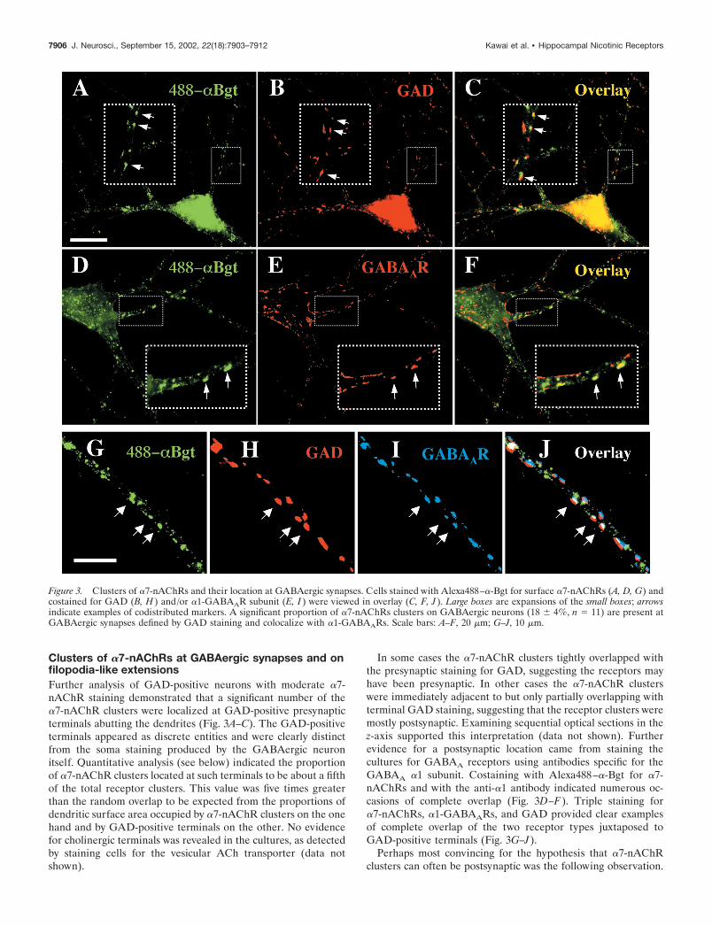
Clusters of �7-nAChRs at GABAergic synapses and onfilopodia-like extensionsFurther analysis of GAD-positive neurons with moderate �7-nAChR staining demonstrated that a significant number of the�7-nAChR clusters were localized at GAD-positive presynapticterminals abutting the dendrites (Fig. 3A–C). The GAD-positiveterminals appeared as discrete entities and were clearly distinctfrom the soma staining produced by the GABAergic neuronitself. Quantitative analysis (see below) indicated the proportionof �7-nAChR clusters located at such terminals to be about a fifthof the total receptor clusters. This value was five times greaterthan the random overlap to be expected from the proportions ofdendritic surface area occupied by �7-nAChR clusters on the onehand and by GAD-positive terminals on the other. No evidencefor cholinergic terminals was revealed in the cultures, as detectedby staining cells for the vesicular ACh transporter (data notshown).
In some cases the �7-nAChR clusters tightly overlapped withthe presynaptic staining for GAD, suggesting the receptors mayhave been presynaptic. In other cases the �7-nAChR clusterswere immediately adjacent to but only partially overlapping withterminal GAD staining, suggesting that the receptor clusters weremostly postsynaptic. Examining sequential optical sections in thez-axis supported this interpretation (data not shown). Furtherevidence for a postsynaptic location came from staining thecultures for GABAA receptors using antibodies specific for theGABAA �1 subunit. Costaining with Alexa488–�-Bgt for �7-nAChRs and with the anti-�1 antibody indicated numerous oc-casions of complete overlap (Fig. 3D–F). Triple staining for�7-nAChRs, �1-GABAARs, and GAD provided clear examplesof complete overlap of the two receptor types juxtaposed toGAD-positive terminals (Fig. 3G–J).
Perhaps most convincing for the hypothesis that �7-nAChRclusters can often be postsynaptic was the following observation.
Figure 3. Clusters of �7-nAChRs and their location at GABAergic synapses. Cells stained with Alexa488–�-Bgt for surface �7-nAChRs (A, D, G) andcostained for GAD (B, H ) and/or �1-GABAAR subunit (E, I ) were viewed in overlay (C, F, J ). Large boxes are expansions of the small boxes; arrowsindicate examples of codistributed markers. A significant proportion of �7-nAChRs clusters on GABAergic neurons (18 � 4%, n � 11) are present atGABAergic synapses defined by GAD staining and colocalize with �1-GABAARs. Scale bars: A–F, 20 �m; G–J, 10 �m.
7906 J. Neurosci., September 15, 2002, 22(18):7903–7912 Kawai et al. • Hippocampal Nicotinic Receptors

GAD-positive neurons that did not themselves express �7-nAChRs (unlabeled category of neurons) lacked �7-nAChR clus-ters even at dendritic sites receiving GAD-positive terminals.Examining a total of 126 dendrites on unlabeled neurons (28neurons, 13 experiments) yielded no detectable �7-nAChR clus-ters, although 2618 GAD-positive terminals were identified asabutting on the dendrites. In contrast, examining 108 dendritesfrom neurons with moderate Alexa488–�-Bgt staining (44 neu-rons, 14 experiments) yielded 3691 receptor clusters, and 664 ofthem were associated with GAD-positive terminals which them-selves numbered 1702 in all. The simplest interpretation is thatmost, if not all, of the �7-nAChR clusters detectable at GABAer-gic synapses on moderate neurons are provided by the postsyn-aptic neuron. Presynaptic �7-nAChRs are likely to be present(Alkondon et al., 1997a; Maggi et al., 2001) but may have beentoo sparse to score as a cluster with the current imaging criteria.A much less likely explanation for the result would be that GADterminals cannot express presynaptic �7-nAChRs for some rea-son if the postsynaptic neuron is unable to do so. Taken together,the present results suggest a postsynaptic location for many of the�7-nAChR clusters observed here at GABAergic synapses, butthis conclusion should be considered tentative until confirmed byultrastructural analysis.
GAD-positive neurons with high �7-nAChR labeling did notpermit resolution of individual receptor clusters in most cases, butexamining the cells yielded an additional interesting result. Inmany cases, �7-nAChRs were found sharply concentrated at thedistal tips of filopodia-like extensions emanating from the den-drites (Fig. 4). Such �7-nAChR clusters were not associated with�1-GABAARs or GAD-stained presynaptic terminals.
Activity-dependent regulation of �7-nAChR levels onGABAergic neuronsElectrical activity was necessary to sustain the �7-nAChR stain-ing levels found on GABAergic hippocampal neurons. This couldbe seen by examining the distribution of cells among �7-nAChRlabeling categories as described above for control neurons. Treat-ing cultures for 3 d with TTX to block voltage-gated sodiumchannels produced a dramatic reduction in the cells with highlabeling for �7-nAChRs and a corresponding increase in theproportion of unlabeled GAD-positive neurons (Fig. 5). Moder-ate cells appeared unchanged in number but did shift to loweroverall levels of labeling, best revealed by quantifying the number
of �7-nAChR clusters (see below). The TTX treatment producedno decrement in the total number of neurons identified by MAP-2staining (99 � 5% of control values; mean � SEM, n � 5experiments with triplicate cultures; total of 1579 cells examined)or in the number of GABAergic neurons identified by GADstaining (98 � 4% of control values).
Blockade of NMDA receptor function produced the sameeffect as TTX. A 3 d exposure to APV almost completely elimi-nated cells from the high category; a concomitant increase oc-curred in unlabeled cells (Fig. 5). APV produced no change in thetotal number of neurons (92 � 8% of control values; n � 3experiments) and no change in the number of GAD-positiveneurons (101 � 8% of control values). Blockade of GABAergictransmission with bicuculline for as long as 7 d, on the other hand,had no effect on the proportions of GAD-positive neurons scoringeither as moderate or high for �7-nAChR labeling. Blockade ofnicotinic transmission with D-TC also had no effect, although lowconcentrations of nicotine either for a 3 or 7 d period increasedthe proportion of GAD-positive neurons with high �7-nAChRlabeling. The results indicate that endogenous activation ofNMDA receptors is essential to sustain the �7-nAChR patternsdetectable here on GABAergic hippocampal neurons. Endoge-nous GABAergic and nicotinic signaling are much less important,although chronic nicotine exposure can increase �7-nAChRstaining.
A different outcome was obtained in one respect when mea-suring the total number of �7-nAChRs on the surface of allhippocampal cells in culture. Using 125I-�-Bgt binding to quan-tify �7-nAChRs on intact cells showed that the 3 d TTX treat-ment produced no change overall (99 � 10% of control values;mean � SEM; n � 3 experiments, three cultures per experiment).The bicuculline treatment (7 d) produced a small nominal in-
Figure 4. Clusters of �7-nAChRs at tips of filopodia-like structuresemanating from dendrites of GABAergic interneurons. A, IsolatedGABAergic neuron with high level of Alexa488–�-Bgt labeling ( green)costained for GAD (red) and shown in overlay. B, The boxed region in Ais shown in expansion. The �7-nAChR clusters present at the tips of thefilopodia-like extensions (arrows) are not associated with GAD staining.Scale bars: A, 20 �m; B, 5 �m.
Figure 5. Activity-dependent regulation of �7-nAChR labeling. Thedistribution of GABAergic neurons among unlabeled, moderate, and high�7-nAChR labeling categories after treatment during the third week ofculture with TTX (1 �M, 3 d), APV (50 �M, 3 d), bicuculline (20 �M, 7 d),nicotine (0.5 �M, 3 and 7 d), or D-TC (10 �M, 3 d). Values are expressedas a percentage of controls from the same experiment (horizontal line) andrepresent the mean � SEM of three to nine experiments. The level of�7-nAChR labeling on GABAergic neurons depends on electrical activityand NMDA receptor activation but not on endogenous GABAA ornicotinic receptor signaling. Addition of nicotine, however, can enhancethe level of labeling. Statistical differences were determined by ANOVA;*p � 0.05, ** p � 0.01, ***p � 0.001.
Kawai et al. • Hippocampal Nicotinic Receptors J. Neurosci., September 15, 2002, 22(18):7903–7912 7907

crease (131 � 21% of control values; n � 3), but the differencewas not significant ( p � 0.1). The divergence between theseresults with TTX and those obtained by fluorescence analysis of�7-nAChRs on GAD-positive neurons may have arisen fromeither of two sources. GAD-positive neurons are a minority ofthe neuronal population and may have been overshadowed byreceptor changes on the population as a whole. Alternatively, thetreatments may have altered the distribution of receptors, chang-ing the proportions that were sufficiently concentrated to bedetected by fluorescence imaging. In any case, the results clearlydemonstrate the importance of monitoring receptor distributionon individual neurons rather than simply measuring overall levelsin heterogeneous cultures.
Effects of activity on �7-nAChR clustersTo obtain a finer grain analysis of activity-dependent �7-nAChRregulation, we again examined �7-nAChR clusters on GAD-positive neurons with moderate receptor labeling. Moderate cellswere chosen because, as stated above, they displayed numerousclusters that could be readily resolved. GAD staining was used todistinguish GABAergic presynaptic terminals contacting the den-drites of such neurons, whereas Alexa488–�-Bgt staining wasused to visualize �7-nAChR clusters on the cells. Thresholdvalues of five contiguous pixels (0.05 �m2) were set both for theminimum area defining an �7-nAChR cluster or GAD-positivepresynaptic terminal and for the minimum area defining overlapor codistribution of the two labels. Threshold values defining theminimum labeling intensity acceptable for a cluster or terminalwere set at half the maximal intensities over background seen formoderate cells in control cultures in the same experiment.
By these criteria the 3 d treatment with either TTX or APVproduced no change in the mean size either of GAD-positiveterminals abutting the neurons or of �7-nAChR clusters them-selves (Fig. 6). Both treatments did, however, produce largedecreases in the number of �7-nAChR clusters (Fig. 6) withoutchanging the number of GAD-positive terminals (data notshown). Bicuculline treatment (7 d) increased the mean size ofGAD-positive terminals but had no effect on the size or numberof �7-nAChR clusters. Nicotine treatment for 3 d had no effect onGAD-positive terminals but increased both the mean size andnumber of �7-AChR clusters (Fig. 6). Neither bicuculline nornicotine significantly altered the proportion of �7-nAChR clus-ters found at GABAergic synapses. Thus, 25 � 4 and 22 � 8%(n � 4 experiments) of the �7-nAChR clusters on GABAergicneurons from bicuculline- and nicotine-treated cultures, respec-tively, were located at GAD-positive terminals, whereas 18 � 4%(n � 11 experiments) were so in control cultures. It was notpossible to determine whether TTX or APV preferentially af-fected �7-nAChR clusters at GABAergic synapses because of thesmall residual numbers of clusters in these cases. When sufficientclusters were present for analysis (e.g., controls and cells treatedwith bicuculline, D-TC, or nicotine), the incidence of colocaliza-tion for �7-nAChR clusters and GAD-positive terminals was atleast five times that expected from random occurrence based onthe proportion of dendritic surface occupied by each population.
The results indicate that elimination of action potentials orblockade of NMDA receptors shifts cells to lower categories oflabeling and reduces the number of detectable �7-nAChR clus-ters. Analyzing the distribution of clusters after triply staining for�7-nAChRs, GAD, and �1-GABAARs yielded an additional con-clusion. On neurons that expressed both classes of receptors,�1-GABAAR clusters were found associated with 58 � 12% of
the GAD-positive terminals abutting the dendrites (eight cells,four experiments). On the same neurons, �7-nAChR clusterswere associated with 38 � 18% of the GAD-positive terminals.Both kinds of receptor clusters co-localized at 29 � 18% of theGAD-positive terminals. This value is consistent with the twoclasses of receptors distributing independently at the synapsesand indicates that their presence is not mutually exclusive.
Neurotrophin-dependent regulation of �7-nAChRsThe neurotrophins BDNF and NGF are both produced by hip-pocampal neurons and play important roles in synaptic develop-ment and plasticity in the hippocampus (McAllister et al., 1999;Schuman, 1999; Schinder and Poo, 2000). Both can enhanceoverall excitation in the cultures (Bolton et al., 2000; Tyler andPozzo-Miller, 2001; Kovalchuk et al., 2001) and are regulated inturn by synaptic activity (Zafra et al., 1991; Lauterborn et al.,2000; Hartmann et al., 2001). Neuregulin upregulates functional�7-nAChRs on hippocampal neurons in culture (Liu et al., 2001),and BDNF can induce neuregulin release in other systems (Loeband Fischbach, 1997; Loeb et al., 2002). These observationsmotivated us to test the effects of neuregulin in the imaging assaysused here and to examine the roles of BDNF and NGF in�7-nAChR regulation. Exposing hippocampal neurons to neu-regulin (1-�1 isoform) for 3 d produced significant increases inthe proportion of GAD-positive neurons expressing high �7-nAChR labeling (Fig. 7). This mirrored the neuregulin effectsreported previously on �7-nAChR-mediated whole-cell re-sponses (Liu et al., 2001). Compensatory decreases were seen inthe unlabeled and moderate categories of GAD-positive neurons(data not shown). Treatment with either BDNF or NGF pro-
Figure 6. Activity-dependent regulation of �7-nAChR clusters. Shownare the normalized values for the mean size of either GAD-positiveterminals or �7-nAChR clusters or the mean number of �7-nAChRclusters distributed along the dendrites of GABAergic neurons havingmoderate levels of �7-nAChR labeling. Cultures were treated for 3 d withTTX (1 �M), APV (50 �M), bicuculline (20 �M), or nicotine (0.5 �M).Both TTX and APV decrease while nicotine increases the number of�7-nAChR clusters; only nicotine increases mean cluster size. Only bicu-culline increases mean GAD terminal size, but it has no effect on themean size or number of �7-nAChR clusters. Values are expressed as apercentage of controls from the same experiment (horizontal line) andrepresent the mean � SEM of four to nine experiments. Controls had45 � 5 �7-nAChR clusters per 100 �m dendritic length and mean sizes of0.18 � 0.02 and 0.34 � 0.02 �m 2 for the �7-nAChR clusters and GAD-positive terminals, respectively (n � 7 experiments). Statistical differenceswere determined by ANOVA; *p � 0.05, ** p � 0.01, ***p � 0.001.
7908 J. Neurosci., September 15, 2002, 22(18):7903–7912 Kawai et al. • Hippocampal Nicotinic Receptors

duced the same increases as neuregulin (Fig. 7). No furtherincrease was seen when BDNF and neuregulin were combined(192 � 22, 175 � 25, and 163 � 13% for BDNF, neuregulin, andBDNF � neuregulin treatments, respectively, normalizing thedata for control values; mean � SEM with n � 4 cultures, 80cells). Anti-BDNF and anti-NGF antibodies each significantlydecreased the proportion of highly labeled GAD-positive neurons(Fig. 7). The results suggest that endogenous BDNF and NGFcontribute to �7-nAChR levels on GABAergic neurons. NeitherBDNF nor NGF, when added to the cultures however, couldovercome the effects of either TTX or APV (Fig. 7). Increasingthe concentration of BDNF 10-fold did not alter the outcome(data not shown). Neuregulin was also ineffective against TTX.
Neurotrophin effects were further evaluated by examining thenumber and location of �7-nAChR clusters. GABAergic neuronswith moderate �7-nAChR labeling were again chosen becauseindividual receptor clusters could be readily quantified. BDNF,NGF, and neuregulin each had little effect on the size or numberof GAD-positive terminals (data not shown), but each increasedthe number of �7-nAChR clusters (Fig. 8). BDNF also caused asmall increase in the mean size of �7-nAChR clusters (data notshown). Treating the neurons with either anti-BDNF or anti-NGF antiserum had the opposite effect, reducing the numbers of�7-nAChR clusters (Fig. 8). Nonimmune IgG used as negativecontrols had no effect. Importantly, neither BDNF nor NGF wereable to overcome the effects of either TTX or APV on thenumber of �7-nAChR clusters (Fig. 8).
A qualitatively similar pattern was seen when counting only�7-nAChR clusters localized at GAD-positive terminals. Whereas
control cells had 8.0 � 1.0 such “co-clusters” per 100 �m dendriticlength (32 neurons, 10 experiments), cells treated with TTX, APV,anti-BDNF, or anti-NGF each had much reduced numbers (2.7 �1.0, 0.5 � 0.3, 2.8 � 0.3, and 1.0 � 0.4, respectively). In fact forseveral of the treatments, the number of �7-nAChR clusters atGAD-positive terminals was not significantly different from thatpredicted for random occurrence based on the total number ofclusters and GAD-positive terminals along the dendrite. BothBDNF and NGF increased the number of clusters at GAD-positiveterminals (16 � 3 and 12 � 2/100 �m, respectively) but neithercould overcome the effects of APV (1.2 � 0.2 and 0.6 � 0.3,respectively). The overall results, both from cluster analysis onmoderate cells and from distribution analysis of cells among label-ing categories, are in agreement. Neurotrophins are necessary, butnot sufficient in the absence of either electrical activity in generalor NMDA receptor activation in particular, to sustain �7-nAChRdistribution patterns on the neurons.
DISCUSSIONThe principal findings reported here are first that fluorescently-labeled �-Bgt can be used to visualize �7-nAChRs on the surfaceof hippocampal neurons in culture and that the receptors arearranged in clusters localized in part at GABAergic synapses.Second, spontaneous electrical activity and NMDA receptor ac-tivation are essential for sustaining the receptor clusters, includ-ing those at GABAergic synapses. Third, endogenous BDNF andNGF also are required, but exogenous BDNF or NGF cannotovercome the downregulation caused by blockade of NMDAreceptors. Several models can account for the results includingthe possibilities that BDNF and NGF act upstream of NMDAreceptors, perhaps increasing excitation in the cultures as sug-gested in other situations (Bolton et al., 2000; Tyler and Pozzo-Miller, 2001; Kovalchuk et al., 2001) or that they are required inparallel with activity. GABAergic neurons have been widelyrecognized as targets of �7-nAChR signaling. Presynaptic �7-
Figure 7. Neurotrophin regulation of �7-nAChR labeling. The distribu-tion of GABAergic neurons among �7-nAChR labeling categories wasanalyzed as in Figure 5 after treating cells for 3 d with neuregulin (5 nM),BDNF (100 ng/ml), NGF (100 ng/ml), chicken anti-BDNF antibodies,sheep anti-NGF antibodies, both chicken and sheep nonimmune IgG(negative controls), and where indicated, either TTX (1 �M) or APV (50�M) as well. Shown here are the results obtained for cells in the highlabeling category. Values are expressed as a percentage of controls fromthe same experiment (horizontal line) and represent the mean � SEM offour to nine experiments. Neuregulin, BDNF, and NGF each elevate thelevel of �7-nAChR labeling while anti-BDNF, and anti-NGF decrease it.The neurotrophins are unable to overcome the negative effects of eitherTTX or APV. Neuregulin was unable to overcome TTX (not significantlydifferent from TTX alone; TTX data from Fig. 5). Statistical differenceswere determined by ANOVA; ** p � 0.01, ***p � 0.001.
Figure 8. Neurotrophin regulation of �7-nAChR clusters. The numbersof �7-nAChR clusters per 100 �m dendritic length on GABAergic neu-rons are expressed as a percentage of controls from the same experiment.Values represent the mean � SEM of four to nine experiments. Condi-tions and agents are the same as in Figure 7. Neuregulin, BDNF, andNGF each increase the total number of �7-nAChR clusters, whereasanti-BDNF and anti-NGF decrease them. The neurotrophins are unableto overcome the negative effects of either TTX or APV. Statisticaldifferences were determined by ANOVA; *p � 0.05, ***p � 0.001.
Kawai et al. • Hippocampal Nicotinic Receptors J. Neurosci., September 15, 2002, 22(18):7903–7912 7909

nAChRs on the neurons modulate GABA release while somato-dendritic �7-nAChRs mediate synaptic input to the neurons andinfluence their participation in hippocampal circuits (Alkondonet al., 1997a, 1998; Frazier et al., 1998; Hefft et al., 1999; Jones etal., 1999; Ji and Dani, 2000; Alkondon and Albuquerque, 2001;Buhler and Dunwiddie, 2001, 2002; Ji et al., 2001; Maggi et al.,2001). Patch-clamp recording suggests that GABAergic interneu-rons have the highest whole-cell �7-nAChR responses in hip-pocampal cultures (Liu et al., 2001). The present studies showthat GABAergic neurons express the highest incidence of surface�7-nAChRs detectable by fluorescence imaging, a feature thatfacilitated the analysis of receptor distribution on the cells. Thereceptors can also be found on glutamatergic neurons, however,and recent ultrastructural studies in situ show them to be localizedat glutamatergic synapses as well (Fabian-Fine et al., 2001; Levyand Aoki, 2002).
Costaining for GAD and �7-nAChRs made clear the nonran-dom distribution of �7-nAChRs clusters at presumed GABAergicsynapses defined by GAD-positive terminals abutting the den-drites. Triply staining for GAD, �7-nAChRs, and �1-GABAARsindicated that the two types of receptors were not mutuallyexclusive in their clustering at individual GABAergic synapses.This is quite different from the pattern reported for misdirectedreceptors on isolated neurons that are constrained to receiveautapses as their only innervation (Rao et al., 2000). In that caseinappropriate receptors could be found at some synapses (e.g.,GABA receptors opposite glutamatergic terminals) but not colo-calized with correct receptors (i.e., glutamate receptors) at thesame synapses. In the present studies the cultures were hetero-geneous, the neurons were multiply innervated, and the �7-nAChR clusters did not exclude the expected transmitterreceptor.
Certainly presynaptic �7-nAChRs would be expected at hip-pocampal GABAergic synapses, judging from electrophysiologi-cal studies. The �7-nAChR clusters imaged here, however,seemed more likely to be postsynaptic for the most part. This wassuggested by the distribution of receptors in three-dimensionalspace, as seen in stacks of optical sections and by the comparativedistributions of �7-nAChRs, GABAA-�1 receptors, and presyn-aptic GAD staining in triple-stain experiments. It was alsostrongly suggested by the finding that detectable �7-nAChR clus-ters were not associated with GAD-positive terminals if theterminals contacted dendrites from neurons not expressing de-tectable �7-nAChRs themselves. Alternative explanations for thislatter finding exist but are less plausible. The imaging studies donot, of course, exclude the presence of presynaptic �7-nAChRsbut do suggest that the staining intensity associated with them isusually less than the threshold set here for defining fluorescentlylabeled �7-nAChR clusters. Indeed, the ultrastructural studiescited above indicate the presence of both presynaptic andpostsynaptic �7-nAChR clusters in the adult hippocampus. Ul-trastructural analysis will probably also be required here to dis-tinguish unambiguously between presynaptic and postsynaptic�7-nAChRs in culture.
Neuronal activity had pronounced effects on the pattern of�7-nAChRs. A 3 d exposure to TTX did not alter the totalnumber of surface �7-nAChRs in culture as measured by 125I-�-Bgt binding, but did markedly reduce �7-nAChR staining levelsand cluster abundance on GABAergic neurons. The treatmentmay have dispersed �7-nAChR clusters, causing receptor densityto be subthreshold for detection by fluorescence. Alternatively,reductions in the number of �7-nAChRs on GABAergic neurons
may have been overshadowed by increases in receptors on othercell populations in the cultures. Blockade of NMDA receptorswith APV produced reductions at least as severe as did TTX, butit would be premature to conclude that all of the regulatory effectsof neuronal activity were mediated through NMDA receptors.
Surprisingly, blockade of GABAA receptors with bicucullinehad no effect on �7-nAChR staining patterns even though asignificant fraction of the �7-nAChR clusters were located atGABAergic synapses. The normal GABA-mediated maturationof the chloride gradient should already have occurred before thebicuculline treatment (Ganguly et al., 2001). Blockade of nicotinicreceptors with D-TC also had no obvious effect, although theculture medium contained sufficient choline to partially desensi-tize the receptors (Alkondon et al., 1997b). Absent from thecultures was the septal input which provides the major cholinergicinnervation of the hippocampus in vivo, and staining for vesicularACh transporter failed to detect cholinergic terminals in thecultures. Chronic nicotine exposure, however, did upregulate thelevel of staining, consistent with other studies on CNS �7-nAChRs (Barrantes et al., 1995; Peng et al., 1997; Kawai andBerg, 2001), and suggests that cholinergic input could play a rolein vivo.
Perhaps most interesting are the effects of BDNF and NGF.Both were attractive candidates for mediating activity-dependentregulation of �7-nAChR clusters. BDNF and NGF are expressedin the hippocampus and both can influence synapse formation(McAllister et al., 1999; Schuman, 1999; Schinder and Poo, 2000).BDNF can enhance neuregulin release at the neuromuscularjunction, and neuregulin upregulates functional �7-nAChRs onhippocampal neurons (Loeb and Fischbach, 1997; Liu et al., 2001;Loeb et al., 2002). Moreover, chronic excitation increases theexpression of BDNF and NGF by hippocampal cells. The presentstudies show that BDNF and NGF can each increase the numberof �7-nAChR clusters on GABAergic hippocampal neurons andthat anti-BDNF and anti-NGF antisera each reduce the number.The latter results argue strongly for a neurotrophin role in reg-ulating �7-nAChRs.
Neither BDNF nor NGF, when supplied exogenously, couldovercome the effects of either TTX or APV. One possibility, ascited above, is that the neurotrophins act upstream of NMDAreceptors. But neurotrophins are known to have multiple effectsand sites of action in the hippocampus (McAllister et al., 1999;Schuman, 1999; Schinder and Poo, 2000). Accordingly, otherpossibilities that must be considered are that the neurotrophinsare necessary but not sufficient or that they must act locally or thattheir signaling is compromised somehow by NMDA receptorblockade (e.g., trk receptor downregulation). Still it is attractiveto consider that a neurotrophin-mediated enhancement of activ-ity might drive neuregulin release which, in turn, regulates �7-nAChRs. BDNF and neuregulin are not additive in their effects,and preliminary results suggest that exogenous neuregulin canovercome the effects of anti-BDNF antiserum on �7-nAChRcluster abundance (W. M. Zago and D. K. Berg, unpublishedresults). These observations support a downstream site of actionfor neuregulin. Where NMDA receptors participate in the path-way and exactly how the neurotrophins and neuregulin exert theireffects have yet to be determined.
Chronic blockade of excitatory activity in hippocampal culturesincreases the capacity for excitation by altering both presynapticand postsynaptic elements at glutamatergic synapses (Schinder andPoo, 2000; Turrigiano and Nelson, 2000; Murthy et al., 2001). Thishomeostatic adaptation depends on AMPA receptor signaling
7910 J. Neurosci., September 15, 2002, 22(18):7903–7912 Kawai et al. • Hippocampal Nicotinic Receptors

rather than on NMDA receptors. NMDA receptors commonlymediate other types of activity-dependent synaptic plasticity in thehippocampus. Nonetheless, the TTX- and APV-mediated reduc-tions seen here in �7-nAChR clusters on GABAergic neuronscould be viewed as homeostatic compensation if the receptorclusters normally enhance GABAergic inhibitory signaling. Al-though this is certainly true for presynaptic �7-nAChRs (Alkon-don et al., 1997a; Maggi et al., 2001), it is less clear for postsynaptic�7-nAChRs at GABAergic synapses. Because GABAergic neu-rons, however, were the focus of the present study, it might beargued that any reduction in excitatory input to the cells, includingthat via �7-nAChRs, could represent a compensatory mechanismbecause it would ultimately reduce output from these inhibitoryneurons.
Key questions remain about the physiological roles of �7-nAChR clusters at noncholinergic synapses. The fact that suchclusters persist in the adult suggests they provide a continuingfunction. Particularly interesting are postsynaptic �7-nAChRclusters at GABAergic synapses because the postsynaptic clustersshould oppose the effect of presynaptic �7-nAChRs on inhibitorysignaling. This opposition would be acute if both classes of re-ceptors are activated in the same time frame, as should occur ifACh participates in volume transmission made possible by thediffuse cholinergic projection of the medial septum to the hip-pocampus (McKinney et al., 1983; Frotscher and Leranth, 1985;Descarries et al., 1997; Descarries, 1998; Zoli et al., 1999) (but seeTurrini et al., 2001). Equally intriguing are the �7-nAChR clus-ters found at the tips of filopodia-like extensions emanating fromGABAergic neuron dendrites. Dendritic filopodia are candidatesfor synaptic precursor structures (Fiala et al., 1998; Jontes andSmith, 2000; Cline, 2001). What �7-nAChRs do for such struc-tures will be interesting to determine.
REFERENCESAlkondon M, Albuquerque EX (2001) Nicotinic acetylcholine receptors
�7 and �4�2 subtypes differentially control GABAergic input to CA1neurons in rat hippocampus. J Neurophysiol 86:3043–3055.
Alkondon M, Pereira EFR, Barbosa CTF, Albuquerque EX (1997a)Neuronal nicotinic acetylcholine receptor activation modulates�-aminobutyric acid release from CA1 neurons of rat hippocampalslices. J Pharmacol Exp Ther 283:1396–1411.
Alkondon M, Pereira EFR, Cortes WS, Maelicke A, Albuquerque EX(1997b) Choline is a selective agonist of �7 nicotinic acetylcholinereceptors in the rat brain neurons. Eur J Neurosci 9:2734–2742.
Alkondon M, Pereira EFR, Albuquerque EX (1998) �-Bungarotoxin-and methyllycaconitine-sensitive nicotinic receptors mediate fast syn-aptic transmission in interneurons of rat hippocampal slices. Brain Res810:257–263.
Barrantes GE, Roger AT, Lindstrom J, Wonnacott S (1995)�-Bungarotoxin binding sites in rat hippocampal and cortical cultures:initial characterization, colocalization with �7 subunits and up-regulation by chronic nicotine treatment. Brain Res 672:228–236.
Bertrand D, Galzi JL, Devillers-Thiery A, Bertrand S, Changeux JP(1993) Mutations at two distinct sites within the channel domain M2alter calcium permeability of neuronal �7 nicotinic receptor. Proc NatlAcad Sci USA 90:6971–6975.
Bolton MM, Pittman AJ, Lo DC (2000) Brain-derived neurotrophicfactor differentially regulates excitatory and inhibitory synaptic trans-mission in hippocampal cultures. J Neurosci 20:3221–3232.
Buhler AV, Dunwiddie TV (2001) Regulation of the activity of hip-pocampal stratum oriens interneurons by �7 nicotinic acetylcholinereceptors. Neuroscience 106:55–67.
Buhler AV, Dunwiddie TV (2002) �7 Nicotinic acetylcholine receptorson GABAergic interneurons evoke dendritic and somatic inhibition ofhippocampal neurons. J Neurophysiol 87:548–557.
Cline HT (2001) Dendritic arbor development and synaptogenesis. CurrOpin Neurobiol 11:118–126.
Descarries L (1998) The hypothesis of an ambient level of acetylcholinein the central nervous system. J Physiol (Paris) 92:215–220.
Descarries L, Gisiger V, Steriade M (1997) Diffuse transmission byacetylcholine in the CNS. Prog Neurobiol 53:603–625.
Fabian-Fine R, Skehel P, Errington ML, Davies HA, Sher E, Stewart
MG, Fine A (2001) Ultrastructural distribution of the �7 nicotinicacetylcholine receptor subunit in rat hippocampus. J Neurosci21:7993–8003.
Fiala JC, Feinberg M, Popov V, Harris KM (1998) Synaptogenesis viadendritic filopodia in developing hippocampal area CA1. J Neurosci18:8900–8911.
Frazier CJ, Buhler AV, Weiner JL, Dunwiddie TV (1998) Synapticpotentials mediated via �-bungarotoxin-sensitive nicotinic acetylcho-line receptors in rat hippocampal interneurons. J Neurosci18:8228–8235.
Freund TF, Buzsaki G (1996) Interneurons of the hippocampus. Hip-pocampus 6:347–470.
Frotscher M, Leranth C (1985) Cholinergic innervation of the rat hip-pocampus as revealed by choline acetyltransferase immunocytochem-istry: a combined light and electron microscopic study. J Comp Neurol239:237–246.
Fujii S, Ji Z, Sumikawa K (2000) Inactivation of �7 ACh receptors andactivation of non-�7 ACh receptors both contribute to long termpotentiation induction in the hippocampal CA1 region. Neurosci Lett286:134–138.
Ganguly K, Schinder AF, Wong ST, Poo Mm (2001) GABA itself pro-motes the developmental switch of neuronal GABAergic responsesfrom excitation to inhibition. Cell 105:521–532.
Gray R, Rajan AS, Radcliffe KA, Yakehiro M, Dani JA (1996) Hip-pocampal synaptic transmission enhanced by low concentrations ofnicotine. Nature 383:713–716.
Hartmann M, Heumann R, Lessmann V (2001) Synaptic secretion ofBDNF after high-frequency stimulation of glutamatergic synapses.EMBO J 20:5887–5897.
Hefft S, Hulo S, Bertrand D, Muller D (1999) Synaptic transmission atnicotinic acetylcholine receptors in rat hippocampal organotypic cul-tures and slices. J Physiol (Lond) 515:769–776.
Ji D, Dani JA (2000) Inhibition and disinhibition of pyramidal neuronsby activation of nicotinic receptors on hippocampal interneurons.J Neurophysiol 83:2682–2690.
Ji D, Lape R, Dani JA (2001) Timing and location of nicotinic activityenhances or depresses hippocampal synaptic plasticity. Neuron31:131–141.
Jones S, Sudweeks S, Yakel JL (1999) Nicotinic receptors in the brain:correlating physiology with function. Trends Neurosci 22:555–561.
Jontes JD, Smith SJ (2000) Filopodia, spines, and the generation ofsynaptic diversity. Neuron 27:11–14.
Kawai H, Berg DK (2001) Nicotinic acetylcholine receptors containing�7 subunits on rat cortical neurons do not undergo long-lasting inacti-vation even when up-regulated by chronic nicotine exposure. J Neuro-chem 78:1367–1378.
Kovalchuk Y, Hanse E, Kafitz KW, Konnerth A (2001) Postsynapticinduction of BDNF-mediated long-term potentiation. Science295:1729–1734.
Lauterborn JC, Lynch G, Vanderklish P, Arai A, Gall CM (2000) Pos-itive modulation of AMPA receptors increases neurotrophin expres-sion by hippocampal and cortical neurons. J Neurosci 20:8–21.
Levy RB, Aoki C (2002) �7 Nicotinic acetylcholine receptors occur atpostsynaptic densities of AMPA receptor-positive and –negative exci-tatory synapses in rat sensory cortex. J Neurosci 22:5001–5015.
Liu Y, Ford B, Mann MA, Fischbach GD (2001) Neuregulins increase�7 nicotinic acetylcholine receptors and enhance excitatory synaptictransmission in GABAergic interneurons of the hippocampus. J Neu-rosci 21:5660–5669.
Loeb JA, Fischbach GD (1997) Neurotrophic factors increase neuregu-lin expression in embryonic ventral spinal cord neurons. J Neurosci17:1416–1424.
Loeb JA, Hmadcha A, Fischbach GD, Land SJ, Zakarian VL (2002)Neuregulin expression at neuromuscular synapses is modulated bysynaptic activity and neurotrophic factors. J Neurosci 22:2206–2214.
Maggi L, Sher E, Cherubini E (2001) Regulation of GABA release bynicotinic acetylcholine receptors in the neonatal rat hippocampus.J Physiol (Lond) 536:89–100.
McAllister AK, Katz LC, Lo DC (1999) Neurotrophins and synapticplasticity. Annu Rev Neurosci 22:295–318.
McKinney M, Coyle JT, Hedreen JC (1983) Topographic analysis of theinnervation of the rat neocortex and hippocampus by the basal fore-brain cholinergic system. J Comp Neurol 217:103–121.
Murthy VN, Schikorski T, Stevens CF, Zhu Y (2001) Inactivity producesincreases in neurotransmitter release and synapse size. Neuron32:673–682.
Olbrich HG, Braak H (1985) Ratio of pyramidal cells versus non-pyramidal cells in sector CA1 of the human Ammon’s horn. AnatEmbryol 173:105–110.
Paysan J, Coggan JS, Conroy WG, Berg DK (2000) The neurofilamentinfrastructure of a developing presynaptic calyx. J Comp Neurol425:284–294.
Peng X, Gerzanich V, Anand R, Wang F, Lindstrom J (1997) Chronicnicotine treatment up-regulates �3 and �7 acetylcholine receptor sub-
Kawai et al. • Hippocampal Nicotinic Receptors J. Neurosci., September 15, 2002, 22(18):7903–7912 7911

types expressed by the human neuroblastoma cell line SH-SY5Y. MolPharmacol 51:776–784.
Radcliffe KA, Dani JA (1998) Nicotinic stimulation produces multipleforms of increased glutamatergic synaptic transmission. J Neurosci18:7075–7083.
Rao A, Cha EM, Craig AM (2000) Mismatched appositions of presyn-aptic and postsynaptic components in isolated hippocampal neurons.J Neurosci 20:8344–8353.
Samuel N, Wonnacott S, Lindstrom J, Futerman AH (1997) Parallelincreases in [�- 125I]bungarotoxin binding and �7 nicotinic subunitimmunoreactivity during the development of rat hippocampal neuronsin culture. Neurosci Lett 222:179–182.
Schinder AF, Poo MM (2000) The neurotrophin hypothesis for synapticplasticity. Trends Neurosci 23:639–645.
Schuman EM (1999) Neurotrophin regulation of synaptic transmission.Curr Opin Neurobiol 9:105–109.
Seguela P, Wadiche J, Dineley-Miller K, Dani JA, Patrick JW (1993)Molecular cloning, functional properties, and distribution of rat brain�7: a nicotinic cation channel highly permeable to calcium. J Neurosci13:596–604.
Turrigiano GG, Nelson SB (2000) Hebb and homeostasis in neuronalplasticity. Curr Opin Neurobiol 10:358–364.
Turrini P, Casu MA, Wong TP, De Koninck Y, Ribeiro-Da-Silva A,Cuello AC (2001) Cholinergic nerve terminals establish classical syn-apses in the rat cerebral cortex: synaptic pattern and age-related atro-phy. Neuroscience 105:277–285.
Tyler WJ, Pozzo-Miller LD (2001) BDNF enhances quantal neurotrans-mitter release and increases the number of docked vesicles at the activezones of hippocampal excitatory synapses. J Neurosci 21:4249–4258.
Zafra F, Castren E, Thoenen H, Lindholm D (1991) Interplay betweenglutamate and gamma-aminobutyric acid transmitter systems in thephysiological regulation of brain-derived neurotrophic factor and nervegrowth factor synthesis in hippocampal neurons. Proc Natl Acad SciUSA 88:10037–10041.
Zarei MM, Radcliffe KA, Chen D, Patrick JW, Dani JA (1999) Distri-butions of nicotinic acetylcholine receptor �7 and �2 subunits oncultured hippocampal neurons. Neuroscience 88:755–764.
Zoli M, Jansson A, Sykova E, Agnati LF, Fuxe K (1999) Volume trans-mission in the CNS and its relevance for neuropsychopharmacology.Trends Pharmacol Sci 20:142–150.
7912 J. Neurosci., September 15, 2002, 22(18):7903–7912 Kawai et al. • Hippocampal Nicotinic Receptors
Copyright © 2022 FDOKUMEN