The role of in vivo Ca2+ signals acting on Ca2+-calmodulin-dependent proteins for skeletal muscle...
11
J Physiol 589.21 (2011) pp 5021–5031 5021 The Journal of Physiology TOPICAL REVIEWS The role of in vivo Ca 2+ signals acting on Ca 2+ –calmodulin-dependent proteins for skeletal muscle plasticity Pasi Tavi 1 and H˚ akan Westerblad 2 1 Department of Biotechnology and Molecular Medicine, A.I. Virtanen Institute for Molecular Sciences, University of Eastern Finland, PO Box 1627, FI-70211 Kuopio, Finland 2 Department of Physiology and Pharmacology, Karolinska Institutet, 171 77 Stockholm, Sweden Abstract Skeletal muscle fibres are highly heterogeneous regarding size, metabolism and contra- ctile function. They also show a large capacity for adaptations in response to alterations in the activation pattern. A major part of this activity-dependent plasticity relies on transcriptional alterations controlled by intracellular Ca 2+ signals. In this review we discuss how intracellular Ca 2+ fluctuations induced by activation patterns likely to occur in vivo control muscle properties via effects on Ca 2+ –calmodulin-dependent proteins. We focus on two such Ca 2+ decoders: calcineurin and Ca 2+ –calmodulin-dependent protein kinase II. Inherent Ca 2+ transients during contractions differ rather little between slow- and fast-twitch muscle fibres and this difference is unlikely to have any significant impact on the activity of Ca 2+ decoders. The major exception to this is fatigue-induced changes in Ca 2+ transients that occur in fast-twitch fibres exposed to high-intensity activation typical of slow-twitch motor units. In conclusion, the cascade from neural stimulation pattern to Ca 2+ -dependent transcription is likely to be central in maintaining the fibre phenotypes in both fast- and slow-twitch fibres. Moreover, changes in Ca 2+ signalling (e.g. induced by endurance training) can result in altered muscle properties (e.g. increased mitochondrial biogenesis) and this plasticity involves other signalling pathways. (Received 24 May 2011; accepted after revision 12 September 2011; first published online 12 September 2011) Corresponding author P. Tavi: Department of Biotechnology and Molecular Medicine, A.I. Virtanen Institute for Molecular Sciences, University of Eastern Finland PO Box 1627 FI-70211 Kuopio, Finland. Email: pasi.tavi@uef.fi Abbreviations CaM, calmodulin; CaMKII, Ca 2+ –calmodulin-dependent protein kinase II; CaN, calcineurin; [Ca 2+ ] i , myoplasmic free [Ca 2+ ]; FDB, flexor digitorum brevis; HDAC, histone deacetylase; MEF2, myocyte enhancer factor-2; NFAT, nuclear factor of activated T-cells; PGC-1α, peroxisome proliferator-activated receptor γ coactivator 1α; RNS, nitrogen species; ROS, reactive oxygen species; RyR, ryanodine receptor; SR, sarcoplasmic reticulum. Introduction Skeletal muscle fibres are heterogeneous with respect to size, metabolism and contractile function. They also have H˚ akan Westerblad (left) is professor in cellular muscle physiology at the Karolinska Institutet in Stockholm, Sweden. He has developed techniques to study contractile function in isolated, intact fibres from mammalian skeletal muscle. A major focus of his research has been on cellular mechanisms of skeletal muscle fatigue. Pasi Tavi (right) is Finnish Academy Research Fellow at the University of Eastern Finland, A.I. Virtanen Institute for molecular sciences, Kuopio, Finland. He has developed several mathematical muscle cell models, and recently his research has focused on understanding the transcriptional control of muscle cell E–C coupling. a striking capability for adaptation and plasticity. This was demonstrated in classical experiments in the early 1960s where it was shown that the activity of the motoneuron defined the properties of the innervated muscle fibres C 2011 The Authors. Journal compilation C 2011 The Physiological Society DOI: 10.1113/jphysiol.2011.212860
-
Upload
independent -
Category
Documents
-
view
1 -
download
0
Transcript of The role of in vivo Ca2+ signals acting on Ca2+-calmodulin-dependent proteins for skeletal muscle...

J Physiol 589.21 (2011) pp 5021–5031 5021
The
Jou
rnal
of
Phys
iolo
gy
TOP ICAL REV IEWS
The role of in vivo Ca2+ signals acting onCa2+–calmodulin-dependent proteins for skeletal muscleplasticity
Pasi Tavi1 and Hakan Westerblad2
1Department of Biotechnology and Molecular Medicine, A.I. Virtanen Institute for Molecular Sciences, University of Eastern Finland, PO Box 1627,FI-70211 Kuopio, Finland2Department of Physiology and Pharmacology, Karolinska Institutet, 171 77 Stockholm, Sweden
Abstract Skeletal muscle fibres are highly heterogeneous regarding size, metabolism and contra-ctile function. They also show a large capacity for adaptations in response to alterations in theactivation pattern. A major part of this activity-dependent plasticity relies on transcriptionalalterations controlled by intracellular Ca2+ signals. In this review we discuss how intracellularCa2+ fluctuations induced by activation patterns likely to occur in vivo control muscle propertiesvia effects on Ca2+–calmodulin-dependent proteins. We focus on two such Ca2+ decoders:calcineurin and Ca2+–calmodulin-dependent protein kinase II. Inherent Ca2+ transients duringcontractions differ rather little between slow- and fast-twitch muscle fibres and this differenceis unlikely to have any significant impact on the activity of Ca2+ decoders. The major exceptionto this is fatigue-induced changes in Ca2+ transients that occur in fast-twitch fibres exposed tohigh-intensity activation typical of slow-twitch motor units. In conclusion, the cascade fromneural stimulation pattern to Ca2+-dependent transcription is likely to be central in maintainingthe fibre phenotypes in both fast- and slow-twitch fibres. Moreover, changes in Ca2+ signalling(e.g. induced by endurance training) can result in altered muscle properties (e.g. increasedmitochondrial biogenesis) and this plasticity involves other signalling pathways.
(Received 24 May 2011; accepted after revision 12 September 2011; first published online 12 September 2011)Corresponding author P. Tavi: Department of Biotechnology and Molecular Medicine, A.I. Virtanen Institute forMolecular Sciences, University of Eastern Finland PO Box 1627 FI-70211 Kuopio, Finland. Email: [email protected]
Abbreviations CaM, calmodulin; CaMKII, Ca2+–calmodulin-dependent protein kinase II; CaN, calcineurin; [Ca2+]i,myoplasmic free [Ca2+]; FDB, flexor digitorum brevis; HDAC, histone deacetylase; MEF2, myocyte enhancer factor-2;NFAT, nuclear factor of activated T-cells; PGC-1α, peroxisome proliferator-activated receptor γ coactivator 1α; RNS,nitrogen species; ROS, reactive oxygen species; RyR, ryanodine receptor; SR, sarcoplasmic reticulum.
Introduction
Skeletal muscle fibres are heterogeneous with respect tosize, metabolism and contractile function. They also have
Hakan Westerblad (left) is professor in cellular muscle physiology at the Karolinska Institutet inStockholm, Sweden. He has developed techniques to study contractile function in isolated, intact fibresfrom mammalian skeletal muscle. A major focus of his research has been on cellular mechanisms of skeletalmuscle fatigue. Pasi Tavi (right) is Finnish Academy Research Fellow at the University of Eastern Finland,A.I. Virtanen Institute for molecular sciences, Kuopio, Finland. He has developed several mathematicalmuscle cell models, and recently his research has focused on understanding the transcriptional controlof muscle cell E–C coupling.
a striking capability for adaptation and plasticity. This wasdemonstrated in classical experiments in the early 1960swhere it was shown that the activity of the motoneurondefined the properties of the innervated muscle fibres
C© 2011 The Authors. Journal compilation C© 2011 The Physiological Society DOI: 10.1113/jphysiol.2011.212860
5022 P. Tavi and H. Westerblad J Physiol 589.21
(Buller et al. 1960; Vrbova, 1963). Subsequently it wasshown that the phenotype of denervated muscles couldbe controlled by direct electrical stimulation; a fastactivation pattern (bursts of high-frequency stimulation atlong intervals) moved muscles towards a fast phenotypeand a slow activation pattern (prolonged low-frequencystimulation) had the opposite effect (Lømo et al. 1974).Ever since the mechanisms behind this activity-dependentplasticity of skeletal muscle have been under intensiveinvestigation (for reviews see e.g. Pette, 2001; Schiaffinoet al. 2007; Gundersen, 2011). It has become evidentthat the matching between phenotype and environmentaldemands utilizes defined programmes of gene expression(Schiaffino & Reggiani, 1996). These muscle specificgenetic programmes are recruited by transcription factors,most of which are affected by Ca2+-dependent signallingcascades (Bassel-Duby & Olson, 2006). It should benoted that the activity of these transcription factors isalso affected by other factors in the cellular environmentand the final result therefore depends on a combinationof Ca2+-dependent and Ca2+-independent signalling(Gundersen, 2011).
Surprisingly little attention has been paid to theproperties of the versatile Ca2+ signals that controlCa2+-dependent signalling cascades. In fact, changes inmyoplasmic free [Ca2+] ([Ca2+]i) is frequently inducedby physiologically rather primitive manoeuvres, such as,increasing baseline [Ca2+]i by Ca2+ ionophores or usageof prolonged continuous tetanic stimulation. Differencesbetween fast and slow muscle fibres are frequentlyattributed to major differences in cellular Ca2+ handlingwhere, for instance, slow-twitch fibre properties aresuggested to be driven by slow [Ca2+]i transients andincreases in baseline [Ca2+]i (Olson & Williams, 2000).However, the popularity of this belief is not a goodguide to its accuracy. In fact, [Ca2+]i transients duringindividual contractions show only modest differencesbetween mammalian fast- and slow-twitch fibres (Carrollet al. 1997; Baylor & Hollingworth, 2003; Calderonet al. 2010), especially considering the slow kinetics ofcellular Ca2+ decoders and their downstream targets.Moreover, baseline [Ca2+]i shows little or no increasein slow-twitch fibres during highly intense fatiguingstimulation (Bruton et al. 2003; Lunde et al. 2006). In fact,facing continuous stimulation, slow type fibres are ablemaintain their contraction and [Ca2+]i signals relativelystable for long periods of time, whereas fast type fibresfatigue in minutes becoming unable to maintain a normal[Ca2+]i balance, excitability and contraction (Allen et al.2008). In this review we discuss how [Ca2+]i changesinduced by stimulation patterns likely to prevail in vivo canaffect cellular Ca2+ decoders and thereby control muscleproperties.
Muscle plasticity
Skeletal muscle fibres are commonly characterized asbeing of two major categories on the basis of theirfatiguability, energy metabolism and speed of contraction.The fast-twitch, type II fibres exert fast contractions,frequently use anaerobic metabolism and fatigue easily.Conversely, the slow-twitch, type I fibres use preferentiallyoxidative metabolism, contract slower and are morefatigue resistant. The plasticity of the muscle cells ismanifested as a large potential to change properties inresponse to altered demands. Well-known adaptations inthis context are, on the one hand, the increase in myo-fibrillar proteins, resulting in larger muscle cross-sectionalarea and increased strength, induced by resistance trainingand, on the other hand, the increase in oxidative capacityand fatigue resistance observed with endurance training.Plasticity may also involve shifts in the expression of agiven protein from one isoform to another isoform withdifferent properties but with the same basic function.For example, human training studies have shown shiftsbetween the two fast-twitch myosin heavy chains, wherethe fastest type IIx isoform and the intermediate IIa iso-form are favoured by inactivity and activity, respectively(Harridge, 2007). However, switching between type IIand type I fibres requires more dramatic changes inthe activation pattern (e.g. denervation or electricalstimulation; Pette, 2001; Schiaffino et al. 2007) andis generally not observed under normal physiologicalconditions (Harridge, 2007).
Ca2+ decoders
Ca2+ decoders are enzymes able to respond to differentkinds of Ca2+ stimuli and then initiate signallingthat leads to, for instance, altered gene transcription.Two central Ca2+ decoders are calcineurin (CaN) andCa2+–calmodulin-dependent protein kinase II (CaMKII).Both these rely on the interaction between Ca2+ andcalmodulin. CaN is the only serine/threonine phosphatasethat is under the control of Ca2+–calmodulin (Klee et al.1998; Sakuma & Yamaguchi, 2010). CaN is a heterodimerconsisting of a calmodulin binding catalytic subunit Aand a Ca2+ binding regulatory subunit B. Upon anincrease in [Ca2+]i, Ca2+ binds to calmodulin formingCa2+–calmodulin complexes, which subsequently activateCaN by binding to the regulatory subunit. The apparentCa2+ dissociation constant (K d) of CaN is stronglydependent on the calmodulin concentration, varying from1.3 to 0.6 μM at calmodulin concentrations from 0.03 to20 μM (Stemmer & Klee, 1994). The deactivation timeconstant of CaN is relatively fast (Stemmer & Klee, 1994)and therefore a sustained CaN activity seems to require a
C© 2011 The Authors. Journal compilation C© 2011 The Physiological Society
J Physiol 589.21 Ca2+ signals and Ca2+ decoders in skeletal muscle 5023
sustained elevation [Ca2+]i or [Ca2+]i transients comingat short intervals. However, CaN can also translate theintervals at which [Ca2+]i transients occur in muscle cellsinto graded levels of activity (Tavi et al. 2004; Saucerman& Bers, 2008).
Ca2+-calmodulin also activates the CaMK family ofserine/threonine protein kinases, of which CaMKII hasbeen given most attention in relation to skeletal muscleplasticity (Chin, 2005). The Ca2+ sensitivity of CaMKIIis considered adequate for decoding [Ca2+]i fluctuationsoccurring in the living cell. As is the case with CaN,CaMKII Ca2+ sensitivity is strongly dependent on thecalmodulin concentration with K d values varying from 0.5to 5 μM depending on the concentration of calmodulin(Chin, 2005; Saucerman & Bers, 2008). The multimericCaMKII is initially activated by Ca2+-calmodulin bindingand this is followed by autonomous inter-subunitautophosphorylation. This complexity results in bothactivation and deactivation of CaMKII being relativelyslow and partly Ca2+ independent. As a result, CaMKIIacts as a frequency decoder, activated by the increasesin [Ca2+]i that occur during prolonged contractions orwhen brief contractions are performed at short inter-vals (Dolmetsch et al. 1997; Aydin et al. 2007; Koivumakiet al. 2009), although it can also be activated by prolongedincreases in basal [Ca2+]i (Wright et al. 2007). Because thedeactivation is slow, CaMKII retains the information fromthe activating signal for prolonged periods and thereby hasthe potential of acting as a memory molecule (Hudmon& Schulman, 2002). Interestingly, activation of CaMKIIcan affect sarcoplasmic reticulum (SR) Ca2+ releaseand inhibition of CaMKII has been shown to decreasetetanic [Ca2+]i during repeated tetanic stimulation ofmouse fast-twitch fibres (Tavi et al. 2003; Aydin et al.2007).
To a large extent, the molecular evidence supportingthe role of CaN and CaMK in muscle plasticity, includingshifts in fibre type composition, comes from genetic
manipulations where these Ca2+ decoders have beenuncoupled form their normal regulation by robust over-or under-expression (Bassel-Duby & Olson, 2006) or byexpression of modified, autonomously active versions ofthe proteins (Naya et al. 2000). Although these elegantapproaches are instrumental in showing that certainsignalling pathways exist, they cannot clarify whether ornot a particular pathway is utilized under physiologicalconditions with normal [Ca2+]i fluctuations.
Differences in Ca2+ signals between fibre types
When linking physiological [Ca2+]i signalling to muscleplasticity, one fundamental question is whether thesame type of stimulation pattern triggers similar ormarkedly different [Ca2+]i signals in different muscle cells.This fundamental question has received relatively littleattention and appears sometimes to be almost completelyignored.
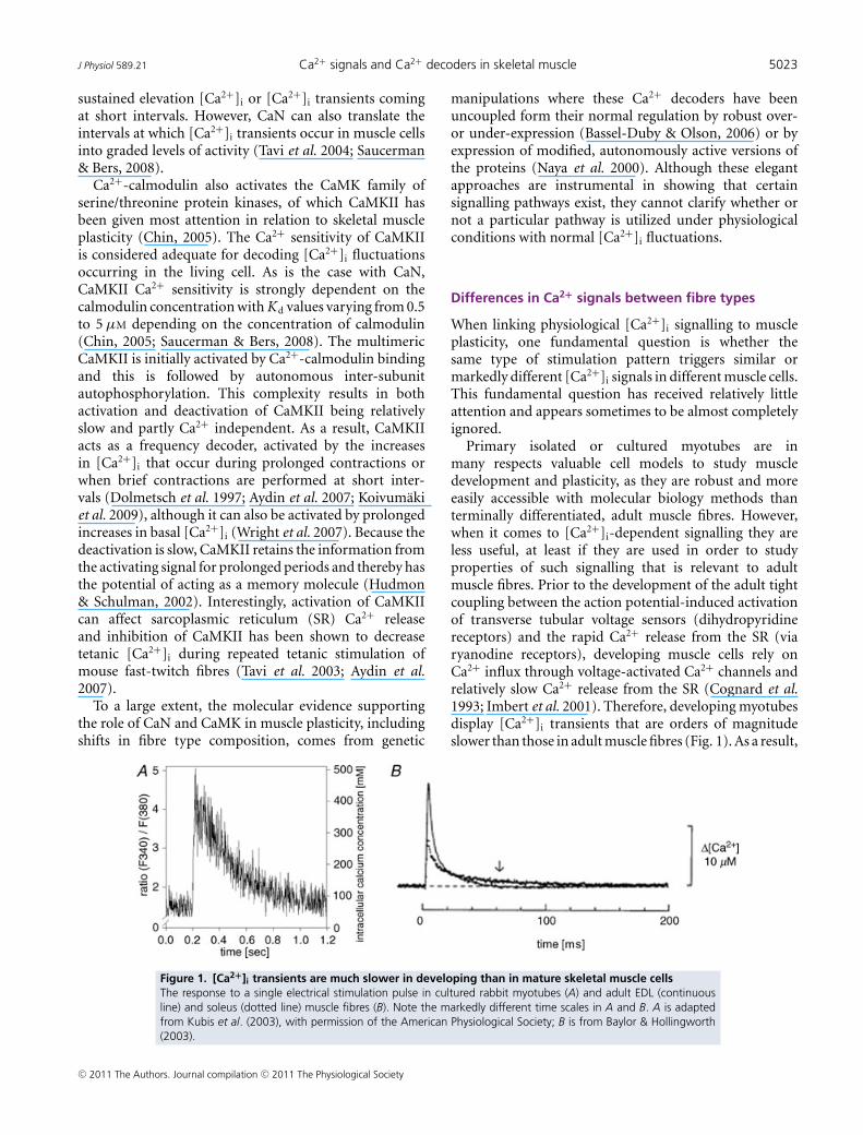
Primary isolated or cultured myotubes are inmany respects valuable cell models to study muscledevelopment and plasticity, as they are robust and moreeasily accessible with molecular biology methods thanterminally differentiated, adult muscle fibres. However,when it comes to [Ca2+]i-dependent signalling they areless useful, at least if they are used in order to studyproperties of such signalling that is relevant to adultmuscle fibres. Prior to the development of the adult tightcoupling between the action potential-induced activationof transverse tubular voltage sensors (dihydropyridinereceptors) and the rapid Ca2+ release from the SR (viaryanodine receptors), developing muscle cells rely onCa2+ influx through voltage-activated Ca2+ channels andrelatively slow Ca2+ release from the SR (Cognard et al.1993; Imbert et al. 2001). Therefore, developing myotubesdisplay [Ca2+]i transients that are orders of magnitudeslower than those in adult muscle fibres (Fig. 1). As a result,
Figure 1. [Ca2+]i transients are much slower in developing than in mature skeletal muscle cellsThe response to a single electrical stimulation pulse in cultured rabbit myotubes (A) and adult EDL (continuousline) and soleus (dotted line) muscle fibres (B). Note the markedly different time scales in A and B. A is adaptedfrom Kubis et al. (2003), with permission of the American Physiological Society; B is from Baylor & Hollingworth(2003).
C© 2011 The Authors. Journal compilation C© 2011 The Physiological Society
5024 P. Tavi and H. Westerblad J Physiol 589.21
Ca2+–calmodulin pathways can be activated in myotubesat much lower stimulation frequencies than in adult fibres;for instance, 1 Hz electrical stimulation activates CaNsignalling in myotubes (Kubis et al. 2002), but not in adultfibres (Liu et al. 2001).
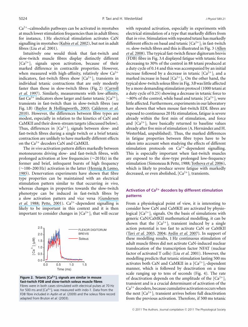
Intuitively one would think that fast-twitch andslow-twitch muscle fibres display distinctly different[Ca2+]i signals upon activation, because of theirmarked difference in contractile properties. However,when measured with high-affinity, relatively slow Ca2+
indicators, fast-twitch fibres show [Ca2+]i transients inindividual tetanic contractions that are only modestlyfaster than those in slow-twitch fibres (Fig. 2) (Carrollet al. 1997). Similarly, measurements with low-affinity,fast Ca2+ indicators show larger and faster tetanic [Ca2+]i
transients in fast-twitch than in slow-twitch fibres (seeFig. 1B) (Baylor & Hollingworth, 2003; Calderon et al.2010). However, the differences between fibre types aremodest, especially in relation to the kinetics of CaN andCaMKII and their down-stream targets (discussed below).Thus, differences in [Ca2+]i signals between slow- andfast-twitch fibres during a single twitch or a brief tetaniccontraction are unlikely to have markedly different effectson the Ca2+ decoders CaN and CaMKII.
The in vivo activation pattern differs markedly betweenmotoneurons driving slow- and fast-twitch fibres, withprolonged activation at low frequencies (∼20 Hz) in theformer and brief, infrequent bursts of high frequency(∼100–200 Hz) activation in the latter (Hennig & Lømo,1985). Denervation experiments have shown that fibretype properties can be maintained with an electricalstimulation pattern similar to that occurring in vivo,whereas changes in properties towards the slow-twitchphenotype can be induced in fast-twitch fibres bya slow activation pattern and vice versa (Gundersenet al. 1988; Pette, 2001). Ca2+-dependent signalling islikely to be important in this context and it is thenimportant to consider changes in [Ca2+]i that will occur
0.0 0.5 1.0 1.50.0
0.5
1.0
1.5
2.0
2.5
[Ca
2+] i
Time (ms)
FLEXOR DIGITORUMBREVIS
Soleus
Figure 2. Tetanic [Ca2+]i signals are similar in mousefast-twitch FDB and slow-twitch soleus muscle fibresFibres were in both cases stimulated with electrical pulses at 70 Hzfor 500 ms and [Ca2+]i was measured with indo-1. Data from theFDB fibre included in Aydin et al. (2009) and the soleus fibre recordadapted from Bruton et al. (2003).
with repeated activation, especially in experiments withelectrical stimulation of a type that markedly differs fromthat in vivo. Stimulation with repeated tetani has markedlydifferent effects on basal and tetanic [Ca2+]i in fast-twitchvs. slow-twitch fibres and this is illustrated in Fig. 3 (Allenet al. 2008). The typical fast-twitch flexor digitorum brevis(FDB) fibre in Fig. 3A displayed fatigue with tetanic forcedecreasing to 30% of the control in 88 tetani produced ata duty cycle of 0.14 and this was accompanied by an initialincrease followed by a decrease in tetanic [Ca2+]i and amarked increase in basal [Ca2+]i. On the other hand, thetypical slow-twitch soleus fibre in Fig. 3B was little affectedby a more demanding stimulation protocol (1000 tetani ata duty cycle of 0.25) showing a decrease in tetanic force to∼80% of the control, while basal and tetanic [Ca2+]i werelittle affected. Furthermore, experiments in our laboratoryhave shown that when mouse fast-twitch EDL fibres areexposed to continuous 20 Hz stimulation, fatigue is severealready within the first min of stimulation, and forceand [Ca2+]i have basically returned to baseline valuesalready after five min of stimulation (A. Hernandez and H.Westerblad, unpublished). Thus, the marked differencesin fatigue properties between fibre types have to betaken into account when studying the effects of differentstimulation protocols on Ca2+-dependent signalling.This is especially important when fast-twitch musclesare exposed to the slow-type prolonged low-frequencystimulation (Simoneau & Pette, 1988; Tothova et al. 2006),which is likely to produce severe fatigue with markedlydecreased, or even abolished, [Ca2+]i transients.
Activation of Ca2+ decoders by different stimulationpatterns
From a physiological point of view, it is interesting toconsider how CaN and CaMKII are activated by physio-logical [Ca2+]i signals. On the basis of simulations withgeneric CaN/CaMKII mathematical modelling, it can beshown that the [Ca2+]i transient induced by a singleaction potential is too fast to activate CaN or CaMKII(Tavi et al. 2003, 2004; Aydin et al. 2007). In support ofthese modelling results, 1 Hz continuous stimulation ofadult muscle fibres did not activate CaN-induced nucleartranslocation of the transcription factor NFAT (nuclearfactor of activated T cells) (Liu et al. 2001). However, themodelling predicts that tetanic stimulation lasting 500 msactivates both CaN and CaMKII in a [Ca2+]i-dependentmanner, which is followed by deactivation on a timescale ranging up to tens of seconds (Fig. 4). The rateof deactivation depends on the amplitude of the [Ca2+]i
transient and is a crucial determinant of activation of theCa2+ decoders, because cumulative activation occurs whenthe next [Ca2+]i transient arrives before full deactivationfrom the previous activation. Therefore, if 500 ms tetanic
C© 2011 The Authors. Journal compilation C© 2011 The Physiological Society

J Physiol 589.21 Ca2+ signals and Ca2+ decoders in skeletal muscle 5025
[Ca2+]i transients are repeated at short intervals, both CaNand CaMKII respond with cumulative activation (Fig. 4).Moreover, the modelling predicts that repeated activationwith same sized tetanic [Ca2+]i transients results in slowerspeed of deactivation with time constants increasing∼3-fold compared to after a single tetanus. Thus, thesemodelling experiments highlight important features ofthe Ca2+ decoders: they are not significantly activated bya fast event such as a single twitch [Ca2+]i transient andconsequently their activity level depends on the patternof repetitive activity; their deactivation is up to 100-foldslower than the decay of the activating [Ca2+]i and hencethey have memory properties determined by the precedingactivity.
In a variety of experimental settings, prolongedlow-frequency stimulation vs. brief high-frequency burstsof stimulation have been used to mimic the activationpattern of slow-twitch and fast-twitch motor units,respectively (Pette & Vrbova, 1999; Gundersen, 2011).Figure 5 shows the activity of CaN and CaMKII predicted
by mathematical modelling in response to different typesof stimulation (Tavi et al. 2003, 2004; Aydin et al. 2007).Figure 5A shows the modelled response to continuous20 Hz stimulation. With this type of stimulation CaNreaches close to maximum activity already after ∼1 sstimulation, whereas the increase in CaMKII activityoccurs more slowly. In this modelling experiment theamplitude of [Ca2+]i transients was kept constant, whichwould be the situation in slow-twitch muscle fibres. On theother hand, in fast-twitch muscle fibres continuous 20 Hzstimulation would lead to severe fatigue with markedlydecreased, or even abolished, [Ca2+]i transients, and hencethe activity of CaN and CaMKII would return towardstheir basal state. Figure 5B and C shows the modelledresponse to the same, brief tetanic [Ca2+]i signal given at1 and 32 s intervals. Due to faster kinetics, CaN respondsmore profoundly to an individual tetanus, rapidly reachinga saturated activity level at short (1 s) stimulation inter-vals, and it becomes fully deactivated during pauses withlong (32 s) intervals. Conversely, CaMKII is activated
Figure 3. Fast-twitch FDB fibres, but not slow-twitch soleus fibres, show major changes in [Ca2+]i duringfatiguing stimulationA, [Ca2+]i of a FDB fibre exposed to repeated 70 Hz, 350 ms tetani given every 2.5 s until force was decreased to30% of the control. Inset shows a comparison between first four (red) and last four (blue) tetani; note the markedincrease in basal [Ca2+]i and decrease in tetanic [Ca2+]i in fatigue. Data from this fibre included in Dahlstedt et al.(2000). B, [Ca2+]i of a soleus fibre during 1000 repeated 70 Hz, 500 ms tetani given every 2 s. Inset shows acomparison between the first five (red) and last five (blue) tetani; note that both basal and tetanic [Ca2+]i werelittle affected by fatiguing stimulation. Data from Bruton et al. (2003).
C© 2011 The Authors. Journal compilation C© 2011 The Physiological Society

5026 P. Tavi and H. Westerblad J Physiol 589.21
and deactivated more slowly and thus shows a graduallydeveloping increase in activity with short (1 s) intervals,whereas it is little affected at long (32 s) intervals.
Ca2+ decoders and gene transcription
CaN and CaMKII mediate their effects on genetranscription by inducing nucleo-cytoplasmic shuttlingof transcription factors (e.g. NFAT and myocyte enhancerfactor-2, MEF2) (Chen et al. 2001; Liu et al. 2001) andtranscription modulators (histone deacetylases, HDACs)(Liu et al. 2005; Shen et al. 2006). In principle, theactivity of CaN and CaMKII is driven by the same changesin [Ca2+]i, but there are exemptions from this rule.For instance, translocation of HDAC4 from the nucleusreportedly involves activation of CaMKII localized in thenucleus and changes in nuclear [Ca2+] occur with sub-stantially slower kinetics than changes in [Ca2+]i (Liu
et al. 2005). This might explain why HDAC nuclear exportcan be activated by lower stimulus frequencies than NFATnuclear import in adult muscle fibres (Liu et al. 2001,2005).
The translocations of transcriptions factors andmodulators are much slower processes than theCa2+-induced activation of CaN and CaMKII. Forinstance, Ca2+-activated CaN promotes translocation ofNFAT from the cytosol to the nucleus in a time frameof tens of minutes (Liu et al. 2001; Tothova et al. 2006).Similarly, CaMK-induced translocation of HDAC fromthe nucleus to the cytosol occurs with a time constant oftens of minutes (Liu et al. 2005). Consequently any [Ca2+]i
signal able to activate Ca2+-dependent transcription hasnot only to be able to activate Ca2+ decoders, but also tomaintain their activity for long enough periods to inducea significant translocation of the transcription factors andmodulators. Thus, in general terms it can be stated that
Figure 4. Modelled activation of CaN and CaMKII in response to tetanic [Ca2+]i transients[Ca2+]i from a single 70 Hz, 500 ms tetanus (A) and from a series of 30 such tetani given at 2 s intervals (B; adaptedfrom Fig. 3B). C, outline of the mathematical model into which the [Ca2+]i records were fed. CaM, calmodulin;PP1, protein phosphatase 1. Arrows indicate activation and punctuated arrow inhibition; for detailed descriptionof the mathematical model see Tavi et al. (2003, 2004) and Aydin et al. (2007). D, modelled activation of CaN (red)and CaMKII (blue) induced by the single tetanus in A. τ values represent modelled deactivation time constants.In order to illustrate the effect of higher tetanic [Ca2+]i (e.g. induced by a higher stimulation frequency), A alsoshows tetanic [Ca2+]i of twice the amplitude (grey line) and the effect on CaN and CaMKII activation is shownin C (lighter colours). E, modelled activation of CaN and CaMKII by the repeated tetanic stimulation in B. Notethat CaN is fully activated after a few tetani whereas CaMKII activity increases throughout the stimulation period.After the end of stimulation, the activity of both enzyme decays about three times slower than after a single 70 Hztetanus.
C© 2011 The Authors. Journal compilation C© 2011 The Physiological Society

J Physiol 589.21 Ca2+ signals and Ca2+ decoders in skeletal muscle 5027
the activation and maintenance of a slow phenotype oftranscription requires relatively constant muscle activity,whereas a fast phenotype is brought about by long lastinglack or infrequent, brief bursts of activity (Huey et al.2001). However, while there are similarities in the responseto complete lack of activity and activation with briefbursts of stimulation at long intervals, there are alsomarked differences. For instance, lack of activation resultsin marked muscle weakness and atrophy and this is notthe situation with the fast pattern of activity (Gundersen,2011). A striking example of this comes from experimentswhere 0.6 s high-frequency pulse trains delivered every100 min resulted in ∼6-fold increase in tetanic force indenervated rat soleus muscle (Westgaard & Lømo, 1988).Moreover, the same study shows that stimulating ratsoleus muscles with brief bursts of 100 Hz stimulation incombination with prolonged 10 Hz stimulation results ina switch towards faster contractile properties (Westgaard& Lømo, 1988). Thus, these latter findings cannot beexplained on the basis of the modelled activity of CaNand CaMKII presented in Figs 4 and 5, which suggeststhe involvement of other important signalling pathwaysand/or that the activation of the Ca2+ decoders may beaffected by other factors than global changes in [Ca2+]i,which will be discussed below.
In general, the prolonged slow-type stimulation ofadult muscle fibres results in activation of both CaNand CaMKII pathways triggering translocation of bothNFAT and HDAC and activation of MEF2 and peroxisomeproliferator-activated receptor γ coactivator 1α (PGC-1α)
(Shen et al. 2006), thereby promoting gene expressiontowards a slow and oxidative phenotype (Chin et al.1998). On the other hand, fast-type stimulation withhigh frequency bursts occurring at long intervals willonly transiently activate the CaN and CaMKII, whichis not sufficient to induce significant translocations andhence it promotes gene expressions towards a fast andanaerobic phenotype (Fig. 6). However, it again needsto be pointed out that the lack of translocations with afast-type stimulation pattern does not result in the samephenotype as is observed with a total lack of activation(Gundersen, 2011).
Physiological significance
There is overwhelming experimental support for CaNand CaMKII being capable of inducing major changes inmuscle fibre properties, even including switches betweenfast-twitch type II and slow-twitch type I myosin heavychain isoforms. However, major changes in muscle fibreproperties directly attributed to CaN and CaMKII havegenerally been observed with rather drastic approaches,for example genetic manipulation of CaN or CaMKII(overexpression, knock-out, expression of constitutivelyactive forms) or interference in the normal activationpattern of muscles (denervation, electrical stimulation).The importance of CaN and CaMKII in muscle plasticityunder more physiological conditions is rather uncertain.Based on the properties of these two Ca2+ decoders and
Figure 5. The activation of CaN and CamKII critically depends on the rate at which contractions areproduced[Ca2+]i record from 20 Hz continuous stimulation (A) and the same representative 500 ms tetanic [Ca2+]i recordgiven at 1 s (B) and 32 s (C) intervals were used as input to the mathematical model (Tavi et al. 2003, 2004; Aydinet al. 2007). The lower part shows the activities of CaN and CaMKII predicted by the model.
C© 2011 The Authors. Journal compilation C© 2011 The Physiological Society

5028 P. Tavi and H. Westerblad J Physiol 589.21
the prevailing [Ca2+]i signals in muscle fibres, it is clearthat inactivity will result in limited activation of CaN andCaMKII and thus contribute to the inactivity-inducedshift towards a fast phenotype with limited aerobiccapacity. It is also evident that the prolonged activityin slow-twitch motoneurones during everyday activitieswill induce sufficient activation of CaN and CaMKII tomaintain the slow, oxidative phenotype of slow-twitchtype I fibres.
However, it is less clear whether increased physicalactivity can activate CaN and CaMKII to such an extentthat this leads to a significant change in muscle fibreproperties. For instance, are CaN and CaMKII involvedin the beneficial muscle effects observed with relativelylow-intensity endurance exercise performed for ∼30 min3–5 days week−1 (American College of Sports MedicinePosition Stand, 1998)? Intuitively, the rather modestand relatively short-lasting increase in [Ca2+]i directlyinduced by 30 min low-intensity exercise would havelittle impact on the overall activity of Ca2+ decoders.Even with the training performed by elite enduranceathletes, the fraction of time that CaN and CaMKII aredirectly activated by exercise-induced increases in [Ca2+]i
is limited; for example, with 4 h training per day, CaNand CaMKII would still be at their basal activity levelfor 20 h. Thus, for CaN and CaMKII to be important inthis context, other modes of activation than the directincrease [Ca2+]i during contractile activity appear tobe required. Accordingly, there are results supportingincreased activity of the Ca2+ decoders also in the restperiods between training bouts (Rose et al. 2007). Onemechanism behind this might be that exercise-inducedchanges in muscle fibre Ca2+ handling amplify the over-all increase in [Ca2+]i. In support of this possibility, itwas recently shown that endurance exercise can inducelong-lasting changes in the RyR channel complex, whichrenders them more leaky (Bellinger et al. 2008). Anincreased SR Ca2+ leak due to modifications of the RyRchannel complex has been associated with increased base-line [Ca2+]i and increases in mitochondrial biogenesisand fatigue resistance (Bruton et al. 2010). Furthermore,an increase in [Ca2+]i below the contraction thresholdinduced by exposing adult fast-twitch fibres to caffeineresulted in increased mitochondrial biogenesis and thiswas inhibited by pharmacological CaMKII inhibition(Wright et al. 2007). Thus, it appears that the Ca2+
Figure 6. Simplified scheme of the stimulus pattern-dependent transcription through Ca2+ activatedpathwaysA, frequent activity mimicking that experienced by slow-twitch muscle fibres in vivo results in a Ca2+ binding tocalmodulin (CaM) that is sufficient to induce a prolonged activation of CaN and CaMKII. Activated CaN controlstranscription by inducing nuclear translocation of NFAT (Bassel-Duby & Olson, 2006) to promote transcription ofslow type-specific genes, such as slow isoforms of myosin heavy chain and troponin I (Dunn et al. 1999; Serranoet al. 2001). Activated CaMKII stimulates transcription by removing repressive HDAC from the nucleus and byphosphorylating MEF2 (Bassel-Duby & Olson, 2006). MEF2 suppresses the myogenesis when it forms a complexwith HDAC, but upon CaMKII-dependent disruption of MEF2-HDAC-complexes, MEF2 activates transcription(McKinsey et al. 2000) in co-operation with NFAT (Wu et al. 2000). When activated, Ca2+-dependent cascadesalso promote mitochondrial biogenesis via activation of PGC-1α (Wu et al. 2002). B, infrequent activation similarto that experienced by fast-twitch muscle fibres in vivo allows CaN and CaMKII deactivation between contractions.As a result, NFAT is not translocated into the nucleus, which suppresses the slow type of gene expression. Inaddition, HDAC remains in the nucleus and forms complexes with MEF2, which further suppresses slow typegene expression. Thus, this type of stimulation favours the expression of fast type-specific protein isoforms and itdoes not stimulate mitochondrial biogenesis; however, it does not promote the same phenotype as total lack ofstimulation, which in addition involves muscle atrophy and weakness. Arrows indicate stimulatory actions, whereasdotted lines indicate lack of activation.
C© 2011 The Authors. Journal compilation C© 2011 The Physiological Society

J Physiol 589.21 Ca2+ signals and Ca2+ decoders in skeletal muscle 5029
decoders are involved in the adaptation of muscle fibresinduced even by rather minor changes in global [Ca2+]i.
According to the established basic properties of CaN orCaMKII small baseline [Ca2+]i changes, such as those seenin cold acclimated muscles (60 to 90 mM) (Bruton et al.2010), would most likely induce only minute activations ofthese enzymes. How can Ca2+ decoders still decode these[Ca2+]i elevations? Several mechanisms for this can beoffered. For instance, when an increase in baseline [Ca2+]i
is induced by increased RyR Ca2+ leak, inevitably the[Ca2+] close to the RYR channel complexes will be higherthan in the cytoplasm as a whole. If Ca2+ decoders arelocated in these subcellular compartments, either throughinteractions with specific anchoring proteins, like αKAP(Bayer et al. 1998; O’Leary et al. 2006), or simply due todiffusion, they would be exposed to a high local [Ca2+].In addition, if calmodulin is enriched in these micro-domains, as reported to be the case in the vicinity of L-typecalcium channels (Mori et al. 2004), local activation ofCa2+ decoders would be more extensive (Saucerman &Bers, 2008) and partly independent of the overall [Ca2+]i.However, the exact details and importance of spatialdecoding of [Ca2+]i gradients for skeletal muscle plasticityare not known and will be an interesting field of futureresearch.
The physiological changes in muscle fibre phenotypethat occur with inactivity/endurance exercise involvenot only the CaN and CaMKII but also several othersignalling pathways, such as AMP-activated protein kinase,mitogen-activated protein kinases and hypoxia induciblefactor 1α (Hawley et al. 2006; Lunde et al. 2011). Inaddition to [Ca2+]i, there are other changes in thecellular environment involved in muscle fibre plasticityand these include changes in energy metabolites andreactive oxygen/nitrogen species (ROS/RNS). There willalso be interactions between these factors. For instance, theincreased SR Ca2+ leak observed with endurance exerciseinvolves ROS/RNS-induced modifications of the RyRchannel complex (Bellinger et al. 2008), and oxidation ofCa2+–calmodulin-activated CaMKII leads to a sustained,Ca2+-independent increase in its activity (Anderson et al.2008; Christensen et al. 2009). Thus, there is evidence foran intricate cross-talk between Ca2+ decoders, [Ca2+]i andROS/RNS, and this constitutes another important area forfuture studies.
Conclusions
In the short term, the activity of the Ca2+ decodersCaN and CaMKII is mainly decided by the activitypattern imposed on muscle fibres by the nervous system,whereas intrinsic differences in [Ca2+]i transients betweenadult muscle fibre types is less important. However,fatigue will develop in fast-twitch fibres if these are
exposed to prolonged, high-intensity stimulation and the[Ca2+]i signals are then not correlated with the stimuluspattern. The cascade from neural stimulation patternto Ca2+-dependent transcription is likely to be centralin maintaining the fibre phenotypes in both fast- andslow-twitch fibres. On the other hand, fibre type switchingby altered activity of this cascade would require drasticchanges in the neural activation pattern of the muscles andsuch drastic changes are unlikely to occur under normalphysiological conditions. In the physiological contextof muscle fibre plasticity, interactions and synergisticactivations of Ca2+-activated cascades and other signallingpathways induced by muscle activity are likely to beimportant, as well as interactions between spatio-temporalchanges in [Ca2+]i and other components of the cellularenvironment.
References
Allen DG, Lamb GD & Westerblad H (2008). Skeletal musclefatigue: cellular mechanisms. Physiol Rev 88, 287–332.
American College of Sports Medicine Position Stand (1998).The recommended quantity and quality of exercise fordeveloping and maintaining cardiorespiratory and muscularfitness, and flexibility in healthy adults. Med Sci Sports Exerc30, 975–991.
Anderson ME, Erickson JR, Joiner MLA, Guan X, Kutschke W,Yang JY, Oddis CV, Bartlett RK, Lowe JS, O’Donnell SE,Aykin-Burns N, Zimmerman MC, Zimmerman K, Ham AJL,Weiss RM, Spitz DR, Shea MA, Colbran RJ & Mohler PJ(2008). A dynamic pathway for calcium-independentactivation of CaMKII by methionine oxidation. Cell 133,462–474.
Aydin J, Andersson DC, Hanninen SL, Wredenberg A, Tavi P,Park CB, Larsson NG, Bruton JD & Westerblad H (2009).Increased mitochondrial Ca2+ and decreased sarcoplasmicreticulum Ca2+ in mitochondrial myopathy. Hum Mol Genet18, 278–288.
Aydin J, Korhonen T, Tavi P, Allen DG, Westerblad H & BrutonJD (2007). Activation of Ca2+-dependent protein kinase IIduring repeated contractions in single muscle fibres frommouse is dependent on the frequency of sarcoplasmicreticulum Ca2+ release. Acta Physiol (Oxf) 191,131–137.
Bassel-Duby R & Olson EN (2006). Signaling pathways inskeletal muscle remodeling. Annu Rev Biochem 75, 19–37.
Bayer KU, Harbers K & Schulman H (1998). αKAP is ananchoring protein for a novel CaM kinase II isoform inskeletal muscle. EMBO J 17, 5598–5605.
Baylor SM & Hollingworth S (2003). Sarcoplasmic reticulumcalcium release compared in slow-twitch and fast-twitchfibres of mouse muscle. J Physiol 551, 125–138.
Bellinger AM, Reiken S, Dura M, Murphy PW, Deng SX,Landry DW, Nieman D, Lehnart SE, Samaru M,LaCampagne A & Marks AR (2008). Remodeling ofryanodine receptor complex causes ‘leaky’ channels: amolecular mechanism for decreased exercise capacity. ProcNatl Acad Sci U S A 105, 2198–2202.
C© 2011 The Authors. Journal compilation C© 2011 The Physiological Society

5030 P. Tavi and H. Westerblad J Physiol 589.21
Bruton J, Tavi P, Aydin J, Westerblad H & Lannergren J (2003).Mitochondrial and myoplasmic [Ca2+] in single fibres frommouse limb muscles during repeated tetanic contractions.J Physiol 551, 179–190.
Bruton JD, Aydin J, Yamada T, Shabalina IG, Ivarsson N, ZhangSJ, Wada M, Tavi P, Nedergaard J, Katz A & Westerblad H(2010). Increased fatigue resistance linked toCa2+-stimulated mitochondrial biogenesis in muscle fibresof cold-acclimated mice. J Physiol 588, 4275–4288.
Buller AJ, Eccles JC & Eccles RM (1960). Interactions betweenmotoneurones and muscles in respect of the characteristicspeeds of their responses. J Physiol 150, 417–439.
Calderon JC, Bolanos P & Caputo C (2010). Myosin heavychain isoform composition and Ca2+ transients in fibresfrom enzymatically dissociated murine soleus and extensordigitorum longus muscles. J Physiol 588, 267–279.
Carroll SL, Klein MG & Schneider MF (1997). Decay ofcalcium transients after electrical stimulation in rat fast- andslow-twitch skeletal muscle fibres. J Physiol 501,573–588.
Chen SL, Wang SC, Hosking B & Muscat GE (2001).Subcellular localization of the steroid receptor coactivators(SRCs) and MEF2 in muscle and rhabdomyosarcoma cells.Mol Endocrinol 15, 783–796.
Chin ER (2005). Role of Ca2+/calmodulin-dependent kinasesin skeletal muscle plasticity. J Appl Physiol 99, 414–423.
Chin ER, Olson EN, Richardson JA, Yang Q, Humphries C,Shelton JM, Wu H, Zhu W, Bassel-Duby R & Williams RS(1998). A calcineurin-dependent transcriptional pathwaycontrols skeletal muscle fiber type. Genes Dev 12, 2499–2509.
Christensen MD, Dun W, Boyden PA, Anderson ME, MohlerPJ & Hund TJ (2009). Oxidized calmodulin kinase IIregulates conduction following myocardial infarction: Acomputational analysis. PLoS Comput Biol 5, e1000583..
Cognard C, Constantin B, Rivet-Bastide M, Imbert N, Besse C& Raymond G (1993). Appearance and evolution of calciumcurrents and contraction during the early post-fusionalstages of rat skeletal muscle cells developing in primaryculture. Development 117, 1153–1161.
Dahlstedt AJ, Katz A, Wieringa B & Westerblad H (2000). Iscreatine kinase responsible for fatigue? Studies of isolatedskeletal muscle deficient in creatine kinase. FASEB J 14,982–990.
Dolmetsch RE, Lewis RS, Goodnow CC & Healy JI (1997).Differential activation of transcription factors induced byCa2+ response amplitude and duration. Nature 386,855–858.
Dunn SE, Burns JL & Michel RN (1999). Calcineurin isrequired for skeletal muscle hypertrophy. J Biol Chem 274,21908–21912.
Gundersen K (2011). Excitation-transcription coupling inskeletal muscle: the molecular pathways of exercise. Biol Rev86, 564–600.
Gundersen K, Leberer E, Lømo T, Pette D & Staron RS (1988).Fibre types, calcium-sequestering proteins and metabolicenzymes in denervated and chronically stimulated musclesof the rat. J Physiol 398, 177–189.
Harridge SD (2007). Plasticity of human skeletal muscle: geneexpression to in vivo function. Exp Physiol 92, 783–797.
Hawley JA, Hargreaves M & Zierath JR (2006). Signallingmechanisms in skeletal muscle: role in substrate selectionand muscle adaptation. Essays Biochem 42, 1–12.
Hennig R & Lømo T (1985). Firing patterns of motor units innormal rats. Nature 314, 164–166.
Hudmon A & Schulman H (2002). Structure-function of themultifunctional Ca2+/calmodulin-dependent protein kinaseII. Biochem J 364, 593–611.
Huey KA, Roy RR, Baldwin KM & Edgerton VR (2001).Temporal effects of inactivty on myosin heavy chain geneexpression in rat slow muscle. Muscle Nerve 24, 517–526.
Imbert N, Vandebrouck C, Duport G, Raymond G, HassoniAA, Constantin B, Cullen MJ & Cognard C (2001). Calciumcurrents and transients in co-cultured contracting normaland Duchenne muscular dystrophy human myotubes.J Physiol 534, 343–355.
Klee CB, Ren H & Wang X (1998). Regulation of thecalmodulin-stimulated protein phosphatase, calcineurin.J Biol Chem 273, 13367–13370.
Koivumaki JT, Korhonen T, Takalo J, Weckstrom M & Tavi P(2009). Regulation of excitation-contraction coupling inmouse cardiac myocytes: integrative analysis withmathematical modelling. BMC Physiol 9, 16.
Kubis HP, Hanke N, Scheibe RJ, Meissner JD & Gros G (2003).Ca2+ transients activate calcineurin/NFATc1 and initiatefast-to-slow transformation in a primary skeletal muscleculture. Am J Physiol Cell Physiol 285, C56–C63.
Kubis HP, Scheibe RJ, Meissner JD, Hornung G & Gros G(2002). Fast-to-slow transformation and nuclearimport/export kinetics of the transcription factor NFATc1during electrostimulation of rabbit muscle cells in culture.J Physiol 541, 835–847.
Liu Y, Cseresnyes Z, Randall WR & Schneider MF (2001).Activity-dependent nuclear translocation and intranucleardistribution of NFATc in adult skeletal muscle fibers. J CellBiol 155, 27–39.
Liu Y, Randall WR & Schneider MF (2005). Activity-dependentand -independent nuclear fluxes of HDAC4 mediated bydifferent kinases in adult skeletal muscle. J Cell Biol 168,887–897.
Lømo T, Westgaard RH & Dahl HA (1974). Contractileproperties of muscle: control by pattern of muscle activity inthe rat. Proc R Soc Lond B Biol Sci 187, 99–103.
Lunde IG, Anton SL, Bruusgaard JC, Rana ZA, Ellefsen S &Gundersen K (2011). Hypoxia inducible factor 1α linksfast-patterned muscle activity and fast muscle phenotype inrats. J Physiol 589, 1443–1454.
Lunde PK, Sejersted OM, Thorud HM, Tonnessen T,Henriksen UL, Christensen G, Westerblad H & Bruton J(2006). Effects of congestive heart failure on Ca2+ handlingin skeletal muscle during fatigue. Circ Res 98, 1514–1519.
McKinsey TA, Zhang CL & Olson EN (2000). Activation of themyocyte enhancer factor-2 transcription factor bycalcium/calmodulin-dependent protein kinase-stimulatedbinding of 14-3-3 to histone deacetylase 5. Proc Natl Acad SciU S A 97, 14400–14405.
Mori MX, Erickson MG & Yue DT (2004). Functionalstoichiometry and local enrichment of calmodulininteracting with Ca2+ channels. Science 304, 432–435.
C© 2011 The Authors. Journal compilation C© 2011 The Physiological Society

J Physiol 589.21 Ca2+ signals and Ca2+ decoders in skeletal muscle 5031
Naya FJ, Mercer B, Shelton J, Richardson JA, Williams RS &Olson EN (2000). Stimulation of slow skeletal muscle fibergene expression by calcineurin in vivo. J Biol Chem 275,4545–4548.
O’Leary H, Sui X, Lin PJ, Volpe P & Bayer KU (2006). Nucleartargeting of the CaMKII anchoring protein αKAP isregulated by alternative splicing and protein kinases. BrainRes 1086, 17–26.
Olson EN & Williams RS (2000). Remodeling muscles withcalcineurin. Bioessays 22, 510–519.
Pette D (2001). Historical Perspectives: plasticity ofmammalian skeletal muscle. J Appl Physiol 90, 1119–1124.
Pette D & Vrbova G (1999). What does chronic electricalstimulation teach us about muscle plasticity? Muscle Nerve22, 666–677.
Rose AJ, Frosig C, Kiens B, Wojtaszewski JF & Richter EA(2007). Effect of endurance exercise training on Ca2+calmodulin-dependent protein kinase II expression andsignalling in skeletal muscle of humans. J Physiol 583,785–795.
Sakuma K & Yamaguchi A (2010). The functional role ofcalcineurin in hypertrophy, regeneration, and disorders ofskeletal muscle. J Biomed Biotech 2010, 721219.
Saucerman JJ & Bers DM (2008). Calmodulin mediatesdifferential sensitivity of CaMKII and calcineurin to localCa2+ in cardiac myocytes. Biophys J 95, 4597–4612.
Schiaffino S & Reggiani C (1996). Molecular diversity ofmyofibrillar proteins: gene regulation and functionalsignificance. Physiol Rev 76, 371–423.
Schiaffino S, Sandri M & Murgia M (2007). Activity-dependentsignaling pathways controlling muscle diversity andplasticity. Physiology (Bethesda) 22, 269–278.
Serrano AL, Murgia M, Pallafacchina G, Calabria E, Coniglio P,Lømo T & Schiaffino S (2001). Calcineurin controls nerveactivity-dependent specification of slow skeletal musclefibers but not muscle growth. Proc Natl Acad Sci U S A 98,13108–13113.
Shen T, Liu Y, Randall WR & Schneider MF (2006). Parallelmechanisms for resting nucleo-cytoplasmic shuttling andactivity dependent translocation provide dual control oftranscriptional regulators HDAC and NFAT in skeletalmuscle fiber type plasticity. J Muscle Res Cell Motil 27,405–411.
Simoneau JA & Pette D (1988). Species-specific effects ofchronic nerve stimulation upon tibialis anterior muscle inmouse, rat, guinea pig, and rabbit. Pflugers Arch 412, 86–92.
Stemmer PM & Klee CB (1994). Dual calcium ion regulation ofcalcineurin by calmodulin and calcineurin B. Biochemistry33, 6859–6866.
Tavi P, Allen DG, Niemela P, Vuolteenaho O, Weckstrom M &Westerblad H (2003). Calmodulin kinase modulates Ca2+release in mouse skeletal muscle. J Physiol 551, 5–12.
Tavi P, Pikkarainen S, Ronkainen J, Niemela P, Ilves M,Weckstrom M, Vuolteenaho O, Bruton J, Westerblad H &Ruskoaho H (2004). Pacing-induced calcineurin activationcontrols cardiac Ca2+ signalling and gene expression.J Physiol 554, 309–320.
Tothova J, Blaauw B, Pallafacchina G, Rudolf R, Argentini C,Reggiani C & Schiaffino S (2006). NFATc1nucleocytoplasmic shuttling is controlled by nerve activity inskeletal muscle. J Cell Sci 119, 1604–1611.
Westgaard RH & Lømo T (1988). Control of contractileproperties within adaptive ranges by patterns of impulseactivity in the rat. J Neurosci 8, 4415–4426.
Vrbova G (1963). The effect of motoneurone activity on thespeed of contraction of striated muscle. J Physiol 169,513–526.
Wright DC, Geiger PC, Han DH, Jones TE & Holloszy JO(2007). Calcium induces increases in peroxisomeproliferator-activated receptor γ coactivator-1α andmitochondrial biogenesis by a pathway leading to p38mitogen-activated protein kinase activation. J Biol Chem282, 18793–18799.
Wu H, Kanatous SB, Thurmond FA, Gallardo T, Isotani E,Bassel-Duby R & Williams RS (2002). Regulation ofmitochondrial biogenesis in skeletal muscle by CaMK.Science 296, 349–352.
Wu H, Naya FJ, McKinsey TA, Mercer B, Shelton JM, Chin ER,Simard AR, Michel RN, Bassel-Duby R, Olson EN &Williams RS (2000). MEF2 responds to multiplecalcium-regulated signals in the control of skeletal musclefiber type. EMBO J 19, 1963–1973.
Acknowledgements
The authors thank T. Korhonen and J. Koivumaki for thehelp with the modelling. The authors acknowledge financialsupport from Academy of Finland, Sigrid Juselius Foundation,the Swedish Research Council, and the Swedish National Centrefor Sports Research.
C© 2011 The Authors. Journal compilation C© 2011 The Physiological Society


![Near-Membrane [Ca2+] Transients Resolved Using the Ca2+ Indicator FFP18](https://static.fdokumen.com/doc/165x107/631286873ed465f0570a4533/near-membrane-ca2-transients-resolved-using-the-ca2-indicator-ffp18.jpg)