A Ca2+/Calmodulin-binding peroxidase from Euphorbia latex: Novel aspects of calcium-hydrogen...
11
A Ca 2+ /Calmodulin-Binding Peroxidase from Euphorbia Latex: Novel Aspects of Calcium-Hydrogen Peroxide Cross-Talk in the Regulation of Plant Defenses ²,‡ Anna Mura, § Rosaria Medda, § Silvia Longu, § Giovanni Floris, § Andrea C. Rinaldi, | and Alessandra Padiglia* ,§ Departments of Applied Sciences in Biosystems and Biomedical Sciences and Technologies, UniVersity of Cagliari, 09042 Monserrato, Cagliari, Italy ReceiVed July 11, 2005; ReVised Manuscript ReceiVed August 31, 2005 ABSTRACT: Calmodulin (CaM) is a ubiquitous Ca 2+ sensor found in all eukaryotes, where it participates in the regulation of diverse calcium-dependent physiological processes. In response to fluctuations of the intracellular concentration of Ca 2+ , CaM binds to a set of unrelated target proteins and modulates their activity. In plants, a growing number of CaM-binding proteins have been identified that apparently do not have a counterpart in animals. Some of these plant-specific Ca 2+ /CaM-activated proteins are known to tune the interaction between calcium and H 2 O 2 in orchestrating plant defenses against biotic and abiotic stresses. We previously characterized a calcium-dependent peroxidase isolated from the latex of the Mediterranean shrub Euphorbia characias (ELP) [Medda et al. (2003) Biochemistry 42, 8909-8918]. Here we report the cDNA nucleotide sequence of Euphorbia latex peroxidase, showing that the derived protein has two distinct amino acid sequences recognized as CaM-binding sites. The cDNA encoding for an E. characias CaM was also found and sequenced, and its protein product was detected in the latex. Results obtained from different CaM-binding assays and the determination of steady-state parameters showed unequivocally that ELP is a CaM-binding protein activated by the Ca 2+ /CaM system. To the best of our knowledge, this is the first example of a peroxidase regulated by this classic signal transduction mechanism. These findings suggest that peroxidase might be another node in the Ca 2+ /H 2 O 2 -mediated plant defense system, having both positive and negative effects in regulating H 2 O 2 homeostasis. In plants, as in all eukaryotes, calcium plays a central role as a second messenger in the regulation of a number of physiological processes. Decodification of Ca 2+ signals is generally devoted to a group of specialized cytosolic proteins that transduce these messages into molecular and cellular responses and integrate calcium signaling into virtually all aspects of plant functioning (2-4). Among plant Ca 2+ - sensing proteins, CaM 1 is increasingly appreciated as a critical player. Indeed, since its discovery about 2 decades ago in peanuts and peas (5), CaM has been involved in the modulation of diverse cellular and whole plant processes, such as cytoskeleton rearrangement, metabolism, and tran- scriptional regulation (6). Much effort has been dedicated in the last years in identifying downstream target proteins activated by CaM in plants and in understanding their function(s). Some of these studies have recently highlighted the role played by CaM-binding proteins in pollen germina- tion and pollen tube growth (7, 8), in floral development and embryogenesis (9, 10), and in disease resistance and cell death (11). Known plant CaM-regulated proteins now include several metabolic enzymes, ion channels, transcription fac- tors, protein kinases/phosphatases, and structural proteins, many of which have no animal counterparts and appear to be unique of plants (6, 12-14). Despite these progresses, however, our picture of plant CaM-binding proteins and of their exact positions in Ca 2+ -mediated signal transduction networks remains largely incomplete. Class III secreted plant peroxidases are a large family of heme-containing enzymes that oxidize a variety of aromatic molecules in the presence of hydrogen peroxide with the generation of aromatic oxyl radicals and reactive oxygen species (ROS; 15). Plant peroxidases are found in the cytosol, vacuole, apoplast, or cell wall and participate in crucial physiological events, such as development and growth induction, polymerization of cell wall lignin and suberin precursors, auxin catabolism, wound healing, and defense against pathogen infection. Although peroxidases are gener- ally reported to participate in the activation of plant defense responses mainly through their contribution to the oxidative burst in which levels of ROS (particularly superoxide and H 2 O 2 ) rapidly increase, an expanded scenario is emerging in which these proteins might interact in more complex ways ² This study was partially supported by FIRB (Fondo per gli Investimenti della Ricerca di Base) funds obtained from the Italian Ministry of Education, University and Research. ‡ The sequences reported in this paper have been deposited in the GenBank database (Euphorbia characias peroxidase, accession number AY586601; E. characias calmodulin, accession number AY297816). * To whom correspondence should be addressed. Tel: +39 070 6754515. Fax: +39 070 6754523. E-mail: [email protected]. § Department of Applied Sciences in Biosystems, University of Cagliari. | Department of Biomedical Sciences and Technologies, University of Cagliari. 1 Abbreviations: ABTS, 2,2′-azinobis(3-ethylbenzothiazoline-6-sul- fonic acid); BBCaM, bovine brain calmodulin; CaM, calmodulin; ECaM, Euphorbia calmodulin cDNA; ELCaM, Euphorbia latex calm- odulin (protein); EP, Euphorbia peroxidase cDNA; ELP, Euphorbia latex peroxidase (protein); MOPS, 3-(N-morpholino)propanesulfonic acid; RT-PCR, reverse transcription polymerase chain reaction; SDS- PAGE, sodium dodecyl sulfate-polyacrylamide gel electrophoresis. 14120 Biochemistry 2005, 44, 14120-14130 10.1021/bi0513251 CCC: $30.25 © 2005 American Chemical Society Published on Web 10/05/2005
Transcript of A Ca2+/Calmodulin-binding peroxidase from Euphorbia latex: Novel aspects of calcium-hydrogen...
A Ca2+/Calmodulin-Binding Peroxidase fromEuphorbiaLatex: Novel Aspects ofCalcium-Hydrogen Peroxide Cross-Talk in the Regulation of Plant Defenses†,‡
Anna Mura,§ Rosaria Medda,§ Silvia Longu,§ Giovanni Floris,§ Andrea C. Rinaldi,| and Alessandra Padiglia*,§
Departments of Applied Sciences in Biosystems and Biomedical Sciences and Technologies, UniVersity of Cagliari,09042 Monserrato, Cagliari, Italy
ReceiVed July 11, 2005; ReVised Manuscript ReceiVed August 31, 2005
ABSTRACT: Calmodulin (CaM) is a ubiquitous Ca2+ sensor found in all eukaryotes, where it participatesin the regulation of diverse calcium-dependent physiological processes. In response to fluctuations of theintracellular concentration of Ca2+, CaM binds to a set of unrelated target proteins and modulates theiractivity. In plants, a growing number of CaM-binding proteins have been identified that apparently donot have a counterpart in animals. Some of these plant-specific Ca2+/CaM-activated proteins are knownto tune the interaction between calcium and H2O2 in orchestrating plant defenses against biotic and abioticstresses. We previously characterized a calcium-dependent peroxidase isolated from the latex of theMediterranean shrubEuphorbia characias(ELP) [Medda et al. (2003)Biochemistry 42, 8909-8918].Here we report the cDNA nucleotide sequence ofEuphorbialatex peroxidase, showing that the derivedprotein has two distinct amino acid sequences recognized as CaM-binding sites. The cDNA encoding foran E. characiasCaM was also found and sequenced, and its protein product was detected in the latex.Results obtained from different CaM-binding assays and the determination of steady-state parametersshowed unequivocally that ELP is a CaM-binding protein activated by the Ca2+/CaM system. To the bestof our knowledge, this is the first example of a peroxidase regulated by this classic signal transductionmechanism. These findings suggest that peroxidase might be another node in the Ca2+/H2O2-mediatedplant defense system, having both positive and negative effects in regulating H2O2 homeostasis.
In plants, as in all eukaryotes, calcium plays a central roleas a second messenger in the regulation of a number ofphysiological processes. Decodification of Ca2+ signals isgenerally devoted to a group of specialized cytosolic proteinsthat transduce these messages into molecular and cellularresponses and integrate calcium signaling into virtually allaspects of plant functioning (2-4). Among plant Ca2+-sensing proteins, CaM1 is increasingly appreciated as acritical player. Indeed, since its discovery about 2 decadesago in peanuts and peas (5), CaM has been involved in themodulation of diverse cellular and whole plant processes,such as cytoskeleton rearrangement, metabolism, and tran-scriptional regulation (6). Much effort has been dedicated
in the last years in identifying downstream target proteinsactivated by CaM in plants and in understanding theirfunction(s). Some of these studies have recently highlightedthe role played by CaM-binding proteins in pollen germina-tion and pollen tube growth (7, 8), in floral developmentand embryogenesis (9, 10), and in disease resistance and celldeath (11). Known plant CaM-regulated proteins now includeseveral metabolic enzymes, ion channels, transcription fac-tors, protein kinases/phosphatases, and structural proteins,many of which have no animal counterparts and appear tobe unique of plants (6, 12-14). Despite these progresses,however, our picture of plant CaM-binding proteins and oftheir exact positions in Ca2+-mediated signal transductionnetworks remains largely incomplete.
Class III secreted plant peroxidases are a large family ofheme-containing enzymes that oxidize a variety of aromaticmolecules in the presence of hydrogen peroxide with thegeneration of aromatic oxyl radicals and reactive oxygenspecies (ROS;15). Plant peroxidases are found in the cytosol,vacuole, apoplast, or cell wall and participate in crucialphysiological events, such as development and growthinduction, polymerization of cell wall lignin and suberinprecursors, auxin catabolism, wound healing, and defenseagainst pathogen infection. Although peroxidases are gener-ally reported to participate in the activation of plant defenseresponses mainly through their contribution to the oxidativeburst in which levels of ROS (particularly superoxide andH2O2) rapidly increase, an expanded scenario is emergingin which these proteins might interact in more complex ways
† This study was partially supported by FIRB (Fondo per gliInvestimenti della Ricerca di Base) funds obtained from the ItalianMinistry of Education, University and Research.
‡ The sequences reported in this paper have been deposited in theGenBank database (Euphorbia characiasperoxidase, accession numberAY586601;E. characiascalmodulin, accession number AY297816).
* To whom correspondence should be addressed. Tel:+39 0706754515. Fax:+39 070 6754523. E-mail: [email protected].
§ Department of Applied Sciences in Biosystems, University ofCagliari.
| Department of Biomedical Sciences and Technologies, Universityof Cagliari.
1 Abbreviations: ABTS, 2,2′-azinobis(3-ethylbenzothiazoline-6-sul-fonic acid); BBCaM, bovine brain calmodulin; CaM, calmodulin;ECaM,Euphorbiacalmodulin cDNA; ELCaM,Euphorbialatex calm-odulin (protein); EP,Euphorbiaperoxidase cDNA; ELP,Euphorbialatex peroxidase (protein); MOPS, 3-(N-morpholino)propanesulfonicacid; RT-PCR, reverse transcription polymerase chain reaction; SDS-PAGE, sodium dodecyl sulfate-polyacrylamide gel electrophoresis.
14120 Biochemistry2005,44, 14120-14130
10.1021/bi0513251 CCC: $30.25 © 2005 American Chemical SocietyPublished on Web 10/05/2005
with defense-related compounds and systems. Peroxidaseshave been recently indicated as components of the salicylicacid signaling pathway, a mechanism responsible for elicitingplant responses that lead to local and systemic diseaseresistance after pathogen attack (16, 17). Furthermore,peroxidases are clearly implicated in the homeostasis ofhydrogen peroxide which, besides being the key componentof the oxidative burst, is believed to act as a secondmessenger for the induction of plant defensive genes (18,19), a messenger whose production/degradation is finelytuned by calcium (refs20 and 21 and references citedtherein).
We have previously reported on the purification andcharacterization of a peroxidase from the latex of theMediterranean shrubEuphorbia characias(1). Contained inthe laticifer-specialized cells forming vessel-like structuresthat permeate various aerial tissues of about 20 plant familiesand present in all Euphorbiaceae (22, 23) latex is a milkysap with a complex composition that includes alkaloids,terpenoid compounds, and a number of enzymes (24) thatcollectively are believed to provide an important contributionto plant defense mechanisms by repelling and killingphytopathogens and sealing wounded areas (25). Euphorbialatex peroxidase (ELP) was found to contain one ferric iron-protoporphyrin IX as the heme prosthetic group plus 1 molof endogenous calcium/mol of enzyme, and its catalyticefficiency was enhanced by 3 orders of magnitude by anexcess of Ca2+ ions in vitro. In this study, we isolated andsequenced the ELP cDNA, showing that the mature proteinharbors two distinct CaM-binding sites. ELP-CaM interac-tion was also determined experimentally and by computer-assisted prediction methods. Subsequently, the presence ofCaM in the latex was demonstrated, and the encoding cDNAwas also found and sequenced. Finally, we showed that Ca2+/CaM enhanced ELP activity. On the basis of these resultswe propose that Ca2+/CaM participates in the regulation ofEuphorbialatex peroxidase activity and, presumably, in theassociated plant defense mechanisms. To the best of ourknowledge, this is the first report of a CaM-binding peroxi-dase.
MATERIALS AND METHODS
Plant Material, Enzymes, and Chemicals.Horseradishperoxidase (HRP), bovine brain calmodulin (BBCaM), and2,2′-azinobis(3-ethylbenzothiazoline-6-sulfonic acid) (ABTS)were purchased from Sigma (St. Louis, MO).o-Dianisidineand hydrogen peroxide were from Merck (Darmstadt,Germany). Anε240 ) 43.6 M-1 cm-1 was used to determineH2O2 concentration. All chemicals were obtained as purecommercial products and used without further purification.E. characiaslatex drawn from cut branches and young leaveswas collected in the four seasons at several locations insouthern Sardinia (Italy), frozen, and stored at-20 °C untiluse.Euphorbia latex peroxidase (ELP; RZ valueA403/A280
) 2.7) was purified from crude latex as previously described(1). ELP concentration was estimated using anε401 ) 130.7mM-1 cm-1 (1). Calcium content was measured by atomicabsorption using a Unicam 969 AA spectrometer Solar(Bournemouth, Dorset, U.K.). The heme content was deter-mined from the absorption spectra of the oxidized andreduced forms of the pyridine hemochromogen derivative,assuming a differential absorption coefficient of∆ε541 (for
the dithionite-reduced enzyme)- ∆ε557 (for the ferricyanide-oxidized enzyme)) 20.7 mM-1 cm-1 (26).
Peroxidase ActiVity. Activity measurements were per-formed in 100 mM MOPS buffer, pH 6.5, at 25°C, usinghydrogen peroxide and the reducing substrate ABTS byfollowing the increase in absorbance at 415 nm resulting fromthe formation of the ABTS cation radical product (ε415 )36 mM-1 cm-1). Activity was calculated in standard enzymeunits (µmol min-1 mg-1), and catalytic center activity (kcat)was defined as (mol of substrate consumed)/(mol of activesites) in 1 s. The value ofKM for ELP using varying reducingsubstrate concentrations at a saturating concentration ofhydrogen peroxide (25 mM) or varying concentrations ofhydrogen peroxide at a saturating concentration of reducingsubstrate (10 mM ABTS) was calculated from doublereciprocal plots by Michaelis-Menten analysis.kcat valueswere compared to that obtained for HRP. The effects of Ca2+
ions on ELP activity were examined in buffers with orwithout CaCl2, and BBCaM was included in the reactionmixture at concentrations indicated in the figure legends.
Western Blot Analysis of Euphorbia Calmodulin.Westernblot was used to verify the presence of ELCaM in the crudelatex and during the purification process of ELP. Proteinfractions from crude latex and from different ELP purificationsteps were resolved by electrophoresis, transferred ontonitrocellulose, and immunolabeled with a monoclonal mouseanti-CaM clone 6D4 IgG (Sigma) and then with a secondaryrabbit anti-mouse IgG HRP-conjugated antibody (Sigma),according to the manufacturer’s recommendations. Detectionwas performed with a ProteoQwest colorimetric westernblotting kit TMB substrate (Sigma).
Isolation of RNA and RT-PCR.Total RNA was isolatedfrom 0.5 g of liquid N2 frozenEuphorbiayoung leaf powderusing the RNacqueous isolation kit (Ambion, Austin, TX)and the plant isolation aid reagent (Ambion) to increase theRNA yield, according to the manufacturer’s instructions. ForRNA extraction from latex,E. characiasbranches weresliced, and the fresh latex (≈5 mL) flowed directly into atube containing 20 mL of 2× RNA extraction buffer (0.1M Tris-HCl, 0.3 M LiCl, 0.01 M EDTA, 10% SDS, pH 9.5)with 5 mL of RNAlater solution (Sigma) to stabilize andprotect RNA. The latex solution was mixed and centrifugedat 8000g for 15 min at 20°C. The supernatant fraction wasprocessed using TRI Reagent RNA isolation reagent (Sigma)according to the manufacturer’s instructions. The quality ofpurified RNA from both leaves and latex was verified bygel electrophoresis using 1% denaturing agarose gel stainedwith ethidium bromide and by the absorbance spectrum from220 to 300 nm. To obtain cDNAs,EuphorbiaRNAs fromlatex and leaf were reverse transcribed with an oligo(dT)primer using an enhanced avian myeloblastosis virus reversetranscriptase enzyme (Sigma). All PCR and nucleic acidblotting experiments described below were carried out usingboth latex and leaf cDNA.
Euphorbia Calmodulin and Peroxidase cDNAs. Amplifica-tion by PCR with Hybrid Primers.Degenerate oligonucle-otide primers were designed using the consensus degeneratehybrid oligonucleotide primer (CODEHOP) strategy (27),starting from the alignment of multiple calmodulin andperoxidase protein sequences from different plant sources.Five sequences for peroxidase and six for calmodulin werechosen from the GenBank database and aligned using
Ca2+/Calmodulin-Binding Peroxidase Biochemistry, Vol. 44, No. 43, 200514121
ClustalW (http://www.ebi.ac.uk/clustalw) and then cut intoblocks using the Block Marker software (http://blocks.fhcrc.org/blocks/). Primers were designed using the default pa-rameters at the CODEHOP server (http://blocks.fhcrc.org/codehop.html), and sets of sense and antisense primers wereselected for each protein. ForEuphorbiaperoxidase (EP)the primers 5′-CGGCTGCACTTCCACgaytgyttygt-3′ and 5′-AGGTCCACGTAGtayttrttrtc-3′ were used in the sense andantisense orientation, respectively. ForEuphorbiacalmodulin(ECaM) one sense primer, 5′-GGCCTTCTCCCTGTTC-gayaargaygg-3′, and three antisense primers, 3′-tytacttyc-trtGGCTGAGGCTCCTCCTC-5′, 3′-ctrgtyttrccGAAGTA-GAGGCGGC-5′, and 3′-ctrctytacyaGGCCCTCCGGCTG-5′, were employed (see Supporting Information). For eachprimer, the consensus clamp is given in upper case, whereasthe degenerate core is in lower case: y) [C,T]; r ) [A,G].In the case of ECaM, the sense primer was used in differentPCR experiments in combination with each of the threeselected antisense primers (see Supporting Information). Forboth EP and ECaM, PCR was performed in a solutioncontaining 1.5 mM MgCl2, 100 mM Tris-HCl, pH 8.3, 50mM KCl, 200 mM dNTP mix, 1 mM sense primer, 1 mMantisense primer, 1 mg ofEuphorbiacDNA, and 1-3 unitsof Jump Start AccuTaq LA DNA polymerase mix (Sigma).Thermal cycles of amplification were carried out in a DNAthermal cycler from Perkin-Elmer (Norwalk, CT) and in aPersonal Eppendorf Mastercycler (Eppendorf, Hamburg,Germany) using slightly different programs for EP andECaM. CODEHOP PCR products of each protein wereidentified by Southern blot using homologous cDNA frag-ments as probes and standard procedures.
Rapid Amplification of cDNA Ends (RACE).Rapid am-plification of the 5′ and 3′ ends was done as reported (28),using antisense-specific primers and the anchor primerprovided in the RACE kit (Roche Diagnostics, Mannheim,Germany). To perform 5′ RACE for ECaM, the antisense-specific primer 5′-TGTCATCACATGACGAAGCTCG-GCAGC-3′ was used in a reverse transcription reaction with2 mg ofE. characiastotal RNA. The first strand cDNA waspurified from unincorporated nucleotides and primers by theHigh Pure PCR purification kit (Roche Diagnostics). Ahomopolymeric tail was added to the 3′ end of RT-PCRproducts, and the obtained cDNA was amplified by PCRusing the nested antisense-specific primer 5′-GCCATCA-GATTAAGGAACTCTGGGAA-3′ and the oligo(dT)-anchorprimer provided in the RACE kit, according to the protocolsupplied. For 3′ RACE, the first strand cDNA was obtainedusing the oligo(dT)-anchor primer and amplified using thesense-specific primer 5′-GGAGGAGCTCAAGGAAGCTTTC-CGTG-3′ or 5′-CTCGGCGAGAAACTCACTGATGAG-GAGG-3′ and the antisense PCR anchor primer provided inthe RACE kit. PCR reactions were performed using 1-3units of Jump Start AccuTaq LA DNA polymerase mix(Sigma) under different experimental conditions. For 5′RACE concerning EP, the antisense-specific primer 5′-GAGCAGGAGACGACGGGGCCACACTCC-3′ was usedin a reverse transcription reaction as reported for ECaM. Theobtained cDNA was amplified by PCR using the nestedantisense-specific primer 5′-GCTCACTGGGTCCACCCGCT-GACCC-3′ and the oligo(dT)-anchor primer provided in theRACE kit. For 3′ RACE, the first strand cDNA was obtainedusing the oligo(dT)-anchor primer and amplified using the
5′ sense-specific primer 5′-GCCCCAACGTGAACACG-GAGAACTC-3′ and the antisense PCR anchor primerprovided in the RACE kit. PCR reactions were performedas described above for ECaM.
cDNA Sequencing and Analysis.cDNA sequencing wasperformed by MWG Biotech (Ebersberg, Germany). Nucleo-tide and deduced amino acid sequence analyses wereperformed either with the OMIGA version 2.0 software(Oxford Molecular, Madison, WI) or with programs acces-sible on the Internet. Translation of nucleotide sequences wasperformed using OMIGA or the ExPASy translate routinesoftware (http://ca.expasy.org/). Similarities were analyzedwith the advanced BLAST algorithm, available at theNational Center for Biotechnology Information website(http://www.ncbi.nlm.nih.gov/), and with the FASTA algo-rithm version 3.0 from the European Bioinformatics Institutewebsite (http://www.ebi.ac.uk/fasta33/index.htlm). Sequenceswere aligned with ClustalW.
Southern Blot Genomic DNA Analysis.DNA was obtainedfrom youngE. characiasleaves using a Plant DNA isolationkit (Roche Diagnostics).Euphorbiagenomic DNA (10µgper assay) was digested with 10-20 units each ofEcoRI,HindIII, andBamHI (Sigma) in the buffer recommended bythe manufacturer at 37°C for 4 h. Digested DNAs wereelectrophoresed in a 1% agarose gel and blotted to Hybondnylon membranes (Amersham Biosciences, Buckingham-shire, U.K.). Filter membranes were then hybridized withfull-length EP or ECaM cDNA, from latex or from leaves,as a probe.
Northern Blot Analysis.Total RNA samples (10µg) weredried in a Speedvac, dissolved in 20µL of RNA sampleloading buffer (Sigma), and heated to 60°C for 10 min. Thesamples were snap-cooled on ice and loaded onto a 1.2%agarose and 1× MOPS (0.2 M MOPS, 10 mM EDTA, 0.5M sodium acetate, pH 7) gel, containing 0.66 M formalde-hyde and ethidium bromide together with 0.3-6.9 kilobaseRNA marker (Roche Diagnostics, Mannheim, Germany). Thegel was run at 5 V/cm in 1 M MOPS buffer, and then RNAwas transferred overnight to a positively charged nylonmembrane (Roche Diagnostics) by capillary blotting in 20× SSC buffer (3 M NaCl, 0.3 M trisodium citrate, pH 7.0).The blot was baked for 30 min at 120°C. The membranewas then hybridized with a full-length ECaM or EP as aprobe. The probe labeling and hybridization process weremade as described in the Southern Blot Genomic DNAAnalysis section.
cDNA Blotting Analysis.cDNA blotting was performedto confirm the results obtained with northern blot analysis.This method resolves several problems associated withnorthern blotting and is especially efficient when applied torecalcitrant plant material likeE. characiastissues (29). ThecDNAs obtained from total RNA samples (5µg) reverse-transcribed were run on a 1.2% agarose gel for 10 h at 18 Vand then stained in ethidium bromide. After two washes inboth denaturing (0.5 M NaOH, 1.5 M NaCl) and neutralizing(0.5 M Tris-HCl, 3 M NaCl, pH 7.5) buffers, the gel wastransferred overnight to a positively charged nylon membrane(Roche Diagnostics) by capillary blotting in 20× SSC buffer.The filter membrane was hybridized with a full-length ECaMor EP cDNA as a probe as described in the Southern Blotsection.
14122 Biochemistry, Vol. 44, No. 43, 2005 Mura et al.
Calmodulin-Binding Assays.Calmodulin-binding assayswere performed as follows:
(i) Affinity Chromatography on Calmodulin-Sepharose.Samples of ELP and ELAO were applied to a calmodulin-Sepharose column (Amersham Pharmacia Biotech, Uppsala,Sweden) equilibrated with a binding buffer composed of 50mM Tris-HCl (pH 7.4) and 5 mM CaCl2 (buffer A). Afterthe column was washed with buffer B composed as (A) butcontaining 1 M NaCl in order to remove nonspecificallybound proteins, ELP was eluted from the column with bufferC containing 25 mM Tris-HCl buffer, pH 7.4, containing 2mM EGTA, a calcium chelating agent. The amount of proteineluted was estimated by Bradford’s method with a proteinassay kit (Bio-Rad, Milan, Italy). In particular, the presenceof ELP was detected spectrophometrically at 401 nm usingε401 ) 130.7 mM-1 cm-1 (1).
(ii) Analytical Polyacrylamide Gel Electrophoresis (PAGE).Purified ELP was incubated with various amounts of bovinebrain calmodulin in the presence and absence of Ca2+ ions.Then PAGE was performed under standard methods. Thegel was stained with Coomassie blue, and protein bands withperoxidase activity were detected by staining the gel afterthe electrophoretic run in buffer containingo-dianisidine andhydrogen peroxide.
RESULTS
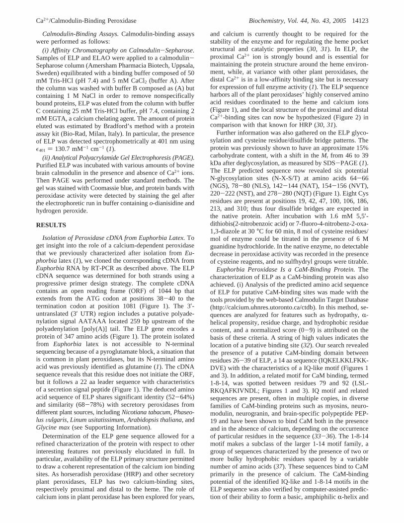
Isolation of Peroxidase cDNA from Euphorbia Latex.Toget insight into the role of a calcium-dependent peroxidasethat we previously characterized after isolation fromEu-phorbia latex (1), we cloned the corresponding cDNA fromEuphorbiaRNA by RT-PCR as described above. The ELPcDNA sequence was determined for both strands using aprogressive primer design strategy. The complete cDNAcontains an open reading frame (ORF) of 1044 bp thatextends from the ATG codon at positions 38-40 to thetermination codon at position 1081 (Figure 1). The 3′-untranslated (3′ UTR) region includes a putative polyade-nylation signal AATAAA located 259 bp upstream of thepolyadenylation [poly(A)] tail. The ELP gene encodes aprotein of 347 amino acids (Figure 1). The protein isolatedfrom Euphorbia latex is not accessible to N-terminalsequencing because of a pyroglutamate block, a situation thatis common in plant peroxidases, but its N-terminal aminoacid was previously identified as glutamine (1). The cDNAsequence reveals that this residue does not initiate the ORF,but it follows a 22 aa leader sequence with characteristicsof a secretion signal peptide (Figure 1). The deduced aminoacid sequence of ELP shares significant identity (52-64%)and similarity (68-78%) with secretory peroxidases fromdifferent plant sources, includingNicotiana tabacum, Phaseo-lusVulgaris, Linum usitatissimum, Arabidopsis thaliana, andGlycine max(see Supporting Information).
Determination of the ELP gene sequence allowed for arefined characterization of the protein with respect to otherinteresting features not previously elucidated in full. Inparticular, availability of the ELP primary structure permittedto draw a coherent representation of the calcium ion bindingsites. As horseradish peroxidase (HRP) and other secretoryplant peroxidases, ELP has two calcium-binding sites,respectively proximal and distal to the heme. The role ofcalcium ions in plant peroxidase has been explored for years,
and calcium is currently thought to be required for thestability of the enzyme and for regulating the heme pocketstructural and catalytic properties (30, 31). In ELP, theproximal Ca2+ ion is strongly bound and is essential formaintaining the protein structure around the heme environ-ment, while, at variance with other plant peroxidases, thedistal Ca2+ is in a low-affinity binding site but is necessaryfor expression of full enzyme activity (1). The ELP sequenceharbors all of the plant peroxidases’ highly conserved aminoacid residues coordinated to the heme and calcium ions(Figure 1), and the local structure of the proximal and distalCa2+-binding sites can now be hypothesized (Figure 2) incomparison with that known for HRP (30, 31).
Further information was also gathered on the ELP glyco-sylation and cysteine residue/disulfide bridge patterns. Theprotein was previously shown to have an approximate 15%carbohydrate content, with a shift in theMr from 46 to 39kDa after deglycosylation, as measured by SDS-PAGE (1).The ELP predicted sequence now revealed six potentialN-glycosylation sites (N-X-S/T) at amino acids 64-66(NGS), 78-80 (NLS), 142-144 (NAT), 154-156 (NVT),220-222 (NST), and 278-280 (NQT) (Figure 1). Eight Cysresidues are present at positions 19, 42, 47, 100, 106, 186,213, and 310; thus four disulfide bridges are expected inthe native protein. After incubation with 1.6 mM 5,5′-dithiobis(2-nitrobenzoic acid) or 7-fluoro-4-nitrobenz-2-oxa-1,3-diazole at 30°C for 60 min, 8 mol of cysteine residues/mol of enzyme could be titrated in the presence of 6 Mguanidine hydrochloride. In the native enzyme, no detectabledecrease in peroxidase activity was recorded in the presenceof cysteine reagents, and no sulfhydryl groups were titrable.
Euphorbia Peroxidase Is a CaM-Binding Protein.Thecharacterization of ELP as a CaM-binding protein was alsoachieved. (i) Analysis of the predicted amino acid sequenceof ELP for putative CaM-binding sites was made with thetools provided by the web-based Calmodulin Target Database(http://calcium.uhnres.utoronto.ca/ctdb). In this method, se-quences are analyzed for features such as hydropathy,R-helical propensity, residue charge, and hydrophobic residuecontent, and a normalized score (0-9) is attributed on thebasis of these criteria. A string of high values indicates thelocation of a putative binding site (32). Our search revealedthe presence of a putative CaM-binding domain betweenresidues 26-39 of ELP, a 14 aa sequence (IQKELKKLFKK-DVE) with the characteristics of a IQ-like motif (Figures 1and 3). In addition, a related motif for CaM binding, termed1-8-14, was spotted between residues 79 and 92 (LSL-RKQAFKIVNDL; Figures 1 and 3). IQ motif and relatedsequences are present, often in multiple copies, in diversefamilies of CaM-binding proteins such as myosins, neuro-modulin, neurogranin, and brain-specific polypeptide PEP-19 and have been shown to bind CaM both in the presenceand in the absence of calcium, depending on the occurrenceof particular residues in the sequence (33-36). The 1-8-14motif makes a subclass of the larger 1-14 motif family, agroup of sequences characterized by the presence of two ormore bulky hydrophobic residues spaced by a variablenumber of amino acids (37). These sequences bind to CaMprimarily in the presence of calcium. The CaM-bindingpotential of the identified IQ-like and 1-8-14 motifs in theELP sequence was also verified by computer-assisted predic-tion of their ability to form a basic, amphiphilicR-helix and
Ca2+/Calmodulin-Binding Peroxidase Biochemistry, Vol. 44, No. 43, 200514123
by calculating the relevant hydrophobicity and hydrophobicmoment. Interaction between CaM and target proteins is
known to be mainly hydrophobic in nature and to requirespecific binding of CaM to short basic peptides in target
FIGURE 1: Nucleotide and deduced amino acid sequence ofE. characiasperoxidase. The nucleotide sequence is numbered in the 5′ to 3′direction. The terminal tga codon is indicated by end. The polyadenylation signal aataaa (nt 1116-1121) is boxed. The signal peptideportion of the protein is underlined. Q corresponds to the N-terminal residue in mature peroxidase. Specific sense (f) and antisense (r)primers used in RACE experiments are in bold. The predicted CaM-binding IQ-like motif (peptide 26-39) and 1-8-14 motif (peptide79-92) are shown in gray. The deduced heme distal and proximal histidine residues, His50 and His179, and the Ca2+ ligands at the distal(Asp51, Val54, Gly56, Asp58, Ser60) and proximal (Thr180, Asp256, Thr259, Ile262, Asp265) metal-binding sites are boxed (see Figure 2 for morestructural details). These residues are highly conserved in all plant peroxidases.
14124 Biochemistry, Vol. 44, No. 43, 2005 Mura et al.

sequences, these peptides having a net propensity to formamphiphilicR-helical structures (12, 38). The helical wheelprojection, propensity forR-helix formation, mean residuehydrophobicity (H), and mean hydrophobic moment (µ) forthe ELP putative CaM-binding peptides are given in Figure3. The potential for these cationic peptides to be structuredas an amphipathicR-helix is manifest, and the calculatedHandµ values and other parameters fit those reported for theIQ and 1-14 classes, respectively (http://calcium.uhnres.utoronto.ca/ctdb). (ii) ELP was found to tightly bind to aCaM-Sepharose column in the presence of calcium andeluted with EGTA, whereas the protein did not bind to acolumn equilibrated in the absence of calcium or in thecontemporary presence of calcium and EGTA, indicating thatCaM binding to ELP is Ca2+ dependent (data not shown).
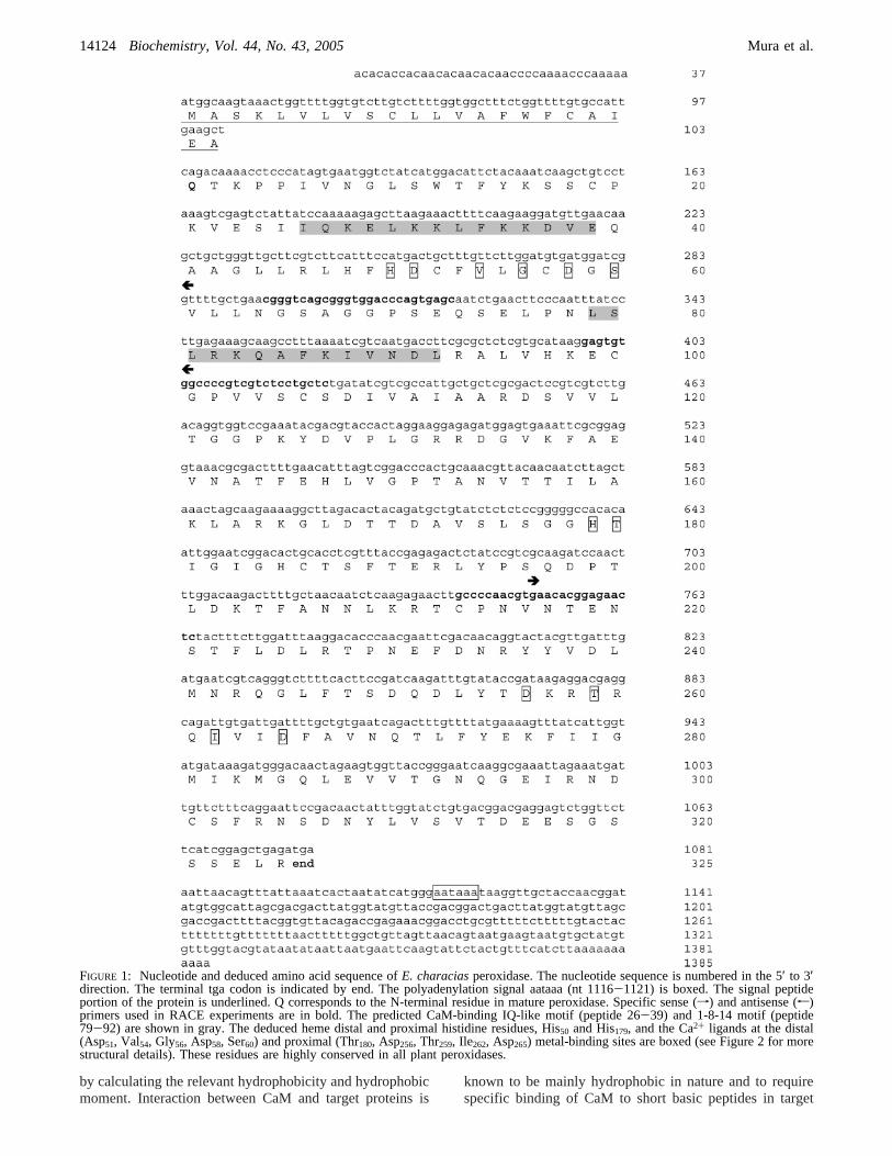
Reaction of ELP with Hydrogen Peroxide and ReducingSubstrates: Kinetic Parameters in the Absence and in thePresence of Calcium Ions.ELP activity was tested in 100mM MOPS buffer, pH 6.5, using ABTS as substrate. Thevalue of KM for ABTS at a saturating concentrations ofhydrogen peroxide was shown to be 1.25 mM whereas theKM for hydrogen peroxide at a saturating concentration ofABTS was calculated to be 3 mM (not shown). When nativeELP was incubated for 10 min in the presence of Ca2+, anactivation was observed (Figure 4) which showed a maxi-mum at 10 mM Ca2+ ion concentration (40-fold) with adrastic increase ofkcat. Under these conditions, calcium isthought to saturate the distal low-affinity binding site,converting the enzyme from an almost inactive form, withonly the proximal Ca2+ ion bound, to a fully active formwith both the proximal and distal calcium ions in situ (1).More in detail, Ca2+ binding to the low-affinity site isbelieved to induce the reorientation of the heme distal His,favoring its action as a general acid-base catalyst in theperoxidase reaction mechanism.
E. characiaslatex contains free calcium ions as determinedby atomic absorption, and the Ca2+ content varies from 2.1((0.1) mM in the winter to 3.5 ((0.15) mM in the summer.Thus we can conclude that the activation of ELP due to thefree calcium in the latex can oscillate between 18- and 24-fold. Crude latex was shown to contain a mean of 26 mg/mL total proteins, as determined by Bradford’s method, andELP activity was 8 nkat/mL (not shown). When ELP activitywas detected in the latex in the presence of 10 mM Ca2+
ions, we showed a 12-fold increase in activity, about three
FIGURE 2: Structure of the calcium-binding sites in ELP. Theproximal and distal calcium-binding sites inEuphorbia latexperoxidase have been hypothesized from analysis of the protein’ssequence and comparison to structural features of HRP (30, 31).Only the direct coordination environment is shown, but several otheramino acids close to the calcium ions are also highly conservedand are probably important for maintaining the correct geometryof the binding sites and of the heme pocket. Both Ca2+ ions areseven coordinated. In ELP, the distal Ca2+ is loosely bound, but itis necessary for expression of the full enzyme’s activity (1).
FIGURE 3: Prediction of CaM-binding sites in ELP. The full aminoacid sequence was analyzed to predict CaM-binding sites using theCaM Target Database (http://calcium.uhnres.utoronto.ca/ctdb). Analy-sis revealed two putative CaM-binding sequences, namely, an IQ-like motif (A) and a 1-8-14 motif (B). The numbers under thesequences indicate normalized scores (0-9) based on the evaluationcriteria for CaM-binding sites (seeEuphorbiaPeroxidase Is a CaM-Binding Protein for details). The predicted IQ-like motif (peptide26-39) and 1-8-14 motif (peptide 79-92) are underlined. Helicalwheel diagrams of the predicted CaM-binding peptides are alsoshown. Charged or hydrophilic residues are shaded in nuances ofred-violet; hydrophobic residues are in blue. Mean residue hydro-phobicity (H) and hydrophobic moment (µ) were calculated usingthe Kyte-Doolittle scale of hydrophobicity (50). The averagepropensity forR-helix formation was calculated using the Chou-Fasman values (51).
Ca2+/Calmodulin-Binding Peroxidase Biochemistry, Vol. 44, No. 43, 200514125
times less than that obtained in successive steps of ELPpurification (1). It may be indicative of a higher basalperoxidase activity of the protein when in the latex environ-ment. Since we used for detection of ELP activity 1 mL ofMOPS containing 50µL of a 200-fold diluted latex, itcorresponded to 2× 10⁻³ nkat/mL of ELP and 0.88µMcalcium ions. This amount of calcium did not perturb per seELP activity when added to samples of purified enzyme(Figure 4). Samples of latex were then dialyzed in MOPSbuffer, pH 6.5, in the presence of 0.4 mM EGTA for 12 hand then dialyzed exhaustively in MOPS buffer withoutEGTA. After this treatment, a lower basal activity was
recorded in ELPsamples but a 40-fold activation of ELPactivity was observed in the presence of 10 mM calciumions. The loss of the strongly bound proximal calcium ionafter treatment with EGTA can be excluded, since this canonly be achieved by incubating the enzyme in 100 mM Tris-HCl buffer, pH 7.2, for 18 h at 25°C with 6 M guanidinehydrochloride and 10 mM EGTA, as previously shown (1).Altogether, these results suggest the presence of an activatingfactor in the latex, and we believe it can be safely assumedthat it might be calmodulin.
Isolation of Calmodulin cDNA from Euphorbia.Asdetailed, the ELP sequence contains a CaM-binding IQ-likemotif and a related motif termed 1-8-14 (Figure 1). Startingfrom this evidence, we thus looked for, found, and cloned aEuphorbiacDNA coding for a CaM protein. We later spottedthis protein as expressed in the plant latex (see below) andtherefore named itEuphorbia latex calmodulin (ELCaM).The ELCaM cDNA contains an ORF of 447 bp which canbe translated to a protein sequence of 149 amino acids(Figure 5). The ATG codon at nucleotides 71-74 mostprobably represents the protein translation initiation site. The3′-untranslated region includes a putative polyadenylationsignal AATAAA located 26 bp upstream of the poly(A) tail.The calculated molecular mass for the predicted protein is16850 kDa, with a pI of 4.1. Not surprisingly, the ELCaMamino acid sequence shows a very high degree of identity(91-100%) and similarity (99-100%) to CaMs isolated fromseveral other higher plants, includingPrunus aVium, Elaeis
FIGURE 4: Response ofEuphorbiaperoxidase activity to Ca2+. ELPactivity in 100 mM MOPS, pH 6.5, and in the presence of calciumions. The continuous curve represents the theoretical bindingisotherm fit to the data.
FIGURE 5: Nucleotide and deduced amino acid sequence of theE. characiasCaM. The nucleotide sequence is numbered in the 5′ to 3′direction. The termination tga codon is indicated by end. The polyadenylation signal AATAAA (nt 742-747) is boxed. M corresponds tothe N-terminal residue in mature CaM. The four EF-hand domains are underlined, and the Ca2+-binding amino acid residues are boxed.Specific sense (f) and antisense (r) primers used in RACE experiments are in bold.
14126 Biochemistry, Vol. 44, No. 43, 2005 Mura et al.
guineensis, Medicago truncatula, Pisum satiVum, P. Vulgaris,andN. tabacum(see Supporting Information).
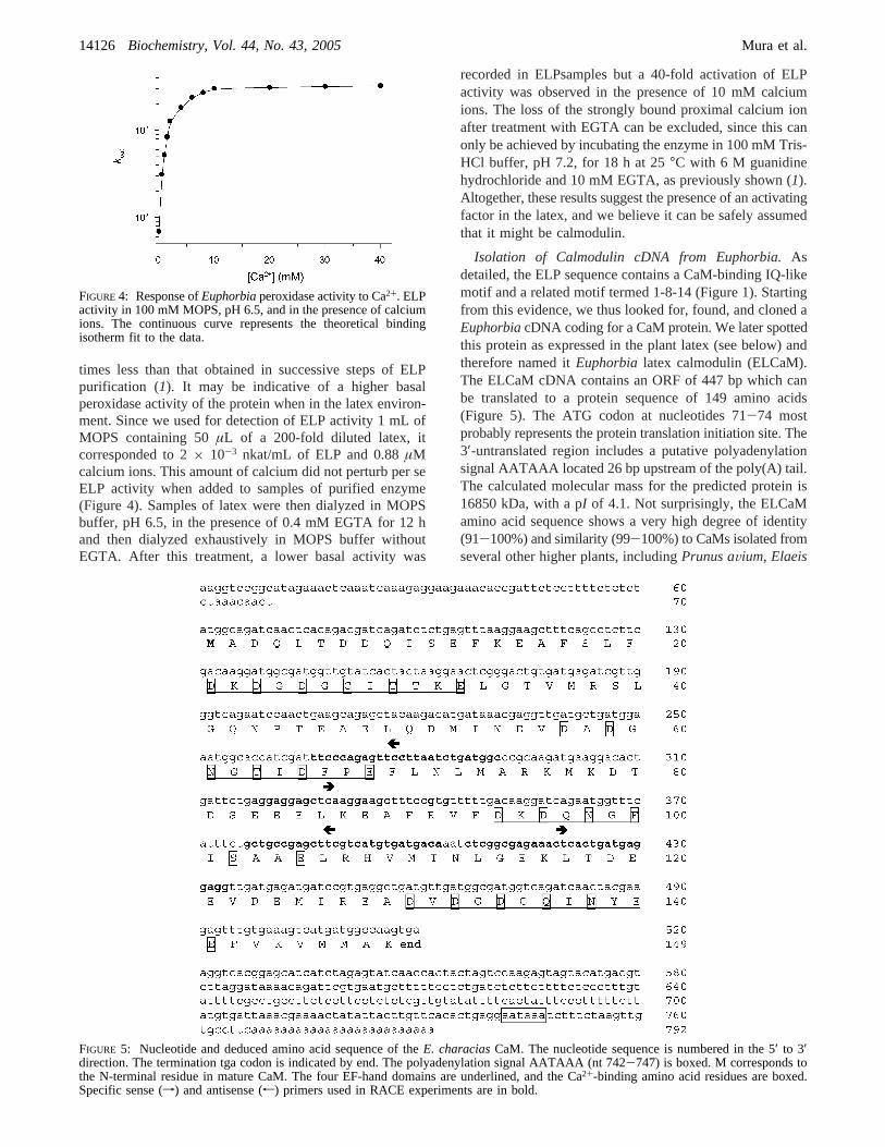
CaM Is Expressed in Euphorbia Latex.To test whetherCaM was present inEuphorbia latex, the crude latex wasimmunoblotted using antibodies specific for the CaM-bindingdomain. A single reactive band was detected (Figure 6),demonstrating thatEuphorbiaCaM is expressed in the latex,where it coexists with ELP. Attempts were also made toverify whether and to which extent ELCaM remains associ-ated to ELP during the latter protein’s purification processfrom the latex, but apparently ELCaM is lost at the first stepof ELP purification and proved itself particularly recalcitrantto isolation in pure form.
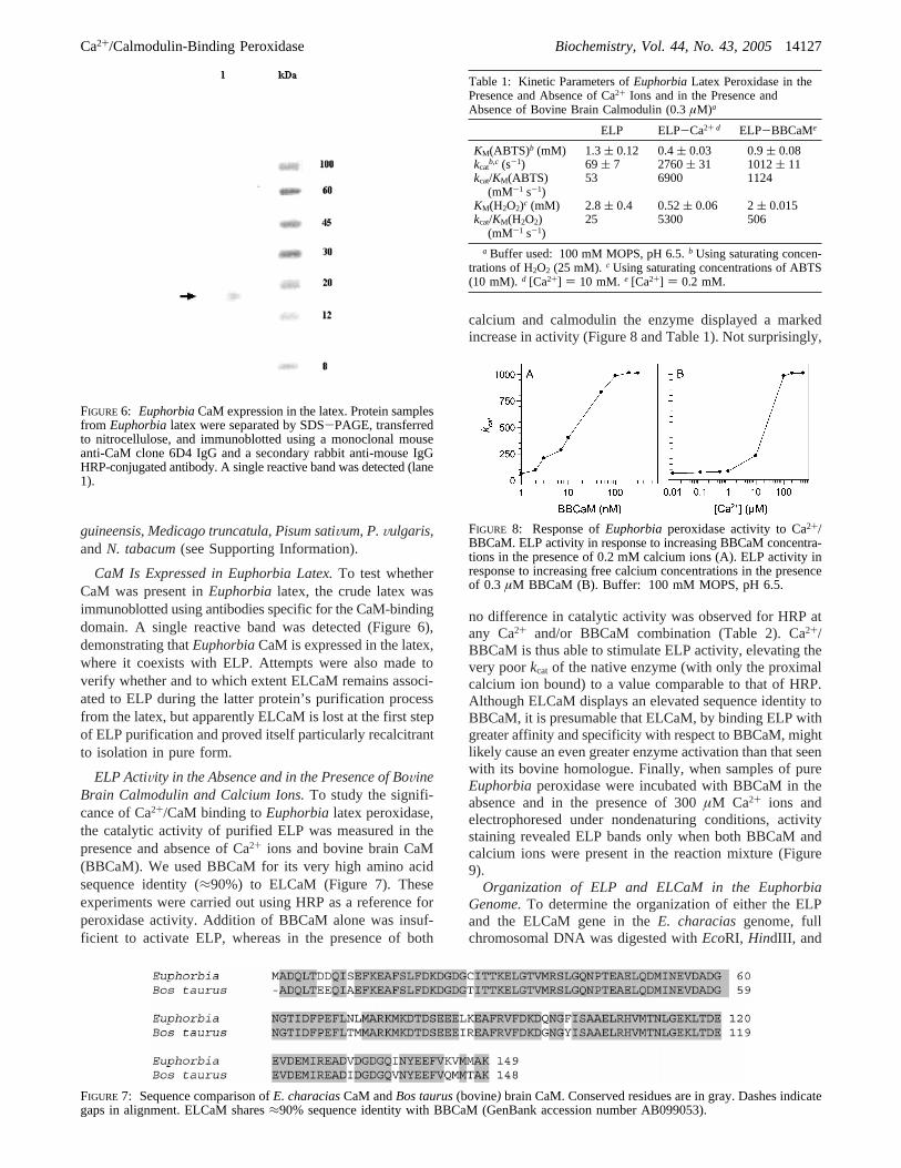
ELP ActiVity in the Absence and in the Presence of BoVineBrain Calmodulin and Calcium Ions.To study the signifi-cance of Ca2+/CaM binding toEuphorbialatex peroxidase,the catalytic activity of purified ELP was measured in thepresence and absence of Ca2+ ions and bovine brain CaM(BBCaM). We used BBCaM for its very high amino acidsequence identity (≈90%) to ELCaM (Figure 7). Theseexperiments were carried out using HRP as a reference forperoxidase activity. Addition of BBCaM alone was insuf-ficient to activate ELP, whereas in the presence of both
calcium and calmodulin the enzyme displayed a markedincrease in activity (Figure 8 and Table 1). Not surprisingly,
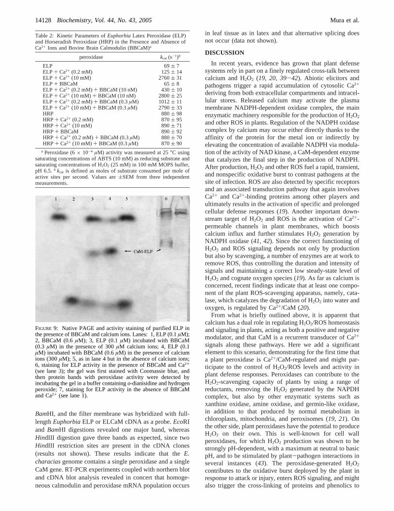
no difference in catalytic activity was observed for HRP atany Ca2+ and/or BBCaM combination (Table 2). Ca2+/BBCaM is thus able to stimulate ELP activity, elevating thevery poorkcat of the native enzyme (with only the proximalcalcium ion bound) to a value comparable to that of HRP.Although ELCaM displays an elevated sequence identity toBBCaM, it is presumable that ELCaM, by binding ELP withgreater affinity and specificity with respect to BBCaM, mightlikely cause an even greater enzyme activation than that seenwith its bovine homologue. Finally, when samples of pureEuphorbiaperoxidase were incubated with BBCaM in theabsence and in the presence of 300µM Ca2+ ions andelectrophoresed under nondenaturing conditions, activitystaining revealed ELP bands only when both BBCaM andcalcium ions were present in the reaction mixture (Figure9).
Organization of ELP and ELCaM in the EuphorbiaGenome.To determine the organization of either the ELPand the ELCaM gene in theE. characiasgenome, fullchromosomal DNA was digested withEcoRI, HindIII, and
FIGURE 6: EuphorbiaCaM expression in the latex. Protein samplesfrom Euphorbialatex were separated by SDS-PAGE, transferredto nitrocellulose, and immunoblotted using a monoclonal mouseanti-CaM clone 6D4 IgG and a secondary rabbit anti-mouse IgGHRP-conjugated antibody. A single reactive band was detected (lane1).
FIGURE 7: Sequence comparison ofE. characiasCaM andBos taurus(bovine) brain CaM. Conserved residues are in gray. Dashes indicategaps in alignment. ELCaM shares≈90% sequence identity with BBCaM (GenBank accession number AB099053).
Table 1: Kinetic Parameters ofEuphorbiaLatex Peroxidase in thePresence and Absence of Ca2+ Ions and in the Presence andAbsence of Bovine Brain Calmodulin (0.3µM)a
ELP ELP-Ca2+ d ELP-BBCaMe
KM(ABTS)b (mM) 1.3( 0.12 0.4( 0.03 0.9( 0.08kcat
b,c (s-1) 69 ( 7 2760( 31 1012( 11kcat/KM(ABTS)
(mM-1 s-1)53 6900 1124
KM(H2O2)c (mM) 2.8( 0.4 0.52( 0.06 2( 0.015kcat/KM(H2O2)
(mM-1 s-1)25 5300 506
a Buffer used: 100 mM MOPS, pH 6.5.b Using saturating concen-trations of H2O2 (25 mM). c Using saturating concentrations of ABTS(10 mM). d [Ca2+] ) 10 mM. e [Ca2+] ) 0.2 mM.
FIGURE 8: Response ofEuphorbia peroxidase activity to Ca2+/BBCaM. ELP activity in response to increasing BBCaM concentra-tions in the presence of 0.2 mM calcium ions (A). ELP activity inresponse to increasing free calcium concentrations in the presenceof 0.3 µM BBCaM (B). Buffer: 100 mM MOPS, pH 6.5.
Ca2+/Calmodulin-Binding Peroxidase Biochemistry, Vol. 44, No. 43, 200514127
BamHI, and the filter membrane was hybridized with full-lengthEuphorbiaELP or ELCaM cDNA as a probe.EcoRIand BamHI digestions revealed one major band, whereasHindIII digestion gave three bands as expected, since twoHindIII restriction sites are present in the cDNA clones(results not shown). These results indicate that theE.characiasgenome contains a single peroxidase and a singleCaM gene. RT-PCR experiments coupled with northern blotand cDNA blot analysis revealed in concert that homoge-neous calmodulin and peroxidase mRNA population occurs
in leaf tissue as in latex and that alternative splicing doesnot occur (data not shown).
DISCUSSION
In recent years, evidence has grown that plant defensesystems rely in part on a finely regulated cross-talk betweencalcium and H2O2 (19, 20, 39-42). Abiotic elicitors andpathogens trigger a rapid accumulation of cytosolic Ca2+
deriving from both extracellular compartments and intracel-lular stores. Released calcium may activate the plasmamembrane NADPH-dependent oxidase complex, the mainenzymatic machinery responsible for the production of H2O2
and other ROS in plants. Regulation of the NADPH oxidasecomplex by calcium may occur either directly thanks to theaffinity of the protein for the metal ion or indirectly byelevating the concentration of available NADPH via modula-tion of the activity of NAD kinase, a CaM-dependent enzymethat catalyzes the final step in the production of NADPH.After production, H2O2 and other ROS fuel a rapid, transient,and nonspecific oxidative burst to contrast pathogens at thesite of infection. ROS are also detected by specific receptorsand an associated transduction pathway that again involvesCa2+ and Ca2+-binding proteins among other players andultimately results in the activation of specific and prolongedcellular defense responses (19). Another important down-stream target of H2O2 and ROS is the activation of Ca2+-permeable channels in plant membranes, which boostscalcium influx and further stimulates H2O2 generation byNADPH oxidase (41, 42). Since the correct functioning ofH2O2 and ROS signaling depends not only by productionbut also by scavenging, a number of enzymes are at work toremove ROS, thus controlling the duration and intensity ofsignals and maintaining a correct low steady-state level ofH2O2 and cognate oxygen species (19). As far as calcium isconcerned, recent findings indicate that at least one compo-nent of the plant ROS-scavenging apparatus, namely, cata-lase, which catalyzes the degradation of H2O2 into water andoxygen, is regulated by Ca2+/CaM (20).
From what is briefly outlined above, it is apparent thatcalcium has a dual role in regulating H2O2/ROS homeostasisand signaling in plants, acting as both a positive and negativemodulator, and that CaM is a recurrent transducer of Ca2+
signals along these pathways. Here we add a significantelement to this scenario, demonstrating for the first time thata plant peroxidase is Ca2+/CaM-regulated and might par-ticipate to the control of H2O2/ROS levels and activity inplant defense responses. Peroxidases can contribute to theH2O2-scavenging capacity of plants by using a range ofreductants, removing the H2O2 generated by the NAPDHcomplex, but also by other enzymatic systems such asxanthine oxidase, amine oxidase, and germin-like oxidase,in addition to that produced by normal metabolism inchloroplasts, mitochondria, and peroxisomes (19, 21). Onthe other side, plant peroxidases have the potential to produceH2O2 on their own. This is well-known for cell wallperoxidases, for which H2O2 production was shown to bestrongly pH-dependent, with a maximum at neutral to basicpH, and to be stimulated by plant-pathogen interactions inseveral instances (43). The peroxidase-generated H2O2
contributes to the oxidative burst deployed by the plant inresponse to attack or injury, enters ROS signaling, and mightalso trigger the cross-linking of proteins and phenolics to
Table 2: Kinetic Parameters ofEuphorbiaLatex Peroxidase (ELP)and Horseradish Peroxidase (HRP) in the Presence and Absence ofCa2+ Ions and Bovine Brain Calmodulin (BBCaM)a
peroxidase kcat (s-1)b
ELP 69( 7ELP + Ca2+ (0.2 mM) 125( 14ELP + Ca2+ (10 mM) 2760( 31ELP + BBCaM 65( 8ELP + Ca2+ (0.2 mM)+ BBCaM (10 nM) 430( 10ELP + Ca2+ (10 mM) + BBCaM (10 nM) 2800( 25ELP + Ca2+ (0.2 mM)+ BBCaM (0.3µM) 1012( 11ELP + Ca2+ (10 mM) + BBCaM (0.3µM) 2790( 33HRP 880( 98HRP+ Ca2+ (0.2 mM) 870( 95HRP+ Ca2+ (10 mM) 890( 71HRP+ BBCaM 890( 92HRP+ Ca2+ (0.2 mM)+ BBCaM (0.3µM) 880 ( 70HRP+ Ca2+ (10 mM) + BBCaM (0.3µM) 870 ( 90
a Peroxidase (6× 10⁻⁴ µM) activity was measured at 25°C usingsaturating concentrations of ABTS (10 mM) as reducing substrate andsaturating concentrations of H2O2 (25 mM) in 100 mM MOPS buffer,pH 6.5. b kcat is defined as moles of substrate consumed per mole ofactive sites per second. Values are(SEM from three independentmeasurements.
FIGURE 9: Native PAGE and activity staining of purified ELP inthe presence of BBCaM and calcium ions. Lanes: 1, ELP (0.1µM);2, BBCaM (0.6µM); 3, ELP (0.1µM) incubated with BBCaM(0.3 µM) in the presence of 300µM calcium ions; 4, ELP (0.1µM) incubated with BBCaM (0.6µM) in the presence of calciumions (300µM); 5, as in lane 4 but in the absence of calcium ions;6, staining for ELP activity in the presence of BBCaM and Ca2+
(see lane 3); the gel was first stained with Coomassie blue, andthen protein bands with peroxidase activity were detected byincubating the gel in a buffer containingo-dianisidine and hydrogenperoxide; 7, staining for ELP activity in the absence of BBCaMand Ca2+ (see lane 1).
14128 Biochemistry, Vol. 44, No. 43, 2005 Mura et al.
reinforce cell walls at the site of infection, which wouldconceivably result in suppression of pathogen ingress (43,44). On the basis of the results reported here, we thereforepropose a model of the interplay between Ca2+ and H2O2
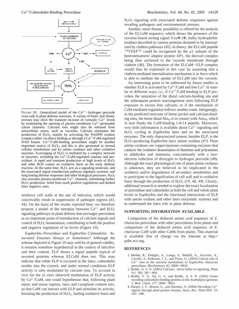
signaling pathways in plant defense that envisages peroxidaseas an important point of transduction of calcium signals andcontrol of H2O2 homeostasis, contributing to both the positiveand negative regulation of its levels (Figure 10).
Euphorbia Peroxidase and Euphorbia Calmodulin: As-sociated Enzymes Always or Sometimes?Although thescheme depicted in Figure 10 may well be of general validity,it remains somehow hypothetical in the context of laticifersand their content. ELP shows a signal peptide typical ofsecreted proteins whereas ELCaM does not. This mayindicate that while ELP is excreted in the latex, calmodulinresides into the cytosol, and under normal conditions ELPactivity is only modulated by calcium ions. To account invivo for the in vitro observed modulation of ELP activityby Ca2+/CaM, one could hypothesize that, following plantinjury and tissue rupture, latex and cytoplasm content mix,so that CaM can interact with ELP and stimulate its activity,boosting the production of H2O2, fueling oxidative burst and
H2O2 signaling with associated defense responses againstinvading pathogens and environmental stresses.
Another, more distant, possibility is offered by the analysisof the ELCaM sequence, which shows the presence of thetyrosine-based sorting signal YxxΦ (Φ, bulky hydrophobicresidue) described in various proteins destined to be internal-ized by clathrin pathways (45). In theory, the ELCaM peptide139YEEF142 could be recognized by theµ1 subunit of theheterotetrameric adaptor protein AP1, the derived complexbeing then anchored to the vacuole membrane throughclathrin (46). The formation of the ELCaM-ELP complexwould thus be explained in this case by assuming that aclathrin-mediated internalization mechanism is in force whichis able to mediate the uptake of ELCaM into the vacuole.
An interesting point to be addressed by future studies iswhether ELP is activated by Ca2+/CaM and free Ca2+ in simi-lar or different ways, i.e., if Ca2+/CaM binding to ELP pro-vokes the saturation of the distal calcium-binding site andthe subsequent protein rearrangement seen following ELPexposure to excess free calcium, or if the mechanism ofCaM-mediated regulation follows another route. Intriguingly,in the predicted structure of heme pocket and calcium-bind-ing sites, the heme distal His50 is in contact with Asn78, whichin turn flanks the CaM-binding 1-8-14 peptide. Moreover,very little information is available about Ca2+ signaling andH2O2 cycling in Euphorbia latex and on the associatedenzymes. The only characterized system, besides ELP, is theH2O2-producingEuphorbialatex amine oxidase (47). Plantamine oxidases are copper/quinone-containing enzymes thatcatalyze the oxidative deamination of diamines and polyaminesto aldehydes and ammonia, concomitantly with a two-electron reduction of dioxygen to hydrogen peroxide (48).Although the exact physiological role of plant amine oxidasesis unknown, they are believed to be implicated in thesynthesis and/or degradation of secondary metabolites andto participate to the lignification of cell wall and to oxidativeburst through the production of H2O2 (19, 48, 49). Clearly,additional research is needed to explore the exact localizationof peroxidase and calmodulin at both the cell and whole plantlevels inEuphorbiaand the functional interactions of ELPwith amine oxidase and other latex enzymatic systems andto understand the latex role in plant defense.
SUPPORTING INFORMATION AVAILABLE
Comparison of the deduced amino acid sequence ofE.characiasperoxidase with other peroxidases from plants andcomparison of the deduced amino acid sequence ofE.characiasCaM with other CaMs from plants. This materialis available free of charge via the Internet at http://pubs.acs.org.
REFERENCES
1. Medda, R., Padiglia, A., Longu, S., Bellelli, A., Arcovito, A.,Cavallo, S., Pedersen, J. Z., and Floris, G. (2003) Critical role ofCa2+ ions in the reaction mechanism ofEuphorbia characiasperoxidase,Biochemistry 42, 8909-8918.
2. Reddy, A. S. N. (2001) Calcium: silver bullet in signaling,PlantSci. 160, 381-404.
3. Reddy, V. S., Ali, G. S., and Reddy, A. S. N. (2002) Genesencoding calmodulin-binding proteins in theArabidopsisgenome,J. Biol. Chem. 277, 9840-9852.
4. Harper, J. F., Breton, G., and Harmon, A. (2004) Decoding Ca2+
signals through plant protein kinases,Annu. ReV. Plant Biol. 55,263-288.
FIGURE 10: Generalized model of the Ca2+-hydrogen peroxidecross-talk in plant defense reactions. A variety of biotic and abioticstresses may elicit the transient increase of cytosolic Ca2+ levelsby modulating the opening of plasma membrane Ca2+-permeablecation channels. Calcium ions might also be released fromintracellular stores, such as vacuoles. Calcium stimulates theproduction of H2O2, mainly by activating the NADPH oxidasecomplex either via direct binding or through a Ca2+/CaM-regulatedNAD kinase. Ca2+/CaM-binding peroxidase might be anotherimportant source of H2O2, and this is also generated in normalcellular metabolism and by amine oxidases and other oxidativeenzymes. Scavenging of H2O2 is mediated by a complex networkof enzymes, including the Ca2+/CaM-regulated catalase and per-oxidase. A rapid and transient production of high levels of H2O2and other ROS causes an oxidative burst as the early defensereaction. At the same time, H2O2 acts as a signaling molecule, andthe associated signal transduction pathway regulates systemic andlong-lasting defense responses and other biological processes. H2O2also activates plasma membrane Ca2+ channels, stimulating furtherH2O2 production. Solid lines mark positive regulations and dashedlines negative ones.
Ca2+/Calmodulin-Binding Peroxidase Biochemistry, Vol. 44, No. 43, 200514129
5. Anderson, J. M., Charbonneau, H., Jones, H. P., McCann, R. O.,and Cormier, M. J. (1980) Characterization of the plant nicoti-namide adenine dinucleotide kinase activator protein and itsidentification as calmodulin,Biochemistry 19, 3113-3120.
6. Snedden, W. A., and Fromm, H. (2001) Calmodulin as a versatilecalcium signal transducer in plants,New Phytol. 151, 35-66.
7. Moutinho, A., Love, J., Trewavas, A. J., and Malho, R. (1998)Distribution of calmodulin protein and mRNA in growing pollentubes,Sex Plant Reprod. 11, 131-139.
8. Golovkin, M., and Reddy, A. S. N. (2003) A calmodulin-bindingprotein fromArabidopsishas an essential role in pollen germina-tion, Proc. Natl. Acad. Sci. U.S.A. 100, 10558-10563.
9. Hua, W., Zhang, L., Liang, S. P., Jones, R. L., and Lu, Y. T.(2004) A tobacco calcium/calmodulin-binding protein kinasefunctions as a negative regulator of flowering,J. Biol. Chem. 279,31483-31494.
10. Hua, W., Li, R. J., Wang, L., and Lu, Y. T. (2004) A tobaccocalmodulin-binding protein kinase (NtCBK2) induced by high-salt/GA treatment and its expression during floral developmentand embryogenesis,Plant Sci. 166, 1253-1259.
11. Kim, C. M., Lee, S. H., Kim, J. K., Chun, H. J., Choi, M. S.,Chung, W. S., Moon, B. C., Kang, C. H., Park, C. Y., Yoo, J. H.,Kang, Y. H., Koo, S. C., Koo, Y. D., Jung, J. C., Kim, S. T.,Schulze-Lefert, P., Lee, S. Y., and Cho, M. J. (2002) Mlo, amodulator of plant defense and cell death, is a novel calmodulin-binding protein. Isolation and characterization of a rice Mlohomologue,J. Biol. Chem. 277, 19304-19314
12. Zielinski, R. E. (1998) Calmodulin and calmodulin-bindingproteins in plants,Annu. ReV. Plant Physiol. Plant Mol. Biol. 49,697-725.
13. Yang, T., and Poovaiah, B. W. (2003) Calcium/calmodulin-mediated signal network in plants,Trends Plant Sci. 8, 505-512.
14. Zhang, L., and Lu, Y. T. (2003) Calmodulin-binding proteinkinases in plants,Trends Plant Sci. 8, 123-127.
15. Hiraga, S., Sasaki, K., Ito, H., Ohashi, Y., and Matsui, H. (2001)A large family of Class III plant peroxidases,Plant Cell Physiol.42, 462-468.
16. Klessig, D. F., Durner, J., Noad, R., Navarre, D. A., Wendehenne,D., Kumar, D., Zhou, J. M., Shah, J., Zhang, S., Kachroo, P.,Trifa, Y., Pontier, D., Lam, E., and Silva, H. (2000) Nitric oxideand salicylic acid signaling in plant defense,Proc. Natl. Acad.Sci. U.S.A. 97, 8849-8855.
17. Kawano, T. (2003) Roles of the reactive oxygen species-generatingperoxidase reactions in plant defense and growth induction,PlantCell. Rep. 21, 829-837.
18. Orozco-Cardenas, M. L., Narvaez-Vasquez, J., and Ryan, C. A.(2001) Hydrogen peroxide acts as a second messenger for theinduction of defense genes in tomato plants in response towounding, systemin, and methyl jasmonate,Plant Cell 13, 179-191.
19. Mittler, R., Vanderauwera, S., Gollery, M., and Van Breusegem,F. (2004) The reactive oxygen gene network of plants,TrendsPlant Sci. 9, 490-498.
20. Yang, T., and Poovaiah, B. W. (2002) Hydrogen peroxidehomeostasis: activation of plant catalase by calcium/calmodulina,Proc. Natl. Acad. Sci. U.S.A. 99, 4097-4102.
21. Neill, S., Desikan, R., and Hancock, J. (2002) Hydrogen peroxidesignaling,Curr. Opin. Plant Biol. 5, 388-395.
22. Rudall, P. (1994) Lactifers in Crotonoideae (Euphorbiaceae):homology and evolution,Ann. MO Bot. Gard. 81, 270-282.
23. Webster, G. L. (1994) Classification of the Euphorbiaceae,Ann.MO Bot. Gard. 81, 3-32.
24. Ko, J. H., Chow, K. S., and Han, K. H. (2003) Transcriptomeanalysis reveals novel features of the molecular events occurringin the laticifers ofHeVea brasiliensis(para rubber tree),PlantMol. Biol. 53, 479-492.
25. Kekwick, R. G. O. (2001)Latex and Laticifers Encyclopedia ofLife Sciences, http://www.els.net.
26. Furhop, J. H., and Smith, K. M. (1975) Laboratory methods inPorphyrins and metalloproteins(Smith, K. M., Ed.) pp 757-869,Elsevier, Amsterdam.
27. Rose, T. M., Henikoff, J. G., and Henikoff, S. (2003) CODEHOP(COnsensus-DEgenerate Hybrid Oligonucleotide Primer) PCRprimer design,Nucleic Acids Res. 31, 3763-3766.
28. Frohman, M. A. (1993) Rapid amplification of complementaryDNA ends for generation of full-length complementary DNAs:thermal RACE,Methods Enzymol. 218, 340-356.
29. Jaakola, L., Pirttila¨, A. M., and Hohtola, A. (2001) cDNA blottingoffers an alternative method for gene expression studies,PlantMol. Biol. Rep. 19, 125-128.
30. Howes, B. D., Feis, A., Raimondi, L., Indiani, C., and Smulevich,G. (2001) The critical role of the proximal calcium ion in thestructural properties of horseradish peroxidase,J. Biol. Chem. 276,40704-40711.
31. Laberge, M., Huang, Q., Schweitzer-Stenner, R., and Fidy, J.(2003) The endogenous calcium ions of horseradish peroxidaseC are required to maintain the functional nonplanarity of the heme,Biophys. J. 84, 2542-2552.
32. Yap, K. L., Kim, J., Truong, K., Sherman, M., Yuan, T., and Ikura,M. (2000) Calmodulin target database,J. Struct. Funct. Genomics1, 8-14.
33. Houdusse, A., and Cohen, C. (1995) Target sequence recognitionby the calmodulin superfamily: Implications from light chainbinding to the regulatory domain of scallop myosin,Proc. Natl.Acad. Sci. U.S.A. 92, 10644-10647.
34. Munshi, H. G., Burks, D. J., Joyal, J. L., White, M. F., and Sacks,D. B. (1996) Ca2+ regulates calmodulin binding to IQ motifs inIRS-1,Biochemistry 35, 15883-15889.
35. Bahler, M., and Rhoads, A. (2002) Calmodulin signaling via theIQ motif, FEBS Lett. 513, 107-113.
36. Putkey, J. A., Kleerekoper, Q., Gaertner, T. R., and Waxham, M.N. (2003) A new role for IQ motif proteins in regulatingcalmodulin function,J. Biol. Chem. 278, 49667-49670.
37. Rhoads, A. R., and Friedberg, F. (1997) Sequence motifs forcalmodulin recognition,FASEB J. 11, 331-340.
38. Erickson-Viitanen, S., and DeGrado, W. F. (1987) Recognitionand characterization of calmodulin-binding sequences,MethodsEnzymol. 139, 455-478.
39. Grant, J. J., and Loake, G. J. (2000) Role of active oxygenintermediates and cognate redox signaling in disease resistance,Plant Physiol. 124, 21-29.
40. Pellinen, R. I., Korhonen, M. S., Tauriainen, A. A., Palva, E. T.,and Kangasja¨rvi, J. (2002) Hydrogen peroxide activates cell deathand defense gene expression in birch,Plant Physiol. 130, 549-560.
41. Demidchik, V., Shabala, S. N., Coutts, K. B., Tester, M. A., andDavies, J. M. (2002) Free oxygen radicals regulate plasmamembrane Ca2+ and K+-permeable channels in plant root cells,J. Cell Sci. 116, 81-88.
42. Foreman, J., Demidchik, V., Bothwell, J. H., Mylona, P., Miedema,H., Torres, M. A., Linstead, P., Costa, S., Brownlee, C., Jones, J.D., Davies, J. M., and Dolan, L. (2003) Reactive oxygen speciesproduced by NADPH oxidase regulate plant cell growth,Nature422, 442-446.
43. Bolwell, G. P., and Wojtaszek, P. (1997) Mechanisms for thegeneration of active oxygen species in plant defence: a broadperspective,Physiol. Mol. Plant Pathol. 51, 347-366.
44. Wu, G., Shortt, B. J., Lawrence, E. B., Leo´n, J., Fitzsimmons, K.C., Levine, E. B., Raskin, I., and Shah, D. M. (1997) Activationof host defense mechanisms by elevated production of H2O2 intransgenic plants,Plant Physiol. 115, 427-435.
45. Ohno, H., Stewart, J., Fournier, M. C., Bosshart, H., Rhee, I.,Miyatake, S., Saito, T., Gallusser, A., Kirchhausen, T., andBonifacino, J. S. (1995) Interaction of tyrosine-based sortingsignals with clathrin-associated proteins,Science 269, 1872-1875.
46. Brett, T. J., Traub, L. M., and Fremont, D. H. (2002) Accessoryprotein recruitment motifs in clathrin-mediated endocytosis,Structure 10, 797-809.
47. Padiglia, A., Medda, R., Lorrai, A., Murgia, B., Pedersen, J. Z.,Finazzi Agro, A., and Floris, G. (1998) Characterization ofEuphorbia characiaslatex amine oxidase,Plant Physiol. 117,1363-1371.
48. Medda, R., Padiglia, A., and Floris, G. (1995) Plant copper-amineoxidases,Phytochemistry 39, 1-9.
49. Laurenzi, M., Tipping, A. J., Marcus, S. E., Knox, J. P., Federico,R., Angelini, R., and McPherson, M. J. (2001) Analysis of thedistribution of copper amine oxidase in cell walls of legumeseedlings,Planta 214, 37-45.
50. Kyte, J., and Doolittle, R. F. (1982) A simple method for displayingthe hydropathic character of a protein,J. Mol. Biol. 157, 105-132.
51. Chou, P. Y., and Fasman, G. D. (1978) Empirical predictions ofprotein conformation,Annu. ReV. Biochem. 47, 251-276.
BI0513251
14130 Biochemistry, Vol. 44, No. 43, 2005 Mura et al.