Streamlined copper defenses make Bordetella pertussis ...
12
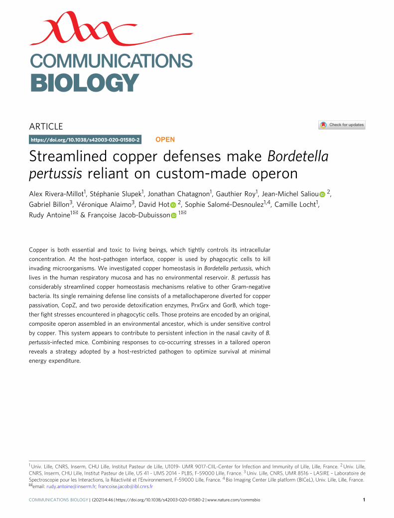
ARTICLE Streamlined copper defenses make Bordetella pertussis reliant on custom-made operon Alex Rivera-Millot 1 , Stéphanie Slupek 1 , Jonathan Chatagnon 1 , Gauthier Roy 1 , Jean-Michel Saliou 2 , Gabriel Billon 3 , Véronique Alaimo 3 , David Hot 2 , Sophie Salomé-Desnoulez 1,4 , Camille Locht 1 , Rudy Antoine 1 ✉ & Françoise Jacob-Dubuisson 1 ✉ Copper is both essential and toxic to living beings, which tightly controls its intracellular concentration. At the host–pathogen interface, copper is used by phagocytic cells to kill invading microorganisms. We investigated copper homeostasis in Bordetella pertussis, which lives in the human respiratory mucosa and has no environmental reservoir. B. pertussis has considerably streamlined copper homeostasis mechanisms relative to other Gram-negative bacteria. Its single remaining defense line consists of a metallochaperone diverted for copper passivation, CopZ, and two peroxide detoxification enzymes, PrxGrx and GorB, which toge- ther fight stresses encountered in phagocytic cells. Those proteins are encoded by an original, composite operon assembled in an environmental ancestor, which is under sensitive control by copper. This system appears to contribute to persistent infection in the nasal cavity of B. pertussis-infected mice. Combining responses to co-occurring stresses in a tailored operon reveals a strategy adopted by a host-restricted pathogen to optimize survival at minimal energy expenditure. https://doi.org/10.1038/s42003-020-01580-2 OPEN 1 Univ. Lille, CNRS, Inserm, CHU Lille, Institut Pasteur de Lille, U1019- UMR 9017-CIIL-Center for Infection and Immunity of Lille, Lille, France. 2 Univ. Lille, CNRS, Inserm, CHU Lille, Institut Pasteur de Lille, US 41 - UMS 2014 - PLBS, F-59000 Lille, France. 3 Univ. Lille, CNRS, UMR 8516 – LASIRE – Laboratoire de Spectroscopie pour les Interactions, la Réactivité et l’Environnement, F-59000 Lille, France. 4 Bio Imaging Center Lille platform (BICeL), Univ. Lille, Lille, France. ✉ email: [email protected]; [email protected] COMMUNICATIONS BIOLOGY | (2021)4:46 | https://doi.org/10.1038/s42003-020-01580-2 | www.nature.com/commsbio 1 1234567890():,;
-
Upload
khangminh22 -
Category
Documents
-
view
1 -
download
0
Transcript of Streamlined copper defenses make Bordetella pertussis ...
ARTICLE
Streamlined copper defenses make Bordetellapertussis reliant on custom-made operonAlex Rivera-Millot1, Stéphanie Slupek1, Jonathan Chatagnon1, Gauthier Roy1, Jean-Michel Saliou 2,
Gabriel Billon3, Véronique Alaimo3, David Hot 2, Sophie Salomé-Desnoulez1,4, Camille Locht1,
Rudy Antoine1✉ & Françoise Jacob-Dubuisson 1✉
Copper is both essential and toxic to living beings, which tightly controls its intracellular
concentration. At the host–pathogen interface, copper is used by phagocytic cells to kill
invading microorganisms. We investigated copper homeostasis in Bordetella pertussis, which
lives in the human respiratory mucosa and has no environmental reservoir. B. pertussis has
considerably streamlined copper homeostasis mechanisms relative to other Gram-negative
bacteria. Its single remaining defense line consists of a metallochaperone diverted for copper
passivation, CopZ, and two peroxide detoxification enzymes, PrxGrx and GorB, which toge-
ther fight stresses encountered in phagocytic cells. Those proteins are encoded by an original,
composite operon assembled in an environmental ancestor, which is under sensitive control
by copper. This system appears to contribute to persistent infection in the nasal cavity of B.
pertussis-infected mice. Combining responses to co-occurring stresses in a tailored operon
reveals a strategy adopted by a host-restricted pathogen to optimize survival at minimal
energy expenditure.
https://doi.org/10.1038/s42003-020-01580-2 OPEN
1 Univ. Lille, CNRS, Inserm, CHU Lille, Institut Pasteur de Lille, U1019- UMR 9017-CIIL-Center for Infection and Immunity of Lille, Lille, France. 2 Univ. Lille,CNRS, Inserm, CHU Lille, Institut Pasteur de Lille, US 41 - UMS 2014 - PLBS, F-59000 Lille, France. 3 Univ. Lille, CNRS, UMR 8516 – LASIRE – Laboratoire deSpectroscopie pour les Interactions, la Réactivité et l’Environnement, F-59000 Lille, France. 4 Bio Imaging Center Lille platform (BICeL), Univ. Lille, Lille, France.✉email: [email protected]; [email protected]
COMMUNICATIONS BIOLOGY | (2021) 4:46 | https://doi.org/10.1038/s42003-020-01580-2 | www.nature.com/commsbio 1
1234
5678
90():,;
Copper is an essential trace element bioavailable in itssoluble Cu2+ form in oxygen-rich environments1. As theredox potential of the Cu1+/Cu2+ pair is exquisitely
tuned for enzymatic reactions, most living organisms use cop-per for oxidation reactions and in electron transfer chains.However, copper excess is highly toxic. In the reducing envir-onment of the cytoplasm, Cu1+ displaces iron and other tran-sition metals from their sites in metalloenzymes, causing loss offunction and deregulating metal import systems2,3. Cu1+ alsoindirectly generates intracellular oxidative stress4,5. Because ofits toxicity, living organisms have evolved a variety of systemsto fend off excess of this metal. As such, ubiquitous copper-specific PIB-type ATPases remove copper from the cytoplasm,in partnership with cytoplasmic copper chaperones in botheukaryotes and prokaryotes1,6,7.
Eukaryotic organisms take advantage of copper toxicity to killinvading microorganisms8,9. Predatory protists, such as amoebae,and phagocytic cells of higher eukaryotes import copper for bac-tericidal purposes10,11. In turn, bacteria have developed variousresistance mechanisms against copper excess12,13. Copper extrusionsystems include CopA-type ATPases, as well as CusABCF-typeresistance-nodulation-cell division (RND) transporters in Gram-negative bacteria, and other less well-characterized proteins1,14.Oxidation of Cu1+ in the periplasm is catalyzed by PcoA-typemulti-copper oxidases (MCO)15. Specific metallochaperones alsocontribute to copper homeostasis as part of export systems or bycopper sequestration16,17.
Environmental bacteria are likely to encounter occasionalcopper excess or predation by protists. Therefore, they havedeveloped large resistance arsenals against copper intoxication.Conversely, bacteria with restricted ecological niches, such aspathogens or commensals, appear to have fewer copper home-ostasis genes18. Here, we investigated copper homeostasis in thewhooping cough agent Bordetella pertussis, a predominantlyextracellular pathogen whose sole natural niche is the humanupper respiratory tract19. Unlike its relative Bordetella bronch-iseptica, whose lifestyles alternate between the environment anda mammalian host, B. pertussis is host-restricted and was thusused here as a model to study how genomic reduction by nichespecialization affects copper homeostasis at the host–pathogeninterface. We discovered that B. pertussis has eliminated mostresistance systems typically found in Gram-negative bacteria.Its only line of defense consists of an original operon to with-stand copper excess and peroxides, both encountered inphagocytic cells.
ResultsLoss of copper homeostasis systems in B. pertussis. Compara-tive genomic analyses revealed that the exclusively humanpathogen B. pertussis and its close relative B. bronchisepticashare common sets of copper-homeostasis genes, includinggenes coding for a CopA ATPase, a PcoA-PcoB-type MCOsystem, and CopZ, CopI, and CusF copper chaperones, but bothlack cusABC genes18. A copper storage protein (csp) gene ispresent in the B. bronchiseptica but not in the B. pertussisgenome18. Both species also have genes coding for CueR-typeregulators and CopRS-type two-component systems that con-trol copper homeostasis genes in other bacteria20,21.
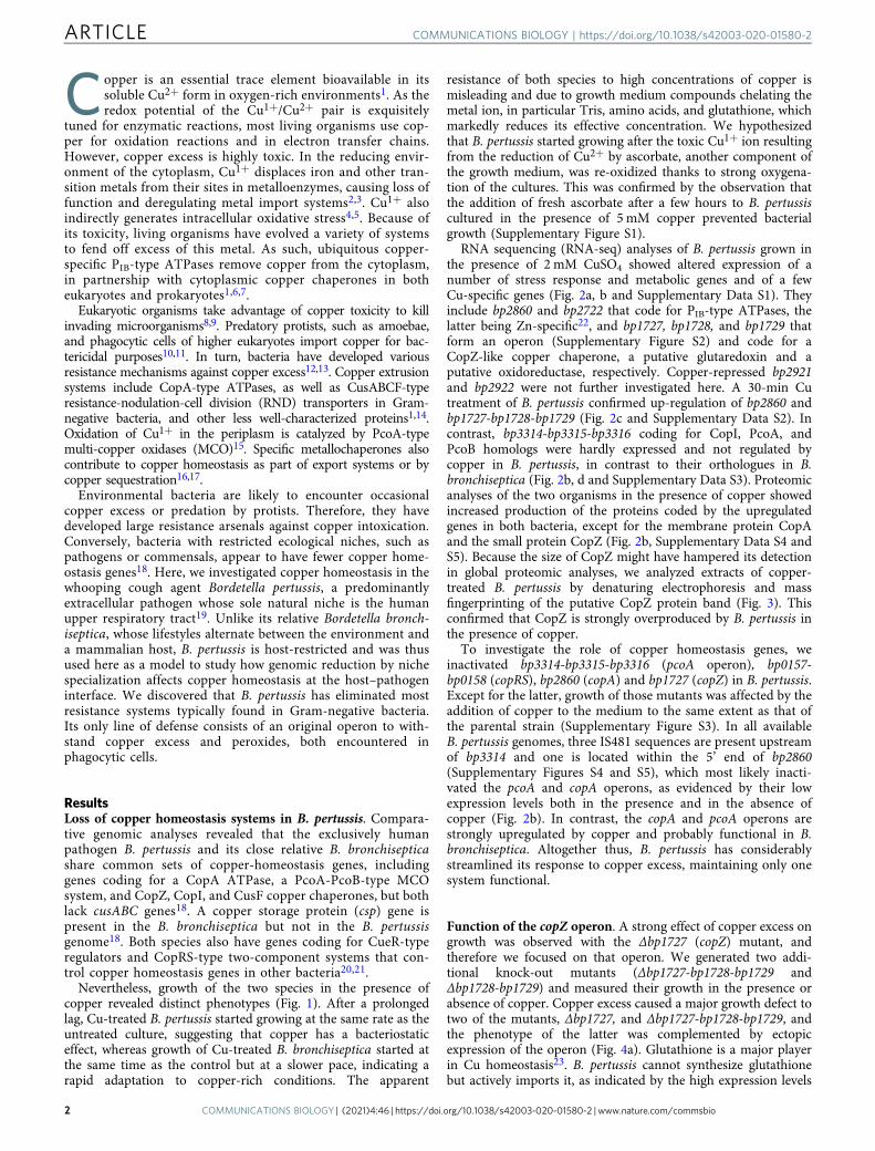
Nevertheless, growth of the two species in the presence ofcopper revealed distinct phenotypes (Fig. 1). After a prolongedlag, Cu-treated B. pertussis started growing at the same rate as theuntreated culture, suggesting that copper has a bacteriostaticeffect, whereas growth of Cu-treated B. bronchiseptica started atthe same time as the control but at a slower pace, indicating arapid adaptation to copper-rich conditions. The apparent
resistance of both species to high concentrations of copper ismisleading and due to growth medium compounds chelating themetal ion, in particular Tris, amino acids, and glutathione, whichmarkedly reduces its effective concentration. We hypothesizedthat B. pertussis started growing after the toxic Cu1+ ion resultingfrom the reduction of Cu2+ by ascorbate, another component ofthe growth medium, was re-oxidized thanks to strong oxygena-tion of the cultures. This was confirmed by the observation thatthe addition of fresh ascorbate after a few hours to B. pertussiscultured in the presence of 5 mM copper prevented bacterialgrowth (Supplementary Figure S1).
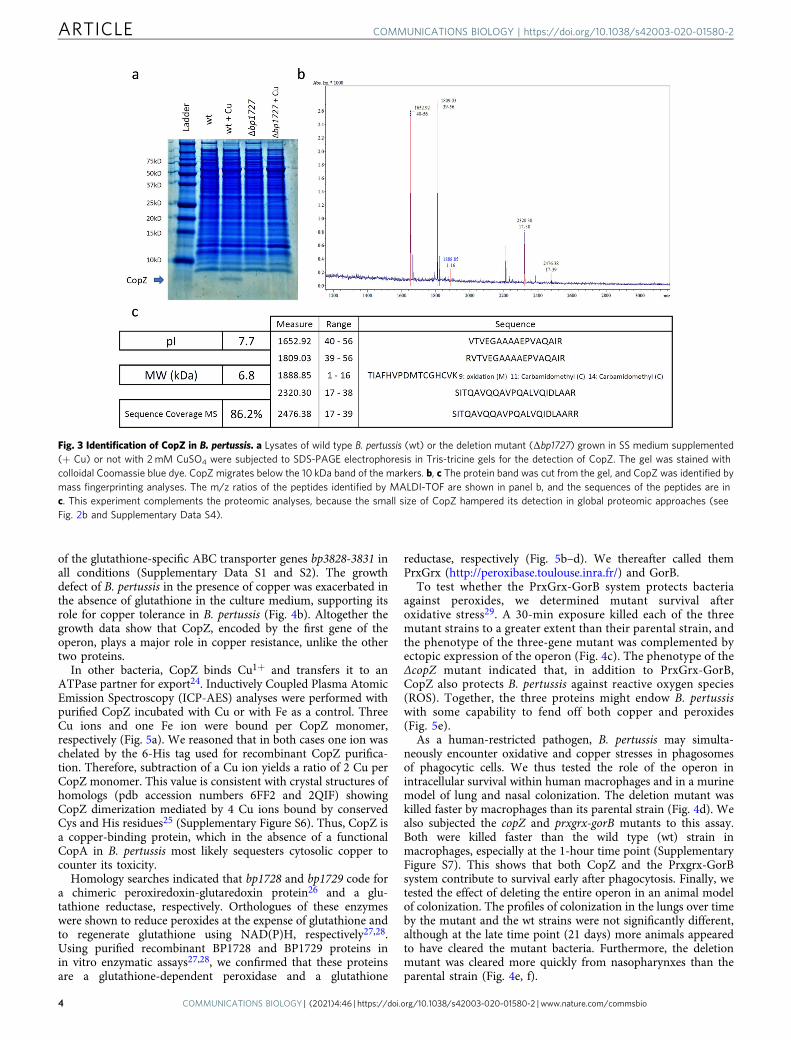
RNA sequencing (RNA-seq) analyses of B. pertussis grown inthe presence of 2 mM CuSO4 showed altered expression of anumber of stress response and metabolic genes and of a fewCu-specific genes (Fig. 2a, b and Supplementary Data S1). Theyinclude bp2860 and bp2722 that code for PIB-type ATPases, thelatter being Zn-specific22, and bp1727, bp1728, and bp1729 thatform an operon (Supplementary Figure S2) and code for aCopZ-like copper chaperone, a putative glutaredoxin and aputative oxidoreductase, respectively. Copper-repressed bp2921and bp2922 were not further investigated here. A 30-min Cutreatment of B. pertussis confirmed up-regulation of bp2860 andbp1727-bp1728-bp1729 (Fig. 2c and Supplementary Data S2). Incontrast, bp3314-bp3315-bp3316 coding for CopI, PcoA, andPcoB homologs were hardly expressed and not regulated bycopper in B. pertussis, in contrast to their orthologues in B.bronchiseptica (Fig. 2b, d and Supplementary Data S3). Proteomicanalyses of the two organisms in the presence of copper showedincreased production of the proteins coded by the upregulatedgenes in both bacteria, except for the membrane protein CopAand the small protein CopZ (Fig. 2b, Supplementary Data S4 andS5). Because the size of CopZ might have hampered its detectionin global proteomic analyses, we analyzed extracts of copper-treated B. pertussis by denaturing electrophoresis and massfingerprinting of the putative CopZ protein band (Fig. 3). Thisconfirmed that CopZ is strongly overproduced by B. pertussis inthe presence of copper.
To investigate the role of copper homeostasis genes, weinactivated bp3314-bp3315-bp3316 (pcoA operon), bp0157-bp0158 (copRS), bp2860 (copA) and bp1727 (copZ) in B. pertussis.Except for the latter, growth of those mutants was affected by theaddition of copper to the medium to the same extent as that ofthe parental strain (Supplementary Figure S3). In all availableB. pertussis genomes, three IS481 sequences are present upstreamof bp3314 and one is located within the 5’ end of bp2860(Supplementary Figures S4 and S5), which most likely inacti-vated the pcoA and copA operons, as evidenced by their lowexpression levels both in the presence and in the absence ofcopper (Fig. 2b). In contrast, the copA and pcoA operons arestrongly upregulated by copper and probably functional in B.bronchiseptica. Altogether thus, B. pertussis has considerablystreamlined its response to copper excess, maintaining only onesystem functional.
Function of the copZ operon. A strong effect of copper excess ongrowth was observed with the Δbp1727 (copZ) mutant, andtherefore we focused on that operon. We generated two addi-tional knock-out mutants (Δbp1727-bp1728-bp1729 andΔbp1728-bp1729) and measured their growth in the presence orabsence of copper. Copper excess caused a major growth defect totwo of the mutants, Δbp1727, and Δbp1727-bp1728-bp1729, andthe phenotype of the latter was complemented by ectopicexpression of the operon (Fig. 4a). Glutathione is a major playerin Cu homeostasis23. B. pertussis cannot synthesize glutathionebut actively imports it, as indicated by the high expression levels
ARTICLE COMMUNICATIONS BIOLOGY | https://doi.org/10.1038/s42003-020-01580-2
2 COMMUNICATIONS BIOLOGY | (2021) 4:46 | https://doi.org/10.1038/s42003-020-01580-2 | www.nature.com/commsbio
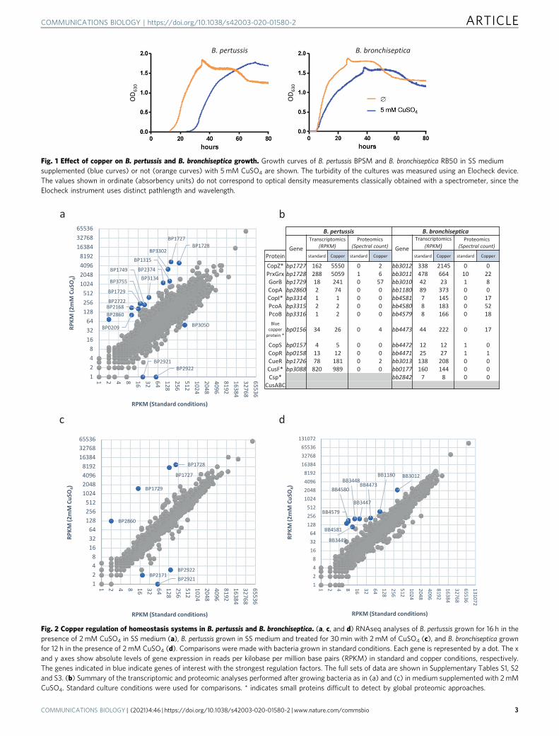
Fig. 1 Effect of copper on B. pertussis and B. bronchiseptica growth. Growth curves of B. pertussis BPSM and B. bronchiseptica RB50 in SS mediumsupplemented (blue curves) or not (orange curves) with 5 mM CuSO4 are shown. The turbidity of the cultures was measured using an Elocheck device.The values shown in ordinate (absorbency units) do not correspond to optical density measurements classically obtained with a spectrometer, since theElocheck instrument uses distinct pathlength and wavelength.
Fig. 2 Copper regulation of homeostasis systems in B. pertussis and B. bronchiseptica. (a, c, and d) RNAseq analyses of B. pertussis grown for 16 h in thepresence of 2 mM CuSO4 in SS medium (a), B. pertussis grown in SS medium and treated for 30min with 2mM of CuSO4 (c), and B. bronchiseptica grownfor 12 h in the presence of 2 mM CuSO4 (d). Comparisons were made with bacteria grown in standard conditions. Each gene is represented by a dot. The xand y axes show absolute levels of gene expression in reads per kilobase per million base pairs (RPKM) in standard and copper conditions, respectively.The genes indicated in blue indicate genes of interest with the strongest regulation factors. The full sets of data are shown in Supplementary Tables S1, S2and S3. (b) Summary of the transcriptomic and proteomic analyses performed after growing bacteria as in (a) and (c) in medium supplemented with 2mMCuSO4. Standard culture conditions were used for comparisons. * indicates small proteins difficult to detect by global proteomic approaches.
COMMUNICATIONS BIOLOGY | https://doi.org/10.1038/s42003-020-01580-2 ARTICLE
COMMUNICATIONS BIOLOGY | (2021) 4:46 | https://doi.org/10.1038/s42003-020-01580-2 | www.nature.com/commsbio 3
of the glutathione-specific ABC transporter genes bp3828-3831 inall conditions (Supplementary Data S1 and S2). The growthdefect of B. pertussis in the presence of copper was exacerbated inthe absence of glutathione in the culture medium, supporting itsrole for copper tolerance in B. pertussis (Fig. 4b). Altogether thegrowth data show that CopZ, encoded by the first gene of theoperon, plays a major role in copper resistance, unlike the othertwo proteins.
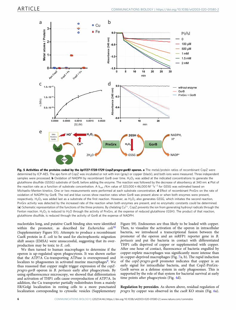
In other bacteria, CopZ binds Cu1+ and transfers it to anATPase partner for export24. Inductively Coupled Plasma AtomicEmission Spectroscopy (ICP-AES) analyses were performed withpurified CopZ incubated with Cu or with Fe as a control. ThreeCu ions and one Fe ion were bound per CopZ monomer,respectively (Fig. 5a). We reasoned that in both cases one ion waschelated by the 6-His tag used for recombinant CopZ purifica-tion. Therefore, subtraction of a Cu ion yields a ratio of 2 Cu perCopZ monomer. This value is consistent with crystal structures ofhomologs (pdb accession numbers 6FF2 and 2QIF) showingCopZ dimerization mediated by 4 Cu ions bound by conservedCys and His residues25 (Supplementary Figure S6). Thus, CopZ isa copper-binding protein, which in the absence of a functionalCopA in B. pertussis most likely sequesters cytosolic copper tocounter its toxicity.
Homology searches indicated that bp1728 and bp1729 code fora chimeric peroxiredoxin-glutaredoxin protein26 and a glu-tathione reductase, respectively. Orthologues of these enzymeswere shown to reduce peroxides at the expense of glutathione andto regenerate glutathione using NAD(P)H, respectively27,28.Using purified recombinant BP1728 and BP1729 proteins inin vitro enzymatic assays27,28, we confirmed that these proteinsare a glutathione-dependent peroxidase and a glutathione
reductase, respectively (Fig. 5b–d). We thereafter called themPrxGrx (http://peroxibase.toulouse.inra.fr/) and GorB.
To test whether the PrxGrx-GorB system protects bacteriaagainst peroxides, we determined mutant survival afteroxidative stress29. A 30-min exposure killed each of the threemutant strains to a greater extent than their parental strain, andthe phenotype of the three-gene mutant was complemented byectopic expression of the operon (Fig. 4c). The phenotype of theΔcopZ mutant indicated that, in addition to PrxGrx-GorB,CopZ also protects B. pertussis against reactive oxygen species(ROS). Together, the three proteins might endow B. pertussiswith some capability to fend off both copper and peroxides(Fig. 5e).
As a human-restricted pathogen, B. pertussis may simulta-neously encounter oxidative and copper stresses in phagosomesof phagocytic cells. We thus tested the role of the operon inintracellular survival within human macrophages and in a murinemodel of lung and nasal colonization. The deletion mutant waskilled faster by macrophages than its parental strain (Fig. 4d). Wealso subjected the copZ and prxgrx-gorB mutants to this assay.Both were killed faster than the wild type (wt) strain inmacrophages, especially at the 1-hour time point (SupplementaryFigure S7). This shows that both CopZ and the Prxgrx-GorBsystem contribute to survival early after phagocytosis. Finally, wetested the effect of deleting the entire operon in an animal modelof colonization. The profiles of colonization in the lungs over timeby the mutant and the wt strains were not significantly different,although at the late time point (21 days) more animals appearedto have cleared the mutant bacteria. Furthermore, the deletionmutant was cleared more quickly from nasopharynxes than theparental strain (Fig. 4e, f).
Fig. 3 Identification of CopZ in B. pertussis. a Lysates of wild type B. pertussis (wt) or the deletion mutant (Δbp1727) grown in SS medium supplemented(+ Cu) or not with 2 mM CuSO4 were subjected to SDS-PAGE electrophoresis in Tris-tricine gels for the detection of CopZ. The gel was stained withcolloidal Coomassie blue dye. CopZ migrates below the 10 kDa band of the markers. b, c The protein band was cut from the gel, and CopZ was identified bymass fingerprinting analyses. The m/z ratios of the peptides identified by MALDI-TOF are shown in panel b, and the sequences of the peptides are inc. This experiment complements the proteomic analyses, because the small size of CopZ hampered its detection in global proteomic approaches (seeFig. 2b and Supplementary Data S4).
ARTICLE COMMUNICATIONS BIOLOGY | https://doi.org/10.1038/s42003-020-01580-2
4 COMMUNICATIONS BIOLOGY | (2021) 4:46 | https://doi.org/10.1038/s42003-020-01580-2 | www.nature.com/commsbio
Copper regulation of the copZ-prxgrx-gorB operon. Quantita-tive RT-PCR (qRT-PCR) on prxgrx showed a 30-fold up-reg-ulation of the operon by copper, even at micromolarconcentrations (Fig. 6a and Supplementary Figure S8). qRT-PCRexperiments on the very small copZ gene were unsuccessful, as noqRT-PCR primer pairs were found to work. Inactivation ofbp1726, in antisense orientation of the copZ-prxgrx-gorB operonand coding for a putative CueR regulator20, resulted in a mark-edly decreased but not abolished transcriptional response ofprxgrx to copper (Fig. 6a). The three genes are co-transcribed
(Supplementary Figure S2) and up-regulated by copper (Fig. 2and Supplementary Data S1 and S2), and we showed that thesecond is controlled by CueR. We can thus reasonably assumethat the entire operon is under the control of CueR. Interestingly,though, the remaining copper-induced up-regulation of prxgrx inthe cueR KO mutant indicates that the operon may also becontrolled by a second system directly or indirectly activated bycopper.
We determined the transcriptional start site of the operon by 5′rapid amplification of cDNA ends (5′ RACE). The 5′ UTR is 61-
Fig. 4 Role of the operon in B. pertussis and in host–pathogen interactions. a, b Growth yields of B. pertussis after 24 h in SS medium (a) or in SS mediumdevoid of glutathione (b), supplemented or not with 2 mM CuSO4. The cultures were inoculated at initial OD600 values of 0.1, and after 24 h the OD600
was determined. Note the different y axis scales between (a) and (b). wt, wild type (parental) strain; Δ27, Δ28-29 and Δ27-29, KO mutants for bp1727(copZ), bp1728-bp1729 (prxgrx-gorB) and the three genes, respectively. The various strains are represented using different symbols and shades of gray.Δ27-29/+27-29 represents the latter mutant complemented by expression of the operon at another chromosomal locus. The horizontal orange linesrepresent mean values. c Survival of the same strains to an oxidative shock of 30min. d Intracellular survival of B. pertussis in THP1 macrophages.The horizontal orange lines represent mean values. See Supplementary Figure S6 for the data of the individual copZ and prxgrx-gorB KO mutants.e, f Colonization of mice lungs (e) and nasopharynxes (f) after nasal infections with the parental strain and the copZ-prxgrx-gorB KO mutant. The numbersof bacteria are indicated for each mouse and organ (black circles: parental bacteria, red squares: mutant). The lines connect the geometric means of thecounts at each time point, and the dotted lines indicate the thresholds of detection. For all the assays, statistical analyses were performed using two-tailedMann-Whitney tests (*, p < 0.05; ** p, <0.005). For panels a and b, 5 biologically independent samples were used. For panels c and d, 4 and 6 biologicalsamples were used, respectively. For panels e and f, 5 and 4 animals per time point were used for the KO and wt strains, respectively.
COMMUNICATIONS BIOLOGY | https://doi.org/10.1038/s42003-020-01580-2 ARTICLE
COMMUNICATIONS BIOLOGY | (2021) 4:46 | https://doi.org/10.1038/s42003-020-01580-2 | www.nature.com/commsbio 5
nucleotides long, and putative CueR binding sites were identifiedwithin the promoter, as described for Escherichia coli30
(Supplementary Figure S5). Attempts to produce a recombinantCueR protein in E. coli to be used for electrophoretic migrationshift assays (EMSA) were unsuccessful, suggesting that its over-production may be toxic to E. coli.
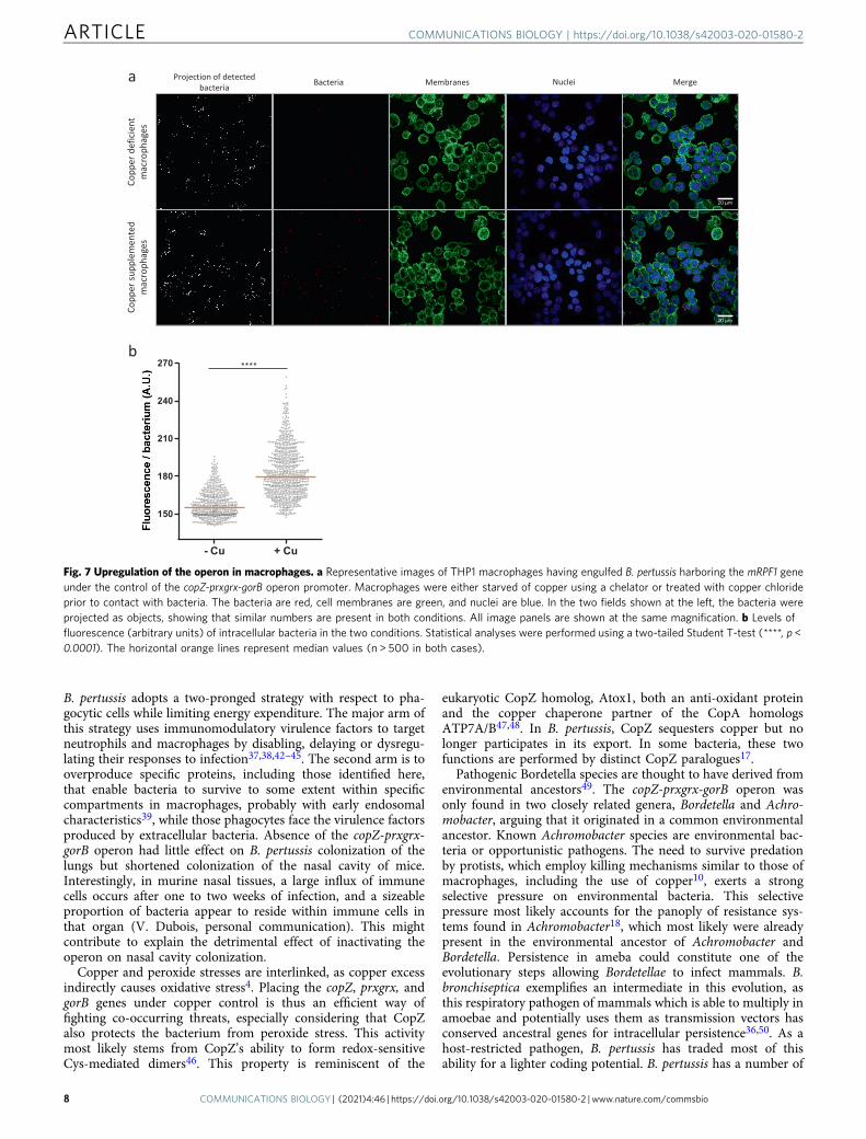
We then turned to human macrophages to determine if theoperon is up-regulated upon phagocytosis. It was shown earlierthat the ATP7A Cu-transporting ATPase is overexpressed andlocalizes to phagosomes in activated murine macrophages9. Wethus reasoned that copper might trigger expression of the copZ-prxgrx-gorB operon in B. pertussis early after phagocytosis. Byusing epifluorescence microscopy, we showed that differentiationand activation of THP1 cells cause overproduction of ATP7A. Inaddition, the Cu transporter partially redistributes from a mainlyER/Golgi localization in resting cells to a more punctuatedlocalization corresponding to cytosolic vesicles (Supplementary
Figure S9). Endosomes are thus likely to be loaded with copper.Then, to visualize the activation of the operon in intracellularbacteria, we introduced a transcriptional fusion between thepromoter of the operon and an mRFP1 reporter gene in B.pertussis and put the bacteria in contact with differentiatedTHP1 cells deprived of copper or supplemented with copper.After one hour of contact, fluorescence of bacteria engulfed bycopper-replete macrophages was significantly more intense thanin copper-deprived macrophages (Fig. 7a, b). The rapid inductionof the copZ-prxgrx-gorB promoter indicates that copper is anearly signal for intracellular bacteria, and that CopZ-PrxGrx-GorB serves as a defense system in early phagosomes. This issupported by the role of that system for bacterial survival at earlytime points after phagocytosis (Fig. 4d).
Regulation by peroxides. As shown above, residual regulation ofprxgrx by copper was observed in the cueR KO strain (Fig. 6a).
Fig. 5 Activities of the proteins coded by the bp1727-1728-1729 (copZ-prxgrx-gorB) operon. a The metal/protein ratios of recombinant CopZ weredetermined by ICP-AES. The apo form of CopZ was incubated or not with iron (gray) or copper (black), and both ions were measured. Three independentsamples were processed. b Oxidation of NADPH by recombinant GorB over time. H2O2 was added at the indicated concentrations to generate theglutathione disulfide (GSSG) substrate of GorB, before adding the enzyme. The reaction was followed by the decrease of absorbency at 340 nm. c Plot ofthe reaction rate as a function of substrate concentration. A kcat /Km value of 323,000 ± 46,000M−1s−1 for GSSG was estimated based onMichaelis–Menten kinetics. One or two measurements were performed at each substrate concentration. d Effect of recombinant PrxGrx on the rate ofoxidation of NADPH by GorB. The red and blue curves show reaction rates when GorB was present alone or when both enzymes were present,respectively. H2O2 was added last as a substrate of the first reaction. However, as H2O2 also generates GSSG, which initiates the second reaction,PrxGrx activity was detected by the increased rate of the reaction when both enzymes are present, and no enzymatic constants could be determined.(e) Schematic representation of the functions of the three proteins. By chelating Cu1+, CopZ prevents the ion from generating hydroxyl radicals through theFenton reaction. H2O2 is reduced to H2O through the activity of PrxGrx, at the expense of reduced glutathione (GSH). The product of that reaction,glutathione disulfide, is reduced through the activity of GorB at the expense of NADPH.
ARTICLE COMMUNICATIONS BIOLOGY | https://doi.org/10.1038/s42003-020-01580-2
6 COMMUNICATIONS BIOLOGY | (2021) 4:46 | https://doi.org/10.1038/s42003-020-01580-2 | www.nature.com/commsbio
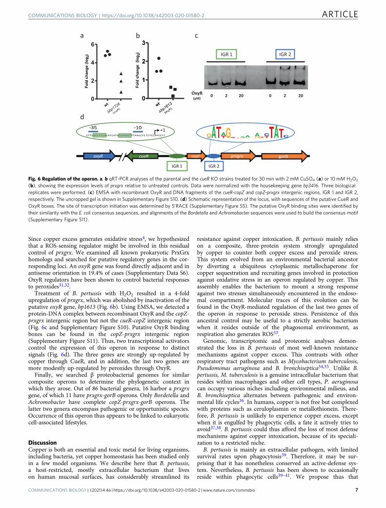
Since copper excess generates oxidative stress4, we hypothesizedthat a ROS-sensing regulator might be involved in this residualcontrol of prxgrx. We examined all known prokaryotic PrxGrxhomologs and searched for putative regulatory genes in the cor-responding loci. An oxyR gene was found directly adjacent and inantisense orientation in 19.4% of cases (Supplementary Data S6).OxyR regulators have been shown to control bacterial responsesto peroxides31,32.
Treatment of B. pertussis with H2O2 resulted in a 4-foldupregulation of prxgrx, which was abolished by inactivation of theputative oxyR gene, bp1613 (Fig. 6b). Using EMSA, we detected aprotein-DNA complex between recombinant OxyR and the copZ-prxgrx intergenic region but not the cueR-copZ intergenic region(Fig. 6c and Supplementary Figure S10). Putative OxyR bindingboxes can be found in the copZ-prxgrx intergenic region(Supplementary Figure S11). Thus, two transcriptional activatorscontrol the expression of this operon in response to distinctsignals (Fig. 6d). The three genes are strongly up-regulated bycopper through CueR, and in addition, the last two genes aremore modestly up-regulated by peroxides through OxyR.
Finally, we searched β proteobacterial genomes for similarcomposite operons to determine the phylogenetic context inwhich they arose. Out of 86 bacterial genera, 16 harbor a prxgrxgene, of which 11 have prxgrx-gorB operons. Only Bordetella andAchromobacter have complete copZ-prxgrx-gorB operons. Thelatter two genera encompass pathogenic or opportunistic species.Occurrence of this operon thus appears to be linked to eukaryoticcell-associated lifestyles.
DiscussionCopper is both an essential and toxic metal for living organisms,including bacteria, yet copper homeostasis has been studied onlyin a few model organisms. We describe here that B. pertussis,a host-restricted, mostly extracellular bacterium that liveson human mucosal surfaces, has considerably streamlined its
resistance against copper intoxication. B. pertussis mainly relieson a composite, three-protein system strongly upregulatedby copper to counter both copper excess and peroxide stress.This system evolved from an environmental bacterial ancestorby diverting a ubiquitous cytoplasmic metallochaperone forcopper sequestration and recruiting genes involved in protectionagainst oxidative stress in an operon regulated by copper. Thisassembly enables the bacterium to mount a strong responseagainst two stresses simultaneously encountered in the endoso-mal compartment. Molecular traces of this evolution can befound in the OxyR-mediated regulation of the last two genes ofthe operon in response to peroxide stress. Persistence of thisancestral control may be useful to a strictly aerobic bacteriumwhen it resides outside of the phagosomal environment, asrespiration also generates ROS33.
Genomic, transcriptomic and proteomic analyses demon-strated the loss in B. pertussis of most well-known resistancemechanisms against copper excess. This contrasts with otherrespiratory tract pathogens such as Mycobacterium tuberculosis,Pseudomonas aeruginosa and B. bronchiseptica34,35. Unlike B.pertussis, M. tuberculosis is a genuine intracellular bacterium thatresides within macrophages and other cell types, P. aeruginosacan occupy various niches including environmental milieus, andB. bronchiseptica alternates between pathogenic and environ-mental life cycles36. In humans, copper is not free but complexedwith proteins such as ceruloplasmin or metallothionein. There-fore, B. pertussis is unlikely to experience copper excess, exceptwhen it is engulfed by phagocytic cells, a fate it actively tries toavoid37,38. B. pertussis could thus afford the loss of most defensemechanisms against copper intoxication, because of its speciali-zation to a restricted niche.
B. pertussis is mainly an extracellular pathogen, with limitedsurvival rates upon phagocytosis39. Therefore, it may be sur-prising that it has nonetheless conserved an active-defense sys-tem. Nevertheless, B. pertussis has been shown to occasionallyreside within phagocytic cells39–41. We propose thus that
Fig. 6 Regulation of the operon. a, b qRT-PCR analyses of the parental and the cueR KO strains treated for 30min with 2mM CuSO4 (a) or 10mM H2O2
(b), showing the expression levels of prxgrx relative to untreated controls. Data were normalized with the housekeeping gene bp3416. Three biologicalreplicates were performed. (c) EMSA with recombinant OxyR and DNA fragments of the cueR-copZ and copZ-prxgrx intergenic regions, IGR 1 and IGR 2,respectively. The uncropped gel is shown in Supplementary Figure S10. (d) Schematic representation of the locus, with sequences of the putative CueR andOxyR boxes. The site of transcription initiation was determined by 5’RACE (Supplementary Figure S5). The putative OxyR binding sites were identified bytheir similarity with the E. coli consensus sequences, and alignments of the Bordetella and Achromobacter sequences were used to build the consensus motif(Supplementary Figure S11).
COMMUNICATIONS BIOLOGY | https://doi.org/10.1038/s42003-020-01580-2 ARTICLE
COMMUNICATIONS BIOLOGY | (2021) 4:46 | https://doi.org/10.1038/s42003-020-01580-2 | www.nature.com/commsbio 7
B. pertussis adopts a two-pronged strategy with respect to pha-gocytic cells while limiting energy expenditure. The major arm ofthis strategy uses immunomodulatory virulence factors to targetneutrophils and macrophages by disabling, delaying or dysregu-lating their responses to infection37,38,42–45. The second arm is tooverproduce specific proteins, including those identified here,that enable bacteria to survive to some extent within specificcompartments in macrophages, probably with early endosomalcharacteristics39, while those phagocytes face the virulence factorsproduced by extracellular bacteria. Absence of the copZ-prxgrx-gorB operon had little effect on B. pertussis colonization of thelungs but shortened colonization of the nasal cavity of mice.Interestingly, in murine nasal tissues, a large influx of immunecells occurs after one to two weeks of infection, and a sizeableproportion of bacteria appear to reside within immune cells inthat organ (V. Dubois, personal communication). This mightcontribute to explain the detrimental effect of inactivating theoperon on nasal cavity colonization.
Copper and peroxide stresses are interlinked, as copper excessindirectly causes oxidative stress4. Placing the copZ, prxgrx, andgorB genes under copper control is thus an efficient way offighting co-occurring threats, especially considering that CopZalso protects the bacterium from peroxide stress. This activitymost likely stems from CopZ’s ability to form redox-sensitiveCys-mediated dimers46. This property is reminiscent of the
eukaryotic CopZ homolog, Atox1, both an anti-oxidant proteinand the copper chaperone partner of the CopA homologsATP7A/B47,48. In B. pertussis, CopZ sequesters copper but nolonger participates in its export. In some bacteria, these twofunctions are performed by distinct CopZ paralogues17.
Pathogenic Bordetella species are thought to have derived fromenvironmental ancestors49. The copZ-prxgrx-gorB operon wasonly found in two closely related genera, Bordetella and Achro-mobacter, arguing that it originated in a common environmentalancestor. Known Achromobacter species are environmental bac-teria or opportunistic pathogens. The need to survive predationby protists, which employ killing mechanisms similar to those ofmacrophages, including the use of copper10, exerts a strongselective pressure on environmental bacteria. This selectivepressure most likely accounts for the panoply of resistance sys-tems found in Achromobacter18, which most likely were alreadypresent in the environmental ancestor of Achromobacter andBordetella. Persistence in ameba could constitute one of theevolutionary steps allowing Bordetellae to infect mammals. B.bronchiseptica exemplifies an intermediate in this evolution, asthis respiratory pathogen of mammals which is able to multiply inamoebae and potentially uses them as transmission vectors hasconserved ancestral genes for intracellular persistence36,50. As ahost-restricted pathogen, B. pertussis has traded most of thisability for a lighter coding potential. B. pertussis has a number of
Fig. 7 Upregulation of the operon in macrophages. a Representative images of THP1 macrophages having engulfed B. pertussis harboring the mRPF1 geneunder the control of the copZ-prxgrx-gorB operon promoter. Macrophages were either starved of copper using a chelator or treated with copper chlorideprior to contact with bacteria. The bacteria are red, cell membranes are green, and nuclei are blue. In the two fields shown at the left, the bacteria wereprojected as objects, showing that similar numbers are present in both conditions. All image panels are shown at the same magnification. b Levels offluorescence (arbitrary units) of intracellular bacteria in the two conditions. Statistical analyses were performed using a two-tailed Student T-test (****, p <0.0001). The horizontal orange lines represent median values (n > 500 in both cases).
ARTICLE COMMUNICATIONS BIOLOGY | https://doi.org/10.1038/s42003-020-01580-2
8 COMMUNICATIONS BIOLOGY | (2021) 4:46 | https://doi.org/10.1038/s42003-020-01580-2 | www.nature.com/commsbio
cuproproteins18 and therefore requires copper for growth. Itmight thus also use CopZ for nutritional passivation51, i.e., as acopper store for future needs.
MethodsCulture conditions. Modified Stainer-Scholte (SS) liquid medium and Bordet-Gengou blood-agar (BGA) medium were used with the appropriate antibiotics togrow B. pertussis BPSM and its derivatives. As copper ions are Lewis acids and B.pertussis requires buffered medium close to pH 7, a stock solution (62 mM ofCuSO4, 100 mM Tris, pH 5.5) in the fraction A of SS medium was used to sup-plement the medium with copper. The control cultures were prepared by addingthe same volumes of 100 mM Tris-HCl (pH 5.5) to SS medium. To record growthcurves, the optical density at 630 nm was continuously measured using an Elocheckdevice (Biotronix). For growth yields, the B. pertussis cultures were started at OD600
of 0.1, and after 24 h of growth optical density values at 600 nm (OD600) weredetermined using an Ultrospec 10 spectrophotometer (Biochrom) with five bio-logical replicates for each condition and strain. Glutathione was omitted from theculture medium where indicated. The turbidities measured using the Elocheck andUltrospec apparatuses, which use different wavelengths and pathlengths, cannot bedirectly compared. For transcriptomic, proteomic and qRT-PCR experiments, theB. pertussis cultures were started at OD600 of 0.1 to reach OD600 values of 1.5 after16–20 h, and B. bronchiseptica cultures were started at OD600 of 0.08 to reachOD600 values of 1.5–1.8 after 10–12 h. To subject B. pertussis to oxidative stress forqRT-PCR analysis, the bacteria were grown to OD600 of 1.5, and 10 mM H2O2 wasadded to the cultures for 30 min. To measure survival to oxidative shock, thebacteria were grown for 36 h on BGA from standardized stocks, resuspended inPBS (pH 7.2) containing 500 μM of hypoxanthine, and then diluted in the samesolution to obtain ~ 50,000 bacteria in 300 µl in 15-ml tubes. 0.05 U.ml−1 xanthineoxidase was added to produce H2O2 and O2
.− 29, and the bacteria were incubatedfor 30 min at 37 °C with shaking. Serial dilutions were plated onto BGA to countCFUs, using control suspensions incubated as above. Three biological replicatesand three technical replicates were made. CFU counts of the initial suspensionswere used as references.
Construction of strains and plasmids. Deletion mutants were obtained by allelicexchange52. Complementation was performed at the ure chromosomal locus52. Theinactivation mutants were constructed using the suicide vector pFUS253. ForpProm1727-mRFP1, the cueR-copZ intergenic region including the first 7 codonsof copZ, and the mRFP1 gene without its initiation codon were amplified by PCRand cloned as BamHI-XbaI and XbaI-HindIII restriction fragments, respectively, togenerate a translational fusion in pBBR1-MCS554. All primers are listed in Sup-plementary Data S7.
RNA sequencing and qRT-PCR. For RNA extractions, the cultures were stoppedby adding 2 mL of a mixture of phenol:ethanol (5:95) to 8 mL of bacterial sus-pensions. Bacteria were pelleted, and total RNA was extracted with TriReagent(InVitrogen)55. Genomic DNA was removed by a DNAse I treatment (SigmaAldrich). DNA-depleted total RNA was treated with the RiboZero rRNA Removalkit (Illumina). The rRNA-depleted RNA was then used to generate the Illuminalibraries using the TrueSeq RNA library, followed with sequencing on an IlluminaNextSeq 500 benchtop sequencer on SR150 high output run mode. The RNA-seqdata were analyzed using Rockhopper v2.0.3 with the default parameters to cal-culate the reads per kilobase per million base pairs (RPKM) values for each codingsequence using the B. pertussis Tohama I BX470248 genome annotation. All RNA-seq experiments were performed on biological duplicates. The results fully sup-ported preliminary microarray analyses for B. pertussis. qRT-PCR were performedon 3 or 4 separate cultures with at least 3 technical replicates for each condition.To map the transcriptional initiation site by rapid amplification of cDNA 5’ ends(RACE), total RNA obtained as described above was treated with the GeneRacer kit(Invitrogen) using appropriate RACE primers (Supplementary Data S7).
Mass spectrometry proteomic analyses. B. bronchiseptica and B. pertussis weregrown in SS medium supplemented or not with 2 mM CuSO4 for 10 h and 16 h,respectively, and the cultures were stopped at OD600 of 1.6–1.8. The bacteria werecollected by centrifugation, resuspended in 50 mM Tris-HCl (pH 7.2), 100 mMNaCl, with Complete protease inhibitor (Roche) and lysed with a French Press.Clarified lysates were ultracentrifuged for 1 h at 100,000 g at 4 °C to separatesoluble and insoluble fractions. Both fractions were heated at 100 °C in 5% SDS, 5%β-mercaptoethanol, 1 mM EDTA, 10% glycerol, 10 mM Tris (pH 8) for 3 min andloaded on a 10% acrylamide SDS-PAGE gel. The migration was stopped soon afterthe samples entered the separating gel. The gel was briefly stained with CoomassieBlue, and one gel slice was taken for each sample. The gel plugs were washed twicein 25 mM NH4HCO3 (50 μL) and acetonitrile (50 μL), the Cys residues werereduced and alkykated by adding 50 μL dithiothreitol (10 mM solution) andincubating at 57 °C before the addition of 50 μL iodoacetamide (55 mM solution).The plugs were dehydrated with acetonitrile, and the proteins were digestedovernight at room temperature using porcine trypsin (12.5 ng/50 μL) in 25 mMNH4HCO3. Tryptic peptides were extracted first with 60% acetonitrile and 5%
formic acid for 1 h, and then with 100% acetonitrile until dehydration of the gelplugs. The extracts were pooled and excess acetonitrile was evaporated beforeanalysis. Peptide fractionation was performed using a 500-mm reversed-phasecolumn at 55 °C and with a gradient separation of 170 min. Eluted peptides wereanalyzed using Q-Extractive instruments (Fisher Scientific)56. Raw data collectedduring nanoLC-MS/MS analyses were processed and converted into *.mgf peak listformat with Proteome Discoverer 1.4 (Thermo Fisher Scientific). MS/MS data wereinterpreted using search engine Mascot (version 2.4.0, Matrix Science, London,UK) installed on a local server. Searches were performed with a tolerance on massmeasurement of 10 ppm for precursor ions and 0.02 Da for fragment ions, againsttwo target decoy databases composed of all potential open reading frames of at least30 residues between stop codons of B. pertussis or B. bronchiseptica (54890 and74418 entries, respectively), plus sequences of recombinant trypsin and classicalcontaminants (118 entries). Cys carbamidomethylation or propionamidation, Metoxidation, and protein N-terminal acetylation were searched as variable mod-ifications. Up to one trypsin missed cleavage was allowed. Peptides were filtered outwith Proline 2.0 according to the cutoff set for proteins hits with 1 or more peptideslarger than 9 residues, ion score > 10 and 2% protein false positive rates56. Spectralcounting analyses were performed with Proline 2.0. The p-values were calculatedwith a Student’s T-test (confidence level: 95%). To obtain q-values, the p-valueswere adjusted for multiple testing correction to control the false discovery rate byfollowing the Benjamini-Hochberg procedure (Q= 5%)57.
To identify CopZ, B. pertussis extracts were separated by electrophoresis usingcommercial Tricine 16% gels (InVitrogen) and stained with colloidal Coomassieblue dye. The fast-migrating protein band was digested in the gel with trypsin, andthe peptide fingerprints were obtained by matrix assisted laser desorption/ionization time-of-flight mass spectrometry.
Protein production. For the production of CopZ, PrxGrx and GorB, recombinantpQE30 derivatives (Qiagen) were introduced into E. coli M15(pREp4). The cellswere grown in LB medium at 37 °C under orbital shaking, and expression wasinduced at OD600 of 0.8 with 1 mM IPTG for 3 h. Production of recombinant OxyRwas done in E. coli SG13009(pREp4) in M9 medium with 2% casaminoacids and250 μM ascorbate. At OD600 of 0.4, 100 μM IPTG was added, and the culture wascontinued for 16 h at 14 °C. The bacteria were collected, and the pellets wereresuspended in 50 mM Tris-HCl (pH 7.5), 100 mM NaCl, 10 mM imidazole, plus1 mM TCEP for CopZ and OxyR. The bacteria were lysed with a French press, andthe lysates were clarified by centrifugation. The proteins were purified by chro-matography on Ni++ columns. Concentrations were measured using the BCAassay kit (ThermoFisher Scientific) for PrxGrx and GorB, and with the Qbit proteinassay kit for CopZ and OxyR (ThermoFisher Scientific).
Metal binding to CopZ. Purified CopZ (30 μM) was incubated in 50 mM Tris-HCl(pH 7.5), 100 mM NaCl, 1 mM TCEP, and 100 mM EDTA for 30 min with stirringat room temperature to remove bound metal ions and then dialyzed against thesame buffer without EDTA, using 3.5-kD cutoff membranes. The apo form of theprotein precipitated in the absence of TCEP. Iron sulfate or copper chloridesolutions were prepared in the same buffer containing 1 mM TCEP and 25 mMascorbate. CopZ was mixed with 125 μM of either metal for 5 min at room tem-perature, the samples were then dialyzed against the same buffer and finallyacidified with 6% nitric acid. Elemental Cu and Fe analyses were performed byICP-AES (Agilent, model 5110) in axial mode. The wavelengths selected for Cu andFe analyses were 327.395 nm and 238.204 nm, respectively. 63Cu and 65Cu con-centrations were both analyzed to evidence potential interferences. To removepolyatomic interferences, a 100 mLmin−1 He flow was added at the collisionreaction interface. Impurity of 129Xe in the Ar gas used for generating the plasmawas chosen as internal standard to detect any instrumental drift. External cali-brations were performed from standard solution at 1 g L−1 (Astasol), adequatelydiluted and acidified to be in the range of the unknown concentrations.
Enzymatic assays. Buffer exchange was performed to obtain the purifiedrecombinant proteins in 0.1 M sodium/potassium phosphate (pH 7.6). Enzymaticactivity of GorB was followed at 25 °C in the same buffer containing 0.3 mMNADPH, 8 mM GSH, and H2O2 at concentrations ranging from 100 µM to 2 mM.The latter was used to generate the corresponding concentrations of oxidizedglutathione, before starting the reaction by adding the enzyme at a concentration of0.5 nM27. The decrease of NADPH concentration was followed by measuring theabsorbance of the solution at 340 nm. Activity of PrxGrx was detected in a coupledassay performed at 25 °C in the same buffer with 0.3 mM NADPH, 6 mM GSH,0.25 nM GorB and 1 µM PrxGrx28. 4 mM H2O2 was added last as the substrate ofthe first reaction to generate the substrate of the second reaction, oxidized glu-tathione. However, as H2O2 also directly generates oxidized glutathione, theactivity of PrxGrx was detected by the increased rate at which NADPH was con-sumed when both enzymes were present in the reaction relative to the reaction ratewith GorB only. No enzymatic constants were determined because of the coupledreaction set up.
EMSA. Amplicons of 127 bp and 139 bp, corresponding to the cueR-copZ (IGR 1)and copZ-prxgrx (IGR 2) intergenic regions, respectively, were mixed at 1 µM with
COMMUNICATIONS BIOLOGY | https://doi.org/10.1038/s42003-020-01580-2 ARTICLE
COMMUNICATIONS BIOLOGY | (2021) 4:46 | https://doi.org/10.1038/s42003-020-01580-2 | www.nature.com/commsbio 9
0, 2 or 20 µM recombinant OxyR, in 50 mM Tris-HCl (pH 7.5), 100 mM NaCl,10 mM MgCl2, 1 mM TCEP. After 30 min at 37 °C, the samples were loaded onto a6% acrylamide gel containing 25 mM Tris-HCl (pH 8.8), 200 mM glycine, 10 mMMgCl2. Electrophoresis was performed on ice for 90 min at 25 mA in the samebuffer. DNA was detected by ethidium bromide staining.
Phagocytosis assays. THP1 cells were cultured in RPMI medium with 10% fetalbovine serum (RPMI/FBS). They were then treated with 50 ng/mL phorbol 12-myristate 13-acetate (PMA, Sigma) for 24 h to induce differentiation into macro-phages and washed in PBS before adding fresh medium. The bacteria were grownat 37 °C on BGA for 48 h, resuspended in PBS and incubated with the macrophagesat a multiplicity of infection (MOI) of 100 for 30 min at 37 °C with 5% CO2. Themacrophages were then washed with PBS containing 100 µg/ml polymyxin B toeliminate extracellular bacteria. For the following incubation the concentration ofpolymyxin was lowered to 5 µg/ml in RPMI/FBS, and one and three hours latermacrophages were washed with PBS and lysed with 0.1% saponine. Serial dilutionswere plated onto BGA for CFU counting. The experiments were performed at leastin biological triplicates.
Fluorescence microscopy. To detect intracellular bacteria, the THP1 cellswere cultured as described above. RPMI/FBS was supplemented with 50 µMbathocuproine disulfonate (BCS) or 20 µM CuCl2 during THP1 differentiationand for the following 12 h, and after washing the cells in RPMI, fresh RPMI/FBSwas added 1 h before contact. Bacteria harboring the reporter plasmidpProm1727-mRFP1 were grown for 12–13 h in SS medium containing 50 µMBCS. They were collected by centrifugation, resuspended in PBS to an OD600 of1 and incubated with macrophages at a MOI of 100. After one hour of contact,extracellular bacteria were eliminated, RPMI/FBS supplemented withcopper or BCS was added, and the incubation was continued for one hour at37 °C. Finally, the cells were fixed with Formalin (Sigma) for 15 min at roomtemperature and washed three times with PBS. The cell nuclei and the plasmamembranes were labeled with DAPI at 30 µg/ml (Sigma) and Wheat GermAgglutinin Alexa Fluor ™ 488 Conjugate (Invitrogen) at 5 µg/ml for 10 min atroom temperature. Cells were then washed twice with PBS. Images were cap-tured by confocal microscopy with a spinning disk microscope Nikon/Gatacasystem csu-w1. The images were analyzed with the Fiji software58. All para-meters were set to the same values on all images. The bacteria were identifiedand counted with the 3D Object Counter module, and the fluorescence intensitywas calculated by evaluating the average gray levels of the red objects. Threeimages containing each between 200 and 266 detectable bacteria were analyzedfor each condition.
For ATP7A labeling, THP1 cells were cultured as above. One million cells wereplaced on top of coverslips in each well of a 24-well plate. The cells were thentreated for 24 h with PMA in RPMI/FBS as above, with PMA+ LPS (500 ng/mL),or with PMA for 24 h and then placed in contact with bacteria at a MOI of 100 for2 h. The cells were then centrifuged for 3 min at 300 g, and the supernatant wasremoved. The centrifugation step was necessary to pellet the non-differentiated,non-adherent control cells. Fixation was performed for 45 min at roomtemperature with the eBioscience Fixation buffer (ThermoFisher Scientic). Thecells were then washed three times by centrifugation and addition of eBiosciencePermeabilization buffer (ThermoFisher Scientic). They were then incubated for35 min with 5% goat serum in the permeabilization buffer, followed by anincubation of 2 h with a monoclonal antibody against ATP7A (Invitrogen MA5-27720) diluted 600 folds. The cells were washed three times as above, before addingAlexa Fluor 488-AffiniPure Donkey Anti-Mouse IgG (Jackson: 715-545-151)diluted 1/500 in permeabilization buffer with 5% goat serum also containing DAPIas described above. After 1 h incubation, the cells were washed three times withPBS. Images were obtained using an Evos M5000 microscope with anapochromatic X60 Olympus objective. The final image is a projection of 100 Zstacks with 0.2-µm steps. All images were captured with the same exposureparameters. The fluorescence intensity of the cells was calculated with the Fijisoftware (ImageJ) in two dimensions.
Animal experiments. The bacteria were grown on BGA for 48 h and resus-pended in sterile PBS to 106 bacteria per 20 µL. Female, 6-weeks-old JAX ™
BALB/ cByJ mice (Charles River) were anesthetized intraperitoneally with amixture of ketamine, atropine, and valium and infected by intranasal inoculationwith 106 bacteria. Groups of 3-5 animals per bacterial strain were sacrificed 3 h,and 4, 7, 14, or 21 days post-inoculation. Their lungs and naso-pharynxes wereremoved in a sterile manner and homogenized using an Ultra Thurax appara-tus59. The suspensions were serially diluted in PBS and plated onto BGA forCFU counting. All the experiments were carried out in accordance with theguidelines of the French Ministry of Research regarding animal experiments, andthe protocols were approved by the Ethical Committees of the Region Nord Pasde Calais and the Ministry of Research (agreement number APAFIS#9107 ±201603311654342 V3).
Bioinformatic analyses. The regions upstream of CueR-regulated genes in E. coliand Rubrivivax gelatinosus (copA and cueO) were aligned, and Glam2 from the
MEME 5.1.0 suite60 was used to define a pattern. This motif was sought on thegenome of B. pertussis using Glam2scan. To identify homologs of PrxGrx, theNCBI nr database was searched with the HMMER software61 for proteins carryingboth Redoxin (Pfam: PF08534) and Glutaredoxin (Pfam: PF00462) domain sig-natures. The corresponding genes and the five upstream and downstream geneswere retrieved from databases of prokaryotic DNA sequences found in the NCBIsite (GenBank, RefSeq, and WGS).
Statistics and Reproducibility. Three to six independent biological samples wereused to determine growth yields and survival to oxidative stress or macrophages.For the animal colonization experiments, 4 and 5 mice per time point were used forthe wt and mutant bacteria, respectively. To determine the Cu/CopZ ratios, threeindividual samples were processed. For experiments using samples of small sizes,statistical tests were performed with the GraphPad Prism software using non-parametric two-tailed Mann-Whitney tests (confidence level 95%). For qRT-PCR 3independent biological samples were used. Given that the cueR and oxyR KObacteria grew poorly in the presence of copper or hydrogen peroxide, respectively,the error bars were large. No statistical analyses were performed in those cases. Forquantification of the fluorescence of intracellular bacteria, large numbers of bac-teria were used (>600). In that case, the sample sizes allowed to use a parametrictwo-tailed Student T-test (confidence level 95%; 1381 degrees of freedom) of theGraphPad Prism software.
Reporting summary. Further information on research design is available in the NatureResearch Reporting Summary linked to this article.
Data availabilityThe transcriptomic data have been deposited in the GEO repository of NCBI (GEOaccession: GSE145049). The mass spectrometry proteomics data have been deposited inthe ProteomeXchange Consortium via the PRIDE62 partner repository with the datasetidentifier PXD020900 and 10.6019/PXD020900. The data used to generate the graphsand charts can be found in Supplementary Data S8.
Code availabilityThe code generated in this work to retrieve the genetic environment of genes of interest isavailable to readers without restriction. It can be accessed using the following link:https://github.com/rudantoine/GeneEnv.git.
Received: 11 February 2020; Accepted: 7 December 2020;
References1. Solioz, M. Copper and Bacteria. Evolution, homeostasis and toxicity, Springer
Nature Switzerland AG, Cham, Switzerland (2018).2. Chillappagari, S. et al. Copper stress affects iron homeostasis by destabilizing
iron-sulfur cluster formation in Bacillus subtilis. J. Bacteriol. 192, 2512–2524(2010).
3. Macomber, L. & Imlay, J. A. The iron-sulfur clusters of dehydratases areprimary intracellular targets of copper toxicity. Proc. Natl Acad. Sci. USA 106,8344–8349 (2009).
4. Dalecki, A. G., Crawford, C. L. & Wolschendorf, F. Copper and antibiotics:discovery, modes of action, and opportunities for medicinal applications. Adv.Micro. Physiol. 70, 193–260 (2017).
5. Hodgkinson, V. & Petris, M. J. Copper homeostasis at the host-pathogeninterface. J. Biol. Chem. 287, 13549–13555 (2012).
6. Migocka, M. Copper-transporting ATPases: The evolutionarily conservedmachineries for balancing copper in living systems. IUBMB Life 67, 737–745(2015).
7. O’Halloran, T. V. & Culotta, V. C. Metallochaperones, an intracellular shuttleservice for metal ions. J. Biol. Chem. 275, 25057–25060 (2000).
8. Stafford, S. L. et al. Metal ions in macrophage antimicrobial pathways:emerging roles for zinc and copper. Biosci Rep. 33, e00049 (2013).
9. White, C., Lee, J., Kambe, T., Fritsche, K. & Petris, M. J. A role for the ATP7Acopper-transporting ATPase in macrophage bactericidal activity. J. Biol.Chem. 284, 33949–33956 (2009).
10. Hao, X. et al. A role for copper in protozoan grazing - two billion yearsselecting for bacterial copper resistance. Mol. Microbiol 102, 628–641 (2016).
11. Sheldon, J. R. & Skaar, E. P. Metals as phagocyte antimicrobial effectors. Curr.Opin. Immunol. 60, 1–9 (2019).
12. Chandrangsu, P., Rensing, C. & Helmann, J. D. Metal homeostasis andresistance in bacteria. Nat. Rev. Microbiol 15, 338–350 (2017).
13. Ladomersky, E. & Petris, M. J. Copper tolerance and virulence in bacteria.Metallomics 7, 957–964 (2015).
ARTICLE COMMUNICATIONS BIOLOGY | https://doi.org/10.1038/s42003-020-01580-2
10 COMMUNICATIONS BIOLOGY | (2021) 4:46 | https://doi.org/10.1038/s42003-020-01580-2 | www.nature.com/commsbio
14. Arguello, J. M., Raimunda, D. & Padilla-Benavides, T. Mechanisms of copperhomeostasis in bacteria. Front Cell Infect. Microbiol 3, 73 (2013).
15. Singh, S. K., Grass, G., Rensing, C. & Montfort, W. R. Cuprous oxidase activityof CueO from Escherichia coli. J. Bacteriol. 186, 7815–7817 (2004).
16. Durand, A. et al. c-Type cytochrome assembly is a key target of copper toxicitywithin the bacterial periplasm. mBio 6, e01007–e01015 (2015).
17. Novoa-Aponte, L., Ramirez, D. & Arguello, J. M. The interplay of themetallosensor CueR with two distinct CopZ chaperones defines copperhomeostasis in Pseudomonas aeruginosa. J. Biol. Chem. 294, 4934–4945(2019).
18. Antoine, R., Rivera-Millot, A., Roy, G. & Jacob-Dubuisson, F. Relationshipsbetween copper-related proteomes and lifestyles in beta proteobacteria. FrontMicrobiol 10, 2217 (2019).
19. Melvin, J. A., Scheller, E. V., Miller, J. F. & Cotter, P. A. Bordetella pertussispathogenesis: current and future challenges. Nat. Rev. Microbiol. 12, 274–288(2014).
20. Capdevila, D. A., Edmonds, K. A. & Giedroc, D. P. Metallochaperones andmetalloregulation in bacteria. Essays Biochem. 61, 177–200 (2017).
21. Rademacher, C. & Masepohl, B. Copper-responsive gene regulation inbacteria. Microbiology 158, 2451–2464 (2012).
22. Kidd, S. P. & Brown, N. L. ZccR–a MerR-like regulator from Bordetellapertussis which responds to zinc, cadmium, and cobalt. Biochem Biophys. ResCommun. 302, 697–702 (2003).
23. Helbig, K., Bleuel, C., Krauss, G. J. & Nies, D. H. Glutathione andtransition-metal homeostasis in Escherichia coli. J. Bacteriol. 190,5431–5438 (2008).
24. Radford, D. S. et al. CopZ from Bacillus subtilis interacts in vivo with a copperexporting CPx-type ATPase CopA. FEMS Microbiol Lett. 220, 105–112(2003).
25. Hearnshaw, S. et al. A tetranuclear Cu(I) cluster in the metallochaperoneprotein CopZ. Biochemistry 48, 9324–9326 (2009).
26. Rouhier, N. & Jacquot, J. P. Molecular and catalytic properties of aperoxiredoxin-glutaredoxin hybrid from Neisseria meningitidis. FEBS Lett.554, 149–153 (2003).
27. Vergauwen, B. et al. Characterization of glutathione amide reductase fromChromatium gracile. Identification of a novel thiol peroxidase (Prx/Grx)fueled by glutathione amide redox cycling. J. Biol. Chem. 276, 20890–20897(2001).
28. Pauwels, F., Vergauwen, B., Vanrobaeys, F., Devreese, B. & Van Beeumen, J. J.Purification and characterization of a chimeric enzyme from Haemophilusinfluenzae Rd that exhibits glutathione-dependent peroxidase activity. J. Biol.Chem. 278, 16658–16666 (2003).
29. Omsland, A., Miranda, K. M., Friedman, R. L. & Boitano, S. Bordetellabronchiseptica responses to physiological reactive nitrogen and oxygenstresses. FEMS Microbiol Lett. 284, 92–101 (2008).
30. Yamamoto, K. & Ishihama, A. Transcriptional response of Escherichia coli toexternal copper. Mol. Microbiol. 56, 215–227 (2005).
31. Green, J. & Paget, M. S. Bacterial redox sensors. Nat. Rev. Microbiol 2,954–966 (2004).
32. Hillion, M. & Antelmann, H. Thiol-based redox switches in prokaryotes. Biol.Chem. 396, 415–444 (2015).
33. Imlay, J. A. Where in the world do bacteria experience oxidative stress?Environ. Microbiol 21, 521–530 (2019).
34. Samanovic, M. I., Ding, C., Thiele, D. J. & Darwin, K. H. Copper inmicrobial pathogenesis: meddling with the metal. Cell Host Microbe 11,106–115 (2012).
35. Quintana, J., Novoa-Aponte, L. & Arguello, J. M. Copper homeostasisnetworks in the bacterium Pseudomonas aeruginosa. J. Biol. Chem. 292,15691–15704 (2017).
36. Taylor-Mulneix, D. L. et al. Bordetella bronchiseptica exploits the complex lifecycle of Dictyostelium discoideum as an amplifying transmission vector. PLoSBiol. 15, e2000420 (2017).
37. Andreasen, C. & Carbonetti, N. H. Pertussis toxin inhibits early chemokineproduction to delay neutrophil recruitment in response to Bordetellapertussisrespiratory tract infection in mice. Infect. Immun. 76, 5139–5148(2008).
38. Ahmad, J. N. et al. Bordetella adenylate cyclase toxin inhibits monocyte-to-macrophage transition and dedifferentiates human alveolar macrophages intomonocyte-like cells. mBio 10, 01743–19 (2019).
39. Lamberti, Y. A., Hayes, J. A., Perez Vidakovics, M. L., Harvill, E. T. &Rodriguez, M. E. Intracellular trafficking of Bordetella pertussis in humanmacrophages. Infect. Immun. 78, 907–913 (2010).
40. Cafiero, J. H., Lamberti, Y. A., Surmann, K., Vecerek, B. & Rodriguez, M. E. ABordetella pertussis MgtC homolog plays a role in the intracellular survival.PLoS One 13, e0203204 (2018).
41. Lamberti, Y., Perez Vidakovics, M. L., van der Pol, L. W. & Rodriguez, M. E.Cholesterol-rich domains are involved in Bordetella pertussis phagocytosis andintracellular survival in neutrophils. Micro. Pathog. 44, 501–511 (2008).
42. Boyd, A. P. et al. Bordetella pertussis adenylate cyclase toxin modulates innateand adaptive immune responses: distinct roles for acylation and enzymaticactivity in immunomodulation and cell death. J. Immunol. 175, 730–738(2005).
43. Carbonetti, N. H. Immunomodulation in the pathogenesis of Bordetellapertussis infection and disease. Curr. Opin. Pharm. 7, 272–278 (2007).
44. Fedele, G., Bianco, M. & Ausiello, C. M. The virulence factors of Bordetellapertussis: talented modulators of host immune response. Arch. Immunol. Ther.Exp. (Warsz.) 61, 445–457 (2013).
45. de Gouw, D., Diavatopoulos, D. A., Bootsma, H. J., Hermans, P. W. & Mooi,F. R. Pertussis: a matter of immune modulation. FEMS Microbiol Rev. 35,441–474 (2011).
46. Utz, M. et al. The Cu chaperone CopZ is required for Cu homeostasis inRhodobacter capsulatus and influences cytochrome cbb3 oxidase assembly.Mol. Microbiol 111, 764–783 (2019).
47. Brose, J., La Fontaine, S., Wedd, A. G. & Xiao, Z. Redox sulfur chemistry of thecopper chaperone Atox1 is regulated by the enzyme glutaredoxin 1, thereduction potential of the glutathione couple GSSG/2GSH and the availabilityof Cu(I). Metallomics 6, 793–808 (2014).
48. Hatori, Y. & Lutsenko, S. An expanding range of functions for the copperchaperone/antioxidant protein Atox1. Antioxid. Redox Signal 19, 945–957 (2013).
49. Hamidou Soumana, I., Linz, B. & Harvill, E. T. Environmental origin of thegenus Bordetella. Front Microbiol 8, 28 (2017).
50. Rivera, I. et al. Conservation of ancient genetic pathways for intracellularpersistence among animal pathogenic bordetellae. Front Microbiol. 10, 2839(2019).
51. Koh, E. I., Robinson, A. E., Bandara, N., Rogers, B. E. & Henderson, J. P.Copper import in Escherichia coli by the yersiniabactin metallophore system.Nat. Chem. Biol. 13, 1016–1021 (2017).
52. Rivera-Millot, A. et al. Characterization of a Bvg-regulated fatty acid methyl-transferase in Bordetella pertussis. PLoS ONE 12, e0176396 (2017).
53. Antoine, R. et al. New virulence-activated and virulence-repressed genesidentified by systematic gene inactivation and generation of transcriptionalfusions in Bordetella pertussis. J. Bacteriol. 182, 5902–5905 (2000).
54. Kovach, M. E. et al. Four new derivatives of the broad-host-range cloningvector pBBR1MCS, carrying different antibiotic-resistance cassettes. Gene 166,175–176 (1995).
55. Lesne, E. et al. Distinct virulence ranges for infection of mice by Bordetellapertussis revealed by engineering of the sensor-kinase BvgS. PLoS ONE 13,e0204861 (2018).
56. Veyron-Churlet, R. et al. Rv0613c/MSMEG_1285 Interacts with HBHA andmediates its proper cell-surface exposure in Mycobacteria. Int. J. Mol. Sci. 19,1673 (2018).
57. Benjamini, Y., Drai, D., Elmer, G., Kafkafi, N. & Golani, I. Controlling the falsediscovery rate in behavior genetics research. Behav. Brain Res 125, 279–284(2001).
58. Schindelin, J. et al. Fiji: an open-source platform for biological-image analysis.Nat. Methods 9, 676–682 (2012).
59. Solans, L. et al. IL-17-dependent SIgA-mediated protection against nasalBordetella pertussis infection by live attenuated BPZE1 vaccine. MucosalImmunol. 11, 1753–1762 (2018).
60. Frith, M. C., Saunders, N. F. W., Kobe, B. & Bailey, T. L. Discovering sequencemotifs with arbitrary insertions and deletions. PLoS Comput. Biol. 4, e1000071(2008).
61. Johnson, L. S., Eddy, S. R. & Portugaly, E. Hidden Markov model speed heuristicand iterative HMM search procedure. BMC Bioinform. 11, 431 (2010).
62. Perez-Riverol, Y. et al. The PRIDE database and related tools and resources in2019: improving support for quantification data. Nucleic Acids Res 47(D1),D442–D450 (2019).
AcknowledgementsWe thank Violaine Dubois for sharing unpublished data, Hervé Drobecq for mass fin-gerprinting analyses, and Blanche Daunou for preliminary work with recombinantproteins. A.R.-M. and G.R. acknowledge the support of doctoral fellowships from theUniversity of Lille- Region Hauts-de-France and from the University of Lille, respec-tively. A. R.-M. also thanks the Fondation de la Recherche Médicale (FRM) for theirsupport. This work was funded by the Institut National de la Santé et de la RechercheMédicale (INSERM) and the University of Lille. ICP-AES measurements were performedon the Chevreul Institute Platform (U-Lille/CNRS). The Region Hauts de France and theFrench government are acknowledged for co-funding this equipment.
Author contributionsA.R.-M., R.A., and F.J.-D. designed the study. A.R.-M., J.C., S.S. G.R., J.-M.S., G.B., V.A.,D.H., S.S.-D. performed the experiments. A.R.-M., R.A., and F.J.-D. analyzed the data.A.R.-M. and F.J.-D. wrote the manuscript. C.L. provided advise. All authors read andmade corrections to the manuscript.
COMMUNICATIONS BIOLOGY | https://doi.org/10.1038/s42003-020-01580-2 ARTICLE
COMMUNICATIONS BIOLOGY | (2021) 4:46 | https://doi.org/10.1038/s42003-020-01580-2 | www.nature.com/commsbio 11

Competing interestsThe authors declare no competing interests.
Additional informationSupplementary information is available for this paper at https://doi.org/10.1038/s42003-020-01580-2.
Correspondence and requests for materials should be addressed to R.A. or F.J.-D.
Reprints and permission information is available at http://www.nature.com/reprints
Publisher’s note Springer Nature remains neutral with regard to jurisdictional claims inpublished maps and institutional affiliations.
Open Access This article is licensed under a Creative CommonsAttribution 4.0 International License, which permits use, sharing,
adaptation, distribution and reproduction in any medium or format, as long as you giveappropriate credit to the original author(s) and the source, provide a link to the CreativeCommons license, and indicate if changes were made. The images or other third partymaterial in this article are included in the article’s Creative Commons license, unlessindicated otherwise in a credit line to the material. If material is not included in thearticle’s Creative Commons license and your intended use is not permitted by statutoryregulation or exceeds the permitted use, you will need to obtain permission directly fromthe copyright holder. To view a copy of this license, visit http://creativecommons.org/licenses/by/4.0/.
© The Author(s) 2021
ARTICLE COMMUNICATIONS BIOLOGY | https://doi.org/10.1038/s42003-020-01580-2
12 COMMUNICATIONS BIOLOGY | (2021) 4:46 | https://doi.org/10.1038/s42003-020-01580-2 | www.nature.com/commsbio