Maternal Immunity Provides Protection against Pertussis in Newborn Piglets
10
10.1128/IAI.74.5.2619-2627.2006. 2006, 74(5):2619. DOI: Infect. Immun. and Volker Gerdts Shokrollah Elahi, Rachelle M. Buchanan, Lorne A. Babiuk against Pertussis in Newborn Piglets Maternal Immunity Provides Protection http://iai.asm.org/content/74/5/2619 Updated information and services can be found at: These include: REFERENCES http://iai.asm.org/content/74/5/2619#ref-list-1 at: This article cites 53 articles, 21 of which can be accessed free CONTENT ALERTS more» articles cite this article), Receive: RSS Feeds, eTOCs, free email alerts (when new http://journals.asm.org/site/misc/reprints.xhtml Information about commercial reprint orders: http://journals.asm.org/site/subscriptions/ To subscribe to to another ASM Journal go to: on May 13, 2014 by guest http://iai.asm.org/ Downloaded from on May 13, 2014 by guest http://iai.asm.org/ Downloaded from
Transcript of Maternal Immunity Provides Protection against Pertussis in Newborn Piglets
10.1128/IAI.74.5.2619-2627.2006.
2006, 74(5):2619. DOI:Infect. Immun. and Volker GerdtsShokrollah Elahi, Rachelle M. Buchanan, Lorne A. Babiuk against Pertussis in Newborn PigletsMaternal Immunity Provides Protection
http://iai.asm.org/content/74/5/2619Updated information and services can be found at:
These include:
REFERENCEShttp://iai.asm.org/content/74/5/2619#ref-list-1at:
This article cites 53 articles, 21 of which can be accessed free
CONTENT ALERTS more»articles cite this article),
Receive: RSS Feeds, eTOCs, free email alerts (when new
http://journals.asm.org/site/misc/reprints.xhtmlInformation about commercial reprint orders: http://journals.asm.org/site/subscriptions/To subscribe to to another ASM Journal go to:
on May 13, 2014 by guest
http://iai.asm.org/
Dow
nloaded from
on May 13, 2014 by guest
http://iai.asm.org/
Dow
nloaded from
INFECTION AND IMMUNITY, May 2006, p. 2619–2627 Vol. 74, No. 50019-9567/06/$08.00�0 doi:10.1128/IAI.74.5.2619–2627.2006Copyright © 2006, American Society for Microbiology. All Rights Reserved.
Maternal Immunity Provides Protection against Pertussisin Newborn Piglets†
Shokrollah Elahi, Rachelle M. Buchanan, Lorne A. Babiuk, and Volker Gerdts*Vaccine & Infectious Disease Organization (VIDO), University of Saskatchewan, Saskatoon, SK, S7N 5E3, Canada
Received 4 December 2005/Returned for modification 26 January 2006/Accepted 22 February 2006
Pertussis continues to be a significant cause of morbidity and mortality in infants and young childrenworldwide. Methods to control the disease are based on vaccination with either whole-cell or acellular vaccinesor treatment with antibiotics. However, despite worldwide vaccination infants are still at the highest risk forthe disease. Here we used our newly developed newborn-piglet model to investigate whether transfer ofmaternal immunity can protect newborn piglets against infection with Bordetella pertussis. Pregnant sows werevaccinated with heat-inactivated B. pertussis or treated with saline (controls). Newborn piglets were allowed tosuckle colostrum and milk for 4 to 5 days before they were challenged with 5 � 109 CFU of bacteriaintrapulmonarily. Elevated levels of B. pertussis-specific secretory immunoglobulin A (S-IgA) and IgG anti-bodies were found in the colostrum and serum of vaccinated sows but not in those of control sows. Subse-quently, significant levels of specific IgG and S-IgA were detected in the serum and bronchoalveolar lavage fluidof piglets born to vaccinated sows. Following infection with 5 � 109 CFU of B. pertussis, clinical symptoms,pathological alterations, and bacterial shedding were significantly reduced in piglets that had received pas-sively transferred immunity. Thus, our results demonstrate that maternal immunization might represent analternative approach to provide protection against pertussis in young infants.
Pertussis (whooping cough) is an acute, highly communica-ble, potentially life-threatening respiratory disease caused bythe gram-negative bacterium Bordetella pertussis and occasion-ally Bordetella parapertussis. Despite continuous extensive child-hood immunization, pertussis has remained a considerable publichealth problem worldwide and the reemergence of the diseasehas been reported even in several industrialized countries with�90% immunization coverage during the past 2 decades (1, 2, 12,22, 36, 46). In particular, pertussis remains a serious threat toinfants as most deaths occur in the first 4 months of life, in thosewho are too young to be vaccinated or who have not receivedcomplete vaccination regimens (9, 8, 25).
The high susceptibility of infants to infectious diseases iscaused by a variety of factors including immaturity of theimmune system, limited functional capacity of the immunesystem, and susceptibility to tolerogenic signals (3, 40). Inparticular, antigen-presenting cells (APCs) (47) and CD4� Tcells were found to display a reduced ability to produce cyto-kines and express cytokine receptors (52, 53), which may resultin decreased cytotoxic effector cell function and a decreasedability to provide adequate B-cell assistance (23, 29). Conse-quently, infancy is a period of high susceptibility to infectiousdiseases. An alternative strategy to provide early protection ofinfants against infectious diseases is maternal immunization,either before or during pregnancy. This approach would beeffective in generating an early and temporal immune responseto allow time for the neonatal immune system to establishmore durable immunity. Advantages of maternal immuniza-tion include the facts that young infants are most susceptible
to infections but least responsive to vaccines, that pregnantwomen are accessible to medical care and respond well tovaccines, that IgG antibodies cross the placenta during thethird trimester, and that immunization of a pregnant womanhas the potential to benefit both the mother and the infant(13). For instance, prenatal administration of tetanus toxoid insome countries has proven to be safe and effective againsttetanus (19). The possibility of protecting newborns againstpertussis by immunizing their mothers during pregnancy hasbeen under investigation since the 1930s (33). In fact, maternalimmunization increased the concentrations of pertussis anti-bodies in infants to as much as 100% of the maternal titers (11,31, 33) and babies born to immunized mothers had 2.9 timesgreater levels of specific antibodies to B. pertussis compared tobabies born to nonimmunized mothers (31). Therefore, itwould be advantageous to immunize pregnant women withappropriate pertussis antigens to provide better passive immu-nity to their infants (28). However, the exact mechanisms ofimmune protection from pertussis are not fully understood andno supportive and clear serological studies have been per-formed to demonstrate that maternally derived antibodies canprotect infants from infection (15). In addition, the half-life ofpassively acquired antibodies and the possible interference ofthese antibodies with subsequent active immunization are notfully understood (15).
Studies with mice, the most commonly used model of per-tussis, demonstrated a protective role for maternal immunityfollowing a challenge with B. pertussis (11). However, thismodel has its limitations in its similarity to humans and limitedaccess to samples such as cells, saliva, serum, colostrum, milk,and bronchoalveolar lavage (BAL) fluid. Pigs have many phys-iological and immunological features that are similar to thoseof humans (14), and large quantities of antibodies (IgG andIgA) are transferred in both colostrum and milk to the off-
* Corresponding author. Mailing address: Vaccine & Infectious Dis-ease Organization (VIDO), University of Saskatchewan, 120 Veteri-nary Road, Saskatoon, SK, S7N 5E3, Canada. Phone: (306) 966-1513.Fax: (306) 966-7478. E-mail: [email protected].
† Report 422 from VIDO, with the permission of the director.
2619
on May 13, 2014 by guest
http://iai.asm.org/
Dow
nloaded from
spring and subsequently are transported to the mucosal sur-faces via a comparable pathway. Moreover, because in pigs allmucosal compartments are accessible, we utilized our newlydeveloped model of pertussis to investigate whether maternalimmunity can be an alternative approach to reduce the vulner-ability of young infants to the disease. The present study wasundertaken to determine the role of maternal immunity inpiglets challenged with B. pertussis.
MATERIALS AND METHODS
Bacterial cultures. Bacterial suspensions of strain Tohama I were stored at�70°C in Casamino Acids plus 10% glycerol. Organisms were initially grown onthe surface of Bordet-Gengou (BG; Becton Dickinson & Co.) agar containing15% (vol/vol) defibrinated sheep blood and 40 �g/ml cephalexin (Sigma-Aldrich)at 37°C for 48 h. After incubation, heavy inocula of bacteria were transferred toStainer-Scholte (SS) medium and grown aerobically at 37°C for 48 h either asliquid cultures at 250 rpm in a Thermo Forma Shaker or as BG agar platecultures. Bacteria were harvested from puddle plates by scraping off and resus-pending bacteria in SS medium. Bacteria were collected by centrifugation at4,500 � g for 10 min. The pellets were resuspended in phosphate-buffered salinewithout Mg2� and Ca2� (PBSA; pH 7.2) and adjusted to the indicated opticaldensity (OD) at 600 nm with a spectrophotometer (Ultrospec 3000; PharmaciaBiotech). The bacterial suspension (adjusted to 50% from liquid culture and 50%from BG agar plates) was kept on ice until it was used for the challenge. Thecorresponding viable counts of these suspensions were determined by platingserial dilutions of the bacterial suspension onto BG agar plates and incubation at37°C for 4 to 5 days.
Animals. Pregnant Landrace sows were purchased from the Saskatoon PrairieSwine Centre, University of Saskatchewan. Sows were induced to farrow by intra-muscular (i.m.) injection of prostaglandin (Planate; Schering, Quebec, Canada) atday 113 of gestation. Piglets were born at day 114 or 115 of gestation. Nursingpiglets were kept within the same room but in separated pens and monitored veryclosely. The piglets were challenged at 3 to 5 days of age. All experiments werecarried out in a double-blind manner. Studies were conducted in accordancewith the ethical guidelines of the University of Saskatchewan and the CanadianCouncil for Animal Care.
Collection of samples. Colostrum and milk samples were collected, and thesolid fraction was removed by adding rennet tablets to a final concentration of125 �g/ml. Samples were stirred and incubated for 3 to 4 h at 37°C for clotformation. In order to collect the whey, samples were centrifuged at 2,000 � g for20 min. This resulted in the formation of three layers, a top layer (fat), a middlelayer (whey), and a bottom solid layer. The middle layer was carefully removedand stored at �20°C until used. Sows were bled prior to priming, boosting, andfarrowing. Newborn piglets were bled before suckling, at the time of challenge,and at euthanizing. Serum was collected from the blood samples and stored at�20°C until used.
Vaccination of pregnant sows. Pregnant sows were prescreened prior to vac-cination by measuring levels of antibody against Bordetella bronchiseptica thatcould cross-react with B. pertussis. Selected sows were vaccinated i.m. in each sideof the neck (trapezius muscle) behind the ear with 2 � 1010 CFU of whole,heat-inactivated B. pertussis in 2 ml of PBSA. Adjuvants were not added to thevaccine. Since intestinal lymphocytes populate the mammary gland at the end ofgestation, we also orally immunized the sows at the same times by feeding thesame number of inactivated bacteria. Control sows received PBSA instead. Sowswere boosted after 2 weeks in the same manner (i.m. and orally).
Intrapulmonary challenge of piglets. Piglets were anesthetized with isofluraneand then challenged intrapulmonarily with 5 � 109 CFU of bacteria. Intubationwas carried out by using a laryngoscope and delivering a Micro-Renathane tube(0.95; Braintree Scientific Inc.) through an endotracheal tube (3 mm; JorgensenLaboratories Inc., Loveland, CO). The bottom end of the Micro-Renathane tubewas sealed, and some holes were made for equal distribution of bacteria insidethe lung. A volume of 1.5 ml/lung was delivered craniodorsally to the bronchialbifurcation.
Postchallenge physiological measurements. Animals were monitored daily,and clinical symptoms such as fever, coughing, and respiratory problems werenoted and the pattern of weight gain or loss was recorded.
Postmortem investigation. Piglets were euthanized by intraperitoneal injectionof sodium barbiturate (Euthanyl) at different time points over a 7-day periodpostchallenge. The thoracic and abdominal cavities were opened and examined.The lungs were removed, BAL fluid samples were taken (in 15 ml of SS me-
dium), and sections of the lung were fixed in 10% buffered formalin and pro-cessed as previously described (14). Histopathological changes including hem-orrhagic necrosis, bronchopneumonia, and pleuropneumonia were evaluated ona scale indicating mild, moderate, and severe damage, respectively. Furthermore,to compare the pathological alterations in the lung, lesions were excised andweighed and compared to the total weight of the lung.
Quantification of bacteria from the lungs. The lungs were removed followingeuthanasia, and the extent of pathological changes was monitored macroscopi-cally. BAL fluid was obtained by filling the lungs with 15 ml of SS medium andwithdrawing as much fluid as possible (this procedure was performed once). Toquantify the presence of B. pertussis in the BAL fluid, fluid samples were cen-trifuged at 310 � g for 10 min to remove debris and host cells; supernatant anddilutions thereof were plated onto BG agar plates in duplicate and incubated at37°C for up to a week. To determine the number of bacteria within the tissues,lesions were excised, weighed, homogenized, and plated onto BG agar plates.
B. pertussis-specific ELISA. Polystyrene microtiter plates (Immulon 2 HB; DynexTechnologies, Chantilly, VA) were coated with 2 �g/ml (100 �l per well) sonicated,heat-killed B. pertussis or B. bronchiseptica and incubated with serially diluted BALfluid, serum, colostrum, or milk whey. Alkaline phosphatase-conjugated goat anti-pig immunoglobulin G (IgG; 1:5,000 dilution; Kirkegaard & Perry Laboratories,Gaithersburg, MD) was used to detect B. pertussis-specific IgG. Mouse anti-pig IgA(1:300 dilution; Serotec) was used to detect B. pertussis-specific IgA in samples. Thereaction was amplified with biotinylated goat anti-mouse IgG (1:5,000 dilution;Zymed). Detection was carried out with streptavidin peroxidase (1:5,000 dilution;Jackson Laboratories), and the reaction was visualized with p-nitrophenylphosphate(Sigma-Aldrich). B. pertussis-specific antibody titers were determined by MicroplateManager 5.0 (Bio-Rad Laboratories) with the assay read at 450 nm by a microplatereader (Bio-Rad Laboratories).
Statistical analysis. All data from this study followed nonnormal distributions.To account for this outcome, data were ranked and then either analysis ofvariance (ANOVA) or Student’s t test was used to detect differences among theexperimental groups. The distributions of the ranked data and the residuals fromeach ANOVA were consistent with the assumptions of procedure. If there weremore than two experimental groups in the analysis and the ANOVA was signif-icant, the means of the ranks were compared with Tukey’s test. Categoricaloutcomes (cough, evidence of clinical symptoms) were compared between ex-perimental groups with Fisher’s exact test. Probabilities less than or equal to 0.05were considered significant.
RESULTS
Three independent animal experiments were performed to an-alyze the role of maternal antibodies in protection against infec-tion with B. pertussis (Table 1). Four pregnant sows were vacci-nated 4 weeks prior to farrowing and boosted at 2 weeks prior tofarrowing. Control sows were treated with PBSA. Antibodies
TABLE 1. Summary of vaccination and challenge experiments
Expt and treatment(no. of sows)
No. ofpiglets
No. withcough/total
No. with clinicalsymptoms/totala
IVaccination (1) 9 4/9b 4/9b
Vaccination (1) 9 2/9b 3/9b
Control (1) 9 9/9 9/9
IIVaccination (1) 11 4/11b 3/11c
Control (1) 11 11/11 11/11
IIIVaccination (1) 9 2/9d 3/9c
Control (1) 11 11/11 11/11
a Clinical symptoms were monitored twice daily. Piglets from vaccinated moth-ers displayed only mild respiratory symptoms or slight rises in body temperature.In contrast, piglets born to control sows displayed fever and severe respiratorysymptoms such as nasal discharge, breathing difficulties, and coughing.
b P � 0.004.c P � 0.002.d P � 0.0005.
2620 ELAHI ET AL. INFECT. IMMUN.
on May 13, 2014 by guest
http://iai.asm.org/
Dow
nloaded from
were measured in sow serum and colostrum. Piglets were bledprior to suckling colostrum and then bled again prior to challengeinfection at 4 to 5 days of age.
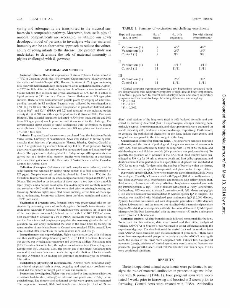
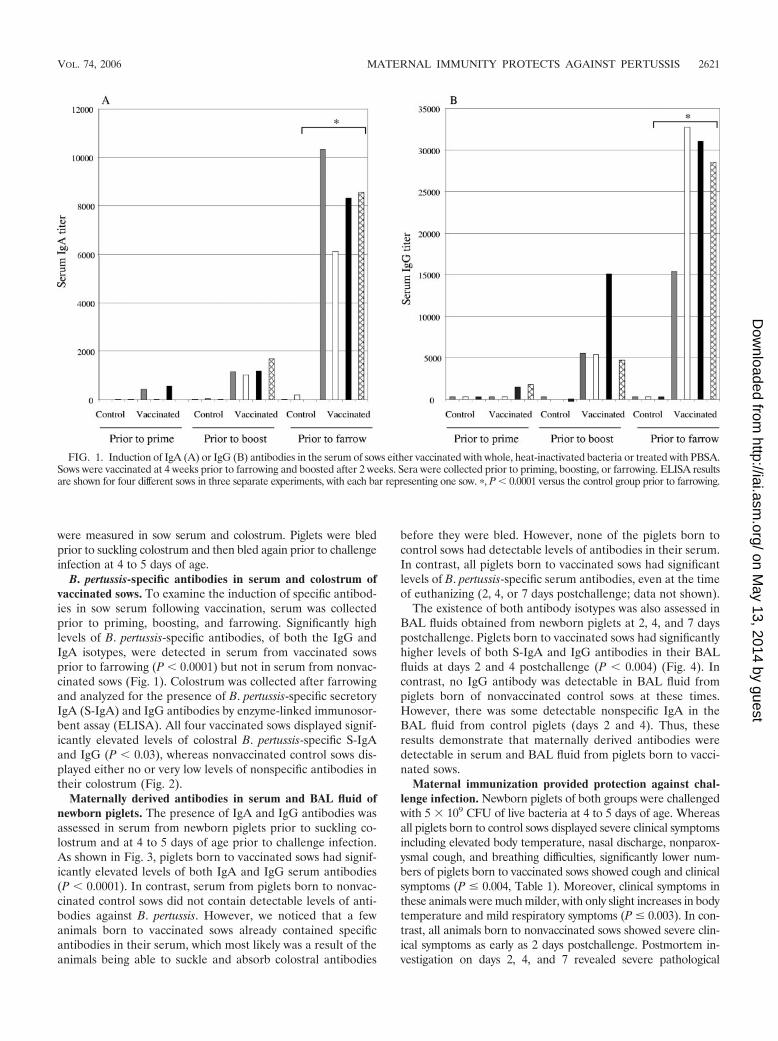
B. pertussis-specific antibodies in serum and colostrum ofvaccinated sows. To examine the induction of specific antibod-ies in sow serum following vaccination, serum was collectedprior to priming, boosting, and farrowing. Significantly highlevels of B. pertussis-specific antibodies, of both the IgG andIgA isotypes, were detected in serum from vaccinated sowsprior to farrowing (P � 0.0001) but not in serum from nonvac-cinated sows (Fig. 1). Colostrum was collected after farrowingand analyzed for the presence of B. pertussis-specific secretoryIgA (S-IgA) and IgG antibodies by enzyme-linked immunosor-bent assay (ELISA). All four vaccinated sows displayed signif-icantly elevated levels of colostral B. pertussis-specific S-IgAand IgG (P � 0.03), whereas nonvaccinated control sows dis-played either no or very low levels of nonspecific antibodies intheir colostrum (Fig. 2).
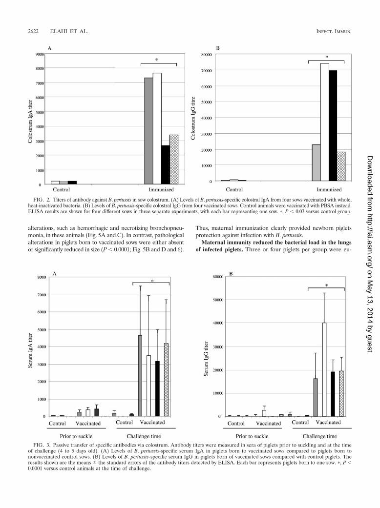
Maternally derived antibodies in serum and BAL fluid ofnewborn piglets. The presence of IgA and IgG antibodies wasassessed in serum from newborn piglets prior to suckling co-lostrum and at 4 to 5 days of age prior to challenge infection.As shown in Fig. 3, piglets born to vaccinated sows had signif-icantly elevated levels of both IgA and IgG serum antibodies(P � 0.0001). In contrast, serum from piglets born to nonvac-cinated control sows did not contain detectable levels of anti-bodies against B. pertussis. However, we noticed that a fewanimals born to vaccinated sows already contained specificantibodies in their serum, which most likely was a result of theanimals being able to suckle and absorb colostral antibodies
before they were bled. However, none of the piglets born tocontrol sows had detectable levels of antibodies in their serum.In contrast, all piglets born to vaccinated sows had significantlevels of B. pertussis-specific serum antibodies, even at the timeof euthanizing (2, 4, or 7 days postchallenge; data not shown).
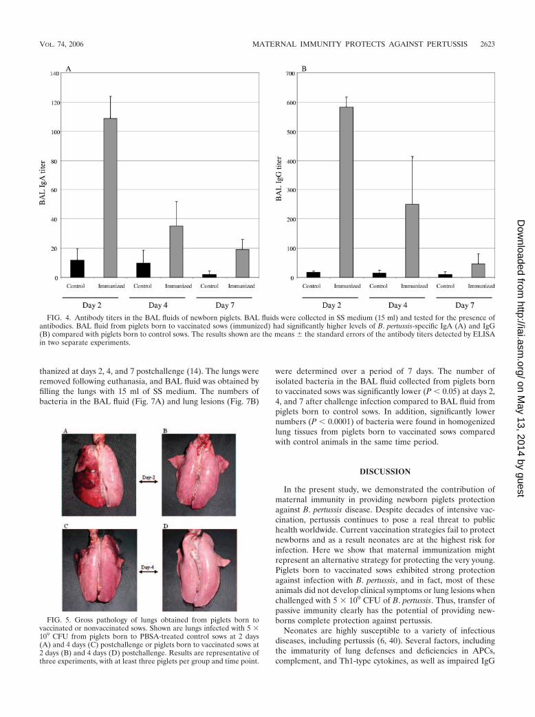
The existence of both antibody isotypes was also assessed inBAL fluids obtained from newborn piglets at 2, 4, and 7 dayspostchallenge. Piglets born to vaccinated sows had significantlyhigher levels of both S-IgA and IgG antibodies in their BALfluids at days 2 and 4 postchallenge (P � 0.004) (Fig. 4). Incontrast, no IgG antibody was detectable in BAL fluid frompiglets born of nonvaccinated control sows at these times.However, there was some detectable nonspecific IgA in theBAL fluid from control piglets (days 2 and 4). Thus, theseresults demonstrate that maternally derived antibodies weredetectable in serum and BAL fluid from piglets born to vacci-nated sows.
Maternal immunization provided protection against chal-lenge infection. Newborn piglets of both groups were challengedwith 5 � 109 CFU of live bacteria at 4 to 5 days of age. Whereasall piglets born to control sows displayed severe clinical symptomsincluding elevated body temperature, nasal discharge, nonparox-ysmal cough, and breathing difficulties, significantly lower num-bers of piglets born to vaccinated sows showed cough and clinicalsymptoms (P � 0.004, Table 1). Moreover, clinical symptoms inthese animals were much milder, with only slight increases in bodytemperature and mild respiratory symptoms (P � 0.003). In con-trast, all animals born to nonvaccinated sows showed severe clin-ical symptoms as early as 2 days postchallenge. Postmortem in-vestigation on days 2, 4, and 7 revealed severe pathological
FIG. 1. Induction of IgA (A) or IgG (B) antibodies in the serum of sows either vaccinated with whole, heat-inactivated bacteria or treated with PBSA.Sows were vaccinated at 4 weeks prior to farrowing and boosted after 2 weeks. Sera were collected prior to priming, boosting, or farrowing. ELISA resultsare shown for four different sows in three separate experiments, with each bar representing one sow. �, P � 0.0001 versus the control group prior to farrowing.
VOL. 74, 2006 MATERNAL IMMUNITY PROTECTS AGAINST PERTUSSIS 2621
on May 13, 2014 by guest
http://iai.asm.org/
Dow
nloaded from
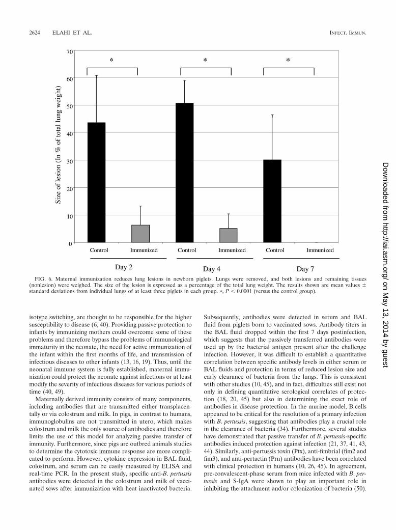
alterations, such as hemorrhagic and necrotizing bronchopneu-monia, in these animals (Fig. 5A and C). In contrast, pathologicalalterations in piglets born to vaccinated sows were either absentor significantly reduced in size (P � 0.0001; Fig. 5B and D and 6).
Thus, maternal immunization clearly provided newborn pigletsprotection against infection with B. pertussis.
Maternal immunity reduced the bacterial load in the lungsof infected piglets. Three or four piglets per group were eu-
FIG. 2. Titers of antibody against B. pertussis in sow colostrum. (A) Levels of B. pertussis-specific colostral IgA from four sows vaccinated with whole,heat-inactivated bacteria. (B) Levels of B. pertussis-specific colostral IgG from four vaccinated sows. Control animals were vaccinated with PBSA instead.ELISA results are shown for four different sows in three separate experiments, with each bar representing one sow. �, P � 0.03 versus control group.
FIG. 3. Passive transfer of specific antibodies via colostrum. Antibody titers were measured in sera of piglets prior to suckling and at the timeof challenge (4 to 5 days old). (A) Levels of B. pertussis-specific serum IgA in piglets born to vaccinated sows compared to piglets born tononvaccinated control sows. (B) Levels of B. pertussis-specific serum IgG in piglets born of vaccinated sows compared with control piglets. Theresults shown are the means � the standard errors of the antibody titers detected by ELISA. Each bar represents piglets born to one sow. �, P �0.0001 versus control animals at the time of challenge.
2622 ELAHI ET AL. INFECT. IMMUN.
on May 13, 2014 by guest
http://iai.asm.org/
Dow
nloaded from
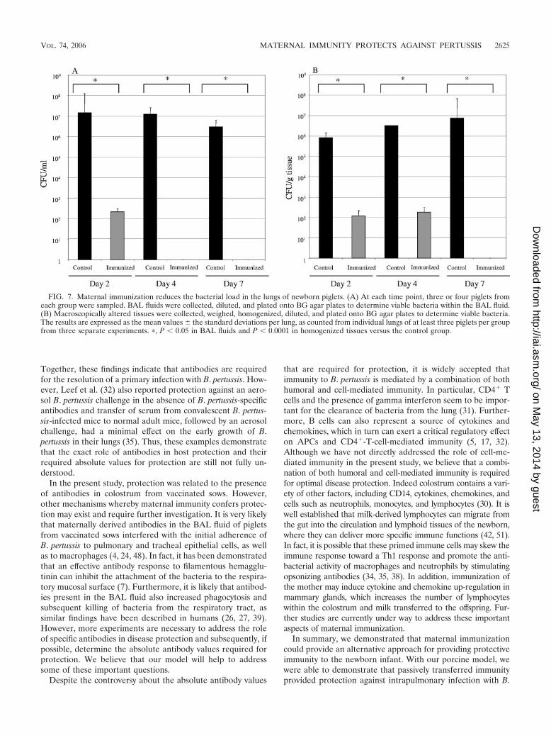
thanized at days 2, 4, and 7 postchallenge (14). The lungs wereremoved following euthanasia, and BAL fluid was obtained byfilling the lungs with 15 ml of SS medium. The numbers ofbacteria in the BAL fluid (Fig. 7A) and lung lesions (Fig. 7B)
were determined over a period of 7 days. The number ofisolated bacteria in the BAL fluid collected from piglets bornto vaccinated sows was significantly lower (P � 0.05) at days 2,4, and 7 after challenge infection compared to BAL fluid frompiglets born to control sows. In addition, significantly lowernumbers (P � 0.0001) of bacteria were found in homogenizedlung tissues from piglets born to vaccinated sows comparedwith control animals in the same time period.
DISCUSSION
In the present study, we demonstrated the contribution ofmaternal immunity in providing newborn piglets protectionagainst B. pertussis disease. Despite decades of intensive vac-cination, pertussis continues to pose a real threat to publichealth worldwide. Current vaccination strategies fail to protectnewborns and as a result neonates are at the highest risk forinfection. Here we show that maternal immunization mightrepresent an alternative strategy for protecting the very young.Piglets born to vaccinated sows exhibited strong protectionagainst infection with B. pertussis, and in fact, most of theseanimals did not develop clinical symptoms or lung lesions whenchallenged with 5 � 109 CFU of B. pertussis. Thus, transfer ofpassive immunity clearly has the potential of providing new-borns complete protection against pertussis.
Neonates are highly susceptible to a variety of infectiousdiseases, including pertussis (6, 40). Several factors, includingthe immaturity of lung defenses and deficiencies in APCs,complement, and Th1-type cytokines, as well as impaired IgG
FIG. 4. Antibody titers in the BAL fluids of newborn piglets. BAL fluids were collected in SS medium (15 ml) and tested for the presence ofantibodies. BAL fluid from piglets born to vaccinated sows (immunized) had significantly higher levels of B. pertussis-specific IgA (A) and IgG(B) compared with piglets born to control sows. The results shown are the means � the standard errors of the antibody titers detected by ELISAin two separate experiments.
FIG. 5. Gross pathology of lungs obtained from piglets born tovaccinated or nonvaccinated sows. Shown are lungs infected with 5 �109 CFU from piglets born to PBSA-treated control sows at 2 days(A) and 4 days (C) postchallenge or piglets born to vaccinated sows at2 days (B) and 4 days (D) postchallenge. Results are representative ofthree experiments, with at least three piglets per group and time point.
VOL. 74, 2006 MATERNAL IMMUNITY PROTECTS AGAINST PERTUSSIS 2623
on May 13, 2014 by guest
http://iai.asm.org/
Dow
nloaded from
isotype switching, are thought to be responsible for the highersusceptibility to disease (6, 40). Providing passive protection toinfants by immunizing mothers could overcome some of theseproblems and therefore bypass the problems of immunologicalimmaturity in the neonate, the need for active immunization ofthe infant within the first months of life, and transmission ofinfectious diseases to other infants (13, 16, 19). Thus, until theneonatal immune system is fully established, maternal immu-nization could protect the neonate against infections or at leastmodify the severity of infectious diseases for various periods oftime (40, 49).
Maternally derived immunity consists of many components,including antibodies that are transmitted either transplacen-tally or via colostrum and milk. In pigs, in contrast to humans,immunoglobulins are not transmitted in utero, which makescolostrum and milk the only source of antibodies and thereforelimits the use of this model for analyzing passive transfer ofimmunity. Furthermore, since pigs are outbred animals studiesto determine the cytotoxic immune response are more compli-cated to perform. However, cytokine expression in BAL fluid,colostrum, and serum can be easily measured by ELISA andreal-time PCR. In the present study, specific anti-B. pertussisantibodies were detected in the colostrum and milk of vacci-nated sows after immunization with heat-inactivated bacteria.
Subsequently, antibodies were detected in serum and BALfluid from piglets born to vaccinated sows. Antibody titers inthe BAL fluid dropped within the first 7 days postinfection,which suggests that the passively transferred antibodies wereused up by the bacterial antigen present after the challengeinfection. However, it was difficult to establish a quantitativecorrelation between specific antibody levels in either serum orBAL fluids and protection in terms of reduced lesion size andearly clearance of bacteria from the lungs. This is consistentwith other studies (10, 45), and in fact, difficulties still exist notonly in defining quantitative serological correlates of protec-tion (18, 20, 45) but also in determining the exact role ofantibodies in disease protection. In the murine model, B cellsappeared to be critical for the resolution of a primary infectionwith B. pertussis, suggesting that antibodies play a crucial rolein the clearance of bacteria (34). Furthermore, several studieshave demonstrated that passive transfer of B. pertussis-specificantibodies induced protection against infection (21, 37, 41, 43,44). Similarly, anti-pertussis toxin (Ptx), anti-fimbrial (fim2 andfim3), and anti-pertactin (Prn) antibodies have been correlatedwith clinical protection in humans (10, 26, 45). In agreement,pre-convalescent-phase serum from mice infected with B. per-tussis and S-IgA were shown to play an important role ininhibiting the attachment and/or colonization of bacteria (50).
FIG. 6. Maternal immunization reduces lung lesions in newborn piglets. Lungs were removed, and both lesions and remaining tissues(nonlesion) were weighed. The size of the lesion is expressed as a percentage of the total lung weight. The results shown are mean values �standard deviations from individual lungs of at least three piglets in each group. �, P � 0.0001 (versus the control group).
2624 ELAHI ET AL. INFECT. IMMUN.
on May 13, 2014 by guest
http://iai.asm.org/
Dow
nloaded from
Together, these findings indicate that antibodies are requiredfor the resolution of a primary infection with B. pertussis. How-ever, Leef et al. (32) also reported protection against an aero-sol B. pertussis challenge in the absence of B. pertussis-specificantibodies and transfer of serum from convalescent B. pertus-sis-infected mice to normal adult mice, followed by an aerosolchallenge, had a minimal effect on the early growth of B.pertussis in their lungs (35). Thus, these examples demonstratethat the exact role of antibodies in host protection and theirrequired absolute values for protection are still not fully un-derstood.
In the present study, protection was related to the presenceof antibodies in colostrum from vaccinated sows. However,other mechanisms whereby maternal immunity confers protec-tion may exist and require further investigation. It is very likelythat maternally derived antibodies in the BAL fluid of pigletsfrom vaccinated sows interfered with the initial adherence ofB. pertussis to pulmonary and tracheal epithelial cells, as wellas to macrophages (4, 24, 48). In fact, it has been demonstratedthat an effective antibody response to filamentous hemagglu-tinin can inhibit the attachment of the bacteria to the respira-tory mucosal surface (7). Furthermore, it is likely that antibod-ies present in the BAL fluid also increased phagocytosis andsubsequent killing of bacteria from the respiratory tract, assimilar findings have been described in humans (26, 27, 39).However, more experiments are necessary to address the roleof specific antibodies in disease protection and subsequently, ifpossible, determine the absolute antibody values required forprotection. We believe that our model will help to addresssome of these important questions.
Despite the controversy about the absolute antibody values
that are required for protection, it is widely accepted thatimmunity to B. pertussis is mediated by a combination of bothhumoral and cell-mediated immunity. In particular, CD4� Tcells and the presence of gamma interferon seem to be impor-tant for the clearance of bacteria from the lung (31). Further-more, B cells can also represent a source of cytokines andchemokines, which in turn can exert a critical regulatory effecton APCs and CD4�-T-cell-mediated immunity (5, 17, 32).Although we have not directly addressed the role of cell-me-diated immunity in the present study, we believe that a combi-nation of both humoral and cell-mediated immunity is requiredfor optimal disease protection. Indeed colostrum contains a vari-ety of other factors, including CD14, cytokines, chemokines, andcells such as neutrophils, monocytes, and lymphocytes (30). It iswell established that milk-derived lymphocytes can migrate fromthe gut into the circulation and lymphoid tissues of the newborn,where they can deliver more specific immune functions (42, 51).In fact, it is possible that these primed immune cells may skew theimmune response toward a Th1 response and promote the anti-bacterial activity of macrophages and neutrophils by stimulatingopsonizing antibodies (34, 35, 38). In addition, immunization ofthe mother may induce cytokine and chemokine up-regulation inmammary glands, which increases the number of lymphocyteswithin the colostrum and milk transferred to the offspring. Fur-ther studies are currently under way to address these importantaspects of maternal immunization.
In summary, we demonstrated that maternal immunizationcould provide an alternative approach for providing protectiveimmunity to the newborn infant. With our porcine model, wewere able to demonstrate that passively transferred immunityprovided protection against intrapulmonary infection with B.
FIG. 7. Maternal immunization reduces the bacterial load in the lungs of newborn piglets. (A) At each time point, three or four piglets fromeach group were sampled. BAL fluids were collected, diluted, and plated onto BG agar plates to determine viable bacteria within the BAL fluid.(B) Macroscopically altered tissues were collected, weighed, homogenized, diluted, and plated onto BG agar plates to determine viable bacteria.The results are expressed as the mean values � the standard deviations per lung, as counted from individual lungs of at least three piglets per groupfrom three separate experiments. �, P � 0.05 in BAL fluids and P � 0.0001 in homogenized tissues versus the control group.
VOL. 74, 2006 MATERNAL IMMUNITY PROTECTS AGAINST PERTUSSIS 2625
on May 13, 2014 by guest
http://iai.asm.org/
Dow
nloaded from
pertussis. However, further studies are necessary to address theissue of immunizing the mother during, before, or even afterpregnancy, as well as understanding the role of cell-mediatedimmunity in disease protection.
ACKNOWLEDGMENTS
We are thankful to Hugh Townsend for help with analyzing the dataand the VIDO Animal Care Staff for assistance with housing, chal-lenging, and monitoring the animals. We are especially thankful toDon Wilson, Kuldip Mirakhur, Amanda Giesbrecht, Sherry Tetland,Lucas Wirth, and Stacy Miskolcszi.
This study was funded by a grant from the Bill & Melinda Gates Foun-dation and the Canadian Institutes of Health Research through the GrandChallenges in Global Health Initiative (http://www.grandchallengesgh.org/),the Canadian Institutes of Health Research (CIHR), the Krembil Foun-dation, and the Saskatchewan Health Research Foundation (SHRF, post-doctoral fellowship to S. Elahi).
REFERENCES
1. Andrews, R., A. Herceg, and C. Roberts. 1997. Pertussis notifications inAustralia, 1991 to 1997. Commun. Dis. Intell. 21:145–148.
2. Baron, S., E. Njamkepo, E. Grimprel, P. Begue, J. C. Desenclos, J. Drucker,and N. Guiso. 1998. Epidemiology of pertussis in French hospitals in 1993and 1994: thirty years after a routine use of vaccination. Pediatr. Infect. Dis.J. 17:412–418.
3. Barrios, C., P. Brawand, M. Berney, C. Brandt, P. H. Lambert, and C. A.Siegrist. 1996. Neonatal and early life immune responses to various forms ofvaccine antigens qualitatively differ from adult responses: predominance of aTh2-biased pattern which persists after adult boosting. Eur. J. Immunol.26:1489–1496.
4. Bassinet, L., P. Gueirard, B. Maitre, B. Housset, P. Gounon, and N. Guiso.2000. Role of adhesins and toxins in invasion of human tracheal epithelialcells by Bordetella pertussis. Infect. Immun. 68:1934–1941.
5. Bayry, J., S. Lacroix-Desmazes, M. D. Kazatchkine, O. Hermine, D. F.Tough, and S. V. Kaveri. 2005. Modulation of dendritic cell maturation andfunction by B lymphocytes. J. Immunol. 175:15–20.
6. Bellanti, J. A., and B. J. Zeligs. 1995. Developmental aspects of pulmonarydefenses in children. Pediatr. Pulmonol Suppl. 11:79–80.
7. Cahill, E. S., D. T. O’Hagan, L. Illum, A. Barnard, K. H. Mills, and K.Redhead. 1995. Immune responses and protection against Bordetella per-tussis infection after intranasal immunization of mice with filamentoushaemagglutinin in solution or incorporated in biodegradable microparticles.Vaccine 13:455–462.
8. Centers for Disease Control and Prevention. 2002. Pertussis deaths—UnitedStates, 2000. Morb. Mortal. Wkly. Rep. 51:616–618.
9. Centers for Disease Control and Prevention. 2002. Pertussis—United States,1997–2000. Morb. Mortal. Wkly. Rep. 51:73–76.
10. Cherry, J. D., J. Gornbein, U. Heininger, and K. Stehr. 1998. A search forserologic correlates of immunity to Bordetella pertussis cough illnesses.Vaccine 16:1901–1906.
11. Cohen, P., and S. J. Scandron. 1943. Placental transmission of protectiveantibodies against whooping cough by inoculation of the pregnant mother.JAMA 121:656–662.
12. de Melker, H. E., J. F. Schellekens, S. E. Neppelenbroek, F. R. Mooi, H. C.Rumke, and M. A. Conyn-van Spaendonck. 2000. Reemergence of pertussisin the highly vaccinated population of the Netherlands: observations onsurveillance data. Emerg. Infect. Dis. 6:348–357.
13. Edwards, K. M. 2003. Pertussis: an important target for maternal immuni-zation. Vaccine 21:3483–3486.
14. Elahi, S., R. Brownlie, J. Korzeniowski, R. Buchanan, B. O’Connor, M. S.Peppler, S. A. Halperin, S. F. Lee, L. A. Babiuk, and V. Gerdts. 2005.Infection of newborn piglets with Bordetella pertussis: a new model for per-tussis. Infect. Immun. 73:3636–3645.
15. Forsyth, K., T. Tan, C. H. von Konig, J. J. Caro, and S. Plotkin. 2005.Potential strategies to reduce the burden of pertussis. Pediatr. Infect. Dis. J.24:S69–S74.
16. Gall, S. A. 2003. Maternal immunization. Obstet. Gynecol. Clin. N. Am.30:623–636.
17. Gonik, B., K. S. Puder, N. Gonik, and M. Kruger. 2005. Seroprevalence ofBordetella pertussis antibodies in mothers and their newborn infants. Infect.Dis. Obstet. Gynecol. 13:59–61.
18. Greco, D., S. Salmaso, P. Mastrantonio, M. Giuliano, A. E. Tozzi, A.Anemona, M. L. Ciofi degli Atti, A. Giammanco, P. Panei, W. C. Black-welder, D. L. Klein, and S. G. Wassilak. 1996. A controlled trial of twoacellular vaccines and one whole-cell vaccine against pertussis. ProgettoPertosse Working Group. N. Engl. J. Med. 334:341–348.
19. Greenwood, B. 2003. Maternal immunisation in developing countries. Vac-cine 21:3436–3441.
20. Gustafsson, L., H. O. Hallander, P. Olin, E. Reizenstein, and J. Storsaeter.1996. A controlled trial of a two-component acellular, a five-componentacellular, and a whole-cell pertussis vaccine. N. Engl. J. Med. 334:349–355.
21. Halperin, S. A., T. B. Issekutz, and A. Kasina. 1991. Modulation of Borde-tella pertussis infection with monoclonal antibodies to pertussis toxin. J. In-fect. Dis. 163:355–361.
22. Halperin, S. A., E. E. Wang, B. Law, E. Mills, R. Morris, P. Dery, M. Lebel,N. MacDonald, T. Jadavji, W. Vaudry, D. Scheifele, G. Delage, and P.Duclos. 1999. Epidemiological features of pertussis in hospitalized patientsin Canada, 1991–1997: report of the Immunization Monitoring Program-Active (IMPACT). Clin. Infect. Dis. 28:1238–1243.
23. Hayward, A. R., and J. Groothuis. 1991. Development of T cells with mem-ory phenotype in infancy. Adv. Exp. Med. Biol. 310:71–76.
24. Hazenbos, W. L., B. M. van den Berg, J. W. van’t Wout, F. R. Mooi, and R.van Furth. 1994. Virulence factors determine attachment and ingestion ofnonopsonized and opsonized Bordetella pertussis by human monocytes. In-fect. Immun. 62:4818–4824.
25. Healy, C. M., F. M. Munoz, M. A. Rench, N. B. Halasa, K. M. Edwards, andC. J. Baker. 2004. Prevalence of pertussis antibodies in maternal delivery,cord, and infant serum. J. Infect. Dis. 190:335–340.
26. Hellwig, S. M., M. E. Rodriguez, G. A. Berbers, J. G. van de Winkel, andF. R. Mooi. 2003. Crucial role of antibodies to pertactin in Bordetella per-tussis immunity. J. Infect. Dis. 188:738–742.
27. Hellwig, S. M., H. F. van Oirschot, W. L. Hazenbos, A. B. van Spriel, F. R.Mooi, and J. G. van De Winkel. 2001. Targeting to Fc� receptors, but notCR3 (CD11b/CD18), increases clearance of Bordetella pertussis. J. Infect.Dis. 183:871–879.
28. Helmy, M. F., M. Hammam, M. S. el Kholy, and N. Guirguis. 1992. Borde-tella pertussis FHA antibodies in maternal/infants sera and colostrum. J.Egypt Public Health Assoc. 67:195–212.
29. Holt, P. G., J. B. Clough, B. J. Holt, M. J. Baron-Hay, A. H. Rose, B. W.Robinson, and W. R. Thomas. 1992. Genetic ‘risk’ for atopy is associatedwith delayed postnatal maturation of T-cell competence. Clin. Exp. Allergy22:1093–1099.
30. Jain, N., N. B. Mathur, V. K. Sharma, and A. M. Dwarkadas. 1991. Cellularcomposition including lymphocyte subsets in preterm and full term humancolostrum and milk. Acta Paediatr. Scand. 80:395–399.
31. Kendrick, P., M. Thompson, and G. Elderling. 1945. Immunity response ofmothers and babies to injections of pertussis vaccine during pregnancy.Am. J. Dis. Child. 70:25–28.
32. Leef, M., K. L. Elkins, J. Barbic, and R. D. Shahin. 2000. Protective immu-nity to Bordetella pertussis requires both B cells and CD4� T cells for keyfunctions other than specific antibody production. J. Exp. Med. 191:1841–1852.
33. Lichty, J. R., B. Slavin, and W. L. Bradford. 1938. Attempt to increaseresistance to pertussis in newborn infants by immunizing their mothersduring pregnancy. J. Clin. Investig. 17:613–621.
34. Mahon, B. P., B. J. Sheahan, F. Griffin, G. Murphy, and K. H. Mills. 1997.Atypical disease after Bordetella pertussis respiratory infection of mice withtargeted disruptions of interferon-gamma receptor or immunoglobulin muchain genes. J. Exp. Med. 186:1843–1851.
35. Mills, K. H., A. Barnard, J. Watkins, and K. Redhead. 1993. Cell-mediatedimmunity to Bordetella pertussis: role of Th1 cells in bacterial clearance in amurine respiratory infection model. Infect. Immun. 61:399–410.
36. Mooi, F. R., I. H. van Loo, and A. J. King. 2001. Adaptation of Bordetellapertussis to vaccination: a cause for its reemergence? Emerg. Infect. Dis.7:526–528.
37. Mountzouros, K. T., A. Kimura, and J. L. Cowell. 1992. A bactericidalmonoclonal antibody specific for the lipooligosaccharide of Bordetella per-tussis reduces colonization of the respiratory tract of mice after aerosolinfection with B. pertussis. Infect. Immun. 60:5316–5318.
38. Redhead, K., J. Watkins, A. Barnard, and K. H. Mills. 1993. Effectiveimmunization against Bordetella pertussis respiratory infection in mice isdependent on induction of cell-mediated immunity. Infect. Immun. 61:3190–3198.
39. Repp, R., T. Valerius, A. Sendler, M. Gramatzki, H. Iro, J. R. Kalden, andE. Platzer. 1991. Neutrophils express the high affinity receptor for IgG (Fc�RI, CD64) after in vivo application of recombinant human granulocytecolony-stimulating factor. Blood 78:885–889.
40. Rowe, J., J. T. Poolman, C. Macaubas, P. D. Sly, R. Loh, and P. G. Holt.2004. Enhancement of vaccine-specific cellular immunity in infants by pas-sively acquired maternal antibody. Vaccine 22:3986–3992.
41. Sato, H., A. Ito, J. Chiba, and Y. Sato. 1984. Monoclonal antibody againstpertussis toxin: effect on toxin activity and pertussis infections. Infect. Im-mun. 46:422–428.
42. Seelig, L. L., Jr., and R. E. Billingham. 1981. Concerning the natural trans-plantation of maternal lymphocytes via milk. Transplant. Proc. 13:1245–1249.
43. Shahin, R. D., M. J. Brennan, Z. M. Li, B. D. Meade, and C. R. Manclark.1990. Characterization of the protective capacity and immunogenicity of the69-kD outer membrane protein of Bordetella pertussis. J. Exp. Med. 171:63–73.
2626 ELAHI ET AL. INFECT. IMMUN.
on May 13, 2014 by guest
http://iai.asm.org/
Dow
nloaded from
44. Shahin, R. D., J. Hamel, M. F. Leef, and B. R. Brodeur. 1994. Analysis ofprotective and nonprotective monoclonal antibodies specific for Bordetellapertussis lipooligosaccharide. Infect. Immun. 62:722–725.
45. Storsaeter, J., H. O. Hallander, L. Gustafsson, and P. Olin. 1998. Levels ofanti-pertussis antibodies related to protection after household exposure toBordetella pertussis. Vaccine 16:1907–1916.
46. Tanaka, M., C. R. Vitek, F. B. Pascual, K. M. Bisgard, J. E. Tate, and T. V.Murphy. 2003. Trends in pertussis among infants in the United States,1980–1999. JAMA 290:2968–2975.
47. Upham, J. W., P. T. Lee, B. J. Holt, T. Heaton, S. L. Prescott, M. J. Sharp,P. D. Sly, and P. G. Holt. 2002. Development of interleukin-12-producingcapacity throughout childhood. Infect. Immun. 70:6583–6588.
48. van den Berg, B. M., H. Beekhuizen, R. J. Willems, F. R. Mooi, and R. vanFurth. 1999. Role of Bordetella pertussis virulence factors in adherence toepithelial cell lines derived from the human respiratory tract. Infect. Immun.67:1056–1062.
49. Van Rie, A., A. M. Wendelboe, and J. A. Englund. 2005. Role of maternalpertussis antibodies in infants. Pediatr. Infect. Dis. J. 24:S62–S65.
50. Watanabe, M., and M. Nagai. 2003. Role of systemic and mucosal immuneresponses in reciprocal protection against Bordetella pertussis and Bordetellaparapertussis in a murine model of respiratory infection. Infect. Immun.71:733–738.
51. Wirt, D. P., L. T. Adkins, K. H. Palkowetz, F. C. Schmalstieg, and A. S.Goldman. 1992. Activated and memory T lymphocytes in human milk. Cy-tometry 13:282–290.
52. Zola, H., M. Fusco, P. J. Macardle, L. Flego, and D. Roberton. 1995. Ex-pression of cytokine receptors by human cord blood lymphocytes: compar-ison with adult blood lymphocytes. Pediatr. Res. 38:397–403.
53. Zola, H., M. Fusco, H. Weedon, P. J. Macardle, J. Ridings, and D. M.Roberton. 1996. Reduced expression of the interleukin-2-receptor gammachain on cord blood lymphocytes: relationship to functional immaturity ofthe neonatal immune response. Immunology 87:86–91.
Editor: A. D. O’Brien
VOL. 74, 2006 MATERNAL IMMUNITY PROTECTS AGAINST PERTUSSIS 2627
on May 13, 2014 by guest
http://iai.asm.org/
Dow
nloaded from