p31comet Induces Cellular Senescence through p21 Accumulation and Mad2 Disruption
Alistair J. Lax and Enrique RozengurtHadriano M. Lacerda, Gill D. Pullinger, Swiss 3T3 Cells Focal Adhesion Kinase and Paxillin in
-dependent Tyrosine Phosphorylation ofrho Induce p21Bordetella bronchisepticafrom
and Dermonecrotic ToxinEscherichia coliCytotoxic Necrotizing Factor 1 from Cell Biology and Metabolism:
doi: 10.1074/jbc.272.14.95871997, 272:9587-9596.J. Biol. Chem.
http://www.jbc.org/content/272/14/9587Access the most updated version of this article at
.JBC Affinity SitesFind articles, minireviews, Reflections and Classics on similar topics on the
Alerts:
When a correction for this article is posted•
When this article is cited•
to choose from all of JBC's e-mail alertsClick here
http://www.jbc.org/content/272/14/9587.full.html#ref-list-1
This article cites 69 references, 45 of which can be accessed free at
by guest on August 1, 2014
http://ww
w.jbc.org/
Dow
nloaded from
by guest on August 1, 2014
http://ww
w.jbc.org/
Dow
nloaded from
Cytotoxic Necrotizing Factor 1 from Escherichia coli andDermonecrotic Toxin from Bordetella bronchisepticaInduce p21rho-dependent Tyrosine Phosphorylation of FocalAdhesion Kinase and Paxillin in Swiss 3T3 Cells*
(Received for publication, December 23, 1996)
Hadriano M. Lacerda‡§, Gill D. Pullinger¶, Alistair J. Laxi, and Enrique Rozengurt‡**
From the ‡Imperial Cancer Research Fund, P. O. Box 123, 44 Lincoln’s Inn Fields, London WC2A 3PX, United Kingdom,the iUMDS Guy’s and St. Thomas’s Medical and Dental School, Floor 28 Guy’s Tower, Guy’s Hospital, London SE1 9RT,United Kingdom, and the ¶Department of Chemical Endocrinology, St. Bartholomews Hospital Medical College, 51-53Bartholomews Close, London EC1A 7BE, United Kingdom
Treatment of Swiss 3T3 cells with cytotoxic necrotiz-ing factor 1 (CNF1) from Escherichia coli and dermon-ecrotic toxin (DNT) from Bordetella bronchiseptica,which directly target and activate p21rho, stimulatedtyrosine phosphorylation of focal adhesion kinase(p125fak) and paxillin. Tyrosine phosphorylation in-duced by CNF1 and DNT occurred after a pronouncedlag period (2 h), and was blocked by either lysosomotro-phic agents or incubation at 22 °C. CNF1 and DNT stim-ulated tyrosine phosphorylation of p125fak and paxillin,actin stress fiber formation, and focal adhesion assem-bly with similar kinetics. Cytochalasin D and high con-centrations of platelet-derived growth factor disruptedthe actin cytoskeleton and completely inhibited CNF1and DNT induced tyrosine phosphorylation. Microinjec-tion of Clostridium botulinum C3 exoenzyme whichADP-ribosylates and inactivates p21rho function, pre-vented tyrosine phosphorylation of focal adhesion pro-teins in response to either CNF1 or DNT. In addition,our results demonstrated that CNF1 and DNT do notinduce protein kinase C activation, inositol phosphateformation, and Ca21 mobilization. Moreover, CNF1 andDNT stimulated DNA synthesis without activation ofp42mapk and p44mapk providing additional evidence for anovel p21rho-dependent signaling pathway that leads toentry into the S phase of the cell cycle in Swiss 3T3.
An increase in the tyrosine phosphorylation of the non-re-ceptor protein tyrosine kinase p125fak (1, 2) and the cytoskel-etal associated protein paxillin (3, 4) has recently been identi-fied as an early event in the action of diverse signalingmolecules that mediate cell growth and differentiation (5), in-cluding mitogenic neuropeptides (6–8), growth factors such asPDGF1 (9, 10), bioactive lipids (11–15), extracellular matrix
proteins (16–20), transforming variants of pp60src (17, 21), andthe potent mitogenic toxin PMT (22). p125fak lacks SH2 andSH3 domains but associates with other proteins including Src,paxillin, and p130cas (1, 2, 23). Paxillin is a multidomain pro-tein that may function as an adaptor capable of complex for-mation with p125fak, Crk, Src, and vinculin (3, 4, 23). Genedisruption experiments indicate a critical role of p125fak inembryonic development and cell locomotion (24). The increasesin p125fak and paxillin tyrosine phosphorylation are accompa-nied by profound alterations in the organization of the actincytoskeleton and in the assembly of focal adhesions (9, 13, 15,25, 26), the distinct areas of the plasma membrane wherep125fak and paxillin are localized (1, 2, 27). The small G proteinp21rho, a member of the Ras superfamily of small GTP-bindingproteins, has been implicated in the mitogen-stimulated forma-tion of focal adhesions and actin stress fibers as well as in thetyrosine phosphorylation of p125fak and paxillin (22, 25, 28–30). These findings suggest the existence of a distinct signaltransduction pathway in which p21rho is upstream of cytoskel-etal reorganization and tyrosine phosphorylation of focal adhe-sion proteins (5).
The mechanism of action of bacterial toxins has providednovel insights into the control of cellular regulatory processes,including signal transduction and cell proliferation. For exam-ple, the Clostridium botulinum C3 exoenzyme and the entero-toxins A and B from Clostridium difficile which selectivelyinactivate members of the Rho subfamily, have provided usefultools to evaluate the role of these small G proteins in signaltransduction and cytoskeletal organization (31–33). In contrastto these clostridial toxins, CNF toxins produced by some path-ogenic strains of Escherichia coli (34) and DNT from Bordetellabronchiseptica (35) directly target and activate p21rho (36, 37).CNF and DNT induce actin reorganization and multinucle-ation in several cell types (37–40) but their effects on signaltransduction pathways, specially on p125fak and paxillin tyro-sine phosphorylation have not been examined.
Here we report that CNF1 and DNT stimulate tyrosine phos-phorylation of p125fak and paxillin and induce a concomitantincrease in the formation of actin stress fibers and in theassembly of focal adhesion plaques in Swiss 3T3 cells. Micro-injection of C3 exoenzyme prevented both the cytoskeletal re-sponses and the increase in tyrosine phosphorylation of focaladhesion proteins. In contrast to most other stimuli that pro-mote tyrosine phosphorylation of p125fak and paxillin (6–15,
* The costs of publication of this article were defrayed in part by thepayment of page charges. This article must therefore be hereby marked“advertisement” in accordance with 18 U.S.C. Section 1734 solely toindicate this fact.
§ Recipient of Research Fellowship from CNPq-Conselho Nacional deDesenvolvimento Cientifico e Tecnologico Brasilia, Brasil.
** To whom all correspondence should be addressed. Tel.: 44-0171-269-3455; Fax: 44-0171-269-3417.
1 The abbreviations used are: PDGF, platelet-derived growth factor;PMT, Pasteurella multocida toxin; CNF, cytotoxic necrotizing factor;DNT, dermonecrotic toxin; anti-Tyr(P), anti-phosphotyrosine; DMEM,Dulbecco’s modified Eagle’s medium; mAb, monoclonal antibody; PBS,phosphate-buffered saline; p125fak, p125 focal adhesion kinase; PKC,protein kinase C; MAPK, mitogen-activated protein kinase; PAGE,polyacrylamide gel electrophoresis; Ins(1,4,5)P3, inositol 1,4,5-trisphos-
phate; PtdIns(4,5)P2, phosphatidylinositol 4,5-bisphosphate; FITC, flu-orescein isothiocyanate; MARCKS, myristoylated alanine-rich C-kinasesubstrate.
THE JOURNAL OF BIOLOGICAL CHEMISTRY Vol. 272, No. 14, Issue of April 4, pp. 9587–9596, 1997© 1997 by The American Society for Biochemistry and Molecular Biology, Inc. Printed in U.S.A.
This paper is available on line at http://www-jbc.stanford.edu/jbc/ 9587
by guest on August 1, 2014
http://ww
w.jbc.org/
Dow
nloaded from
22), CNF1 and DNT do not activate phospholipase C-mediatedevents including inositol phosphate production, Ca21 mobiliza-tion, and PKC activation. In addition, CNF1 and DNT bothstimulated reinitiation of DNA synthesis but neither induceactivation of p42mapk (ERK2). Thus, our results demonstratingthat CNF1 and DNT induce a specific subset of molecular andcytoskeletal responses in Swiss 3T3 cells provide novel evi-dence for the existence of a signal transduction pathway thatlinks p21rho activation to tyrosine phosphorylation of focal ad-hesion proteins.
EXPERIMENTAL PROCEDURES
Cell Culture—Stock cultures of Swiss 3T3 fibroblasts were main-tained in DMEM supplemented with 10% fetal bovine serum in ahumidified atmosphere containing 10% CO2 and 90% air at 37 °C. Forexperimental purposes, cells were plated either in 30-mm Nunc Petridishes at 105 cells/dish, or in 90-mm dishes at 6 3 105 cells/dish, inDMEM containing 10% fetal bovine serum and used after 6–8 dayswhen the cells were confluent and quiescent (41).
DNA Synthesis Measurements—Quiescent cultures of Swiss 3T3 cellswere washed twice with DMEM and incubated with DMEM/Way-mouth’s medium 1:1 (v/v) containing [3H]thymidine (1 mCi/ml) andvarious factors as indicated. After 40 h, the cultures were washed twicewith ice-cold phosphate-buffered saline and incubated in 5% trichloro-acetic acid for 30 min at 4 °C. Trichloroacetic acid was then removedand the cultures were washed twice with ethanol and extracted in 1 mlof 2% Na2CO3, 0.1 M NaOH, 1% SDS. Incorporation of [3H]thymidinewas determined by scintillation counting in 6 ml of scintillation fluid.The proportion of cells in the G0/G1, S, G2, and M phases of the cell cyclewere determined by fluorescence-activated cell sorter analysis as de-scribed previously (42).
Immunoprecipitation—Quiescent cultures of Swiss 3T3 cells (1–2 3106) were washed twice with DMEM, treated with CNF1, DNT, or PMTor other factors in 10 ml of DMEM/Waymouth (1:1, v/v) for the timesindicated and lysed at 4 °C in 1 ml of a lysis buffer solution containing10 mM Tris/HCl, pH 7.6, 5 mM EDTA, 50 mM NaCl, 30 mM sodiumpyrophosphate, 50 mM sodium fluoride, 100 mM Na3VO4, and 1% TritonX-100. Proteins were immunoprecipitated at 4 °C overnight with agar-ose-linked mAbs directed against phosphotyrosine, paxillin, or p125fak
as indicated. Immunoprecipitates were washed three times with lysisbuffer and extracted for 10 min at 95 °C in 2 3 SDS-PAGE samplebuffer (200 mM Tris-HCl, 6% SDS, 2 mM EDTA, 4% 2-mercaptoethanol,10% glycerol, pH 6.8) and analyzed by SDS-PAGE.
Western Blotting—Treatment of quiescent cultures of cells with fac-tors, cell lysis, and immunoprecipitations were performed as describedabove. After separation by SDS-PAGE, proteins were transferred toImmobilon membranes (43). Membranes were blocked using 5% nonfatdried milk in PBS, pH 7.2, and incubated for 3–5 h at 22 °C with PY72or 4G10 anti-Tyr(P) mAbs (1 mg/ml). Immunoreactive bands were visu-alized using 125I-labeled sheep anti-mouse IgG followed by autoradiog-raphy. Autoradiograms were scanned using an LKB Ultrascan XLinternal integrator. The values expressed represent percentages of themaximum increase in tyrosine phosphorylation above control values.
32P Labeling of Cells and Analysis of 80K/MARCKS Phosphoryla-tion—Quiescent and confluent cultures of Swiss 3T3 cells in 30-mmdishes were washed twice in phosphate-free DMEM and incubated at37 °C with this medium containing 50 mCi/ml carrier-free [32P]Pi. After12 h, various factors were added for the indicated times. The cells weresubsequently lysed and the lysates were immunoprecipitated with spe-cific anti-80K/MARCKS antibody (44).
Analysis of Total Inositol Phosphates—Cultures of Swiss 3T3 cells in33-mm dishes were labeled for 16–18 h in 1 ml of DMEM/Waymouth’smedium (1:1) containing 10 mCi of [2-3H]inositol. Additions were madeto the cells as described for each experiment and LiCl was added to afinal concentration of 20 mM for the last 30 min of the incubation.Inositol phosphates were extracted by replacing the medium with 1 mlof ice-cold 3% HClO4. After 20 min at 4 °C the extract was neutralizedwith 0.5 M KOH containing 25 mM HEPES, 5 mM EDTA, and 0.01%phenol red. Precipitated KClO4 was removed by centrifugation. Analy-sis of total inositol phosphates was by anion-exchange column chroma-tography (45). Samples were diluted to 10 ml with water and thenloaded onto 1 ml of Dowex AG1-X8 (100–200 mesh, HCOO2 form) inBio-Rad Econo-columns. After 3 washes with 3 3 10 ml of H2O and 2 310 ml of 60 mM NH4COOH, 5 mM Na2B4O7 the inositol phosphates wereeluted with 7 ml of 1 M NH4COOH, 0.1 M HCOOH. An aliquot (1 ml) ofeluate was counted in 10 ml of scintillation fluid.
Intracellular Concentration of Ca21—To determine the intracellularconcentration of Ca21, [Ca21]i, 5 3 105 cells were subcultured with 10ml of DMEM containing 10% fetal bovine serum in 90-mm Nunc dishes.After 6–8 days, when the cells were confluent and quiescent, themedium was replaced with 5 ml of DMEM/Waymouth’s medium (1:1).The cells were pretreated as indicated, and then 5 ml of 1 mM fura-2/AM(46–48) was added directly to the medium. After a further 10 min thecells were washed twice at 37 °C with 3 ml of electrolyte solution,comprising 140 mM NaCl, 5 mM KCl, 1.8 mM CaCl2, 0.9 mM MgCl2, 25mM glucose, 16 mM HEPES, 6 mM Tris, pH 7.2, and the amino acidcontent of DMEM. The cells were then gently removed from the dish byscraping into 2 ml of the electrolyte solution. The cell suspension wastransferred to a 1-cm2 quartz cuvette and stirred at 37 °C for 4 minprior to any additions. Fluorescence was measured in a Perkin-ElmerLS-5 fluorometer with an excitation wavelength of 510 nm. [Ca21]i wascalculated from the maximum and minimum fluorescence of the fura-2,as described (46, 49).
Immunostaining of Cells—Quiescent Swiss 3T3 cells were washedtwice with DMEM and incubated for the indicated times in DMEM/Waymouth’s medium (1:1, v/v) at 37 °C with CNF1, DNT, or PMT. Foractin staining, cells were washed once with PBS, fixed in 4% paraform-aldehyde in PBS for 10 min at room temperature, and permeabilizedwith PBS containing 0.2% Triton X-100 for 8 min at room temperature.The cells were then incubated with FITC-conjugated phalloidin (0.25mg/ml) in PBS for 30 min at room temperature and visualized using aZeiss Axiophot immunofluorescence microscope. In experiments inwhich quiescent Swiss 3T3 cells were labeled with both FITC-conju-gated phalloidin and anti-vinculin antibody, the cells were fixed andpermeabilized as described above and then stained with a mixture ofFITC-conjugated phalloidin (0.25 mg/ml) and anti-vinculin antibody(dilution 1:100) for 30 min at room temperature. Cells were subse-quently washed three times in PBS and then incubated with Cy3-labeled rabbit anti-mouse IgG as a secondary antibody at a dilution of1:100 for another 30 min at room temperature.
Microinjection of C3 Exoenzyme—For microinjection experimentsSwiss 3T3 cells were plated in 33-mm Nunc Petri dishes at 105 cells/dish in DMEM containing 10% fetal bovine serum and used after 6–8days when the cells were confluent and quiescent. Approximately 50cells were microinjected with 100 mg/ml recombinant C3 exoenzyme.The efficiency of injection was determined by co-injecting guinea pigimmunoglobulin at 0.5 mg/ml followed by staining with FITC-linkedgoat anti-guinea pig IgG. A single batch of C3 transferase was used inall experiments described.
Confocal Microscopy—Confocal imaging was performed using a Bio-Rad MRC 600 laser scanning head fitted to a Nikon Optiphot micro-scope. A 60X N.A./1.4 planapochromat oil immersion lens (Nikon) wasused for all imaging. FITC-conjugated phalloidin and Cy3 anti-mouseIgG fluorochromes were excited at 488 and 568 nm, respectively, usinga krypton/argon mixed gas laser (Bio-Rad). Two filter blocks were used,K1 and K2. K1 is a double dichromic filter enabling excitation at thewavelengths of 488 and 568 nm, whereas the K2 filter is a 560-nmdichromic combined with 522-nm green emission and 585-nm red emis-sion filters. Images were collected using the Kalman filter. Care wastaken to ensure that the FITC-conjugated phalloidin channel was suf-ficiently bright relative to the fluorescein signal to minimize the con-tribution of bleed-through from the green channel into the red channel(approximately 10%). Correction of images for bleed-through and otherprocessing was carried out using COMOS and SOM programs (Bio-Rad)run on a Compaq Deskpro 66M 486 computer (66 MHz). Data arepresented as projections of sequential optical sections. For Z-series,optical sections were recorded at 0.5-mm intervals. Final images werephotographed directly from the VDU screen.
Shift Assay for MAP Kinase Activation—Activation of p42mapk andp44mapk was determined by the appearance of slower migrating forms ingel electrophoresis due to phosphorylation of specific threonine andtyrosine residues (50). Lysates from quiescent cells in 33-mm dishesprepared as above were subjected to SDS-PAGE and transferred toImmobilon membranes. Membranes were blocked using 5% nonfatdried milk in phosphate-buffered saline. Rabbit polyclonal antibodiesraised against COOH-terminal peptides (EETARFQPGYRS for p42mapk
and IFQETARFQPGAPE for the p44mapk) were used at 1/1000 dilution,and 125I-protein A was used to visualize immunoreactive bands.
Constructs Bearing CNF1, DNT, and PMT—Strain pISS392 is E. coliDH5a containing plasmid pISS392 which consists of a 3.5-kilobasefragment encoding only CNF1 inserted into the vector pGEM3. Controlstrain pGEM3 is E. coli DH5a containing a plasmid derived frompISS392 by deletion of a 3.5-kilobase insert. It is pGEM3 with a deletionin the multiple cloning site. Strain DNT1 is E. coli XL1-Blue containing
CNF1 and DNT Stimulation of Tyrosine Phosphorylation9588
by guest on August 1, 2014
http://ww
w.jbc.org/
Dow
nloaded from
plasmid pDNT1 which consists of a 5-kilobase fragment encoding theDNT gene (B. bronchiseptica B58) inserted into the vector pBluescript.Control strain pBlue is E. coli XL1-Blue with plasmid pBluescript (51).Strain pTOX2 is E. coli XL1-Blue with plasmid pTOX2 which consistsof the vector pBluescript with an approximately 4-kilobase pair insertencoding PMT. Control strain pBlue is E. coli XL1-Blue with plasmidpBluescript here nominated PC12.
Preparation of Samples of CNF1, DNT, and PMT—The E. coli strainswere grown overnight from a fresh colony. They were lysed in the lysisbuffer (50 mg/ml lysozyme, 5 mM EDTA, 0.1% toluene, 0.05 M Tris-Cl, pH7.2, 0.1 M NaCl) treated with RNase and DNase at 10 mg/ml each toremove nucleic acid and filtered through a 0.2-mm nitrocellulose filter.
Materials—Bombesin, cytochalasin D, FITC-conjugated phalloidin,monoclonal anti-vinculin mAb, Cy3-linked anti-mouse IgG, guinea pigIgG, and FITC-conjugated goat anti-guinea pig IgG were obtained fromSigma. Recombinant PDGF (BB homodimer), 125I-sheep anti-mouseIgG (15 mCi/mg), carrier-free [32P]Pi, and [2-3H]inositol (18.8 Ci/mmol,1 Ci 5 37 GBq) were all supplied by Amersham Corp., United Kingdom.Agarose-linked anti-Tyr(P) mAb was purchased from Oncogene ScienceInc., New York. 4G10 anti-Tyr(P) mAb was from UBI, Lake Placid, NY.mAb 2A7 directed against p125fak was from TCS Biologicals Ltd., Buck-ingham, United Kingdom. p125fak immunoblotting was performed withmAb from Transduction Laboratories, Lexington, KY. The anti-Tyr(P)mAb PY72 was obtained from the hybridoma development unit, Impe-rial Cancer Research Fund. Clone pISS392 was kindly supplied by Dr.A. Caprioli, Instituto Superiore di Sanita, Rome, Italy. All other re-agents were of the highest grade commercially available. The C3 C.botulinum exoenzyme was a gift from Dr. N. Morii and Professor S.Narumiya, Department of Pharmacology, Kyoto University Faculty ofMedicine, Sakyo-ku 606, Japan.
RESULTS
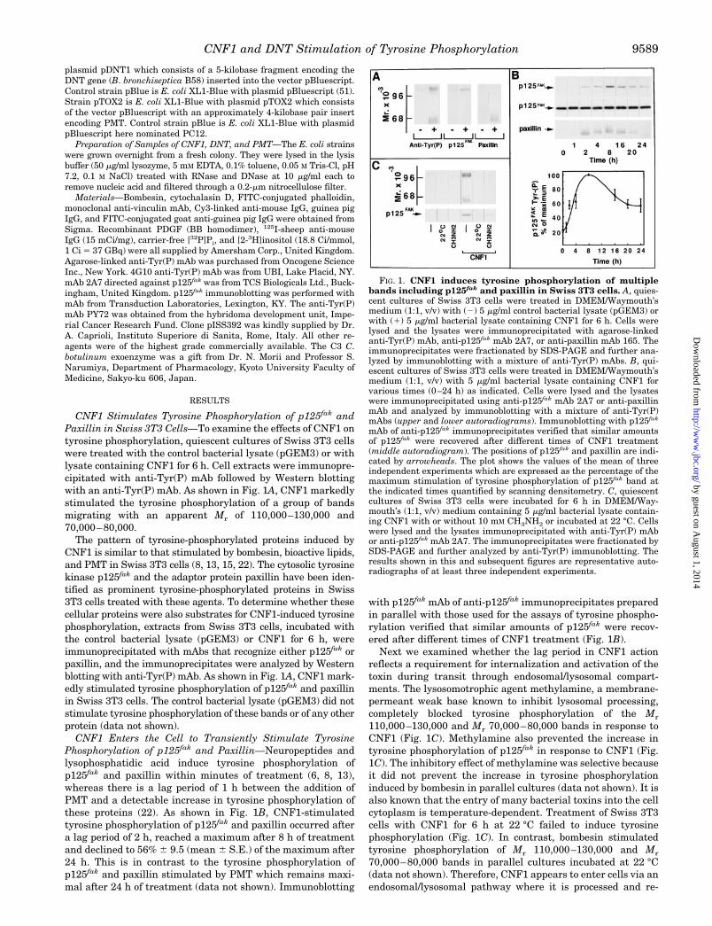
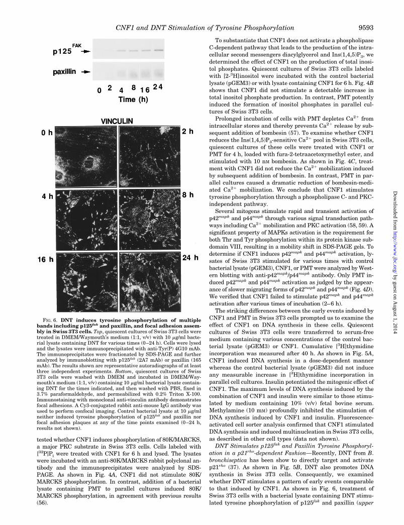
CNF1 Stimulates Tyrosine Phosphorylation of p125fak andPaxillin in Swiss 3T3 Cells—To examine the effects of CNF1 ontyrosine phosphorylation, quiescent cultures of Swiss 3T3 cellswere treated with the control bacterial lysate (pGEM3) or withlysate containing CNF1 for 6 h. Cell extracts were immunopre-cipitated with anti-Tyr(P) mAb followed by Western blottingwith an anti-Tyr(P) mAb. As shown in Fig. 1A, CNF1 markedlystimulated the tyrosine phosphorylation of a group of bandsmigrating with an apparent Mr of 110,000–130,000 and70,000–80,000.
The pattern of tyrosine-phosphorylated proteins induced byCNF1 is similar to that stimulated by bombesin, bioactive lipids,and PMT in Swiss 3T3 cells (8, 13, 15, 22). The cytosolic tyrosinekinase p125fak and the adaptor protein paxillin have been iden-tified as prominent tyrosine-phosphorylated proteins in Swiss3T3 cells treated with these agents. To determine whether thesecellular proteins were also substrates for CNF1-induced tyrosinephosphorylation, extracts from Swiss 3T3 cells, incubated withthe control bacterial lysate (pGEM3) or CNF1 for 6 h, wereimmunoprecipitated with mAbs that recognize either p125fak orpaxillin, and the immunoprecipitates were analyzed by Westernblotting with anti-Tyr(P) mAb. As shown in Fig. 1A, CNF1 mark-edly stimulated tyrosine phosphorylation of p125fak and paxillinin Swiss 3T3 cells. The control bacterial lysate (pGEM3) did notstimulate tyrosine phosphorylation of these bands or of any otherprotein (data not shown).
CNF1 Enters the Cell to Transiently Stimulate TyrosinePhosphorylation of p125fak and Paxillin—Neuropeptides andlysophosphatidic acid induce tyrosine phosphorylation ofp125fak and paxillin within minutes of treatment (6, 8, 13),whereas there is a lag period of 1 h between the addition ofPMT and a detectable increase in tyrosine phosphorylation ofthese proteins (22). As shown in Fig. 1B, CNF1-stimulatedtyrosine phosphorylation of p125fak and paxillin occurred aftera lag period of 2 h, reached a maximum after 8 h of treatmentand declined to 56% 6 9.5 (mean 6 S.E.) of the maximum after24 h. This is in contrast to the tyrosine phosphorylation ofp125fak and paxillin stimulated by PMT which remains maxi-mal after 24 h of treatment (data not shown). Immunoblotting
with p125fak mAb of anti-p125fak immunoprecipitates preparedin parallel with those used for the assays of tyrosine phospho-rylation verified that similar amounts of p125fak were recov-ered after different times of CNF1 treatment (Fig. 1B).
Next we examined whether the lag period in CNF1 actionreflects a requirement for internalization and activation of thetoxin during transit through endosomal/lysosomal compart-ments. The lysosomotrophic agent methylamine, a membrane-permeant weak base known to inhibit lysosomal processing,completely blocked tyrosine phosphorylation of the Mr
110,000–130,000 and Mr 70,000–80,000 bands in response toCNF1 (Fig. 1C). Methylamine also prevented the increase intyrosine phosphorylation of p125fak in response to CNF1 (Fig.1C). The inhibitory effect of methylamine was selective becauseit did not prevent the increase in tyrosine phosphorylationinduced by bombesin in parallel cultures (data not shown). It isalso known that the entry of many bacterial toxins into the cellcytoplasm is temperature-dependent. Treatment of Swiss 3T3cells with CNF1 for 6 h at 22 °C failed to induce tyrosinephosphorylation (Fig. 1C). In contrast, bombesin stimulatedtyrosine phosphorylation of Mr 110,000–130,000 and Mr
70,000–80,000 bands in parallel cultures incubated at 22 °C(data not shown). Therefore, CNF1 appears to enter cells via anendosomal/lysosomal pathway where it is processed and re-
FIG. 1. CNF1 induces tyrosine phosphorylation of multiplebands including p125fak and paxillin in Swiss 3T3 cells. A, quies-cent cultures of Swiss 3T3 cells were treated in DMEM/Waymouth’smedium (1:1, v/v) with (2) 5 mg/ml control bacterial lysate (pGEM3) orwith (1) 5 mg/ml bacterial lysate containing CNF1 for 6 h. Cells werelysed and the lysates were immunoprecipitated with agarose-linkedanti-Tyr(P) mAb, anti-p125fak mAb 2A7, or anti-paxillin mAb 165. Theimmunoprecipitates were fractionated by SDS-PAGE and further ana-lyzed by immunoblotting with a mixture of anti-Tyr(P) mAbs. B, qui-escent cultures of Swiss 3T3 cells were treated in DMEM/Waymouth’smedium (1:1, v/v) with 5 mg/ml bacterial lysate containing CNF1 forvarious times (0–24 h) as indicated. Cells were lysed and the lysateswere immunoprecipitated using anti-p125fak mAb 2A7 or anti-paxillinmAb and analyzed by immunoblotting with a mixture of anti-Tyr(P)mAbs (upper and lower autoradiograms). Immunoblotting with p125fak
mAb of anti-p125fak immunoprecipitates verified that similar amountsof p125fak were recovered after different times of CNF1 treatment(middle autoradiogram). The positions of p125fak and paxillin are indi-cated by arrowheads. The plot shows the values of the mean of threeindependent experiments which are expressed as the percentage of themaximum stimulation of tyrosine phosphorylation of p125fak band atthe indicated times quantified by scanning densitometry. C, quiescentcultures of Swiss 3T3 cells were incubated for 6 h in DMEM/Way-mouth’s (1:1, v/v) medium containing 5 mg/ml bacterial lysate contain-ing CNF1 with or without 10 mM CH3NH2 or incubated at 22 °C. Cellswere lysed and the lysates immunoprecipitated with anti-Tyr(P) mAbor anti-p125fak mAb 2A7. The immunoprecipitates were fractionated bySDS-PAGE and further analyzed by anti-Tyr(P) immunoblotting. Theresults shown in this and subsequent figures are representative auto-radiographs of at least three independent experiments.
CNF1 and DNT Stimulation of Tyrosine Phosphorylation 9589
by guest on August 1, 2014
http://ww
w.jbc.org/
Dow
nloaded from
leased into the cytosol in an active form to stimulate tyrosinephosphorylation of p125fak and paxillin.
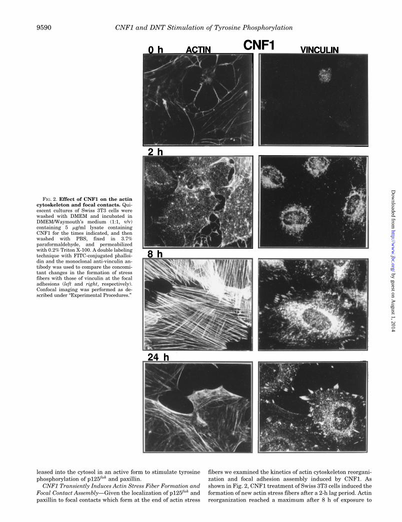
CNF1 Transiently Induces Actin Stress Fiber Formation andFocal Contact Assembly—Given the localization of p125fak andpaxillin to focal contacts which form at the end of actin stress
fibers we examined the kinetics of actin cytoskeleton reorgani-zation and focal adhesion assembly induced by CNF1. Asshown in Fig. 2, CNF1 treatment of Swiss 3T3 cells induced theformation of new actin stress fibers after a 2-h lag period. Actinreorganization reached a maximum after 8 h of exposure to
FIG. 2. Effect of CNF1 on the actincytoskeleton and focal contacts. Qui-escent cultures of Swiss 3T3 cells werewashed with DMEM and incubated inDMEM/Waymouth’s medium (1:1, v/v)containing 5 mg/ml lysate containingCNF1 for the times indicated, and thenwashed with PBS, fixed in 3.7%paraformaldehyde, and permeabilizedwith 0.2% Triton X-100. A double labelingtechnique with FITC-conjugated phalloi-din and the monoclonal anti-vinculin an-tibody was used to compare the concomi-tant changes in the formation of stressfibers with those of vinculin at the focaladhesions (left and right, respectively).Confocal imaging was performed as de-scribed under “Experimental Procedures.”
CNF1 and DNT Stimulation of Tyrosine Phosphorylation9590
by guest on August 1, 2014
http://ww
w.jbc.org/
Dow
nloaded from

CNF1. Interestingly, after 24 h of CNF1 treatment, the numberof actin stress fibers decreased and actin appeared to accumu-late in a cortical fashion. Vinculin staining showed the recruit-
ment of this protein to focal adhesions formed at the ends ofactin stress fibers. The number of focal adhesions also reacheda maximum after 8 h and decreased after 24 h treatment (Fig.
FIG. 3. Effect of cytochalasin D,PDGF, and C3 exoenzyme on CNF1-stimulated tyrosine phosphorylationand focal adhesion assembly. Upperpanel, effect of cytochalasin D and PDGFon CNF1-stimulated focal adhesion as-sembly. Cells were treated with (A) 5mg/ml control bacterial lysate (pGEM3)for 4 h, (B) 5 mg/ml of bacterial lysatecontaining CNF1 for 4 h, (C) 1.2 mM cy-tochalasin D (CYT.D) for 1 h and subse-quently with 5 mg/ml bacterial lysate con-taining CNF1 for 4 h, or (D) 5 mg/mlbacterial lysate containing CNF1 for 4 hand 30 ng/ml PDGF for a further 10 min.Cells were then washed with PBS andfixed in 4% paraformaldehyde, permeabi-lized, and focal adhesion were then visu-alized with mouse anti-vinculin mAb andCy3-conjugated rabbit anti-mouse IgG.Middle panel, effect of cytochalasin D andPDGF on CNF1 stimulated p125fak tyro-sine phosphorylation. Quiescent Swiss3T3 cells were treated with (2) 5 mg/ml ofthe control bacterial lysate (pGEM3) ortreated with 1.2 mM cytochalasin D for 1 hand then incubated with 5 mg/ml bacteriallysate containing CNF1 for a further 4 h.Parallel cell cultures were treated with 5mg/ml bacterial lysate containing CNF1for 4 h and subsequently with 30 ng/mlPDGF for a further 10 min. Cells werethen lysed, and the lysates were furtheranalyzed by immunoprecipitation usingmAb 2A7 directed against p125fak fol-lowed by anti-Tyr(P) Western blotting.Lower panel, microinjection of C3 exoen-zyme prevents the increase of tyrosinephosphorylation of focal adhesion pro-teins. Quiescent cultures of Swiss 3T3cells in 30-mm dishes were washed twiceand incubated in DMEM/Waymouth’smedium (1:1, v/v). Cells were microin-jected with 0.5 mg/ml guinea pig IgG (Aand B) or 0.5 mg/ml guinea pig IgG 1 100mg/ml C3 exoenzyme (C and D). Cellswere then treated with 5 mg/ml bacteriallysate containing CNF1. After 4 h of incu-bation, the cells were stained with 4G10mAb directed against phosphotyrosineresidues and visualized by confocal mi-croscopy using Cy3-linked rabbit anti-mouse IgG (B and D). Microinjected cells(shown in A and C) were identified bystaining with FITC-conjugated goat anti-guinea pig IgG Ab.
CNF1 and DNT Stimulation of Tyrosine Phosphorylation 9591
by guest on August 1, 2014
http://ww
w.jbc.org/
Dow
nloaded from
2). The control bacterial lysate did not induce actin stress fiberformation or focal adhesion assembly (2–24 h, data not shown).
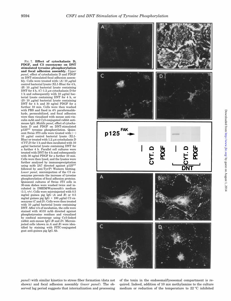
CNF1-stimulated Tyrosine Phosphorylation Requires Integ-rity of the Actin Cytoskeleton and Functional p21rho—The time-dependent effects of CNF1 on actin stress fiber formation andfocal adhesion assembly depicted in Fig. 2 prompted us toexamine whether the integrity of the actin cytoskeleton wasnecessary for CNF1-induced tyrosine phosphorylation. Quies-cent cultures of Swiss 3T3 cells were pretreated with 1.2 mM
cytochalasin D for 1 h and then stimulated with CNF1 for 6 h.As shown in Fig. 3, cytochalasin D prevented the assembly offocal adhesions (upper panel C) and completely blocked tyro-sine phosphorylation of p125fak (middle panel) and paxillin(data not shown) in response to CNF1.
Recent data from our laboratory have shown that PDGF, ata high concentration (30 ng/ml), completely abolishes bomb-esin-, lysophosphatidic acid-, and PMT-induced actin stressfiber formation and focal adhesions assembly. Similarly, themarked increase in focal adhesion assembly and actin stressfibers induced by CNF1 was prevented by the addition of PDGFat 30 ng/ml (Fig. 3, upper panel D, and results not shown).Since CNF1 stimulated p125fak tyrosine phosphorylation by amechanism dependent on the integrity of the actin cytoskele-ton, we examined whether CNF1-stimulated tyrosine phospho-rylation of p125fak could be affected by high concentrations ofPDGF. As shown in Fig. 3 (middle panel), PDGF (30 ng/ml)markedly reduced CNF1-stimulated tyrosine phosphorylationof p125fak.
The rho gene product, p21rho, has been implicated in theassembly of focal adhesions and in tyrosine phosphorylation of
p125fak and paxillin (9, 25, 29, 30). It has been suggested thatCNF1 directly targets and activates p21rho (36, 52). To inves-tigate the role of p21rho in the CNF1-stimulated tyrosine phos-phorylation of focal adhesion-associated proteins we utilizedthe C. botulinum C3 exoenzyme which ADP-ribosylates Asn41
of p21rho and thereby prevents its function (53–55). Recombi-nant C3 exoenzyme and pure guinea pig IgG were comicroin-jected into confluent and quiescent Swiss 3T3 cells, and thecultures were further treated with CNF1 for 4 h. Cells werethen fixed, permeabilized, and stained for tyrosine-phosphoryl-ated proteins which are predominantly localized at the focalcontacts in CNF1-treated cells (Fig. 3, lower panel B). Thetyrosine phosphorylation of focal adhesion proteins in responseto CNF1 was profoundly inhibited in cells microinjected withC3 exoenzyme (Fig. 3, lower panel D). Microinjection itself didnot interfere with CNF1-induced tyrosine phosphorylation offocal adhesion proteins (Fig. 3, lower panels A and B).
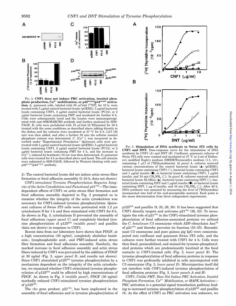
CNF1, Unlike PMT, Does Not Induce PKC Activation, InositolPhosphate Formation, Ca21 Mobilization, or MAPK Activation—PKC activation is a potential signal transduction pathway lead-ing to increased tyrosine phosphorylation of p125fak and paxillin(8). As the effect of CNF1 on PKC activation was unknown, we
FIG. 4. CNF1 does not induce PKC activation, inositol phos-phate production, Ca21 mobilization, or p42mapk/p44mapk activa-tion. A, quiescent cells, labeled with 50 mCi/ml [32P]Pi for 18 h, weretreated with 5 mg/ml control bacterial lysate (pGEM3), 5 mg/ml bacteriallysate containing CNF1, 2 mg/ml control bacterial lysate (PC12), or 2mg/ml bacterial lysate containing PMT and incubated for further 6 h.Cells were subsequently lysed and the lysates were immunoprecipi-tated with anti-80K/MARCKS antibody and further analyzed by SDS-PAGE. B, cells were prelabeled with 10 mCi/ml [2-3H]inositol for 16 htreated with the same conditions as described above adding directly tothe dishes and the cultures were incubated at 37 °C for 6 h. LiCl (20mM) was then added, and after a further 30 min the cellular inositolphosphate content was determined. C, [Ca21]i was measured as de-scribed under “Experimental Procedures.” Quiescent cells were pre-treated with 5 mg/ml control bacterial lysate (pGEM3), 5 mg/ml bacteriallysate containing CNF1, 2 mg/ml control bacterial lysate (PC12), or 2mg/ml bacterial lysate containing PMT for 4 h, and the increase in[Ca21]i induced by bombesin (10 nM) was then determined. D, quiescentcells were treated for 4 h as described above and lysed. The cell extractswere subjected to SDS-PAGE, followed by Western blotting with anti-p42mapk/p44mapk antibody.
FIG. 5. Stimulation of DNA synthesis in Swiss 3T3 cells byCNF1 and DNT. Dose-response curve for the stimulation of DNAsynthesis by CNF1 (A) and DNT (B). Confluent, quiescent cultures ofSwiss 3T3 cells were washed and incubated at 37 °C in 2 ml of Dulbec-co’s modified Eagle’s medium DMEM/Waymouth’s medium (1:1, v/v),containing 1 mCi of [3H]thymidine/ml. In panel A, cultures receivedvarious concentrations of the control bacterial lysate (å) (pGEM3),bacterial lysate containing CNF1 (E), bacterial lysate containing CNF1and 1 mg/ml insulin (●), or bacterial lysate containing CNF1, 1 mg/mlinsulin, and 10 mM CH3NH2 (M). In panel B, cultures received controlbacterial lysate XL1Blue (å), bacterial lysate containing DNT (E), bac-terial lysate containing DNT and 1 mg/ml insulin (●), or bacterial lysatecontaining DNT, 1 mg of insulin, and 10 mM CH3NH2 (M). After 40 h,DNA synthesis was assessed by measuring the level of [3H]thymidineincorporated into half of the acid-precipitable material. Each point isthe mean determination from three independent experiments.
CNF1 and DNT Stimulation of Tyrosine Phosphorylation9592
by guest on August 1, 2014
http://ww
w.jbc.org/
Dow
nloaded from
tested whether CNF1 induces phosphorylation of 80K/MARCKS,a major PKC substrate in Swiss 3T3 cells. Cells labeled with[32P]Pi were treated with CNF1 for 6 h and lysed. The lysateswere incubated with an anti-80K/MARCKS rabbit polyclonal an-tibody and the immunoprecipitates were analyzed by SDS-PAGE. As shown in Fig. 4A, CNF1 did not stimulate 80K/MARCKS phosphorylation. In contrast, addition of a bacteriallysate containing PMT to parallel cultures induced 80K/MARCKS phosphorylation, in agreement with previous results(56).
To substantiate that CNF1 does not activate a phospholipaseC-dependent pathway that leads to the production of the intra-cellular second messengers diacylglycerol and Ins(1,4,5)P3, wedetermined the effect of CNF1 on the production of total inosi-tol phosphates. Quiescent cultures of Swiss 3T3 cells labeledwith [2-3H]inositol were incubated with the control bacteriallysate (pGEM3) or with lysate containing CNF1 for 6 h. Fig. 4Bshows that CNF1 did not stimulate a detectable increase intotal inositol phosphate production. In contrast, PMT potentlyinduced the formation of inositol phosphates in parallel cul-tures of Swiss 3T3 cells.
Prolonged incubation of cells with PMT depletes Ca21 fromintracellular stores and thereby prevents Ca21 release by sub-sequent addition of bombesin (57). To examine whether CNF1reduces the Ins(1,4,5)P3-sensitive Ca21 pool in Swiss 3T3 cells,quiescent cultures of these cells were treated with CNF1 orPMT for 4 h, loaded with fura-2-tetraacetoxymethyl ester, andstimulated with 10 nM bombesin. As shown in Fig. 4C, treat-ment with CNF1 did not reduce the Ca21 mobilization inducedby subsequent addition of bombesin. In contrast, PMT in par-allel cultures caused a dramatic reduction of bombesin-medi-ated Ca21 mobilization. We conclude that CNF1 stimulatestyrosine phosphorylation through a phospholipase C- and PKC-independent pathway.
Several mitogens stimulate rapid and transient activation ofp42mapk and p44mapk through various signal transduction path-ways including Ca21 mobilization and PKC activation (58, 59). Asignificant property of MAPKs activation is the requirement forboth Thr and Tyr phosphorylation within its protein kinase sub-domain VIII, resulting in a mobility shift in SDS-PAGE gels. Todetermine if CNF1 induces p42mapk and p44mapk activation, ly-sates of Swiss 3T3 stimulated for various times with controlbacterial lysate (pGEM3), CNF1, or PMT were analyzed by West-ern blotting with anti-p42mapk/p44mapk antibody. Only PMT in-duced p42mapk and p44mapk activation as judged by the appear-ance of slower migrating forms of p42mapk and p44mapk (Fig. 4D).We verified that CNF1 failed to stimulate p42mapk and p44mapk
activation after various times of incubation (2–6 h).The striking differences between the early events induced by
CNF1 and PMT in Swiss 3T3 cells prompted us to examine theeffect of CNF1 on DNA synthesis in these cells. Quiescentcultures of Swiss 3T3 cells were transferred to serum-freemedium containing various concentrations of the control bac-terial lysate (pGEM3) or CNF1. Cumulative [3H]thymidineincorporation was measured after 40 h. As shown in Fig. 5A,CNF1 induced DNA synthesis in a dose-dependent mannerwhereas the control bacterial lysate (pGEM3) did not induceany measurable increase in [3H]thymidine incorporation inparallel cell cultures. Insulin potentiated the mitogenic effect ofCNF1. The maximum levels of DNA synthesis induced by thecombination of CNF1 and insulin were similar to those stimu-lated by medium containing 10% (v/v) fetal bovine serum.Methylamine (10 mM) profoundly inhibited the stimulation ofDNA synthesis induced by CNF1 and insulin. Fluorescence-activated cell sorter analysis confirmed that CNF1 stimulatedDNA synthesis and induced multinucleation in Swiss 3T3 cells,as described in other cell types (data not shown).
DNT Stimulates p125fak and Paxillin Tyrosine Phosphoryl-ation in a p21rho-dependent Fashion—Recently, DNT from B.bronchiseptica has been show to directly target and activatep21rho (37). As shown in Fig. 5B, DNT also promotes DNAsynthesis in Swiss 3T3 cells. Consequently, we examinedwhether DNT stimulates a pattern of early events comparableto that induced by CNF1. As shown in Fig. 6, treatment ofSwiss 3T3 cells with a bacterial lysate containing DNT stimu-lated tyrosine phosphorylation of p125fak and paxillin (upper
FIG. 6. DNT induces tyrosine phosphorylation of multiplebands including p125fak and paxillin, and focal adhesion assem-bly in Swiss 3T3 cells. Top, quiescent cultures of Swiss 3T3 cells weretreated in DMEM/Waymouth’s medium (1:1, v/v) with 10 mg/ml bacte-rial lysate containing DNT for various times (0–24 h). Cells were lysedand the lysates were immunoprecipitated with anti-Tyr(P) 4G10 mAb.The immunoprecipitates were fractionated by SDS-PAGE and furtheranalyzed by immunoblotting with p125fak (2A7 mAb) or paxillin (165mAb). The results shown are representative autoradiographs of at leastthree independent experiments. Bottom, quiescent cultures of Swiss3T3 cells were washed with DMEM and incubated in DMEM/Way-mouth’s medium (1:1, v/v) containing 10 mg/ml bacterial lysate contain-ing DNT for the times indicated, and then washed with PBS, fixed in3.7% paraformaldehyde, and permeabilized with 0.2% Triton X-100.Immunostaining with monoclonal anti-vinculin antibody demonstratesfocal adhesions. A Cy3-conjugated rabbit anti-mouse IgG antibody wasused to perform confocal imaging. Control bacterial lysate at 10 mg/mlneither induced tyrosine phosphorylation of p125fak and paxillin norfocal adhesion plaques at any of the time points examined (0–24 h,results not shown).
CNF1 and DNT Stimulation of Tyrosine Phosphorylation 9593
by guest on August 1, 2014
http://ww
w.jbc.org/
Dow
nloaded from
panel) with similar kinetics to stress fiber formation (data notshown) and focal adhesion assembly (lower panel). The ob-served lag period suggests that internalization and processing
of the toxin in the endosomal/lysosomal compartment is re-quired. Indeed, addition of 10 mM methylamine to the culturemedium or reduction of the temperature to 22 °C inhibited
FIG. 7. Effect of cytochalasin D,PDGF, and C3 exoenzyme on DNTstimulated tyrosine phosphorylationand focal adhesion assembly. Upperpanel, effect of cytochalasin D and PDGFon DNT-stimulated focal adhesion assem-bly. Cells were treated with: (A) 10 mg/mlcontrol bacterial lysate (XL1-Blue) for 4 h,(B) 10 mg/ml bacterial lysate containingDNT for 4 h, (C) 1.2 mM cytochalasin D for1 h and subsequently with 10 mg/ml bac-terial lysate containing DNT for 4 h, or(D) 10 mg/ml bacterial lysate containingDNT for 4 h and 30 ng/ml PDGF for afurther 10 min. Cells were then washedwith PBS and fixed in 4% paraformalde-hyde, permeabilized, and focal adhesionwere then visualized with mouse anti-vin-culin mAb and Cy3-conjugated rabbit anti-mouse IgG. Middle panel, effect of cytocha-lasin D and PDGF on DNT-stimulatedp125fak tyrosine phosphorylation. Quies-cent Swiss 3T3 cells were treated with (2)10 mg/ml control bacterial lysate (XL1-Blue) or treated with 1.2 mM cytochalasin D(CYT.D) for 1 h and then incubated with 10mg/ml bacterial lysate containing DNT fora further 4 h. Parallel cell cultures weretreated with DNT for 4 h and subsequentlywith 30 ng/ml PDGF for a further 10 min.Cells were then lysed, and the lysates werefurther analyzed by immunoprecipitationusing mAb 2A7 directed against p125fak
followed by anti-Tyr(P) Western blotting.Lower panel, microinjection of the C3 ex-oenzyme prevents the increase of tyrosinephosphorylation of focal adhesion proteins.Quiescent cultures of Swiss 3T3 cells in30-mm dishes were washed twice and in-cubated in DMEM/Waymouth’s medium(1:1, v/v). Cells were microinjected with 0.5mg/ml guinea pig IgG (A and B) or 0.5mg/ml guinea pig IgG 1 100 mg/ml C3 ex-oenzyme (C and D). Cells were then treatedwith 10 mg/ml bacterial lysate containingDNT. After 4 h of incubation, the cells werestained with 4G10 mAb directed againstphosphotyrosine residues and visualizedby confocal microscopy using Cy3-linkedrabbit anti-mouse IgG (B and D). Microin-jected cells (shown in A and B) were iden-tified by staining with FITC-conjugatedgoat anti-guinea pig IgG Ab.
CNF1 and DNT Stimulation of Tyrosine Phosphorylation9594
by guest on August 1, 2014
http://ww
w.jbc.org/
Dow
nloaded from
DNT-stimulated tyrosine phosphorylation (data not shown).Next, we tested whether the integrity of the cytoskeleton was
necessary for DNT-stimulated tyrosine phosphorylation. Asshown in Fig. 7, cytochalasin D or PDGF blocked DNT-stimu-lated stress fiber formation (data not shown), focal adhesionassembly (upper panel), and tyrosine phosphorylation ofp125fak (middle panel). In addition, microinjection of C3 exoen-zyme profoundly inhibited tyrosine phosphorylation of focaladhesion proteins in response to DNT (lower panel).
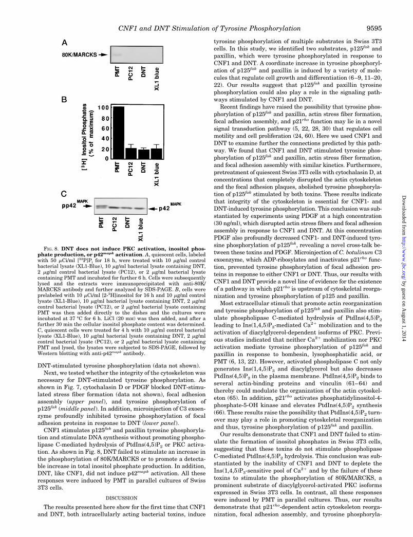
CNF1 stimulates p125fak and paxillin tyrosine phosphoryla-tion and stimulate DNA synthesis without promoting phospho-lipase C-mediated hydrolysis of PtdIns(4,5)P2 or PKC activa-tion. As shown in Fig. 8, DNT failed to stimulate an increase inthe phosphorylation of 80K/MARCKS or to promote a detecta-ble increase in total inositol phosphate production. In addition,DNT, like CNF1, did not induce p42mapk activation. All theseresponses were induced by PMT in parallel cultures of Swiss3T3 cells.
DISCUSSION
The results presented here show for the first time that CNF1and DNT, both intracellularly acting bacterial toxins, induce
tyrosine phosphorylation of multiple substrates in Swiss 3T3cells. In this study, we identified two substrates, p125fak andpaxillin, which were tyrosine phosphorylated in response toCNF1 and DNT. A coordinate increase in tyrosine phosphoryl-ation of p125fak and paxillin is induced by a variety of mole-cules that regulate cell growth and differentiation (6–9, 11–20,22). Our results suggest that p125fak and paxillin tyrosinephosphorylation could also play a role in the signaling path-ways stimulated by CNF1 and DNT.
Recent findings have raised the possibility that tyrosine phos-phorylation of p125fak and paxillin, actin stress fiber formation,focal adhesion assembly, and p21rho function may lie in a novelsignal transduction pathway (5, 22, 28, 30) that regulates cellmotility and cell proliferation (24, 60). Here we used CNF1 andDNT to examine further the connections predicted by this path-way. We found that CNF1 and DNT stimulated tyrosine phos-phorylation of p125fak and paxillin, actin stress fiber formation,and focal adhesion assembly with similar kinetics. Furthermore,pretreatment of quiescent Swiss 3T3 cells with cytochalasin D, atconcentrations that completely disrupted the actin cytoskeletonand the focal adhesion plaques, abolished tyrosine phosphoryla-tion of p125fak stimulated by both toxins. These results indicatethat integrity of the cytoskeleton is essential for CNF1- andDNT-induced tyrosine phosphorylation. This conclusion was sub-stantiated by experiments using PDGF at a high concentration(30 ng/ml), which disrupted actin stress fibers and focal adhesionassembly in response to CNF1 and DNT. At this concentrationPDGF also profoundly decreased CNF1- and DNT-induced tyro-sine phosphorylation of p125fak, revealing a novel cross-talk be-tween these toxins and PDGF. Microinjection of C. botulinum C3exoenzyme, which ADP-ribosylates and inactivates p21rho func-tion, prevented tyrosine phosphorylation of focal adhesion pro-teins in response to either CNF1 or DNT. Thus, our results withCNF1 and DNT provide a novel line of evidence for the existenceof a pathway in which p21rho is upstream of cytoskeletal reorga-nization and tyrosine phosphorylation of p125 and paxillin.
Most extracellular stimuli that promote actin reorganizationand tyrosine phosphorylation of p125fak and paxillin also stim-ulate phospholipase C-mediated hydrolysis of PtdIns(4,5)P2
leading to Ins(1,4,5)P3-mediated Ca21 mobilization and to theactivation of diacylglycerol-dependent isoforms of PKC. Previ-ous studies indicated that neither Ca21 mobilization nor PKCactivation mediate tyrosine phosphorylation of p125fak andpaxillin in response to bombesin, lysophosphatidic acid, orPMT (6, 13, 22). However, activated phospholipase C not onlygenerates Ins(1,4,5)P3 and diacylglycerol but also decreasesPtdIns(4,5)P2 in the plasma membrane. PtdIns(4,5)P2 binds toseveral actin-binding proteins and vinculin (61–64) andthereby could modulate the organization of the actin cytoskel-eton (65). In addition, p21rho activates phosphatidylinositol-4-phosphate-5-OH kinase and elevates PtdIns(4,5)P2 synthesis(66). These results raise the possibility that PtdIns(4,5)P2 turn-over may play a role in promoting cytoskeletal reorganizationand thus, tyrosine phosphorylation of p125fak and paxillin.
Our results demonstrate that CNF1 and DNT failed to stim-ulate the formation of inositol phosphates in Swiss 3T3 cells,suggesting that these toxins do not stimulate phospholipaseC-mediated PtdIns(4,5)P2 hydrolysis. This conclusion was sub-stantiated by the inability of CNF1 and DNT to deplete theIns(1,4,5)P3-sensitive pool of Ca21 and by the failure of thesetoxins to stimulate the phosphorylation of 80K/MARCKS, aprominent substrate of diacylglycerol-activated PKC isoformsexpressed in Swiss 3T3 cells. In contrast, all these responseswere induced by PMT in parallel cultures. Thus, our resultsdemonstrate that p21rho-dependent actin cytoskeleton reorga-nization, focal adhesion assembly, and tyrosine phosphoryla-
FIG. 8. DNT does not induce PKC activation, inositol phos-phate production, or p42mapk activation. A, quiescent cells, labeledwith 50 mCi/ml [32P]Pi for 18 h, were treated with 10 mg/ml controlbacterial lysate (XL1-Blue), 10 mg/ml bacterial lysate containing DNT,2 mg/ml control bacterial lysate (PC12), or 2 mg/ml bacterial lysatecontaining PMT and incubated for further 6 h. Cells were subsequentlylysed and the extracts were immunoprecipitated with anti-80K/MARCKS antibody and further analyzed by SDS-PAGE. B, cells wereprelabeled with 10 mCi/ml [2-3H]inositol for 16 h and 10 mg/ml controllysate (XL1-Blue), 10 mg/ml bacterial lysate containing DNT, 2 mg/mlcontrol bacterial lysate (PC12), or 2 mg/ml bacterial lysate containingPMT was then added directly to the dishes and the cultures wereincubated at 37 °C for 6 h. LiCl (20 mM) was then added, and after afurther 30 min the cellular inositol phosphate content was determined.C, quiescent cells were treated for 4 h with 10 mg/ml control bacteriallysate (XL1-Blue), 10 mg/ml bacterial lysate containing DNT, 2 mg/mlcontrol bacterial lysate (PC12), or 2 mg/ml bacterial lysate containingPMT and lysed, the lysates were subjected to SDS-PAGE, followed byWestern blotting with anti-p42mapk antibody.
CNF1 and DNT Stimulation of Tyrosine Phosphorylation 9595
by guest on August 1, 2014
http://ww
w.jbc.org/
Dow
nloaded from
tion of p125fak and paxillin can by dissociated from hydrolysisof PtdIns(4,5)P2 in CNF1- and DNT-treated Swiss 3T3 cells.
The findings presented here have another interesting impli-cation. A wide range of extracellular signals including growthfactors and mitogenic neuropeptides activate one or more mem-bers of the family of the highly conserved serine/threoninemitogen-activated protein (MAP) kinases (ERKs) (58, 59, 67).MAP kinases, important intermediates in signal transductionpathways leading to mitogenesis or differentiation, are acti-vated predominantly via PKC in Swiss 3T3 cells stimulated bybombesin. Once activated p42mapk (ERK2) and p44mapk (ERK1)phosphorylate an array of cellular proteins including proteinkinases, transcription factors, and proteins involved in theregulation of cell growth (68–71). Because CNF1 and DNTstimulate DNA synthesis in Swiss 3T3 cells, we investigatedthe ability of these toxins to induce activation of p42mapk andp44mapk. Our results demonstrated that unlike PMT, CNF1and DNT did not stimulate p42mapk in these cells. These resultssuggest that CNF1 and DNT can signal entry into S phasewithout activating p42mapk and p44mapk in Swiss 3T3 cells.
The signaling events triggered by CNF and DNT appear tobe very similar which suggest that both toxins trigger the sameinitial event, and indeed there is evidence that CNF and DNTeach directly target and modify Rho (36, 37). CNF and DNT arehomologous over a region of 100 amino acids near the C termi-nus (72) which in DNT but not in CNF contains a putativeP-loop nucleotide-binding site. We have identified a lysine res-idue within this motif that is essential for the action of DNT,which suggests that the motif, and by inference this region, isinvolved in the catalytic function of DNT (51). Thus both CNFand DNT activate Rho but possibly through a differentmechanism.
In conclusion, our results with CNF1 and DNT provide furtherevidence for a p21rho-dependent pathway leading to tyrosinephosphorylation of p125fak and paxillin. In particular, actinstress fiber formation, focal adhesion assembly, and tyrosinephosphorylation of p125fak were dissociated from PtdIns(4,5)P2
hydrolysis in CNF1- and DNT-treated cells. Furthermore, CNF1and DNT induce DNA synthesis in the absence of detectableactivation of p42mapk and p44mapk providing additional evidencefor a novel p21rho-dependent signaling pathway that leads toentry into the S phase of the cell cycle.
Acknowledgments—We thank Drs. Toni Adams and Isobel Hoskin forproduction of bacterial lysates and Dr. Alfredo Caprioli for supplyingthe CNF1 clone.
REFERENCES
1. Hanks, S. K., Calalb, M. B., Harper, M. C., and Patel, S. K. (1992) Proc. Natl.Acad. Sci. U. S. A. 89, 8487–8491
2. Schaller, M. D., Borgman, C. A., Cobb, B. S., Vines, R. R., Reynolds, A. B., andParsons, J. T. (1992) Proc. Natl. Acad. Sci. U. S. A. 89, 5192–5196
3. Salgia, R., Li, J.-L., Lo, S. H., Brunkhorst, B., Kansas, G. S., Sobhany, E. S.,Sun, Y., Pisick, E., Hallek, M., Ernst, T., Tantravahi, R., Chen, L. B., andGriffin, J. D. (1995) J. Biol. Chem. 270, 5039–5047
4. Turner, C. E., and Miller, J. T. (1994) J. Cell Sci. 107, 1583–15915. Rozengurt, E. (1995) Cancer Surveys 24, 81–966. Zachary, I., Sinnett-Smith, J., and Rozengurt, E. (1992) J. Biol. Chem. 267,
19031–190347. Sinnett-Smith, J., Zachary, I., Valverde, A. M., and Rozengurt, E. (1993)
J. Biol. Chem. 268, 14261–142688. Zachary, I., Sinnett-Smith, J., Turner, C. E., and Rozengurt, E. (1993) J. Biol.
Chem. 268, 22060–220659. Rankin, S., and Rozengurt, E. (1994) J. Biol. Chem. 269, 704–710
10. Rankin, S., Hooshmand-Rad, R., Claesson-Welsh, L., and Rozengurt, E. (1996)J. Biol. Chem. 271, 7829–7834
11. Kumagai, N., Morii, N., Fujisawa, K., Yoshimasa, T., Nakao, K., and Naru-miya, S. (1993) FEBS Lett. 329, 273–276
12. Hordijk, P. L., Verlaan, I., van Corven, E. J., and Moolenaar, W. H. (1994)J. Biol. Chem. 269, 645–651
13. Seufferlein, T., and Rozengurt, E. (1994) J. Biol. Chem. 269, 9345–935114. Seufferlein, T., and Rozengurt, E. (1994) J. Biol. Chem. 269, 27610–2761715. Seufferlein, T., and Rozengurt, E. (1995) J. Biol. Chem. 270, 1–916. Kornberg, L., Earp, H. S., Parsons, J. T., Schaller, M., and Juliano, R. L. (1992)
J. Biol. Chem. 267, 23439–2344217. Guan, J. L., and Shalloway, D. (1992) Nature 358, 690–692
18. Burridge, K., Turner, C. E., and Romer, L. H. (1992) J. Cell Biol. 119, 893–90319. Lipfert, L., Haimovich, B., Schaller, M. D., Cobb, B. S., Parsons, J. T., and
Brugge, J. S. (1992) J. Cell Biol. 119, 905–91220. Vuori, K., and Ruoslahti, E. (1993) J. Biol. Chem. 268, 21459–2146221. Kanner, S. B., Reynolds, A. B., Vines, R. R., and Parsons, J. T. (1990) Proc.
Natl. Acad. Sci. U. S. A. 87, 3328–333222. Lacerda, H. M., Lax, A. J., and Rozengurt, E. (1996) J. Biol. Chem. 271,
439–44523. Schaller, M. D., and Parsons, J. T. (1995) Mol. Cell. Biol. 15, 2635–264524. Ilic, D., Furuta, Y., Kanazawa, S., Takeda, N., Sobue, K., Nakatsuji, J., No-
mura, S., Fujimoto, J., Okada, M., Yamamoto, T., and Aizawa, S. (1995)Nature 377, 539–544
25. Ridley, A. J., and Hall, A. (1992) Cell 70, 389–39926. Hynes, R. O. (1992) Cell 69, 11–2527. Turner, C. E. (1991) J. Cell Biol. 115, 201–20728. Rankin, S., Morii, N., Narumiya, S., and Rozengurt, E. (1994) FEBS Lett. 354,
315–31929. Kumagai, N., Morii, N., Fujisawa, K., Nemoto, Y., and Narumiya, S. (1993)
J. Biol. Chem. 268, 24535–2453830. Seckl, M. J., Morii, N., Narumiya, S., and Rozengurt, E. (1995) J. Biol. Chem.
270, 6984–699031. Aktories, K., Braun, U., Habermann, B., and Rosener, S. (1990) in ADP-
ribosylating Toxins and G Proteins: Insights into Signal Transduction(Moss, J., and Vaughn, M., ed) pp. 97–115, American Society for Microbi-ology, Washington, D. C.
32. Just, I., Wilm, M., Selzer, J., Rex, G., von Eichel-Streiber, C., Mann, M., andAktories, K. (1995) J. Biol. Chem. 270, 13932–13936
33. Just, I., Selzer, J., Wilm, M., von Eichel-Streiber, C., Mann, M., and Aktories,K. (1995) Nature 375, 500–503
34. Falbo, V., Pace, T., Picci, L., Pizzi, E., and Caprioli, A. (1993) Infect. Immun.61, 4909–4914
35. Horiguchi, Y., Nakai, T., and Kume, K. (1989) Microbiol. Pathogens 6, 361–36836. Oswald, E., Sugai, M., Labigne, A., Wu, W., Fiorentini, C., Boquet, P., and
O’Brien, A. D. (1994) Proc. Natl. Acad. Sci. U. S. A. 91, 3814–381837. Horiguchi, Y., Senda, T., Sugimoto, N., Katahira, J., and Matsuda, M. (1995)
J. Cell Sci. 108, 3243–325138. Caprioli, A., Falbo, V., Roda, L. G., Ruggeri, F. M., and Zona, C. (1983) Infect.
Immun. 39, 1300–130639. Caprioli, A., Donelli, G., Falbo, V., Possenti, R., and Roda, L. G. (1984) Bio-
chem. Biophys. Res. Commun. 118, 587–59340. Horiguchi, Y., Sugimoto, N., and Matsuda, M. (1993) Infect. Immun. 61,
3611–361541. Rozengurt, E., and Sinnett-Smith, J. (1983) Proc. Natl. Acad. Sci. U. S. A. 80,
2936–294042. Herget, T., Brooks, S. F., Broad, S., and Rozengurt, H. (1993) Proc. Natl. Acad.
Sci U. S. A. 909, 2945–294943. Kamps, M. P., and Sefton, B. M. (1989) Anal. Biochem. 176, 22–2744. Erusalimsky, J. D., Brooks, S. F., Herget, T., Morris, C., and Rozengurt, E.
(1991) J. Biol. Chem. 266, 7073–708045. Berridge, M. J., Dawson, R. M. C., Downes, C. P., Heslop, J. P., and Irvine, R.
F. (1983) Biochem. J. 212, 473–48246. Grynkiewicz, G., Poenie, M., and Tsien, R. Y. (1985) J. Biol. Chem. 260,
3440–345047. Lopez-Rivas, A., Mendoza, S. A., Nanberg, E., Sinnett-Smith, J., and Rozen-
gurt, E. (1987) Proc. Natl. Acad. Sci. U. S. A. 84, 5768–577248. Nanberg, E., and Rozengurt, E. (1988) EMBO J. 7, 2741–274749. Tsien, R. Y., Pozzan, T., and Rink, T. J. (1982) J. Cell Biol. 94, 325–33450. Leevers, S. J., and Marshall, C. J. (1992) EMBO J. 11, 569–57451. Pullinger, G. D., Adams, T. E., Mullan, P. B., Garrod, T. I., and Lax, A. J.
(1996) Infect. Immun. 64, 4163–417152. Fiorentini, C., Donelli, G., Matarrese, P., Fabbri, A., Paradisi, S., and Boquet,
P. (1995) Infect. Immun. 63, 3936–394453. Nemoto, Y., Namba, T., Kozaki, S., and Narumiya, S. (1991) J. Biol. Chem.
266, 19312–1931954. Aktories, K., Weller, U., and Chatwal, G. S. (1987) FEBS Lett. 212, 109–11355. Sekine, A., Fujiwara, M., and Narumiya, S. (1989) J. Biol. Chem. 264,
8602–860556. Staddon, J. M., Chanter, N., Lax, A. J., Higgins, T. E., and Rozengurt, E. (1990)
J. Biol. Chem. 265, 11841–1184857. Staddon, J. M., Barker, C. J., Murphy, A. C., Chanter, N., Lax, A. J., Michell,
R. H., and Rozengurt, E. (1991) J. Biol. Chem. 266, 4840–484758. Pelech, S. L., and Sanghera, J. S. (1992) Science 257, 1355–135659. Posada, J., and Cooper, J. A. (1992) Mol. Biol. Cell 3, 383–39260. Gilmore, A. P., and Romer, L. H. (1996) Mol. Biol. Cell 7, 1209–122461. Lee, S. B., and Rhee, S. G. (1995) Curr. Opin. Cell Biol. 7, 183–18962. Lassing, I., and Lindberg, U. (1985) Nature 314, 472–47463. Janmey, P. A., and Stossel, T. P. (1987) Nature 325, 362–36464. Fukami, K. (1992) Nature 359, 150–15265. Gilmore, A. P., and Burridge, K. (1996) Nature 381, 531–53566. Chong, L. D., Traynor-Kaplan, A., Bokoch, G. M., and Schwartz, M. A. (1994)
Cell 79,67. Cobb, M., Boulton, T. G., and Robbins, D. J. (1991) Mol. Biol. Cell 2, 965–96868. Sturgill, T. W., and Wu, J. (1991) Biochim. Biophys. Acta 1092, 350–35769. Gille, H., Sharrock, A. D., and Shaw, P. E. (1992) Nature 358, 414–41770. Seth, A., Alvarez, E., Gupta, S., and Davis, R. J. (1991) J. Biol. Chem. 266,
23521–2352471. Lin, L.-L., Wartman, M., Lin, Y. A., Knopf, J. L., Seth, A., and Davies, R. J.
(1993) Cell 72, 269–27872. Walker, K. E., and Weiss, A. A. (1994) Infect Immun 62, 3817–3828
CNF1 and DNT Stimulation of Tyrosine Phosphorylation9596
by guest on August 1, 2014
http://ww
w.jbc.org/
Dow
nloaded from
Copyright © 2022 FDOKUMEN