Evolution and Adaptation of Australian Bordetella ... - UNSWorks
Upload
independentCategory
view
1download
0
Biochimica et Biophysica Acta 1788 (2009) 1249–1254
Contents lists available at ScienceDirect
Biochimica et Biophysica Acta
j ourna l homepage: www.e lsev ie r.com/ locate /bbamem
Different modes of membrane permeabilization by two RTX toxins: HlyA fromEscherichia coli and CyaA from Bordetella pertussis
Radovan Fišer, Ivo Konopásek ⁎Department of Genetics and Microbiology, Faculty of Science, Charles University in Prague, Viničná 5, CZ-128 44, Prague 2, Czech Republic
Abbreviations: ANTS, 8-Aminonaphthalene-1,3,6-trimembranes; CyaA, adenylate cyclase toxin from Bordetelpyridinium bromide; FITC, fluorescein-5-isothiocyanEscherichia coli; LUV100, LUV400, LUV1000, large unilamefilters of 100, 400 and 1000 nm pore-size, respectively; Ppolymyxa⁎ Corresponding author. Tel.: +420 221 951 711; fax:
E-mail addresses: [email protected] (R. Fišer), kon(I. Konopásek).
0005-2736/$ – see front matter © 2009 Elsevier B.V. Adoi:10.1016/j.bbamem.2009.03.019
a b s t r a c t
a r t i c l e i n f oArticle history:Received 6 November 2008Received in revised form 18 March 2009Accepted 29 March 2009Available online 5 April 2009
Keywords:CyaAHlyAANTSDPXFluorescence requenching methodLiposome disruption
This study clarifies the membrane disruption mechanisms of two bacterial RTX toxins: αhemolysin (HlyA)from Escherichia coli and a highly homologous adenylate cyclase toxin (CyaA) from Bordetella pertussis. Forthis purpose, we employed a fluorescence requenching method using liposomes (extruded through filters ofdifferent pore size — 1000 nm, 400 nm or 100 nm) with encapsulated fluorescent dye/quencher pair ANTS/DPX. We showed that both toxins induced a graded leakage of liposome content with different selectivities αfor DPX and ANTS. In contrast to HlyA, CyaA exhibited a higher selectivity for cationic quencher DPX, whichincreased with vesicle diameter. Large unilamellar vesicles (LUV1000) were found to be more suitable fordistinguishing between high α values whereas smaller ones (LUV100) were more appropriate fordiscriminating an all-or-none leakage (α=0) from the graded leakage with low values of α. Whiledisrupting LUV1000, CyaA caused a highly cation-selective leakage (α~15) whereas its mutated form withdecreased channel K+/Cl− selectivity due to two substitutions in a predicted transmembrane segment(CyaA-E509K+E516K) exhibited much lower selectivity (α∼6). We concluded that the fluorescencerequenching method in combination with different size of liposomes is a valuable tool for characterization ofpore-forming toxins and their variants.
© 2009 Elsevier B.V. All rights reserved.
1. Introduction
Both adenylate cyclase toxin of Bordetella pertussis (CyaA, 177 kDa)and αhemolysin of Escherichia coli (HlyA, 117 kDa) belong to the RTXtoxin (RTX, Repeat in ToXin) family. Their C-terminal hemolysin parts,RTX cytolysin moieties, are highly homologous [1] (see Fig. 1). RTXcytolysin part is known for its ability to damage biological membraneseven without the presence of specific cellular receptors [2–4]. Never-theless, cellular receptors for both toxins have been discovered [5,6].
RTX repeat domain bears multiple Ca2+ binding sites that arenecessary for the full hemolytic activity.N-terminalpartof thehemolysinis responsible for the binding to biological membranes and channelformation due to its high hydrophobicity and double acylation.
Whereas HlyA is known mainly for its hemolytic capacity, CyaAinteracts with its target membrane in a more complex way. Afterbinding to the target cell membrane it translocates its N-terminaladenylate cyclase domain into the cytoplasmwhere it rapidly converts
sulfonic acid; BLM, black lipidla pertussis; DPX, p-xylene-bis-ate; HlyA, αhemolysin fromllar vesicles extruded througholyB, Polymyxin B from Bacillus
+420 221 951 [email protected]
ll rights reserved.
ATP to cAMP upon activation by cellular calmodulin [7,8], thus causingrapid ATP depletion and cell death [9,10]. In parallel, the RTXhemolysin moiety of CyaA forms small cation-selective membranechannels that allow the entry of monovalent cations and areresponsible for osmotic lysis of the cell [11].
In black lipid membranes, CyaA forms very narrow channels withan estimated diameter of 0.6–0.8 nm [12,13] whereas HlyA formsmuch wider channels (approx. 2–3 nm) with a lifetime much longerthan those of CyaA [14]. Both CyaA and HlyA channels are supposed tobe formed by oligomers of the toxin subunits [3,11,14–17]. Both toxinswere recently shown to induce the formation of non-lamellar phasesin biological membranes [18,19].
An “all-or-none” mechanism (see below) by which toxin releasesmodel lipid vesicle content was found for HlyA from E. coli [3]. Theauthors used the method developed by Parente et al. [20] in whichliposomes loadedwith fluorescence probe ANTS and its quencher DPXwere subjected to toxin-induced lysis. The mechanism by which CyaAfrom Bordetella pertussis disrupts a membrane, however, has not yetbeen investigated.
When liposomes loaded with ANTS/DPX pair are subjected totoxin-induced lysis there are in general two possible differentmechanisms of membrane disruption:
1) “Graded leakage”means that all of thevesicles affectedby the toxinarelosingcontinuouslysomepartof their innercontents. This correspondstothe formationof transientnarrowporeswhichdonotallowone-steprelease of their inner contents. This release can theoretically be more
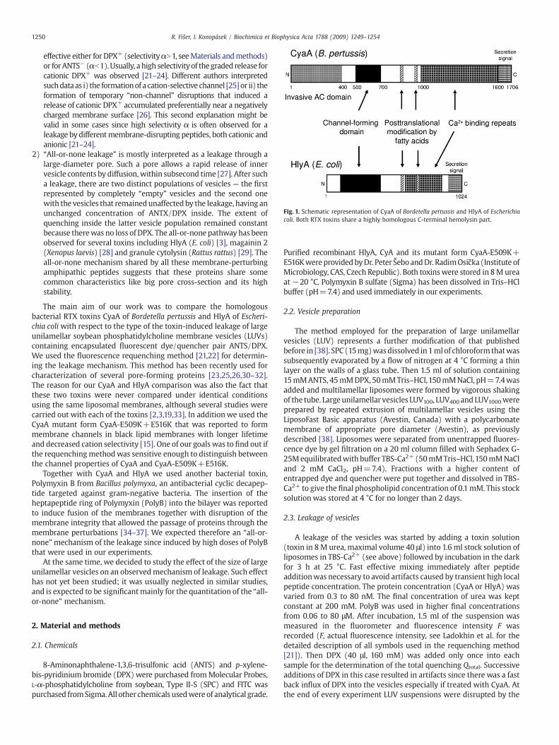
Fig. 1. Schematic representation of CyaA of Bordetella pertussis and HlyA of Escherichiacoli. Both RTX toxins share a highly homologous C-terminal hemolysin part.
1250 R. Fišer, I. Konopásek / Biochimica et Biophysica Acta 1788 (2009) 1249–1254
effective either for DPX+ (selectivityαN1, seeMaterials andmethods)or forANTS− (αb1).Usually, a high selectivityof thegraded release forcationic DPX+ was observed [21–24]. Different authors interpretedsuchdata as i) the formationof a cation-selective channel [25] or ii) theformation of temporary “non-channel” disruptions that induced arelease of cationic DPX+ accumulated preferentially near a negativelycharged membrane surface [26]. This second explanation might bevalid in some cases since high selectivity α is often observed for aleakage by differentmembrane-disrupting peptides, both cationic andanionic [21–24].
2) “All-or-none leakage” is mostly interpreted as a leakage through alarge-diameter pore. Such a pore allows a rapid release of innervesicle contents by diffusion,within subsecond time [27]. After sucha leakage, there are two distinct populations of vesicles — the firstrepresented by completely “empty” vesicles and the second onewith the vesicles that remainedunaffected by the leakage, having anunchanged concentration of ANTX/DPX inside. The extent ofquenching inside the latter vesicle population remained constantbecause therewas no loss of DPX. The all-or-none pathway has beenobserved for several toxins including HlyA (E. coli) [3], magainin 2(Xenopus laevis) [28] and granule cytolysin (Rattus rattus) [29]. Theall-or-none mechanism shared by all these membrane-perturbingamphipathic peptides suggests that these proteins share somecommon characteristics like big pore cross-section and its highstability.
The main aim of our work was to compare the homologousbacterial RTX toxins CyaA of Bordetella pertussis and HlyA of Escheri-chia coliwith respect to the type of the toxin-induced leakage of largeunilamellar soybean phosphatidylcholine membrane vesicles (LUVs)containing encapsulated fluorescent dye/quencher pair ANTS/DPX.We used the fluorescence requenching method [21,22] for determin-ing the leakage mechanism. This method has been recently used forcharacterization of several pore-forming proteins [23,25,26,30–32].The reason for our CyaA and HlyA comparison was also the fact thatthese two toxins were never compared under identical conditionsusing the same liposomal membranes, although several studies werecarried out with each of the toxins [2,3,19,33]. In additionwe used theCyaA mutant form CyaA-E509K+E516K that was reported to formmembrane channels in black lipid membranes with longer lifetimeand decreased cation selectivity [15]. One of our goals was to find out ifthe requenchingmethodwas sensitive enough to distinguish betweenthe channel properties of CyaA and CyaA-E509K+E516K.
Together with CyaA and HlyA we used another bacterial toxin,Polymyxin B from Bacillus polymyxa, an antibacterial cyclic decapep-tide targeted against gram-negative bacteria. The insertion of theheptapeptide ring of Polymyxin (PolyB) into the bilayer was reportedto induce fusion of the membranes together with disruption of themembrane integrity that allowed the passage of proteins through themembrane perturbations [34–37]. We expected therefore an “all-or-none”mechanism of the leakage since induced by high doses of PolyBthat were used in our experiments.
At the same time, we decided to study the effect of the size of largeunilamellar vesicles on an observedmechanism of leakage. Such effecthas not yet been studied; it was usually neglected in similar studies,and is expected to be significantmainly for the quantitation of the “all-or-none” mechanism.
2. Material and methods
2.1. Chemicals
8-Aminonaphthalene-1,3,6-trisulfonic acid (ANTS) and p-xylene-bis-pyridinium bromide (DPX) were purchased fromMolecular Probes,L-α-phosphatidylcholine from soybean, Type II-S (SPC) and FITC waspurchased fromSigma.All other chemicals usedwere of analytical grade.
Purified recombinant HlyA, CyA and its mutant form CyaA-E509K+E516KwereprovidedbyDr. Peter ŠeboandDr. RadimOsička (Institute ofMicrobiology, CAS, Czech Republic). Both toxinswere stored in 8M ureaat −20 °C. Polymyxin B sulfate (Sigma) has been dissolved in Tris–HClbuffer (pH=7.4) and used immediately in our experiments.
2.2. Vesicle preparation
The method employed for the preparation of large unilamellarvesicles (LUV) represents a further modification of that publishedbefore in [38]. SPC (15mg)wasdissolved in 1ml of chloroform thatwassubsequently evaporated by a flow of nitrogen at 4 °C forming a thinlayer on the walls of a glass tube. Then 1.5 ml of solution containing15mMANTS, 45mMDPX, 50mMTris–HCl,150mMNaCl, pH=7.4wasadded and multilamellar liposomes were formed by vigorous shakingof the tube. Largeunilamellar vesicles LUV100, LUV400 and LUV1000wereprepared by repeated extrusion of multilamellar vesicles using theLiposoFast Basic apparatus (Avestin, Canada) with a polycarbonatemembrane of appropriate pore diameter (Avestin), as previouslydescribed [38]. Liposomes were separated from unentrapped fluores-cence dye by gel filtration on a 20 ml column filled with Sephadex G-25Mequilibratedwith buffer TBS-Ca2+ (50mMTris–HCl,150mMNaCland 2 mM CaCl2, pH=7.4). Fractions with a higher content ofentrapped dye and quencher were put together and dissolved in TBS-Ca2+ to give the final phospholipid concentration of 0.1mM. This stocksolution was stored at 4 °C for no longer than 2 days.
2.3. Leakage of vesicles
A leakage of the vesicles was started by adding a toxin solution(toxin in 8M urea, maximal volume 40 μl) into 1.6 ml stock solution ofliposomes in TBS-Ca2+ (see above) followed by incubation in the darkfor 3 h at 25 °C. Fast effective mixing immediately after peptideadditionwas necessary to avoid artifacts caused by transient high localpeptide concentration. The protein concentration (CyaA or HlyA) wasvaried from 0.3 to 80 nM. The final concentration of urea was keptconstant at 200 mM. PolyB was used in higher final concentrationsfrom 0.06 to 80 μM. After incubation, 1.5 ml of the suspension wasmeasured in the fluorometer and fluorescence intensity F wasrecorded (F, actual fluorescence intensity, see Ladokhin et al. for thedetailed description of all symbols used in the requenching method[21]). Then DPX (40 μl, 160 mM) was added only once into eachsample for the determination of the total quenching Qtotal. Successiveadditions of DPX in this case resulted in artifacts since there was a fastback influx of DPX into the vesicles especially if treated with CyaA. Atthe end of every experiment LUV suspensions were disrupted by the

Fig. 2. Leakage of the fluorescence probe ANTS from LUV1000 induced by bacterial RTXtoxins CyaA and HlyA. The leakage exhibited no positive cooperativity. Concentrationdependence of ANTS leakage (fout) showed Hill coefficients nCyaA=0.92±0.06 for CyaAand nHlyA=0.57±0.02 for HlyA. Similar values were obtained for liposomes of differentsize LUV100 and LUV400 and for DPX release (not shown).
1251R. Fišer, I. Konopásek / Biochimica et Biophysica Acta 1788 (2009) 1249–1254
addition of 10 μl of Triton X-100 (10% v/v) for determination of thesecond value of Qtotal which allowed calculation of quenching insideLUVs (Qin) and the fraction of ANTS outside (fout). Maximal value offluorescence intensity Fmax was obtained after addition of Triton X-100 to untreated vesicles. Subsequent addition of DPX (40 μl, 160mM)allowed the characterization of quenching of fluorescence outsidevesicles Qout (see [21]). Urea (final concentration 200 mM) was usedas a blank sample for HlyA and CyaA experiments whereas TBS wasused for PolyB in every leakage run. Leakage runs weremeasured withat least two independent toxin purifications in case of CyaA, CyaA-E509K+E516K and HlyA and were found to be highly reproducible.
2.4. Fluorescence spectroscopy measurement
Fluorescence measurements were performed at 25 °C usingFluoroMax-3 (Jobin Yvon, Horriba) fluorometer. Excitation andemission wavelengths were 370 nm and 505 nm, respectively (bothbandwidths of 4 nm). Suspension of vesicles (1.5 ml) was placed into1×1 cm quartz cuvette and recorded fluorescence intensities werecorrected for background (vesicles without ANTS and DPX, about 2% oftotal intensity) and for dilution due to addition of the toxins, urea, DPXand Triton X-100.
2.5. Mathematical analysis
Fitting of the experimental data was done with Origin 7.5-basedsoftware FluorEssence 2.0 (Horriba Jobin Yvon) using Levenberg–Marquardt iterations without weighting. Error of parameters wasestimated using bootstrap analysis. The following form of Hill functionwas used for fitting our data:
fout =tox½ �n
Kn50 + tox½ �n ð1Þ
where fout is the fraction of ANTS outside vesicles, [tox] is theconcentration of the toxin, K50 is the concentration of the toxinnecessary for the half-maximum leakage and n is the Hill number.
2.6. Mechanism of leakage — requenching method
There are two possible simplified mechanisms of a vesicle leakage— a graded process in which each vesicle releases some part of itscontents or an all-or-none mechanism in which some of vesicles
release all of their contents and others remain intact. The mechanismsof release of LUV vesicles content were determined using thefluorescence requenching method [21,22] based on the followingassumptions: a) when DPX is added into the vesicle suspension, thepopulations of ANTS molecules inside and outside the vesicles displaydifferent susceptibilities to DPX quenching; b) DPXwhich leaked fromvesicles is so diluted that it does not contribute to the quenching ofANTS in solution [21]. Briefly, using this method one measures thedependence of ANTS quenching inside the vesicles Qin (100%quenching corresponds to Qin=0) as a function of the fraction ofANTS that has leaked out of the vesicles fout. In our experiments, thisexternal fraction fout was changed by alternating the amount of toxinadded to vesicles whereas the incubation period remained constant.
If Qin observed for different toxin concentrations is independent offout, then the leakage is interpreted as all-or-none. If Qin increases withfout then the leakage is considered to be graded. For the graded release,Qin depends on fout as follows [21]:
Qin = 1 + KD � DPX½ �0 � 1− foutð Þα� � � 1 + KS � DPX½ �0 � 1− foutð Þα� �� �−1
ð2Þ
where [DPX]0 is the initial concentration of DPX in the vesicles and α isthe selectivity defined as the ratio of the rates of release of DPX andANTS. The constant KD is the dynamic quenching constant and KS is thestatic quenching constant for the pair ANTS and DPX. In ourexperiments we found the following values for KD and KS: 60±5 M−1
and 115±10M−1, respectively. Fitting Eq. (2) to the experimental datausing non-linear least squaresmethods gives parameter α, a selectivityof a leakage for givenprotein. Using this approach itwas found formanypreparations of vesicles loaded with ANTS and DPX that about 20% ofthe dye ANTS was outside the vesicles before toxin addition. Althoughthe same osmotic conditions were carefully maintained, therewas alsosome spontaneous decrease of DPX concentration inside the vesiclesduring their preparation. We also tested the nonreleasable fraction fNR,the amount of ANTS entrapped in multilamellar vesicles, (see [21]).However, the fraction fNRwas found to be negligible and this parameterhas not been taken into account in this work.
3. Results
3.1. Pores formed by both RTX toxins CyaA and HlyA showedpseudo-first-order kinetics of a leakage
We tested the lytic activities of CyaA and HlyA toxins on LUV1000 byvarying toxin concentration and leaving both the lipid concentrationand incubation time constant (see Material and methods). Both CyaAand HlyA induced efficient escape of ANTS and DPX from LUV1000. Ourleakage kinetics data for HlyA and CyaA were comparable with thosereported previously [3,4,19]. Fig. 2 shows the fraction of ANTS outsideLUVs (fout) as a function of toxin concentration. We fitted our data byHill equation (Eq. (1)) with Hill coefficients nCyaA=0.92±0.06 andnHlyA=0.57±0.02 for CyaA and HlyA, respectively. Using thissimplified approach, no positive cooperativity in toxin action wasobserved implying there was no oligomer formation involved at thestage of LUV membrane permeabilization within the range of toxinsconcentrations represented on Fig. 2.We also calculated the fraction ofDPX which leaked outside (f DPXout) according to the recentlydescribed requenching method [21] and these f DPXout data werefitted with Hill function with similar results in terms of cooperativity.Hill coefficients n≤1were found for both CyaA and HlyA (not shown).
3.2. CyaA induced a highly cation-selective leakage, while HlyA andPolyB induced a leakage with a low selectivity α
We tested the properties of CyaA pores on LUV in terms ofcation selectivity using the requenching method (see Material and

Fig. 3. Mode of action and pore selectivity of different toxins CyaA, HlyA and PolyB onLUV1000 and LUV100 vesicles. Toxin concentration was varied from 0.3 to 80 nM, lipidconcentration was kept constant at 0.1 mM (see Section 2.3 Leakage of vesicles). (A)Experiments with LUV1000. Pore selectivity of CyaA was much higher (α∼15.70±2.01)compared with its mutated form CyaA-E509K+E516K (α∼4.97±0.12), with substitu-tions within a predicted amphipathic α-helix. Mode of action of HlyA and PolyB wasvery similar, graded, and non-preferential (α=0.80±0.01 and α=1.37±0.17 for HlyAand PolyB, respectively). (B) Experiments with LUV100. The data of wild type CyaA andCyaA-E509K+E516K overlapped and exhibited in both cases a graded leakage with ahigher selectivity for DPX (α=6.35±0.40 and α=5.98±0.31, respectively). Incontrast, the data of HlyA and PolyB were better resolved showing a non-preferentialleakage for HlyA (α=0.65±0.02) and an all-or-none leakage (α∼0) for PolyB.
Fig. 4. Agarose gel electrophoresis of fluorescent probes ANTS (50 mM), FITC (5 mM)and the quencher DPX (1 M) under UV illumination, without (A) and with (B) stainingwith ANTS. Additional staining with ANTS was used to visualize the mobility ofquencher DPX. (A) ANTS showed higher mobility toward the positive electrode thanFITC. Such FITC behavior might be explained by a different net charge, hydrophobiccharacter of the probe and/or formation of FITC aggregates. (B) After staining of the gelwith 50 mM ANTS, quencher DPX was observed as a quenched dark band moving tonegative electrode (arrow). Diffusion rates of ANTS and DPX look similar. Agarose gelelectrophoresis (2% agarose) was carried out in Tris–HCl 50 mM, NaCl 150 mM, pH=8,at 5 V/cm for 20 min.
1252 R. Fišer, I. Konopásek / Biochimica et Biophysica Acta 1788 (2009) 1249–1254
methods for details). CyaA caused a graded leakage of LUV1000
encapsulated material with high selectivity for DPX (α=15.70±2.01) when compared to ANTS (Fig. 3A). A significantly different αvalue was found for the mutated form CyaA-E509K+E516K(α=4.97±0.12, Fig. 3A) which was previously reported to formmembrane channels with about five times (2.3 versus 10.8)reduced ion selectivity for K+ versus Cl− compared to CyaA [15].This finding was not surprising because, similarly as in the case ofK+ and Cl− ions, ANTS and DPX molecules can also be consideredas an anion/cation pair as was shown in our simple electrophoreticmobility experiment (Fig. 4). When LUV100 with much smallerdiameter were used to study the selectivity difference betweenCyaA and its CyaA-E509K+E516K mutated form, almost nodifference between their selectivities was observed (α=6.35±0.40 and α=5.98±0.31, respectively, see Fig. 3B). Note that the fitof the CyaA data in Fig. 3A did no match the data well, especiallyat higher toxin concentrations (and higher fout) where the Qin
values are located below the fitting curve. This could be attributedto a putative change in pore characteristics at high toxin doses. Asimilar effect, namely a change in release mechanism after mem-brane protein aggregation, has already been described [31].
We compared the type of leakage and corresponding selectivitiesof CyaA toxin and its mutant form CyaA-E509K+E516K with that ofHlyA, a related RTX toxin from E. coli. HlyA induced a non-preferential graded leakage on both LUV100 and LUV1000 vesicles(α=0.65±0.02 and α=0.80±0.01, respectively, Fig. 3). Values ofαb1 can be in fact explained in two ways: i) an improbably highselectivity for ANTS or ii) a combination of an all-or-none and non-selective graded leakage [21]. Rather than a higher selectivity ofleakage for ANTS (as could be expected from αb1) we explain thisresult as a combination of all-or-none and non-selective gradedleakage since an all-or-none mechanism for HlyA has already beenfound by Ostolaza et al. [3] who used the different approach ofParente et al. [39] for the determination of leakage mechanism.When we used PolyB toxin at an extremely high peptide:lipid ratio(up to 1:40) we expected a more dramatic effect on LUV100 andLUV1000 in comparison with CyaA and HlyA RTX toxins (Fig. 3). PolyBclearly induced an all-or-none leakage of LUV100 (α∼0) and a non-selective graded leakage of LUV1000 (α=1.37±0.17).
3.3. Large LUV1000 were more appropriate for evaluation of alphaparameters while small LUV100 were more suitable for determination ofleakage mechanism
Our data clearly indicated that the LUV mean size markedlyaffected our results in terms of both selectivity (α value) and themechanism of leakage, i.e. graded vs. all-or-none leakage. To examinehow the LUV diameter alone affects the resulting leakage mechanismof the toxins CyaA, HlyA and PolyB, we complemented previousresults with the data obtained using LUV400. The data shown in Fig. 5indicate that large unilamellar vesicles (LUV1000) were more suitablefor distinguishing between high α values (i.e. for different selectivitiesexhibited by CyaA and CyaA-E509K+E516K) whereas smallest ones

Fig. 5. Selectivity of the leakage of ANTS and DPX from LUV induced by CyaA, CyaA-E509K+E516K, HlyA and PolyB as a function of vesicle size. LUV1000 were found to bemore suitable for distinguishing between high values of α (CyaA vs. CyaA-E509K+E516K). Smaller LUV100, on the other hand, allowed a discrimination between an all-or-none leakage (α=0) and the graded leakage with low values of α (HlyA vs. PolyB).
1253R. Fišer, I. Konopásek / Biochimica et Biophysica Acta 1788 (2009) 1249–1254
(LUV100) allowed us to discriminate the all-or-none leakage (α=0)from the graded leakage with low values of αN0 (HlyA vs. PolyB).
4. Discussion
In our work we studied the mechanism of disruption of largeunilamellar vesicles of different sizes (LUV100, LUV400, LUV1000) bytwo bacterial RTX toxins— CyaA and HlyA and by polymyxin B, a toxinwith well-known characteristics of vesicle disruption. Using arequenching method [21] we were able to study the cooperativity ofpossible toxin oligomers, type of leakage of LUV inner contents(graded or all-or-none leakage) induced by these toxins andselectivity of the toxin channels for anions/cations.
We used a very simplified approach for studying toxin coopera-tivity, namely the Hill function that is used very often for this purpose.The main disadvantage of the Hill function rests in the fact that it doesnot describe the nature of the pore formation and the vesicle leakage.In general, one can conclude that toxin oligomers are involved in lysisonly when observing sigmoidal dependence of vesicle leakage ontoxin concentration. However, data for CyaA or HlyA presented inFig. 2 indicated that this was not the case. There are other modelsdescribing toxin oligomerization and toxin-induced vesicle leakagebut they usually expect an a priori all-or-none leakage mechanism.Due to this assumptionwe could not use suchmodels since in the caseof RTX toxins we observed a graded leakage of the vesicle contents.
A recent model of CyaA interactionwith membrane suggested thathemolytic and translocation activities are based on the equilibriumbetween two conformational isomers — oligomers with hemolyticactivity andmonomers with AC domain translocating activity [15]. Weexpect that pores formed by CyaA on liposomes are comparable tothose observed by these authors since we observed reasonablechanges in channel ion selectivity when we used a mutated form ofCyaA (CyaA-E509K+E516K) with two substitutions within a pre-dicted transmembrane segment. This mutant toxin exhibited alowered selectivity of leakage α (leakage of DPX+ compared withthat of ANTS−) induced by the toxin on LUV1000. This finding was inaccordance with the modified channel properties of CyaA-E509K+E516K observed on BLM, namely with a reduced ion selectivity for K+
versus Cl− compared to CyaA [15,40]. The prolonged lifetime of themutated channel observed by these authors could be also partiallyresponsible for the differences in α values between CyaA and itsmutant observed in our experiments. Such effect of lifetime on αvalues could be expected according to the model described elsewhere[41]. The authors modeled different modes of dye efflux while varyingthe ratio of theoretical pore lifetime and vesicle retention time. Theypredicted the relationship between vesicle diameter and the timenecessary for the leakage of vesicle content by diffusion through
water-filled pore. According to their model, one could in generalexpect lower α values for vesicles with smaller diameter. In ourexperiments, such an effect was observed for CyaA but not for CyaA-E509K+E516K mutant that showed almost a constant value of αindependently of vesicle diameter (Fig. 5). We suggest that thesuccessive vesicle leakage with relatively high DPX+ selectivityinduced the formation of membrane potential on LUVs with CyaA,the effect already observed with HlyA [42]. Formation of membranepotential would subsequently lead to a transient slow-down of theDPX efflux thus reducing the α value mainly on larger liposomesLUV1000. Such decrease of α due to the membrane potential formedcould also be more pronounced in case of pores with a longer lifetime(CyaA-E509K+E516K). This hypothesis has to be, however, furthertested e.g. with some fluorescent probe sensitive to membranepotential.
Mašín et al. proposed recently that CyaA caused the leakage of FITCfrom LUVs by large-scale membrane disruptions since the rate of FITCrelease was similar to that of FITC-labeled dextrans [33]. Thecomposition of LUV and other conditions of leakage experimentswere the same as the ones used in our work. Their finding, however,contrasts with our results and we therefore compared the efficiency ofFITC and ANTS/DPX release induced by CyaA. We found that for acomparable leakage of the probe one needs about 10× lower toxinconcentration in case of ANTS/DPX compared to FITC (not shown). Inorder to show the net charges of ANTS and DPX we performed a semi-quantitative electrophoretic analysis of the charge and diffusion rateof ANTS, FITC and quencher DPX (Fig. 4). We confirmed our predictionthat the quencher DPX is positively charged and both dyes ANTS andFITC behave as anions in our experimental conditions. The mobility ofFITC was clearly the lowest in all directions, i.e. its electrophoreticbehavior was affected not only by its different charge but also by itsmolecular shape and size (Fig. 4C). Themost probable explanation of aslow FITC leakage besides the low permeability of CyaA toxin pores foranions is therefore the formation of poorly soluble aggregates of FITCin liposomes that can be released from LUVs only at high toxinconcentration able to disintegrate themembrane. CyaA channels werefound to be permeable for solutes with molecular size comparable orsmaller than that of FITC, ANTS or DPX [13]. Therefore, the leakage ofFITC-labeled dextrans [33] must be interpreted as some large-scalemembrane perturbation that was also able to release FITC aggregates.Aggregation of fluorescein based probes was already reported [43].
In experiments with another RTX toxin, HlyA, we found a pseudo-first-order leakage with low selectivity α. Such lack of cooperativity ofa dye leakage induced by HlyA was also observed previously [4].Contradictory results in terms of HlyA cooperativity were obtained onBLM; Menestrina et al. found linear dependence of the membraneconductance on HlyA concentration [44] whereas other authorsobserved a steep curve with a slope ∼3 in a double logarithmic plot[14]. For low doses of the toxins the value of this slope shouldcorrespond to a Hill number. However, we concluded that moresophisticated model for description of pore formation must probablybe used to yield relevant outcome.
An all-or-none leakage of LUVs by HlyA was reported elsewhere[3]. Their result together with our finding (non-selective gradedleakage) could be compatible with the formation of large channelswith long lifetimes observed by other authors on BLM [14]. On theother hand, the reported all-or-none mechanism mentioned above isin contrast with the data published by other authors [42] whosuggested that HlyA formed cation-selective pores directly generatingelectric membrane potential on the membrane. Such effect is clearlycompatible only with a graded leakage.
Our results show that the pore-forming properties of both RTXtoxins, CyaA and HlyA can be effectively compared using therequenching method with a set of LUV of different diameters. Thedifferences in crucial characteristics of the pores formed by toxins, i.e.the pore size or ion selectivity, are both manifested in the differences

1254 R. Fišer, I. Konopásek / Biochimica et Biophysica Acta 1788 (2009) 1249–1254
of parameter α. The effects of pore lifetime and vesicle size onparameter α have to be studied in more detail in order to develop anew model of vesicle leakage that would include these parameters.Such improvement of the model would greatly enhance the ability ofthe fluorescence requenching method to describe the interactionbetween the protein toxin and biological membrane.
Acknowledgements
We thank Dr. Peter Šebo and Dr. Radim Osička for providing CyaAand HlyA toxins. This work was supported by a grant of the Ministry ofEducation LC 06034, Research PlanMSM0021620858 and by Academyof Sciences of the Czech Republic Grant IAA500200914.
References
[1] R.A. Welch, RTX toxin structure and function: a story of numerous anomalies andfew analogies in toxin biology, Curr. Top. Microbiol. Immunol. 257 (2001) 85–111.
[2] V.M. Gordon, W.W. Young Jr., S.M. Lechler, M.C. Gray, S.H. Leppla, E.L. Hewlett,Adenylate cyclase toxins from Bacillus anthracis and Bordetella pertussis. Differentprocesses for interactionwith and entry into target cells,, J. Biol. Chem. 264 (1989)14792–14796.
[3] H. Ostolaza, B. Bartolome, I. Ortiz de Zarate, F. de la Cruz, F.M. Goni, Release of lipidvesicle contents by the bacterial protein toxin alpha-haemolysin, Biochim.Biophys. Acta 1147 (1993) 81–88.
[4] G. Menestrina, Escherichia coli hemolysin permeabilizes small unilamellar vesiclesloaded with calcein by a single-hit mechanism, FEBS Lett. 232 (1988) 217–220.
[5] A.L. Cortajarena, F.M. Goni, H. Ostolaza, Glycophorin as a receptor for Escherichiacoli alpha-hemolysin in erythrocytes, J. Biol. Chem. 276 (2001) 12513–12519.
[6] P. Guermonprez, N. Khelef, E. Blouin, P. Rieu, P. Ricciardi-Castagnoli, N. Guiso, D.Ladant, C. Leclerc, The adenylate cyclase toxin of Bordetella pertussis binds totarget cells via the alpha(M)beta(2) integrin (CD11b/CD18), J. Exp. Med. 193(2001) 1035–1044.
[7] D.L. Confer, J.W. Eaton, Phagocyte impotence caused by an invasive bacterialadenylate-cyclase, Science 217 (1982) 948–950.
[8] E. Hanski, Z. Farfel, Bordetella pertussis invasive adenylate cyclase: partial resolu-tion and properties of its cellular penetration, J. Biol. Chem. 260 (1985) 5526–5532.
[9] M. Basler, H. Mašín, R. Osička, P. Šebo, Pore-forming and enzymatic activities ofBordetella pertussis adenylate cyclase toxin synergize in promoting lysis ofmonocytes, Infect. Immun. 74 (2006) 2207–2214.
[10] E.L. Hewlett, G.M. Donato, M.C. Gray, Macrophage cytotoxicity produced byadenylate cyclase toxin from Bordetella pertussis: more than just making cyclicAMP! Mol. Microbiol. 59 (2006) 447–459.
[11] M. Gray, G. Szabo, A. Otero, L. Gray, E. Hewlett, Distinct mechanisms for K+ efflux,intoxication, and hemolysis by Bordetella pertussis AC toxin, J. Biol. Chem. 273(1998) 18260–18267.
[12] R. Benz, E. Maier, D. Ladant, A. Ullmann, P. Šebo, Adenylate cyclase toxin (CyaA) ofBordetella pertussis. Evidence for the formation of small ion-permeable channelsand comparisonwithHlyAofEscherichia coli, J. Biol. Chem. 269 (1994) 27231–27239.
[13] I.E. Ehrmann, M.C. Gray, V.M. Gordon, L.S. Gray, E.L. Hewlett, Hemolytic activity ofadenylate cyclase toxin from Bordetella pertussis, FEBS Lett. 278 (1991) 79–83.
[14] R. Benz, A. Schmid, W. Wagner, W. Goebel, Pore formation by the Escherichia colihaemolysin: evidence for an association-dissociation equilibrium of the pore-forming aggregates, Infect. Immun. 57 (1989) 887–895.
[15] A. Osičková, R. Osička, E. Maier, R. Benz, P. Šebo, An amphipathic alpha-helixincluding glutamates 509 and 516 is crucial for membrane translocation ofadenylate cyclase toxin and modulates formation and cation selectivity of itsmembrane channels, J. Biol. Chem. 274 (1999) 37644–37650.
[16] A. Ludwig, R. Benz, W. Goebel, Oligomerization of Escherichia coli haemolysin(HlyA) is involved in pore formation, Mol. Gen. Genet. 241 (1993) 89–96.
[17] G. Szabo, M.C. Gray, E.L. Hewlett, Adenylate cyclase toxin from Bordetella pertussisproduces ion conductance across artificial lipid bilayers in a calcium and polarity-dependent manner, J. Biol. Chem. 269 (1994) 22496–22499.
[18] L. Bakas, A. Chanturiya, V. Herlax, J. Zimmerberg, Paradoxical lipid dependence ofpores formed by the Escherichia coli alpha-hemolysin in planar phospholipidbilayer membranes, Biophys. J. 91 (2006) 3748–3755.
[19] C. Martin, M.A. Requero, J. Mašín, I. Konopásek, F.M. Goni, P. Šebo, H. Ostolaza,Membrane restructuring by Bordetella pertussis adenylate cyclase toxin, a memberof the RTX toxin family, J. Bacteriol. 186 (2004) 3760–3765.
[20] R.A. Parente, S. Nir, F.C. Szoka, Mechanism of leakage of phospholipid vesiclecontents induced by the peptide gala, Biochemistry 29 (1990) 8720–8728.
[21] A.S. Ladokhin, W.C. Wimley, S.H. White, Leakage of membrane vesicle contents:determination of mechanism using fluorescence requenching, Biophys. J. 69(1995) 1964–1971.
[22] A.S. Ladokhin, W.C. Wimley, K. Hristova, S.H. White, Mechanism of leakage ofcontents of membrane vesicles determined by fluorescence requenching,Methods Enzymol. 278 (1997) 474–486.
[23] W.C. Wimley, M.E. Selsted, S.H. White, Interactions between human defensins andlipid bilayers: evidence for formation of multimeric pores, Protein Sci. 3 (1994)1362–1373.
[24] K. Hristova, M.E. Selsted, S.H. White, Interactions of monomeric rabbit neutrophildefensins with bilayers: comparison with dimeric human defensin HNP-2,Biochemistry 35 (1996) 11888–11894.
[25] N. Uematsu, K. Matsuzaki, Polar angle as a determinant of amphipathic alpha-helix-lipid interactions: a model peptide study, Biophys. J. 79 (2000) 2075–2083.
[26] K. Hristova, M.E. Selsted, S.H. White, Critical role of lipid composition inmembrane permeabilization by rabbit neutrophil defensins, J. Biol. Chem. 272(1997) 24224–24233.
[27] S. Rex, G. Schwarz, Quantitative studies on the melittin-induced leakagemechanism of lipid vesicles, Biochemistry 37 (1998) 2336–2345.
[28] E. Grant, T.J. Beeler, K.M.P. Taylor, K. Gable, M.A. Roseman, Mechanism ofmagainin-2a induced permeabilization of phospholipid-vesicles, Biochemistry31 (1992) 9912–9918.
[29] R. Blumenthal, P.J. Millard, M.P. Henkart, C.W. Reynolds, P.A. Henkart, Liposomes astargets for granule cytolysin from cyto-toxic large granular lymphocyte tumors,Proc. Natl. Acad. Sci. U. S. Am.-Biol. Sci. 81 (1984) 5551–5555.
[30] J.M. Mancheno, M. Onaderra, A.M. delPozo, P. DiazAchirica, D. Andreu, L. Rivas, J.G.Gavilanes, Release of lipid vesicle contents by an antibacterial cecropin A-melittinhybrid peptide, Biochemistry 35 (1996) 9892–9899.
[31] W.C. Wimley, K. Hristova, A.S. Ladokhin, L. Silvestro, P.H. Axelsen, S.H. White,Folding of beta-sheet membrane proteins: a hydrophobic hexapeptide model, J.Mol. Biol. 277 (1998) 1091–1110.
[32] L. Silvestro, K. Gupta, J.N. Weiser, P.H. Axelsen, The concentration-dependentmembrane activity of cecropin A, Biochemistry 36 (1997) 11452–11460.
[33] J. Mašín, I. Konopásek, J. Svobodová, P. Šebo, Different structural requirements foradenylate cyclase toxin interactions with erythrocyte and liposome membranes,Biochim. Biophys. Acta, Biomembr. 1660 (2004) 144–154.
[34] R.A. Dixon, I. Chopra, Polymyxin-B and polymyxin-B nonapeptide alter cytoplas-mic membrane-permeability in Escherichia-coli, J. Antimicrob. Chemother. 18(1986) 557–563.
[35] R.A. Dixon, I. Chopra, Leakage of periplasmic proteins from Escherichia-colimediated by polymyxin-B nonapeptide, Antimicrob. Agents Chemother. 29 (1986)781–788.
[36] M. Koike, K. Iida, T. Matsuo, Electron microscopic studies on mode of action ofpolymyxin, J. Bacteriol. 97 (1969) 448–452.
[37] P. Kubesch, J. Boggs, L. Luciano, G. Maass, B. Tummler, Interaction of polymyxin-Bnonapeptide with anionic phospholipids, Biochemistry 26 (1987) 2139–2149.
[38] R.C. MacDonald, R.I. MacDonald, B.P. Menco, K. Takeshita, N.K. Subbarao, L.R. Hu,Small-volume extrusion apparatus for preparation of large, unilamellar vesicles,Biochim. Biophys. Acta 1061 (1991) 297–303.
[39] R.A. Parente, S. Nir, F.C. Szoka Jr., Mechanism of leakage of phospholipid vesiclecontents induced by the peptide GALA, Biochemistry 29 (1990) 8720–8728.
[40] M. Basler, O. Knapp, J. Mašín, R. Fišer, E. Maier, R. Benz, P. Šebo, R. Osička, Segmentscrucial for membrane translocation and pore-forming activity of Bordetellaadenylate cyclase toxin, J. Biol. Chem. 282 (2007) 12419–12429.
[41] A. Arbuzova, G. Schwarz, Pore-forming action of mastoparan peptides onliposomes: a quantitative analysis, Biochim. Biophys. Acta 1420 (1999) 139–152.
[42] G. Menestrina, C. Pederzolli, M. DallaSerra, M. Bregante, F. Gambale, Permeabilityincrease induced by Escherichia coli hemolysin A in human macrophages is due tothe formation of ionic pores: a patch clamp characterization, J. Membr. Biol. 149(1996) 113–121.
[43] I.L. Arbeloa, Dimeric and trimeric states of the fluorescein dianion, J. Chem. Soc.,Faraday Trans. 277 (1981) 1725–1733.
[44] G. Menestrina, N. Mackman, I.B. Holland, S. Bhakdi, Escherichia coli haemolysinforms voltage-dependent ion channels in lipid membranes, Biochim. Biophys.Acta 905 (1987) 109–117.
Copyright © 2022 FDOKUMEN