Bordetella adenylate cyclase toxin induces a cascade of morphological changes of sheep erythrocytes...
11
Bordetella Adenylate Cyclase Toxin Induces a Cascade of Morphological Changes of Sheep Erythrocytes and Localizes Into Clusters in Erythrocyte Membranes JANA VOJTOVA ´ , OLGA KOFRON ˇ OVA ´ , PETER S ˇ EBO, AND OLDR ˇ ICH BENADA* Institute of Microbiology, Academy of Sciences of the Czech Republic, 142 20, Prague 4, Czech Republic KEY WORDS CyaA; scanning electron microscopy; transmission electron microscopy; immunolabeling; membrane projection; membrane protrusions ABSTRACT Adenylate cyclase toxin (CyaA) of Bordetella pertussis penetrates the membrane of eukaryotic cells, producing high levels of intracellular cAMP, as well as hemolysis that results from the formation of cation-selective toxin channels in the membrane. Using several microscopical approaches we studied the effects of CyaA action on the morphology of sheep erythrocytes during early phases preceding lysis and examined localization of CyaA molecules within the erythrocyte membrane. CyaA induced a cascade of morphological changes of erythrocytes, such as shrinkage, formation of membrane projections, and blebs and swelling. The use of an enzymatically inactive CyaA-AC toxoid that is unable to produce cAMP and of a CyaA-E581K mutant exhibiting higher hemolytic activity than with CyaA showed that the hemolytic activity is responsible for the induc- tion of morphological changes of erythrocytes. Further, immunolabeling of inserted CyaA-232/ FLAG molecules with specific anti-FLAG antibodies and IgG-gold particles indicated a clustered distribution of CyaA molecules in erythrocyte membrane. This was confirmed by immunofluores- cence and confocal microscopy, which revealed uniform stoichiometry of CyaA clusters, suggesting CyaA binding into specific domains in erythrocyte membrane. Indeed, a decrease of CyaA binding after cholesterol depletion of erythrocytes suggests toxin targeting and binding to membrane microdomains (rafts). Microsc. Res. Tech. 69:119–129, 2006. V V C 2006 Wiley-Liss, Inc. INTRODUCTION The adenylate cyclase toxin (CyaA, referred to also as ACT, AC-toxin, or AC-Hly) is produced by Bordetella pertussis, the etiological agent of whooping cough. CyaA plays an important role in the early stages of bacterial infection, since it paralyzes the host immune defense by inhibiting bactericidal functions of phagocytes and allows Bordetella to colonize the respiratory tract epi- thelium (Confer and Eaton, 1982; Goodwin and Weiss, 1990; Harvill et al., 1999; Khelef et al., 1992). CyaA is a 1706 residue-long bifunctional protein exhibiting a cell-invasive enzymatic adenylate cyclase activity and a hemolytic activity (Bellalou et al., 1990; Confer and Eaton, 1982; Glaser et al., 1988). It is syn- thesized as an inactive precursor, which is converted to the active toxin by palmitoylation of the Lys 983 (Hack- ett et al., 1994) and Lys 860 residues (Havlicek et al., 2001). CyaA consists of an amino-terminal adenylate cyclase (AC) domain, comprising about 400 N-terminal residues, and of a hemolysin moiety of about 1306 resi- dues (Glaser et al., 1988). The toxin can translocate its AC domain into the cytosol of target cell (a cell-invasive activity) by a poorly understood mechanism that re- quires the intact hemolysin moiety. In the cytosol the AC domain binds calmodulin and catalyzes the conver- sion of ATP to cAMP, thereby subverting cellular sig- naling (Confer and Eaton, 1982; Guiso et al., 1991). The hemolysin moiety displays structural characteris- tics that link CyaA to the family of bacterial pore-form- ing toxins known as the RTX ( Repeat in To Xin) family, the prototype of which is HlyA, the Escherichia coli a- hemolysin (Welch, 1991). Upon insertion of CyaA into the target membrane, the hemolysin moiety can form small cation-selective channels of 0.6–0.8 nm in dia- meter (Benz et al., 1994; Szabo et al., 1994), which can cause colloid-osmotic lysis of erythrocytes (Bellalou et al., 1990; Glaser et al., 1988). This results from the formation of pores that allow free movement of solutes across cell membrane and influx of water, inducing cell swelling and lysis (Chou and Fitch, 1981; Harris et al., 1991). Both the translocation of the AC domain into the cytosol (cell-invasive activity) and formation of hemolytic channels (hemolytic or channel-forming ac- tivity) absolutely require binding of calcium ions into the RTX-hemolysin moiety and the subsequent confor- mational changes that promote the penetration of CyaA into cells (Hewlett et al., 1991; Rose et al., 1995). The toxin binds to target cells primarily via the CD11b/CD18 (a M b 2 ) integrin receptor (Guermonprez et al., 2001), which is expressed by myeloid phagocytic *Correspondence to: Oldr ˇich Benada, Institute of Microbiology, Academy of Sciences of the Czech Republic, Vı ´den ˇ ska ´ 1083, CZ-142 20 Prague 4, Czech Republic. E-mail: [email protected] Received 1 September 2005; accepted in revised form 5 December 2005 Contract grant sponsor: EU; Contract grant numbers: LSHB-CT-2003-503582, AVZ50200510; Contract grant sponsor: Academy of Sciences; Contract grant number: A5020406 of the Academy of Sciences; Contract grant sponsor: Howard Hughes Medical Institute; Contract grant number: 55000334. DOI 10.1002/jemt.20277 Published online in Wiley InterScience (www.interscience.wiley.com). V V C 2006 WILEY-LISS, INC. MICROSCOPY RESEARCH AND TECHNIQUE 69:119–129 (2006)
-
Upload
independent -
Category
Documents
-
view
0 -
download
0
Transcript of Bordetella adenylate cyclase toxin induces a cascade of morphological changes of sheep erythrocytes...

Bordetella Adenylate Cyclase Toxin Induces a Cascadeof Morphological Changes of Sheep Erythrocytes andLocalizes Into Clusters in Erythrocyte MembranesJANAVOJTOVA, OLGA KOFRONOVA, PETER SEBO, AND OLDRICH BENADA*Institute of Microbiology, Academy of Sciences of the Czech Republic, 142 20, Prague 4, Czech Republic
KEY WORDS CyaA; scanning electron microscopy; transmission electron microscopy;immunolabeling; membrane projection; membrane protrusions
ABSTRACT Adenylate cyclase toxin (CyaA) of Bordetella pertussis penetrates the membraneof eukaryotic cells, producing high levels of intracellular cAMP, as well as hemolysis that resultsfrom the formation of cation-selective toxin channels in the membrane. Using several microscopicalapproaches we studied the effects of CyaA action on the morphology of sheep erythrocytes duringearly phases preceding lysis and examined localization of CyaA molecules within the erythrocytemembrane. CyaA induced a cascade of morphological changes of erythrocytes, such as shrinkage,formation of membrane projections, and blebs and swelling. The use of an enzymatically inactiveCyaA-AC� toxoid that is unable to produce cAMP and of a CyaA-E581K mutant exhibiting higherhemolytic activity than with CyaA showed that the hemolytic activity is responsible for the induc-tion of morphological changes of erythrocytes. Further, immunolabeling of inserted CyaA-232/FLAG molecules with specific anti-FLAG antibodies and IgG-gold particles indicated a clustereddistribution of CyaA molecules in erythrocyte membrane. This was confirmed by immunofluores-cence and confocal microscopy, which revealed uniform stoichiometry of CyaA clusters, suggestingCyaA binding into specific domains in erythrocyte membrane. Indeed, a decrease of CyaA bindingafter cholesterol depletion of erythrocytes suggests toxin targeting and binding to membranemicrodomains (rafts). Microsc. Res. Tech. 69:119–129, 2006. VVC 2006 Wiley-Liss, Inc.
INTRODUCTION
The adenylate cyclase toxin (CyaA, referred to also asACT, AC-toxin, or AC-Hly) is produced by Bordetellapertussis, the etiological agent of whooping cough. CyaAplays an important role in the early stages of bacterialinfection, since it paralyzes the host immune defenseby inhibiting bactericidal functions of phagocytes andallows Bordetella to colonize the respiratory tract epi-thelium (Confer and Eaton, 1982; Goodwin and Weiss,1990; Harvill et al., 1999; Khelef et al., 1992).
CyaA is a 1706 residue-long bifunctional proteinexhibiting a cell-invasive enzymatic adenylate cyclaseactivity and a hemolytic activity (Bellalou et al., 1990;Confer and Eaton, 1982; Glaser et al., 1988). It is syn-thesized as an inactive precursor, which is converted tothe active toxin by palmitoylation of the Lys983 (Hack-ett et al., 1994) and Lys860 residues (Havlicek et al.,2001). CyaA consists of an amino-terminal adenylatecyclase (AC) domain, comprising about 400 N-terminalresidues, and of a hemolysin moiety of about 1306 resi-dues (Glaser et al., 1988). The toxin can translocate itsAC domain into the cytosol of target cell (a cell-invasiveactivity) by a poorly understood mechanism that re-quires the intact hemolysin moiety. In the cytosol theAC domain binds calmodulin and catalyzes the conver-sion of ATP to cAMP, thereby subverting cellular sig-naling (Confer and Eaton, 1982; Guiso et al., 1991).The hemolysin moiety displays structural characteris-tics that link CyaA to the family of bacterial pore-form-ing toxins known as the RTX (Repeat in ToXin) family,
the prototype of which is HlyA, the Escherichia coli a-hemolysin (Welch, 1991). Upon insertion of CyaA intothe target membrane, the hemolysin moiety can formsmall cation-selective channels of 0.6–0.8 nm in dia-meter (Benz et al., 1994; Szabo et al., 1994), which cancause colloid-osmotic lysis of erythrocytes (Bellalouet al., 1990; Glaser et al., 1988). This results from theformation of pores that allow free movement of solutesacross cell membrane and influx of water, inducing cellswelling and lysis (Chou and Fitch, 1981; Harris et al.,1991). Both the translocation of the AC domain intothe cytosol (cell-invasive activity) and formation ofhemolytic channels (hemolytic or channel-forming ac-tivity) absolutely require binding of calcium ions intothe RTX-hemolysin moiety and the subsequent confor-mational changes that promote the penetration ofCyaA into cells (Hewlett et al., 1991; Rose et al., 1995).
The toxin binds to target cells primarily via theCD11b/CD18 (aMb2) integrin receptor (Guermonprezet al., 2001), which is expressed by myeloid phagocytic
*Correspondence to: Oldrich Benada, Institute of Microbiology, Academy ofSciences of the Czech Republic, Vıdenska 1083, CZ-142 20 Prague 4, CzechRepublic. E-mail: [email protected]
Received 1 September 2005; accepted in revised form 5 December 2005
Contract grant sponsor: EU; Contract grant numbers: LSHB-CT-2003-503582,AVZ50200510; Contract grant sponsor: Academy of Sciences; Contract grantnumber: A5020406 of the Academy of Sciences; Contract grant sponsor: HowardHughes Medical Institute; Contract grant number: 55000334.
DOI 10.1002/jemt.20277
Published online inWiley InterScience (www.interscience.wiley.com).
VVC 2006 WILEY-LISS, INC.
MICROSCOPY RESEARCH AND TECHNIQUE 69:119–129 (2006)

cells. However, CyaA can also bind to and penetrate, toa lower extent, a variety of other cell types lackingthe protein receptor or endocytic mechanisms, such asmammalian erythrocytes (Bellalou et al., 1990; Gordonet al., 1989; Hanski, 1989). This shows that the toxinis capable of a direct translocation of the AC domainacross the cytoplasmic membrane. While translocationof the AC domain (cell-invasive activity) appears to bea monomolecular process, formation of the CyaA chan-nels likely involves oligomerization of several CyaAmonomers (Bejerano et al., 1999; Betsou et al., 1993;Gray et al., 1998; Iwaki et al., 1995, Osickova et al.,1999; Szabo et al., 1994). This and also other resultssuggest that translocation of the AC domain across themembrane and formation of the CyaA channels are twoindependent and separable membrane activities result-ing from the membrane insertion of CyaA (Betsouet al., 1993; Gray et al., 1998; Osickova et al., 1999;Rogel and Hanski, 1992; Rogel et al., 1989, 1991; Roseet al., 1995; Schlecht et al., 2004; Szabo et al., 1994).Membrane insertion, translocation of the AC domaininto cytosol, and formation of cation-selective channelsin the membrane remain, however, poorly understood.
Here we used several microscopical approaches toinvestigate the effect of CyaA activity on the morpho-logy of sheep erythrocytes and to examine the localiza-tion of CyaA molecules in erythrocyte membrane. Ourresults show that the hemolytic activity, not the ade-nylate cyclase activity, of CyaA accounts for the induc-tion of morphological changes of erythrocytes and thatCyaA molecules are localized in clusters in erythrocytemembranes.
MATERIALS AND METHODSChemicals and Antibodies
NaBH4, methyl-b-cyclodextrin, mouse monoclonalM2 anti-FLAG antibody, antimouse IgG-FITC, anti-mouse IgG-Cy3 and antimouse IgG-gold 5 nm par-ticles, PC (egg phosphatidylcholine), cholesterol, andsphingomyelin (from egg yolk) were purchased fromSigma. F(ab)2-FITC was purchased from KPL Labora-tories and glutaraldehyde (electron microscopy grade)was purchased from Merck.
Production and Purification of theCyaA-Derived Proteins
The wild-type CyaA, CyaA-AC�, CyaA-E581K, andCyaA-232/FLAG were produced in the presence of theactivating protein CyaC, using the E. coli strain XL1-Blue (Stratagene) transformed with the appropriateplasmid, derived from pCACT3 (wt CyaA and CyaA-E581K) or pT7CACT1 (CyaA-232/FLAG and CyaA-AC�), respectively (Basler et al., submitted; El-Azami-El-Idrissi et al., 2003; Osicka et al., 2000). Exponential500-mL cultures were induced with isopropyl-b-D-thio-galactopyranoside (1 mM), cells were disrupted byultrasound, and the insoluble cell debris was extractedwith 8 M urea, 50 mM Tris–HCl (pH 8.0), and 0.2 mMCaCl2, as described previously (Sebo et al., 1991). Theproteins were purified by ion-exchange chromatogra-phy on DEAE-Sepharose and by hydrophobic chroma-tography on Phenyl Sepharose as previously described(Karimova et al., 1998). In the final step, the proteinswere eluted with 50 mM Tris–HCl (pH 8.0), 8 M urea,and 2 mM EDTA and stored at �208C.
Preparation of Sheep Erythrocytes
Fresh sheep blood (BioTest, Konarovice, CzechRepublic) was drawn into flasks containing Alsever’santicoagulant solution (1 part blood to 1 part Alsever’ssolution) and stored at 48C. The blood was centrifugedto remove serum and buffy coat and the erythrocyteswere washed five times with 20 mM Tris–HCl (pH 8.0)and 150 mM NaCl, and resuspended at 5 3 108 eryth-rocytes/mL.
Assay of Adenylate Cyclase (AC), Binding,Cell-invasive and Hemolytic Activities
Adenylate cyclase activity was determined in thepresence of 1 lM calmodulin as described previously(Ladant, 1988). One unit (U) of AC activity correspondsto the formation of 1 lmol of cAMP formed per minuteat 308C at pH 8.0. Binding activity was determined afterincubation of washed sheep erythrocytes (5 3 108 cells/mL) with 1 U/mL of purified toxin for 30 min at 378C in20 mM Tris–HCl (pH 8.0), 150 mM NaCl, and 2 mMCa2þ. Under these conditions no hemolysis of sheeperythrocytes was observed. The cell-associated AC activ-ity was determined as described previously (Masinet al., 2004). The number of bound CyaA molecules pererythrocyte (N) was calculated according to the formula:N ¼ ((ACbound/ACspecific)3 NA)/(M3Nery), where ACbound
corresponds to cell-associated AC activity (U/mL),ACspecific to specific AC activity of used CyaA (U/g), NA
to Avogadro’s constant (6.022 3 1023 mol�1), M to molarmass of acylated CyaA (178 017 g.mol�1) and Nery tonumber of erythrocytes per mL (5 3 108 cells/mL). Thecell-invasive enzymatic AC activity was determined asthe capacity to raise intracellular cAMP level in sheeperythrocytes (53 108/mL) upon incubation with 1 U/mLof purified CyaA at 378C for 30 min. The reaction wasstopped by addition of 0.2% Tween-20 in 100 lM HCland the samples were boiled for 15 min at 1008C (cAMPis heat and acid resistant). The samples were neutral-ized by the addition of 150 lL of unbuffered imidazoleand cAMP concentration was determined by an anti-body competition immunoassay as described previously(Karimova et al., 1998). The hemolytic activity was mea-sured by photometric (A541) determination of the amountof hemoglobin released upon incubation of washed sheeperythrocytes with the toxin (5 lg/mL) in 20 mM Tris–HCl (pH 8.0), 150 mM NaCl, and 2 mM Ca2þ at 378C asdescribed by Iwaki et al., 1995.
Transmission and Scanning ElectronMicroscopy
Sheep erythrocytes (5 3 108 cells/mL) were incubatedwith CyaA (25 lg/mL, 30 min, 378C) in 10 mM HEPES(pH 7.4), 140 mM NaCl, and 1% BSA in the presence of2 mM Ca2þor 5 mM EDTA. Then the erythrocytes werewashed and incubated with a fresh input of the toxin(25 lg/mL, 30 min, 378C) to raise the amount of boundCyaA. This step was repeated two times, since sheeperythrocytes do not have the CD11b/CD18 receptor forCyaA and CyaA loses its activity in solution. The ery-throcytes were then hypotonicaly lysed with 10 mMHEPES (pH 7.4) and erythrocyte ghosts were depositedonto glow-discharge activated grids coated with for-mvar-carbon film (Benada and Pokorny, 1990). Sampleswere negatively stained with 2% uranyl acetate andexamined using a Philips CM 100 electron microscope
120 J. VOJTOVA ET AL.

(FEI CompanyTM, Hillsboro, USA, previously Philips EO,Eindhoven, The Netherlands). Images were recorded ei-ther onto Kodak 4489 film or digitally recorded usingslow scan camera Mega View II (Soft Imaging System,GmbH, Germany).
For scanning electron microscopy, the toxin-treatederythrocytes were fixed with 3% glutaraldehyde,washed with a cacodylate buffer, and allowed to sedi-ment for 48 h at 48C onto SPI-pore filter (0.2 lm)treated with poly-L-lysine (Sanders et al., 1975). Thesamples were dehydrated in alcohol series followed byabsolute acetone, and dried in a critical-point device(Balzers 010). Finally, the samples were sputter-coatedwith gold in a Polaron sputter-coater (Series 11HD)and examined in a Philips CM12/STEM electron micro-scope (FEI CompanyTM, Hillsboro, USA) at 80 kV.
Immunolabeling with IgG-Gold
Sheep erythrocytes (5 3 108 cells/mL) were treatedwith CyaA-232/FLAG (80 lg/mL, 30 min, 378C) in10 mM HEPES (pH 7.4), 140 mM NaCl, 1% BSA con-taining either 2 mM Ca2þor 5 mM EDTA (negative con-trol), and supplemented with 75 mM sucrose to avoiderythrocyte lysis (Ehrmann et al., 1991). Then theerythrocytes were washed with 10 mM HEPES (pH7.4), 140 mM NaCl, 75 mM sucrose, and 5 mM EDTA,and stripped with 100 mM Na2CO3 (pH 11.5) and150 mM NaCl to remove loosely bound toxin moleculesthat were not integrated into the membrane (Fujikiet al., 1982; Rogel and Hanski, 1992). The membraneinserted CyaA-232/FLAG molecules were immunola-beled with specific mouse anti-FLAG antibody (1:100,60 min, 48C) and then decorated by IgG coupled to5 nm gold particles (1:20, 60 min, 48C) in 10 mMHEPES (pH 7.4), 140 mM NaCl, 75 mM sucrose, and1% BSA. The control erythrocytes incubated withCyaA-232/FLAG in the presence of EDTA were treatedin the same way. After immunolabeling, the erythro-cytes were hypotonically lysed in 10 mM HEPES (pH7.4). The erythrocyte ghosts were deposited onto glow-discharge activated grids coated with formvar-carbonfilm and unstained samples were examined in a PhilipsCM 100 electron microscope. When samples were stainedwith 2% uranyl acetate to check the quality of erythro-cyte ghosts, we first performed postfixation of erythro-cyte ghosts with 0.1% glutaraldehyde to prevent disso-ciation of IgG-gold particles upon negative staining.
Immunofluorescence Microscopyand Confocal Microscopy
Sheep erythrocytes (5 3 108 cells/mL) were treatedwith CyaA-232/FLAG (25 lg/mL, 30 min) at 48C or378C and washed with 100 mM Na2CO3 (pH 11.5) asdescribed earlier. An aliquot of toxin-treated erythro-cytes was fixed with 0.1% glutaraldehyde (10 min, 48C)and then incubated with NaBH4 (0.5 mg/mL, 10 min,48C) to reduce free glutaraldehyde groups (Weber et al.,1978), prior to immunolabeling with specific mouse anti-FLAG antibody (1:100, 60 min, 48C) and subsequentdecoration by IgG-FITC. Another aliquot of the samplewas not fixed by glutaraldehyde and was subjectedto immunolabeling of CyaA in parallel as describedearlier. The fixed erythrocytes were mounted in moviolþ DABCO (1,4-diazabicyclo[2.2.2]octane) and samples
were examined in an Olympus fluorescence microscopeBX60 equipped with an F-View CCD camera (SoftImaging System).
For confocal microscopy, sheep erythrocytes wereincubated with CyaA-232/FLAG (65 lg/mL, 378C) for10, 20, and 30 min in the presence of 2 mM Ca2þ or5 mMEDTA as described earlier. The bound CyaA mole-cules were immunolabeled with anti-FLAG mouse M2antibody (1:100, 48C, 1 h) and antimouse IgG-Cy3(1:100, 48C, 1 h) and the erythrocytes were then fixedwith 0.1% glutaraldehyde as described earlier. Sampleswere examined in a Leica TCS NT confocal microscopeusing a helium-neon laser at 543 nm, an oil-immersionlens (PL APO, 1003, NA 1.4), and a pinhole size of 0.7Airy. Ten randomly selected erythrocytes from eachsample were optically sectioned and the number of spotsper erythrocyte and the diameter of spots were analyzedin AnalySIS 3.1 software (Soft Imaging System).
Depletion of Cholesterol FromErythrocyte Membranes
Sheep erythrocytes (5 3 108 cells/mL) were incu-bated with methyl-b-cyclodextrin (1, 2, or 4 mM) in10 mM HEPES (pH 7.4), 150 mM NaCl, and 75 mM su-crose at 378C on a rocking plate for 30 min. After treat-ment, the erythrocytes were washed, incubated withCyaA-232/FLAG (65 lg/mL, 30 min, 378C), and theamount of bound CyaA was determined by the assayfor the adenylate cyclase enzyme activity.
Binding of CyaA to Liposomes
Liposomes from pure lipids (2 mg/mL) were preparedby sonication (Stolz et al., 1994) of multilamellar hand-shaken liposome vesicles in 20 mM Tris–HCl buffer(pH 8.0) and 150 mM NaCl. The liposomes were incu-bated with CyaA (1 lg/mL) in the presence of 2 mMCa2þ or 5 mM EDTA for 30 min at room temperature.Upon incubation, 3 mL of 20 mM Tris–HCl (pH 8.0),150 mM NaCl, and 5 mM EDTA was added to 1 mL ofthe CyaA-treated liposomes and the liposomes werecollected by centrifugation for 40 min at 16,00003g at48C. The same washing steps were then repeated twotimes using 0.1 M Na2CO3, pH 11.5 (Fujiki et al.,1982). Potential sedimentation and carry-over of aggre-gated CyaA was controlled by omitting the liposomesand it was found to be nil. The amount of membraneassociated CyaA was determined by the assay for ade-nylate cyclase enzyme activity as described previously(Ladant, 1988; Masin et al., 2004).
RESULTSAdenylate Cyclase Toxin Induces Morphological
Changes of Sheep Erythrocytes
We have investigated the effect of the CyaA on themorphology of sheep erythrocytes during the earlyphases preceding cell lysis. Scanning electron micros-copy (SEM) revealed an induction of morphologicalchanges of toxin-treated erythrocytes, such as forma-tion of membrane projections and erythrocyte swelling(Fig. 1). The occurrence of the morphological changesof erythrocytes was dependent on the number of re-peated incubation steps with toxin and did not occur inthe negative control experiment, where the erythro-cytes were treated with CyaA in the presence of EDTA
121CyaA-INDUCED CHANGES OF ERYTHROCYTES

(Figs. 1A and 1B), to chelate Ca2þ ions required for tox-in binding. After the first incubation with CyaA, thesheep erythrocytes exhibited ‘‘echinocyte’’-like shapebecause of shrinkage of erythrocytes and formation ofmembrane projections (Figs. 1C and 1D). After the sec-ond incubation with freshly added CyaA, the toxininduced erythrocyte swelling (Figs. 1E and 1F) andupon third incubation with CyaA, the majority oferythrocytes lyzed and the erythrocyte ghosts wereglobular with membrane projections and vesicles pro-truding out of the membrane (Figs. 1G and 1H). Inter-estingly, also small blebs were observed at the endsof some membrane projections (Fig. 2), which wereformed during the first and primarily during the sec-ond incubation with CyaA.
Transformation of erythrocytes to echinocytes hasbeen reported to be associated with ATP depletion(Backman, 1986; Palek et al., 1978). Besides the chan-
nel-forming activity, CyaA also has an adenylate cyclaseenzymatic activity that was shown to induce ATP deple-tion (Bachelet et al., 2002). To test whether the adenyl-ate cyclase activity of CyaA had any impact on morpho-logical changes of erythrocytes we used an enzymati-cally inactive CyaA-AC� toxoid that fully retained thecapacity to lyze erythrocytes (Table 1). In addition, aCyaA-E581K mutant was used that exhibits a several-fold higher specific hemolytic activity than wt CyaA,while having a specifically reduced capacity to deliverthe AC domain into cell cytosol and to elevate intracellu-lar cAMP (Basler et al. submitted). As shown in Figure 3,in contrast to untreated sheep erythrocytes that pre-served a biconcave shape (Fig. 3A), the sheep erythro-cytes treated with the CyaA-AC� toxoid (Fig. 3B) exhib-ited morphological changes that were similar to thoseinduced by wild type CyaA (Fig. 3C). On the other hand,sheep erythrocytes treated with the superhemolyticCyaA-E581K mutant (Fig. 3D) exhibited an enhancedformation of membrane projections and swelling, com-pared with erythrocytes treated with wild type CyaA(Fig. 3C). Altogether, these results demonstrate that thehemolytic (channel-forming) activity of CyaA (and notthe enzymatic activity) accounts for the morphologicalchanges of toxin-treated sheep erythrocytes.
CyaAMolecules are Nonrandomly Distributedin Erythrocyte Membranes
The SEM observations showed that CyaA inducedthe formation of membrane projections in sheep eryth-rocyte membranes because of its channel-forming ac-tivity. Since pore-forming toxins were shown to inducedirect damage of membrane by formation of clustersof pores (Nguyen et al., 2003; Yamaji-Hasegawa et al.,2003), we next examined the morphology of CyaA-induced erythrocyte ghosts and distribution of CyaAmolecule in the membrane by negative staining trans-mission electron microscopy (TEM). As shown in Fig-ures 4A and 4B, control erythrocytes that were treatedwith CyaA in the presence of EDTA exhibited a smoothshape. However, in the membranes of sheep erythro-cytes treated with CyaA in the presence of Ca2þ (con-taining inserted toxin) we observed compact globularstructures termed here ‘‘membrane protrusions’’ (Figs.4C and 4D, indicated by arrows) according to denomi-nation used for very similar structures observed inerythrocytes treated with another pore-forming toxin,lysenin (Yamaji-Hasegawa et al., 2003). In additionto the membrane protrusions, membrane projections
Fig. 1. Scanning electron microscopy of CyaA-induced erythrocyteswelling and formation of membrane projections. A, B: Morphology ofcontrol erythrocytes treated with CyaA (25 lg/mL, 30 min, 378C) inthe presence of 5 mM EDTA. C, D: Erythrocytes after incubation withCyaA (25 lg/mL, 30 min, 378C) in the presence of 2 mM Ca2þ. E, F:Erythrocytes after a second incubation with CyaA (60 min, 378C). G,H: Erythrocytes after a third incubation with CyaA (90 min, 378C).The experiment was repeated three times with the same result.
Fig. 2. CyaA-induced formation of blebs at the ends of membraneprojections. A: Scanning electron micrograph of a sheep erythrocyteafter second incubation with CyaA (25 lg/mL, 30 min, 378C) in thepresence of Ca2þ. B: Magnified view of the rectangular region in A.
122 J. VOJTOVA ET AL.

protruding out of the edge of some ghosts were alsoobserved (Fig. 4E, indicated by asterisks). While themembrane projections appear to correspond to the pro-jections observed by SEM (Fig. 1), the globular mem-brane protrusions (Figs. 4C and 4D) likely representdifferent structures, since they have an apparentlymore compact structure than membrane projections.When the distribution of CyaA molecules was exam-ined in the erythrocyte membranes, no CyaA moleculescould be visualized by negative staining in high-magni-fication images (data not shown), therefore, immuno-detection of CyaA was performed. Sheep erythrocyteswere treated in the presence or absence of Ca2þ withCyaA-232/FLAG possessing an inserted FLAG epitope
TABLE 1. Specific activities of the toxins on sheep erythrocytes
ProteinSpecific AC
activityb (U/mg)
% Activity of wild-type CyaAa
Binding activityc Cell-invasive activityd Hemolytic activitye
CyaA 357 100 6 10 100 6 15 1006 8CyaA-AC�f <0.01 nd nd 1006 11CyaA-E581Kg 334 72 6 5 15 6 5 3206 11
aThe activities expressed as percentages of wild-type CyaA activity and represent average values 6 standard deviations (n ¼ 6) from at least three independent deter-minations performed in duplicate.bSpecific AC (adenylate cyclase) activity was measured by the assay of adenylate cyclase enzyme activity. One unit (U) of AC activity corresponds to the formation of1 lmol of cAMP formed per min at 308C and at pH 8.0.cSheep erythrocytes (5 3 108/mL) were incubated at 378C with 1 U/mL of purified CyaA proteins and, after 30 min, aliquots were taken for determination of the cell-associated AC activity by the assay for adenylate cyclase activity. nd, not detectable due to ablated enzymatic adenylate cyclase activity.dInvasive activity was determined as the capacity of CyaA to penetrate cells and raise the intracellular cAMP levels in sheep erythrocytes upon incubation with1 U/mL of intact CyaA or its derivates for 30 min at 378C.eThe hemolytic activity was measured photometrically at 541 nm, as the amount of hemoglobin released after 270 min of incubation of the toxin (5 lg/mL) with sheeperythrocytes (Iwaki et al., 1995).fCyaA-AC�, CyaA toxoid in which the enzymatic activity was ablated by a CysThr dipeptide insertion between residues Asp188 and Ile198 of the ATP binding sitewithin the AC catalytic domain (Osicka et al., 2000).gThe CyaA-E581K mutant was constructed and characterized by Basler et al. (submitted).
Fig. 3. Morphological changes of sheep erythrocytes are inducedby hemolytic (channel-forming) activity of CyaA. A: An example ofan untreated control erythrocyte. B: An example of a sheep erythro-cyte after treatment with enzymatically inactive CyaA-AC� toxoid(25 lg/mL, 30 min, 378C). C: An example of a sheep erythrocyte aftertreatment with wt CyaA (25 lg/mL, 30 min, 378C). D: Morphology ofsheep erythrocytes after treatment with the superhemolytic mutantprotein CyaA-E581K (25 lg/mL, 30 min, 378C). Erythrocytes wereincubated in the presence of 2 mM Ca2þ. Scanning electron micro-graphs. The experiment was repeated twice with the same result.
Fig. 4. TEM of CyaA-induced erythrocyte ghosts. A: Electronmicrograph of a negatively stained ghost of control sheep erythrocyterepeatedly (33) treated with CyaA (25 lg/mL, 30 min, 378C) in thepresence of EDTA. B: Enlarged section of the erythrocyte membraneshown in A. C: An example of erythrocyte repeatedly (33) treatedwith CyaA (25 lg/mL, 30 min, 378C) in the presence of Ca2þ. D:Enlarged section of the erythrocyte membrane shown in C. Arrowsindicate globular membrane protrusions. E: Example of the ghostmembrane of an erythrocyte treated with CyaA in the presence ofCa2þ as indicated earlier. Asterisks indicate membrane projectionsprotruding at the edge of the ghost.
123CyaA-INDUCED CHANGES OF ERYTHROCYTES

in the adenylate cyclase domain (El-Azami-El-Idrissiet al., 2003) and upon thorough washing-out of un-bound toxin molecules, the membrane-inserted CyaAmolecules were labeled with a specific mouse anti-FLAGantibody and antimouse IgG coupled to 5 nm gold par-ticles. As shown in Figure 5, no gold particles weredetected on control erythrocyte membranes (Fig. 5A),while in the membrane of erythrocytes treated withthe toxin in the presence of Ca2þ, the gold particles,decorating the CyaA molecules, were distributed inclusters (Fig. 5B, indicated by arrows). Moreover, someclusters of gold particles were also found in membranestructures that appeared dark in unstained specimens(Fig. 5B) and likely represented damaged areas of eryth-rocyte membranes.
Clusters of CyaAMolecules in ErythrocyteMembranes Appear to Have a
Uniform Stoichiometry
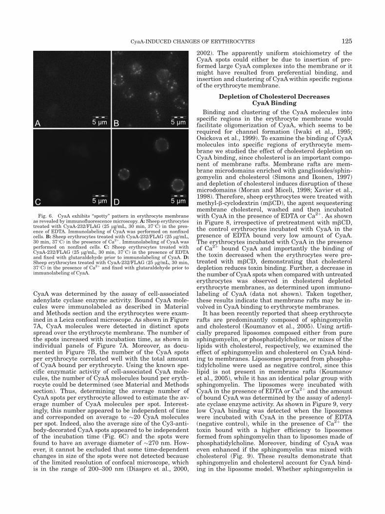
Clustered distribution of CyaA molecules in sheeperythrocyte membranes was further examined by indi-rect immunofluorescence microscopy. Sheep erythro-cytes were treated with CyaA-232/FLAG and boundCyaA molecules were immunolabeled with specificmouse anti-FLAG antibodies that were subsequentlydecorated by antimouse IgG-FITC. As shown in Figure 6,in contrast to control erythrocytes that were treatedwith CyaA-232/FLAG in the presence of 5 mM EDTA
and exhibited no fluorescence (Fig. 6A), the erythro-cytes treated with CyaA-232/FLAG in the presence of2 mM Ca2þ exhibited a ‘‘spotty’’ fluorescence pattern(Fig. 6B) indicating a clustered distribution of CyaA.However, it was shown previously that clustered distri-bution of membrane proteins observed by an indirectimmunofluorescence microscopy may result from anantibody-induced patching (Santoso et al., 1986). There-fore, prior to labeling, erythrocytes were fixed with glu-taraldehyde and then immunolabeling was applied inthe same way as described earlier. Again, no fluores-cence was observed on control erythrocytes that weretreated with CyaA-232/FLAG in the presence of 5 mMEDTA (Fig. 6C), while the spotty fluorescence patternwas observed also on erythrocytes treated with CyaA-232/FLAG in the presence of 2 mM Ca2þ and fixed priorto labeling (Fig. 6D). Moreover, the spotty distributionof CyaA in the membrane was also observed when theerythrocytes were treated with CyaA at 48C (data notshown). Altogether, these results demonstrate thatCyaA molecules formed clusters in the erythrocytemembrane and that CyaA clustering was not inducedby an antibody-induced patching.
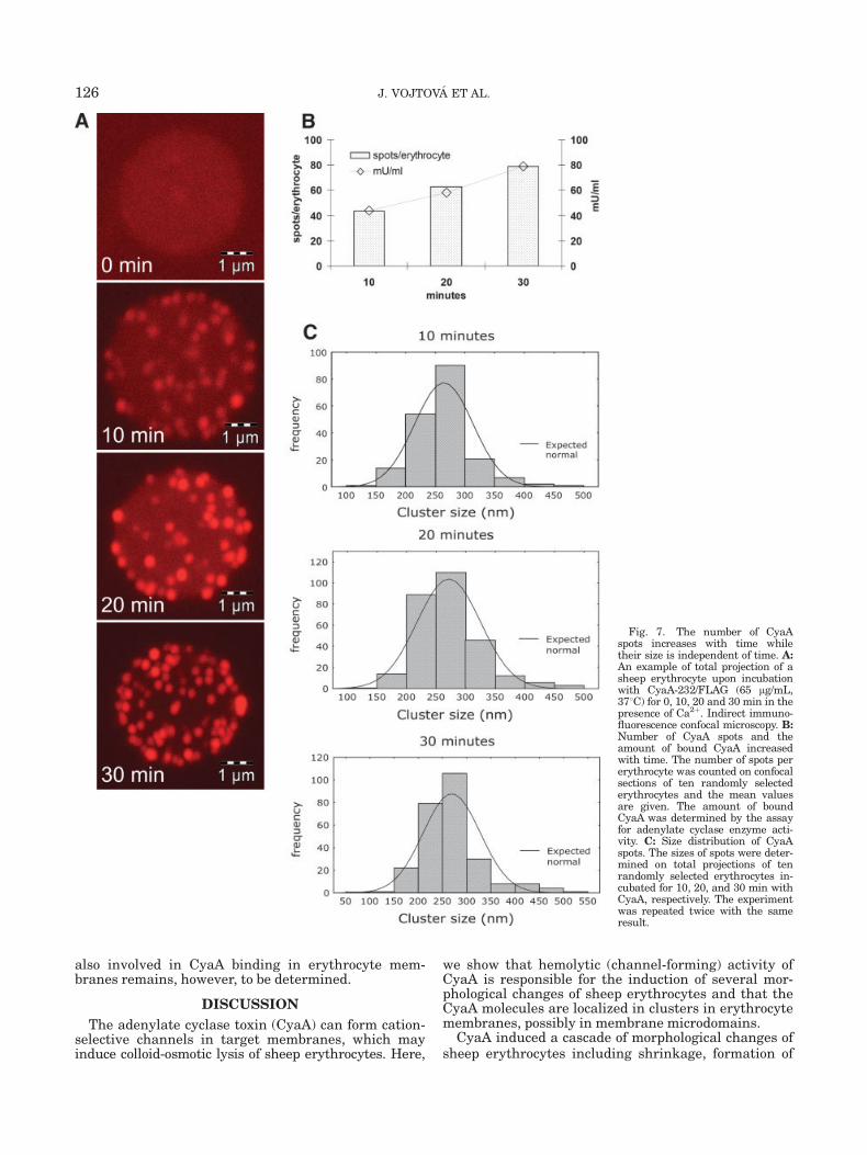
Next, we studied the formation and size of CyaAspots in erythrocyte membrane over the time of incuba-tion with toxin. Sheep erythrocytes were incubatedwith CyaA-232/FLAG at 378C for 0, 10, 20, and 30 minin the presence EDTA or Ca2þ and the amount of bound
Fig. 5. CyaA molecules are distributed in clusters in the erythro-cyte membrane A: Negative control of immunodetection. Sheep eryth-rocytes were treated with CyaA-232/FLAG (80 lg/mL, 30 min, 378C)in the presence of EDTA and immunolabeled with anti-FLAG anti-body and IgG-5 nm gold particles. Unstained sample. B: Indirect
immunodetection of CyaA in erythrocyte membrane. Sheep erythro-cytes were treated with CyaA-232/FLAG (80 lg/mL, 30 min, 378C) inthe presence of Ca2þ and immunolabeled as described earlier. Arrowsindicate clusters of gold particles. Unstained sample. The experimentwas repeated twice with the same result.
124 J. VOJTOVA ET AL.
CyaA was determined by the assay of cell-associatedadenylate cyclase enzyme activity. Bound CyaA mole-cules were immunolabeled as described in Materialand Methods section and the erythrocytes were exam-ined in a Leica confocal microscope. As shown in Figure7A, CyaA molecules were detected in distinct spotsspread over the erythrocyte membrane. The number ofthe spots increased with incubation time, as shown inindividual panels of Figure 7A. Moreover, as docu-mented in Figure 7B, the number of the CyaA spotsper erythrocyte correlated well with the total amountof CyaA bound per erythrocyte. Using the known spe-cific enzymatic activity of cell-associated CyaA mole-cules, the number of CyaA molecules bound per eryth-rocyte could be determined (see Material and Methodssection). Thus, determining the average number ofCyaA spots per erythrocyte allowed to estimate the av-erage number of CyaA molecules per spot. Interest-ingly, this number appeared to be independent of timeand corresponded on average to �20 CyaA moleculesper spot. Indeed, also the average size of the Cy3-anti-body-decorated CyaA spots appeared to be independentof the incubation time (Fig. 6C) and the spots werefound to have an average diameter of �270 nm. How-ever, it cannot be excluded that some time-dependentchanges in size of the spots were not detected becauseof the limited resolution of confocal microscope, whichis in the range of 200–300 nm (Diaspro et al., 2000,
2002). The apparently uniform stoichiometry of theCyaA spots could either be due to insertion of pre-formed large CyaA complexes into the membrane or itmight have resulted from preferential binding, andinsertion and clustering of CyaAwithin specific regionsof the erythrocyte membrane.
Depletion of Cholesterol DecreasesCyaA Binding
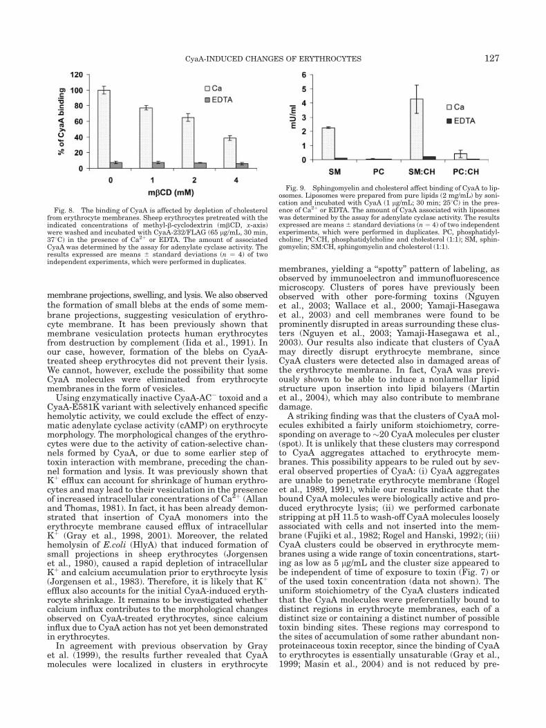
Binding and clustering of the CyaA molecules intospecific regions in the erythrocyte membrane wouldfacilitate oligomerization of CyaA, which seems to berequired for channel formation (Iwaki et al., 1995;Osickova et al., 1999). To examine the binding of CyaAmolecules into specific regions of erythrocyte mem-brane we studied the effect of cholesterol depletion onCyaA binding, since cholesterol is an important compo-nent of membrane rafts. Membrane rafts are mem-brane microdomains enriched with gangliosides/sphin-gomyelin and cholesterol (Simons and Ikonen, 1997)and depletion of cholesterol induces disruption of thesemicrodomains (Moran and Miceli, 1998; Xavier et al.,1998). Therefore, sheep erythrocytes were treated withmethyl-b-cyclodextrin (mbCD), the agent sequesteringmembrane cholesterol, washed and then incubatedwith CyaA in the presence of EDTA or Ca2þ. As shownin Figure 8, irrespective of pretreatment with mbCD,the control erythrocytes incubated with CyaA in thepresence of EDTA bound very low amount of CyaA.The erythrocytes incubated with CyaA in the presenceof Ca2þ bound CyaA and importantly the binding ofthe toxin decreased when the erythrocytes were pre-treated with mbCD, demonstrating that cholesteroldepletion reduces toxin binding. Further, a decrease inthe number of CyaA spots when compared with untreatederythrocytes was observed in cholesterol depletederythrocyte membranes, as determined upon immuno-labeling of CyaA (data not shown). Taken together,these results indicate that membrane rafts may be in-volved in CyaA binding to erythrocyte membranes.
It has been recently reported that sheep erythrocyterafts are predominantly composed of sphingomyelinand cholesterol (Koumanov et al., 2005). Using artifi-cially prepared liposomes composed either from puresphingomyelin, or phosphatidylcholine, or mixes of thelipids with cholesterol, respectively, we examined theeffect of sphingomyelin and cholesterol on CyaA bind-ing to membranes. Liposomes prepared from phospha-tidylcholine were used as negative control, since thislipid is not present in membrane rafts (Koumanovet al., 2005), while it has an identical polar group withsphingomyelin. The liposomes were incubated withCyaA in the presence of EDTA or Ca2þ and the amountof bound CyaA was determined by the assay of adenyl-ate cyclase enzyme activity. As shown in Figure 9, verylow CyaA binding was detected when the liposomeswere incubated with CyaA in the presence of EDTA(negative control), while in the presence of Ca2þ thetoxin bound with a higher efficiency to liposomesformed from sphingomyelin than to liposomes made ofphosphatidylcholine. Moreover, binding of CyaA waseven enhanced if the sphingomyelin was mixed withcholesterol (Fig. 9). These results demonstrate thatsphingomyelin and cholesterol account for CyaA bind-ing in the liposome model. Whether sphingomyelin is
Fig. 6. CyaA exhibits ‘‘spotty’’ pattern in erythrocyte membraneas revealed by immunofluorescence microscopy. A: Sheep erythrocytestreated with CyaA-232/FLAG (25 lg/mL, 30 min, 378C) in the pres-ence of EDTA. Immunolabeling of CyaA was performed on nonfixedcells. B: Sheep erythrocytes treated with CyaA-232/FLAG (25 lg/mL,30 min, 378C) in the presence of Ca2þ. Immunolabeling of CyaA wasperformed on nonfixed cells. C: Sheep erythrocytes treated withCyaA-232/FLAG (25 lg/mL, 30 min, 378C) in the presence of EDTAand fixed with glutaraldehyde prior to immunolabeling of CyaA. D:Sheep erythrocytes treated with CyaA-232/FLAG (25 lg/mL, 30 min,378C) in the presence of Ca2þ and fixed with glutaraldehyde prior toimmunolabeling of CyaA.
125CyaA-INDUCED CHANGES OF ERYTHROCYTES
also involved in CyaA binding in erythrocyte mem-branes remains, however, to be determined.
DISCUSSION
The adenylate cyclase toxin (CyaA) can form cation-selective channels in target membranes, which mayinduce colloid-osmotic lysis of sheep erythrocytes. Here,
we show that hemolytic (channel-forming) activity ofCyaA is responsible for the induction of several mor-phological changes of sheep erythrocytes and that theCyaA molecules are localized in clusters in erythrocytemembranes, possibly in membrane microdomains.
CyaA induced a cascade of morphological changes ofsheep erythrocytes including shrinkage, formation of
Fig. 7. The number of CyaAspots increases with time whiletheir size is independent of time. A:An example of total projection of asheep erythrocyte upon incubationwith CyaA-232/FLAG (65 lg/mL,378C) for 0, 10, 20 and 30 min in thepresence of Ca2þ. Indirect immuno-fluorescence confocal microscopy. B:Number of CyaA spots and theamount of bound CyaA increasedwith time. The number of spots pererythrocyte was counted on confocalsections of ten randomly selectederythrocytes and the mean valuesare given. The amount of boundCyaA was determined by the assayfor adenylate cyclase enzyme acti-vity. C: Size distribution of CyaAspots. The sizes of spots were deter-mined on total projections of tenrandomly selected erythrocytes in-cubated for 10, 20, and 30 min withCyaA, respectively. The experimentwas repeated twice with the sameresult.
126 J. VOJTOVA ET AL.
membrane projections, swelling, and lysis. We also observedthe formation of small blebs at the ends of some mem-brane projections, suggesting vesiculation of erythro-cyte membrane. It has been previously shown thatmembrane vesiculation protects human erythrocytesfrom destruction by complement (Iida et al., 1991). Inour case, however, formation of the blebs on CyaA-treated sheep erythrocytes did not prevent their lysis.We cannot, however, exclude the possibility that someCyaA molecules were eliminated from erythrocytemembranes in the form of vesicles.
Using enzymatically inactive CyaA-AC� toxoid and aCyaA-E581K variant with selectively enhanced specifichemolytic activity, we could exclude the effect of enzy-matic adenylate cyclase activity (cAMP) on erythrocytemorphology. The morphological changes of the erythro-cytes were due to the activity of cation-selective chan-nels formed by CyaA, or due to some earlier step oftoxin interaction with membrane, preceding the chan-nel formation and lysis. It was previously shown thatKþ efflux can account for shrinkage of human erythro-cytes and may lead to their vesiculation in the presenceof increased intracellular concentrations of Ca2þ (Allanand Thomas, 1981). In fact, it has been already demon-strated that insertion of CyaA monomers into theerythrocyte membrane caused efflux of intracellularKþ (Gray et al., 1998, 2001). Moreover, the relatedhemolysin of E.coli (HlyA) that induced formation ofsmall projections in sheep erythrocytes (Jorgensenet al., 1980), caused a rapid depletion of intracellularKþ and calcium accumulation prior to erythrocyte lysis(Jorgensen et al., 1983). Therefore, it is likely that Kþ
efflux also accounts for the initial CyaA-induced eryth-rocyte shrinkage. It remains to be investigated whethercalcium influx contributes to the morphological changesobserved on CyaA-treated erythrocytes, since calciuminflux due to CyaA action has not yet been demonstratedin erythrocytes.
In agreement with previous observation by Grayet al. (1999), the results further revealed that CyaAmolecules were localized in clusters in erythrocyte
membranes, yielding a ‘‘spotty’’ pattern of labeling, asobserved by immunoelectron and immunofluorescencemicroscopy. Clusters of pores have previously beenobserved with other pore-forming toxins (Nguyenet al., 2003; Wallace et al., 2000; Yamaji-Hasegawaet al., 2003) and cell membranes were found to beprominently disrupted in areas surrounding these clus-ters (Nguyen et al., 2003; Yamaji-Hasegawa et al.,2003). Our results also indicate that clusters of CyaAmay directly disrupt erythrocyte membrane, sinceCyaA clusters were detected also in damaged areas ofthe erythrocyte membrane. In fact, CyaA was previ-ously shown to be able to induce a nonlamellar lipidstructure upon insertion into lipid bilayers (Martinet al., 2004), which may also contribute to membranedamage.
A striking finding was that the clusters of CyaA mol-ecules exhibited a fairly uniform stoichiometry, corre-sponding on average to �20 CyaA molecules per cluster(spot). It is unlikely that these clusters may correspondto CyaA aggregates attached to erythrocyte mem-branes. This possibility appears to be ruled out by sev-eral observed properties of CyaA: (i) CyaA aggregatesare unable to penetrate erythrocyte membrane (Rogelet al., 1989, 1991), while our results indicate that thebound CyaA molecules were biologically active and pro-duced erythrocyte lysis; (ii) we performed carbonatestripping at pH 11.5 to wash-off CyaA molecules looselyassociated with cells and not inserted into the mem-brane (Fujiki et al., 1982; Rogel and Hanski, 1992); (iii)CyaA clusters could be observed in erythrocyte mem-branes using a wide range of toxin concentrations, start-ing as low as 5 lg/mL and the cluster size appeared tobe independent of time of exposure to toxin (Fig. 7) orof the used toxin concentration (data not shown). Theuniform stoichiometry of the CyaA clusters indicatedthat the CyaA molecules were preferentially bound todistinct regions in erythrocyte membranes, each of adistinct size or containing a distinct number of possibletoxin binding sites. These regions may correspond tothe sites of accumulation of some rather abundant non-proteinaceous toxin receptor, since the binding of CyaAto erythrocytes is essentially unsaturable (Gray et al.,1999; Masin et al., 2004) and is not reduced by pre-
Fig. 8. The binding of CyaA is affected by depletion of cholesterolfrom erythrocyte membranes. Sheep erythrocytes pretreated with theindicated concentrations of methyl-b-cyclodextrin (mbCD, x-axis)were washed and incubated with CyaA-232/FLAG (65 lg/mL, 30 min,378C) in the presence of Ca2þ or EDTA. The amount of associatedCyaA was determined by the assay for adenylate cyclase activity. Theresults expressed are means 6 standard deviations (n ¼ 4) of twoindependent experiments, which were performed in duplicates.
Fig. 9. Sphingomyelin and cholesterol affect binding of CyaA to lip-osomes. Liposomes were prepared from pure lipids (2 mg/mL) by soni-cation and incubated with CyaA (1 lg/mL; 30 min; 258C) in the pres-ence of Ca2þ or EDTA. The amount of CyaA associated with liposomeswas determined by the assay for adenylate cyclase activity. The resultsexpressed are means6 standard deviations (n ¼ 4) of two independentexperiments, which were performed in duplicates. PC, phosphatidyl-choline; PC:CH, phosphatidylcholine and cholesterol (1:1); SM, sphin-gomyelin; SM:CH, sphingomyelin and cholesterol (1:1).
127CyaA-INDUCED CHANGES OF ERYTHROCYTES

treatment of the erythrocytes with proteases (Masinet al., 2004). Previously, a role of lipids (or glycolipids)in the interaction of CyaA with cell membranes hasbeen suggested (Gordon et al., 1989). These authorsdemonstrated that the incubation of CyaA with gan-gliosides prior to addition to target cells inhibitedCyaA-mediated cAMP accumulation in cells (Gordonet al., 1989). Our results show that binding of CyaA tocells decreases upon cholesterol depletion from eryth-rocyte membranes. This suggests that CyaA may bindinto membrane microdomains (rafts) that are typicallyenriched in gangliosides, sphingomyelin, and choles-terol (Simons and Ikonen, 1997) and are disruptedupon cholesterol extraction. Role of microdomains inCyaA binding would further be supported by the obser-vation that liposomes formed from sphingomyelin (SM)and cholesterol, the two most abundant lipids of sheeperythrocyte microdomains (Koumanov et al., 2005),bind CyaA with a much higher efficiency than lipo-somes made of pure phospholipids, such as phosphati-dylcholine.
Taken together, the results would suggest that thetargeting of CyaA to specific domains in erythrocytemembranes may favor protein–protein interactionsamong CyaA molecules and hence facilitate toxin oligo-merization and channel formation. Further, morpho-logical changes of sheep erythrocytes resulting fromCyaA hemolytic (channel-forming) activity tend to sug-gest that fluxes of Kþ and possibly also fluxes of Ca2þ
ions are the early steps preceding colloid-osmotic swel-ling and lysis of erythrocytes induced by CyaA.
ACKNOWLEDGMENTS
The authors are grateful to Marek Basler for the giftof the CyaA-E581K construct.
REFERENCES
Allan D, Thomas P. 1981. Ca2þ-induced biochemical changes inhuman erythrocytes and their relation to microvesiculation. Bio-chem J 198:433–440.
Bachelet M, Richard MJ, Francois D, Polla BS. 2002. Mitochondrialalterations precede Bordetella pertussis-induced apoptosis. FEMSImmunol Med Microbiol 32:125–131.
Backman L. 1986. Shape control in the human red cell. J Cell Sci 80:281–298.
Bejerano M, Nisan I, Ludwig A, Goebel W, Hanski E. 1999. Character-ization of the C-terminal domain essential for toxic activity of ade-nylate cyclase toxin. Mol Microbiol 31:381–392.
Bellalou J, Ladant D, Sakamoto H. 1990. Synthesis and secretion ofBordetella pertussis adenylate cyclase as a 200-kilodalton protein.Infect Immun 58:1195–1200.
Benada O, Pokorny V. 1990. Modification of the Polaron sputter-coater unit for glow-discharge activation of carbon support films.J Electron Microsc Tech 16:235–239.
Benz R, Maier E, Ladant D, Ullmann A, Sebo P. 1994. Adenylatecyclase toxin (CyaA) of Bordetella pertussis. Evidence for the forma-tion of small ion-permeable channels and comparison with HlyA ofEscherichia coli. J Biol Chem 269:27231–27239.
Betsou F, Sebo P, Guiso N. 1993. CyaC-mediated activation is impor-tant not only for toxic but also for protective activities of Bordetellapertussis adenylate cyclase-hemolysin. Infect Immun 61:3583–3589.
Chou AC, Fitch CD. 1981. Mechanism of hemolysis induced by ferri-protoporphyrin IX. J Clin Invest 68:672–677.
Confer DL, Eaton JW. 1982. Phagocyte impotence caused by an inva-sive bacterial adenylate cyclase. Science 217:948–950.
Diaspro A, Annunziata S, Robello M. 2000. Single-pinhole confocal imag-ing of sub-resolution sparse objects using experimental point spreadfunction and image restoration. Microsc Res Tech 51:464–468.
Diaspro A, Federici F, Robello M. 2002. Influence of refractive-indexmismatch in high-resolution three-dimensional confocal microsco-py. Appl Opt 41:685–690.
Ehrmann IE, Gray MC, Gordon VM, Gray LS, Hewlett EL. 1991.Hemolytic activity of adenylate cyclase toxin from Bordetella per-tussis. FEBS Lett. 278:79–83.
El-Azami-El-Idrissi M, Bauche C, Loucka J, Osicka R, Sebo P, LadantD, Leclerc C. 2003. Interaction of Bordetella pertussis adenylatecyclase with CD11b/CD18: Role of toxin acylation and identificationof the main integrin interaction domain. J Biol Chem 278:38514–38521.
Fujiki Y, Hubbard AL, Fowler S, Lazarow PB. 1982. Isolation of intra-cellular membranes by means of sodium carbonate treatment:application to endoplasmic reticulum. J Cell Biol 93:97–102.
Glaser P, Sakamoto H, Bellalou J, Ullmann A, Danchin A. 1988.Secretion of cyclolysin, the calmodulin-sensitive adenylate cyclase-haemolysin bifunctional protein of Bordetella pertussis. Embo J7:3997–4004.
Goodwin MS, Weiss AA. 1990. Adenylate cyclase toxin is critical forcolonization and pertussis toxin is critical for lethal infection byBordetella pertussis in infant mice. Infect Immun 58:3445–3447.
Gordon VM, Young WW, Jr, Lechler SM, Gray MC, Leppla SH, Hew-lett EL. 1989. Adenylate cyclase toxins from Bacillus anthracis andBordetella pertussis. Different processes for interaction with andentry into target cells. J Biol Chem 264:14792–14796.
Gray M, Szabo G, Otero AS, Gray L, Hewlett E. 1998. Distinct mecha-nisms for Kþ efflux, intoxication, and hemolysis by Bordetella per-tussis AC toxin. J Biol Chem 273:18260–18267.
Gray MC, Ross W, Kim K, Hewlett EL. 1999. Characterization ofbinding of adenylate cyclase toxin to target cells by flow cytometry.Infect Immun 67:4393–4399.
Gray MC, Lee SJ, Gray LS, Zaretzky FR, Otero AS, Szabo G, HewlettEL. 2001. Translocation-specific conformation of adenylate cyclasetoxin from Bordetella pertussis inhibits toxin-mediated hemolysis.J Bacteriol 183:5904–5910.
Guermonprez P, Khelef N, Blouin E, Rieu P, Ricciardi-Castagnoli P,Guiso N, Ladant D, Leclerc C. 2001. The adenylate cyclase toxin ofBordetella pertussis binds to target cells via the alpha(M)beta(2)integrin (CD11b/CD18). J Exp Med 193:1035–1044.
Guiso N, Szatanik M, Rocancourt M. 1991. Protective activity of Bor-detella adenylate cyclase-hemolysin against bacterial colonization.Microb Pathog 11:423–431.
Hackett M, Guo L, Shabanowitz J, Hunt DF, Hewlett EL. 1994. Inter-nal lysine palmitoylation in adenylate cyclase toxin from Bordetellapertussis. Science 266:433–435.
Hanski E. 1989. Invasive adenylate cyclase toxin of Bordetella pertus-sis. Trends Biochem Sci 14:459–463.
Harris RW, Sims PJ, Tweten RK. 1991. Evidence that Clostridiumperfringens theta-toxin induces colloid-osmotic lysis of erythrocytes.Infect Immun 59:2499–2501.
Harvill ET, Cotter PA, Yuk MH, Miller JF. 1999. Probing the functionof Bordetella bronchiseptica adenylate cyclase toxin by manipulat-ing host immunity. Infect Immun 67:1493–1500.
Havlicek V, Higgins L, Chen W, Halada P, Sebo P, Sakamoto H, Hack-ett M. 2001. Mass spectrometric analysis of recombinant adenylatecyclase toxin from Bordetella pertussis strain 18323/pHSP9. J MassSpectrom 36:384–391.
Hewlett EL, Gray L, Allietta M, Ehrmann I, Gordon VM, Gray MC.1991. Adenylate cyclase toxin from Bordetella pertussis. Confor-mational change associated with toxin activity. J Biol Chem 266:17503–17508.
Iida K, Whitlow MB, Nussenzweig V. 1991. Membrane vesiculationprotects erythrocytes from destruction by complement. J Immunol147:2638–2642.
Iwaki M, Ullmann A, Sebo P. 1995. Identification by in vitro comple-mentation of regions required for cell-invasive activity of Borde-tella pertussis adenylate cyclase toxin. Mol Microbiol 17:1015–1024.
Jorgensen SE, Hammer RF, Wu GK. 1980. Effects of a single hit fromthe alpha hemolysin produced by Escherichia coli on the morpholo-gy of sheep erythrocytes. Infect Immun 27:988–994.
Jorgensen SE, Mulcahy PF, Wu GK, Louis CF. 1983. Calcium accumu-lation in human and sheep erythrocytes that is induced by Esche-richia coli hemolysin. Toxicon 21:717–727.
Karimova G, Fayolle C, Gmira S, Ullmann A, Leclerc C, Ladant D.1998. Charge-dependent translocation of Bordetella pertussis ade-nylate cyclase toxin into eukaryotic cells: implication for the in vivodelivery of CD8(þ) T cell epitopes into antigen-presenting cells.Proc Natl Acad Sci USA 95:12532–12537.
Khelef N, Sakamoto H, Guiso N. 1992. Both adenylate cyclase and he-molytic activities are required by Bordetella pertussis to initiateinfection. Microb Pathog 12:227–235.
Koumanov KS, Tessier C, Momchilova AB, Rainteau D, Wolf C, QuinnPJ. 2005. Comparative lipid analysis and structure of detergent-
128 J. VOJTOVA ET AL.

resistant membrane raft fractions isolated from human and rumi-nant erythrocytes. Arch Biochem Biophys 434:150–158.
Ladant D. 1988. Interaction of Bordetella pertussis adenylate cyclasewith calmodulin. Identification of two separated calmodulin-bind-ing domains. J Biol Chem 263:2612–2618.
Martin C, Requero MA, Masin J, Konopasek I, Goni FM, Sebo P, Osto-laza H. 2004. Membrane restructuring by Bordetella pertussis ade-nylate cyclase toxin, a member of the RTX toxin family. J Bacteriol186:3760–3765.
Masin J, Konopasek I, Svobodova J, Sebo P, 2004. Different structuralrequirements for adenylate cyclase toxin interactions with erythro-cyte and liposome membranes. Biochim Biophys Acta 1660:144–154.
Moran M, Miceli MC. 1998. Engagement of GPI-linked CD48 contrib-utes to TCR signals and cytoskeletal reorganization: a role for lipidrafts in T cell activation. Immunity 9:787–796.
Nguyen VT, Kamio Y, Higuchi H. 2003. Single-molecule imaging of co-operative assembly of gamma-hemolysin on erythrocyte mem-branes. Embo J 22:4968–4979.
Osicka R, Osickova A, Basar T, Guermonprez P, RojasM, Leclerc C, SeboP. 2000. Delivery of CD8(þ) T-cell epitopes into major histocompatibil-ity complex class I antigen presentation pathway by Bordetella pertus-sis adenylate cyclase: delineation of cell invasive structures and per-missive insertion sites. Infect Immun 68: 247–256.
Osickova A, Osicka R, Maier E, Benz R, Sebo P. 1999. An amphipathicalpha-helix including glutamates 509 and 516 is crucial for mem-brane translocation of adenylate cyclase toxin and modulates for-mation and cation selectivity of its membrane channels. J BiolChem 274:37644–37650.
Palek J, Liu SC, Snyder LM. 1978. Metabolic dependence of proteinarrangement in human erythrocyte membranes. I. Analysis of spec-trin-rich complexes in ATP-depleted red cells. Blood 51:385–395.
Rogel A, Hanski E. 1992. Distinct steps in the penetration of adenyl-ate cyclase toxin of Bordetella pertussis into sheep erythrocytes.Translocation of the toxin across the membrane. J Biol Chem 267:22599–22605.
Rogel A, Schultz JE, Brownlie RM, Coote JG, Parton R, Hanski E.1989. Bordetella pertussis adenylate cyclase: purification and char-acterization of the toxic form of the enzyme. Embo J 8:2755–2760.
Rogel A, Meller R, Hanski E. 1991. Adenylate cyclase toxin from Bor-detella pertussis. The relationship between induction of cAMP andhemolysis. J Biol Chem 266:3154–161.
Rose T, Sebo P, Bellalou J, Ladant D. 1995. Interaction of calciumwith Bordetella pertussis adenylate cyclase toxin. Characterization
of multiple calcium-binding sites and calcium-induced conforma-tional changes. J Biol Chem 270:26370–26376.
Sanders SK, Alexander EL, Braylan RC. 1975. A high-yield techniquefor preparing cells fixed in suspension for scanning electron micros-copy. J Cell Biol 67(2PT. 1):476–480.
Santoso S, Zimmermann U, Neppert J, Mueller-Eckhardt C. 1986.Receptor patching and capping of platelet membranes induced bymonoclonal antibodies. Blood 67:343–349.
Schlecht G, Loucka J, Najar H, Sebo P, Leclerc C. 2004. Antigen tar-geting to CD11b allows efficient presentation of CD4þ and CD8þ Tcell epitopes and in vivo Th1-polarized T cell priming. J Immunol173:6089–6097.
Sebo P, Glaser P, Sakamoto H, Ullmann A. 1991. High-level synthesisof active adenylate cyclase toxin of Bordetella pertussis in a recon-structed Escherichia coli system. Gene 104:19–24.
Simons K, Ikonen E. 1997. Functional rafts in cell membranes.Nature 387:569–572.
Stolz J, Stadler R, Opekarova M, Sauer N. 1994. Functional reconsti-tution of the solubilized Arabidopsis thaliana STP1 monosaccha-ride-Hþ symporter in lipid vesicles and purification of the histidinetagged protein from transgenic Saccharomyces cerevisiae. Plant J6:225–233.
Szabo G, Gray MC, Hewlett EL. 1994. Adenylate cyclase toxin fromBordetella pertussis produces ion conductance across artificial lipidbilayers in a calcium- and polarity-dependent manner. J Biol Chem269:22496–22499.
Wallace AJ, Stillman TJ, Atkins A, Jamieson SJ, Bullough PA, GreenJ, Artymiuk PJ. 2000. E. coli hemolysin E (HlyE, ClyA, SheA):X-ray crystal structure of the toxin and observation of membranepores by electron microscopy. Cell 100:265–276.
Weber K, Rathke PC, Osborn M. 1978. Cytoplasmic microtubu-lar images in glutaraldehyde-fixed tissue culture cells by electronmicroscopy and by immunofluorescence microscopy. Proc Natl AcadSci USA 75:1820–1824.
Welch RA. 1991. Pore-forming cytolysins of gram-negative bacteria.Mol Microbiol 5:521–528.
Xavier R, Brennan T, Li Q, McCormack C, Seed B. 1998. Membranecompartmentation is required for efficient T cell activation. Immu-nity 8:723–732.
Yamaji-Hasegawa A, Makino A, Baba T, Senoh Y, Kimura-Suda H,Sato SB, Terada N, Ohno S, Kiyokawa E, Umeda M, Kobayashi T.2003. Oligomerization and pore formation of a sphingomyelin-specific toxin, lysenin. J Biol Chem 278:22762–22770.
129CyaA-INDUCED CHANGES OF ERYTHROCYTES