Human Cytomegalovirus pp28 (UL99) Localizes to a Cytoplasmic Compartment Which Overlaps the...
10
JOURNAL OF VIROLOGY, 0022-538X/00/$04.0010 Apr. 2000, p. 3842–3851 Vol. 74, No. 8 Copyright © 2000, American Society for Microbiology. All Rights Reserved. Human Cytomegalovirus pp28 (UL99) Localizes to a Cytoplasmic Compartment Which Overlaps the Endoplasmic Reticulum-Golgi-Intermediate Compartment VERONICA SANCHEZ, 1 ² ELIZABETH SZTUL, 2 AND WILLIAM J. BRITT 1,3 * Departments of Pediatrics, 1 Cell Biology, 2 and Microbiology, 3 The University of Alabama at Birmingham, Birmingham, Alabama Received 9 November 1999/Accepted 18 January 2000 Although the assembly of herpesviruses has remained an active area of investigation, considerable contro- versy continues to surround the cellular location of tegument and envelope acquisition. This controversy is particularly evident when the proposed pathways for a- and b-herpesvirus assembly are compared. We have approached this aspect of human cytomegalovirus (HCMV) assembly, specifically, envelopment, by investi- gating the intracellular trafficking of viral tegument proteins which localize in the cytoplasms of infected cells. In this study we have demonstrated that the virion tegument protein pp28 (UL99), a true late protein, was membrane associated as a result of myristoylation. A mutation in this protein which prevented incorporation of [ 3 H]myristic acid also altered the detergent solubility and intracellular distribution of the protein when it was expressed in transfected cells. Using a panel of markers for intracellular compartments, we could localize the expression of wild-type pp28 to an intracellular compartment which colocalized with the endoplasmic reticulum-Golgi-intermediate compartment (ERGIC), a dynamic compartment of the secretory pathway which interfaces with both the ER and Golgi apparatus. The localization of this viral tegument protein within an early secretory compartment of the cell provided further evidence that the assembly of the HCMV tegument likely includes a cytoplasmic phase. Because pp28 has been shown to be localized to a cytoplasmic assembly compartment in HCMV-infected cells, our findings also suggested that viral tegument protein interactions within the secretory pathway may have an important role in the assembly of the virion. The assembly of human herpesviruses remains an active area of investigation. Several different models of virion assembly have been proposed (12, 16, 21, 30, 34, 38, 47, 48). Although all models include steps of capsid formation and tegumentation within the nucleus of an infected cell, the intracellular loca- tions of virion tegumentation and envelopment remain con- tentious. Early models of envelopment suggested that tegu- mented particles budded through the inner leaflet of the nuclear envelope and entered the secretory pathway through the endoplasmic reticulum (ER) (9, 34, 38, 39). Evidence sup- porting this model has come from electron microscopic anal- ysis of herpes simplex virus (HSV)- and human cytomegalovi- rus (HCMV)-infected cells, studies using inhibitors of glycoprotein processing, and characterizations of viruses with mutations in envelope glycoprotein genes (10, 18, 19, 34, 38, 40). More recently, several laboratories have provided evi- dence that the final envelopment of a-herpesviruses occurs in the cytoplasm following transient envelopment and de-envel- opment at the nuclear membrane (7, 16, 47–49). These studies have led to an alternative model of envelopment which appears to be more consistent with recently published findings, yet the intracellular compartment in which the final envelopment of herpesviruses takes place remains incompletely defined and may differ for different members of this family of viruses. The final envelopment of varicella-zoster virus appears to take place in the Golgi apparatus and trans-Golgi network (TGN), based on studies of the trafficking of the major glycoprotein of varicella-zoster virus, gE (12, 16, 21, 31, 51, 52). Similarly, results of early studies were consistent with a similar site of envelopment for pseudorabies virus, and although more recent studies have suggested that the site of virion envelopment might include the TGN, additional compartments within the infected cell may also serve as assembly compartments (15, 44, 47, 48). Our study of the assembly of HCMV was consistent with envelopment occurring in the TGN or an intracellular site contiguous with the TGN (36). This proposed site of HCMV virion assembly was also consistent with several previous stud- ies which indicated that the major glycoprotein of the envelope of HCMV, gB, was cleaved into its mature form by the cellular enzyme furin (28, 45, 46). Thus, evidence from several sources has suggested that the final envelopment of b-herpesviruses likely takes place in the cytoplasm, perhaps in the TGN or in a compartment contiguous to this intracellular compartment. We have studied the envelopment of HCMV using a differ- ent approach. It was our hypothesis that understanding envel- opment and assembly could be accomplished by studying the intracellular trafficking of HCMV virion tegument proteins, as these proteins are major constituents of the extracellular par- ticle. Furthermore, a large number of different studies of as- sembly of several different enveloped RNA viruses have indi- cated that budding of the subviral particle involves passage of a capsid through a viral glycoprotein anchored within a bio- logical membrane (11). In many cases this step was facilitated by interaction of the capsid and/or envelope glycoprotein with a viral matrix protein (11). Thus, if HCMV was enveloped in the cytoplasm as existing evidence suggested, then it was pos- sible that one or more virion tegument proteins were partici- pating in this intracellular budding event. Interestingly, early ultrastructural studies of HSV and HCMV consistently noted that nonenveloped cytoplasmic particles in HCMV-infected * Corresponding author. Mailing address: Department of Pediatrics, 1600 7th Ave. South, Suite 752, Birmingham, AL 35233. Phone: (205) 939-9590. Fax: (205) 975-6549. E-mail: [email protected]. ² Present address: Department of Biology, University of California, San Diego, La Jolla, Calif. 3842 on December 16, 2015 by guest http://jvi.asm.org/ Downloaded from
-
Upload
independent -
Category
Documents
-
view
3 -
download
0
Transcript of Human Cytomegalovirus pp28 (UL99) Localizes to a Cytoplasmic Compartment Which Overlaps the...
JOURNAL OF VIROLOGY,0022-538X/00/$04.0010
Apr. 2000, p. 3842–3851 Vol. 74, No. 8
Copyright © 2000, American Society for Microbiology. All Rights Reserved.
Human Cytomegalovirus pp28 (UL99) Localizes to a CytoplasmicCompartment Which Overlaps the Endoplasmic
Reticulum-Golgi-Intermediate CompartmentVERONICA SANCHEZ,1† ELIZABETH SZTUL,2 AND WILLIAM J. BRITT1,3*
Departments of Pediatrics,1 Cell Biology,2 and Microbiology,3 The University ofAlabama at Birmingham, Birmingham, Alabama
Received 9 November 1999/Accepted 18 January 2000
Although the assembly of herpesviruses has remained an active area of investigation, considerable contro-versy continues to surround the cellular location of tegument and envelope acquisition. This controversy isparticularly evident when the proposed pathways for a- and b-herpesvirus assembly are compared. We haveapproached this aspect of human cytomegalovirus (HCMV) assembly, specifically, envelopment, by investi-gating the intracellular trafficking of viral tegument proteins which localize in the cytoplasms of infected cells.In this study we have demonstrated that the virion tegument protein pp28 (UL99), a true late protein, wasmembrane associated as a result of myristoylation. A mutation in this protein which prevented incorporationof [3H]myristic acid also altered the detergent solubility and intracellular distribution of the protein when itwas expressed in transfected cells. Using a panel of markers for intracellular compartments, we could localizethe expression of wild-type pp28 to an intracellular compartment which colocalized with the endoplasmicreticulum-Golgi-intermediate compartment (ERGIC), a dynamic compartment of the secretory pathway whichinterfaces with both the ER and Golgi apparatus. The localization of this viral tegument protein within an earlysecretory compartment of the cell provided further evidence that the assembly of the HCMV tegument likelyincludes a cytoplasmic phase. Because pp28 has been shown to be localized to a cytoplasmic assemblycompartment in HCMV-infected cells, our findings also suggested that viral tegument protein interactionswithin the secretory pathway may have an important role in the assembly of the virion.
The assembly of human herpesviruses remains an active areaof investigation. Several different models of virion assemblyhave been proposed (12, 16, 21, 30, 34, 38, 47, 48). Although allmodels include steps of capsid formation and tegumentationwithin the nucleus of an infected cell, the intracellular loca-tions of virion tegumentation and envelopment remain con-tentious. Early models of envelopment suggested that tegu-mented particles budded through the inner leaflet of thenuclear envelope and entered the secretory pathway throughthe endoplasmic reticulum (ER) (9, 34, 38, 39). Evidence sup-porting this model has come from electron microscopic anal-ysis of herpes simplex virus (HSV)- and human cytomegalovi-rus (HCMV)-infected cells, studies using inhibitors ofglycoprotein processing, and characterizations of viruses withmutations in envelope glycoprotein genes (10, 18, 19, 34, 38,40). More recently, several laboratories have provided evi-dence that the final envelopment of a-herpesviruses occurs inthe cytoplasm following transient envelopment and de-envel-opment at the nuclear membrane (7, 16, 47–49). These studieshave led to an alternative model of envelopment which appearsto be more consistent with recently published findings, yet theintracellular compartment in which the final envelopment ofherpesviruses takes place remains incompletely defined andmay differ for different members of this family of viruses. Thefinal envelopment of varicella-zoster virus appears to takeplace in the Golgi apparatus and trans-Golgi network (TGN),based on studies of the trafficking of the major glycoprotein of
varicella-zoster virus, gE (12, 16, 21, 31, 51, 52). Similarly,results of early studies were consistent with a similar site ofenvelopment for pseudorabies virus, and although more recentstudies have suggested that the site of virion envelopmentmight include the TGN, additional compartments within theinfected cell may also serve as assembly compartments (15, 44,47, 48). Our study of the assembly of HCMV was consistentwith envelopment occurring in the TGN or an intracellular sitecontiguous with the TGN (36). This proposed site of HCMVvirion assembly was also consistent with several previous stud-ies which indicated that the major glycoprotein of the envelopeof HCMV, gB, was cleaved into its mature form by the cellularenzyme furin (28, 45, 46). Thus, evidence from several sourceshas suggested that the final envelopment of b-herpesviruseslikely takes place in the cytoplasm, perhaps in the TGN or ina compartment contiguous to this intracellular compartment.
We have studied the envelopment of HCMV using a differ-ent approach. It was our hypothesis that understanding envel-opment and assembly could be accomplished by studying theintracellular trafficking of HCMV virion tegument proteins, asthese proteins are major constituents of the extracellular par-ticle. Furthermore, a large number of different studies of as-sembly of several different enveloped RNA viruses have indi-cated that budding of the subviral particle involves passage ofa capsid through a viral glycoprotein anchored within a bio-logical membrane (11). In many cases this step was facilitatedby interaction of the capsid and/or envelope glycoprotein witha viral matrix protein (11). Thus, if HCMV was enveloped inthe cytoplasm as existing evidence suggested, then it was pos-sible that one or more virion tegument proteins were partici-pating in this intracellular budding event. Interestingly, earlyultrastructural studies of HSV and HCMV consistently notedthat nonenveloped cytoplasmic particles in HCMV-infected
* Corresponding author. Mailing address: Department of Pediatrics,1600 7th Ave. South, Suite 752, Birmingham, AL 35233. Phone: (205)939-9590. Fax: (205) 975-6549. E-mail: [email protected].
† Present address: Department of Biology, University of California,San Diego, La Jolla, Calif.
3842
on Decem
ber 16, 2015 by guesthttp://jvi.asm
.org/D
ownloaded from
cells were coated with a thick tegument layer but that nonen-veloped HSV particles often had the appearance of nakedcapsids (38). Whether these HSV cytoplasmic capsids wereactually devoid of tegument or coated with a layer of unrec-ognized tegument was not addressed in this study. In the caseof HCMV, at least four different tegument or matrix proteinshave been shown to localize within the cytoplasms of infectedcells in the late phases of the replicative cycle, a time of max-imal production of progeny virions (3, 24, 36). We have shownthat three of these tegument proteins, pp150 (UL32), pp65(UL83), and pp28 (UL99), accumulated in a stable jux-tanuclear, membranous cytoplasmic structure together withthree envelope glycoproteins (36). Thus, any of these threetegument proteins or an as yet unstudied tegument proteinmay facilitate the cytoplasmic budding and envelopment of thesubviral particle of HCMV.
In this study we have characterized the membrane associa-tion and intracellular trafficking of pp28 (UL99), one of thefirst HCMV virion tegument proteins which was shown to beexpressed exclusively in the cytoplasms of infected cells (24).This viral protein is an example of a true late protein and isexpressed only very late in the infectious cycle during the timeof maximal virus production (22, 24). It is a phosphorylatedprotein component of the virion which can be demonstrated invacuole-like structures in HCMV-infected human fibroblasts,suggesting that it is membrane associated (24, 36). Interest-ingly, the HSV protein homolog of HCMV UL99, UL11, hasbeen suggested to be essential for replication of HSV in vitro(1, 26). A UL11 null mutant virus exhibited impaired growth,and in one study it was noted that the UL11 null mutantexhibited deficits in the nuclear egress of the capsid, suggestingthat this tegument protein may have a role in virion envelop-ment (1, 26, 39).
In the present investigation, we studied the intracellularlocalization of HCMV pp28 (UL99). We have demonstratedthat pp28 was a myristoylated protein in infected cells and thatthis modification accounted for its membrane association ininfected cells and in virions. In the absence of other viralproteins, pp28 was not associated with the ER, Golgi appara-tus, TGN, or lysosomal compartments but was restricted to theER-Golgi-intermediate compartment (ERGIC). Additionalevidence of the location of pp28 within the ERGIC was pro-vided by colocalization of pp28 with ERGIC 53, a proteinwhich cycles within the ERGIC, when cells were incubated at15°C or treated with nocadazole. This cellular localization wasdependent on myristoylation of pp28. Together, these resultsdemonstrated that this major virion tegument protein wasmembrane associated and localized to an early compartmentof the cellular secretory pathway when it was expressed in theabsence of other viral proteins. Because this abundant viriontegument protein is located beneath the virion envelope, ourfindings provided additional evidence that budding of the sub-viral particle likely takes place in a nonnuclear cellular mem-brane derived from the secretory pathway. Furthermore, thelocalization of pp28 to the ERGIC in the absence of other viralproteins suggested that a viral function was required for local-ization of pp28 to the cytoplasmic assembly compartment ob-served in HCMV-infected HF cells (36). Thus, it appears thatthe assembly program of HCMV has a very complex cytoplas-mic phase which likely involves the interaction between tegu-ment proteins and envelope proteins localized to the cellularsecretory pathway.
MATERIALS AND METHODS
Cells, recombinant vaccinia viruses, plasmids, and antibodies. Cos7 and babyhamster kidney (BHK) cells were obtained from Eric Hunter (University of
Alabama at Birmingham, Birmingham, Ala.) and propagated as described pre-viously (35). 293T cells were also propagated as described previously (36). Plas-mid DNA was introduced into cells by using a modification of the calciumchloride-mediated transfection process (35). Recombinant vaccinia viruses weregenerated as we have described previously (6). The pp28 (UL99) open readingframe was cloned into the expression plasmid pcDNA3 (Invitrogen, Carlsbad,Calif.). The G2A mutant pp28 was generated by PCR using a mutagenic oligo-nucleotide which altered the second codon from glycine to alanine (G2A). AllPCR products were subjected to nucleotide sequencing, and their sequenceswere compared to the published sequence of UL99 prior to use.
Monoclonal antibodies (MAbs) reactive with pp28 (UL99) included MAbs41-18 and 21-21 (36). A rabbit anti-pp28 antiserum which was produced byimmunization of rabbits with Escherichia coli-derived pp28 fusion protein waskindly provided by Michael Mach (University of Erlangen, Erlangen, Germany)(27). Antibodies reactive with cellular markers were as follows: (i) RAP, aresident ER protein (29); (ii) ERGIC 53, a recycling ERGIC protein (PeterHauri, University of Basel, Basel, Switzerland); (iii) GM130, a Golgi protein(29); (iv) LAMP-1, a lysosomal membrane protein (M. Fukuda, La Jolla CancerResearch Center, La Jolla, Calif.); and (v) polyclonal rabbit anti-calreticulin(Affinity BioReagents, Golden, Colo.). Texas red-conjugated wheat germ agglu-tinin (WGA) was purchased from Molecular Probes, Eugene, Oreg. Fluoro-chrome-conjugated secondary antibodies were purchased from Southern Bio-technology Associates, Birmingham, Ala.
SDS-PAGE, immunoblotting, immunoprecipitation, and radiolabeling of in-fected cells. Electrophoresis under reducing conditions and immunoblottingwere carried out as previously described (35). Immunoprecipitations using MAband formalin-fixed staphylococcal bacteria to collect antigen-antibody complexeswere done as described previously (4). Human fibroblasts infected with HCMVstrain AD169 were radiolabeled for 16 h with 100 mCi of [3H]myristic acid (NewEngland Nuclear, Boston, Mass.) per ml in medium supplemented with 1%delipidated calf serum (Sigma Chemical Co., St. Louis, Mo.). Following solubi-lization and preclearing with a nonreactive antibody, the radiolabeled proteinswere precipitated overnight. The antigen-antibody complexes were collected,washed, and then analyzed by sodium dodecyl sulfate-polyacrylamide gel elec-trophoresis (SDS-PAGE). Following fixation, radiolabeled proteins were de-tected by fluorography using Biomax film (Kodak, Rochester, N.Y.).
TX114 solubilization. We modified a phase-separation method originally de-scribed by Bordier (5). Infected cells were washed initially in PBS (phosphate-buffered saline, 0.15 M NaCl [pH 7.4]) and then with Tris-buffered saline (TBS)(0.05 M Tris, 0.15 M NaCl, 0.001 M EDTA [pH 7.4]). The cell pellet wasresuspended in TBS containing 1.0% Triton X-114 (TX114; Sigma ChemicalCo.) and incubated at 4°C for 1 h. Solubilized proteins were then subjected tophase separation by layering the supernatant over a cushion of 7% sucrose inTBS which contained 0.1% TX114. The tube was incubated for 10 min at 30°Cand then centrifuged at 400 3 g for 4 min. The detergent phase (cloudy phase)pelleted as a droplet on the bottom of the tube. The aqueous phase was carefullyremoved and extracted a second time with TX114 to a final concentration of1.0%. The detergent-phase droplet from the second extraction was discarded.The detergent-phase pellet following the first extraction and the pooled aqueousphases were then analyzed by SDS-PAGE followed by immunoblotting.
Immunofluorescence microscopy. Cos7 cells were grown on 12-mm-diametercoverslips and transfected 20 h after being seeded by a calcium chloride protocolfor introduction of DNA (35). The cells on the coverslips were harvested 48 hlater and fixed in 2% paraformaldehyde in PBS. The cells were permeabilized inPBS containing 0.05% NP-40 for 10 min at 4°C and then blocked with 20%normal goat serum for 30 min. Primary antibody and secondary antibody devel-opment was carried out as described previously (35). The coverslips were thenpostfixed in 2% paraformaldehyde, and images were captured and digitalized asdescribed previously (36). In some cases transfected cells were incubated in 2 mMnocadazole (Sigma) or 2 mg of Brefeldin A (Sigma) per ml for 1 to 2 h prior tofixation.
RESULTS
pp28 (UL99) is myristoylated and membrane associated ininfected cells. Previous studies have demonstrated that the latevirion structural protein pp28 (UL99) remained localized tocytoplasmic vacuoles and membrane structures during produc-tive infection of human fibroblasts (24, 36). Because theseearlier findings were based primarily on electron and immu-nofluorescence microscopy, we investigated the biochemicalproperties of pp28 within infected cells using detergent solu-bility in TX114 to provide further evidence for pp28’s associ-ation with cellular membranes (5). HCMV-infected fibroblastswere harvested 6 days postinfection and solubilized in TX114at 4°C. The clarified detergent phase (membranes) was thenwarmed to 30°C and partitioned into a detergent and aqueousphase by low-speed centrifugation. An aliquot from each phase
VOL. 74, 2000 HCMV pp28 LOCALIZES TO THE ERGIC 3843
on Decem
ber 16, 2015 by guesthttp://jvi.asm
.org/D
ownloaded from
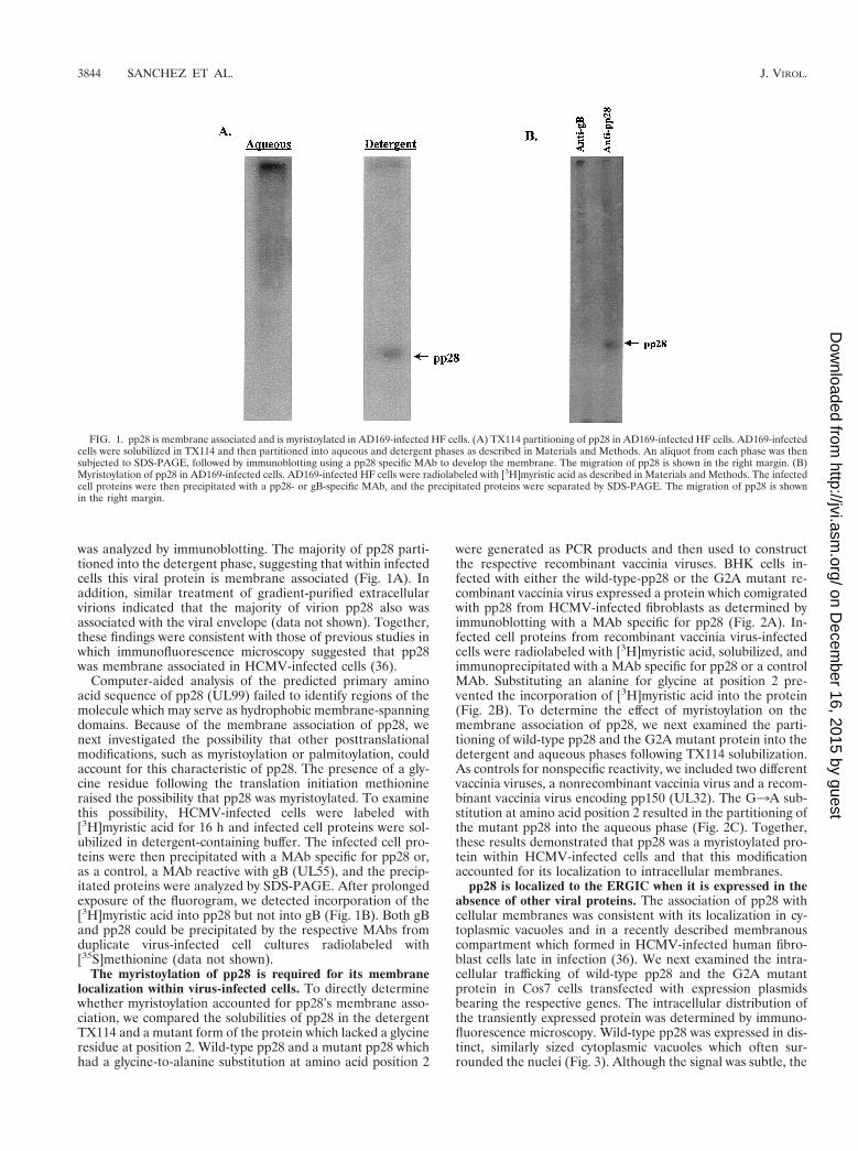
was analyzed by immunoblotting. The majority of pp28 parti-tioned into the detergent phase, suggesting that within infectedcells this viral protein is membrane associated (Fig. 1A). Inaddition, similar treatment of gradient-purified extracellularvirions indicated that the majority of virion pp28 also wasassociated with the viral envelope (data not shown). Together,these findings were consistent with those of previous studies inwhich immunofluorescence microscopy suggested that pp28was membrane associated in HCMV-infected cells (36).
Computer-aided analysis of the predicted primary aminoacid sequence of pp28 (UL99) failed to identify regions of themolecule which may serve as hydrophobic membrane-spanningdomains. Because of the membrane association of pp28, wenext investigated the possibility that other posttranslationalmodifications, such as myristoylation or palmitoylation, couldaccount for this characteristic of pp28. The presence of a gly-cine residue following the translation initiation methionineraised the possibility that pp28 was myristoylated. To examinethis possibility, HCMV-infected cells were labeled with[3H]myristic acid for 16 h and infected cell proteins were sol-ubilized in detergent-containing buffer. The infected cell pro-teins were then precipitated with a MAb specific for pp28 or,as a control, a MAb reactive with gB (UL55), and the precip-itated proteins were analyzed by SDS-PAGE. After prolongedexposure of the fluorogram, we detected incorporation of the[3H]myristic acid into pp28 but not into gB (Fig. 1B). Both gBand pp28 could be precipitated by the respective MAbs fromduplicate virus-infected cell cultures radiolabeled with[35S]methionine (data not shown).
The myristoylation of pp28 is required for its membranelocalization within virus-infected cells. To directly determinewhether myristoylation accounted for pp28’s membrane asso-ciation, we compared the solubilities of pp28 in the detergentTX114 and a mutant form of the protein which lacked a glycineresidue at position 2. Wild-type pp28 and a mutant pp28 whichhad a glycine-to-alanine substitution at amino acid position 2
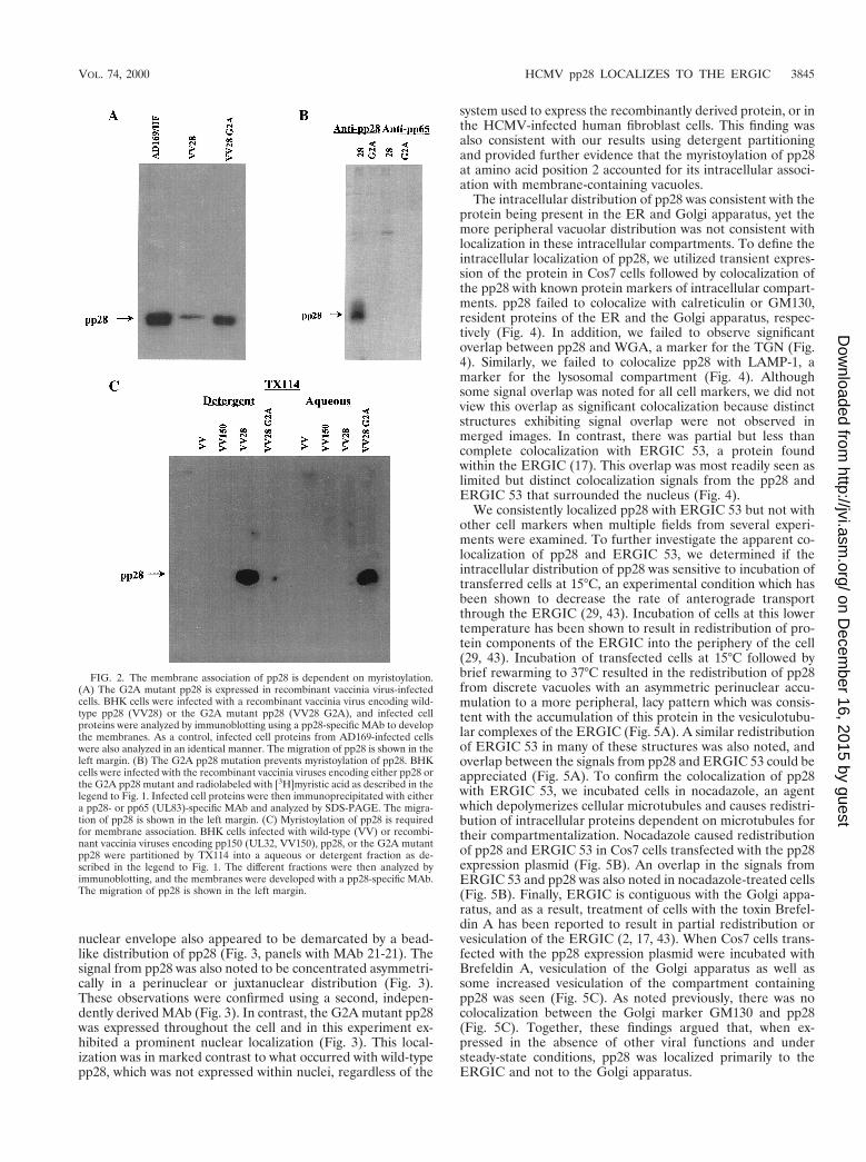
were generated as PCR products and then used to constructthe respective recombinant vaccinia viruses. BHK cells in-fected with either the wild-type-pp28 or the G2A mutant re-combinant vaccinia virus expressed a protein which comigratedwith pp28 from HCMV-infected fibroblasts as determined byimmunoblotting with a MAb specific for pp28 (Fig. 2A). In-fected cell proteins from recombinant vaccinia virus-infectedcells were radiolabeled with [3H]myristic acid, solubilized, andimmunoprecipitated with a MAb specific for pp28 or a controlMAb. Substituting an alanine for glycine at position 2 pre-vented the incorporation of [3H]myristic acid into the protein(Fig. 2B). To determine the effect of myristoylation on themembrane association of pp28, we next examined the parti-tioning of wild-type pp28 and the G2A mutant protein into thedetergent and aqueous phases following TX114 solubilization.As controls for nonspecific reactivity, we included two differentvaccinia viruses, a nonrecombinant vaccinia virus and a recom-binant vaccinia virus encoding pp150 (UL32). The G3A sub-stitution at amino acid position 2 resulted in the partitioning ofthe mutant pp28 into the aqueous phase (Fig. 2C). Together,these results demonstrated that pp28 was a myristoylated pro-tein within HCMV-infected cells and that this modificationaccounted for its localization to intracellular membranes.
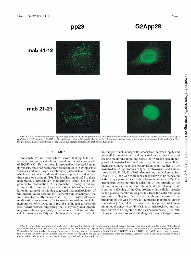
pp28 is localized to the ERGIC when it is expressed in theabsence of other viral proteins. The association of pp28 withcellular membranes was consistent with its localization in cy-toplasmic vacuoles and in a recently described membranouscompartment which formed in HCMV-infected human fibro-blast cells late in infection (36). We next examined the intra-cellular trafficking of wild-type pp28 and the G2A mutantprotein in Cos7 cells transfected with expression plasmidsbearing the respective genes. The intracellular distribution ofthe transiently expressed protein was determined by immuno-fluorescence microscopy. Wild-type pp28 was expressed in dis-tinct, similarly sized cytoplasmic vacuoles which often sur-rounded the nuclei (Fig. 3). Although the signal was subtle, the
FIG. 1. pp28 is membrane associated and is myristoylated in AD169-infected HF cells. (A) TX114 partitioning of pp28 in AD169-infected HF cells. AD169-infectedcells were solubilized in TX114 and then partitioned into aqueous and detergent phases as described in Materials and Methods. An aliquot from each phase was thensubjected to SDS-PAGE, followed by immunoblotting using a pp28 specific MAb to develop the membrane. The migration of pp28 is shown in the right margin. (B)Myristoylation of pp28 in AD169-infected cells. AD169-infected HF cells were radiolabeled with [3H]myristic acid as described in Materials and Methods. The infectedcell proteins were then precipitated with a pp28- or gB-specific MAb, and the precipitated proteins were separated by SDS-PAGE. The migration of pp28 is shownin the right margin.
3844 SANCHEZ ET AL. J. VIROL.
on Decem
ber 16, 2015 by guesthttp://jvi.asm
.org/D
ownloaded from
nuclear envelope also appeared to be demarcated by a bead-like distribution of pp28 (Fig. 3, panels with MAb 21-21). Thesignal from pp28 was also noted to be concentrated asymmetri-cally in a perinuclear or juxtanuclear distribution (Fig. 3).These observations were confirmed using a second, indepen-dently derived MAb (Fig. 3). In contrast, the G2A mutant pp28was expressed throughout the cell and in this experiment ex-hibited a prominent nuclear localization (Fig. 3). This local-ization was in marked contrast to what occurred with wild-typepp28, which was not expressed within nuclei, regardless of the
system used to express the recombinantly derived protein, or inthe HCMV-infected human fibroblast cells. This finding wasalso consistent with our results using detergent partitioningand provided further evidence that the myristoylation of pp28at amino acid position 2 accounted for its intracellular associ-ation with membrane-containing vacuoles.
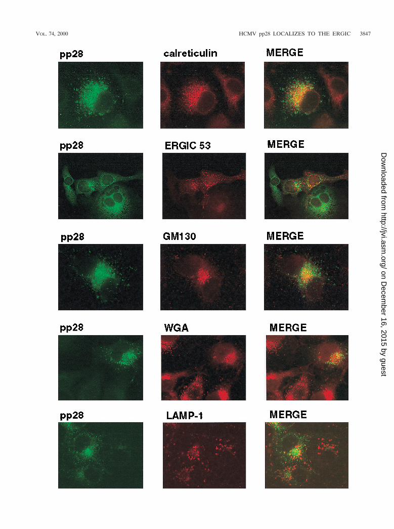
The intracellular distribution of pp28 was consistent with theprotein being present in the ER and Golgi apparatus, yet themore peripheral vacuolar distribution was not consistent withlocalization in these intracellular compartments. To define theintracellular localization of pp28, we utilized transient expres-sion of the protein in Cos7 cells followed by colocalization ofthe pp28 with known protein markers of intracellular compart-ments. pp28 failed to colocalize with calreticulin or GM130,resident proteins of the ER and the Golgi apparatus, respec-tively (Fig. 4). In addition, we failed to observe significantoverlap between pp28 and WGA, a marker for the TGN (Fig.4). Similarly, we failed to colocalize pp28 with LAMP-1, amarker for the lysosomal compartment (Fig. 4). Althoughsome signal overlap was noted for all cell markers, we did notview this overlap as significant colocalization because distinctstructures exhibiting signal overlap were not observed inmerged images. In contrast, there was partial but less thancomplete colocalization with ERGIC 53, a protein foundwithin the ERGIC (17). This overlap was most readily seen aslimited but distinct colocalization signals from the pp28 andERGIC 53 that surrounded the nucleus (Fig. 4).
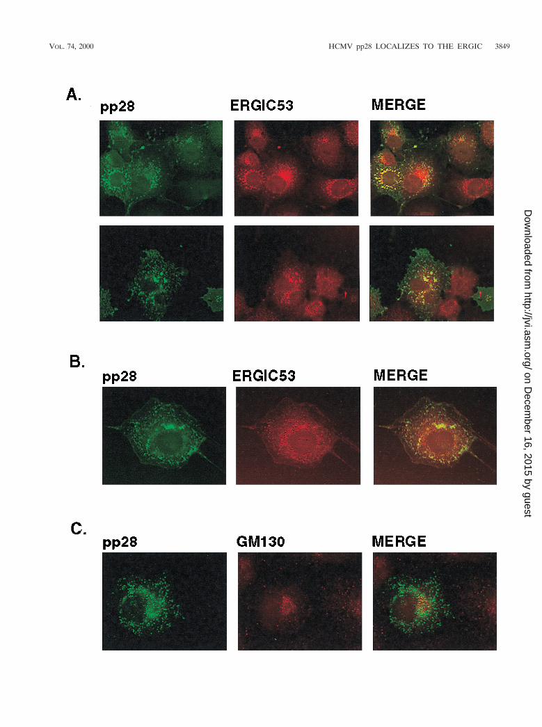
We consistently localized pp28 with ERGIC 53 but not withother cell markers when multiple fields from several experi-ments were examined. To further investigate the apparent co-localization of pp28 and ERGIC 53, we determined if theintracellular distribution of pp28 was sensitive to incubation oftransferred cells at 15°C, an experimental condition which hasbeen shown to decrease the rate of anterograde transportthrough the ERGIC (29, 43). Incubation of cells at this lowertemperature has been shown to result in redistribution of pro-tein components of the ERGIC into the periphery of the cell(29, 43). Incubation of transfected cells at 15°C followed bybrief rewarming to 37°C resulted in the redistribution of pp28from discrete vacuoles with an asymmetric perinuclear accu-mulation to a more peripheral, lacy pattern which was consis-tent with the accumulation of this protein in the vesiculotubu-lar complexes of the ERGIC (Fig. 5A). A similar redistributionof ERGIC 53 in many of these structures was also noted, andoverlap between the signals from pp28 and ERGIC 53 could beappreciated (Fig. 5A). To confirm the colocalization of pp28with ERGIC 53, we incubated cells in nocadazole, an agentwhich depolymerizes cellular microtubules and causes redistri-bution of intracellular proteins dependent on microtubules fortheir compartmentalization. Nocadazole caused redistributionof pp28 and ERGIC 53 in Cos7 cells transfected with the pp28expression plasmid (Fig. 5B). An overlap in the signals fromERGIC 53 and pp28 was also noted in nocadazole-treated cells(Fig. 5B). Finally, ERGIC is contiguous with the Golgi appa-ratus, and as a result, treatment of cells with the toxin Brefel-din A has been reported to result in partial redistribution orvesiculation of the ERGIC (2, 17, 43). When Cos7 cells trans-fected with the pp28 expression plasmid were incubated withBrefeldin A, vesiculation of the Golgi apparatus as well assome increased vesiculation of the compartment containingpp28 was seen (Fig. 5C). As noted previously, there was nocolocalization between the Golgi marker GM130 and pp28(Fig. 5C). Together, these findings argued that, when ex-pressed in the absence of other viral functions and understeady-state conditions, pp28 was localized primarily to theERGIC and not to the Golgi apparatus.
FIG. 2. The membrane association of pp28 is dependent on myristoylation.(A) The G2A mutant pp28 is expressed in recombinant vaccinia virus-infectedcells. BHK cells were infected with a recombinant vaccinia virus encoding wild-type pp28 (VV28) or the G2A mutant pp28 (VV28 G2A), and infected cellproteins were analyzed by immunoblotting using a pp28-specific MAb to developthe membranes. As a control, infected cell proteins from AD169-infected cellswere also analyzed in an identical manner. The migration of pp28 is shown in theleft margin. (B) The G2A pp28 mutation prevents myristoylation of pp28. BHKcells were infected with the recombinant vaccinia viruses encoding either pp28 orthe G2A pp28 mutant and radiolabeled with [3H]myristic acid as described in thelegend to Fig. 1. Infected cell proteins were then immunoprecipitated with eithera pp28- or pp65 (UL83)-specific MAb and analyzed by SDS-PAGE. The migra-tion of pp28 is shown in the left margin. (C) Myristoylation of pp28 is requiredfor membrane association. BHK cells infected with wild-type (VV) or recombi-nant vaccinia viruses encoding pp150 (UL32, VV150), pp28, or the G2A mutantpp28 were partitioned by TX114 into a aqueous or detergent fraction as de-scribed in the legend to Fig. 1. The different fractions were then analyzed byimmunoblotting, and the membranes were developed with a pp28-specific MAb.The migration of pp28 is shown in the left margin.
VOL. 74, 2000 HCMV pp28 LOCALIZES TO THE ERGIC 3845
on Decem
ber 16, 2015 by guesthttp://jvi.asm
.org/D
ownloaded from
DISCUSSION
Previously we and others have shown that pp28 (UL99)remained within the cytoplasm throughout the infectious cycleof HCMV (36). Furthermore, in productively infected humanfibroblasts, pp28 has been shown to accumulate in cytoplasmicvacuoles and in a large, membranous juxtanuclear structurewhich also contained additional tegument proteins and at leastthree envelope proteins (36). The localization of pp28 to thesemembranous intracellular compartments could not be ex-plained by examination of its predicted primary sequence.However, the presence of a glycine residue following the trans-lation initiation of methionine suggested that myristoylation ofthe protein could account for its membrane association. Wewere able to directly demonstrate that this posttranslationalmodification was necessary for its association with intracellularmembranes. Myristoylation of proteins is thought to occur onfree polyribosomes, suggesting that a myristoylated proteinmay associate nonspecifically with a number of different intra-cellular membranes (20). Our findings from image analysis did
not support such nonspecific association between pp28 andintracellular membranes and indicated more restricted andspecific membrane targeting. Consistent with the specific tar-geting of myristoylated viral matrix proteins to intracellularmembranes have been the observations from studies of themyristoylated Gag proteins of type C retroviruses and lentivi-ruses (8, 14, 33, 37, 42). With Moloney murine leukemia virus(Mo-MuLV), the Gag protein has been shown to be associatedwith the cytoplasmic face of the plasma membrane (42). Themechanism which permits localization of this protein to theplasma membrane is not entirely understood but may resultfrom the trafficking of the Gag protein with a cellular proteinto the plasma membrane or possibly from the cotranslationalinsertion of Gag into the plasma membrane because of theproximity of the Gag mRNA to the plasma membrane duringtranslation (13, 41, 42). Likewise, the Gag protein of humanimmunodeficiency virus (HIV) is also myristoylated and hasbeen shown to be targeted to the plasma membrane (8, 14, 50).However, in contrast to the findings with some C-type retro-
FIG. 3. Intracellular localization of pp28 is dependent on its myristoylation. Cos7 cells were transfected with an expression plasmid bearing either wild-type pp28(pp28) or the G2A mutant pp28 (G2App28) and imaged with pp28-specific MAbs and then fluorescein isothiocyanate anti-murine immunoglobulin G antibodies. Notethe prominent nuclear distribution of the G2A pp28 protein compared to that of wild-type pp28.
FIG. 4. Intracellular localization of pp28. Cos7 cells were transfected with an expression plasmid bearing the gene encoding pp28 and then fixed 48 h later asdescribed in Materials and Methods. The cells were reacted with either pp28-specific MAb or polyclonal rabbit anti-pp28 antibodies and the corresponding cell marker.We used the following markers for compartments in the secretory system: (i) calreticulin for the ER, (ii) ERGIC 53 for the ERGIC, (iii) GM130 for the Golgi apparatus,(iv) WGA for the TGN, and (v) LAMP-1 for lysosomes. Colocalization was considered significant when the merged signal revealed a yellow color associated with adistinct cellular site or structure and not in a more generalized location within the cell.
3846 SANCHEZ ET AL. J. VIROL.
on Decem
ber 16, 2015 by guesthttp://jvi.asm
.org/D
ownloaded from
VOL. 74, 2000 HCMV pp28 LOCALIZES TO THE ERGIC 3847
on Decem
ber 16, 2015 by guesthttp://jvi.asm
.org/D
ownloaded from
viruses, it appeared that HIV Gag may require an interactionwith the envelope glycoprotein for efficient targeting to theplasma membrane (32, 42). Interestingly, mutations in the Gagprotein of HIV-1 which prevented myristoylation also pre-vented assembly of capsid structures and budding of virus, yethad no effect on the proteolytic processing of the Gag polypro-tein (14). Thus, it appears that myristoylation of viral matrixprotein provides at least one mechanism of membrane associ-ation; however, this modification alone is insufficient for tar-geting the viral matrix protein to a specific intracellular mem-brane. Although several sequences within the primarysequence of pp28 resemble known intracellular targeting se-quences, our preliminary studies with pp28 deletion mutantshave failed to identify a specific targeting sequence within thisprotein.
Previously, studies have shown that the HCMV pp28 ho-molog of HSV, UL11, was also myristoylated (25). The impor-tance of myristoylation to the intracellular targeting of theHSV UL11 protein has recently been suggested by Bowzard etal. (J. B. Bowzard, R. J. Visalli, C. B. Wilson, E. M. Callahan,J. S. Loomis, R. J. Courtney, and J. W. Wills, Abstr. 24th Int.Herpesvirus Workshop, abstr. 7.021, 1999). Similar to the re-quirements for the targeting of Mo-MuLV to the plasma mem-brane, both myristoylation and a signal within the NH2 termi-nus of this protein have been postulated to be required fortargeting to intracellular membranes, specifically the TGN(Bowzard et al., 24th Int. Herpesvirus Workshop). A cluster ofbasic amino acids in the NH2 terminus of the MoMuLV Gagprotein has been suggested to be responsible for the interac-tion of MoMuLV Gag with the plasma membrane (42). Bow-zard et al. noted that an acidic cluster in the amino terminus ofHSV UL11 might have a similar role in the intracellular tar-geting of UL11 to the TGN (Bowzard et al., 24th Int. Herpes-virus Workshop). Those investigators further suggested thatthis cluster was conserved in UL11 homologs of several differ-ent herpesviruses and that it thus may represent a consensussignal for localization to the TGN. Although our results wereconsistent with specific intracellular targeting of pp28 and al-though pp28 contained an acidic cluster of amino acids in alocation similar to that of the acidic cluster of HSV UL11, theHCMV pp28 (UL99) protein was not localized to the TGNwhen it was expressed in the absence of other viral proteins.Thus, this region in HCMV pp28 did not appear to have thesame targeting function as the homologous region of HSVUL11, at least when UL99 was expressed in the absence ofother viral proteins. Additional experiments will be directedtowards determining the function of different domains of pp28in its intracellular trafficking; however, some caution must beapplied to the interpretation of these studies, because theintracellular trafficking of these herpesvirus matrix proteinswas defined in the absence of other viral functions which couldmarkedly alter the localization of these proteins in infectedcells.
Several of our results indicated that pp28 was located in amembranous compartment contiguous or interfacing with theERGIC. These included the finding that pp28 partially colo-
calized with ERGIC 53, perhaps the most well-studied markerof the ERGIC, but not with markers for the ER, Golgi appa-ratus, TGN, or lysosomal compartments (17). ERGIC 53 is alectin-like protein of 510 amino acids which contains a signalsequence, a transmembrane region, and a dilysine ER retrievalsignal at the C terminus (17). Studies of this protein haveshown that it cycles between the ER and the cis-Golgi appa-ratus but that under steady-state conditions it is concentratedwithin a compartment intermediate between the ER and Golgiapparatus, i.e., the ERGIC (2, 17, 29, 43). Under conditionswhich inhibited normal intracellular trafficking of ERGIC 53,such as incubation of cells at 15°C followed by rapid rewarmingto 37°C, the protein has been shown to exhibit a more periph-eral, spotty distribution which often completely surrounds thenucleus (23, 43). This is thought to occur secondarily to redis-tribution of ERGIC 53 containing vesiculotubular structures toan ER-like peripheral location. Incubation of Cos7 cells trans-fected with an expression plasmid bearing the gene encodingpp28 at 15°C resulted in a similar dispersion of pp28 intoperipheral sites in the cell. In the same experiment, ERGIC 53was similarly distributed in cells incubated at 15°C and partiallycolocalized with pp28 under these conditions. Following treat-ment of cells with nocadazole, pp28 and ERGIC 53 wereredistributed into an intracellular compartment which resem-bled the distribution which was observed following incubationat 15°C, and again colocalization of these proteins was ob-served. The latter result suggested that the distribution of pp28was dependent on the integrity of microtubules. Previous stud-ies which have compared the intracellular localizations of theKDEL-R protein and ERGIC 53 have shown that nocadazoleinhibited anterograde transport from the ERGIC to the Golgiapparatus but did not prevent redistribution of ERGIC 53 tothe ER (43). Finally, treatments with Brefeldin A caused asubtle redistribution of pp28, suggesting that at least some ofthe intracellular pp28 was associated with the cis-Golgi appa-ratus, yet not within the same structures as the Golgi proteinGM130. This finding was consistent with the distribution ofproteins cycling within the ERGIC as has been shown for TAP(p115) and ERGIC 53, proteins localized to the ERGIC (29).
The finding that a herpesvirus tegument protein was tar-geted to a compartment which interfaced with the ERGIC wasunexpected and further underscored the likelihood thatHCMV did not acquire its final envelope at the nuclear mem-brane as was previously proposed for a-herpesviruses. Thecellular ERGIC represents an early and very dynamic com-partment of the secretory system (2, 17, 23, 29). Althoughseveral proteins which under steady-state conditions concen-trate within the ERGIC have been described, none has beendescribed to be a resident of this compartment because of thenature of the ERGIC. This compartment likely represents anearly maturational stage of the Golgi apparatus, and proteinswhich do not contain ER retrieval signals, such as the C-terminal dilysine present in ERGIC 53, leave this compart-ment and concentrate within the Golgi stacks (2, 17). Thepredicted amino acid sequence of pp28 has a dilysine motif;however, it is positioned at residues 8 and 9 from the C ter-
FIG. 5. pp28 colocalizes with the ERGIC 53 protein. (A) Cos7 cells transfected with the expression plasmid bearing the gene encoding pp28 were incubated for2 h at 15°C, rewarmed briefly to 37°C, and then fixed as described in Materials and Methods. The cells were then reacted with a rabbit anti-pp28 antiserum or witha MAb reactive with ERGIC 53. Colocalization can be appreciated by the yellow signal surrounding the nucleus. The two panels represent the results of twoindependent experiments. (B) Cos7 cells transiently expressing pp28 were incubated in medium containing 2 mM nocadazole for 2 h and then fixed and processed asdescribed above. Colocalization can be observed in structures surrounding the nucleus. (C) pp28 does not colocalize with the Golgi apparatus after Brefeldin Atreatment. Cos7 cells expressing pp28 were treated with 2 mg of Brefeldin A per ml for 2 to 3 h and then fixed as described in Materials and Methods. The fixed cellswere then reacted with a pp28-specific MAb and a rabbit serum reactive with the Golgi marker, GM130, and developed with fluorochrome-conjugated secondaryantibodies. Note the vesiculation of the signals from both pp28 and GM130 compared to that of the corresponding panels in Fig. 4, but colocalization was not observed.
3848 SANCHEZ ET AL. J. VIROL.
on Decem
ber 16, 2015 by guesthttp://jvi.asm
.org/D
ownloaded from
VOL. 74, 2000 HCMV pp28 LOCALIZES TO THE ERGIC 3849
on Decem
ber 16, 2015 by guesthttp://jvi.asm
.org/D
ownloaded from
minus and therefore is unlikely to function as an ER retrievalsignal. Although our experimental findings were consistentwith the possibility that another ER retrieval signal was re-sponsible for the apparent cycling of pp28 in the ERGIC, wecannot rule out the equally likely alternative possibilities thatpp28 was interacting with a cellular protein which was cyclingwithin the ERGIC and that this protein-protein interactionaccounted for the intracellular distribution of pp28 in the ab-sence of other viral proteins.
Previously we have shown that in virus-infected cells, threedifferent tegument proteins (including pp28) and three differ-ent envelope proteins of HCMV accumulated late in infectionin a membranous, juxtanuclear structure which could not becolocalized to the ER, ERGIC, or Golgi apparatus (36). Thisstructure was dependent on the integrity of microtubules asdemonstrated by its sensitivity to the microtubule-destabilizingdrug nocadazole (36). Therefore, it was of interest that pp28remained in a distinct, ERGIC-linked compartment and failedto traffic to a similar intracellular location when it was ex-pressed in the absence of other viral proteins. This result in-dicated that either HCMV infection induced alteration of in-tracellular trafficking within the secretory pathway or that aspecific viral protein or viral function was required for local-ization of pp28 to the juxtanuclear compartment in produc-tively infected HF cells. We favor the explanation that thetransit of pp28 in infected cells was facilitated by other viralproteins because this unique viral protein containing an intra-cellular structure failed to develop in the absence of productivevirus infection. Because pp28 has been classified as a lateprotein during productive infection, the number of candidateviral proteins which could mediate transport of pp28 to thejuxtanuclear site of viral protein localization was extensive. Wepropose that it was most likely a viral protein which also traf-ficked through the secretory pathway. If this was the case, thenit follows that pp28 interacted with an envelope protein whichwas eventually targeted to a site of envelopment and virionassembly. Such an interaction would then suggest a role forpp28 in the intracellular budding of HCMV, perhaps similar tothe role postulated for myristoylated viral matrix proteins inthe assembly and budding of retroviruses.
ACKNOWLEDGMENTS
We thank Michael Mach for critical discussions and Dana Pinson forassistance in preparing the manuscript.
This work was supported by the National Institutes of Healththrough NIAID grant R01 AI35602 (W.J.B.).
REFERENCES
1. Baines, J. D., and B. Roizman. 1992. The UL11 gene of herpes simplex virus1 encodes a function that facilitates nucleocapsid envelopment and egressfrom cells. J. Virol. 66:5168–5174.
2. Bannykh, S. I., and W. E. Balch. 1997. Membrane dynamics at the endo-plasmic reticulum-Golgi interface. J. Cell Biol. 138:1–4.
3. Battista, M. C., G. Bergamini, M. C. Boccuni, F. Campanini, A. Ripalti, andM. P. Landini. 1999. Expression and characterization of a novel structuralprotein of human cytomegalovirus, pUL25. J. Virol. 73:3800–3809.
4. Billstrom, M. A., and W. J. Britt. 1995. Postoligomerization folding of hu-man cytomegalovirus glycoprotein B: identification of folding intermediatesand importance of disulfide bonding. J. Virol. 69:7015–7022.
5. Bordier, C. 1981. Phase separation of integral membrane proteins in TritonX-114 solution. J. Biol. Chem. 256:1604–1607.
6. Britt, W. J., L. Vugler, E. J. Butfiloski, and E. B. Stephens. 1990. Cell surfaceexpression of human cytomegalovirus (HCMV) gp55-116 (gB): use ofHCMV-recombinant vaccinia virus-infected cells in analysis of the humanneutralizing antibody response. J. Virol. 64:1079–1085.
7. Browne, H., S. Bell, T. Minson, and D. W. Wilson. 1996. An endoplasmicreticulum-retained herpes simplex virus glycoprotein H is absent from se-creted virions: evidence for reenvelopment during egress. J. Virol. 70:4311–4316.
8. Bryant, M., and L. Ratner. 1990. Myristoylation-dependent replication and
assembly of human immunodeficiency virus 1. Proc. Natl. Acad. Sci. USA87:523–527.
9. Darlington, R. W., and L. H. Moss. 1968. Herpesvirus envelopment. J. Virol.2:48–55.
10. Di Lazarro, C., G. Campadelli-Fiume, and M. R. Torrisi. 1995. Intermediateforms of glycoconjugates are present in the envelope of herpes simplexvirions during their transport along the exocytic pathway. Virology 214:619–623.
11. Garoff, H., R. Hewson, and D. J. E. Opstelten. 1998. Virus maturation bybudding. Microbiol. Mol. Biol. Rev. 62:1171–1190.
12. Gershon, A. A., D. L. Sherman, Z. Zhu, C. A. Gabel, R. T. Ambron, andM. D. Gershon. 1994. Intracellular transport of newly synthesized varicella-zoster virus: final envelopment in the trans-Golgi network. J. Virol. 68:6372–6390.
13. Gielkens, A. L. J., M. H. L. Salden, and H. Bloemendal. 1974. Virus-specificmessenger RNA on free and membrane-bound polyribosomes from cellsinfected with Rauscher leukemia virus. Proc. Natl. Acad. Sci. USA 71:1093–1097.
14. Gottlinger, H. F., J. G. Sodroski, and W. A. Haseltine. 1989. Role of capsidprecursor processing and myristoylation in morphogenesis and infectivity ofhuman immunodeficiency virus type 1. Proc. Natl. Acad. Sci. USA 86:5781–5785.
15. Granzow, H., F. Weiland, A. Jons, B. G. Klupp, A. Karger, and T. C.Mettenleitner. 1997. Ultrastructural analysis of the replication cycle of pseu-dorabies virus in cell culture: a reassessment. J. Virol. 71:2072–2082.
16. Grose, C. 1990. Glycoproteins encoded by varicella-zoster virus: biosynthesis,phosphorylation, and intracellular trafficking. Annu. Rev. Microbiol. 44:59–80.
17. Itin, C., R. Schindler, and H. P. Hauri. 1995. Targeting of protein ERGIC-53to the ER/ERGIC/cis-Golgi recycling pathway. J. Cell Biol. 131:57–67.
18. Johnson, D. C., and P. G. Spear. 1982. Monesin inhibits the processing ofherpes simplex virus glycoproteins, their transport to the cell surface, andegress of virions from infected cells. J. Virol. 43:1102–1112.
19. Johnson, D. C., and P. G. Spear. 1983. O-linked oligosaccharides are ac-quired by herpes simplex virus glycoproteins in the Golgi apparatus. Cell32:987–997.
20. Johnson, D. R., R. S. Bhatnagar, L. J. Knoll, and J. I. Gordon. 1994. Geneticand biochemical studies of protein N-myristoylation. Annu. Rev. Biochem.63:869–914.
21. Jones, F., and C. Grose. 1988. Role of cytoplasmic vacuoles in varicella-zoster virus glycoprotein trafficking and virion envelopment. J. Virol. 62:2701–2711.
22. Kerry, J. A., M. A. Priddy, C. P. Kohler, T. L. Staley, D. Weber, T. R. Jones,and R. M. Stenberg. 1997. Translational regulation of the human cytomeg-alovirus pp28 (UL99) late gene. J. Virol. 71:981–987.
23. Klumperman, J., A. Schweizer, H. Clausen, B. L. Tang, W. Hong, V. Oor-schot, and H. P. Hauri. 1998. The recycling pathway of protein ERGIC-53and dynamics of the ER-Golgi intermediate compartment. J. Cell Sci. 111:3411–3425.
24. Landini, M. P., B. Severi, G. Furlini, and D. G. L. Badiali. 1987. Humancytomegalovirus structural components: intracellular and intraviral localiza-tion of p28 and p65-69 by immunoelectron microscopy. Virus Res. 8:15–23.
25. MacLean, C. A., B. Clark, and D. J. McGeoch. 1989. Gene UL11 of herpessimplex virus type 1 encodes a virion protein which is myristylated. J. Gen.Virol. 70:3147–3157.
26. MacLean, C. A., A. Dolan, F. E. Jamieson, and D. J. McGeoch. 1992. Themyristylated virion proteins of herpes simplex virus type 1: investigation oftheir role in the virus life cycle. J. Gen. Virol. 73:539–547.
27. Meyer, H., A. Bankier, M. P. Landini, and R. Ruger. 1988. Identification andprocaryotic expression of the gene coding for the highly immunogenic 28-kilodalton structural phosphoprotein (pp28) of human cytomegalovirus.J. Virol. 62:2243–2250.
28. Molloy, S. S., E. D. Anderson, F. Jean, and G. Thomas. 1999. Bi-cycling thefurin pathway: from TGN localization to pathogen activation and embryo-genesis. Trends Cell Biol. 9:28–35.
29. Nelson, D. S., C. Alvarez, Y. S. Gao, R. Garcia-Mata, E. Fialkowski, and E.Sztul. 1998. The membrane transport factor TAP/p115 cycles between theGolgi and earlier secretory compartments and contains distinct domainsrequired for its localization and function. J. Cell Biol. 143:319–331.
30. Olson, J. K., and C. Grose. 1998. Complex formation facilitates endocytosisof the varicella-zoster viral gE:gI Fc receptor. J. Virol. 72:1542–1551.
31. Olson, J. K., and C. Grose. 1997. Endocytosis and recycling of varicella-zoster virus Fc receptor glycoprotein gE: internalization mediated by aYXXL motif in the cytoplasmic tail. J. Virol. 71:4042–4054.
32. Owens, R. J., J. W. Dubay, E. Hunter, and R. W. Compans. 1991. Humanimmunodeficiency virus envelope protein determines the site of virus releasein polarized epithelial cells. Proc. Natl. Acad. Sci. USA 88:3987–3991.
33. Rein, A., M. R. McClure, N. R. Rice, R. B. Luftig, and A. M. Schultz. 1986.Myristylation site in Pr65gag is essential for virus particle formation byMoloney murine leukemia virus. Proc. Natl. Acad. Sci. USA 83:7246–7250.
34. Roizman, B. 1996. Herpesviridae, p. 2221–2230. In B. N. Fields, D. M. Knipe,P. M. Howley, R. M. Chanock, J. L. Melnick, T. P. Monath, B. Roizman, and
3850 SANCHEZ ET AL. J. VIROL.
on Decem
ber 16, 2015 by guesthttp://jvi.asm
.org/D
ownloaded from
S. E. Straus (ed.), Fields virology, 3rd ed. Lippincott-Raven, Philadelphia,Pa.
35. Sanchez, V., P. C. Angeletti, J. A. Engler, and W. J. Britt. 1998. Localizationof human cytomegalovirus structural proteins to the nuclear matrix of in-fected human fibroblasts. J. Virol. 72:3321–3329.
36. Sanchez, V., K. D. Greis, E. Sztul, and W. J. Britt. 2000. Accumulation ofvirion tegument and envelope proteins in a stable cytoplasmic compartmentduring human cytomegalovirus replication: characterization of a potentialsite of virus assembly. J. Virol. 74:975–986.
37. Schultz, A. M., and A. Rein. 1989. Unmyristylated Moloney murine leukemiavirus Pr65 gag is excluded from virus assembly and maturation events. J. Vi-rol. 63:2370–2373.
38. Smith, J. D., and E. DeHarven. 1973. Herpes simplex virus and humancytomegalovirus replication in WI-38 cells. I. Sequence of viral replication.J. Virol. 12:919–930.
39. Spear, P. G. 1985. Glycoproteins specified by herpes simplex viruses, p.315–356. In B. Roizman (ed.), The herpesviruses, vol. 3. Plenum Press, NewYork, N.Y.
40. Steven, A. C., and P. G. Spear. 1997. Herpes capsid assembly and envelop-ment, p. 312–351. In W. Chiu, R. M. Burnett, and R. L. Garcea (ed.),Structural biology of viruses. Oxford University Press, New York, N.Y.
41. St. Johnston, D. 1995. The intracellular localization of messenger RNAs.Cell 81:161–170.
42. Suomalainen, M., K. Hultenby, and H. Garoff. 1996. Targeting of Moloneymurine leukemia virus gag precursor to the site of virus binding. J. Cell Biol.135:1841–1852.
43. Tang, B. L., S. H. Low, H. P. Hauri, and W. Hong. 1995. Segregation ofERGIC53 and the mammalian KDEL receptor upon exit from the 15 de-grees C compartment. Eur. J. Cell Biol. 68:398–410.
44. Tirabassi, R. S., and L. W. Enquist. 1999. Mutation of the YXXL endocy-
tosis motif in the cytoplasmic tail of pseudorabies virus gE. J. Virol. 73:2717–2728.
45. Vey, M., W. Schafer, B. Reis, R. Ohuchi, W. Britt, W. Garten, H. D. Klenk,and K. Radsak. 1995. Proteolytic processing of human cytomegalovirus gly-coprotein B (gpUL55) is mediated by the human endoprotease furin. Virol-ogy 206:746–749.
46. Wan, L., S. S. Molloy, L. Thomas, G. Liu, Y. Xiang, S. L. Rybak, and G.Thomas. 1998. PACS-1 defines a novel gene family of cytosolic sortingproteins required for trans-Golgi network localization. Cell 94:205–216.
47. Whealy, M. E., J. P. Card, R. P. Meade, A. K. Robbins, and L. W. Enquist.1991. Effect of Brefeldin A on alphaherpesvirus membrane protein glyco-sylation and virus egress. J. Virol. 65:1066–1081.
48. Whealy, M. E., A. K. Robbins, and L. W. Enquist. 1990. The export pathwayof the pseudorabies virus gB homolog gII involves oligomer formation in theendoplasmic reticulum and protease processing in the Golgi apparatus. J. Vi-rol. 64:1946–1955.
49. Whitley, A., B. Bruun, T. Minson, and H. Browne. 1999. Effects of targetingherpes simplex virus type 1 gD to the endoplasmic reticulum and trans-Golginetwork. J. Virol. 73:9515–9520.
50. Zhou, W., L. J. Parent, J. W. Wills, and M. D. Resh. 1994. Identification ofa membrane-binding domain within the amino-terminal region of humanimmunodeficiency virus type 1 Gag protein which interacts with acidic phos-pholipids. J. Virol. 68:2556–2569.
51. Zhu, Z., M. D. Gershon, Y. Hao, R. T. Ambron, C. A. Gabel, and A. A.Gershon. 1995. Envelopment of varicella-zoster virus: targeting of viral gly-coproteins to the trans-Golgi network. J. Virol. 69:7951–7959.
52. Zhu, Z., Y. Hao, M. D. Gershon, R. T. Ambron, and A. A. Gershon. 1996.Targeting of glycoprotein I (gE) of varicella-zoster virus to the trans-Golginetwork by an AYRV sequence and an acidic amino acid-rich patch in thecytosolic domain of the molecule. J. Virol. 70:6563–6575.
VOL. 74, 2000 HCMV pp28 LOCALIZES TO THE ERGIC 3851
on Decem
ber 16, 2015 by guesthttp://jvi.asm
.org/D
ownloaded from