UVI31+ Is a DNA Endonuclease That Dynamically Localizes to Chloroplast Pyrenoids in C. reinhardtii
13
UVI31+ Is a DNA Endonuclease That Dynamically Localizes to Chloroplast Pyrenoids in C. reinhardtii Manish Shukla 1 , Renu Minda 1 , Himanshu Singh 2 , Srikanth Tirumani 1 , Kandala V. R. Chary 2 , Basuthkar J. Rao 1 * 1 Department of Biological Sciences, Tata Institute of Fundamental Research, Colaba, Mumbai, India, 2 Department of Chemical Sciences, Tata Institute of Fundamental Research, Colaba, Mumbai, India Abstract UVI31+ is an evolutionarily conserved BolA family protein. In this study we examine the presence, localization and possible functions of this protein in the context of a unicellular alga, Chlamydomonas reinhardtii. UVI31+ in C. reinhardtii exhibits DNA endonuclease activity and is induced upon UV stress. Further, UVI31+ that normally localizes to the cell wall and pyrenoid regions gets redistributed into punctate foci within the whole chloroplast, away from the pyrenoid, upon UV stress. The observed induction upon UV-stress as well as the endonuclease activity suggests plausible role of this protein in DNA repair. We have also observed that UV31+ is induced in C. reinhardtii grown in dark conditions, whereby the protein localization is enhanced in the pyrenoid. Biomolecular interaction between the purified pyrenoids and UVI31+ studied by NMR demonstrates the involvement of the disordered loop domain of the protein in its interaction. Citation: Shukla M, Minda R, Singh H, Tirumani S, Chary KVR, et al. (2012) UVI31+ Is a DNA Endonuclease That Dynamically Localizes to Chloroplast Pyrenoids in C. reinhardtii. PLoS ONE 7(12): e51913. doi:10.1371/journal.pone.0051913 Editor: Rajagopal Subramanyam, University of Hyderabad, India Received September 3, 2012; Accepted November 7, 2012; Published December 17, 2012 Copyright: ß 2012 Shukla et al. This is an open-access article distributed under the terms of the Creative Commons Attribution License, which permits unrestricted use, distribution, and reproduction in any medium, provided the original author and source are credited. Funding: This work was funded by Department of Atomic Energy at TIFR. The funders had no role in study design, data collection and analysis, decision to publish, or preparation of the manuscript. Competing Interests: The authors have declared that no competing interests exist. * E-mail: [email protected] Introduction C. reinhardtii, a unicellular green alga, undergoes apoptosis in response to UV-C irradiation [1]. In order to understand the process of UV mediated apoptosis in C. reinhardtii, we undertook an in-silico global genome analysis, which revealed the presence of a UV inducible gene, UVI31+ from C. reinhardtii (C_2020005). This gene showed strong homology with a similar gene from S. pombe. UV inducible gene, UVI31+ was first identified in fission yeast S. pombe. UVI31+ gene was shown to get up regulated by about 5 to 10 fold within an hour of UV treatment (120 J/m 2 )of the cells [2]. However its expression was unaltered by other DNA damaging or cytotoxic agents, and has no significant homology to the known DNA repair genes [3]. During normal cell cycle, uvi31+ transcript increases during G1 phase before septation and also increases during diauxic shift. A null mutant of UVI31+ in S. pombe showed sensitivity to UV-light, defects inseptation and cytokinesis during the resumption of cell division from the UV damage-induced cell cycle arrest [4]. Protein sequence analyses revealed the presence of a ubiquitous BolA domain, rendering UVI31+ as a member of the BolA protein family. This family consists of the morphogene bolA from E. coli and its various homologs, which are ubiquitous and conserved from prokaryotes to eukaryotes including humans. Biological function of BolA domain in higher eukaryotes including humans is largely unknown. It is very likely that such conserved domain might be involved with diverse cellular functions depending up on its context. Commonly, BolA proteins have a helix turn helix motif, which is a major structural motif with an ability to bind DNA [5]. Further, most of the members of the BolA family are annotated as secretory proteins [6]. In E. coli, bolA transcript level increases in response to general stress [7] where the protein has the ability to cause osmotically stable round cells [8] and promote biofilm formation when over expressed [9]. In addition, cells lacking bolA do not undergo shape alteration in nutrient restrictive poor medium (M9 medium) at the onset of stationary phase or in response to stress as compared to the wild type cells [8]. On the other hand, BolA protein of P. fluorescens is implicated in the metabolism of sulphur containing amino acids and has no effect on bacterial cell morphology and biofilm formation, unlike E. coli BolA protein [10]. Here, we report that UVI31+ is a UV and dark inducible gene, which gets differentially regulated, during the light-dark (12 hr:12 hr) cycle of C. reinhardtii. Interestingly, purified UVI31+ protein as well as endogenous protein from C. reinhardtii cells is endowed with DNA endonuclease activity and causes about 1000 fold higher resistance to UV in E. coli cells over expressing UVI31+ protein. The protein gets localized in the cell wall and pyrenoid compartments of C. reinhardtii cell, the endonuclease activity is retained in these sites. Pyrenoids are the sub-organellar structures in the chloroplast of algae, which specialize in carbondioxide concentration and fixation during photosynthesis in the cell. It has been shown that Pyrenoids contain DNA [11] and are also associated with RNA processing in the cell [11,12]. Further, UVI31+ gets redistributed into punctate foci within the whole chloroplast, away from the pyrenoid, upon exposure to UV. Biomolecular interaction between the purified pyrenoids and UVI31+ studied by NMR demonstrates the involvement of the disordered loop domain of the protein in its interaction. This result PLOS ONE | www.plosone.org 1 December 2012 | Volume 7 | Issue 12 | e51913
Transcript of UVI31+ Is a DNA Endonuclease That Dynamically Localizes to Chloroplast Pyrenoids in C. reinhardtii

UVI31+ Is a DNA Endonuclease That DynamicallyLocalizes to Chloroplast Pyrenoids in C. reinhardtiiManish Shukla1, Renu Minda1, Himanshu Singh2, Srikanth Tirumani1, Kandala V. R. Chary2,
Basuthkar J. Rao1*
1 Department of Biological Sciences, Tata Institute of Fundamental Research, Colaba, Mumbai, India, 2 Department of Chemical Sciences, Tata Institute of Fundamental
Research, Colaba, Mumbai, India
Abstract
UVI31+ is an evolutionarily conserved BolA family protein. In this study we examine the presence, localization and possiblefunctions of this protein in the context of a unicellular alga, Chlamydomonas reinhardtii. UVI31+ in C. reinhardtii exhibits DNAendonuclease activity and is induced upon UV stress. Further, UVI31+ that normally localizes to the cell wall and pyrenoidregions gets redistributed into punctate foci within the whole chloroplast, away from the pyrenoid, upon UV stress. Theobserved induction upon UV-stress as well as the endonuclease activity suggests plausible role of this protein in DNA repair.We have also observed that UV31+ is induced in C. reinhardtii grown in dark conditions, whereby the protein localization isenhanced in the pyrenoid. Biomolecular interaction between the purified pyrenoids and UVI31+ studied by NMRdemonstrates the involvement of the disordered loop domain of the protein in its interaction.
Citation: Shukla M, Minda R, Singh H, Tirumani S, Chary KVR, et al. (2012) UVI31+ Is a DNA Endonuclease That Dynamically Localizes to Chloroplast Pyrenoids in C.reinhardtii. PLoS ONE 7(12): e51913. doi:10.1371/journal.pone.0051913
Editor: Rajagopal Subramanyam, University of Hyderabad, India
Received September 3, 2012; Accepted November 7, 2012; Published December 17, 2012
Copyright: � 2012 Shukla et al. This is an open-access article distributed under the terms of the Creative Commons Attribution License, which permitsunrestricted use, distribution, and reproduction in any medium, provided the original author and source are credited.
Funding: This work was funded by Department of Atomic Energy at TIFR. The funders had no role in study design, data collection and analysis, decision topublish, or preparation of the manuscript.
Competing Interests: The authors have declared that no competing interests exist.
* E-mail: [email protected]
Introduction
C. reinhardtii, a unicellular green alga, undergoes apoptosis in
response to UV-C irradiation [1]. In order to understand the
process of UV mediated apoptosis in C. reinhardtii, we undertook an
in-silico global genome analysis, which revealed the presence of a
UV inducible gene, UVI31+ from C. reinhardtii (C_2020005). This
gene showed strong homology with a similar gene from S. pombe.
UV inducible gene, UVI31+ was first identified in fission yeast S.
pombe. UVI31+ gene was shown to get up regulated by about 5 to
10 fold within an hour of UV treatment (120 J/m2)of the cells [2].
However its expression was unaltered by other DNA damaging or
cytotoxic agents, and has no significant homology to the known
DNA repair genes [3]. During normal cell cycle, uvi31+ transcript
increases during G1 phase before septation and also increases
during diauxic shift. A null mutant of UVI31+ in S. pombe showed
sensitivity to UV-light, defects inseptation and cytokinesis during
the resumption of cell division from the UV damage-induced cell
cycle arrest [4].
Protein sequence analyses revealed the presence of a ubiquitous
BolA domain, rendering UVI31+ as a member of the BolA protein
family. This family consists of the morphogene bolA from E. coli
and its various homologs, which are ubiquitous and conserved
from prokaryotes to eukaryotes including humans. Biological
function of BolA domain in higher eukaryotes including humans is
largely unknown. It is very likely that such conserved domain
might be involved with diverse cellular functions depending up on
its context. Commonly, BolA proteins have a helix turn helix
motif, which is a major structural motif with an ability to bind
DNA [5]. Further, most of the members of the BolA family are
annotated as secretory proteins [6]. In E. coli, bolA transcript level
increases in response to general stress [7] where the protein has the
ability to cause osmotically stable round cells [8] and promote
biofilm formation when over expressed [9]. In addition, cells
lacking bolA do not undergo shape alteration in nutrient restrictive
poor medium (M9 medium) at the onset of stationary phase or in
response to stress as compared to the wild type cells [8]. On the
other hand, BolA protein of P. fluorescens is implicated in the
metabolism of sulphur containing amino acids and has no effect on
bacterial cell morphology and biofilm formation, unlike E. coli
BolA protein [10].
Here, we report that UVI31+ is a UV and dark inducible gene,
which gets differentially regulated, during the light-dark
(12 hr:12 hr) cycle of C. reinhardtii. Interestingly, purified
UVI31+ protein as well as endogenous protein from C. reinhardtii
cells is endowed with DNA endonuclease activity and causes about
1000 fold higher resistance to UV in E. coli cells over expressing
UVI31+ protein. The protein gets localized in the cell wall and
pyrenoid compartments of C. reinhardtii cell, the endonuclease
activity is retained in these sites. Pyrenoids are the sub-organellar
structures in the chloroplast of algae, which specialize in
carbondioxide concentration and fixation during photosynthesis
in the cell. It has been shown that Pyrenoids contain DNA [11]
and are also associated with RNA processing in the cell [11,12].
Further, UVI31+ gets redistributed into punctate foci within the
whole chloroplast, away from the pyrenoid, upon exposure to UV.
Biomolecular interaction between the purified pyrenoids and
UVI31+ studied by NMR demonstrates the involvement of the
disordered loop domain of the protein in its interaction. This result
PLOS ONE | www.plosone.org 1 December 2012 | Volume 7 | Issue 12 | e51913

can rationalize localization changes involving dynamic re-associ-
ation of UVI31+ protein with pyrenoid in C. reinhardtii cells.
Results
UVI31+, a UV and Dark Inducible Gene from C. reinhardtiiOur in-silico global genome analysis had revealed the presence of
a UV inducible, UVI31+ protein from C. reinhardtii
(XP_001702905) that showed strong homology with a similar
gene from S. pombe (CAB_16898.1) (Table S1). We observed that
there were three distinct homologies (UVI31+ A gene ID 5728473,
UVI31+ B gene ID 5720138, UVI31+ C gene ID 5716816) of
UVI31+ in C. reinhardtii genome. The UVI31+ transcript level in C.
reinhardtii increased as a function of UV-C fluence and incubation
of cells in dark (Figure 1). There was about 12-fold increase in the
transcript level when the cells were exposed to UV-C (160 J/m2)
as compared to unexposed control. In addition, incubation of C.
reinhardtii in dark also led to induction of UVI31+ by about 6-fold.
The C. reinhardtii actin gene was used as an internal control to
assess the UVI31+ transcript levels semi-quantitatively. The cloned
gene corresponding to experimentally detected UV and dark
induced transcript matched fully with the homologue UVI31+ A,
both at DNA and protein sequence level. This gene (1063 bp)
comprises of three introns and four exons, encodes a transcript of
303 bases and a protein of 100 amino acid length (Protein ID-
XP_001702905.1). We searched for upstream regulatory elements
of UVI31+ gene that are relevant for DNA damage response and
cell cycle control. The analysis revealed a TATATAA box-like
signature at 2375 position, a DNA damage responsive like
element (DRE) TCTTGAA at 270, two Mlu1 cell cycle box
(MCB) like elements (AGGCGC and TCGTGA) at 2793 and
2646 positions respectively and two elements containing signature
sequences of SWI4/6 dependent cell cycle box (SCB) (GACAA
and AAAGAAAA) at 2738 and 2181 respectively. Such DRE-
like sequences are found in the upstream of several genes induced
during DNA damage response conditions [13]. Furthermore,
MCB and SCB-like elements are found in the promoter regions of
genes over-expressed in S. cerevisiae during the late G1 phase of cell
cycle [3]. The sequence signatures such as DRE, MCB and SCB
are consistent with a gene that is DNA damage inducible and cell
cycle regulated, expected of known phenotypes of UVI31+ gene
homologue in S. pombe [3].
Interestingly, in silico search for similar motifs showed that the
sequence of UVI31+ has a BolA-like domain from 16th to 100th
amino acid residues. This prompted us to align different BolA and
BolA-like sequences from various organisms with the C. reinhardtii
UVI31+. The percentage of amino acid residue identity was the
highest with C. albicans (48%) and the least with E. coli BolA protein
(27%), thus ascribing this protein as being closely related to the
fungal proteins (Table S1).
UVI31+ Protein of C. reinhardtii Reveals Properties of BothE. coli BolA and UVI31+ of S. pombe
Over expression of bola gene in E. coli causes formation of
osmotically stable spherical cells [8]. In spite of low sequence
homology (identity: 27% and similarity: 54%), C. reinhardtii
UVI31+ protein shows substantial structural homology with the
known tertiary structure of E. Coli BolA ([14] and unpublished
observations). With this in mind and gain an insight into the BolA
domain of UVI31+, we tested whether UVI31+ also causes round
morphology in bacterial cells. Our data suggests that E. coli cells
harboring the plasmid pRKM201 that over expresses UVI31+protein are predominantly round in shape (65%; Fig. 2A) as
opposed to E. coli cells that harbor control vector (pQE30) lacking
UVI31+ insert (27%; Figure 2A). Further, UVI31+ protein
expressing round cells showed normal growth kinetics and colony
forming units.
The sequence homolog of UVI31+ in S. pombe is specifically
induced by UV stress [2] and its null mutant is also sensitive to
UV-light [15]. We tested E. coli BL21 (DE3) cells over expressing
C. reinhardtii UVI31+ protein for its UV-sensitivity phenotype.
Cells over expressing UVI31+ protein showed ,1000 fold more
resistance to UV in the range of 35 to 70 J/m2 compared to cells
harboring only the control vector (pQE30) (Figure 2B). The round
cell morphology and UV-resistance conferred by UVI31+ over
expression in E. coli cells was specific to UVI31+ protein. A control
protein (human Translin) over expression does not cause these
phenotypes (data not shown).
Purification and Biochemical Characterization of UVI31+from E. coli
Sequence analysis of the cDNA (303 bp) confirmed the presence
of an uninterrupted open reading frame. Recombinant UVI31+protein was purified from E. coli by Ni2+-NTA agarose column,
followed by gel filtration chromatography [14]. The protein eluted
as a major peak at 87 ml, a volume corresponding to a monomer
of approximately 13 kDa. However, a small hump was also
observed between 68th–75th ml indicating a dimeric form. SDS-
PAGE combined with Comassie Brilliant Blue, silver nitrate
staining and western blot analysis using anti-Histidine antibody
confirmed the purity of the protein eluted (Figure S1). Assessing
Figure 1. Expression of UVI31+ gene from C. reinhardtii usingsemi quantitative RT-PCR. (A) Levels of UVI31+ (I) and actin (II) cDNAin control and UV-C irradiated cells. (B) Levels of UVI31+ (I) and actin (II)cDNA in control and dark incubated cells.doi:10.1371/journal.pone.0051913.g001
Role of UVI31+ in C. reinhardtii Physiology
PLOS ONE | www.plosone.org 2 December 2012 | Volume 7 | Issue 12 | e51913

the molecular mass and protein sequence of the protein elute
(13,404633 Da) by mass spectrometry further confirmed it to be
UVI31+.
Most BolA proteins have two conserved basic regions harboring
a helix-loop-helix domain predicted to be involved in DNA
binding. We therefore checked the DNA binding ability of purified
UVI31+ protein. Different plasmids were incubated with UVI31+pure protein and checked for electrophoretic mobility shift.
Surprisingly, we found that UVI31+ showed DNA nicking instead
of binding activity (Figure 2C). It acts as a nonspecific
endonuclease on both UV-irradiated and un-irradiated super-
coiled and nicked circular forms of double stranded plasmid DNA,
converting them into linear form. Endonuclease action was also
evident in the time-course analyses performed in a separate
electrophoretic run (Figure 2C). Under the experimental condi-
tions described here, the DNA did not fragment further. However,
when the reaction was performed at higher protein concentrations
and/or for prolonged incubation times, we did see additional
fragmentation of full-length linear DNA (data not shown). Lack of
smearing from full-length linear band suggested relative absence of
dsDNA-specific exonuclease activity. The reaction required Mg
(II) and was inhibited in the presence of EDTA and was
independent of ATP or GTP hydrolysis. Purified protein showed
a gel filtration profile that contained monomer and dimer forms.
DNA endonuclease activity profile scaled in proportion with
protein absorbance and the activity associated with the dimer peak
was relatively higher than that of monomer (data not shown).
Detailed characterization of DNA endonuclease activity, study of
mutations that modulate the same is part of a separate
investigation. In this paper, we describe the protein localization
and its modulation in C. reinhardtii cells, as described below.
UVI31+ Protein Localization in C. reinhardtii CellsPolyclonal antibodies were raised against purified UVI31+
protein. Purified antibody preparation as well as control antibodies
(neutralized with purified UVI31+ protein) (see Methods) were
used to perform immunofluorescence experiments against C.
reinhardtii cells as well as purified cellular fractions (as described
below). A diffuse signal of anti-UVI31+ antibody was seen
throughout the cell, but it was more intense in the cell wall and
Figure 2. UVI31+ overexpression physiology in E. coli and characterization of its endonuclease activity. (A) Morphology of E. coli BL21cells. Phase contrast micrographs of BL21 (pQE30); and BL21 over expressing UVI31+ (pRKM201). (B) UV survival of E. coli BL21 strains harboring theplasmid pRKM201 over expressing UVI31+ (&)and pQE30 (N). (C) Endonuclease activity of UVI31+.Plasmid DNA un-irradiated (lane 1) or irradiatedwith 7.2 J/m2 of UV-C (lane 3) was incubated with 13 mM of UVI31+ respectively (lanes 2 and 4), for 30 min at 37uC. Plasmid DNA (lane 5) wasincubated with 13 mM of UVI31+ (lanes 6–8) for 5, 15 and 30 min, respectively. M, 1 kb DNA ladder.doi:10.1371/journal.pone.0051913.g002
Role of UVI31+ in C. reinhardtii Physiology
PLOS ONE | www.plosone.org 3 December 2012 | Volume 7 | Issue 12 | e51913
pyrenoid locations (Figure 3). We ascertained the cell wall signal
by using a cell wall minus mutant (CC3395) where the peripheral
staining was absent. However, this mutant showed localization of
UVI31+ in the pyrenoid region, which was identified by bright
field image of the cells (Figure 3). Cell wall associated antibody
staining was variable in different growth and culture conditions.
However pyrenoid staining was more consistent in wild type
(CC125) and cell wall minus strain (CC3395). UVI31+ neutralized
antiserum showed the loss of signal at pyrenoid as well as cell wall
locations. In fact, this control revealed a complete loss of diffuse
staining associated with UVI31+ protein in the cells, thereby
indicating that antibody staining was specific to UVI31+ protein.
Since UVI31+ gene is UV inducible, we wanted to check UV-stress
associated changes in the cell. At the UV dosage of 160 J/m2
(about 30 to 40% cell lethality), we observed a significant loss of
the pyrenoid-associated signal in wild type (CC125) and cell wall
minus strain (CC3395). However, UV exposure did not affect cell
wall associated signal in CC125. Other intracellular distributional
changes of UVI31+ protein in CC125 were less distinct. In
CC3395 cells (cell wall minus), UV exposure seems to have
generated a punctate distribution of UVI31+ protein. In our
experience, CC3395 cells were better than CC125 for imaging
UVI31+ protein changes. The punctate staining of UVI31+protein in UV treated CC3395 cells were further corroborated by
confocal imaging of cells. Confocal images of immunostained cells
(Digital slices in Figure 2) and 3D rendered version of the same
(Figure 3C & 3D) revealed that pyrenoid staining is reduced
accompanied by increase in punctate staining of UVI31+ in cells
that were UV-treated. Punctate staining is visible across all the
slices, thereby suggesting that UVI31+ protein spreads throughout
the chloroplast following UV-treatment.
Localization and DNA-endonuclease Activity of UVI31+Protein in Purified Pyrenoid and Cell Wall Fractions
We purified pyrenoid and cell wall fractions according to a
standard procedure ([16] and [17], see Methods). The purified
fractions showed expected morphological characteristics in bright
field imaging: pyrenoid fraction showed spherical bodies of about
3–5 micron size; cell wall fraction revealed largely empty spherical
shells, devoid of cell contents (Figure 4A & B). When the fixed
preparations of the cell wall and pyrenoid fractions were analyzed
by immunofluorescence, bright staining of anti UVI31+ antibodies
was seen (Figure 4A & B). This pattern corroborated the staining
seen for C. reinhardtii cells. Though whole cell protein western
showed no signal for antiUVI31+ the purified cellwall and
pyrenoid western were positive (Figure S3). Western blot analyses
of these fractions were performed along with purified UVI31+protein as a standard control for comparison. The standard
protein lane showed a strong western signal at 13.4 kDa
(monomer position) while the same at dimer position was rather
weak. However, the dimer specific signal was strong in pyrenoid
and cell wall fractions (Figure 4C). Monomer levels were too low to
be detected in these cell fractions. Western blot analyses of whole
cell extracts (control & UV-treated) had failed to detect UVI31+protein signal (Figure S3), suggesting that anti-UVI31+ Ab is not
only unable to detect endogenous UVI31+ protein but also is
unable to spuriously cross-react with any other cellular proteins in
the whole cell extract sample, perhaps due to insufficient titer
strength of the Ab preparation. However, we succeeded in
obtaining the western signal for UVI31+ in samples, which are
enriched with this protein (Figure 4D). We therefore conclude that
anti-UVI31+ Ab signal in the western blot analyses performed on
pyrenoid and cell wall samples is specific.
We wanted to verify whether the protein present in pyrenoid
and cell wall fractions also shows DNA endonuclease activity,
consistent with the purified UVI31+ protein. DNA endonuclease
activity was tested by a zymogram assay, following renaturation of
proteins from pyrenoid and cell wall fractions resolved in SDS-
PAGE. Both the monomer and dimer forms of the purified
UVI31+ protein showed the endonuclease activity, where the
dimer was relatively more active compared to monomer
(Figure 4D). Interestingly, zymogram assay being more sensitive
than western blot enabled us to detect the trace levels of UVI31+monomers in pyrenoid fractions. This observation corroborated
our earlier result where dimeric form of protein from gel-filtration
profile had shown higher endonuclease activity (data not shown).
Interestingly, the cell wall and the pyrenoid fractions also showed
the endonuclease activity in protein bands corresponding to the
dimer form. The activity associated with monomer form of the
protein was faint, but discernible.
UVI31+ localization Changes During Light-dark Cycle andConcomitant Effects Following UV-C Exposure
It is the 12 hr dark period that is central to division processes in
C. reinhardtii during which UVI31+ protein function is likely to be
paramount, as inferred from the studies reported on UVI31+homologue in S. pombe [3,4,18]. We therefore set out analyzing
changes in UVI31+ transcript levels as well as UVI31+ localization
in C. reinhardtii cells during different time points of growth in
synchronized cells from light-dark (12 hr:12 hr) cycles. Experi-
ments were carried out on CC3395 cells grown photoautotrophi-
cally in acetate-free TP medium. We performed the immunoflu-
orescence analyses on synchronized cells harvested at four
different time points, mid-dark (MD; 6 hr into dark), complete
dark (CD; 12 hr into dark), mid-light (ML; 6 hr into light) and
complete light (CL; 12 hr into light) (see Methods). Pyrenoid
specific location of UVI31+ protein was evident only in dark phase
cells (MD & CD, Figure 5A). The imaging resolution does not
allow us to discern if there is any punctate pattern in pyrenoid in
any conditions. The staining was weak and diffuse, with much
reduced localization at pyrenoids, in light phase cells (ML & CL,
Figure 5A). UV-C exposure caused changes in protein localization
(as shown earlier, Figure 3): pyrenoid specific staining in dark-
phase cells was reduced, with concomitant appearance of punctate
and diffuse staining in the cell (Figure 5B). Light-phase cells also
exhibited similar punctate and diffuse staining following UV-C
exposure, where the intensity was less as compared to equivalent
dark-phase cells. We believe that the weaker IF signal in UV-
treated cells in Figure 5 (compared to that in Figure 1 & 3) is
perhaps due to reduced UV-C as well as other differences in the
conditions (see Methods). The focus of the experiment described in
Figure 5 is on control samples that are not exposed to UV-C,
where we see dramatic changes in UVi31+ protein localization at
different stages of light-dark cycle. Based on the confocal imaging
data described in Fig. 3, we believe that the diffuse and punctate
staining of protein observed in light phase cells and UV-treated
dark phase cells relate to the spreading of protein into chloroplast
compartment. We also carried out RT-PCR analyses of UVI31+transcript level changes. We found that the UVI31+ transcripts
reach a maximum in CD, followed by a drop through ML/CL
and the start of a surge at MD that peaks at CD (Figure 5C).
UVI 31+ Interacts with Pyrenoids via Specific StructuralDomains of the Protein
Prompted by our observations that UVI31+ protein exhibits
localization changes that involve dynamic association and
Role of UVI31+ in C. reinhardtii Physiology
PLOS ONE | www.plosone.org 4 December 2012 | Volume 7 | Issue 12 | e51913

dissociation of the protein with pyrenoids in C. reinhardtii cells
(Figure 3C & D; Figure 5), we tested the ability of purified UVI31+protein to directly interact with purified pyrenoids, in vitro. We
used a solution NMR technique to study the biomolecular
interaction between the purified pyrenoids and UVI31+. We
wanted to probe UVI31+ binding to pyrenoid at amino acid
Figure 3. Localisation of UVI31+ protein in C. reinhardtii cells. Immunofluorescence staining of UVI31+ protein in C. reinhardtii CC125 andCC3395 (from dark phase) and 160 J/m2 UV treated cells (A) and bright field images (B) (Bars 5 mm). 3D rendering of UVI31+ immunofluorescenceconfocal images of CC3395 control (C) and 160j UV treated cells (D).doi:10.1371/journal.pone.0051913.g003
Role of UVI31+ in C. reinhardtii Physiology
PLOS ONE | www.plosone.org 5 December 2012 | Volume 7 | Issue 12 | e51913
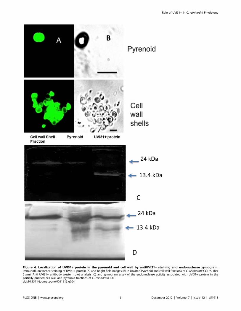
Figure 4. Localization of UVI31+ protein in the pyrenoid and cell wall by antiUVI31+ staining and endonuclease zymogram.Immunofluorescence staining of UVI31+ protein (A) and bright field images (B) in isolated Pyrenoid and cell wall fractions of C. reinhardtii CC125. (Bar5 mm). Anti UVI31+ antibody western blot analysis (C) and zymogram assay of the endonuclease activity associated with UVI31+ protein in thepartially purified cell wall and pyrenoid fractions of C. reinhardtii (D).doi:10.1371/journal.pone.0051913.g004
Role of UVI31+ in C. reinhardtii Physiology
PLOS ONE | www.plosone.org 6 December 2012 | Volume 7 | Issue 12 | e51913

residue level resolution. We eventually intend to map the UVI31+protein domain that interacts with pyrenoid for designing
appropriate genetic perturbations, in vivo. This is feasible by
mapping the interacting residues using uniformly 13C and 15N
labeled UVI31+ protein complexed with unlabeled pyrenoids. The
heteronuclear single quantum coherence/correlation (HSQC) is
the most frequently used experiment in such a study. The resulting
two-dimensional (2D) spectrum correlates 1H spins with its directly
attached heteronucleus, an NMR active nucleus such as 13C and15N. The spectrum provides residue level information. In such an
interaction study, one can isotopically label one of the interacting
molecules, leaving the other one without label. Thus, when one
records the HSQC spectrum of such a complex, one observes the
spectral signatures arising from the isotopically labeled molecule
alone, and hence, it turns out to be less complex for analysis. A
comparison of the spectral signatures of the labeled molecule in its
Figure 5. Localisation changes of UVI31+ protein in C. reinhardtii cells during light dark regime and UV exposure. Immunofluorescencestaining of UVI31+ protein (A) and bright field images (B) of C. reinhardtii CC3395 control and UV treated (40 J/m2)cells at various intervals of the darkand light regime (Bar 5 mm). Expression analyses of UVI31+ gene from C. reinhardtii CC3395at various intervals of the dark and light regime by RT-PCR(C).doi:10.1371/journal.pone.0051913.g005
Role of UVI31+ in C. reinhardtii Physiology
PLOS ONE | www.plosone.org 7 December 2012 | Volume 7 | Issue 12 | e51913

free state and as part of the complex throws light on the
biomolecular interaction. Though the HSQC experiment can be
performed using natural abundance of the 15N isotope, one
normally uses an isotopically labeled protein, which is produced by
expressing it in bacterial cells grown in 15N-labelled medium. The
2D [15N-1H] HSQC provides the correlation between the
backbone 15N and its directly attached amide proton (1HN) of
each residue with an exception of Pro residue, which lacks 1HN.
When a protein interacts strongly with high molecular weight
cellular component such as DNA, heat shock protein complex, cell
membrane, pyrenoid organelle etc, the bound residues of the
protein can cause line broadening up to the complete loss of the
respective NMR signals due to their slowing in tumbling motion
[19]. Alternately, binding that does not significantly reduce
tumbling motion of residues, but alter their chemical environment,
leads to chemical shift perturbations [20].
In the present study, we used 2D [15N-1H] HSQC to
characterize the biomolecular interaction between the purified
pyrenoids and uniformly 15N-labeled UVI31+ (200 mM). Earlier,
we had carried out almost complete sequence specific 1H, 13C and15N resonance assignments of UVI31+ [14] and the 3D solution
structure of the protein was determined by NMR spectroscopy
(Manuscript under preparation). With this in the backdrop, when
we added the protein to purified pyrenoids fraction, we observed
distinct chemical shift perturbations in the HSQC spectrum of the
complex (Figure 6). Visual inspection of the spectrum revealed
subtle changes in the spectrum. In the complex, some of the
original peaks broadened out and some showed distinct changes in
their chemical shifts, while many remained unperturbed. Figure 6
shows the overlay of the 2D [15N-1H] HSQC spectrum of the
complex (formed after mixing u-15N-labelled UVI31+ with
pyrenoids) with that of u-15N-labelled UVI31+ alone. The
broadened peaks included the spectral signatures of the N-
terminal amino acid residues belonging to the entire polypeptide
stretch between the residues at positions G11 and M24 (Figure 6).
The peaks, which showed substantial perturbations in the
chemical shifts, included those of A25, E26, Q28, L29, F49,
H56, K57, H58, A59, H61, A63, S67, A72, L79 and R94. Rest of
them does not undergo any chemical shift perturbation. UVI31+protein thus interacts with pyrenoids largely through the segment
G11–M24, as evidenced by the missing peaks due to signal
broadening (Figure 6). Further, significant chemical shift pertur-
bations seen near the N-terminal polypeptide stretch of the protein
(A25, E26, Q28 and L29) and in the disordered loop region of the
protein (H56, K57, H58, A59, H61, A63, S67, A72, L79 and
R94), which is in a close proximity of the N-terminal polypeptide
stretch, support the biomolecular interaction between UVI31+and pyrenoid. We therefore, conclude that the biomolecular
interaction is specific and perhaps is of physiological relevance.
That will be tested further by in vivo genetic perturbation
experiments as a part of separate study. Complete sequence
specific 1H, 13C and 15N resonance assignments and 3D structural
characterization of the complex formed after mixing u-15N-
labelled UVI31+ with pyrenoids is in progress.
Discussion
UVI31+ protein has been shown to be important in regulating
the cell division function in S. pombe, especially after the cells
resume back following cell-cycle arrest [4]. More specifically, the
protein seems to negatively regulate septum formation and
cytokinesis during the onset of cell division [4]. UVI31+ null
mutant cells show higher tendency to form spurious septa and
undergo enhanced cell proliferation yielding smaller size cells,
compared to wild type control cells [4]. In addition, UVI31+transcript levels go up following UV-exposure of cells and show
cell growth phase dependent cyclic changes in expression levels
where transient increase is seen during diauxic shift of growth
phase [3]. In this study, we corroborate the basic features of
UVI31+ protein as described above from S. pombe in another single
celled organism, C. reinhardtii, and shed light on some additional
features of the protein. After the previous elaboration of UVI31+ in
S. pombe, the only organism where this protein function has been
studied so far, our study perhaps represents the second system
being investigated to gain more understanding of cellular function
of UVI31+ protein. C. reinhardtii offers a unique system where cell
division is regulated by light-dark (12 hr:12 hr) cycles: cells grow in
size, reach ‘‘commit to divide’’ stage after reaching a critical cell
size in 12 hr light period followed by 2 or 3 successive divisions
yielding 4 or 8 daughter cell clusters in the 12 hr dark period
(Harris 2009). It is the 12 hr dark period that is central to division
processes in C. reinhardtii during which UVI31+ protein function is
likely to be paramount.
Firstly, we were encouraged by the observation that UVI31+gene transcript levels go up when the C. reinhardtii cells are grown
in dark (division phase in C. reinhardtii) and also when the cells are
exposed to UV-C (Figure 1& 5C), The sequence signatures such as
DRE, MCB and SCB that we detected at upstream region of
UVI31+ gene also add credence to our conclusion that the gene is
DNA damage inducible and cell cycle regulated, just as the
UVI31+ of in S. pombe [3]. All these observations, put together,
were in line with the lessons learned in S. pombe system, which
prompted us to study UVI31+ function further. In addition,
UVI31+ protein from C. reinhardtii, when over-expressed in E. coli
cells, conferred several hundred fold higher resistance to UV-C
compared to control cells. Moreover, as in UVI31+ gene of S.
pombe, the C. reinhardtii gene also showed a discernible BolA domain
sequence (Table S1). Importantly, E. coli cells over expressing
UVI31+ protein from C. reinhardtii conferred round cell morphol-
ogy in the bacterial cells, a phenotype that is ascribed to BolA
domain proteins [8]. Therefore all the hallmarks of UVI31+protein from C. reinhardtii seem to fit the features well described for
S.pombe protein.
Our current study extends the analyses further, in the context of
C. reinhardtii biology, as described below. 1. The protein shows
dominant localization to cell wall and pyrenoid fractions (Figure 3).
2. Protein localizes to pyrenoids only when the cells are grown in
dark (Figure 5A). Contrarily, cells grown in light exhibit
distributed localization of UVI31+ protein, not associated with
pyrenoids (Figure 5A). 3. Intriguingly, over expressed-purified
protein as well as the endogenous protein from C. reinhardtii
exhibits DNA endonuclease activity. The protein localization to
cell wall region has been enigmatic. A comparison using the
UVI31+ protein structure solved by solution NMR [21] (Rout, A.
K. et. al., Unpublished), revealed that there is considerable
structural similarity with Serratia family endonuclease structural
motifs. Such enzymes with unusual localization might also perform
functions other than that in replication, repair and recombination
[22] and thereby have roles in host defense [23], apoptosis [24]
and cell division [25]. One could speculate that cell wall
localization of a nuclease might poise for its release extracellularly
in stress responses. Alternately, cell wall localization of UVI31+protein possibly places it right for rendering it as cell shape
reorganizer expected of a BolA domain (morphogene) containing
protein.
All these attributes suggest that UVI31+ protein is associated
with important functions whose specific relevance is unclear. Even
though, we have been yet unsuccessful in either over expressing
Role of UVI31+ in C. reinhardtii Physiology
PLOS ONE | www.plosone.org 8 December 2012 | Volume 7 | Issue 12 | e51913

the protein in C. reinhardtii cells or knocking it down by an RNAi
approach, efforts are on to gain cell biological insights on these
aspects. Nevertheless, as a first pass, we have uncovered some
interesting aspects of the protein based on which we speculate that
UVI31+ protein may be involved in UV-damage response and
repair under the regulation of light-dark cycles.
It is important to note that we still do not fully understand the
transit peptide sequence motifs that are required for several
Figure 6. An overlay of 2D [15N-1H] HSQC spectrum of the complex (formed between u-15N-labelled UVI31+ and pyrenoids) (shownin black) with that of u-15N-labelled UVI31+ alone (shown in red). Most red spots overlap with black spots in the spectrum (residues that donot change in the complex, also shown as black letter code of the amino acid residues). Signals of a few residues are lost in the complex (shown asgreen letter code of the amino acid residues). Signals of a few residues are shifted due to chemical shift perturbations (shown as blue letter code ofthe amino acid residues). The amine-group peaks from the side-chains of Asn and Gln appear as doublets near the top right corner of the spectrum asconnected by horizontal lines. The experiments were recorded at pH 6.4 and 298 K. The spectra were recorded on Bruker Avance 800 MHz NMRspectrometer with 256 and 2048 complex points along t1 and t2 dimensions, respectively.doi:10.1371/journal.pone.0051913.g006
Role of UVI31+ in C. reinhardtii Physiology
PLOS ONE | www.plosone.org 9 December 2012 | Volume 7 | Issue 12 | e51913
thousands of proteins that are nuclear coded but are subsequently
imported into organelles such as chloroplast, mitochondria and
peroxisomes etc [26]. The problem is more compounded when it
relates to chloroplast transit peptide signatures, which appear
highly diverse [26]. Since the protein sequence showed no
chloroplast-specific import tags, it is likely that UVI31+ protein
reaches pyrenoid location in chloroplast by the help of an
unknown interactor protein.
mRNA transcripts of UVI31+ show a twelve and six fold
increase in expression during UV stress and dark incubation
respectively in C. reinhardtii cells (Fig. 1). The presence of the
upstream DRE elements reflects the role of these sequences in
the over expression of the UVI31+ transcripts in the DNA
damaging conditions of UV exposures [13]. The presence of
DRE elements and the possible up regulation of these elements
after UV exposure underscores UVI31+ role in UV survival
response as shown in over expressing E. coli (Figure 2B). The
cells enter into S phase of cell cycle as soon as they go into the
dark phase, where in the cell cycle regulation genes including
that of UVI31+ get up regulated. Thus the presence of MCB
and SCB elements in the upstream region explains the over
expression of UVI31+ transcripts during transition to dark
(Figure 1 & 5C) when the cell prepares for division cycles and
enters into G1-S phase of cell cycle.
Pyrenoid localization of UVI31+ is interesting, but also
intriguing. NMR is a powerful, high structural resolution
technique that is used to demonstrate the interaction between
protein-protein and protein with other target moieties. Moreover,
we probed UVI31+ binding to pyrenoid at amino acid residue
level resolution to map the UVI31+ protein domain that
interacts with pyrenoid with an intention of designing appropri-
ate genetic perturbations, in vivo. We therefore used this ability of
NMR to decipher the interaction of the labeled protein with the
isolated Pyrenoid from cells. Specific interaction of UVI31+protein via its N-terminal flexible domain with exogenously
added pyrenoid fractions (Figure 6 & 7) was observed and is
consistent with its localization to pyrenoids in vivo. Recent studies
have clearly demonstrated that pyrenoid in C. reinhardtii
chloroplasts are associated with processing of RNA stress
granules [12] and may also be de novo sites of RNA translation
and DNA association [11]. However the functional and
physiological implications of these findings are unclear. Studies
hint towards the fact that pyrenoid proteome represents a
dynamic hub of regulation involving carbon concentrating
mechanism associated proteins as well as regulatory components
associated with RNA and DNA processing [11,12]. Perhaps
UVI31+ protein is a part of such protein network. We speculate
that its DNA endonuclease activity might form a part of hitherto
unknown DNA repair/modulation in pyrenoid fraction. If
pyrenoids play a role during chloroplast cup division, UVI31+protein component might be a regulator of chloroplast septation,
as it specifically localizes to pyrenoids in dark phase when cells
go through successive divisions. Our current efforts are focused
on uncovering the functional significance of these changes in
UVI31+ protein dynamics. Dynamic nature of protein associa-
tion and dissociation with pyrenoids is already well exemplified:
RuBisCo shuttles between pyrenoids and stroma in C. reinhardtii
chloroplasts as a function of light and dark cues [27]. There are
indications that lipid biogenesis ensues in pyrenoids when cells
are subjected to lipogenic conditions, suggesting that lipogenic
proteins/enzymes might be dynamically recruited there [28]. It is
very likely that pyrenoids in C. reinhardtii chloroplasts are
intensely active dynamic ‘‘hubs’’ of cellular regulation.
Materials and Methods
Strains, Cell CulturesAll the C. reinhardtii strains were obtained from Chlamy
Database Duke University, Durham, North Carolina, USA. Cells
were grown in TAP (Tris acetate phosphate) or TP (Tris
phosphate) culture medium [29] and supplemented with arginine
for strain CC3395.
Growth ConditionsWe inoculated C. reinhardtii colonies from TAP plates into
150 mL of TP liquid medium, grew the culture with continuous
shaking in light-dark (12 hr: 12 hr X for 3 or 4 days) until the cells
reach mid-log phase and get synchronized, which we consider as
primary culture or inoculum. We used this inoculum in 600 mL of
TP medium, continued the growth in a regime of 12 hr light and
by 12 hr dark with continuous shaking (Bernstein, 1960). Cultures
are grown photo autotrophically at an intensity of 90 mmol/m2/s.
Light intensity was measured using LI-250A light meter from LI –
COR Biosciences. When the cells were harvested for RNA
analyses, the culture had reached mid-log phase of growth with
cell density of about , 86105 to 1.06106 cells/ml. All analyses
were performed on four types of samples growing at light-dark
(12 hr: 12 hr) regime from where the cultures were retrieved at
6 hr of growth in light (Mid-light; ML), 12 hr of growth in light
(Complete-light; CL), 6 hr of growth in dark (Mid-dark; MD) and
12 hr of growth in dark (Complete-dark; CD).
UV DosageCells grown in continuous light were exposed to UV-C light
(4 J/m2/s) for 40 s, followed by incubation in dark for 1 hr and
harvesting the cells for RT-PCR (Fig. 1) or immunofluorescence
assay (Fig. 3). Cell viability in this condition was about 70%.
One and half hour prior to their harvesting, cells belonging to
ML, CL, MD and CD samples were exposed to UV-C Light (4 J/
m2/s) for 10 s, reverted back to their pre-UV conditions of growth
and incubated for an additional 1.5 hr [30]. Thereby, this protocol
generated UV-treated samples that were exposed to a short pulse
of UV during ML, CL, MD and CD incubations. There was no
measurable cell lethality observed in any sample following UV
treatment in this protocol. Cell viability following UV-C exposure
was more than 90%. UV-C light intensity was measured using
ILT 77 germicidal radiometer from International Light Technol-
ogies. Cells were harvested for RT-PCR and immunofluorescence
assay (Fig. 5).
RT-PCR, Cloning of Wild-type uvi31+ Coding Sequences,Sequence Analysis and Alignments
The C. reinhardtii UVI31+ coding sequence was amplified by
PCR using cDNA as template. cDNA was prepared from DNase-
treated total RNA prepared from logarithmic phase culture as
described [31]. Gene specific primers (Fw-59GCGGATCCAT-
GAGAGGATCGCATCAC 39; and Rv-59
TTACTGCTCTGCCGGTGTCTTTG 39) were used for PCR
amplification. In order to semi-quantify the transcript, C. reinhardtii
actin gene was used as an internal reference. The primers used for
amplifying actin cDNA were; Fw-59ATGATCACCATCGG-
CAACG39; and Rv-59TGTTGTTGTAGAGGTCCTTGCG 39.
The UVI31+ cDNA was cloned into two N-terminal His-tagged
vectors; pQE30-UA and pET28a. pQE30-UA is a pre-linearized
expression vector (Qiagen) which has a U-overhang in its multiple
cloning site and allows direct cloning of PCR products, which have
an A-overhang. In case of cloning in pET28a (Novagen), the
UVI31+ cDNA was amplified using Pwo DNA polymerase
Role of UVI31+ in C. reinhardtii Physiology
PLOS ONE | www.plosone.org 10 December 2012 | Volume 7 | Issue 12 | e51913
(Roche), which produces blunt-ended PCR products. The cDNA
was cloned into the NdeI cleaved and blunted (using DNA
polymerase I large Klenow fragment from Promega) pET28a
expression vector. The ligated products were used to transform
competent E. coli BL21 (DE3) cells. Recombinant clones were
selected on LB plates containing ampicillin (100 mg/ml) or
kanamycin (50 mg/ml) as per requirement. The presence and
orientation of the insert in the vector was confirmed by colony
PCR using a combination of plasmid specific and gene specific
primers (data not shown). It was further confirmed by sequencing.
The resulting plasmids were named pRKM201 and pRKM202,
respectively.
Expression and Purification of UVI31+E. coli strain BL21 (DE3) harboring the vector pRKM201 or
pRKM202 was grown in LB medium containing ampicillin
(100 mg/ml) or kanamycin (50 mg/ml) to an absorbance A600 of
0.5 at 33uC, followed by induction with 1 mM IPTG at 25uC for
4 h. Cells were collected by centrifugation, resuspended in lysis
buffer [50 mM sodium phosphate (pH 7.6), 50 mM NaCl, 1 mM
PMSF, 5 mM Imidazole, 2% Tween 20, and 1 mg/ml lysozyme]
and incubated on ice for 30 mins. Cells were disrupted by
ultrasonication. The cell debris was removed by centrifugation
(15,000 rpm for 20 min at 4uC). UVI31+ protein was purified
from the resulting supernatant using Ni-NTA (Ni2+-nitrilotriace-
tate) agarose (Qiagen, Hilden, Germany). His6-tagged UVI31+was eluted with 250 mM imidazole in 50 mM sodium phosphate
(pH 7.6), 50 mM NaCl. The eluted fractions were dialyzed
overnight against 50 mM sodium phosphate (pH 7.6), 50 mM
NaCl at 4uC. The protein was further purified by gel filtration
using a Sephadex G75 column (GE healthcare, USA) equilibrated
with 50 mM sodium phosphate (pH 7.6), 50 mM NaCl. N-
terminal amino acid sequence analysis was performed by the
Proteomics International Pty Ltd, Australia. UVI31+ was quan-
titated by Bradford with bovine serum albumin as a standard [32],
and by measuring absorbance at 280 nm. Typical yields were 25
to 30 mg/l of culture.
UV Survival TestE. coli BL21 (DE3) cells over-expressing UVI31+ protein (E. coli
pRKM201) and control strain (E. coli pQE30) were grown in LB
medium containing ampicillin (100 mg/ml) to an absorbance A600
of 0.3 at 33uC, followed by induction with 1 mM IPTG at 33uCfor 1 h. Cells were harvested and resuspended in an equal volume
of 0.85% saline. Aliquots (100 ml) of appropriate dilutions (in
saline) were spread on LB agar plates and immediately exposed to
UV light from a TUV15Wgermicidal Mercury vapor lamp
(Philips). The intensity of irradiation was 0.70 J/m2/s1. Survivors
were determined after an overnight incubation at 37uC. All
procedures were carried out in dark to prevent photo-reactivation.
Endonuclease and Zymogram AssayFor agarose gel assay Each reaction mixture (20 ml total volume)
contained 300 ng of negatively supercoiled pBR322 DNA, non-
irradiated or irradiated at the indicated dose, 50 mM sodium
phosphate (pH 7.6), 50 mM NaCl, 1 mMMgCl2 and UVI31+,
followed by incubation at 37uC for 30 min. Reaction was stopped
by adding 5 ml of stop solution (10% glycerol, 0.005% bromo-
phenol blue, 0.1% SDS). DNA samples were analyzed by
electrophoresis at 3V/cm for 4 hr on a 0.8% agarose gel in
Tris-acetate-EDTA buffer (40 mM Tris-acetate, 1 mM EDTA
pH 8.0). The gel was stained in ethidium bromide solution
(0.5 mg/ml) for 30 min, and finally visualized in a UV transillu-
minator.
The DNAse zymogram was performed by running the gel on an
SDS page containing 0.1 mg/ml denatured Salmon sperm DNA.
The protein in the gel was allowed to renature in PEB buffer
[Phophate buffer (pH 7.6), 5 mM NaCl, 0.5 mM EDTA] for
48 hrs during which the gel was washed three times. The gel was
then incubated in 0.05% EtBr solution, followed by visualization
of the gel on a UV transilluminator. The endonuclease activity was
observed as dark band reflecting absence of DNA due to DNAse
activity on an ErBr fluorescing background caused by DNA
wherever endonuclease was absent.
mRNA Expression AnalysisCells were pelleted from ML, CL, MD and CD cultures; RNA
was extracted using Trizol reagent following the guidelines
provided (Bangalore Genei). The RNA concentrations were
determined with Nanovue plus from GE health care, Life sciences
and the integrity of the RNA was confirmed by Agarose gel
electrophoresis. mRNA was further isolated using an oligotex
mRNA isolation kit (Qiagen). In each reaction, 50 ng of mRNA
was used for cDNA synthesis following the manufacturer’s
instructions (RT-PCR kit, Bangalore Genei) using poly(dT)
primers. This cDNA was amplified using kappa master mix using
the UVI31+ gene specific primers shown above. Actin cDNA
amplification was used as an internal control in each sample. ‘No
template controls’ (NTC) and ‘no-RT’ controls were included in
all runs to exclude potential DNA contamination. RT-PCR
products analyzed by agarose gel electrophoresis matched with
expected RT-PCR product of UVI31+ rather than its genomic
PCR product. We sequenced RT-PCR products to validate the
nature of products under study. Following gel analyses, RT-PCR
DNA band intensities were measured by Image J intensity
measurement program. The intensity of UVI31+gene transcript
was expressed by its RT-PCR DNA band intensity divided by that
of actin band intensity. Fold enhancement of transcript is
computed by comparing such normalized values.
Isolation of Cell Wall Shells and PyrenoidsCell wall shells were isolated from a one-liter culture of CC125
grown in TAP medium under continuous light by the mechanical
disruption method followed by Imam et al (1985) [17]. The cells
were deflagellated by pH-shock method [33]. Cell wall shell pellet
was obtained from deflagellated cells after glass bead mediated
mechanical disruption, followed by high-speed centrifugation.
Pyrenoids were isolated according to the method described by
Kuchitsu et al (1988) using one-liter culture of cell wall less mutant
(CC3395) cells [16]. Themethod essentially involved sonication of
the cellwall less mutant, treatment of the lysate with 1% NP40
detergent, followed by density gradient centrifugation that pelleted
pyrenoid-rich fraction.
Western BlottingProteins were resolved by SDS-PAGE, transferred to a
nitrocellulose membrane (GE health care), followed by incubation
with primary antibodies for 1 hr, according standard conditions.
The primary antibody (1:1000) was raised in rabbit, purified by gel
exclusion chromatography followed by affinity purification using
purified UVI31+ protein. The membrane was probed by
secondary antibodies (1:1000) according to the standard protocol,
followed by developing the blot by DAB reagent(metal enhanced)
(Amersham, Piscataway, NJ). Purified primary antibody was
neutralized by incubating it with UVI31+ protein (100 mM)
overnight in PBST-BSA buffer. Such neutralized antibody was
used for control experiments. Anti-tubulin antibody incubated
Role of UVI31+ in C. reinhardtii Physiology
PLOS ONE | www.plosone.org 11 December 2012 | Volume 7 | Issue 12 | e51913

with UVI31+ protein was used as a control to show that such
neutralization protocol does not reduce its titer.
ImmunofluorescenceStandard protocol of immunostaining was followed [34].
Briefly, immunofluorescence staining was carried out by immobi-
lizing the cells on a 0.1% poly-lysine coated slide, followed by
fixation with chilled methanol (10 min) and acetone (6 min). The
cells were blocked by immersion for one hour in PBST-3% BSA
blocking buffer. The slides were further incubated with anti-
UVI31+ antibody (1:200), followed washing (thrice of five min
each) in the blocking buffer and incubation with anti-rabbit
secondary antibody tagged with FITC (1:200). The cells were
observed in a Ziessepi fluorescence or Zeiss 510 laser confocal
microscope after immersion in a DAPI containing mounting
medium.
NMR Studies on UVI31+ and PyrenoidFor NMR experiments, uniformly 15N-labeled protein (u-15N-
UVI31+) sample was produced in minimal (M9) media supple-
mented with 15NH4Cl as the sole source of nitrogen [14]. Purified
UVI31+ protein (200 mM) was added to pyrenoids fraction
prepared from 100 ml C. reinhardtii culture and incubated for
1 hour at 4uC. Free UVI31+ protein and UVI31+ bound to
pyrenoids fraction were prepared in a mixed solvent of 90% H2O
and 10% 2H2O [50 mM sodium phosphate (pH 6.4), 100 mM
NaCl]. NMR experiments were recorded at 298 K on a Bruker
Avance 800 MHz NMR spectrometer equipped with a 5 mm
triple-resonance cryogenically cooled probe at 298 K. Experi-
ments recorded with u-15N UVI31+ included sensitivity enhanced
2D [15N-1H]-HSQC using water-flip-back for minimizing water
saturation. 1H chemical shifts were referenced to the external
standard 2,2-dimethyl-2-silapentene-5-sulfonates (DSS). 15N and13C chemical shifts were calibrated indirectly from DSS.
Supporting Information
Figure S1 SDS-PAGE analysis of expression and purification of
UVI31+ protein. Molecular weight marker (lane 1); Total cell
lysate of E. coli BL21 cells over expressing UVI31+ in absence (lane
2) and presence (lane 3) of IPTG; Purified UVI31+ protein (lanes 4
& 5); Western blot analysis of purified UVI31+ protein probed
with anti-Histidine antibody (lane 6). Coomassie Brilliant Blue
stained gel (lanes 1–4); Silver nitrate stained gel (lane 5).
(TIF)
Figure S2 UVI31+ immunofluorescence confocal image stacks
(twelve stacks arranged from left to right in three rows) of CC3395
control (A) and 160 J/m2 UV treated cells (B).
(TIF)
Figure S3 Western blot analysis using combined anti UVI31+ &
anti tubulin antibody mixture on the standard UVI31+ protein
(lane 1), whole cell extracts from control (lane 3) and UV-treated
cells(160 J/m2) (lane 4). The dimer and monomer positions from
UVI31+ are shown at 24 and 13.4 kDa respectively. Whole cell
extract lanes (3 & 4) show only tubulin signal at expected location.
(TIF)
Table S1 Percent identity of UVI31+ with BolA-like proteins
from other organisms
(DOCX)
Acknowledgments
The facilities provided by National Facility for High Field NMR at TIFR
supported by Department of Science and Technology (DST), Department
of Biotechnology (DBT), Council of Scientific and Industrial Research
(CSIR) and Tata Institute of Fundamental Research, Mumbai, India are
gratefully acknowledged. We also acknowledge Dr. Ishita Mehta for a
critical reading of the manuscript. BJR and KVRC thank DST for their JC
Bose Fellowships.
Author Contributions
Conceived and designed the experiments: BJR MS RM. Performed the
experiments: MS RM HS ST. Analyzed the data: BJR MS RM KVRC.
Wrote the paper: BJR MS.
References
1. Moharikar S, D’Souza JS, Kulkarni AB, Rao BJ (2006) Apoptotic-like cell death
pathway is induced in unicellular chlorophyte Chlamydomonas reinhardtii(Chlor-
ophyceae) cells following UV irradiation: detection and functional analyses.
J Phycol 42: 423–433.
2. Lee JK, Park EJ, Chung HK, Hong SH, Joe CO, et al. (1994) Isolation of UV-
inducible transcripts from Schizosaccharomyces pombe. Biochem Biophys Res
Commun 202: 1113–1119.
3. Kim SH, Kim M, Lee JK, Kim MJ, Jin YH, et al. (1997) Identification and
expression of uvi31+, a UV-inducible gene from Schizosaccharomyces pombe.
Environ Mol Mutagen 30: 72–81.
4. Kim MJ, Kim HS, Lee JK, Lee CB, Park SD (2002) Regulation of septation and
cytokinesis during resumption of cell division requires uvi31+, a UV-inducible
gene of fission yeast. Mol Cells 14: 425–430.
5. Kasai T, Inoue M, Koshiba S, Yabuki T, Aoki M, et al. (2004) Solution structure
of a BolA-like protein from Mus musculus. Protein Science 13: 545–548.
6. Zhou YB, Cao JB, Wan BB, Wang XR, Ding GH, et al. (2008) hBolA, novel
non-classical secreted proteins, belonging to different BolA family with
functional divergence. Molecular and Cellular Biochemistry 317: 61–68.
7. Santos JM, Freire P, Vicente M, Arraiano CM (1999) The stationary-phase
morphogene bolA from Escherichia coli is induced by stress during early stages of
growth. Mol Microbiol 32: 789–798.
8. Freire P, Moreira RN, Arraiano CM (2009) BolA inhibits cell elongation and
regulates MreB expression levels. J Mol Biol 385: 1345–1351.
9. Adnan M, Morton G, Singh J, Hadi S (2010) Contribution of rpoSand
bolAgenes in biofilm formation in Escherichia coli K-12 MG1655. Mol Cell
Biochem 342: 207–213.
10. Koch B, Nybroe O (2006) Initial characterization of a bolA homologue from
Pseudomonas fluorescens indicates different roles for BolA-like proteins in P.
fluorescens and Escherichia coli. FEMS Microbiol Lett 262: 48–56.
11. Miyamura S, Hori T (1991) DNA is present in the pyrenoid core of the
siphonous green algae of the genus Caulerpnand yellow-green algae of the genus
Pseudodichotomosiphon. Protoplasma 161: 192–196.
12. Uniacke J, Zerges W (2008) Stress induces the assembly of RNA granules in the
chloroplast of C. reinhardtii. J Cell Biol 182: 641–646.
13. Silva WD, Cavalcanti AD, Guimaraes KS, Moraisjr MA (2005) Identification in
silico of putative damage responsive elements (dre) in promoter regions of the
yeast genome. Genet Mol Biol 28: 814–820.
14. Rout AK, Minda R, Peri D, Ramakrishnan V, Bhattacharjee SK, et al. (2010)
Sequence specific 1H, 13C and 15N backbone resonance assignments of
UVI31+ from Chlamydomonas reinhardtii. Biomol NMR Assign 4: 171–174.
15. Kim MJ, Kim HS, Lee JK, Lee CB, Park SD (2002) Regulation of septation and
cytokinesis during resumption of cell division requires uvi31+, a UV-inducible
gene of fission yeast. Mol Cells 14: 425–430.
16. Kuchitsu K, Tsuzuki M, Miyachi S (1988) Characterization of the Pyrenoid
Isolated from Unicellular Green Alga Chlamydomonas reinhardtii: Particulate
Form of RuBisCO Protein. Protoplasma 144: 17–24.
17. Imam SH, Buchnan MJ, Hyung-c S, Snell WJ (1985 ) The Chlamydomonas Cell
Wall: Characterization of the Wall Framework. Journal of Cell Biology 101:
1599–1607.
18. Lee JK, Park EJ, Chung HK, Hong SH, Joe CO, et al. (1994) Isolation of UV-
inducible transcripts from Schizosaccharomycespombe. BiochemBiophys Res
Commun 202: 1113–1119.
19. Selenko P, Wagner G (2007) Looking into live cells with in-cell NMR
spectroscopy. J Struct Biol 158: 244–253.
20. Lou YC, Wei SY, Rajasekaran M, Chou CC, Hsu HM, et al. (2009) NMR
structural analysis of DNA recognition by a novel Myb1 DNA-binding domain
in the protozoan parasite Trichomonas vaginalis. Nucleic Acids Res 37: 2381–
2394.
Role of UVI31+ in C. reinhardtii Physiology
PLOS ONE | www.plosone.org 12 December 2012 | Volume 7 | Issue 12 | e51913
21. Rout AK, Minda R, Peri D, Ramakrishnan V, Bhattacharjee SK, et al. (2010)
Sequence specific 1H, 13C and 15N backbone resonance assignments ofUVI31+ from Chlamydomonas reinhardtii. Biomol NMR Assign 4: 171–174.
22. Rangarajan ES, Shankar V (2001) Sugar non-specific endonucleases. FEMS
Microbiol Rev 25: 583–613.23. Hsia KC, Li CL, Yuan HS (2005) Structural and functional insight into sugar-
nonspecific nucleases in host defense. Curr Opin Struct Biol 15: 126–134.24. Schafer P, Scholz SR, Gimadutdinow O, Cymerman IA, Bujnicki JM, et al.
(2004) Structural and functional characterization of mitochondrial EndoG, a
sugar non-specific nuclease which plays an important role during apoptosis.J Mol Biol 338: 217–228.
25. Grafi G, Larkins BA (1995) Activity of single-stranded DNA endonucleases inmung bean is associated with cell division. Plant Mol Biol 29: 703–710.
26. Bruce BD (2000) Chloroplast transit peptides: structure, function and evolution.trends in CELL BIOLOGY 10: 440–447.
27. Lin S, Carpenter EJ (1997) Rubisco of Dunaliella tertiolecta is redistributed
between the pyrenoid and the stroma as a light/shade response. Marine Biology127: 521–529.
28. Gibbs SP (1962) The ultrastructure of the pyrenoids of algae exclusive of the
green algae. J Ultra struc Res 7: 247–261.29. Harris EH (2009) Chlamydomonas in the laboratory Amsterdam Elsevier. 243–
251 p.
30. Nikaido SS, Johnson CH (2000) Daily and circadian variation in survival fromultraviolet radiation in Chlamydomonas reinhardtii. Photochem Photobiol 71:
758–765.31. Moharikar S, D’Souza JS, Rao BJ (2007) A homologue of the defender against
the apoptotic death gene (dad1 )in UV-exposed Chlamydomonas cells is
downregulated with the onset of programmed cell death. J Biosci 32: 261–270.32. Bradford MM (1976) A rapid and sensitive method for the quantitation of
microgram quantities of protein utilizing the principle of protein-dye binding.Anal Biochem 72: 248–254.
33. Witman GBK, Carlson J, Berliner P, Rosenbaum JL (1972) Chlamydomonasflagella. 1. Isolation and electrophoretic analysis of microtubules, matrix,
membrane, and mastigonemes. J Cell Biol 54: 507–539.
34. Sanders MA, Salisbury JL (1995) Immunofluorescence microscopy of cilia andflagella. Meth Cell Biol 47: 163–169.
Role of UVI31+ in C. reinhardtii Physiology
PLOS ONE | www.plosone.org 13 December 2012 | Volume 7 | Issue 12 | e51913