All Trypanosoma cruzi developmental forms present lysosome-related organelles
Upload
independentCategory
view
2download
0
A Solanesyl-diphosphate Synthase Localizes in Glycosomesof Trypanosoma cruzi*
Received for publication, August 4, 2006, and in revised form, October 23, 2006 Published, JBC Papers in Press, October 24, 2006, DOI 10.1074/jbc.M607451200
Marcela Ferella‡§¶�, Andrea Montalvetti¶, Peter Rohloff ¶, Kildare Miranda**, Jianmin Fang**, Silvia Reina‡¶,Makoto Kawamukai‡‡, Jacqueline Bua‡, Daniel Nilsson�, Carlos Pravia‡, Alejandro Katzin§§, Maria B. Cassera§§,Lena Åslund§, Bjorn Andersson�**, Roberto Docampo¶**1, and Esteban J. Bontempi‡§�2
From the ‡Instituto Nacional de Parasitologıa Dr. M. Fatala Chaben, Av. Paseo Colon 568, Administracion Nacional deLaboratorios e Institutos de Salud, Ministerio de Salud, Buenos Aires 1063, Argentina, the §Department of Genetics and Pathology,Uppsala University, Uppsala SE751 85, Sweden, the ¶Department of Pathobiology, University of Illinois at Urbana-Champaign,Urbana 61802, Illinois, the �Center for Genomics and Bioinformatics, Karolinska Institute, Stockholm SE171 77, Sweden, the**Center for Tropical and Emerging Global Diseases and Department of Cellular Biology, University of Georgia, Athens, Georgia30602-2607, the ‡‡Department of Applied Bioscience and Biotechnology, Faculty of Life and Environmental Science, ShimaneUniversity Matsue 690-8540, Japan, and the §§Departamento de Parasitologia, Instituto de Ciencias Biomedicas,Universidade de Sao Paulo, Sao Paulo 05508-900, Brazil
We report the cloning of a Trypanosoma cruzi gene encodinga solanesyl-diphosphate synthase, TcSPPS. The amino acidsequence (molecular mass � 39 kDa) is homologous to polypre-nyl-diphosphate synthases from different organisms, showingthe seven conserved motifs and the typical hydrophobic profile.TcSPPSpreferred geranylgeranyl diphosphate as the allylic sub-strate. The final product, as determined by TLC, had nine iso-prene units. This suggests that the parasite synthesizes mainlyubiquinone-9 (UQ-9), as described forTrypanosoma brucei andLeishmaniamajor. In fact, that was the length of the ubiquinoneextracted from epimastigotes, as determined by high-perform-ance liquid chromatography. Expression of TcSPPS was able tocomplement an Escherichia coli ispBmutant. A punctuated pat-tern in the cytoplasm of the parasite was detected by immuno-fluorescence analysis with a specific polyclonal antibody againstTcSPPS. An overlapping fluorescence pattern was observedusing an antibody directed against the glycosomalmarker pyru-vate phosphate dikinase, suggesting that this step of the isopre-noid biosynthetic pathway is located in the glycosomes. Co-lo-calization in glycosomes was confirmed by immunogoldelectron microscopy and subcellular fractionation. Because UQhas a central role in energy production and in reoxidation of
reduction equivalents, TcSPPS is promising as a new chemo-therapeutic target.
Trypanosoma cruzi is the etiological agent of Chagas diseaseor American trypanosomiasis, which is the leading cause ofcardiac death in endemic areas throughout Latin America.More than 18million people are infected with the parasite, andsome 40 million more are at risk (1).Chemotherapy of Chagas disease is unsatisfactory because of
toxicity and lack of efficacy of existing drugs, and it is importantto identify enzymes and metabolic processes in T. cruzi thatmight be potential targets for drug development. One pathwaythat has been particularly useful for the identification of newtargets is the isoprenoid pathway. Enzymes studied so farinvolved in the synthesis of sterols (2), farnesyl diphosphate (3),and protein prenylation (4) have been reported to be good drugtargets against this parasite. The farnesyl-diphosphate syn-thase, for example, has been demonstrated to be the target ofbisphosphonates that have activity in vitro and in vivo againstT. cruzi (3, 5–9).Polyprenyl-diphosphate synthases are responsible for chain
elongation in isoprenoid biosynthesis and catalyze the sequen-tial condensation of isopentenyl diphosphate (IPP,3 C5) withallylic prenyl diphosphates (10). These condensations are cata-lyzed by a family of prenyltransferases, which are classified intotwo groups according to the stereochemistry of the E or Z dou-ble bond that is formed (10). Z-Polyprenyl-diphosphate syn-thases are used for the synthesis of dolichols for N-linked gly-coprotein biosynthesis, Z-polyprenols for peptidoglycanbiosynthesis in bacteria, and natural rubber, whereas E-poly-
* This work was supported in part by National Institutes of Health GrantsAI-68647 and GM-65307 (to R. D.), the Programa de Nanociencia e Nano-tecnologia, MCT/CNPq, Brazil (to K. M), the NASA/ChagaSpace network,Consejo Nacional de Investigaciones Cientıficas y Tecnicas (CONICET,Argentina), the Network for Research and Training in Parasitic Diseases atthe Southern Cone of Latin America Swedish International DevelopmentAgency/Swedish Agency for Research Cooperation and Wallenberg Con-sortium North, and by the Instituto Nacional de Parasitologıa Dr. MarioFatala Chaben, Administracion Nacional de Laboratorios e Institutos deSalud, Dr. Carlos G. Malbran. The costs of publication of this article weredefrayed in part by the payment of page charges. This article must there-fore be hereby marked “advertisement” in accordance with 18 U.S.C. Sec-tion 1734 solely to indicate this fact.
The nucleotide sequence(s) reported in this paper has been submitted to theGenBankTM/EBI Data Bank with accession number(s) AF282771.
1 To whom correspondence may be addressed: Center for Tropical andEmerging Global Diseases and Dept. of Cellular Biology, 350 Paul D. Cov-erdell Center, University of Georgia, Athens, GA 30602-2607. Tel.: 706-542-3310; Fax: 706-583-0181; E-mail: [email protected].
2 To whom correspondence may be addressed. Tel.: 54-11-4331-4019; Fax:54-11-4331-7142; E-mail: [email protected].
3 The abbreviations used are: IPP, isopentenyl diphosphate; DMAPP, dimeth-ylallyl diphosphate; GPP, geranyl diphosphate; FPP, farnesyl diphosphate;GGPP, geranylgeranyl diphosphate; UQ, ubiquinone; SPP, solanesyldiphosphate; SPPS, solanesyl-diphosphate synthase; TcSPPS, T. cruzisolanesyl-diphosphate synthase; PPDK, pyruvate phosphate dikinase;HMG-CoA, 3-hydroxy-3-methylglutaryl coenzyme A; HPLC, high-perfor-mance liquid chromatography; MOPS, 4-morpholinepropanesulfonicacid; RT, reverse transcription; GAPDH, glyceraldehyde-3-phosphatedehydrogenase.
THE JOURNAL OF BIOLOGICAL CHEMISTRY VOL. 281, NO. 51, pp. 39339 –39348, December 22, 2006© 2006 by The American Society for Biochemistry and Molecular Biology, Inc. Printed in the U.S.A.
DECEMBER 22, 2006 • VOLUME 281 • NUMBER 51 JOURNAL OF BIOLOGICAL CHEMISTRY 39339
at University of G
eorgia - Athens on D
ecember 15, 2006
ww
w.jbc.org
Dow
nloaded from

prenyl-diphosphate synthases are used for the synthesis of avast variety of important natural isoprenoids, such as steroids,cholesterol, sesquiterpenes, heme a, dolichols, farnesylatedproteins, carotenoids, diterpenes, geranylgeranylated proteins,chlorophylls, and archaebacterial ether-linked lipids (10). LongE-polyprenyl-diphosphate synthases producing compoundswith chain lengths from C30 to C50 are involved in respiratoryquinone biosynthesis (10).So far, only the genes encoding farnesyl diphosphate (FPP)
synthases have been studied in trypanosomatids (5, 11). This isdespite the presence of ubiquinone 9 (UQ-9), the product of abiosynthetic pathway beginningwith the condensation of p-hy-droxybenzoic acid and solanesyl diphosphate (SPP, C45), inLeishmania (12–14), T. brucei (15, 16), Crithida fasciculata(17), andCrithidia oncopelti (18) and the finding that, at least inL. major and T. brucei (12, 16), labeled precursors (acetate andmevalonate, and mevalonate, respectively) are incorporatedinto UQ. These results imply the presence of a solanesyl-diphosphate synthase (SPPS) in these parasites.The localization of the trypanosomatid enzymes involved in
isoprenoid metabolism has been little studied, although someof them, like the T. cruzi FPP synthase (5), bear predicted tar-geting signals for the glycosomes. Glycosomes are specializedperoxisomes that, like them, contain several enzymes in path-ways of ether lipid synthesis, fatty acid �-oxidation, and perox-ide metabolism, and, in addition, contain the Embden-Meyer-hof segment of glycolysis (19).In the present study, we report the cloning, sequencing, and
heterologous expression of a T. cruzi gene designated TcSPPSthat encodes a functional SPPS. The expressed TcSPPS genecould complement the function of the corresponding polypre-nyl-diphosphate synthase of Escherichia coli, and the cells pro-ducedmainly UQ-9. The kinetic properties of the recombinantTcSPPS were analyzed, and the enzyme was shown to localizein the glycosomes, supporting the role of these organelles inisoprenoid synthesis.
EXPERIMENTAL PROCEDURES
Materials—Newborn calf serum, Dulbecco’s phosphate-buffered saline, protease inhibitor mixture, dimethylallyldiphosphate (DMAPP), geranyl diphosphate (GPP), FPP, gera-nylgeranyl diphosphate (GGPP), and IPP were purchased fromSigma. [4-14C]IPP (57.5mCi/mmol) was fromPerkinElmer LifeSciences. Adsorbosil RP HPTLC plates were from Alltech(Deerfield, IL). BenzonaseTM nuclease was from Novagen(Madison, WI). Nickel-nitrilotriacetic acid-agarose wasobtained fromQiagen (Valencia, CA). PD-10 desalting columnwas from Amersham Biosciences. Plasmid and cosmid DNAwas obtained using the Wizard miniprep kits (Promega, Madi-son, WI). PCR products were purified using the Concert kit(Life Technologies, Rockville, MD). Affinity purified T. cruziSPPS antibodies were obtained as described previously (11).Anti T. brucei pyruvate phosphate dikinase (PPDK)-producingmouse hybridoma culture supernatant was a gift from Fred-erique Bringaud (University of Bordeaux, France); rabbit anti-TbgGAPDH antibody was provided by Fred Opperdoes (Uni-versity of Louvain, Belgium); anti-T. brucei vacuolarpyrophosphatase (TbVP1) was a gift from Norbert Bakalara
(Ecole Nationale Superieure de Chimie de Montpellier,France); MitoTracker Red CMXRos, anti-mouse Alexa 488,and anti-rabbit Alexa 546 were from Molecular Probes(Eugene, OR). Co-enzyme Q10 was purchased from Sigma. Co-enzyme Q8 was isolated from E. coli by extraction with hexaneand further purification by high-performance liquid chroma-tography (HPLC) as described by Okamoto and co-workers(20). All solvents were HPLC grade.Culture Methods and Cell Extraction—T. cruzi amastigotes
and trypomastigotes (Y strain) were obtained from the culturemediumof L6E9myoblasts as described previously (21).T. cruziepimastigotes (Y strain) were grown at 28 °C in liver infusiontryptose medium (22) supplemented with 10% newborn calfserum. T. cruzi epimastigotes (CL Brener clone) were grown asdescribed before (23).DNA Sequencing and Bioinformatics—Sequencing grade
DNAwas obtained using aQiagen kit. Sequencingwas performedon an ABI 377 using a BigDye Terminator Cycle Sequencing Kit(PerkinElmer Life Sciences), or on a MegaBACE 1000 using theDYEnamic ET dye terminator kit (Amersham Biosciences). Vec-tor primers and the following sequencing primers were used: Fwd(antisense), 5�-CACGTGCCACCATGGCAAAC-3�; Fwd2 (anti-sense), 5�-CAATGCCTTCTGCCATGTC-3�. Chromatogramswere analyzed using Bio Edit software (24). Homology searcheswere performed at theNCBIBlast server (25), and sequenceswerealigned using ClustalX VI 1.81. The theoretical molecular weightand isoelectric point were obtained from the ExPASy Server(cn.expasy.org). The superimposed hydrophobicity profileswere calculated using the Kyte-Doolittle hydropathy algorithm(26) at bioinformatics.weizmann.ac.il/hydroph. The presenceof a signal peptide was assessed by the SignalP 3.0 software(www.cbs.dtu.dk/) (27).Hybridization to Cosmid Filters—Cosmid filters from a CL
Brener cosmid library were used (28). The whole codingsequence of the gene was generated by PCR, purified from aga-rose gels using DEAE membranes (29), and 30 ng was labeledwith [�-32P]dCTP by random priming (Prime a Gene, Pro-mega). Cosmid filters were prehybridized and hybridized asdescribed (28), using a Micro 4 oven (Hybaid, UK). Two of thepositive clones (20i8 and 69i5) were further studied.Hybridization to Pulsed Field Gel Electrophoresis and North-
ern Filters—Chromosomes from the T. cruzi CL Brener clonewere separated by pulsed field gel electrophoresis using differ-ent running conditions (30) and transferred to nylon filters(kindly provided by Mario Galindo, Instituto de Ciencias Bio-medicas, Facultad deMedicina, Universidad de Chile). Schizos-accharomyces pombe and Saccharomyces cerevisiae chromo-somes were used as markers (Bio-Rad). Total RNA fromepimastigotes was isolated using an SV Prep Total RNA kit(Sigma), according to the manufacturer’s instructions. ForNorthern blot analysis, epimastigotes total RNA was subjectedto electrophoresis in 1% agarose gel containing 1� MOPSbuffer and 6.29% (v/v) formaldehyde after boiling for 10 min in50% (v/v) formamide, 1� MOPS buffer, and 5.9% (v/v) formal-dehyde. The RNA was transferred to a Hybond-N filter. AT. cruzi probe encoding the 19-kDa cyclophilin, TcCyP19, wasused as a positive control.
Solanesyl-diphosphate Synthase from T. cruzi
39340 JOURNAL OF BIOLOGICAL CHEMISTRY VOLUME 281 • NUMBER 51 • DECEMBER 22, 2006
at University of G
eorgia - Athens on D
ecember 15, 2006
ww
w.jbc.org
Dow
nloaded from
RT-PCR—T. cruzi CL Brener epimastigote mRNA was iso-lated by using a QuickPrep Micro mRNA kit (GE HealthcareBio-Sciences), andRT-PCRwas performedwith theAccess RT-PCR System (Promega) using the following primers at 1 �Mfinal concentration: Miniexon (sense), 5�-AACGCTATTATT-GATACAGTTTCTGTACTATATTG-3�, Fwd2 (antisense).As an internal positive control TcCyP19 was amplified.Southern Blot Analysis and Genome Organization—T. cruzi
CL Brener genomic DNA (3 �g) was digested by NcoI andAatII (Fermentas), separated on a 1% agarose gel and trans-ferred to Hybond-N� membrane (Amersham Biosciences).Efficient transfer was confirmed by methylene blue staining(Sigma). Probe generation and target detection was per-formed using the Gene Images AlkPhos Direct Labeling &Detection system (Amersham Biosciences) following themanufacturer’s instructions. Blast searches of the T. cruzigenome (www.genedb.org/genedb/tcruzi/) were performedwith TcSPPS nucleotide sequence. In silico restriction anal-ysis was performed at The SequenceManipulation Suite website (bioinformatics.org/sms/).Expression and Purification of TcSPPS from E. coli—For
expression in E. coli, the entire coding sequence of the TcSPPSgene was amplified by PCR using primers (PS5, 5�-CCGGAT-CCATGCTGAAAACAGGCCTTT-3�; PS3, 5�-CCAAGCTT-CATACTTGTCGCGTTAAAA-3�) that introduced BamHIand HindIII restriction sites for convenient cloning into theexpression vector pET-28a� to yield pET-TcSPPS. The joiningregion was sequenced for confirmation. E. coli BL21(DE3) bac-terial cells transformed with pET-TcSPPS were induced, andthe recombinant protein was purified by nickel-nitrilotriaceticacid-agarose, following the standard Qiagen procedure. Theeluted fraction was desalted with a PD-10 desalting column.Proteins were quantified by the Bradford method (31) withbovine serum albumin as a standard and the absence of proteincontaminants was checked by SDS-PAGE.Measurement of Activity and Product Analysis—Enzyme
activity was measured by determination of the amount of[4-14C]IPP incorporated into butanol-extractable polyprenyldiphosphates. Because removal of the polyhistidine tag resultedin complete loss of activity of other prenyltransferases fromtrypanosomatids (5, 11), this was not done. The standard assaymixture contained, in a total volume of 100 �l, 100 mM Tris-HCl buffer (at physiological pH 7.4), 1 mM MgCl2, 1% (v/v)Triton X-100, 100 �M [4-14C]IPP (1 �Ci/�mol), allylic sub-strate (400 �M DMAPP, 400 �M GPP, 30 �M FPP, or 50 �MGGPP), and 0.5–3 �g of the purified protein. The mixture wasincubated at 37 °C for 30 min, and the reaction was stopped bychilling quickly in an ice bath. The reaction products were thenextracted with 1 ml of 1-butanol saturated with water. The1-butanol layer was washed with water saturated with NaCl,and radioactivity in the butanol extract was determined with aliquid scintillation counter. One unit of enzyme activity wasdefined as the activity required to incorporate 1 nmol of[4-14C]IPP into extracted product in 1 min. To identify thereaction products after the enzymatic reaction, the radioactiveprenyl diphosphates in themixture were hydrolyzed to the cor-responding alcohols with potato acid phosphatase as describedbefore (32). The alcohols were extracted with n-pentane and
analyzed by TLC on a reversed-phase Adsorbosil HPTLC platewith a solvent system of acetone/water (12:1, v/v). The posi-tions of authentic standards were visualized by iodine vapors.The radioactivity was visualized by autoradiography.Glycosome Enrichment—T. cruzi CL Brener epimastigotes
(�109 cells) were centrifuged for 10 min at 2,000 � g, andwashed twice in TEDS buffer (25 mM Tris-HCl, pH 7.4, 1 mMEDTA, 250mM sucrose, 1 mM dithiothreitol) containing prote-ase inhibitors (P8340, Sigma). After freezing at �80 °C for 20min and thawing at 37 °C, cells were centrifuged and resus-pended in homogenization buffer (250 mM sucrose, 1 mMEDTA, 0.1% v/v ethanol, 5 mM MOPS, pH 7.2, and proteaseinhibitors). The parasites were grinded in a pre-chilled mortarwith 1� wet weight silicon carbide until no intact cells wereobserved under the light microscope. The lysate was centri-fuged at 100� g for 10min to remove the silicon carbide, whichwas washed in homogenization buffer, and both supernatantswere combined (Fraction A). A centrifugation at 1,000 � g for15 min was performed to remove the nuclei, and the superna-tant (Fraction B) was centrifuged at 33,000 � g to enrich inglycosomes. The supernatant was Fraction C (cytoplasm) andthe pellet (Fraction D) was the glycosomal enriched fraction.The whole procedure was performed twice. Protein concentra-tion of each fractionation step was measured by a colorimetricassay (Protein Assay, Bio-Rad).Western Blot Analysis—To investigate for protein expression
in the different stages, total trypanosome proteins (30 �g ofprotein/lane) were separated by SDS-polyacrylamide gel (10%)and transferred to nitrocellulose.Membraneswere probedwith1:3,000 dilution of a rabbit anti-SPPS and thenwith horseradishperoxidase-conjugated anti-rabbit IgG antibody (1:10,000).Immunoblots were developed using the ECLTM chemilumines-cent detection kit (Amersham Biosciences).For Western blot analysis of the different subcellular frac-
tions, the blots were sequentially probed with a rabbit anti-TbgGAPDH antibody as a marker for glycosomes at a dilutionof 1:3,000, and after a stripping step, with rabbit anti-TcSPPSantibody.Complementation Analysis—The TcSPPS was tested for its
capacity to complement the ispB gene of E. coli. Strain KO229,whose essential ispB gene was disrupted and complemented bythe ispB expression vector pKA3 (spectinomycin-resistant),was subjected to a plasmid-swapping experiment (33). Becausethe pET construct would not be inducible in strain KO229, thegene was subcloned into pQE30 vector (pQE-TcSPPS). Aftertransformation with pQE-TcSPPS, the colonies were grown andpassaged for several days in LB medium (1% tryptone, 0.5% yeastextract, 1% sodium chloride, pH 7.5) supplemented with ampicil-lin (to selectpQE-TcSPPS-carryingcolonies) and isopropyl 1-thio-�-D-galactopyranoside (to induce expression of the His6-TcSPPSfusion protein). Isolated colonies were checked for ampicillinresistance and spectinomycin sensitivity.Ubiquinone Extraction and Measurement—Ubiquinone was
extracted as previously described (34). The crude extract of UQwas analyzed by normal-phase TLC with authentic standardUQ-10. Normal-phase TLCwas carried out on a Kieselgel 60 F254plate (Merck)withbenzene/acetone (93:7, v/v).Thebandcontain-ing UQwas collected from the TLC plate following UV visualiza-
Solanesyl-diphosphate Synthase from T. cruzi
DECEMBER 22, 2006 • VOLUME 281 • NUMBER 51 JOURNAL OF BIOLOGICAL CHEMISTRY 39341
at University of G
eorgia - Athens on D
ecember 15, 2006
ww
w.jbc.org
Dow
nloaded from
tion and extracted with chloroform/methanol (1:1, v/v). Sampleswere dried and re-dissolved in ethanol. The purified UQ was fur-ther analyzed by HPLCwith ethanol as the solvent.Fluorescence Microscopy—For co-localization with a glyco-
somal marker, T. cruzi Y strain epimastigotes slides were pre-pared as previously described (35). Antibody concentrationswere as follows: affinity-purified rabbit anti-TcSPPS antibodyat 1:4,000; supernatant from an anti-TbPPDK producingmouse hybridoma culture at 1:10; anti-mouse Alexa 488 at1:1,000; anti-rabbit Alexa 546 at 1:1,000. For co-localizationstudies with MitoTracker and the vacuolar pyrophosphatase,epimastigotes were fixed for 30min in 4% paraformaldehyde in0.1 M cacodylate buffer, washed twice in Dulbecco’s phosphate-buffered saline, pH 7.2, adhered to poly-L-lysine-coated cover-slips, and permeabilized for 3minwith 0.3%TritonX-100. Cellswere blocked for 30min in 50mMNH4Cl and 3% bovine serumalbumin in phosphate-buffered saline, pH 8.0, and incubatedfor 1 h with polyclonal primary antibodies raised againstT. cruzi SPPS (1:1000), and monoclonal antibodies raisedagainst T. brucei VP1 (1:200). For mitochondrial staining, cellswere previously incubated for 30 min in culture medium con-taining 100 nM MitoTracker before fixation. Cells were thenwashed in 3% bovine serum albumin, incubated with secondaryantibodies anti-mouse Alexa 488 (1:1,000), anti-rabbit Alexa488, and anti-rabbit Alexa 546 (1:1,000) and mounted in pro-long Antifade. Cells were observed in a Deltavision fluores-cence microscope. Images were recorded with a PhotometricsCoolSnapHQcamera and deconvolved for 15 cycles using Soft-warx deconvolution software.Immunogold Electron Microscopy—Immunogold electron
microscopy experiments were performed as described previ-ously (35) using the rabbit anti-SPPS antibody (1:100) and amonoclonal antibody against T. brucei pyruvate-phosphatedikinase (1:10). After washing, the grids were incubated with 18nm colloidal gold-AffiniPure-conjugated anti-rabbit IgG (H �L) and 12 nmcolloidal gold-conjugated goat anti-mouse IgG (H� L). Images were acquired on a Phillips CM-200 transmissionelectron microscope operating at 120 kV.
RESULTS
Identification of T. cruzi SPPS—Wedetermined the completesequence of the T. cruzi cDNA clone TENU4155 (accessionnumber AW324852) (36), which showed similarities to poly-prenyl synthases. The sequence surrounding the first ATGcomplied with the published rules for start codons in protozoa(37). To obtain further upstream sequences a cDNA probe washybridized to high density cosmid filters, and the sequenceobtained from two of the positive clones (20i8 and 69i5) withthe forward primer displayed a stop codon in the same readingframe as the first putative ATG, confirming the protein codingregion. This sequence has been submitted to the GenBankTMunder the accession number AF282771.Translation of the open reading frame of 1092 bp yielded a
polypeptide of 363 amino acids with a predicted molecularmass of 39 kDa and an isoelectric point of 6.01. A small residue(Ala) is found at position �5 before the first aspartate-richmotif. This position is diagnostic, determining the final productlength (for a review, see Ref. 38). Bulky amino acids do not allow
nascent long chains to extend further inside the hydrophobiccavity of the enzyme. A BLAST search of the protein data baseshowed that the amino acid sequence fromT. cruzi shared up to38% identity and up to 61% similarity with other polyprenylsynthases. Considering specifically the human homologue(accession number NP_055132), the identity reached 33%.The amino acid sequence from the T. cruzi enzyme was
aligned with other representative polyprenyl synthases (Fig.1A). All the conserved motifs involved in catalysis or binding(regions I–VII) identified in other polyprenyl synthases (39) arepresent in the T. cruzi enzyme. The functional residues con-form also several motifs present in databases, pfam00348among them, related to trans-isoprenyl-diphosphate synthases,as well as motif COG0142, IspA, related to farnesyl-diphos-phate synthases.Hydrophobicity analysis of the protein showedthe characteristic pattern of this family of enzymes (40), con-sisting of alternating hydrophobic regions, which in Fig. 1B issuperimposed to the pattern of the human homologue forcomparison.Four features were observed when comparing the putative
T. cruzi polyprenyl synthase to those from other species: ashorter N terminus, an insertion, a “correct length” and a “cor-rect charge” of the C terminus (Fig. 1A). The N-terminal lengthvariation does not seem relevant, because some species alsohave a longer N terminus (Homo sapiens, Capsicum annum,and both SPPS from Arabidopsis thaliana) (41, 42). Theobserved 15-amino acid insertion, based on the proposed struc-ture of polyprenyl synthases (40), could be located in loop 2,which seems not to be involved in binding to the substrate. Thisshould minimize the effect of this difference in the overallstructure and in the activity. The C terminus length seems to besignificant, because it is claimed to form a flexible flap that sealsthe active site upon substrate binding (40). Regarding the Cterminus charge, the majority of these proteins have positiveside-chain amino acids in some of the last three positions (40),which is also the case for TcSPPS (Fig. 1A).The genes encoding TcSPPS were located in homologue
chromosomes of sizes 800 and 1100 kb, as assessed by hybrid-ization to pulsed field gel electrophoresis membranes (data notshown). By analysis of the codon usage (bioinformatics.org/sms/) a preference for A- and T-ending codons was detected.The gene could then be assigned to groups TC2 or TC3, con-formed by genes using non-optimal codons, which are nothighly expressed (43).Southern Blot and Genome Analysis—Four sequences with
homology with TcSPPS were present in the contigs generatedby the Genome Project. Three of the contigs contained in-complete genes. Two of them were identical in the overlap,had some differences in comparison to the whole gene, andcould represent one allele. The third contig was a mix of thetwo alleles and could arise from an assembly problem. Thefourth and longest contig (GenBankTM accession numberAAHK01002353) contained within its 6233 bp the completeTcSPPS coding region (locus tag Tc00.1047053509445.30),with identical sequence to TENU4155. The size of the frag-ments generated by complete digestion of total genomic DNAwith NcoI (bands of�1000 and 2500 bp, Fig. 2) agreed with thetheoretical restrictionmap of this contig (data not shown). The
Solanesyl-diphosphate Synthase from T. cruzi
39342 JOURNAL OF BIOLOGICAL CHEMISTRY VOLUME 281 • NUMBER 51 • DECEMBER 22, 2006
at University of G
eorgia - Athens on D
ecember 15, 2006
ww
w.jbc.org
Dow
nloaded from
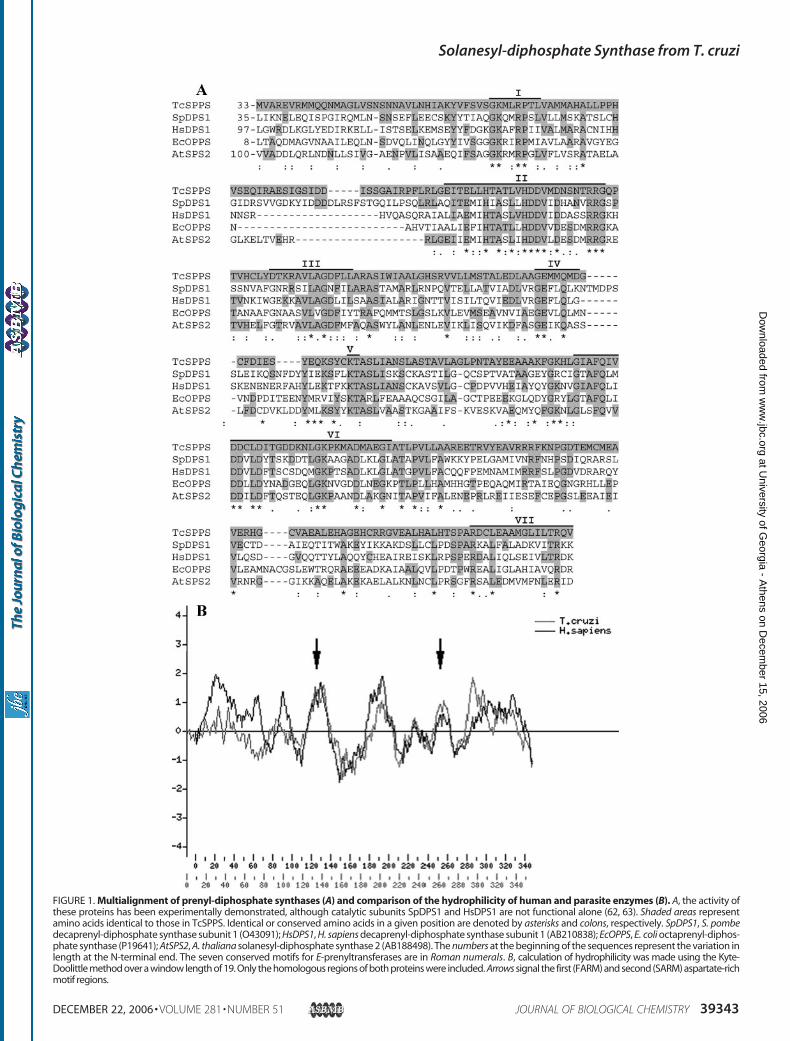
FIGURE 1. Multialignment of prenyl-diphosphate synthases (A) and comparison of the hydrophilicity of human and parasite enzymes (B). A, the activity ofthese proteins has been experimentally demonstrated, although catalytic subunits SpDPS1 and HsDPS1 are not functional alone (62, 63). Shaded areas representamino acids identical to those in TcSPPS. Identical or conserved amino acids in a given position are denoted by asterisks and colons, respectively. SpDPS1, S. pombedecaprenyl-diphosphate synthase subunit 1 (O43091); HsDPS1, H. sapiens decaprenyl-diphosphate synthase subunit 1 (AB210838); EcOPPS, E. coli octaprenyl-diphos-phate synthase (P19641); AtSPS2, A. thaliana solanesyl-diphosphate synthase 2 (AB188498). The numbers at the beginning of the sequences represent the variation inlength at the N-terminal end. The seven conserved motifs for E-prenyltransferases are in Roman numerals. B, calculation of hydrophilicity was made using the Kyte-Doolittle method over a window length of 19. Only the homologous regions of both proteins were included. Arrows signal the first (FARM) and second (SARM) aspartate-richmotif regions.
Solanesyl-diphosphate Synthase from T. cruzi
DECEMBER 22, 2006 • VOLUME 281 • NUMBER 51 JOURNAL OF BIOLOGICAL CHEMISTRY 39343
at University of G
eorgia - Athens on D
ecember 15, 2006
ww
w.jbc.org
Dow
nloaded from

same was observed with the AatII digestion pattern (band of�1000 bp, data not shown). These experiments support theidea that TcSPPS is a single copy gene.Ubiquinone Detection in Complemented E. coli and in
T. cruzi Epimastigotes—E. coli has an octaprenyl-diphosphatesynthase (44). To test whether the final product of TcSPPS,UQ-9, was able to replace the essential functions of UQ-8, aplasmid-swapping test was carried out using the insertionmutant strain KO229. Several colonies that were ampicillin-resistant (carrying the TcSPPS-expressing plasmid) and sensi-tive to spectinomycin (free of the ispB-expressing plasmid)were obtained, suggesting that TcSPPS was fully functional inbacteria. When the UQ of complemented colonies were iso-lated and their length determined, UQ-9 was the main productdetected (Fig. 3a). This is in contrast to KO229 harboringpKA3, which produced mainly UQ-8 (Fig. 3b). To establish thelength of the native molecule in the parasite, UQ was extractedfrom 1 g of T. cruzi epimastigotes and run in HPLC (Fig. 3c).The peak corresponded toUQ-9, as reported for other trypano-somatids (12–15).Functional Analysis of the Candidate Long-chain Prenyl-
diphosphate Synthase—For functional analysis, the protein washeterologously expressed in E. coli cells as a fusion protein withan N-terminal polyhistidine tag and purified by affinity chro-matography (Fig. 4A). The purified protein runs close to itspredicted molecular mass.
The enzymatic activity assay was performed in the presenceof different concentrations of Mg2� and Mn2�, to determinetheir effect on theTcSPPSwhen the allylic substratewasGGPP.Mg2� andMn2�were added to the reactionmixture at concen-trations between 0.5 and 20 mM. As shown in Table 1, optimallevels of activity were obtained by the addition of 0.5–1 mMMg2�. The addition of 10 mM EDTA abolished SPPS activity.Enzymatic activity was not detected when the divalent cationwas Mn2�. The T. cruzi enzyme activity was also assayedbetween 0.5 and 5% (v/v) Triton X-100. Maximum activity wasobserved at 1% (v/v) TritonX-100 (usingDMAPP, FPP,GPP, orGGPP as primer) (data not shown).Four kinds of allylic diphosphates were tested as a primer
substrate with [4-14C]IPP as described under “ExperimentalProcedures.” The enzyme utilized the four allylic diphosphatesas a primer substrate, however, the enzymatic activity usingDMAPP as substrate was one order of magnitude lower thanthe enzymatic activity using FPP, GPP, or GGPP (Table 2). Thereaction products were dephosphorylated and then analyzed by
FIGURE 3. Determination of UQ by HPLC. a, E. coli insertion mutant strainKO229 complemented by pQE-TcSPPS recombinant plasmid; b, KO229 har-boring its essential plasmid pKA3; c, T. cruzi CL Brener clone epimastigotes.
FIGURE 4. Expression and purification of TcSPPS (A) and TLC autoradio-grams of prenyl alcohols obtained by enzymatic hydrolysis of the prod-ucts formed by TcSPPS (B). A, recombinant E. coli BL21(DE3) (non-induced,lane 2) was induced with 0.5 mM isopropyl 1-thio-�-D-galactopyranoside for3 h at 30 °C (lane 3). After sonication the supernatant (lane 4) was purified byHis tag affinity chromatography (lane 5). TcSPPS displayed the expectedmolecular weight, as evaluated with the protein ladder 10 –200 kDa (MBIFermentas, lane 1). B, the products obtained from incubation of 100 �M
[4-14C]IPP (10 �Ci/�mol) and 10 �M DMAPP, GPP, FPP, or GGPP were analyzedby reversed-phase HPTLC as described under “Experimental Procedures.”Ori., origin; S.F., solvent front.
FIGURE 2. Determination of TcSPPS gene copy number. Southern blotanalysis of NcoI digestion of CL Brener clone genomic DNA. Digestion timeswere as follows: Lane 1, starting material; 2, 10 min; 3, 45 min; 4, 90 min; 5, 3 h;6, 6 h; 7, overnight (16 –18 h). Only two expected fragments were identified byhybridization, showing a single copy gene. Molecular weight markers areindicated on the left in kilobases.
Solanesyl-diphosphate Synthase from T. cruzi
39344 JOURNAL OF BIOLOGICAL CHEMISTRY VOLUME 281 • NUMBER 51 • DECEMBER 22, 2006
at University of G
eorgia - Athens on D
ecember 15, 2006
ww
w.jbc.org
Dow
nloaded from

reversed-phaseTLC.When either FPP,GPP, orGGPPwas usedas the primer substrate, solanesol (C45) was predominantlydetected by TLC analysis indicating that the protein generatedsolanesyl diphosphate (C45) as the major product (Fig. 4B).When the primer substrate was DMAPP the labeled productswere almost undetectable by TLC.Kinetic Analysis—Standard procedures were used to deter-
mine kinetic parameters. Km and Vmax values were obtained bya non-linear regression fit of the data to the Michaelis-Mentenequation (SigmaPlot 2000 for Windows), and the results aresummarized in Table 3. TcSPPS showed a similar Km value forFPP and GGPP, however the Km value for GPP was 7.7-foldhigher than that for GGPP. Moreover, the kcat value for GGPPwas 2.7-fold higher than that for FPP and slightly higher thanthat for GPP. Consequently the kcat/Km value for GGPP was1.8-fold higher than that for FPP and 10.4-fold higher than thatfor GPP, indicating that TcSPPS prefers GGPP to FPP or GPP.Moreover, TcSPPS showed a similarKm value for IPP when theallylic substratewas FPP,GPP, orGGPP.However, whenGGPPwas used as primer substrate, the enzyme showed a 1.6-foldhigher kcat value for IPP than that for IPP with GPP, and a5.0-fold higher than that for IPP with FPP. Consequently, thekcat/Km values for IPP with GGPP or GPP were similar,although the kcat/Km value for IPP with GGPP was 6.3-foldhigher than that for IPP with FPP. These results suggest thepreference of TcSPPS for GGPP over FPP or GPP.Presence of mRNA and Protein in T. cruzi—Several assays
were used in an attempt to establish the presence of the specific
mRNA in CL Brener clone epimastigotes. Neither Northernblot analysis nor RT-PCR amplification allowed the detectionof even a negligible amount of mRNA. On the other hand, themRNA for the positive control used (TcCyP19 gene) wasdetected in the Northern and RT-PCR assays. A search wasthen done on an expressed sequence tag clusters data base(TcruziDB Version 4.1). A cluster (99127) composed of oneread (TcEST_NCBI_AW324852.1) was identified. Its sequence(316 bp) was identical to the 3�-end of the original expressedsequence tag clone, TENU4155 (1262 bp).Immunoblot analysis with affinity-purified antibody against
TcSPPS showed a band of �36–37 kDa present in all develop-mental stages of the Y strain (Fig. 5). The antibody also recog-nized a 85-kDa band in all three stages and more weakly a40-kDa band. These proteins might share a few epitopes withSPPS in the motif regions.To further characterize the anti-TcSPPS cross-reaction to
other proteins in the parasite, we performed subcellular frac-tionation experiments to enrich for glycosomes. Fraction D,enriched for glycosomes, showed the strongest reaction byWestern blot analysis with antibodies against TcSPPS (Fig. 6A)and TbgGAPDH, a glycosomal marker (Fig. 6B). In this enrich-ment experiment, the 40- and 85-kDa bands were not detected in
FIGURE 5. Immunoblot analysis with antibodies against T. cruzi SPPS.Detection of SPPS by immunoblotting using affinity-purified polyclonal anti-body against SPPS. Epimastigote (E), trypomastigote (T), and amastigote (A)protein (Y strain, 30 �g/lane) were separated by SDS-PAGE and transferred toa polyvinylidene difluoride membrane.
TABLE 1Effect of divalent cations on TcSPPSSPPS activity wasmeasured in the presence of the different concentrations ofMgCl2indicated in a reaction medium containing 100 mM Tris-HCl buffer (pH 7.4), 1%(v/v) Triton X-100, 100 �M �4-14C�IPP (1 �Ci/ �mol), 50 �M GGPP, and 0.5 �g ofrecombinant protein (final volume of 100 �l). Reactions were incubated for 30 minat 37 °C and stopped by chilling in an ice bath. The radioactive prenyl products werethen extracted with 1-butanol as described under “Experimental Procedures.” Noactivity was detected in the absence of MgCl2 or presence of 10 mM EDTA. Valuesshown are means � S.D. of two experiments in duplicate.
MgCl2 SPPS activitymM nmol/min/mg0 00.5 116.75 � 0.331 121.99 � 15.782 107.53 � 5.715 76.72 � 12.3610 47.32 � 2.56
TABLE 2Allylic substrate specificity of TcSPPSSPPS activity was measured in the presence of the different allylic substrates (400�M DMAPP, 400 �M GPP, 30 �M FPP, 50 �M GGPP) in a reaction medium con-taining 100 mM Tris-HCl buffer (pH 7.4), 1 mM MgCl2, 1% (v/v) Triton X-100, 100�M �4-14C�IPP (1 �Ci/ �mol), and 0.5–3 �g of the purified protein (final volume of100 �l). Reactions were incubated for 30 min at 37 °C and stopped by chilling in anice bath. The radioactive prenyl products were then extracted with 1-butanol asdescribed under “Experimental Procedures.” Values shown are means � S.D. ofthree experiments in duplicate. A lower activity of the enzyme as compared withthatmeasured in Table 1 indicates a lower amount of active protein permilligram ofprotein.
Allylic substrate SPP synthase activitynmol/min/mg
DMAPP 3.91 � 0.07GPP 34.35 � 2.29FPP 17.99 � 2.09GGPP 64.34 � 13.03
TABLE 3Kinetic parameters of TcSPPSThe kinetic parameters of TcSPPSwere determined at 37 °C for 30min, as describedunder “Experimental Procedures.” The kcat value was defined by units of nanomolesof IPP converted into products per 1 nmol of dimer enzyme per second. Valuesshown are means � S.D. of three experiments in duplicate.
Substrate Km kcat kcat/Km
�M s�1 s�1�nM�1
GPP 54.83 � 5.92 0.065 � 0.020 1.19FPP 4.59 � 0.48 0.032 � 0.004 7.01GGPP 7.07 � 1.08 0.087 � 0.007 12.35IPP (400 �M GPP) 19.18 � 1.16 0.131 � 0.026 6.85IPP ( 30 �M FPP) 30.67 � 8.44 0.043 � 0.007 1.40IPP ( 50 �M GGPP) 24.33 � 5.45 0.217 � 0.032 8.93
Solanesyl-diphosphate Synthase from T. cruzi
DECEMBER 22, 2006 • VOLUME 281 • NUMBER 51 JOURNAL OF BIOLOGICAL CHEMISTRY 39345
at University of G
eorgia - Athens on D
ecember 15, 2006
ww
w.jbc.org
Dow
nloaded from
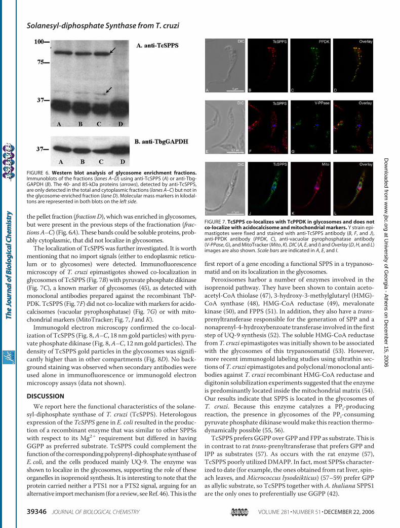
the pellet fraction (fractionD), whichwas enriched in glycosomes,but were present in the previous steps of the fractionation (frac-tions A–C) (Fig. 6A). These bands could be soluble proteins, prob-ably cytoplasmic, that did not localize in glycosomes.The localization of TcSPPSwas further investigated. It is worth
mentioning that no import signals (either to endoplasmic reticu-lum or to glycosomes) were detected. Immunofluorescencemicroscopy of T. cruzi epimastigotes showed co-localization inglycosomes of TcSPPS (Fig. 7B) with pyruvate phosphate dikinase(Fig. 7C), a known marker of glycosomes (45), as detected withmonoclonal antibodies prepared against the recombinant TbP-PDK. TcSPPS (Fig. 7F) did not co-localize withmarkers for acido-calcisomes (vacuolar pyrophosphatase) (Fig. 7G) or with mito-chondrial markers (MitoTracker; Fig. 7, J andK).Immunogold electron microscopy confirmed the co-local-
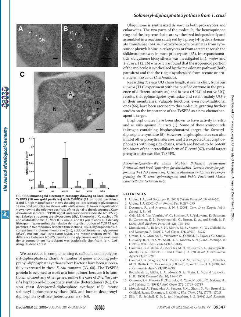
ization of TcSPPS (Fig. 8,A–C, 18 nm gold particles) with pyru-vate phosphate dikinase (Fig. 8,A–C, 12 nmgold particles). Thedensity of TcSPPS gold particles in the glycosomes was signifi-cantly higher than in other compartments (Fig. 8D). No back-ground staining was observed when secondary antibodies wereused alone in immunofluorescence or immunogold electronmicroscopy assays (data not shown).
DISCUSSION
We report here the functional characteristics of the solane-syl-diphosphate synthase of T. cruzi (TcSPPS). Heterologousexpression of the TcSPPS gene in E. coli resulted in the produc-tion of a recombinant enzyme that was similar to other SPPSswith respect to its Mg2� requirement but differed in havingGGPP as preferred substrate. TcSPPS could complement thefunctionof thecorrespondingpolyprenyl-diphosphate synthaseofE. coli, and the cells produced mainly UQ-9. The enzyme wasshown to localize in the glycosomes, supporting the role of theseorganelles in isoprenoid synthesis. It is interesting to note that theprotein carried neither a PTS1 nor a PTS2 signal, arguing for analternative importmechanism(fora review, seeRef. 46).This is the
first report of a gene encoding a functional SPPS in a trypanoso-matid and on its localization in the glycosomes.Peroxisomes harbor a number of enzymes involved in the
isoprenoid pathway. They have been shown to contain aceto-acetyl-CoA thiolase (47), 3-hydroxy-3-methylglutaryl (HMG)-CoA synthase (48), HMG-CoA reductase (49), mevalonatekinase (50), and FPPS (51). In addition, they also have a trans-prenyltransferase responsible for the generation of SPP and anonaprenyl-4-hydroxybenzoate transferase involved in the firststep of UQ-9 synthesis (52). The soluble HMG-CoA reductasefromT. cruzi epimastigotes was initially shown to be associatedwith the glycosomes of this trypanosomatid (53). However,more recent immunogold labeling studies using ultrathin sec-tions ofT. cruzi epimastigotes and polyclonal/monoclonal anti-bodies against T. cruzi recombinant HMG-CoA reductase anddigitonin solubilization experiments suggested that the enzymeis predominantly located inside the mitochondrial matrix (54).Our results indicate that SPPS is located in the glycosomes ofT. cruzi. Because this enzyme catalyzes a PPi-producingreaction, the presence in glycosomes of the PPi-consumingpyruvate phosphate dikinase wouldmake this reaction thermo-dynamically possible (55, 56).TcSPPS prefers GGPP overGPP and FPP as substrate. This is
in contrast to rat trans-prenyltransferase that prefers GPP andIPP as substrates (57). As occurs with the rat enzyme (57),TcSPPS poorly utilizedDMAPP. In fact, most SPPSs character-ized to date (for example, the ones obtained from rat liver, spin-ach leaves, and Micrococcus lysodeikticus) (57–59) prefer GPPas allylic substrate, so TcSPPS together with A. thaliana SPPS1are the only ones to preferentially use GGPP (42).
FIGURE 6. Western blot analysis of glycosome enrichment fractions.Immunoblots of the fractions (lanes A–D) using anti-TcSPPS (A) or anti-Tbg-GAPDH (B). The 40- and 85-kDa proteins (arrows), detected by anti-TcSPPS,are only detected in the total and cytoplasmic fractions (lanes A–C) but not inthe glycosome-enriched fraction (lane D). Molecular mass markers in kilodal-tons are represented in both blots on the left side.
FIGURE 7. TcSPPS co-localizes with TcPPDK in glycosomes and does notco-localize with acidocalcisome and mitochondrial markers. Y strain epi-mastigotes were fixed and stained with anti-TcSPPS antibody (B, F, and J),anti-PPDK antibody (PPDK, C), anti-vacuolar pyrophosphatase antibody(V-PPase, G), and MitoTracker (Mito, K). DIC (A, E, and I) and Overlay (D, H, and L)images are also shown. Scale bars are indicated in A, E, and I.
Solanesyl-diphosphate Synthase from T. cruzi
39346 JOURNAL OF BIOLOGICAL CHEMISTRY VOLUME 281 • NUMBER 51 • DECEMBER 22, 2006
at University of G
eorgia - Athens on D
ecember 15, 2006
ww
w.jbc.org
Dow
nloaded from
We succeeded in complementing E. coli deficient in polypre-nyl-diphosphate synthase. A number of genes encoding poly-prenyl-diphosphate synthases frombacteria have been success-fully expressed in these E. coli mutants (33, 60). The TcSPPSprotein is assumed to work as a homodimer, because it is func-tional without any other genes, unlike the case of Bacillus sub-tilis heptaprenyl-diphosphate synthase (heterodimer) (61), fis-sion yeast decaprenyl-diphosphate synthase (62), mousesolanesyl-diphosphate synthase (63), and human decaprenyl-diphosphate synthase (heterotetramers) (63).
Ubiquinone is synthesized de novo in both prokaryotes andeukaryotes. The two parts of the molecule, the benzoquinonering and the isoprene chain, are synthesized independently andassembled in a reaction catalyzed by a prenyl-4-hydroxybenzo-ate transferase (64). 4-Hydroxybenzoate originates from tyro-sine or phenylalanine in eukaryotes or from acetate through theshikimate pathway in most prokaryotes (65). In trypanosoma-tids, ubiquinone biosynthesis was investigated in L. major andT. brucei (12, 16)where it was found that the isoprenoid portionof themolecule is synthesized by themevalonate pathway (bothparasites) and that the ring is synthesized from acetate or aro-matic amino acids (Leishmania).Regarding T. cruziUQ chain length, it seems clear, from our
in vitro (TLC experiment with the purified enzyme in the pres-ence of different substrates) and in vivo (HPLC of native UQ)results, that epimastigotes synthesize and retain mainly UQ-9in their membranes. Valuable functions, even non-traditionalones (66), have been ascribed to this molecule, granting furtherstudies on the importance of the TcSPPS as a new chemother-apeutic target.Bisphosphonates have been shown to have activity in vitro
and in vivo against T. cruzi (1). Some of these compounds(nitrogen-containing bisphosphonates) target the farnesyl-diphosphate synthase (5). However, bisphosphonates can alsoinhibit other prenyltransferases, and it is expected that bisphos-phonates with long side chains, which are known to be potentinhibitors of the intracellular form of T. cruzi (67), could targetprenyltransferases like TcSPPS.
Acknowledgments—We thank Norbert Bakalara, FrederiqueBringaud, and Fred Opperdoes for antibodies, Octavio Fusco for per-forming theDNA sequencing, CristinaMaidana and LindaBrown forgrowing the T. cruzi epimastigotes, and Pablo Fazio and MartaLauricella for technical help.
REFERENCES1. Urbina, J. A., and Docampo, R. (2003) Trends Parasitol. 19, 495–5012. Urbina, J. A. (2002) Curr. Pharm. Des. 8, 287–2953. Docampo, R., and Moreno, S. N. J. (2001) Curr. Drug Targets Infect.
Disord. 1, 51–614. Gelb, M. H., Van Voorhis, W. C., Buckner, F. S., Yokoyama, K., Eastman,
R., Carpenter, E. P., Panethymitaki, C., Brown, K. A., and Smith, D. F.(2003)Mol. Biochem. Parasitol. 126, 155–163
5. Montalvetti, A., Bailey, B. N., Martin, M. B., Severin, G. W., Oldfield, E.,and Docampo, R. (2001) J. Biol. Chem. 276, 33930–33937
6. Urbina, J. A., Moreno, B., Vierkotter, S., Oldfield, E., Payares, G., Sanoja,C., Bailey, B. N., Yan, W., Scott, D. A., Moreno, S. N. J., and Docampo, R.(1999) J. Biol. Chem. 274, 33609–33615
7. Garzoni, L. R., Caldera, A., Meirelles, M. N., de Castro, S. L., Docampo, R.,Meints, G. A., Oldfield, E., and Urbina, J. A. (2004) Int. J. Antimicrob.Agents 23, 273–285
8. Garzoni, L. R.,Waghabi, M. C., Baptista, M.M., de Castro, S. L., Meirelles,M. N., Britto, C. C., Docampo, R., Oldfield, E., and Urbina, J. A. (2004) Int.J. Antimicrob. Agents 23, 286–290
9. Bouzahzah, B., Jelicks, L. A., Morris, S. A., Weiss, L. M., and Tanowitz,H. B. (2005) Parasitol. Res. 96, 184–187
10. Ohnuma, S.-i., Hirooka, K., Tsuruoka, N., Yano, M., Ohto, C., Nakane, H.,and Nishino, T. (1998) J. Biol. Chem. 273, 26705–26713
11. Montalvetti, A., Fernandez, A., Sanders, J. M., Ghosh, S., Van Brussel, E.,Oldfield, E., and Docampo, R. (2003) J. Biol. Chem. 278, 17075–17083
12. Ellis, J. E., Setchell, K. D. R., and Kaneshiro, E. S. (1994) Mol. Biochem.
FIGURE 8. Immunogold electron microscopy showing co-localization ofTcSPPS (18 nm gold particles) with TcPPDK (12 nm gold particles).A and B, high magnification views showing co-localization to glycosomes.12 nm gold particles are shown with white arrows. C, lower magnificationview showing the relative specificity of the signal to the glycosomes. Openarrowheads indicate TcPPDK signal, and black arrows indicate TcSPPS sig-nal. Labeled structures are glycosomes (Gly), kinetoplast (K), nucleus (N),and acidocalcisome (A). Bars: 0.05 �m (A) and 0.1 �m (B and C). D, densityhistogram representing the relative density distribution of TcSPPS goldparticles in five randomly selected thin sections (�S.D.) by organellar sub-compartments: plasma membrane (pm), acidocalcisome (ac), glycosome(glyco), nucleus (nuc), cytoplasm (cyto), and mitochondrion (mito). Thedifference between TcSPPS density in the glycosome and the next mostdense compartment (cytoplasm) was statistically significant (p 0.05)using Student’s t test.
Solanesyl-diphosphate Synthase from T. cruzi
DECEMBER 22, 2006 • VOLUME 281 • NUMBER 51 JOURNAL OF BIOLOGICAL CHEMISTRY 39347
at University of G
eorgia - Athens on D
ecember 15, 2006
ww
w.jbc.org
Dow
nloaded from
Parasitol. 65, 213–22413. Ranganathan, G., and Mukkada, A. J. (1995) Int. J. Parasitol. 25, 279–28414. Rassam, M. B., Shanshal, M., and Gargees, G. S. (1988) Mol. Biochem.
Parasitol. 29, 61–6415. Clarkson, A. B., Bienen, E. J., Pollakis, G., and Grady, R. W. (1989) J. Biol.
Chem. 264, 17770–1777616. Low, P., Dallner, G., Mayor, S., Cohen, S., Chait, B. T., and Menon, A. K.
(1991) J. Biol. Chem. 266, 19250–1925717. Kusel, J. P., andWeber, M.M. (1965) Biochim. Biophys. Acta 98, 632–63918. Vakirtzi-Lemonias, V. C., Kidder, G. W., and Dewey, V. C. (1963) Comp.
Biochem. Physiol. 8, 133–13619. Michels, P.A., Moyersoen, J., Krazy, H., Galland, N., Herman, M., and
Hannaert, V. (2005)Mol. Membr. Biol. 22, 133–14520. Okamoto, T., Fukui, K., Nakamoto, M., Kishi, T., Okishio, T., Yamagami,
T., Kanamori, N., Kishi, H., and Hiraoka, E. (1985) J. Chromatogr. 342,35–46
21. Furuya, T., Kashuba, C., Docampo, R., and Moreno, S. N. (2000) J. Biol.Chem., 275, 6428–6438
22. Bone, G. J., and Steinert, M. (1956) Nature 178, 308–30923. Gerez de Burgos, N. M., Burgos, C., Blanco, A., Paulone, I., and Segura,
E. L. (1976) Acta Physiol. Lat. Am. 26, 10–1924. Hall, T. A. (1999) Nucl. Acids Symp. Ser. 41, 95–9825. Altschul, S. F., Gish,W.,Miller,W.,Myers, E.W., and Lipman, D. J. (1990)
J. Mol. Biol. 215, 403–41026. Kyte, J., and Doolittle, R. F. (1982) J. Mol. Biol. 157, 105–13227. Bendtsen, J. D., Nielsen, H., von Heijne, G., and Brunak, S. (2004) J. Mol.
Biol. 340, 783–79528. Hanke, J., Sanchez, D. O., Henriksson, J., Åslund, L. C., Pettersson, U.,
Frasch, A. C. C., and Hoheisel, J. (1996) BioTechniques 21, 686–69329. Dretzen, G., Bellard, M., Sassone-Corsi, P., and Chambon, P. (1981) Anal.
Biochem. 112, 295–29830. Bua, J., Garcia, G. A., Galindo, M., Galanti, N., and Ruiz, A. M. (1999)
Medicina (B Aires) 59, Suppl. 2, 11–1731. Bradford, M. M. (1976) Anal. Biochem. 72, 248–25432. Koyama, T., Fujii, H., and Ogura, K. (1985) Methods Enzymol. 110,
153–15533. Okada, K., Minehira, M., Zhu, X., Suzuki, K., Nakagawa, T., Matsuda, H.,
and Kawamukai, M. (1997) J. Bacteriol. 179, 3058–306034. Okada, K., Kainou, T., Tanaka, K., Nakagawa, T., Matsuda, H., and
Kawamukai, M. (1998) Eur. J. Biochem. 255, 52–5935. Rohloff, P., Montalvetti, A., and Docampo, R. (2004) J. Biol. Chem. 279,
52270–5228136. Porcel, B.M., Tran, A. N., Tammi,M., Nyarady, Z., Rydaker,M., Urmenyi,
T. P., Rondinelli, E., Pettersson, U., Andersson, B., and Aslund, L. (2000)Genome Res. 10, 1103–1107
37. Yamauchi, K. (1991) Nucleic Acids Res. 19, 2715–272038. Liang, P.-H., Ko, T.-P., and Wang, A. H.-J. (2002) Eur. J. Biochem. 269,
3339–335439. Koyama, T. (1999) Biosci. Biotechnol. Biochem. 63, 1671–167640. Chen, A., Kroon, P. A., Poulter, C. D. (1994) Prot. Sci. 3, 600–60741. Jun, L., Saiki, R., Tatsumi, K., Nakagawa, T., and Kawamukai, M. (2004)
Plant Cell Physiol. 45, 1882–188842. Hirooka, K., Bamba, T., Fukusaki, E., and Kobayashi, A. (2003) Biochem. J.
370, 679–68643. Alvarez, F., Robello, C., and Vignali, M. (1994) Mol. Biol. Evol. 11,
790–80244. Asai, K., Fujisaki, S., Nishimura, Y., Nishino, T., Okada, K., Nakagawa, T.,
Kawamukai,M., andMatsuda, H. (1994) Biochem. Biophys. Res. Commun.202, 340–345
45. Parsons, M. (2004)Mol. Microbiol. 53, 717–72446. Purdue, P. E., and Lazarow, P. B. (2001) Annu. Rev. Cell Dev. Biol. 17,
701–75247. Thompson, S. L., and Krisans, S. K. (1990) J. Biol. Chem. 265, 5731–573548. Olivier, L. M., and Krisans, S. K. (2000) Biochim. Biophys. Acta 1529,
89–10249. Keller, G. A., Barton, M. C., Shapiro, D. J., and Singer, S. J. (1985) Proc.
Natl. Acad. Sci. U. S. A. 82, 770–77450. Stamellos, K. D., Shackelford, J. E., Tanaka, R. D., and Krisans, S. K. (1992)
J. Biol. Chem. 267, 5560–556851. Krisans, S. K., Ericsson, J., Edwards, P. A., and Keller, G. A. (1994) J. Biol.
Chem. 269, 14165–1416952. Tekle, M., Bentinger, M., Nordman, T., Appelkvist, E.-L., Chojnacki, T.,
andOlsson, J.M. (2002)Biochem. Biophys. Res. Commun.291, 1128–113353. Concepcion, J. L., Gonzalez-Pacanowska,D., andUrbina, J. A. (1998)Arch.
Biochem. Biophys. 352, 114–12054. Pena-Diaz, J., Montalvetti, A., Flores, C. L., Constan, A., Hurtado-Guer-
rero, R., De Souza, W., Gancedo, C., Ruiz-Perez, L. M., and Gonzalez-Pacanowska, D. (2004)Mol. Biol. Cell 15, 1356–1363
55. Bringaud, F., Baltz, D., and Baltz, T. (1998) Proc. Natl. Acad. Sci. U. S. A.95, 7963–7968
56. Acosta, H., Dubuordieu, M., Quinones, W., Caceres, A., Bringaud, F., andConcepcion, J. L. (2004) Comp. Biochem. Physiol. B 138, 347–356
57. Teclebrhan, H., Olsson, J., Swiezewska, E., and Dallner, G. (1993) J. Biol.Chem. 268, 23081–23086
58. Swiezewaska, E., Dallner, G., Andersson, B., and Ernster, L. (1993) J. Biol.Chem. 268, 1494–1499
59. Sagami, H., Ogura, K., and Seto, S. (1977) Biochemistry 16, 4616–462260. Okada, K., Kamiya, Y., Zhu, X., Suzuki, K., Tanaka, K., Nakagawa, T.,
Matsuda, H., and Kawamukai, M. (1997) J. Bacteriol. 179, 5992–599861. Zhang, Y. W., Koyama, T., and Ogura, K. (1997) J. Bacteriol. 179,
1417–141962. Saiki, R., Nagata, A., Uchida, N., Kainou, T.,Matsuda, H., and Kawamukai,
M. (2003) Eur. J. Biochem. 270, 4113–412163. Saiki, R., Nagata, A., Kainou, T., Matsuda, H., and Kawamukai, M. (2005)
FEBS J. 272, 5606–562264. Turunen, M., Olsson, J., and Dallner, G. (2004) Biochim. Biophys. Acta
1660, 171–19965. Ellis, J. E. (1994) Parasitol. Today 10, 296–30166. Kawamukai, M. (2002) J. Biosci. Bioeng. 94, 511–51767. Szajnman, S. H., Montalvetti, A., Wang, Y., Docampo, R., and Rodriguez,
J. B. (2003) Bioorg. Med. Chem. Lett. 13, 3231–3235
Solanesyl-diphosphate Synthase from T. cruzi
39348 JOURNAL OF BIOLOGICAL CHEMISTRY VOLUME 281 • NUMBER 51 • DECEMBER 22, 2006
at University of G
eorgia - Athens on D
ecember 15, 2006
ww
w.jbc.org
Dow
nloaded from
Copyright © 2022 FDOKUMEN