All Trypanosoma cruzi developmental forms present lysosome-related organelles
12
Histochem Cell Biol (2008) 130:1187–1198 DOI 10.1007/s00418-008-0486-8 123 ORIGINAL PAPER All Trypanosoma cruzi developmental forms present lysosome-related organelles Celso Sant’Anna · Fabiola Parussini · Daniela Lourenço · Wanderley de Souza · Juan Jose Cazzulo · Narcisa Leal Cunha-e-Silva Accepted: 27 July 2008 / Published online: 12 August 2008 © Springer-Verlag 2008 Abstract Trypanosoma cruzi epimastigote forms concen- trate their major protease, cruzipain, in the same compart- ment where these parasites store macromolecules obtained from medium and for this ability these organelles were named as reservosomes. Intracellular digestion occurs mainly inside reservosomes and seems to be modulated by cruzipain and its natural inhibitor chagasin that also con- centrates in reservosomes. T. cruzi mammalian forms, try- pomastigotes and amastigotes, are unable to capture macromolecules by endocytosis, but also express cruzipain and chagasin, whose role in infectivity has been described. In this paper, we demonstrate that trypomastigotes and amastigotes also concentrate cruzipain, chagasin as well as serine carboxypeptidase in hydrolase-rich compartments of acidic nature. The presence of P-type proton ATPase indicates that this compartment is acidiWed by the same enzyme as epimastigote endocytic compartments. Elec- tron microscopy analyzes showed that these organelles are placed at the posterior region of the parasite body, are single membrane bound and possess an electron-dense matrix with electronlucent inclusions. Three-dimensional reconstruction showed that these compartments have diVerent size and shape in trypomastigotes and amastigotes. Based on these evidences, we suggest that all T. cruzi developmental stages present lysosome-related organelles that in epimastigotes have the additional and unique ability of storing cargo. Keywords Lysosome-related organelles · Reservosomes · Serine carboxypeptidase · Trypomastigotes · Amastigotes · Trypanosoma cruzi Introduction The trypanosomatid parasites have diVerent developmental stages that represent an adaptation to the extra and intra- cellular environments encountered by the parasites within their two hosts, the mammalian host and the insect vector. Endocytosis and protein turnover are essential metabolic pathways for life cycle development of Trypanosoma cruzi, the etiologic agent of Chagas disease (De Souza et al. 2002). The endocytic pathway is under strict developmental control in the diVerent life cycle stages of the parasite. Epimastigotes, proliferative forms found inside insect host, are highly active in Xuid phase and receptor mediated path- ways, and store macromolecules up taken from the medium inside the reservosomes (Soares and De Souza 1991). The pathway has not been observed in either trypomastigotes, C. Sant’Anna · D. Lourenço · W. de Souza · N. L. Cunha-e-Silva (&) Laboratório de Ultraestrutura Celular Hertha Meyer, Instituto de Biofísica Carlos Chagas Filho, Universidade Federal do Rio de Janeiro, CCS, Bloco G subsolo, Cidade Universitária, Ilha do Fundão, Rio de Janeiro 21949-900, Brazil e-mail: [email protected] C. Sant’Anna · W. de Souza Diretoria de Programas (DIPRO), INMETRO, Rio de Janeiro, Brazil F. Parussini · J. J. Cazzulo Instituto de Investigaciones Biotecnológicas, Instituto Tecnológico de Chascomús, Universidad Nacional de General San Martín, CONICET, Av. General Paz 5445, INTI, EdiWcio 24, Casilla de Correo 30, 1650 San Martín, Provincia de Buenos Aires, Argentina Present Address: F. Parussini Microbiology and Molecular Genetics, University of Vermont, 316 StaVord Hall, 95 Carrigan Drive, Burlington, VT 05405, USA
-
Upload
independent -
Category
Documents
-
view
1 -
download
0
Transcript of All Trypanosoma cruzi developmental forms present lysosome-related organelles

Histochem Cell Biol (2008) 130:1187–1198
DOI 10.1007/s00418-008-0486-8ORIGINAL PAPER
All Trypanosoma cruzi developmental forms present lysosome-related organelles
Celso Sant’Anna · Fabiola Parussini · Daniela Lourenço · Wanderley de Souza · Juan Jose Cazzulo · Narcisa Leal Cunha-e-Silva
Accepted: 27 July 2008 / Published online: 12 August 2008© Springer-Verlag 2008
Abstract Trypanosoma cruzi epimastigote forms concen-trate their major protease, cruzipain, in the same compart-ment where these parasites store macromolecules obtainedfrom medium and for this ability these organelles werenamed as reservosomes. Intracellular digestion occursmainly inside reservosomes and seems to be modulated bycruzipain and its natural inhibitor chagasin that also con-centrates in reservosomes. T. cruzi mammalian forms, try-pomastigotes and amastigotes, are unable to capturemacromolecules by endocytosis, but also express cruzipainand chagasin, whose role in infectivity has been described.In this paper, we demonstrate that trypomastigotes andamastigotes also concentrate cruzipain, chagasin as well as
serine carboxypeptidase in hydrolase-rich compartmentsof acidic nature. The presence of P-type proton ATPaseindicates that this compartment is acidiWed by the sameenzyme as epimastigote endocytic compartments. Elec-tron microscopy analyzes showed that these organelles areplaced at the posterior region of the parasite body, are singlemembrane bound and possess an electron-dense matrix withelectronlucent inclusions. Three-dimensional reconstructionshowed that these compartments have diVerent size andshape in trypomastigotes and amastigotes. Based on theseevidences, we suggest that all T. cruzi developmental stagespresent lysosome-related organelles that in epimastigoteshave the additional and unique ability of storing cargo.
Keywords Lysosome-related organelles · Reservosomes · Serine carboxypeptidase · Trypomastigotes · Amastigotes · Trypanosoma cruzi
Introduction
The trypanosomatid parasites have diVerent developmentalstages that represent an adaptation to the extra and intra-cellular environments encountered by the parasites withintheir two hosts, the mammalian host and the insect vector.Endocytosis and protein turnover are essential metabolicpathways for life cycle development of Trypanosoma cruzi,the etiologic agent of Chagas disease (De Souza et al.2002). The endocytic pathway is under strict developmentalcontrol in the diVerent life cycle stages of the parasite.Epimastigotes, proliferative forms found inside insect host,are highly active in Xuid phase and receptor mediated path-ways, and store macromolecules up taken from the mediuminside the reservosomes (Soares and De Souza 1991). Thepathway has not been observed in either trypomastigotes,
C. Sant’Anna · D. Lourenço · W. de Souza · N. L. Cunha-e-Silva (&)Laboratório de Ultraestrutura Celular Hertha Meyer, Instituto de Biofísica Carlos Chagas Filho, Universidade Federal do Rio de Janeiro, CCS, Bloco G subsolo, Cidade Universitária, Ilha do Fundão, Rio de Janeiro 21949-900, Brazile-mail: [email protected]
C. Sant’Anna · W. de SouzaDiretoria de Programas (DIPRO),INMETRO, Rio de Janeiro, Brazil
F. Parussini · J. J. CazzuloInstituto de Investigaciones Biotecnológicas, Instituto Tecnológico de Chascomús, Universidad Nacional de General San Martín, CONICET, Av. General Paz 5445, INTI, EdiWcio 24, Casilla de Correo 30, 1650 San Martín, Provincia de Buenos Aires, Argentina
Present Address:F. ParussiniMicrobiology and Molecular Genetics, University of Vermont, 316 StaVord Hall, 95 Carrigan Drive, Burlington, VT 05405, USA
123

1188 Histochem Cell Biol (2008) 130:1187–1198
infective forms found in insect feces and vertebrate blood,or amastigotes, proliferative forms found inside vertebratehost cells (Soares and De Souza 1991). The diVerentiationprocesses that convert the proliferative into the infectiveforms must be energy consuming and dependent on theaminoacid availability, which probably results from the useof endocytic cargo as substrate at least for the diVerentia-tion of epimastigotes into metacyclic trypomastigotes(Soares et al. 1989). Lipid cargo processing has not beeninvestigated yet, but the knowledge about protein degrada-tion has increased in the last years. Several enzymesinvolved in protein degradation were already identiWed,although their Wne modulation and subcellular site of actionare poorly known (Cazzulo et al. 2001).
Two lysosomal proteases have been well characterized inT. cruzi. The Wrst one was cruzipain (CZP), the major prote-ase found in epimastigote forms (Cazzulo et al. 1990). It is acathepsin L-like cysteine protease synthesized as a zymogen(pre-pro-enzyme) (Campetella et al. 1992; Eakin et al.1992). CZP seems to mature in Golgi complex (Engel et al.1998, 2000) and is highly accumulated (Soares et al. 1992)and active (Cunha-e-Silva et al. 2002) in reservosomes. Thepathway that targets cruzipain to reservosomes is not fullyunderstood. Previous work suggested that this enzyme istransported from Golgi complex through secretory vesiclesto reservosomes (Soares et al. 1992) in a mannose 6-phos-phate independent pathway (Cazzulo et al. 1990). Using animmunocytochemical approach, cruzipain was also local-ized in the parasite plasma membrane and Xagellar pocket(Souto-Padrón et al. 1990). Therefore, it is possible that atleast a fraction of CZP is Wrst delivered to parasite surfaceand then internalized as part of the endocytic pathway anddelivered to reservosomes. The second well-describedhydrolase is a serine carboxypeptidase (TcCBP), member ofthe �/� hydrolase family that catalyzes the hydrolysis of thecarboxyterminal bond in peptides and proteins (Parussiniet al. 2003). In mammalian cells, CBP has lysosomallocalization, while in yeast and plant it is concentrated inendosomal/vacuolar compartments. Lysosomal serine car-boxypeptidase was puriWed, cloned and characterized in T.cruzi (Parussini et al. 2003). This enzyme is a monomericN-linked glycoprotein encoded by genes arranged in tan-dem. Gene sequencing showed that TcCBP belongs to theS10 family with high similarity with its orthologues in plantand yeast cells. Moreover, the enzyme displays optimumactivity at low pH and is speciWc to carboxyterminal peptideresidues. Subcellular localization of TcCBP in epimastigoteswas assayed using a biochemical approach. It was suggestedthat serine carboxypeptidase is localized in the reservo-somes as it co-localized with other lysosomal enzymes ascruzipain and acid �-mannosidase (Parussini et al. 2003).
Reservosomes are the last compartment of the endocyticpathway found exclusively in epimastigotes from the
Schyzotrypanum sub-genus of Trypanosoma, such asTrypanosoma vespertilionis, Trypanosoma dionisii andTrypanosoma cruzi. They are absent in T. brucei and also inLeishmania, the other two trypanosomatids that display aremarkable endocytic activity. Reservosomes were namedon basis of their unusual characteristic of storing exogenousmacromolecules endocytosed by the parasite (Soares and DeSouza 1988) that diverges epimastigote reservosomes fromeither mammalian lysosomes or yeast vacuole. Morphologi-cally, reservosomes are large and round organelles with adiameter of 400–600 nm, bound by a membrane unit andpositioned at the posterior portion of epimastigote cell body.Recently, we demonstrated the presence of internal vesicles,as well as long membrane proWles that cross reservosomelumen. Inner vesicles, always in a few number, which arenot accessible to endocytosed macromolecules was evi-denced. Their nature remains to be determined. Anothernoticeable structure found was the rod-shaped electron-lucent body bound by a membrane monolayer, which makesreservosome morphology complex and very typical(Sant’Anna et al. 2008). Reservosomes were initially char-acterized as pre-lysosomal organelles due to: (1) absence oflysosomal makers (LAMP1, LAMP2, lgp120 and acid phos-phatase) and (2) evaluation of organelle internal pH: theiracid character was determined by incubation of epim-astigotes with acridine orange (Soares and De Souza 1991),a weak base that concentrates in acidic compartments. Later,using the DAMP technique for pH evaluation in electronmicroscopy, reservosome pH was determined as 6.0 (Soareset al. 1992). Interestingly, the acid character of reservo-somes is generated by the action of two P-type H+-ATPaseisoforms (TcHA1 and TcHA2) (Vieira et al. 2005), protonpumps found mainly in the plasma membrane of plant andyeast cells (Serrano et al. 1992). In contrast, the endosomalpH in mammalian cells is driven by the action of a V-typeH+-ATPase (Beyenbach and Wieczorek 2006). Recently,chagasin, a natural inhibitor of cruzipain, was localized inGolgi complex and reservosomes (Santos et al. 2005).Besides, a T. cruzi tyrosine phosphatase was characterizedand localized in epimastigote endocytic pathway, includingreservosomes (Cuevas et al. 2005). Despite the fact thatreservosomes have been the subject of several studies, areservosome molecular marker has not been determined sofar (Cunha-e-Silva et al. 2006).
Although the diVerentiation of epimastigotes duringmetacyclogenesis directly generates metacyclic trypom-astigotes, no reservosome or other lysosome-like structurewas described in these forms, neither in the bloodstreamtrypomastigotes nor in the amastigotes found in vertebratehost, mainly because they do not possess a substantiallyactive endocytic pathway (Soares and De Souza 1991).In this paper, we demonstrate that the lysosomal hydro-lases cruzipain and serine carboxypeptidase co-localize in
123

Histochem Cell Biol (2008) 130:1187–1198 1189
epimastigote reservosomes and in intracellular compart-ments localized at the posterior region of trypomastigotesand amastigotes. These compartments display typical lyso-somal characteristics, although not related to degradation ofexogenous macromolecules, indicating that, as reservo-somes, they are lysosome-related organelles (LROs).
Material and methods
Parasites
Trypanosoma cruzi epimastigotes of Y strain were culti-vated at 28°C in LIT medium (liver infusion tryptosemedium), supplemented with 10% fetal calf serum (Cam-argo 1964). Bloodstream trypomastigote forms, derivedfrom the blood of Swiss mice infected by intraperitonealinjection 7 days before, were used to infect LLC-MK2 celltype. Five to 7 days after infection, trypomastigotes andamastigotes were collected from the supernatant of theinfected cultures and puriWed by centrifugation as previ-ously described (Carvalho and De Souza 1983).
Antibodies
Polyclonal antibody against T. cruzi serine carboxypeptidasewas obtained by inoculating mice with recombinant TcCBPcontaining a poly-His tag at the C-terminus. Rabbit polyclonalanti-cruzipain antibodies were produced against the enzymepuriWed from epimastigotes (Souto-Padrón et al. 1990). Rab-bit polyclonal anti-P type proton ATPase was producedagainst a recombinant fragment of T. cruzi enzyme (Vieiraet al. 2005) and recognizes a protein of 100 kDa in all parasitedevelopmental forms. Rabbit polyclonal anti-chagasin wasobtained using the recombinant protein (Santos et al. 2005).
Fluorescence microscopy
Parasites were harvested from medium by centrifugation,washed in PBS (50 mM NaCl in 10 mM sodium phosphatebuVer, pH 7.3) and Wxed in 4% formaldehyde in PBS for1 h. Washed parasites were adhered to 0.1% poly-L-lysinecoated glass coverslips, incubated with 50 mM NH4Cl inPBS and permeabilized with 0.5% Triton X-100 for 5 min.After blocking in 1.5% bovine albumin, 0.5% Wsh gelatin,in PBS for 30 min at room temperature, parasites wereincubated with anti-serine carboxypeptidase (1:200 dilu-tion), anti-cruzipain (1:1,000 dilution), anti-chagasin (1:100dilution) or anti-P-type H+ ATPase (1:100 dilution) inblocking buVer for 1 h, washed and incubated with 1:400Alexa 488 goat anti-mouse IgG and/or Alexa 546 goat anti-rabbit IgG (Molecular Probes, OR, USA) for 1 h. In order tolabel acid compartments, trypomastigotes and amastigotes
were washed twice in serum free RPMI medium andincubated with 1 �M Lysotracker green (Molecular Probes,OR, USA) in RPMI for 10 min at 37°C. Samples weremounted on 0.2 M n-propylgallate in glycerol:PBS (9:1)and Xuorescence images were acquired in a Zeiss AxioplanXuorescence microscope coupled with colored HamamatsuCCD camera C5810. Images were processed using AdobePhotoshop CS2 (Adobe Systems, Inc.).
Transmission electron microscopy
Trypomastigotes and amastigotes were collected by centri-fugation, washed in PBS, Wxed in 2.5% glutaraldehyde in0.1 M phosphate buVer, pH 7.2, postWxed in 1% osmiumtetroxide, 0.8% potassium ferrocyanide, 5 mM calciumchloride in 0.1 M cacodylate buVer, pH 7.2, dehydrated inan increasing acetone series and embedded in Epon resin.Ultrathin sections were stained with uranyl acetate and leadcitrate and observed in a JEOL 1200 EX transmission elec-tron microscope operating at 80 kV.
Immunocytochemistry
Parasites were harvested by centrifugation, washed in PBS,Wxed in 0.2% glutaraldehyde, 4% formaldehyde, 3.5%sucrose in 0.1 M phosphate buVer, pH 7.3, dehydrated inethanol at 4°C, inWltrated in Unicryl resin (Pelco) at ¡20°Cand polymerized under UV radiation for 96 h. Ultrathinsections of Unicryl-embedded parasites were collected onnickel grids, incubated in 50 mM ammonium chloride inPBS for 30 min, to quench free aldehyde groups and trans-ferred to blocking buVer for 1 h at room temperature. Thinsections were subsequently incubated with 1:200 anti-ser-ine carboxypeptidase in blocking buVer for 1 h, washed andincubated with 15 nm gold-labeled goat anti-mouse IgGsecondary antibody (BBI, England) diluted 1:100. Afterextensive washing, grids were incubated with 1:500 anti-cruzipain for 1 h, followed by washing and incubation with5 nm gold-labeled goat anti-mouse IgG secondary antibody(BBI, England) diluted 1:100. Control grids were per-formed omitting incubation with the primary antibodies.The sections were successively washed in PBS and water,stained in uranyl acetate and lead citrate and observed in aJEOL 1200 EX transmission electron microscope.
Three-dimensional reconstruction
Trypomastigotes and amastigotes were Wxed and processed fortransmission electron microscopy as described above. Seriesof ultra-thin sections were archived, collected on copper gridsand deposited on Formvar/carbon Wlms. After staining in ura-nyl acetate and lead citrate they were observed in a JEOL 1200EX transmission electron microscope. Recorded images were
123

1190 Histochem Cell Biol (2008) 130:1187–1198
aligned after establishment of Wducial markers. In each plane,the proWles of interest were traced using a digitalizing plate(Muminics) and software from Young et al. (1987). The dataobtained were transferred to SYNU program (Hessler et al.1992) to generate the 3D model.
Results
Previous work using digitonin extraction in epimastigotesshowed that the extraction pattern of serine carboxypepti-dase is similar to that described for other lysosomal hydro-lases such as cruzipain and acid �-mannosidase (Parussiniet al. 2003), suggesting that TcCBP is concentrated inreservosomes. In the current paper, the sub-cellular localiza-tion of TcCBP was studied using antibodies raised againstthe puriWed recombinant enzyme. Labeling of roundedstructures localized at posterior region, typical of reservo-somes, was observed by immunoXuorescence microscopyanalysis of epimastigotes (Fig. 1b, d). The reservosomallocalization was supported by the co-localization with cruzi-pain (Fig. 1c, d), an enzyme previously shown to be highlyconcentrated in these organelles (Cunha-e-Silva et al. 2002;Soares et al. 1992; Souto-Padrón et al. 1990) and conWrmedby immunoelectronmicroscopy (Fig. 2a). These resultsdemonstrate that there is no segregation between these twoenzymes that follow the same traYcking pathway leading tothe accumulation in the same organelles.
In trypomastigote forms, immunoXuorescence analysisshowed that CZP and TcCBP were observed in organellesconWned between nucleus and kinetoplast (Fig. 1e–h).Using ultrastructural immunoelectronmicroscopy with gold-labeled secondary antibody, serine carboxypeptidase wasfound highly concentrated in round membrane boundedcompartments (Fig. 2b, c) between nucleus and kinetoplast,close to the Xagellar pocket, where cruzipain was alsoenriched. ImmunoXuorescence analysis of amastigotesshowed that the enzymes co-localized in structures placed atthe posterior region (Fig. 1i–m). Ultra-thin sections ofamastigotes, immunolabeled with CZP and TcCBP showedco-localization between these enzymes in heterogeneousmembrane-bound organelles (Fig. 2 d, e). Co-localizationbetween CZP and TcCBP mainly in the posterior region wasalso observed in intracellular amastigotes (Fig. 1n–q).
The morphology of the distinct intracellular compart-ments that store lysosomal hydrolases in trypomastigotesand amastigotes was determined by transmission electronmicroscopy. In trypomastigotes, round organelles wereobserved between nucleus and kinetoplast (Fig. 3a–c).Tubular structures were also observed in trypomastigotes(Fig. 3d). Interestingly, many trypomastigote organellespresent typical reservosome morphology (Fig. 3c). Thesecompartments were surrounded by a membrane unit and
have slightly electron-dense lumen with electron-lucentinclusions. Also, some organelles were rich in intralumenalmembranes (Fig. 3a, b) of unknown nature and function. Inamastigotes, the membrane-bound organelles concentratedat the posterior region and displayed diVerent size andshape (Fig. 3e–f). These amastigote compartments had aparticular inner structure, an amorphous slightly electron-dense portion limited by an intraorganellar membrane, butelectronlucent inclusions were absent. Besides, large elec-tronlucent rods were observed (Fig. 3e, f). These unknownstructures are largely found in reservosomes of epimastig-otes (Sant’Anna et al. 2008). The spatial distribution andshape of the hydrolase-rich compartments of trypomastig-otes and amastigotes were evaluated by 3D reconstruction.Three-dimensional model of a trypomastigote (Fig. 4a, b)revealed a large number of these organelles distributedbetween nucleus and kinetoplast. In addition, they showeda round uniform shape. Three-dimensional reconstructionof amastigotes (Fig. 4c, d) showed organelles conWned tothe posterior region and heterogeneous in shape and in size.
In order to understand the biological function of hydrolase-rich compartments in T. cruzi mammalian stages, we searchedfor characteristics that had already been well-established inhydrolase-rich compartments of epimastigotes, the reservo-somes. We incubated both trypomastigotes and amastigoteswith diVerent Xuorescent endocytic tracers such as BSA,transferrin or ConA, however no intracellular staining wasdetected. These results corroborate previously reportedabsence of endocytic activity in T. cruzi infective stages(Soares and De Souza 1991). To probe the acid character oftrypomastigote and amastigote hydrolase-rich compartments,live parasites were incubated with Lysotracker. This Xuores-cent-weak base stained structures at the posterior region oftrypomastigotes (Fig. 5a, b) and amastigotes (Fig. 5c, d). Thislocalization is consistent with serine carboxypeptidase andcruzipain-positive compartments. The acid pH of epimastigotereservosomes is maintained by P-type H+-ATPase isoforms,TcHA1 and TcHA2 (Vieira et al. 2005). TcHA1 was local-ized in the plasma membrane and also in the endocytic path-way, while TcHA2 was found exclusively in reservosomes. Inthe same work, TcHAs antibodies stained the plasma mem-brane and intracellular compartments localized in their poster-ior region of trypomastigotes and amastigotes. In the currentwork, we observed that the P-type H+-ATPase-positive intra-cellular compartments co-localized partially with serine car-boxypeptidase in trypomastigotes (Fig. 5e–h) and amastigotes(Fig. 5i–m).
Chagasin, a recently characterized natural inhibitor ofcruzipain, was localized in Golgi complex and reservo-somes in epimastigotes (Santos et al. 2005). It has beensuggested that it is an endogenous regulator of cruzipainactivity in the reservosomes. In trypomastigotes andamastigotes, immunogold analyses showed chagasin in the
123

Histochem Cell Biol (2008) 130:1187–1198 1191
plasma membrane and Xagellar pocket. Besides, chagasinwas observed in intracellular compartments in trypomastig-otes (Monteiro et al. 2001). In this paper, we demonstratedchagasin labeling in the plasma membrane and partial co-localization with serine carboxypeptidase in the intracellu-lar organelles localized in the trypomastigotes (Fig. 5n–q)and amastigotes (Fig. 5r–u) posterior region.
Discussion
Lysosomes are deWned as organelles containing a large setof soluble and membrane-associated hydrolases involved inintracellular degradation of protein from endocytosis,phagocytosis or autophagy (De Duve 1973). In trypanoso-matids, lysosomes have been characterized mainly in
Fig. 1 ImmunoXorescence of serine carboxypeptidase (TcCBP) andcruzipain (CZP) in epimastigotes, trypomastigotes and amastigotes. Inepimastigotes, both the enzymes were co-localized in Golgi complexand in reservosomes in the posterior region (a–d). In trypomastigotes(e–h) and amastigotes (i–m) derived from tissue culture supernatant,compartments stained with both serine carboxypeptidase (f, j) or cruzi-
pain (g, l) were observed in the posterior region. n–q show co-localiza-tion between CPZ and TcCBP in intracellular amastigotes. These dataindicate coincident localization (h, m) of these acid hydrolases. a, e, i,n Images of parasites by diVerential interferential contrast. DAPI stain-ing indicates kinetoplast and nucleus position, including nucleus fromhost cell in n. Bars: a–d 8 �m, e–h 5 �m, i–m 3.3 �m and n–q 1.6 �m
123

1192 Histochem Cell Biol (2008) 130:1187–1198
T. brucei stages (Shimamura et al. 2001; Hall et al. 2004).In the case of T. cruzi epimastigotes, it has been described anorganelle where endocytic pathway reaches the end of the
line and that does not function exclusively in protein degra-dation, although they concentrate lysosomal hydrolases. Onthe contrary, its main function is the storage of macromole-
Fig. 2 Immunocytochemistry of serine carboxypeptidase and cruzi-pain in T. cruzi. a Serine carboxypeptidase is localized in reservosomes(10 nm-gold particles, arrowheads). b, c Show co-localization of cru-zipain (5 nm-gold particles, arrows) and serine carboxypeptidase(15 nm-gold particles, arrowheads) in round compartments localized
between nucleus (N) and kinetoplast (K) in trypomastigotes. d Double-labeled round and tubular organelles are observed localized in amasti-gote posterior region. e is a higher magniWcation of amastigote poster-ior region delimited in d. N Nucleus, K kinetoplast. Bars: a 0.5 �m, b0.3 �m, c 0.1 �m, d 0.4 �m and e 0.2 �m
123

Histochem Cell Biol (2008) 130:1187–1198 1193
cules ingested by endocytosis. Based on this unusualcharacteristic, these organelles were designated as“reservosomes” (Soares and De Souza 1988). Therefore,they are particular organelles found in epimastigotes from
trypanosomatids of the Schyzotrypanum sub-genus, whichcannot be classiWed as typical lysosomes.
In this work, we focused on detailing the morphologicalaspects of T. cruzi lysosomal hydrolase-rich organelles by
Fig. 3 Ultrastructural analysis of CZP and TcCBP-positive compartments. a–d Ultra-thin section of trypomastigotes showing membrane-bound organelles in the posterior re-gion. Organelles resembling re-servosomes were frequently observed (c, asterisk). They have a slight electron-dense ma-trix and electronlucent inclusion. In some cases, it was possible to visualize organelles rich in inner membranes (a, b). b is a higher magniWcation of trypomastigote organelles delimited in a. Tubu-lar compartments full of TcCBP-gold particles were also found (d). e, f are ultrathin section of an amastigote showing organ-elles concentrated at the poster-ior region. They have heterogeneous morphology with slight electron-dense portion limited by a membrane unit. f is a higher magniWcation of amas-tigote organelles shown in e. FP Flagellar pocket, N nucleus, K kinetoplast. Bars: a 0.5 �m, b 0.23 �m, c 0.4 �m, d 0.3 �m, e 0.6 �m, and f 0.2 �m
123

1194 Histochem Cell Biol (2008) 130:1187–1198
light and electron microscopy techniques, pointing to newstructures found. In this way, it was possible to ready dem-onstrate that they are pleomorphic organelles, conWned atposterior region and present resident reservosomal proteins.Besides, the low internal pH driven by the action of aP-type proton pump was evidenced. However, lysosomalhydrolase-rich organelles from trypomastigotes/amastig-otes diVer from reservosomes in a signiWcant aspect, exoge-nous nutrient storage capability. In mammalian stages ofT. cruzi, neither endocytic activity nor endocytic organelleshave been reported so far. Due to the absence of endocyticactivity, also stated in this paper, we concluded that, in con-trast to reservosomes, lysosomal hydrolase-rich organellesin these stages are not related to external macromoleculesstorage. Cruzipain (CZP) has been considered as a T. cruzivirulence factor fundamental in parasite host cell invasionand intracellular survival. Previous work has demonstratedthat trypomastigotes treated with selective membrane-per-meable cruzipain inhibitors arrest the host cell invasion
process. Moreover, they impair amastigote replication anddiVerentiation into trypomastigotes (Meirelles et al. 1992).It was also shown that the cruzipain secreted by trypom-astigotes has an important function in parasite host cellinvasion (Aparicio et al. 2004; Scharfstein et al. 2000,Todorov et al. 2003). The role of serine carboxypeptidase(TcCBP) in these processes was not addressed so far,
Fig. 4 Spatial distribution of lysosomes related organelles in trypom-astigotes and amastigotes. Three-dimensional reconstruction of try-pomastigotes (a, b) showing the spatial distribution of CZP andTcCBP-positive organelles between nucleus and kinetoplast. c, dThree-dimensional reconstruction of amastigotes showing the spatialdistribution of CZP and TcCBP-positive organelles. These compart-
ments have polymorphic shape and size. N Nucleus, K kinetoplast, redTcCBP and CZP-positive compartments, yellow Golgi complex, bluenucleus and kinetoplast. Plasma membrane is shown in gray in a andc. In b and d, plasma membrane was omitted to evidence internalorganelles
Fig. 5 Characterization of intracellular compartments in trypomastig-otes and amastigotes. Trypomastigotes (a, b) and amastigotes (c, d)incubated with weak base Lysotracker showing concentration of thedye in the structures placed in the posterior region. This localization isconsistent with TcCBP and CZP-positive compartments (Fig. 1e–l). e–h show co-localization between P-type +H-ATPase (TcHA; g) and ser-ine carboxypeptidase (f) in trypomastigote. The complete overlappingof both the stainings was also observed in amastigotes (i–m). In n andp, it is possible to observe that TcCBP-positive compartments (o, s) arepartially co-localized with chagasin (p, t) in trypomastigotes (q) andamastigotes (u). DAPI staining indicates kinetoplast and nucleus posi-tion. Bars: a, b 3.3 �m, c, d 3.3 �m, e–h 3.3 �m, i–m, n–q 3.3 �m andr–u 2.5 �m
�
123

Histochem Cell Biol (2008) 130:1187–1198 1195
123
1196 Histochem Cell Biol (2008) 130:1187–1198
mainly for the limitation to obtain speciWc small inhibitorsfor this type of enzyme.
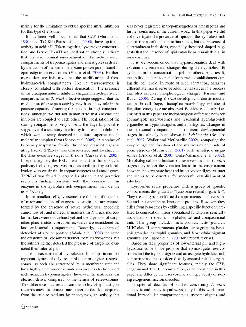
It has been well documented that CZP (Murta et al.1990) and TcCBP (Parussini et al. 2003), have optimumactivity in acid pH. Taken together, lysotracker concentra-tion and P-type H+-ATPase localization strongly indicatethat the acid luminal environment of the hydrolase-richcompartments of trypomastigotes and amastigotes is drivenby the action of the same membrane proton pump found inepimastigote reservosomes (Vieira et al. 2005). Further-more, they are indicative that the acidiWcation of thesehydrolase-rich compartments, like in reservosomes, isclosely correlated with protein degradation. The presenceof the cruzipain natural inhibitor chagasin in hydrolase-richcompartments of T. cruzi infective stage suggests that themodulation of cruzipain activity may have a key role in theparasite capacity of storing the enzyme in high concentra-tions, although we did not demonstrate that enzyme andinhibitor are coupled to each other. The localization of thestoring compartments, very close to the Xagellar pocket issuggestive of a secretory fate for hydrolases and inhibitors,which were already detected in culture supernatants inmolecular complex form (Santos et al. 2005). A member oftyrosine phosphatase family, the phosphatase of regener-ating liver-1 (PRL-1), was characterized and localized inthe three evolutive stages of T. cruzi (Cuevas et al. 2005).In epimastigotes, the PRL-1 was found in the endocyticpathway including reservosomes, as conWrmed by co-local-ization with cruzipain. In trypomastigotes and amastigotes,TcPRL-1 was found in organelles placed in the posteriorregion, a Wnding consistent with the presence of thisenzyme in the hydrolase-rich compartments that we arenow focusing.
In mammalian cells, lysosomes are the site of digestionof macromolecules of exogenous origin and are charac-terized by the presence of active hydrolases, endocyticcargo, low pH and molecular markers. In T. cruzi, molecu-lar markers were not deWned yet and the digestion of cargotakes place inside reservosomes, which are considered thelast endosomal compartment. Recently, cytochemicaldetection of aryl sulphatase (Adade et al. 2007) indicatedthe existence of lysosomes distinct from reservosomes, butthe authors neither detected the presence of cargo nor eval-uated their internal pH.
The ultrastructure of hydrolase-rich compartments oftrypomastigotes closely resembles epimastigote reservo-somes, as both are surrounded by a membrane unit andhave highly electron-dense matrix as well as electronlucentinclusions. In trypomastigotes, however, the matrix is lesselectron-dense, compared to the lumen of reservosomes.This diVerence may result from the ability of epimastigotereservosomes to concentrate macromolecules acquiredfrom the culture medium by endocytosis, an activity that
was never registered in trypomastigotes or amastigotes andfurther conWrmed in the current work. In this paper we didnot investigate the presence of lipids in the hydrolase-richcompartments of the mammalian stages, but the presence ofelectronlucent inclusions, especially those rod shaped, sug-gests that the presence of lipids may be as remarkable as inreservosomes.
It is well-documented that trypanosomatids deal withextreme environmental changes during their complex lifecycle, as in ion concentration, pH and others. As a result,the ability to adapt is crucial for parasite establishment dur-ing the cell cycle. In route of such adaptation, parasitesdiVerentiate into diverse developmental stages in a processthat also involves morphological changes (Parsons andRuben 2000). During T. cruzi development, drastic modiW-cations in cell shape, kinetoplast morphology and site ofXagellum emergence are observed. Besides, we clearly doc-umented in this paper the morphological diVerence betweenepimastigote reservosomes and lysosomal hydrolase-richorganelles in trypomastigotes and amastigotes. Changes inthe lysosomal compartment in diVerent developmentalstages has already been shown in Leishmania (Besteiroet al. 2007; Waller and McConville 2002), comparing themorphology and function of the multivesicular tubule ofpromastigotes (Mullin et al. 2001) with amastigote mega-somes (Brooks et al. 2000, Ueda-Nakamura et al. 2002).Morphological modiWcation of reservosomes in T. cruzistages may reXect the variation found in the environmentbetween the vertebrate host and insect vector digestive tractand seems to be essential for successful establishment ofinfection.
Lysosomes share properties with a group of speciWccompartments designated as “lysosome-related organelles”.They are cell type-speciWc acid compartments and have solu-ble and transmembrane lysosomal proteins. However, theydiVer from lysosomes by exhibiting a speciWc function unre-lated to degradation. Their specialized function is generallyassociated to a speciWc morphological and compositionaltrait. This group includes melanosomes, lytic granules,MHC class II compartments, platelet-dense granules, baso-phil granules, azurophil granules, and Drosophila pigmentgranules (see Raposo et al. 2007 for a recent review).
Based on their properties of low-internal pH and high-hydrolase content, we propose that epimastigote reservo-somes and the trypomastigote and amastigote hydrolase-richcompartments are considered as lysosomal-related organ-elles. They share signiWcant features, mainly the CZP,chagasin and TcCBP accumulation, as demonstrated in thispaper and diVer by the reservosome’s unique ability of stor-ing exogenous macromolecules.
In spite of decades of studies concerning T. cruziendocytic and exocytic pathways, only in this work func-tional intracellular compartments in trypomastigotes and
123

Histochem Cell Biol (2008) 130:1187–1198 1197
amastigotes were described. These structures are distinctfrom lysosomes and comprise the Wrst characterization of“lysosome-related organelles (LROs)” in a protozoan para-site. Changes in the structure of the LROs in T. cruzi stagesmay reXect diVerent ability to degrade but also adaptationof the organelles to play diVerent functions. The presenceof such organelles in the infective stages of T. cruzi mayhave signiWcant implication in the host cell invasion andmay contribute to a better understanding of this vital pro-cess for the parasite. Also, TcLROs may be considered apromising chemotherapy target against Chagas’s disease.
Acknowledgments The authors are grateful to Dr. Ana Paula C. A.Lima and Dr. Roberto Docampo for kindly providing the antibodiesagainst chagasin and P-type +H-ATPase, respectively. Also, we wouldlike to thank Mr. Antonio Bosco Carlos for technical support. Thiswork was supported by Conselho Nacional de Desenvolvimento Cient-íWco e Tecnológico (CNPq), Fundação Carlos Chagas Filho de Am-paro à Pesquisa do Estado do Rio de Janeiro (FAPERJ) e Coordenaçãode Aperfeiçoamento de Pessoal de Nível Superior (CAPES).
References
Adade CM, de Castro SL, Soares MJ (2007) Ultrastructural localiza-tion of Trypanosoma cruzi lysosomes by aryl sulphatase cyto-chemistry. Micron 38:252–256
Aparicio IM, Scharfstein J, Lima APCA (2004) A new cruzipain depen-dent pathway of human cell invasion by Trypanosoma cruzi re-quires trypomastigote membranes. Infect Immun 72:5892–5902
Besteiro S, Williams RAM, Coombs GH, Mottram JC (2007) Proteinturnover and diVerentiation in Leishmania. Int J Parasitol37:1063–1075
Beyenbach KW, Wieczorek H (2006) The V-type H+ ATPase: molec-ular structure and function, physiological roles and regulation.J Exp Biol 209:577–589
Brooks DR, Tetley L, Coombs GH, Mottram JC (2000) Processing andtraYcking of cysteine proteases in Leishmania mexicana. J CellSci 113:4035–4041
Camargo EP (1964) Growth and diVerentiation of Trypanosoma cruzi.I. Origin of metacyclic trypanosomes in liquid media. Rev InstMed Trop São Paulo 6:93–100
Campetella O, Henriksson J, Åslund L, Frasch ACC, Pettersson U, Ca-zzulo JJ (1992) The major cysteine proteinase (Cruzipain) fromTrypanosoma cruzi is encoded by multiple polymorphic tandemlyorganized genes located on diVerent chromosomes. Mol BiochemParasitol 50:225–234
Carvalho TMU, De Souza W (1983) Separation of amastigotes and try-pomastigotes of Trypanosoma cruzi from cultured cells. Z Para-sitenkd 69:571–575
Cazzulo JJ, Cazzulo Franke MC, Martinez J, Franke De Cazzulo BM(1990) Some kinetic properties of a cysteine proteinase (cruzi-pain) from Trypanosoma cruzi. Biochim Biophys Acta.1037:186–191
Cazzulo JJ, Stoka V, Turk V (2001) The major cysteine proteinase ofTrypanosoma cruzi: a valid target for chemotherapy of Chagasdisease. Current Pharm Des 7:1143–1156
Cuevas IC, RohloV P, Sanchez DO, Docampo R (2005) Characteriza-tion of farnesylated protein tyrosine phosphatase TcPRL-1 fromTrypanosoma cruzi. Eukaryot Cell 4:1550–1561
Cunha-e-Silva NL, Atella GC, Porto-Carreiro IA, Morgado-Diaz JA,Pereira MG, De Souza W (2002) Isolation and characterization of
a reservosome fraction from Trypanosoma cruzi. FEMS Micro-biol Lett 214:7–12
Cunha-e-Silva NL, Sant’Anna C, Pereira MG, Porto-Carreiro I, Jeova-nio AL, De Souza W (2006) Reservosomes: multipurpose organ-elles? Parasitol Res 99:325–327
De Duve C (1973) The lysosome in retrospect. In: Dingle JT, Fell HB(eds) Lysosomes in biology and pathology, vol 1. North-Holland,Amsterdam, pp 3–40
De Souza W (2002) Basic cell biology of Trypanosoma cruzi. CurrPharm Des 8:269–285
Eakin AE, Mills AA, Harth G, McKerrow JH, Craik CS (1992) The se-quence, organization, and expression of the major cysteine protease(cruzain) from Trypanosoma cruzi. J Biol Chem 267:7411–7420
Engel JC, Doyle PS, Palmer J, Hsieh I, Bainton DF, Mckerrow JH(1998) Cysteine protease inhibitors alter Golgi complex ultra-structure and function in Trypanosoma cruzi. J Cell Sci 111:597–606
Engel JC, García CT, Hsieh I, Doyle PS, Mckerrow JH (2000) Upreg-ulation of the secretory pathway in cysteine protease inhibitor-resistant Trypanosoma cruzi. J Cell Sci 113:1345–1354
Hall BS, Pal A, Goulding D, Field MC (2004) Rab4 is an essential reg-ulator of lysosomal traYcking in trypanosomes. J Biol Chem279:45047–45056
Hessler D, Young SJ, Carragher BO, Martone ME, Hinshaw JE, Mil-ligan RA, Masliah E, Whittaker M, Lamont S, Ellisman MH(1992) SYNU: software for visualization of three-dimensionalbiological structures. Microscopy 22:72–82
Meirelles MN, Juliano L, Carmona E, Silva SG, Costa EM, Murta AC,Scharfstein J (1992) Inhibitors of the major cysteinyl proteinase(GP57/51) impair host cell invasion and arrest the intracellulardevelopment of Trypanosoma cruzi in vitro. Mol Biochem Paras-itol 52:175–184
Monteiro AC, Abrahamson M, Lima AP, Vannier-Santos MA, Scharf-stein J (2001) IdentiWcation, characterization and localization ofchagasin, a tight-binding cysteine protease inhibitor in Trypano-soma cruzi. J Cell Sci 114:3933–3942
Mullin KA, Foth B, Ilgoutz SM, Callaghan J, McFadden GM, McCon-ville MJ (2001) Regulated degradation of ER membrane proteinsin a novel tubular lysosome in Leishmania mexicana. Mol BiolCell 12:2364–2377
Murta AC, Persechini PM, Souto Padron T, De Souza W, GuimarãesJA, Scharfstein J (1990) Structural and functional identiWcation ofGP57/51 antigen of Trypanosoma cruzi as a cysteine proteinase.Mol Biochem Parasitol 43:27–38
Parsons M, Ruben L (2000) Pathways involved in environmental sens-ing in trypanosomatids. Parasitol Today 16:56–62
Parussini F, Garcia M, Mucci J, Aguero F, Sanchez D, Hellman U, Asl-und L, Cazzulo JJ (2003) Characterization of a lysosomal serinecarboxypeptidase from Trypanosoma cruzi. Mol Biochem Paras-itol 131:11–23
Raposo G, Marks MS, Cutler DF (2007) Lysosome related organelles:driving post-Golgi compartments into specialization. Curr OpinCell Biol 19:394–401
Sant’Anna C, Pereira MG, Lemgruber L, De Souza W, Cunha-e-SilvaN (2008) New insights into the morphology of Trypanosoma cru-zi reservosome. Microsc Res Tech 71(8):599–605
Santos CC, Sant’Anna C, Terres A, Cunha-e-Silva NL, Scharfstein J,Lima AP (2005) Chagasin, the endogenous cysteine-proteaseinhibitor of Trypanosoma cruzi, modulates parasite diVerentiationand invasion of mammalian cells. J Cell Sci 118:901–915
Scharfstein J, Schmitz V, Morandi V, Capella MM, Lima APCA, Mor-rot A, Juliano L, Muller-Esterl W (2000) Host cell invasion byTrypanosoma cruzi is potentiated by activation of bradykinin B2receptors. J Exp Med 192:1289–1330
Serrano R, Villalba JM, Palmgren MG, Portillo F, Parets-Soler A,Roldan M, Ferguson C, Montesinos C (1992) Studies of the
123

1198 Histochem Cell Biol (2008) 130:1187–1198
plasma membrane H(+)-ATPase of yeast and plants. Biochem SocTrans 20:562–566
Shimamura M, Hager KM, Hajduk SL (2001) The lysosomal targetingand intracellular metabolism of trypanosome lytic factor by Try-panosoma brucei brucei. Mol Biochem Parasitol 115:227–237
Soares MJ, De Souza W (1991) Endocytosis of gold-labeled proteinsand LDL by Trypanosoma cruzi. Parasitol Res 77:461–469
Soares MJ, De Souza W (1988) Cytoplasmic organelles of trypanoso-matids. A cytochemical and stereological stereological study.J Submicrosc Cytol Pathol 20:349–363
Soares MJ, Souto-Padrón T, Bonaldo MC, Goldenberg S, de Souza W(1989) A stereological study of the diVerentiation process in Try-panosoma cruzi. Parasitol Res 75:522–527
Soares MJ, Souto-Padrón T, De Souza W (1992) IdentiWcation of alarge pre-lysosomal compartment in the patogenic protozoan Try-panosoma cruzi. J Cell Sci 102:157–167
Souto-Padrón T, Campetella OE, Cazzulo JJ, De Souza W (1990)Cysteine proteinase in Trypanosoma cruzi: immunocytochemicallocalization and involvement in parasite-host cell interation.J Cell Sci 96:485–490
Todorov AG, Andrade DS, Pesquero JB, Araújo RC, Bader M, StewartJ, Gera L, Muller-Esterl W, Morandi V, Goldenberg RC, NetoHC, Scharfstein J (2003) Trypanosoma cruzi induces edemato-genic responses in mice and invades cardiomyocytes and endo-thelial cells in vitro by activating distinct kinin receptor (B1/B2)subtypes. FASEB J 7:73–75
Ueda-Nakamura T, a Sampaio M, Cunha-e-Silva N, Traub-Cseko YM,De Souza W (2002) Expression and processing of megasome cys-teine proteinases during Leishmania amazonensis diVerentiation.Parasitol Res 88:332–337
Vieira M, RohloV P, Luo S, Cunha-e-Silva NL, De Souza W, DocampoR (2005) Role for a P-type H+-ATPase in the acidiWcation of theendocytic pathway of Trypanosoma cruzi. Biochem J 392:467–474
Waller RF, McConville MJ (2002) Developmental changes in lyso-some morphology and function Leishmania parasites. Int J Paras-itol 32:1435–1445
Young SJ, Royer SM, Groves PM, Kinnamon JC (1987) Three-dimen-sional reconstruction from serial micrographs using an IBM PC.J Elect Microsc Tech 6:207–215
123