Flowcytometric assessment of the effect of drugs on Giardia lamblia trophozoites in vitro
Upload
independentCategory
view
3download
0
..............................................................
Mitochondrial remnant organelles ofGiardia function in iron-sulphurprotein maturation
Jorge Tovar1, Gloria Leon-Avila1, Lidya B Sanchez2*, Robert Sutak3,Jan Tachezy3, Mark van der Giezen1, Manuel Hernandez1,Miklos Muller2 & John M. Lucocq4
1School of Biological Sciences, Royal Holloway, University of London, Egham,Surrey TW20 0EX, UK2The Rockefeller University, 1230 York Avenue, New York, New York 10021, USA3Department of Parasitology, Faculty of Science, Charles University,12844 Prague 2, Czech Republic4School of Life Sciences, WTB/MSI complex, University of Dundee, DundeeDD1 5EH, UK
* Present address: Public Health Research Institute, Newark, New Jersey 07103, USA
.............................................................................................................................................................................
Giardia intestinalis (syn. lamblia) is one of the most widespreadintestinal protozoan pathogens worldwide, causing hundreds ofthousands of cases of diarrhoea each year1. Giardia is a memberof the diplomonads, often described as an ancient protist groupwhose primitive nature is suggested by the lack of typicaleukaryotic organelles (for example, mitochondria, peroxisomes),the presence of a poorly developed endomembrane system and bytheir early branching in a number of gene phylogenies1,2. Thediscovery of nuclear genes of putative mitochondrial ancestry inGiardia3–7 and the recent identification of mitochondrial rem-nant organelles in amitochondrial protists such as Entamoebahistolytica8,9 and Trachipleistophora hominis10 suggest that theeukaryotic amitochondrial state is not a primitive condition butis rather the result of reductive evolution. Using an in vitroprotein reconstitution assay and specific antibodies against IscSand IscU—two mitochondrial marker proteins involved in iron–sulphur cluster biosynthesis—here we demonstrate that Giardia
contains mitochondrial remnant organelles (mitosomes)bounded by double membranes that function in iron–sulphurprotein maturation. Our results indicate that Giardia is notprimitively amitochondrial and that it has retained a functionalorganelle derived from the original mitochondrialendosymbiont.
The assembly and maturation of iron–sulphur (Fe–S) proteins isa recently identified critical function of the mitochondrion11.Proteins containing Fe–S centres are widely distributed in natureand operate in metalloenzyme catalysis and electron transport.Although several Fe–S proteins are important in energy metabolismin amitochondriate organisms (for example, pyruvate:ferredoxinoxidoreductase, ferredoxin, hydrogenase)12, almost nothing isknown about the maturation of these proteins into functionalenzymes or about the biosynthesis of their essential Fe–S reactivecentres. Genes encoding the soluble enzyme cysteine desulphurase(IscS), a central component of the Fe–S cluster assembly system ofprokaryotic and eukaryotic cells, were recently cloned from theamitochondrial protists Trichomonas vaginalis and Giardia intesti-nalis5. Because phylogenetic analyses of these genes support theirmitochondrial ancestry, it is possible that their encoded proteins arestill compartmentalized into a mitochondrion-derived organelle.While the mitochondrion-related hydrogenosome of Trichomonasis a good candidate for this function13,14, a mitochondrion-derivedintracellular compartment in Giardia has not been identified,despite mounting evidence suggesting its existence3–7,15,16.
We cloned and characterized a second Giardia gene involved inthe biosynthesis of Fe–S clusters, the GiiscU gene. This gene encodesthe soluble iron-binding protein IscU, a functional partner of IscSthought to participate as a scaffold protein that forms Fe–Smolecular complexes in conjunction with IscS (ref. 11). Aminoacid sequence comparison of the Giardia IscU with bacterial andeukaryotic homologues revealed a high degree of similarity alongthe entire length of the protein (63–69% similarity, 46–51%identity), with conservation of cysteine residues essential for invivo functionality17. Similar to IscS (ref. 5), phylogenetic analysesprovide significant support for the clustering of Giardia IscU with
Figure 1 Distribution of IscU and IscS in Giardia cell extracts. a, Comparison of N-terminal
regions of prokaryotic and eukaryotic IscU proteins aligned by CLUSTALW. Hydroxylated
and basic amino acids of the Giardia sequence are underlined. E27, the proposed
N-terminal cleavage site of the Giardia sequence and Y35, the first residue of the mature
human IscU (ref. 18), are in bold typeface. b, Distribution of Isc proteins in Giardia
fractions obtained by differential centrifugation. Trophozoite extracts fractionated as
described in Methods were electrophoresed, blotted and probed with specific antibodies
against IscS and IscU. Sizes of molecular markers are given in kDa.
letters to nature
NATURE | VOL 426 | 13 NOVEMBER 2003 | www.nature.com/nature172 © 2003 Nature Publishing Group

mitochondrial homologues (not shown). The amino-terminalsequence of IscU is rich in hydroxylated and basic amino acidsreminiscent of mitochondrial-targeting presequences (Fig. 1a).Analysis of this sequence using MitoProt predicts the mitochondriallocalization of Giardia IscU with high confidence (P . 0.95). Thepredicted N-terminal cleavage site at E27 coincides with the firstamino acid residue of all prokaryotic IscU sequences and is tworesidues upstream of Y35, the cleavage site of the human sequence18.These findings suggest that the N-terminal sequence might targetIscU to a mitochondrion-derived organelle in this organism.
To test this hypothesis we overexpressed Giardia IscU and IscS inEscherichia coli and generated antibodies against the recombinantproteins. The specific anti-IscU and anti-IscS antibodies were usedfor immunoblotting of subcellular fractions obtained by differentialcentrifugation. Protein bands of around 49 and 19 kDa wereidentified, in good agreement with the sizes of IscS and IscUrespectively, predicted from their genomic sequences. Both proteinswere found predominantly in the low-speed sediment of unbrokencells and cell debris (see Methods for details) and in the high-speedpellet containing sedimentable particulate matter such as mem-branes and membrane-bounded organelles (Fig. 1b, lanes 1 and 3).Little or no label was found in the cytosol, suggesting that bothproteins are compartmentalized in this organism.
We tested the functionality of Giardia Fe–S protein maturationusing an in vitro reconstitution assay that monitors the assembly ofde novo synthesized radiolabelled Fe–S clusters into recombinantferredoxin apoprotein lacking Fe–S moieties. Formation of fullyassembled ferredoxin occurs exclusively when the high-speed sedi-ment is used in the reaction (Fig. 2). Incubation with cytosolicproteins did not produce significant incorporation of label intoferredoxin and the addition of EDTA, a metal chelator, completelyinhibited ferredoxin maturation. These results are consistent withthe observed cellular distribution of Isc proteins in Giardia anddemonstrate that Fe–S cluster biosynthesis, a mitochondrial bio-synthetic pathway, is fully operational in this amitochondrialorganism.
The compartmentalized distribution of IscS and IscU in Giardiawas demonstrated by confocal scanning fluorescence microscopy.Fixed trophozoites treated with anti-IscS and anti-IscU antibodiesdisplayed localized staining in small cellular structures distributedthroughout the cytoplasm, including an accumulation of labelaround basal bodies and the base of flagellar axonemes (Fig. 3a;see also Fig. 4f). Figure 3a shows representative maximum projec-tion images for both proteins merged with the correspondingdifferential interference contrast (DIC) images. The number oforganelles detected per cell ranged from 25 to well over 100, asestimated by counting labelled structures in optical stacks of 300 nmthickness, suggesting a significant physiological role for this orga-
nelle in Giardia. Double labelling for IscS and IscU revealed thatboth proteins are present in the same intracellular compartment(Fig. 3b; see also Figs 4c, d), as would be expected from theirfunctional partnership in Fe–S cluster biosynthesis. Interestingly,chaperonin Cpn60 was not found to colocalize with IscS in doublelabelling experiments (Fig. 3c), indicating that the current functionof Cpn60 in Giardia is not conducted in the same compartment asFe–S cluster biosynthesis. Unlike IscU, Giardia IscS lacks a recog-nizable putative mitochondrial targeting signal5 but its clear com-partmentalized localization suggests that organellar protein importmechanisms in Giardia are not solely dependent on N-terminalpresequences. A similar observation was recently made in Trachi-pleistophora, where mtHsp70 is efficiently targeted into mito-
Figure 2 Functional analysis of Fe–S protein maturation in Giardia. Incorporation of
radioactive Fe–S clusters into recombinant apoferredoxin was assayed as described in
Methods. Assembly of the de novo synthesized Fe–S clusters into apoferredoxin is
observed exclusively when the high-speed sediment is used in the reaction. Ferredoxin
maturation is inhibited by metal ion chelation with EDTA.
Figure 3 Localization of IscS and IscU in Giardia trophozoites by confocal
immunofluorescence microscopy. a, Giardia trophozoites, fixed and permeabilized as
described in Methods, were probed independently with antibodies specific for IscU and
IscS. The label is located in foci throughout the cytoplasm; distinctive accumulation is also
observed around the base of flagellar axonemes and basal bodies (indicated by
arrowheads). b, Double labelling of Giardia trophozoites with anti-IscS and anti-IscU
antibodies; colocalization of label is readily apparent. c, Double labelling of trophozoites
with anti-IscS and anti-Cpn60 antibodies; no colocalization of label is observed.
letters to nature
NATURE | VOL 426 | 13 NOVEMBER 2003 | www.nature.com/nature 173© 2003 Nature Publishing Group

chondrial remnant organelles despite the absence of a recognizableorganelle targeting presequence10.
We used immunoelectron microscopy to investigate the structureof the organelle harbouring the Giardia Isc proteins. Immunogoldlabelling of IscS and IscU was localized over small organellesbounded by two limiting membranes (Fig. 4a–d). Labelling den-sities for IscS and IscU were at least 200 and 110 fold higher thanover cytosol or peripherally located endolysosomes respectively(Fig. 4i). Double labelling for IscS and IscU localized both ofthese antigens in the same class of double membrane-boundedstructures (Fig. 4c, d), and similar structures were found in epoxyresin sections (Figs 4e–h). In cryosections, gold-labelled double-membrane-bounded organelles were elongated in profile andmeasured an average of 142 £ 61 nm while in epoxy resin sectionsthese structures measured an average of 137 £ 68 nm (Fig. 4j).These organelles were found distributed throughout the cytosoland in close proximity to flagellar axonemes (Fig. 4f), in agreementwith the distribution observed by confocal microscopy.
Immunogold labelling of Cpn60 did not produce significantsignal over membrane-bounded structures (not shown), in agree-ment with a previous report on the lack of association of GiardiaCpn60 with biological membranes16 and consistent with our con-focal microscopy observations. The differential distribution ofCpn60 and IscS is noteworthy because the mitochondrial ancestryof both proteins is strongly supported by phylogenetic analyses.
However, the fact that only Isc proteins are associated with mem-brane-bounded organelles suggests that Cpn60 has been relocatedfrom its original mitochondrial compartment as part of an ongoingprocess of reductive evolution. Alternatively, the possibility thatGiardia Cpn60 might have been acquired by lateral transfer from aprokaryotic organism related to the mitochondrial endosymbiontcannot be excluded19,20. The precise nature of the Cpn60 aggregatesthat render the characteristic punctate distribution in Giardiaremains to be elucidated.
Overall, our data provide direct physical evidence for the pres-ence of mitochondrial remnant organelles (mitosomes) in Giardia,as postulated on the basis of previous phylogenetic and biochemicalstudies3–7,15,21. Mitochondrial remnant organelles have also beenobserved in other amitochondrial protists including the micro-sporidian Trachipleistophora hominis, the apicomplexan Crypto-sporidium parvum and the entamoebid Entamoeba histolytica8–10,22.Giardia has traditionally been considered to be one of the earliest-branching eukaryotes, although the significance of its basal positionin phylogenetic reconstructions has been disputed1,2,23. The identi-fication of mitosomes in this organism places the endosymbioticevent that led to mitochondria firmly before the evolutionarydivergence of the diplomonads.
Energy generation and recycling of resources are thought to havedriven the original endosymbiotic event. Whether ATP generationin the heterotrophic mitochondrial endosymbiont involved aerobic
Figure 4 Localization of IscS and IscU in Giardia trophozoites by transmission electron
immunomicroscopy. a–d, Thawed frozen cryosections of glutaraldehyde-fixed Giardia
were labelled with anti-IscS (a) and anti-IscU antibodies (b) or double-labelled (c and d) for
anti-IscU (8 nm gold particles) followed by anti-IscS antibodies (12 nm gold particles).
Arrows indicate double-membrane-bounded structures. Membranes appear as white
profiles with this methodology. e–h, Epoxy resin sections of glutaraldehyde-fixed
trophozoites. f, Double-membrane-bounded structures are shown in close proximity to
flagellar axonemes. g, h, High-magnification images showing organelle double
membranes. Arrows point towards outer membranes while arrowheads identify inner
membranes. i, Densities of labelling from typical labelling experiments were obtained as
explained in Methods (n ¼ 8 for IscS and n ¼ 7 for IscU; n ¼ number of scanned
micrographs). j, Sizes of double membrane bounded organelles in epoxy resin and
cryosections labelled with antibodies to either IscS or IscU (n ¼ 43 for epoxy resin,
n ¼ 13 for IscS and n ¼ 11 for IscU; n ¼ number of organelles). Intermembrane spacing
in epoxy resin was 9.36 ^ 0.49 nm (n ¼ 29). Scale bars, 100 nm (a–f) and 50 nm (g, h).
letters to nature
NATURE | VOL 426 | 13 NOVEMBER 2003 | www.nature.com/nature174 © 2003 Nature Publishing Group

or anaerobic respiration—as proposed by current theories ofeukaryotic evolution (reviewed in ref. 24)—all electron transportchains and modes of heterotrophic oxidative metabolismshare a common feature: several of their key enzymes rely on Fe–Scentres for their catalytic activities (for example, ferredoxins,pyruvate:ferredoxin oxidoreductase, succinate dehydrogenase,cytochrome b-associated Rieske protein, aconitase, hydrogenase)12.Thus, the original endosymbiont must have possessed the capacity tosynthesize Fe–S clusters and to assemble them into functional redoxand electron transport proteins. These crucial metabolic functionscould have secured the permanent establishment of the mitochon-drial endosymbiont in the newly formed eukaryotic cell.
Our data also directly demonstrate an essential metabolic func-tion for the mitosomes of amitochondriate protists. The apparentubiquity of Fe–S cluster assembly proteins in the genomes ofall amitochondriate protists sequenced until now (for example,Encephalitozoon25, Giardia26, Cryptosporidium27, Entamoeba khttp://www.sanger.ac.uk/Projects/E_histolytica/l, khttp://www.tigr.org/tdb/e2k1/eha1/l) suggests that Fe–S cluster biosynthesis and Fe–Sprotein maturation might have been the only biochemical featuresof the mitochondrial endosymbiont essential for eukaryotic cellsurvival under conditions of oxygen deprivation. Interestingly,hydrogen production by Giardia has recently been detected understrict anaerobic conditions28. The possibility that this process mightalso occur in the double-membrane-bounded organelles describedhere awaits the unequivocal localization of hydrogenase in thisorganism. We suggest that the requirement for the assembly andmaturation of Fe–S proteins may have provided, for hundreds ofmillions of years, the selective pressure required for the retention ofthe mitochondrial endosymbiont in both aerobic and anaerobicenvironments. A
MethodsGene cloning, protein expression and antibody generationThe entire coding regions of the Giardia iscS and iscU genes were amplified by PCR usingGiardia genomic DNA as template and the primer pairs 5
0-TATTTTTTAATGAGGTACC
TGGACAACCAGGCCACTACG-30
/ 50-AGTGGTGTGAGTCGACTAGTCATGCTT
CCACTCTATGCTC-3 0 and 5 0 -AATTCTAATTGGATCCCAAGCCTTCAGCTCTCTAGCACT-3
0/ 5
0-CAGGAGGCACCCGGGTCAAGAAGACTTTGATACCTGTATC-3
0
designed on the basis of partial sequences retrieved from the Giardia genome database26.These incorporated unique KpnI and SalI sites and BamHI and XmaI sites (italics) into iscSand iscU respectively. PCR products were cloned directionally into plasmid pQE-32(Qiagen) and transformed into E. coli M15 [pREP4]. N-terminal His-tagged proteins werepurified under denaturing conditions by affinity chromatography following themanufacturer’s protocol (Qiagen) and used to generate rabbit polyclonal anti-IscS andanti-IscU antibodies (Lampire Biological Laboratories). Mouse polyclonal anti-Cpn60antibodies were generated using recombinant Cpn60 protein from E. histolytica preparedas described9.
Organism, cell fractionation and western blottingGiardia intestinalis (WB strain, ATCC No. 30957) was used throughout this study.Trophozoites were partially lysed to a maximum 50% cell lysis in isotonic buffer (0.2 Mmannitol, 50 mM sucrose, 10 mM KCl, 1 mM EDTA, 10 mM HEPES pH 7.4 supplementedwith 70 mM E64 and 0.75 mg ml21 leupeptin). A low-speed sediment fraction composed ofcell debris and unbroken cells was obtained by centrifugation at 1,000g for 10 min at 4 8C.The supernatant was further centrifuged at 108,000g for 30 min. The resulting high-speedsediment was resuspended in isotonic buffer. The high-speed supernatant was used as thesoluble cytosolic fraction. Equivalent protein aliquots (10 mg) were electrophoresed on10% or 12.5% polyacrylamide gels for detection of IscS and IscU respectively. Proteinswere electroblotted onto nitrocellulose membranes, blocked by incubation in 2%skimmed milk in PBS and probed with anti-IscS (1:1,000) or anti-IscU (1:750) antibodiesfor 1 h at room temperature. Membranes were further incubated with anti-rabbit IgGantibodies conjugated to peroxidase (1:25,000) for 1 h at room temperature. Blots weredeveloped using an ECL detection system (Amersham Biosciences).
Reconstitution of Fe–S clusters in vitroTrophozoites were mechanically disrupted and fractionated as described above. Fe–Scluster formation by cytosolic and high-speed sediment fractions was determined usingrecombinant apoferredoxin from Trichomonas vaginalis as a reporter protein.Recombinant ferredoxin was prepared as described29. The standard reaction mixturecontained 30–100 mg protein of tested fraction, 7 mg of apo-ferredoxin, 20 mM HEPES,0.2% Triton-X100, 250 mM saccharose, 50 mM ferrous ascorbate, 20 mM DTT, 25 mML-cysteine and 10 mCi of 35S-L-cysteine. To observe inhibition of the reaction, 5 mM EDTAwas added to the reaction mixture before the incubation. In controls, the cell fractions
were omitted. Reactions proceeded in an anaerobic jar under atmosphere of 95% nitrogenand 5% hydrogen gas for 90 min at 25 8C. Samples were separated on a 15% non-denaturing polyacrylamide gel at 4 8C, followed by gel vacuum-drying andautoradiography.
Immunofluorescence confocal microscopyGiardia trophozoites were fixed in 4% paraformaldehyde in PBS for 30 min at 37 8C andpermeabilized with cold acetone or 0.2% Triton X-100 in PBS for 5 min. After blockingwith 1% BSA in PBS, slides were incubated overnight with anti-Isc rabbit antibodies(1:200 dilution) and subsequently with secondary anti-rabbit IgG-TRITC conjugate(1:300 dilution) for 1 h at 37 8C. In Isc colocalization experiments, slides treated with anti-IscU antibodies were further incubated with 250 ml of purified rabbit IgGimmunoglobulins (40 mg ml21) for 1 h at 37 8C followed by incubation with FITC-conjugated anti-IscS antibodies (Fluoro Tag FITC Conjugation Kit, Sigma). Slides weremounted with anti-fade mounting medium (Vectashield) and observed under a laserscanning confocal microscope (Radiance 2100, Bio-Rad). Images were collected usingBio-Rad LaserSharp 2000 software and processed with Adobe PhotoShop 7.0.
Immunoelectron microscopyOrganisms were fixed in 0.5% glutaraldehyde or 4% paraformaldehyde/0.1%glutaraldehyde each in 0.2 M PIPES buffer pH 7.2 for 30 min at room temperature,washed in PBS and stored at 4 8C. Cells were pelleted and cryoprotected in 2.3 Msucrose in PBS and frozen on cryosectioning stubs in liquid nitrogen and cryosectionedon a Leica ultracryomicrotome at 50–100 nm. Thawed cryosections were labelled usingrabbit antisera against Isc proteins followed by 8 nm protein A gold30, contrasted inmethyl cellulose/uranyl acetate and observed in a JEOL 1200EX transmission electronmicroscope. For quantitation, elongated or round profiles with evidence of doublemembranes and measuring less than 110 nm minor diameters were photographed at anominal magnification of 40,000. Micrographs were recorded on phosphoimagingplates (4,342 £ 4,912 pixels) (DITABIS, Germany) and stored as TIFF files and furthermagnified in Adobe Photoshop 5.5. Square lattice grids with spacing of 25 nm, 250 nmand 125 nm were generated on screen and point counting was used to estimate areas ofdouble membrane structures, cytosol and peripherally located endolysosomesrespectively. Double labelling using 8 nm and 12 nm gold particles was carried out asdescribed elsewhere30. Standard errors of ratio estimates were computed according toref. 31.
Received 7 April; accepted 22 July 2003; doi:10.1038/nature01945.
1. Adam, R. D. Biology of Giardia lamblia. Clin. Microbiol. Rev. 14, 447–475 (2001).
2. Sogin, M. L. & Silberman, J. D. Evolution of the protists and protistan parasites from the perspective of
molecular systematics. Int. J. Parasitol. 28, 11–20 (1998).
3. Hashimoto, T., Sanchez, L. B., Shirakura, T., Muller, M. & Hasegawa, M. Secondary absence of
mitochondria in Giardia lamblia and Trichomonas vaginalis revealed by valyl-tRNA synthetase
phylogeny. Proc. Natl Acad. Sci. USA 95, 6860–6865 (1998).
4. Roger, A. J. et al. A mitochondrial-like chaperonin 60 gene in Giardia lamblia: evidence that
diplomonads once harbored an endosymbiont related to the progenitor of mitochondria. Proc. Natl
Acad. Sci. USA 95, 229–234 (1998).
5. Tachezy, J., Sanchez, L. B. & Muller, M. Mitochondrial type iron-sulfur cluster assembly in the
amitochondriate eukaryotes Trichomonas vaginalis and Giardia intestinalis, as indicated by the
phylogeny of IscS. Mol. Biol. Evol. 18, 1919–1928 (2001).
6. Morrison, H. G., Roger, A. J., Nystul, T. G., Gillin, F. D. & Sogin, M. L. Giardia lamblia expresses a
proteobacterial-like DnaK homolog. Mol. Biol. Evol. 18, 530–541 (2001).
7. Arisue, N., Sanchez, L. B., Weiss, L. M., Muller, M. & Hashimoto, T. Mitochondrial-type hsp70 genes
of the amitochondriate protists Giardia intestinalis, Entamoeba histolytica and two microsporidians.
Parasitol. Int. 51, 9–16 (2002).
8. Mai, Z. et al. Hsp60 is targeted to a cryptic mitochondrion-derived organelle (“crypton”) in the
microaerophilic protozoan parasite Entamoeba histolytica. Mol. Cell. Biol. 19, 2198–2205 (1999).
9. Tovar, J., Fischer, A. & Clark, C. G. The mitosome, a novel organelle related to mitochondria in the
amitochondrial parasite Entamoeba histolytica. Mol. Microbiol. 32, 1013–1021 (1999).
10. Williams, B. A. P., Hirt, R. P., Lucocq, J. M. & Embley, T. M. A mitochondrial remnant in the
microsporidian Trachipleistophora hominis. Nature 418, 865–869 (2002).
11. Lill, R. & Kispal, G. Maturation of cellular Fe-S proteins: an essential function of mitochondria. Trends
Biochem. Sci. 25, 352–356 (2000).
12. Muller, M. in Molecular Medical Parasitology (eds Marr, J., Nilsen, T. & Komuniecki, R.) 125–139
(Academic, London, 2003).
13. Dyall, S. D. & Johnson, P. J. Origins of hydrogenosomes and mitochondria: evolution and organelle
biogenesis. Curr. Opin. Microbiol. 3, 404–411 (2000).
14. Embley, T. M., van der Giezen, M., Horner, D. S., Dyal, P. L. & Foster, P. Mitochondria and
hydrogenosomes are two forms of the same fundamental organelle. Phil. Trans. R. Soc. Lond. 358,
191–203 (2003).
15. Lloyd, D. & Harris, J. C. Giardia: highly evolved parasite or early branching eukaryote? Trends
Microbiol. 10, 122–127 (2002).
16. Soltys, B. J. & Gupta, R. S. Presence and cellular distribution of a 60-kDa protein related to
mitochondrial hsp60 in Giardia lamblia. J. Parasitol. 80, 580–590 (1994).
17. Tokumoto, U. et al. Network of protein-protein interactions among iron-sulfur cluster assembly
proteins in Escherichia coli. J. Biochem. (Tokyo) 131, 713–719 (2002).
18. Tong, W. H. & Rouault, T. Distinct iron-sulfur cluster assembly complexes exist in the cytosol and
mitochondria of human cells. EMBO J. 19, 5692–5700 (2000).
19. Andersson, J. O., Sjogren, A. M., Davis, L. A., Embley, T. M. & Roger, A. J. Phylogenetic analyses of
diplomonad genes reveal frequent lateral gene transfers affecting eukaryotes. Curr. Biol. 13, 94–104
(2003).
20. Doolittle, W. F. You are what you eat: a gene transfer ratchet could account for bacterial genes in
eukaryotic nuclear genomes. Trends Genet. 14, 307–311 (1998).
21. Lloyd, D. et al. The ‘primitive’ microaerophile Giardia intestinalis (syn. lamblia, duodenalis) has
letters to nature
NATURE | VOL 426 | 13 NOVEMBER 2003 | www.nature.com/nature 175© 2003 Nature Publishing Group

specialized membranes with electron transport and membrane-potential-generating functions.
Microbiology 148, 1349–1354 (2002).
22. Riordan, C. E., Ault, J. G., Langreth, S. G. & Keithly, J. S. Cryptosporidium parvum Cpn60 targets a
relict organelle. Curr. Genet. advance online publication, 20 August 2003 (doi:10.1007/s00294-003-
0432-1).
23. Philippe, H. et al. Early-branching or fast-evolving eukaryotes? An answer based on slowly evolving
positions. Proc. R. Soc. Lond. B 267, 1213–1221 (2000).
24. Martin, W., Hoffmeister, M., Rotte, C. & Henze, K. An overview of endosymbiotic models for the
origins of eukaryotes, their ATP-producing organelles (mitochondria and hydrogenosomes), and
their heterotrophic lifestyle. Biol. Chem. 382, 1521–1539 (2001).
25. Katinka, M. D. et al. Genome sequence and gene compaction of the eukaryote parasite
Encephalitozoon cuniculi. Nature 414, 450–453 (2001).
26. McArthur, A. G. et al. The Giardia genome project database. FEMS Microbiol. Lett. 189, 271–273
(2000).
27. Strong, W. B. & Nelson, R. G. Preliminary profile of the Cryptosporidium parvum genome: an
expressed sequence tag and genome survey sequence analysis. Mol. Biochem. Parasitol. 107, 1–32
(2000).
28. Lloyd, D., Ralphs, J. R. & Harris, J. C. Giardia intestinalis, a eukaryote without hydrogenosomes,
produces hydrogen. Microbiology 148, 727–733 (2002).
29. Vidakovic, M. S., Fraczkiewicz, G. & Germanas, J. P. Expression and spectroscopic characterization of
the hydrogenosomal [2Fe-2S] ferredoxin from the protozoan Trichomonas vaginalis. J. Biol. Chem.
271, 14734–14739 (1996).
30. Prescott, A. R., Lucocq, J. M., James, J., Lister, J. M. & Ponnambalam, S. Distinct
compartmentalization of TGN46 and beta 1,4-galactosyl transferase in HeLa cells. Eur. J. Cell Biol. 72,
238–246 (1997).
31. Cochran, W. G. Sampling Techniques (John Wiley and Sons, London, 1977).
Acknowledgements We thank G. Clark, J. Bowyer and S. Cutting for critically reading the
manuscript. A recombinant plasmid containing the T. vaginalis ferredoxin gene was provided by
J. P. Germanas and K. Krause. The use of partial genome sequence information from the Giardia
Genome Project Database26 is acknowledged. The technical assistance of J. James and
N. Sommerville is also acknowledged. M.H. is a sabbatical visitor supported by CINVESTAV,
Mexico. Research at the Rockefeller University (gene cloning, antibody generation) was
supported by a NIH grant to M.M. Research at Charles University (in vitro assembly of Fe–S
clusters) was supported by a grant from FIRCA to J.Tachezy. J.M.L. (electron microscopy) was
supported by a Research Leave Fellowship from the Wellcome Trust and by Tenovus Scotland.
Research at Royal Holloway (bioinformatics, cell fractionation, fluorescence confocal microscopy,
manuscript writing, project coordination) was supported by a Wellcome Trust grant to J.Tovar.
Competing interests statement The authors declare that they have no competing financial
interests.
Correspondence and requests for materials should be addressed to J.Tovar ([email protected]).
The sequence of GiiscU has been deposited with GenBank under accession number AY040612.
..............................................................
Allele substitution at a flowercolour locus produces apollinator shift in monkeyflowersH. D. Bradshaw Jr1 & Douglas W. Schemske2
1Department of Biology, University of Washington, Seattle, Washington 98195,USA2Department of Plant Biology, Michigan State University, East Lansing,Michigan 48824, and W. K. Kellogg Biological Station, Hickory Corners,Michigan 49060, USA.............................................................................................................................................................................
The role of major mutations in adaptive evolution has beendebated for more than a century1,2. The classical view is thatadaptive mutations are nearly infinite in number with infinite-simally small phenotypic effect3, but recent theory suggestsotherwise4. To provide empirical estimates of the magnitude ofadaptive mutations in wild plants, we conducted field studies todetermine the adaptive value of alternative alleles at a single locus,YELLOW UPPER5–7 (YUP). YUP controls the presence or absenceof yellow carotenoid pigments in the petals of pink-floweredMimulus lewisii, which is pollinated by bumblebees5,8–10, andits red-flowered sister species11 M. cardinalis, which is pollinatedby hummingbirds5,8–10. We bred near-isogenic lines (NILs) inwhich the YUP allele from each species was substituted into the
other. M. cardinalis NILs with the M. lewisii YUP allele had darkpink flowers and received 74-fold more bee visits than the wildtype, whereas M. lewisii NILs with the M. cardinalis yup allelehad yellow-orange flowers and received 68-fold more humming-bird visits than the wild type. These results indicate that anadaptive shift in pollinator preference may be initiated by a singlemajor mutation.
Where their ranges overlap, the monkeyflowers M. lewisii andM. cardinalis are .99% reproductively isolated by the difference intheir pollinator guilds8,9. In previous studies of artificial F2 hybridsbetween M. lewisii and M. cardinalis, we showed that flower colourhas marked effects on pollinator visitation8, and that yellow pig-ment concentration is controlled in part by the major quantitativetrait locus (QTL; reviewed in ref. 12), YUP6,7. Although F2 popu-lations are useful for mapping QTLs controlling differences in floraltraits between species6,7,13, they are less than ideal for assessing theadaptive effect of a single mutation. The many intermediate flowerphenotypes in an F2 population6–8,13 may provide a bridge forpollinators to develop learned visitation patterns completely unlikethose that would occur as a result of a single-locus mutational stepin an adaptive walk.
By substituting one allele for another using repeated backcrosses,NILs more closely mimic the effect of a single mutation likely to bepart of an adaptive pollinator shift; that is, from bumblebee-pollinated to hummingbird-pollinated, or vice versa. The dominantM. lewisii YUP allele prevents carotenoid deposition, so thepetals show only their pink anthocyanin pigments. The recessiveM. cardinalis yup allele allows carotenoid deposition in the petalsand produces red flowers when present in conjunction with a highconcentration of anthocyanins5–7. Although phylogenetic evidencesuggests that the hummingbird pollination syndrome of M. cardi-nalis is derived from a bee-pollinated ancestor similar to M. lewisii11,we constructed YUP NILs in both species (Fig. 1). The wild-typeM. lewisii NIL is pink-flowered (Fig. 1a), whereas the ‘mutant’ NILhomozygous for the introgressed M. cardinalis yup allele has paleyellow-orange flowers (Fig. 1b). The wild-type M. cardinalis NIL isred-flowered (Fig. 1c), but the presence of a dominant M. lewisiiYUP allele produces a dark-pink-flowered NIL (Fig. 1d).
Pollinator visitation rates were determined by field observation ofNIL experimental arrays near a zone of sympatry between M. lewisiiand M. cardinalis5,8 to ensure that pollinators were familiar withboth species in their natural habitat. Bumblebees strongly preferpink-flowered NILs carrying the YUP allele (Fig. 1a, d) in boththe M. lewisii and M. cardinalis genetic backgrounds (Table 1).Hummingbirds prefer yellow-orange- or red-flowered NILs homo-zygous for the yup allele (Fig. 1b, c) in both backgrounds (Table 1).
The striking effect of flower colour on pollinator specificity isevidence for the adaptation of both monkeyflower species to theircurrent pollinators (Table 1). A wild-type pink M. lewisii flower(Fig. 1a) is.700 times more likely to be visited by a bumblebee thanby a hummingbird, whereas the yellow-orange-flowered ‘mutant’(Fig. 1b) is only 1.8 times as likely to be visited by a bumblebee. Inthe M. cardinalis background, a wild-type red flower (Fig. 1c) is.1,200 times more likely to be visited by a hummingbird than by abumblebee, but the pink-flowered ‘mutant’ (Fig. 1d) is visited only15 times as frequently by hummingbirds.
When these visitation rates are compared with the results fromour previous F2 QTL mapping population8, we find that the F2
experiments accurately predict pollinator visitation when we con-sider only bumblebees visiting M. lewisii NILs, and hummingbirdsvisiting M. cardinalis NILs. In M. lewisii NILs and the F2, the wild-type pink flowers were visited by bumblebees at about a fivefoldhigher rate than were the ‘mutant’ yellow-orange flowers (Table 1and ref. 8). In M. cardinalis NILs, hummingbirds showed a slight1.1-fold preference for wild-type red flowers over the pink-flowered‘mutants’, similar to that found in the F2 population (Table 1 andref. 8). The close correspondence of the results from these indepen-
letters to nature
NATURE | VOL 426 | 13 NOVEMBER 2003 | www.nature.com/nature176 © 2003 Nature Publishing Group

A ll known life-forms are eitherprokaryotes or eukaryotes; there isnothing in between. Eukaryotic cells
have a nucleus and compartments that are surrounded by two membranes, butprokaryotic cells never do. The prokaryotescame first; eukaryotes (all plants, animals,fungi and protists) evolved from them, andto this day biologists hotly debate how thistransition took place,with about 20 differenttheories on the go1. In most textbookaccounts, an ancient prokaryotic lineagesupposedly evolved a nucleus,giving rise to aeukaryote, which then acquired mitochon-dria — the double-membrane-boundedpowerhouses of eukaryotic cells. A corner-stone of this view has been the single-celledeukaryote Giardia intestinalis, which manyconsider to be a primitive intermediate2,3,a ‘living fossil’ from the time of the pro-karyote-to-eukaryote transition, because it possesses a nucleus but lacks mitochondria.Or so we thought.
The paper on page 172 of this issue willsurprise many people. There, Jorge Tovarand colleagues4 show that Giardia (Fig. 1)does have mitochondria after all. They arehighly reduced, and are called mitosomes.Unlike the mitochondria familiar to mostbiologists, Giardia’s mitosomes do not gen-erate ATP (energy). Rather, they are factoriesfor the assembly of iron–sulphur (Fe–S)clusters,which Giardia requires to make ATP.These findings mark a turning point forviews of early eukaryotic and mitochondrialevolution: Giardia’s place as an intermediatestage in standard schemes of eukaryotic evo-lutionary history2,3 is no longer tenable.
Giardia intestinalis (formerly G. lamblia)is a pathogen that shuns oxygenated envir-onments and causes diarrhoea when it infectsthe human intestine.Like all cells, it requires aconstant supply of ATP. Treatments for Giardia infection exploit the fact that its ATP-synthesizing biochemistry differs fromours.Whereas human cells make their ATP inmitochondria and require oxygen to do so,Giardia makes all of its ATP in the cytosolwith the help of simple anaerobic pathways5,6.Several enzymes of Giardia’s ATP-generatingmachinery contain Fe–S clusters5–9,which areubiquitous cofactors in the electron-transferreactions involved in ATP production. Theyare essential for the parasite’s survival.Among Giardia’s proteins that contain Fe–Sclusters are hydrogenases7–9, ferredoxins and
pyruvate:ferredoxin oxidoreductase5,8,9 (PFO,which incidentally is the target for anti-Giardia drugs).
In other eukaryotes — yeast and humans,for example — the synthesis of Fe–S clustersalways begins in mitochondria10. But if, asalmost everyone believes,Giardia lacks mito-chondria yet has Fe–S clusters, where are theclusters synthesized? Tovar et al.4 have inves-tigated this question, and find the answer to be that they are made in curious little mem-brane-bounded structures, several dozen ofwhich are present in each Giardia cell. Theseorganelles are surrounded by two mem-branes, a decisive observation that leaves only two reasonable possibilities as to whatthe organelles might be. This is because, in a century of delving into cells, biologists havediscovered only two kinds of organelle thatare surrounded by a double membrane:chloroplasts and mitochondria.
Chloroplasts are the organelles of photo-synthesis in plants, and are descendants of
news and views
NATURE | VOL 426 | 13 NOVEMBER 2003 | www.nature.com/nature 127
cyanobacteria (photosynthetic prokaryotes)that, deep in evolutionary history, took upresidence inside a eukaryotic host, forming asymbiotic partnership. But the Giardiaorganelles are almost certainly not chloro-plasts, because no trace of chloroplast-specific biochemistry has ever been found inthis protist. So are they mitochondria?Indeed they are.
Mitochondria are hallmarks of eukaryoticcells, and can be traced to another prokary-ote — an �-proteobacterium11 — that tookup residence in its host long before chloro-plasts arose. Mitochondria comprise adiverse family of organelles, including manyanaerobic forms12. Most notable amongthese are hydrogenosomes, hydrogen-pro-ducing forms of mitochondria discovered 30years ago by Miklós Müller. As Tovar et al.4
explain, mitosomes are the most recent addition to the mitochondrial family oforganelles. They are small, functionallyreduced forms that were first found in the
Essence of mitochondriaKatrin Henze and William Martin
For years, a unicellular creature called Giardia has occupied a special placein biology because it was thought to lack mitochondria. But it does havethem — though tiny, they pack a surprising anaerobic punch.
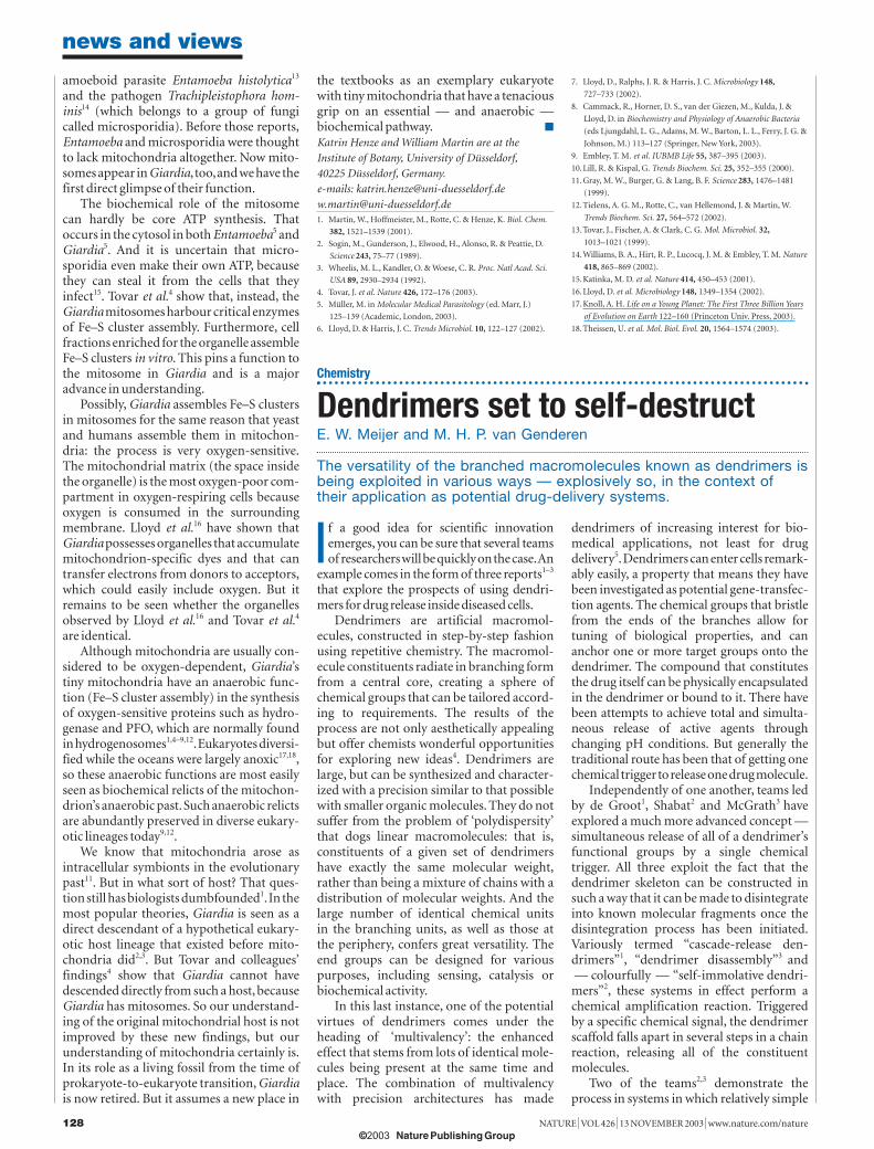
Figure 1 Mitochondria where least expected. Giardia has been the textbook example of a single-celledeukaryote that lacks mitochondria. Now, with the paper by Tovar and colleagues4, it becomes a primeexample of an anaerobic eukaryote that possesses mitosomes — highly reduced mitochondria thatdo not function in core ATP synthesis, but are essential for the assembly of iron–sulphur clusters.The image is about 100,000 times life size.
HO
SSLE
R,C
UST
OM
ME
D.S
TO
CK
PH
OT
O/S
PL
13.11 N&V 127 MH 7/11/03 5:55 pm Page 127
© 2003 NaturePublishing Group
© 2003 Nature Publishing Group
amoeboid parasite Entamoeba histolytica13
and the pathogen Trachipleistophora hom-inis14 (which belongs to a group of fungicalled microsporidia). Before those reports,Entamoeba and microsporidia were thoughtto lack mitochondria altogether. Now mito-somes appear in Giardia, too,and we have thefirst direct glimpse of their function.
The biochemical role of the mitosomecan hardly be core ATP synthesis. Thatoccurs in the cytosol in both Entamoeba5 andGiardia5. And it is uncertain that micro-sporidia even make their own ATP, becausethey can steal it from the cells that theyinfect15. Tovar et al.4 show that, instead, theGiardia mitosomes harbour critical enzymesof Fe–S cluster assembly. Furthermore, cellfractions enriched for the organelle assembleFe–S clusters in vitro. This pins a function tothe mitosome in Giardia and is a majoradvance in understanding.
Possibly, Giardia assembles Fe–S clustersin mitosomes for the same reason that yeastand humans assemble them in mitochon-dria: the process is very oxygen-sensitive.The mitochondrial matrix (the space insidethe organelle) is the most oxygen-poor com-partment in oxygen-respiring cells becauseoxygen is consumed in the surroundingmembrane. Lloyd et al.16 have shown thatGiardia possesses organelles that accumulatemitochondrion-specific dyes and that cantransfer electrons from donors to acceptors,which could easily include oxygen. But itremains to be seen whether the organellesobserved by Lloyd et al.16 and Tovar et al.4
are identical.Although mitochondria are usually con-
sidered to be oxygen-dependent, Giardia’stiny mitochondria have an anaerobic func-tion (Fe–S cluster assembly) in the synthesisof oxygen-sensitive proteins such as hydro-genase and PFO, which are normally foundin hydrogenosomes1,4–9,12.Eukaryotes diversi-fied while the oceans were largely anoxic17,18,so these anaerobic functions are most easilyseen as biochemical relicts of the mitochon-drion’s anaerobic past.Such anaerobic relictsare abundantly preserved in diverse eukary-otic lineages today9,12.
We know that mitochondria arose asintracellular symbionts in the evolutionarypast11. But in what sort of host? That ques-tion still has biologists dumbfounded1.In themost popular theories, Giardia is seen as adirect descendant of a hypothetical eukary-otic host lineage that existed before mito-chondria did2,3. But Tovar and colleagues’findings4 show that Giardia cannot havedescended directly from such a host, becauseGiardia has mitosomes. So our understand-ing of the original mitochondrial host is notimproved by these new findings, but ourunderstanding of mitochondria certainly is.In its role as a living fossil from the time ofprokaryote-to-eukaryote transition, Giardiais now retired. But it assumes a new place in
news and views
128 NATURE | VOL 426 | 13 NOVEMBER 2003 | www.nature.com/nature
the textbooks as an exemplary eukaryotewith tiny mitochondria that have a tenaciousgrip on an essential — and anaerobic — biochemical pathway. ■
Katrin Henze and William Martin are at theInstitute of Botany, University of Düsseldorf,40225 Düsseldorf, Germany.e-mails: [email protected]@uni-duesseldorf.de1. Martin, W., Hoffmeister, M., Rotte, C. & Henze, K. Biol. Chem.
382, 1521–1539 (2001).
2. Sogin, M., Gunderson, J., Elwood, H., Alonso, R. & Peattie, D.
Science 243, 75–77 (1989).
3. Wheelis, M. L., Kandler, O. & Woese, C. R. Proc. Natl Acad. Sci.
USA 89, 2930–2934 (1992).
4. Tovar, J. et al. Nature 426, 172–176 (2003).
5. Müller, M. in Molecular Medical Parasitology (ed. Marr, J.)
125–139 (Academic, London, 2003).
6. Lloyd, D. & Harris, J. C. Trends Microbiol. 10, 122–127 (2002).
Chemistry
Dendrimers set to self-destruct E. W. Meijer and M. H. P. van Genderen
The versatility of the branched macromolecules known as dendrimers isbeing exploited in various ways — explosively so, in the context oftheir application as potential drug-delivery systems.
If a good idea for scientific innovationemerges, you can be sure that several teamsof researchers will be quickly on the case.An
example comes in the form of three reports1–3
that explore the prospects of using dendri-mers for drug release inside diseased cells.
Dendrimers are artificial macromol-ecules, constructed in step-by-step fashionusing repetitive chemistry. The macromol-ecule constituents radiate in branching formfrom a central core, creating a sphere ofchemical groups that can be tailored accord-ing to requirements. The results of theprocess are not only aesthetically appealingbut offer chemists wonderful opportunitiesfor exploring new ideas4. Dendrimers arelarge, but can be synthesized and character-ized with a precision similar to that possiblewith smaller organic molecules. They do not suffer from the problem of ‘polydispersity’that dogs linear macromolecules: that is,constituents of a given set of dendrimershave exactly the same molecular weight,rather than being a mixture of chains with adistribution of molecular weights. And thelarge number of identical chemical units in the branching units, as well as those at the periphery, confers great versatility. Theend groups can be designed for various purposes, including sensing, catalysis or biochemical activity.
In this last instance, one of the potentialvirtues of dendrimers comes under theheading of ‘multivalency’: the enhancedeffect that stems from lots of identical mole-cules being present at the same time andplace. The combination of multivalency with precision architectures has made
dendrimers of increasing interest for bio-medical applications, not least for drugdelivery5.Dendrimers can enter cells remark-ably easily, a property that means they havebeen investigated as potential gene-transfec-tion agents. The chemical groups that bristlefrom the ends of the branches allow for tuning of biological properties, and cananchor one or more target groups onto thedendrimer. The compound that constitutesthe drug itself can be physically encapsulatedin the dendrimer or bound to it. There havebeen attempts to achieve total and simulta-neous release of active agents through changing pH conditions. But generally thetraditional route has been that of getting onechemical trigger to release one drug molecule.
Independently of one another, teams ledby de Groot1, Shabat2 and McGrath3 haveexplored a much more advanced concept —simultaneous release of all of a dendrimer’sfunctional groups by a single chemical trigger. All three exploit the fact that the dendrimer skeleton can be constructed insuch a way that it can be made to disintegrateinto known molecular fragments once thedisintegration process has been initiated.Variously termed “cascade-release den-drimers”1, “dendrimer disassembly”3 and— colourfully — “self-immolative dendri-mers”2, these systems in effect perform achemical amplification reaction. Triggeredby a specific chemical signal, the dendrimerscaffold falls apart in several steps in a chainreaction, releasing all of the constituent molecules.
Two of the teams2,3 demonstrate theprocess in systems in which relatively simple
7. Lloyd, D., Ralphs, J. R. & Harris, J. C. Microbiology 148,
727–733 (2002).
8. Cammack, R., Horner, D. S., van der Giezen, M., Kulda, J. &
Lloyd, D. in Biochemistry and Physiology of Anaerobic Bacteria
(eds Ljungdahl, L. G., Adams, M. W., Barton, L. L., Ferry, J. G. &
Johnson, M.) 113–127 (Springer, New York, 2003).
9. Embley, T. M. et al. IUBMB Life 55, 387–395 (2003).
10.Lill, R. & Kispal, G. Trends Biochem. Sci. 25, 352–355 (2000).
11.Gray, M. W., Burger, G. & Lang, B. F. Science 283, 1476–1481
(1999).
12.Tielens, A. G. M., Rotte, C., van Hellemond, J. & Martin, W.
Trends Biochem. Sci. 27, 564–572 (2002).
13.Tovar, J., Fischer, A. & Clark, C. G. Mol. Microbiol. 32,
1013–1021 (1999).
14.Williams, B. A., Hirt, R. P., Lucocq, J. M. & Embley, T. M. Nature
418, 865–869 (2002).
15.Katinka, M. D. et al. Nature 414, 450–453 (2001).
16.Lloyd, D. et al. Microbiology 148, 1349–1354 (2002).
17.Knoll, A. H. Life on a Young Planet: The First Three Billion Years
of Evolution on Earth 122–160 (Princeton Univ. Press, 2003).
18.Theissen, U. et al. Mol. Biol. Evol. 20, 1564–1574 (2003).
13.11 N&V 127 MH 7/11/03 5:55 pm Page 128
© 2003 NaturePublishing Group
© 2003 Nature Publishing Group
Copyright © 2022 FDOKUMEN