Have Philosophical Accusations Of Talking Nonsense Been Treated With Unmerited Respect?
Upload
independentCategory
view
6download
0
UPF1, a Conserved Nonsense-Mediated mRNA DecayFactor, Regulates Cyst Wall Protein Transcripts in GiardialambliaYi-Hsiu Chen, Li-Hsin Su, Yu-Chang Huang, Yi-Ting Wang, Yu-Yun Kao, Chin-Hung Sun*
Department of Parasitology, College of Medicine, National Taiwan University, Taipei, Taiwan, Republic of China
Abstract
The Giardia lamblia cyst wall is required for survival outside the host and infection. Three cyst wall protein (cwp) genesidentified to date are highly up-regulated during encystation. However, little is known of the molecular mechanismsgoverning their gene regulation. Messenger RNAs containing premature stop codons are rapidly degraded by a nonsense-mediated mRNA decay (NMD) system to avoid production of non-functional proteins. In addition to RNA surveillance, NMDalso regulates thousands of naturally occurring transcripts through a variety of mechanisms. It is interesting to know theNMD pathway in the primitive eukaryotes. Previously, we have found that the giardial homologue of a conserved NMDfactor, UPF1, may be functionally conserved and involved in NMD and in preventing nonsense suppression. In this study, wetested the hypothesis that NMD factors can regulate some naturally occurring transcripts in G. lamblia. We found thatoverexpression of UPF1 resulted in a significant decrease of the levels of CWP1 and cyst formation and of the endogenouscwp1-3, and myb2 mRNA levels and stability. This indicates that NMD could contribute to the regulation of the cwp1-3 andmyb2 transcripts, which are key to G. lamblia differentiation into cyst. Interestingly, we also found that UPF1 may beinvolved in regulation of eight other endogenous genes, including up-regulation of the translation elongation factor gene,whose product increases translation which is required for NMD. Our results indicate that NMD factor could contribute to theregulation of not only nonsense containing mRNAs, but also mRNAs of the key encystation-induced genes and otherendogenous genes in the early-diverging eukaryote, G. lamblia.
Citation: Chen Y-H, Su L-H, Huang Y-C, Wang Y-T, Kao Y-Y, et al. (2008) UPF1, a Conserved Nonsense-Mediated mRNA Decay Factor, Regulates Cyst Wall ProteinTranscripts in Giardia lamblia. PLoS ONE 3(10): e3609. doi:10.1371/journal.pone.0003609
Editor: Jurg Bahler, Wellcome Trust Sanger Institute, United Kingdom
Received July 14, 2008; Accepted October 15, 2008; Published October 31, 2008
Copyright: � 2008 Chen et al. This is an open-access article distributed under the terms of the Creative Commons Attribution License, which permitsunrestricted use, distribution, and reproduction in any medium, provided the original author and source are credited.
Funding: This work was supported by grants from the National Science Council (NSC 96-2320-B-002-040-MY3) and the National Health Research Institutes (NHRI-EX96-9510NC and NHRI-EX97-9510NC) in Taiwan. The funders had no role in study design, data collection and analysis, decision to publish, or preparation of themanuscript.
Competing Interests: The authors have declared that no competing interests exist.
* E-mail: chinhsun@ ntu.edu.tw
Introduction
Giardia lamblia is a major cause of outbreaks of waterborne
diarrheal disease worldwide, which contributes greatly to malnu-
trition and malabsorption in children [1–3]. Like Entamoeba
histolytica and other intestinal protozoan parasites, G. lamblia
undergoes differentiation from a pathogenic trophozoite form
into a resistant infectious cyst form [3,4]. Cyst can survive in the
hostile environment and infect a new host because they have a
resistant extracellular wall.
Because of the importance of the cyst wall, a lot of researches are
focusing on identifying and understanding its key components, cyst
wall proteins (CWPs) [5–7]. Interestingly, three cwp genes identified
to date are highly up-regulated during encystation [5–7]. There is
little understanding of the molecular mechanisms governing their
transcriptional regulation. A Myb family transcription factor (Myb2)
is encystation-induced and is involved in coordinating upregulation
of the cwp1-3 genes [8]. Two GARP family transcription factors may
be involved in transcriptional regulation of many different genes
including the encystation-induced cwp1 gene and constitutive ran
gene [9]. An ARID family transcription factor can bind to specific
AT-rich Inr sequences and function as an important transactivator in
the regulation of the cwp1 gene [10]. However, little is known about
regulation of mRNA stability of the cwp genes during giardial growth
and differentiation.
In late-branching eukaryotes, either a frameshift or a nonsense
mutation often leads to rapid degradation of the gene’s mRNA by
a nonsense-mediated mRNA decay (NMD) pathway [11–13]. This
surveillance system protects cells from the production of non-
functional proteins by eliminating mutant mRNAs. The NMD
pathway is present in yeast, plants, Caenorhabditis elegans and
mammals [11–13]. NMD factors such as up-frameshift 1 (UPF1),
UPF2 and UPF3 have been identified in yeast, C. elegans, mice and
humans [14–19]. They have been shown to interact together and
form a complex [20,21]. Mutations in upf genes stabilize mRNAs
with nonsense mutations [22,23]. UPF1 is one of the most
conserved NMD factors [11–13]. NMD is a translation-dependent
event since its mechanism depends on the recognition of the
nonsense mutations by the translational machinery [13]. Studies
have shown that NMD factors including UPF1 enhance
translation termination at a nonsense codon through interaction
with the termination release factors [20,24–26].
In addition to RNA surveillance, NMD factors also function in
regulating the abundance of some naturally occurring mRNAs
[22,27]. Hundreds or thousands of wild-type NMD targets have
been identified in yeast and humans [28,29]. Most of them are up-
PLoS ONE | www.plosone.org 1 October 2008 | Volume 3 | Issue 10 | e3609

regulated, some of them are down-regulated in upf null mutant
[29].
G. lamblia is of biological interest in understanding the progress of
eukaryotic evolution [30–33]. It has fewer cellular components for
DNA synthesis, transcription and RNA processing, possibly due to
their divergence or their functional redundancy with other proteins
in some pathways [33]. Blast searches of the G. lamblia genome
databases identified matches for UPF1, which is the most conserved
NMD factor but no matches for UPF2 and UPF3 and some other
NMD factors [34]. Interestingly, our previous results showed that: i)
the levels of the nonsense transcripts were lower in G. lamblia; ii) the
aminoglycoside G418 had an inhibiting effect on NMD in G. lamblia,
similar to the effect of aminoglycosides on inhibiting NMD in late-
branching eukaryotes; iii) Giardial UPF1 functioned in reducing the
levels of nonsense-containing transcripts and in enhancing fidelity of
translation termination [34]. Therefore, the NMD phenomenon
could be present in G. lamblia. However, G. lamblia does not have
some of the components of the NMD pathway and the reduction
levels of the nonsense transcripts observed in G. lamblia are lower
than those in late-branching eukaryotes, suggesting that the NMD
system in G. lamblia may be less-functional.
Previously, we have found that the expression of the upf1 gene
was induced in cells expressing the luciferase gene with a nonsense
mutation [34]. In this study, we found that the nonsense mutation
triggered a decrease in cwp1 and cwp2 mRNA levels and there was
a reverse correlation between the expression levels of the cwp1/2
and upf1 mRNA. We also found that overexpression of UPF1
reduced the levels of CWP1 and cyst formation and reduced the
mRNA levels and stability of the cwp1, cwp2, cwp3, and myb2 genes.
In addition, we found that the expression of other five genes was
increased and that of other three genes was decreased by the UPF1
overexpression. For example, the translation elongation factor mRNA
was increased by the UPF1 overexpression. This could be a new
example of an NMD target whose product increases translation
which is required for NMD. Our findings provide new insights
into regulation of the giardial cyst wall protein genes and other
endogenous genes.
Results
NMD can be monitored by a constitutive promotersystem
NMD is related to the presence of a premature stop codon or
not [11–13]. We have tested NMD effect using a luciferase gene
with or without a premature stop codon under the control of the
encystation-induced cwp1 promoter [34]. We found that NMD
could be present in G. lamblia because the results showed that the
mRNA produced from the luciferase gene with a stop codon under
the control of the cwp1 promoter was decayed compared with wild
type luciferase mRNA [34]. We further used a constitutive
promoter to test the NMD. We prepared puromycin-based
constructs pPT5 and pPT5m in which the wild type luciferase
gene (luc+) and the luciferase gene with a stop codon (luc+m) are
controlled by the a2-tubulin promoter, respectively (Fig. 1A), and
stably transfected them into G. lamblia. The level of luc+m activity
in the pPT5m cell line was reduced by approximately 5,000-fold,
relative to that of the wild-type luc+ in the pPT5 cell line (Fig. 1B),
indicating that luc+m could be non-functional. This result is
similar to the data we previously reported for the luc+m under the
control of the cwp1 promoter [34]. The level of luciferase mRNA
was lower by ,50% (P,0.05) in the pPT5m transfectants
compared with that in the pPT5 transfectants (Fig. 1C, lanes 1
and 2), indicating that the presence of a nonsense mutation in
luc+m triggered a decrease in mRNA levels (NMD). Therefore,
NMD can be monitored by a constitutive promoter system. The
results from the a2-tubulin and the cwp1 promoters [34] similarly
indicate that NMD could be present in G. lamblia. As a control, the
ran mRNA levels did not change in the pPT5m transfectants
compared with the pPT5 transfectants (Fig. 1C, lanes 1 and 2).
Reverse correlation between the expression level of thecwp1/2 and upf1 mRNA
Previously we found that the nonsense mutation triggered an
increase in upf1 mRNA levels [34]. In this study, we also found
that the levels of the upf1 mRNA increased by ,2-fold (P,0.05) in
the pPT5m cell line compared with the pPT5 cell line (Fig. 1C).
Interestingly, the levels of the cwp1 and cwp2 mRNA were lower by
,30% (P,0.05) in the pPT5m cell line compared with the pPT5
cell line (Fig. 1C). Similarly decreased cwp1 or cwp2 mRNA levels
were also detected in the pPW1m cell line that expressed the
luciferase gene with a premature stop codon under the control of the
cwp1 promoter (data not shown) [34].
Previously we found that the levels of the upf1 mRNA decreased
with increasing levels of the cwp1 and cwp2 mRNA during
encystation [34]. We also demonstrated that stable transfection
systems can increase the levels of the cwp1, cwp2 and cwp3
transcripts during vegetative growth [35]. We further asked
whether stable transfection influenced the upf1 gene expression.
We found that the level of the upf1 mRNA decreased by ,50–70%
(P,0.05) in cells stably transfected with the pRANneo or 59D5N-
Pac, which contain the neomycin or puromycin selective marker,
relative to untransfected cells (Fig. 2). As reported previously, the
levels of the cwp1/2 mRNA increased significantly and the levels of
the ran and ribosomal mRNA did not change significantly in the
transfected cell lines (Fig. 2) [35].
Overexpression of UPF1 reduced the levels of cyst wallprotein 1 and cyst formation
Because of the reverse correlation between the levels of the cwp1/2
and upf1 mRNAs as described above, we further investigated the
effect of giardial UPF1 on cyst formation. We stably transfected the
UPF1 expression plasmid pPUPF1HA into G. lamblia (Fig. 3A). The
UPF1-HA protein was expressed in the stable transfectants as
detected in immunofluorescence assays [34]. No significant change
in growth rate was observed in the pPUPF1HA cell line (data not
shown). We found that the cyst number in the vegetative or encysting
pPUPF1HA cell line decreased by ,30% or ,50% (P,0.05)
relative to the control cell line which only carries the pac gene
(59D5N-Pac) (Fig. 3B). We further asked whether the levels of cyst
wall protein 1 decreased with the decrease of the cyst formation. As
shown by Western blot analysis, UPF1 overexpression also resulted
in a reduction of the levels of CWP1 protein (Fig. 3C). As a control,
similar levels of intensity of the giardial RAN protein (,30 kDa)
were detected by anti-human RAN antibody (Fig. 3C). The results
suggest that UPF1 may function in reducing the level of CWP1 and
cyst formation.
Overexpression of UPF1 decreased cwp1-3 and myb2mRNA stability
We further investigated whether UPF1 overexpression can
influence the expression of cwp and other endogenous genes. We
found that the levels of native cwp1, cwp2, cwp3, or myb2 mRNA in
the vegetative pPUPF1HA cell line decreased by ,85% or ,50%
(P,0.05) relative to the control cell line which only carries the pac
gene (Fig. 4A). The ran mRNA levels did not change significantly in
the pPUPF1HA cell line (Fig. 4A). The upf1 mRNA levels increased
significantly (P,0.05) in the pPUPF1HA cell line (Fig. 4A).
upf1 Regulates Giardia mRNAs
PLoS ONE | www.plosone.org 2 October 2008 | Volume 3 | Issue 10 | e3609
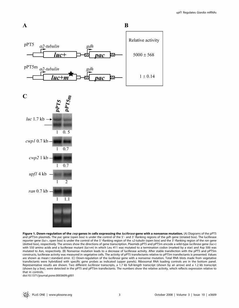
Figure 1. Down-regulation of the cwp genes in cells expressing the luciferase gene with a nonsense mutation. (A) Diagrams of the pPT5and pPT5m plasmids. The pac gene (open box) is under the control of the 59- and 39-flanking regions of the gdh gene (striated box). The luciferasereporter gene (luc+, open box) is under the control of the 59-flanking region of the a2-tubulin (open box) and the 39-flanking region of the ran gene(dotted box), respectively. The arrows show the directions of gene transcription. Plasmids pPT5 and pPT5m encode a wild-type luciferase gene (luc+)with 550 amino acids and a luciferase mutant (luc+m) in which Leu 411 was mutated to a termination codon (marked by a star) and Asp 500 wasmutated to Asn, respectively. (B) Nonsense mutation leads to a decrease of luciferase activity. After stable transfection with the pPT5 and pPT5mconstructs, luciferase activity was measured in vegetative cells. The activity of pPT5 transfectants relative to pPT5m transfectants is presented. Valuesare shown as mean6standard error. (C) Down-regulation of the luciferase gene with a nonsense mutation. Total RNA blots made from vegetativetransfectants were hybridized with specific gene probes as indicated (upper panels). Ribosomal RNA loading controls are in the bottom panel.Representative results are shown. Two different luciferase transcripts, a 1.7 kb full-length transcript (shown by an arrow) and a 1.2 kb transcript(shown by a line), were detected in the pPT5 and pPT5m transfectants. The numbers show the relative activity, which reflects expression relative tothat in controls.doi:10.1371/journal.pone.0003609.g001
upf1 Regulates Giardia mRNAs
PLoS ONE | www.plosone.org 3 October 2008 | Volume 3 | Issue 10 | e3609

We further investigated whether the decrease of cwp mRNA
levels in the pPUPF1HA transfectants was due to the decrease of
mRNA stability. We found that treatment of the pPUPF1HA
transfectants with 45 mg/ml of actinomycin D for 13 min
decreased mRNA levels of the cwp1 and cwp2 genes by ,90%
(P,0.05) (Fig. 4C). As a control, treatment of the control cell line
with actinomycin D for 13 min decreased mRNA levels of the
cwp1 and cwp2 genes by 30–40% (P,0.05) (Fig. 4B). Therefore, the
cwp1 and cwp2 mRNAs exhibited a half-life of ,13 min when the
upf1 was overexpressed, while these mRNAs exhibited a half-life of
.13 min in the control cell line. The cwp3 and myb2 mRNA
stability also decreased significantly in the pPUPF1HA transfec-
tants as compared with that in the control cell line (data not
shown). The ran mRNA stability did not change significantly in the
pPUPF1HA transfectants as compared with that in the control cell
line (Fig. 4C). The results indicate that UPF1 can decrease cwp1-3
and myb2 mRNA stability.
Overexpression of UPF1 changed transcript levels ofother endogenous genes
In the previous studies, we have found that the expression of the
cwp1, cwp2, myb2 genes was upregulated in stable cell line with
drug selection and that the expression of other eight genes was also
upregulated [35]. They encodes enzymes involved in anaerobic
glycolysis, phosphoglycerate kinase (PGK) and glyceraldehyde-3-
phosphate dehydrogenase (G3PD), enzymes for arginine hydroly-
sis, ornithine carbamoyltransferase (OCT) and carbamate kinase
(CK), enzymes involved in protein folding, cyclophilin (CY), co-
chaperone-like protein p21, and Bip, and open reading frame
(ORF) 16424 with unknown function [1,35,36]. We wished to
understand the importance of UPF1 for expression of these genes.
To achieve this goal, we compared expression of these genes in the
UPF1 overexpressed cell line and the control cell line. Of these
eight genes, three were upregulated by UPF1 overexpression,
including cy, p21, and bip (P,0.05) (Fig. 5). Interestingly, one gene
was downregulated by UPF1 overexpression (g3pd, P,0.05)
(Fig. 5). The transcript levels of the other four genes, pgk, oct, ck,
and orf 16424, were not changed significantly by UPF1
overexpression (Fig. 5). We also found that two other genes with
no change in mRNA levels in the stable cell lines, glutamate
dehydrogenase (gdh), and thioredoxin peroxidase (tp), were
downregulated by UPF1 overexpression (P,0.05) (Fig. 5). In
addition, we found two newly identified genes, translation elongation
factor (tef, c subunit of translation elongation factor 1B) [37] and
arginine deiminase (ad) [1], were upregulated by UPF1 overexpression
(P,0.05) (Fig. 5).
Figure 2. Reverse correlation between the expression levels ofthe cwp1/2 and upf1 mRNA in stable cell lines. Diagrams of thepRANneo and 59D5N-Pac plasmids for stable transfection are shown ininset. The neo or pac gene (open box) is under the control of the 59- and39-flanking regions of the ran (dotted box) or gdh gene (striated box).Total RNA blots made from vegetative untransfected cells (UT),pRANneo or 59D5N-Pac transfectants were hybridized with specificprobes as indicated. Hybridization signals were imaged and quantifiedas indicated in Materials and Methods. The results are expressed asrelative expression level over untransfected control. Values are shownas mean6standard error.doi:10.1371/journal.pone.0003609.g002
Figure 3. Overexpression of UPF1 reduced the levels of CWP1and cyst formation. (A) Diagrams of the pPUPF1HA plasmid. The pacgene (open box) expression cassette is the same as in Fig. 2. The upf1gene is under the control of its own 59-flanking region (open box) andthe 39-flanking region of the ran gene (dotted box). The filled black boxindicates the coding sequence of the HA epitope tag. (B) Overexpres-sion of UPF1 reduced the levels of cyst formation. The 59D5N-Pac andpPUPF1HA stable transfectants were cultured in growth medium to latelog/early stationary phase (Veg). Cyst count was performed on theselate log/early stationary phase cultures (1.56106 cells/ml). In anotherstudy, the 59D5N-Pac and pPUPF1HA stable transfectants were culturedin encystation medium for 24 h and then subjected to cyst count (Enc).The sum of total cysts is expressed as relative expression level overcontrol. Values are shown as mean6standard error. (C) Overexpressionof UPF1 reduced the CWP1 level. The 59D5N-Pac and pPUPF1HA stabletransfectants were cultured in encystation medium for 24 h and thensubjected to SDS-PAGE and Western blot. The blot was probed by anti-CWP1 antibody. Equal amounts of proteins loaded were confirmed bydetection of giardial RAN protein. Representative results are shown.doi:10.1371/journal.pone.0003609.g003
upf1 Regulates Giardia mRNAs
PLoS ONE | www.plosone.org 4 October 2008 | Volume 3 | Issue 10 | e3609

Overexpression of UPF1 decreased mRNA levels ofvector-expressed cwp1 gene
We next tested whether overexpression of UPF1 influenced
vector-expressed cwp1 gene. We stably transfected the UPF1
expression plasmid pNUPF1HA (Fig. 6A) together with construct
pPC1 in which the cwp1 gene is controlled by its own promoter
and contains an AU5 epitope tag at their C terminus (Fig. 6A).
The CWP1-AU5 protein was expressed in the stable transfectants
as detected in immunofluorescence assays and Western blot
analysis (data not shown). Northern blot analysis showed that the
levels of the cwp1-au5 mRNA decreased by ,80% (P,0.05) in the
pPC1+pNUPF1HA co-transfectants relative to the pPC1+pRAN-
neo control cell line during vegetative growth (Fig. 6B). The levels
of the cwp1 (including endogenous cwp1 and vector-derived cwp1-
au5) and cwp2 mRNA also decreased by ,60–70% (P,0.05) in the
UPF1 overexpressed cell line, pPC1+pNUPF1HA (Fig. 6B). The
results indicate that overexpression of UPF1 not only can decrease
the expression of the endogenous cwp genes but also can decrease
the expression of the vector-expressed cwp1 gene.
Mapping of the region in the cwp1 gene needed for theUPF1 dependent decay
We further determined the region within the cwp1 mRNA
responsible for the UPF1 dependent decay by constructing a series
of deletions (Fig. 7A). Deletion of the sequence encoding the first,
second or third to fourth LRRs (nucleotides 193–264, construct
D2; nucleotides 265–345, construct D3; nucleotides 346–474,
construct D4, Fig. 7A) still resulted in a significant decrease of
cwp1-au5 mRNA in the pNUPF1HA co-transfectants relative to
the control cell line (Fig. 7B). However, deletion of the sequence
encoding the region N terminal to the LRRs (nucleotides 4–192,
construct D1, Fig. 7A), the fifth LRR (nucleotides 475–552,
construct D5, Fig. 7A) or the region C terminal to the LRRs
(nucleotides 553–723, construct D6, Fig. 7A) resulted in an
Figure 4. Effect of UPF1 overexpression on cwp gene expression. (A) Overexpression of UPF1 decreased the cwp1-3 and myb2 mRNA levels.Total RNA was harvested from vegetative 59D5N-Pac and pPUPF1HA transfectants. Northern blots were hybridized with specific probes as indicated(upper panels). (B)(C) Overexpression of UPF1 decreased the cwp1 and cwp2 mRNA stability. Total RNA was harvested from either 59D5N-Pac (B) orpPUPF1HA (C) transfectants during vegetative growth. The cells were treated without (0 min) or with 45 mg/ml actinomycin D for 13 min to arrestmRNA synthesis. Northern blot were hybridized with specific gene probes as indicated (upper panels). Ribosomal RNA loading controls are in thebottom panels. Representative results are shown. The numbers show the relative activity, which reflects expression relative to that in controls. Thecwp1 and cwp2 signals from Fig. 4B and C were a long exposure to show the difference in the AcD treated and untreated samples.doi:10.1371/journal.pone.0003609.g004
upf1 Regulates Giardia mRNAs
PLoS ONE | www.plosone.org 5 October 2008 | Volume 3 | Issue 10 | e3609

increase of the levels of the cwp1-au5 mRNA to ,1.4 or 1.7-fold
(P,0.05) in the pNUPF1HA co-transfectants relative to the
control cell line (Fig. 7B). The results indicate that 59 (nucleotides
4–192) or 39 (nucleotides 475–723) sequence of the cwp1 gene may
contain the sequence responsible for the UPF1 dependent decay.
Discussion
In addition to RNA surveillance, NMD factors also function in
regulating the abundance of some naturally occurring mRNAs
[23,28,38]. Hundreds or thousands of wild-type NMD targets that
may be up-regulated or down-regulated by NMD factors have
been identified in yeast and humans [28,29,39]. Their changes in
mRNA abundance may be correlated (direct targets of NMD) or
not correlated (indirect targets of NMD) with changes in the
mRNA stability [28,39]. For example, abundance of one subset of
wild type mRNAs increased in upf1 null mutant, including PPR1,
URA1, URA3, and URA4 mRNAs [28,40]. However, only PPR1
is the direct target of NMD because only PPR1 mRNA stability
increased in upf1 null mutant [28,40]. PPR1 is a positive
transcriptional activator for these URA genes. Altered half-life of
this regulatory protein could indirectly influence the abundance of
the mRNAs of the downstream targets (URA genes) [28,40]. An
UPF1 dependent destabilizing element (UDE) was mapped to a
region located within the 59-untranslated region and the first 92
bases of the PPR1 coding region [40]. Similarly, one of the UDEs
was mapped to a region located within the 59 (nucleotides 4–192)
sequence of the cwp1 gene.
In G. lamblia, we identified the four encystation-induced genes,
cwp1, cwp2, cwp3, and myb2, as wild-type targets of NMD. These
four genes may be the direct targets of NMD because
overexpression of UPF1 led to decreased levels of their mRNA
stability. In addition, UDEs were mapped to a region located
within the 59 (nucleotides 4–192) or 39 (nucleotides 475–723)
sequence of the cwp1 gene. It is possible that the cwp2, cwp3 and
myb2 genes also have UDEs and that their mRNAs are targeted by
Figure 5. Effect of UPF1 overexpression on endogenous geneexpression. Total RNA blots made from the 59D5N-Pac and pPUPF1HAtransfectants were hybridized with specific gene probes as described.Equal RNA loading was confirmed by ethidium bromide staining ofribosomal RNA (data not shown). Representative results are shown. Thenumbers show the relative activity, which reflects expression relative tothat in controls.doi:10.1371/journal.pone.0003609.g005
Figure 6. Overexpression of UPF1 decreased mRNA levels ofvector-based cwp1 gene. (A) Diagrams of the pPC1 and pNUPF1HAplasmids. The neo or pac gene (open box) expression cassette is the sameas in Fig. 2. In pPC1, the cwp1 gene (open boxes) is flanked by its own 59-flanking region (gray box) and 39-flanking region of the ran gene (dottedbox) and the filled black box indicates the coding sequence of the AU5epitope tag. In pNUPF1HA, the upf1 gene is under the control of its ownpromoter (open box) and the 39-flanking of the ran gene (dotted box) andthe filled black box indicates the coding sequence of the HA epitope tag.(B) Northern blot analysis of au5 tagged cwp1 transcripts in vegetativecells (upper panel). Total RNA blots made from pPC1+pRANneo orpPC1+pNUPF1HA transfectants were hybridized with the au5 specificprobe (au5hyb), and specific gene probes as indicated (upper panels).Ribosomal RNA loading controls are in the bottom panel. Representativeresults are shown. The numbers show the relative activity, which reflectsexpression relative to that in controls.doi:10.1371/journal.pone.0003609.g006
upf1 Regulates Giardia mRNAs
PLoS ONE | www.plosone.org 6 October 2008 | Volume 3 | Issue 10 | e3609

UPF1-dependent decay. UPF1 may coordinately down-regulate
the encystation-induced cwp1-3 and myb2 genes during vegetative
growth. Because Myb2 acts as a positive transcriptional activator for
these cwp genes [8], a decrease of the level of the myb2 transcripts that
may lead to a decrease of the levels of the Myb2 protein and then
results in a decrease of the cwp transcripts. In addition, we have
shown that the levels of UPF1 decreased significantly during
encystation [34]. During encystation, the up-regulation of these
encystation-induced genes may be correlated with the presence of
the lower level of UPF1 protein. Interestingly, our results show that
the levels of the cwp1 and cwp2 mRNAs were lower in the cells
containing luciferase nonsense transcripts. This could be due to the
down-regulation of the cwp genes by the presence of the higher level
of UPF1 protein upon NMD induction.
It is interesting to know how NMD factors recognize their
targets. In yeast, it has been thought that NMD promotes rapid
decay of the nonsense-containing mRNA through interaction of a
RNA-binding protein(s) with specific RNA elements [12].
Heterogeneous nuclear ribonucleoprotein 1 (HRP1) may be a
marker protein that binds to the downstream element of the
nonsense mutation and interacts with NMD factors [12]. On the
other hand, wild-type mRNAs without premature stop codon were
also regulated by a UPF1-dependent mechanism [38,41]. Four
natural targets for an RNA-binding protein, Stau1, were identified
in humans, including ADP ribosylation factor 1, c-JUN,
SERPINE1, IL7R, and GAP43 mRNAs. Stau1 can bind to the
39-untranslated region of these targets’ mRNAs for Stau1-
mediated mRNA decay, which depends on translation and
Figure 7. Mapping of the region in the cwp1 gene needed for the UPF1 dependent decay. (A) Diagrams of the pPC1 and plasmids forCWP1 deletion mapping. The pac or cwp1 gene (open box) expression cassette is the same as in Fig. 2 and Fig. 6, respectively. The predicted signalpeptide is in gray. The LRRs are indicated as open boxes labeled ‘‘L’’. (B). Analysis of au5 tagged cwp1 transcripts in vegetative cells. The pPC1D1-6+pRANneo and pPC1D1-6+pNUPF1HA transfectants were cultured in encystation medium for 24 h and then subjected to Northern blot analysis.Total RNA blots were hybridized with the au5 specific probe (au5hyb), and ran gene probe (upper panels). Ribosomal RNA loading controls are in thebottom panel. Representative results are shown. The numbers show the relative activity, which reflects expression relative to that in controls.doi:10.1371/journal.pone.0003609.g007
upf1 Regulates Giardia mRNAs
PLoS ONE | www.plosone.org 7 October 2008 | Volume 3 | Issue 10 | e3609

recruitment of the NMD factor UPF1 [38,41]. Short untranslated
regions are typical of giardial transcripts [3]. Interestingly, one of
the UDEs was mapped to a region located within the 39
(nucleotides 475–723) sequence of the cwp1 gene. It is possible
that G. lamblia may have similar RNA binding proteins as marker
proteins for abnormal or natural NMD targets. Blast searches of
the G. lamblia genome databases identified a match for HRP1,
which has five RNA binding domains [34]. However, it is very
different from the yeast HRP1, which has two RNA binding
domains. Further studies will be required to identify the RNA
binding proteins and elucidate their roles in NMD.
In addition to encystation-induced genes, we also found that
UPF1 may be involved in regulating transcripts of many different
genes. Overexpression of UPF1 led to enhanced levels of five of
twelve genes, including cy, p21, bip, tef and ad. Three genes were
downregulated, including g3pd, gdh, and tp. The other four genes
tested were not changed, including pgk, oct, ck, and orf 16424. The
affected genes encode proteins involved in protein translation
(TEF) [37], protein folding (CY, p21, and Bip) [35], cell
metabolism (AD, G3PD, GDH, and TP) [1,36]. They may be
direct or indirect targets of NMD and this requires further
investigation.
NMD may increase translation accuracy because NMD factors
including UPF1-3 could enhance translation termination at a
nonsense codon through interaction with the termination release
factors in yeast [20,24–26]. NMD requires translation since its
mechanism depends on the recognition of the nonsense mutations
by the translational machinery [13]. NMD factors including
UPF1-3 also function in stimulating translation in human [42]. In
addition, NMD factors including UPF1-3 may increase translation
through down-regulation of Ebs1p, which is a global inhibitor of
translation in yeast [43]. This occurs without a change in the EBS1
mRNA stability, indicating that EBS1 is an indirect target of NMD
and it may be targeted by NMD-regulated transcription factors. In
this study, we also found that UPF1 may be involved in
upregulating transcripts of translation elongation factor (tef, c subunit
of translation elongation factor 1B) [37]. This suggests that the
giardial NMD factors may function in enhancing translation
through upregulation of tef. Therefore, we provided a new
example of an NMD target whose product increases translation
initiation which is required for NMD.
Our results indicate that NMD can affect some endogenous
genes involved in differentiation, metabolism, and protein
translation and folding in the early-diverging protozoan G. lamblia.
Our findings also lead to greater understanding of the regulation
of mRNA stability of the genes involved in cyst wall pathway and
provide a model to investigate the mechanism of cell differenti-
ation.
Materials and Methods
G. lamblia cultureTrophozoites of G. lamblia WB (ATCC 30957) clone C6 were
cultured in modified TYI-S33 medium [44] and encysted as
previously described [7]. Cyst count was performed on vegetative
cultures as previously described [35]. Cyst count was also
performed on 24 h encysting cultures. In experiments exposing
G. lamblia vegetative trophozoites to actinomycin D, trophozoites
were cultured in medium 45 mg/ml actinomycin D (in PBS) for
indicated time in the legends of Figures 4B and C.
RNA extraction and Northern blot analysisTotal RNA was extracted from G. lamblia clones C6 at the
indicated differentiation stages in the legends of Figures 1, and 3–7
using TRIzol reagent (Invitrogen). For Northern blot analysis,
10 mg total RNA was fractionated and transferred to charged
Nylon membranes (Biodyne B membrane, Pall). Full-length coding
region probes of luciferase, cwp1 (GenBank accession
no. U09330), cwp2 (GenBank accession no. U28965), cwp3
(GenBank accession no. AY061927), myb2 (GenBank accession
no. AY082882), ran (GenBank accession no. U02589), upf1
(GenBank accession no. DQ861427), phosphoglycerate kinase (pgk,
GenBank accession no. for genomic DNA XM_762975),
glyceraldehyde-3-phosphate dehydrogenase (g3pd, GenBank accession no.
for genomic DNA M88062), ornithine carbamoyltransferase (oct,
GenBank accession no. for genomic DNA XM_765341), carbamate
kinase (ck, GenBank accession no. for genomic DNA
XM_765099), orf 16424 (GenBank accession no. for genomic
DNA XM_764168; orf 16424 in G. lamblia genome database,
http://www.giardiadb.org/giardiadb/)(Morrison et al., 2007),
cyclophilin (cy, GenBank accession no. for genomic DNA
XM_774688), p21 (GenBank accession no. for genomic DNA
XM_762782, for protein XP_767875), bip (GenBank accession
no. for genomic DNA XM_766560, for protein XP_771653),
glutamate dehydrogenase (gdh, GenBank accession no. for genomic
DNA XM_773614), thioredoxin peroxidase (tp, GenBank accession
no. for genomic DNA XM_774576)[36], c subunit of translation
elongation factor 1B (tef, GenBank accession no. for genomicDNA XP_778603), and arginine deiminase (ad, GenBank acces-sion no. for genomic DNA U49236) genes were prepared by
PCR amplification of genomic DNA using primers lucF (ATGG-
AAGACGCCAAAAAC) and lucR (TTACACGGCGATCTTT-
CC), cwp1F (ATGATGCTCGCTCTCCTT) and cwp1R (TCA-
AGGCGGGGTGAGGCA), cwp2F (ATGATCGCAGCCCTT-
GTT) and cwp2R (TCACCTTCTGCGGACAAT), cwp3F
(ATGTTTTCTCTGCTTCTT) and cwp3R (TTATCTGTAG-
TAGGGCGG), myb2F (ATGTTACCGGTACCTTCT) and
myb2R (TCAGGGTAGCTTCTCACG), ranF (ATGTCTGA-
CCCAATCAGC) and ranR (TCAATCATCGTCGGGAAG),
upf1F (ATGGAGCCTTGTGCATTG) and upf1R (CTATGCC-
TTAGGAATTAC), pgkF (ATGTCCTTAGCGAAGCTCTCC)
and pgkR (CTTCTTGTCAGACAGTCTGAT), g3pdF (ATGC-
CTATTCGCCTCGGAATC) and g3pdR (GCAGCCCTTG-
GACCCGACGTA), octF (ATGCCGTTCAAGCAGACCCGC)
and octR (CTCCATCTTGCAGTCATGCAA), ckF (ATGTC-
GGCAGGGAAAACGGTT) and ckR (ATCCTTGATGATG-
CGGGTCCC), 16424F (CACCATGAGTAGAACGCCAAAC)
and 16424R (GTAGCGACGATTACCGGA), cyF (ATGAAC-
TCTCCAGTTTCTGAC) and cyR (CTGGAGCACGCCA-
CAGTCGGC), p21F (ATGCACCATCCGACGATCTA) and
p21R (CTCCTCTGCCTTCTCTTCGCC), bipF (ATGACGT-
CTAGTCACGTTAA) and bipR (GAGTTCATCTTTTTCTG-
CAT), gdhF (ATGCCTGCCCAGACGATCGAG) and gdhR
(CACGCAGCCCTGCTCGATCAT), tpF (ATGCCCGTCCC-
CATCCCCGGC) and tpR (CTTCTTGAACGTCTTGGA-
GAA), tefF (ATGCAGATCACAGGCAGTCAG) and tefR
(CTAGTGCCAAGTCTCCCCATC), adF (ATGACTGACT-
TCTCCAAGGAT) and adR (TCACTTGATATCGACGCA-
GAT), respectively. Radiolabeled probes were prepared using
the Rediprime II kit (GE Healthcare). An oligonucleotide probe
complementary to the AU5 tag coding sequence and its flanking
region (au5hyb, GAATTCTCACTTGAGGTAGAAATCGG-
TAGGCGGGGTGAGG, AU5 tag coding sequence is under-
lined) was end-labeled using [c-32P] ATP and T4 polynucleotide
kinase. The membranes were hybridized and washed as previously
described [45]. Equal loading was confirmed by reprobing the
Northern blots with radiolabeled ribosomal DNA. The ribosomal
DNA fragment for large subunit ribosomal RNA (X05397) was
upf1 Regulates Giardia mRNAs
PLoS ONE | www.plosone.org 8 October 2008 | Volume 3 | Issue 10 | e3609

amplified by PCR using primers RIBOF (GGCCTGCCCC-
TCGCCCGC) and RIBOR (CCCCTCAGTCCTCCGGGG)
and a genomic DNA template. Radiolabeled ribosomal DNA
probes were prepared as described above. For detection, the blots
were exposed to a storage phosphor screen and the radioactive
signals were quantitated using a Typhoon TrioTM Variable Mode
Imager (GE Healthcare). Two independently generated stably
transfected lines were made from each construct and each of these
cell lines was assayed three separate times. The results are
expressed as relative expression level over control. Student’s t-tests
were used to determine statistical significance of differences
between samples.
Plasmid constructionAll constructs were verified by DNA sequencing with a BigDye
Terminator 3.1 DNA Sequencing kit and an ABI 3100 DNA
Analyser (Applied Biosystems). Plasmid 59D5N-Pac was a gift from
Dr. Steven Singer and Dr. Theodore Nash [46]. Plasmids
pRANneo, pPW1, pPW1m, pPC1, pPUPF1HA, and pNUPF1HA
have been described previously [7,34,35,47]. A NheI/ClaI
fragment containing the luciferase gene, 32-bp ran promoter and
two copies of a 19-bp tet operator sequence from pPop2N [34] was
replaced by the NheI/ClaI excised luciferase gene and a2-tubulin
promoter from pNT5, resulting in pPT5. An NcoI/ClaI fragment
containing the wild type luciferase gene from pPT5 [34] was
replaced by the NcoI/ClaI excised mutated luciferase gene from
pPW1m, resulting in pPT5m. For constructing pPC1D1, a PCR
with oligonucleotide cwp1D1NF (GGCGCCATGGATGCCCT-
GGATCTTTCGGACATG) and ran3C (GCGGATCGATG-
TAACGAACCGCTAGAAG) generated a 0.8-kb product that
was digested with NcoI and ClaI. Another PCR with primers T3
(ATTAACCCTCACTAAAG) and cwp1D1NR (GGCGCCAT-
GGCCCTGATATTTTATTTCTGTG) generated a 0.3-kb PCR
product that was digested with NcoI and NheI and cloned into
NheI/ClaI digested pPop2N with the 0.8-kb NcoI/ClaI fragment.
The resulting pPC1D1 contains a cwp1 gene lacking the coding
sequence for the predicted signal peptide sequence and the
sequence N terminal to LRRs (nucleotides 4–192). For construct-
ing pPC1D2, a PCR with oligonucleotide cwp1D2BF (GGCGG-
GATCCACCCTTTACTTGAGCAACAAC) and ran3C gener-
ated a 0.6-kb product that was digested with BamHI and ClaI.
Another PCR with primers T3 and cwp1D2BR (GGCGGG-
ATCCGATAACGTAGTTATTCGAGGC) generated a 0.4-kb
PCR product that was digested with BamHI and NheI and cloned
into NheI/ClaI digested pPop2N with the 0.6-kb BamHI/ClaI
fragment. The resulting pPC1D2 contains a cwp1 gene lacking the
coding sequence for the first LRR (nucleotides 193–264). Similar
strategy was used to constructs pPC1D3, pPC1D4, pPC1D5, and
pPC1D6, which contain cwp1 gene with deletion of the coding
sequence for the second LRR (nucleotides 265–345), third to
fourth LRR (nucleotides 346–474), fifth LRR (nucleotides 475–
552), and the sequence C terminal to LRRs (nucleotides 553–723),
respectively.
Transfection, luciferase assay, and Western blot analysisCells transfected with pN series plasmids were selected with
G418 as described [47]. Stable transfectants were maintained at
150 mg/ml G418. Cells transfected with pP series plasmids
containing the pac gene were selected and maintained with
54 mg/ml puromycin. For co-transfection assays (see Figs. 6 and
7), G. lamblia cells were first transfected with pP series plasmids and
selected in 54 mg/ml puromycin. The stable transfectants were
transfected with pN series plasmids and then the cells were doubly
selected in both 150 mg/ml G418 and 54 mg/ml puromycin. After
stable transfection with specific constructs, luciferase activity was
determined in vegetative cells at late log/stationary phase
(1.56106 cells/ml) or in 24 h encysting cells as described [45]
and was measured with an Optocomp I luminometer (MGM
Instruments). Two independently generated stably transfected
lines were made from each construct and each of these lines was
assayed three separate times. Western blots were probed with anti-
CWP1 antibody (1/10,000) [48] or anti-human RAN antibody (1/
5,000) (Santa Cruz Biotechnology), and detected with peroxidase-
conjugated goat anti-mouse IgG (Pierce, 1/5,000) and enhanced
chemiluminescence (GE Healthcare).
Acknowledgments
We thank the researchers and administrators of the G. lamblia Genome
database. We also thank Dr. Ming-Shyue Lee, Dr. Tsai-Kun Li, and Dr.
Chien-Kuo Lee for helpful comments, and Ms Yi-Li Liu and I-Ching
Huang and Mr. Chao-Cheng Cho for technical support.
Author Contributions
Conceived and designed the experiments: CHS. Performed the exper-
iments: YHC LHS YCH YTW YYK. Analyzed the data: YHC CHS.
Contributed reagents/materials/analysis tools: CHS. Wrote the paper:
CHS.
References
1. Wolfe MS (1992) Giardiasis. Clin Microbiol Rev 5: 93–100.
2. Marshall MM, Naumovitz D, Ortega Y, Sterling CR (1997) Waterborne
protozoan pathogens. Clin Microbiol Rev 10: 67–85.
3. Adam RD (2001) Biology of Giardia lamblia. Clin Microbiol Rev 14:
447–475.
4. Ondarza RN (2007) Drug targets from human pathogenic amoebas: Entamoeba
histolytica, Acanthamoeba polyphaga and Naegleria fowleri. Infect Disord Drug Targets
7: 266–280.
5. Lujan HD, Mowatt MR, Conrad JT, Bowers B, Nash TE (1995) Identification
of a novel Giardia lamblia cyst wall protein with leucine-rich repeats. Implications
for secretory granule formation and protein assembly into the cyst wall. J Biol
Chem 270: 29307–29313.
6. Mowatt MR, Lujan HD, Cotton DB, Bower B, Yee J, et al. (1995)
Developmentally regulated expression of a Giardia lamblia cyst wall protein
gene. Mol Microbiol 15: 955–963.
7. Sun CH, McCaffery JM, Reiner DS, Gillin FD (2003) Mining the Giardia lamblia
genome for new cyst wall proteins. J Biol Chem 278: 21701–21708.
8. Sun CH, Palm D, McArthur AG, Svard SG, Gillin FD (2002) A novel Myb-
related protein involved in transcriptional activation of encystation genes in
Giardia lamblia. Mol Microbiol 46: 971–984.
9. Sun CH, Su LH, Gillin FD (2006) Novel plant-GARP-like transcription factors
in Giardia lamblia. Mol Biochem Parasitol 146: 45–57.
10. Wang CH, Su LH, Sun CH (2007) A novel ARID/Bright-like protein involved
in transcriptional activation of cyst wall protein 1 gene in Giardia lamblia. J Biol
Chem 282: 8905–8914.
11. Nagy E, Maquat LE (1998) A rule for termination-codon position within intron-
containing genes: when nonsense affects RNA abundance. Trends Biochem Sci
23: 198–199.
12. Gonzalez CI, Bhattacharya A, Wang W, Peltz SW (2001) Nonsense-mediated
mRNA decay in Saccharomyces cerevisiae. Gene 274: 15–25.
13. Wilkinson MF (2005) A new function for nonsense-mediated mRNA-decay
factors. Trends in Genetics 21: 143–148.
14. Perlick HA, Medghalchi SM, Spencer FA, Kendzior RJ Jr, Dietz HC (1996)
Mammalian orthologues of a yeast regulator of nonsense transcript stability.
Proc Natl Acad Sci USA 93: 10928–10932.
15. Applequist SE, Selg M, Raman C, Jack HM (1997) Cloning and characteriza-
tion of HUPF1, a human homolog of the Saccharomyces cerevisiae nonsense mRNA-
reducing UPF1 protein. Nucleic Acids Res 25: 814–821.
16. Page MF, Carr B, Anders KR, Grimson A, Anderson P (1999) SMG-2 is a
phosphorylated protein required for mRNA surveillance in Caenorhabditis elegans
and related to Upf1p of yeast. Mol Cell Biol 19: 5943–5951.
17. Lykke-Andersen J, Shu MD, Steitz JA (2000) Human Upf proteins target an
mRNA for nonsense-mediated decay when bound downstream of a termination
codon. Cell 103: 1121–1131.
upf1 Regulates Giardia mRNAs
PLoS ONE | www.plosone.org 9 October 2008 | Volume 3 | Issue 10 | e3609

18. Mendell JT, Medghalchi SM, Lake RG, Noensie EN, Dietz HC (2000) Novel
Upf2p orthologues suggest a functional link between translation initiation andnonsense surveillance complexes. Mol Cell Biol 20: 8944–8957.
19. Serin G, Gersappe A, Black JD, Aronoff R, Maquat LE (2001) Identification and
characterization of human orthologues to Saccharomyces cerevisiae Upf2 protein andUpf3 protein (Caenorhabditis elegans SMG-4). Mol Cell Biol 21: 209–223.
20. Weng Y, Czaplinski K, Peltz SW (1996) Genetic and biochemical character-ization of mutations in the ATPase and helicase regions of the Upf1 protein. Mol
Cell Biol 16: 5477–5490.
21. He F, Brown AH, Jacobson A (1997) Upf1p, Nmd2p, and Upf3p are interactingcomponents of the yeast nonsense-mediated mRNA decay pathway. Mol Cell
Biol 117: 1580–1594.22. Leeds P, Peltz SW, Jacobson A, Culbertson MR (1991) The product of the yeast
UPF1 gene is required for rapid turnover of mRNAs containing a prematuretranslational termination codon. Genes Dev 5: 2303–2314.
23. Leeds P, Wood JM, Lee BS, Culbertson MR (1992) Gene products that promote
mRNA turnover in Saccharomyces cerevisiae. Mol Cell Biol 12: 2165–2177.24. Czaplinski K, Ruiz-Echevarria MJ, Paushkin SV, Han X, Weng Y, et al. (1998)
The surveillance complex interacts with the translation release factors toenhance termination and degrade aberrant mRNAs. Genes Dev 12: 1665–1677.
25. Maderazo AB, He F, Mangus DA, Jacobson A (2000) Upf1p control of nonsense
mRNA translation is regulated by Nmd2p and Upf3p. Mol Cell Biol 20:4591–4603.
26. Wang W, Cajigas IJ, Peltz SW, Wilkinson MF, Gonzalez CI (2006) Role forUpf2p phosphorylation in Saccharomyces cerevisiae nonsense-mediated mRNA
decay. Mol Cell Biol 26: 3390–3400.27. Dahlseid JN, Puziss J, Shirley RL, Atkin AL, Hieter P, et al. (1998)
Accumulation of mRNA coding for the ctf13p kinetochore subunit of
Saccharomyces cerevisiae depends on the same factors that promote rapid decay ofnonsense mRNAs. Genetics 150: 1019–1035.
28. Lelivelt MJ, Culbertson MR (1999) Yeast Upf proteins required for RNAsurveillance affect global expression of the yeast transcriptome. Mol Cell Biol 19:
6710–6719.
29. Mendell JT, Sharifi NA, Meyers JL, Martinez-Murillo F, Dietz HC (2004)Nonsense surveillance regulates expression of diverse classes of mammalian
transcripts and mutes genomic noise. Nat Genet 36: 1073–1078.30. Sogin ML, Gunderson JH, Elwood HJ, Alonso RA, Peattie DA (1989)
Phylogenetic meaning of the kingdom concept: an unusual ribosomal RNAfrom Giardia lamblia. Science 243: 75–77.
31. Hashimoto T, Nakamura Y, Nakamura F, Shirakura T, Adachi J, et al. (1994)
Protein phylogeny gives a robust estimation for early divergences of eukaryotes:phylogenetic place of a mitochondria-lacking protozoan, Giardia lamblia. Mol Biol
Evol 11: 65–71.32. Hashimoto T, Nakamura Y, Kamaishi T, Nakamura F, Adachi J, et al. (1995)
Phylogenetic place of mitochondrion-lacking protozoan, Giardia lamblia, inferred
from amino acid sequences of elongation factor 2. Mol Biol Evol 12: 782–793.
33. Morrison HG, McArthur AG, Gillin FD, Aley SB, Adam RD, et al. (2007)
Genomic minimalism in the early diverging intestinal parasite Giardia lamblia.
Science 317: 1921–1926.
34. Chen YH, Su LH, Sun CH (2008) Incomplete nonsense-mediated mRNA decay
in Giardia lamblia. Int J Parasitol;doi:10.1016/j.ijpara.2008.02.006.
35. Su LH, Lee GA, Huang YC, Chen YH, Sun CH (2007) Neomycin and
puromycin affect gene expression in Giardia lamblia stable transfection. Mol
Biochem Parasitol 156: 124–135.
36. Li L, Wang CC (2006) A likely molecular basis of the susceptibility of Giardia
lamblia towards oxygen. Mol Microbiol 59: 202–211.
37. Sanders J, Brandsma M, Janssen GM, Dijk J, Moller W (1996) Immunofluo-
rescence studies of human fibroblasts demonstrate the presence of the complex
of elongation factor-1 beta gamma delta in the endoplasmic reticulum. J Cell Sci
109: 1113–1117.
38. Kim YK, Furic L, Desgroseillers L, Maquat LE (2005) Mammalian Staufen1
recruits Upf1 to specific mRNA 39UTRs so as to elicit mRNA decay. Cell 120:
195–208.
39. Taylor R, Kebaara BW, Nazarenus T, Jones A, Yamanaka R (2005) Gene set
coregulated by the Saccharomyces cerevisiae nonsense-mediated mRNA decay
pathway. Eukaryot Cell 4: 2066–2077.
40. Kebaara B, Nazarenus T, Taylor R, Forch A, Atkin AL (2003) The Upf-
dependent decay of wild-type PPR1 mRNA depends on its 59-UTR and first 92
ORF nucleotides. Nucleic Acids Res 31: 3157–3165.
41. Kim YK, Furic L, Parisien M, Major F, DesGroseillers L, et al. (2007) Staufen1
regulates diverse classes of mammalian transcripts. EMBO J 26: 2670–2681.
42. Nott A, Le Hir H, Moore MJ (2004) Splicing enhances translation in
mammalian cells: an additional function of the exon junction complex. Genes
Dev 18: 210–222.
43. Ford AS, Guan Q, Neeno-Eckwall E, Culbertson MR (2006) Ebs1p, a negative
regulator of gene expression controlled by the Upf proteins in the yeast
Saccharomyces cerevisiae. Eukaryot Cell 5: 301–312.
44. Keister DB (1983) Axenic culture of Giardia lamblia in TYI-S-33 medium
supplemented with bile. Trans R Soc Trop Med Hyg 77: 487–488.
45. Knodler LA, Svard SG, Silberman JD, Davids BJ, Gillin FD (1999)
Developmental gene regulation in Giardia lamblia: first evidence for an
encystation-specific promoter and differential 59 mRNA processing. Mol
Microbiol 34: 327–340.
46. Singer SM, Yee J, Nash TE (1998) Episomal and integrated maintenance of
foreign DNA in Giardia lamblia. Mol Biochem Parasitol 92: 59–69.
47. Sun CH, Chou CF, Tai JH (1998) Stable DNA transfection of the primitive
protozoan pathogen Giardia lamblia. Mol Biochem Parasitol 92: 123–132.
48. Huang YC, Su LH, Lee GA, Chiu PW, Cho CC, Wu JY, Sun CH (2008)
Regulation of cyst wall protein promoters by Myb2 in Giardia lamblia. J Biol
Chem; Sep 2008; doi:10.1074/jbc.M805023200.
upf1 Regulates Giardia mRNAs
PLoS ONE | www.plosone.org 10 October 2008 | Volume 3 | Issue 10 | e3609
Copyright © 2022 FDOKUMEN


![[The protein complex Ppz1p/Hal3p and nonsense suppression efficiency in the yeast Saccharomyces cerevisiae]](https://static.fdokumen.com/doc/165x107/6345a970f474639c9b04f289/the-protein-complex-ppz1phal3p-and-nonsense-suppression-efficiency-in-the-yeast.jpg)