Immune effector responses to an excretory-secretory product of Giardia lamblia
13
Immune e¡ector responses to an excretory-secretory product of Giardia lamblia H. Kaur a , H. Samra a , S. Ghosh a , Virender K. Vinayak b , Nirmal K. Ganguly a; * a Division of Experimental Parasitology and Parasitic Immunology, Department of Experimental Medicine and Biotechnology, Postgraduate Institute of Medical Education and Research, Chandigarh 160 012, India b Department of Biotechnology, Government of India, New Delhi, India Received 9 June 1998; received in revised form 3 November 1998; accepted 3 November 1998 Abstract The prior immunisation of mice with purified excretory-secretory product (ESP) led to a complete failure of Giardia lamblia colonisation following challenge inoculation of these animals with trophozoites. The prior immunisation of mice with ESP resulted in a significant stimulation of local immunity as evidenced by a significant enhancement of T helper/inducer activity along with a significant increase in immunoglobulin A-bearing cells. Further, the presence of anti-ESP antibodies in the serum of immunised as well as immunised-challenged animals indicated the stimulation of the systemic lymphoid system. This suggests that the ESP is highly immunogenic and it could be one of the major antigens of G. lamblia responsible for protection against the infection. z 1999 Federation of European Microbiological Societies. Published by Elsevier Science B.V. All rights reserved. Keywords : Giardia lamblia ; Excretory-secretory product; Mouse; Lymphocyte subsetting; Flow cytometry 1. Introduction Giardia lamblia has been implicated as one of the causative agents of diarrhoea and malabsorption es- pecially in children [1]. Identi¢cation of the parasite antigen responsible for initiating protective immunity is essential to complete our understanding of the natural history of G. lamblia infection and the devel- opment of an enteric vaccine. Several groups have investigated the antigenic determinants of Giardia [2^11]. These studies provide evidence that surface molecules serve as anti-parasite targets for the host immune system. However, the importance of these antigens in promoting clearance of G. lamblia from the intestine and the development of protective im- munity remains to be established. The immune sys- tem appears to have a pivotal role in not only a¡ect- ing parasite clearance and protective immunity but possibly also in the pathogenesis of the T cell-medi- ated mucosal damage. Since G. lamblia is primarily a gut parasite, alter- ations of gut-associated immune e¡ector mechanisms would be important in permitting G. lamblia troph- ozoites to colonise while a stimulation of such re- sponses would eliminate the parasite from the gut. 0928-8244 / 99 / $19.00 ß 1999 Federation of European Microbiological Societies. Published by Elsevier Science B.V. All rights reserved. PII:S0928-8244(98)00123-0 * Corresponding author. Present address: Indian Council of Medical Research, Ansari Nagar, New Delhi 110029, India. Tel.: +91 (11) 6517204, 6963980 ext. 264; Fax: (Off.) +91 (11) 6868662. FEMS Immunology and Medical Microbiology 23 (1999) 93^105
-
Upload
independent -
Category
Documents
-
view
0 -
download
0
Transcript of Immune effector responses to an excretory-secretory product of Giardia lamblia

Immune e¡ector responses to an excretory-secretory product ofGiardia lamblia
H. Kaur a, H. Samra a, S. Ghosh a, Virender K. Vinayak b, Nirmal K. Ganguly a;*a Division of Experimental Parasitology and Parasitic Immunology, Department of Experimental Medicine and Biotechnology,
Postgraduate Institute of Medical Education and Research, Chandigarh 160 012, Indiab Department of Biotechnology, Government of India, New Delhi, India
Received 9 June 1998; received in revised form 3 November 1998; accepted 3 November 1998
Abstract
The prior immunisation of mice with purified excretory-secretory product (ESP) led to a complete failure of Giardia lambliacolonisation following challenge inoculation of these animals with trophozoites. The prior immunisation of mice with ESPresulted in a significant stimulation of local immunity as evidenced by a significant enhancement of T helper/inducer activityalong with a significant increase in immunoglobulin A-bearing cells. Further, the presence of anti-ESP antibodies in the serumof immunised as well as immunised-challenged animals indicated the stimulation of the systemic lymphoid system. Thissuggests that the ESP is highly immunogenic and it could be one of the major antigens of G. lamblia responsible for protectionagainst the infection. z 1999 Federation of European Microbiological Societies. Published by Elsevier Science B.V. Allrights reserved.
Keywords: Giardia lamblia ; Excretory-secretory product; Mouse; Lymphocyte subsetting; Flow cytometry
1. Introduction
Giardia lamblia has been implicated as one of thecausative agents of diarrhoea and malabsorption es-pecially in children [1]. Identi¢cation of the parasiteantigen responsible for initiating protective immunityis essential to complete our understanding of thenatural history of G. lamblia infection and the devel-opment of an enteric vaccine. Several groups haveinvestigated the antigenic determinants of Giardia
[2^11]. These studies provide evidence that surfacemolecules serve as anti-parasite targets for the hostimmune system. However, the importance of theseantigens in promoting clearance of G. lamblia fromthe intestine and the development of protective im-munity remains to be established. The immune sys-tem appears to have a pivotal role in not only a¡ect-ing parasite clearance and protective immunity butpossibly also in the pathogenesis of the T cell-medi-ated mucosal damage.
Since G. lamblia is primarily a gut parasite, alter-ations of gut-associated immune e¡ector mechanismswould be important in permitting G. lamblia troph-ozoites to colonise while a stimulation of such re-sponses would eliminate the parasite from the gut.
0928-8244 / 99 / $19.00 ß 1999 Federation of European Microbiological Societies. Published by Elsevier Science B.V. All rights reserved.PII: S 0 9 2 8 - 8 2 4 4 ( 9 8 ) 0 0 1 2 3 - 0
FEMSIM 960 8-2-99
* Corresponding author. Present address: Indian Council ofMedical Research, Ansari Nagar, New Delhi 110029, India.Tel. : +91 (11) 6517204, 6963980 ext. 264;Fax: (Off.) +91 (11) 6868662.
FEMS Immunology and Medical Microbiology 23 (1999) 93^105
The factors that mediate the clearance of G. lambliainfection in some individuals while allowing persist-ence of infection in others remain elusive. Currentevidence suggests that both humoral and cellular im-mune responses are important in clearing the para-site and providing immunity to reinfection [12^15].Though the trophozoites of G. lamblia have a com-plex mosaic of antigens, the surface-associated anti-gens would be important for initiation of host im-mune responses and activation of immunologicale¡ector mechanisms. Such responses may destroythe parasites or inhibit their multiplication [16,17].Therefore knowledge of the antigenic compositionof the parasite and the role that these antigens playin the immune response during infection is importantfor understanding the pathogenesis of the disease.We have earlier identi¢ed and characterised an im-munodominant excretory-secretory (ES) antigen(ESP, 58 kDa) of G. lamblia (paper communicated:Article No. IAI 1179-98) and the main aim of thepresent study was to investigate the biological signif-icance of this antigen in gut-associated e¡ector im-mune responses leading to the clearance of the para-site. This paper thus focuses its attention to theimmune responses developed against the puri¢edES product of G. lamblia in an experimental mousemodel to understand the clearance of infection.
2. Materials and methods
2.1. Puri¢cation of ESP
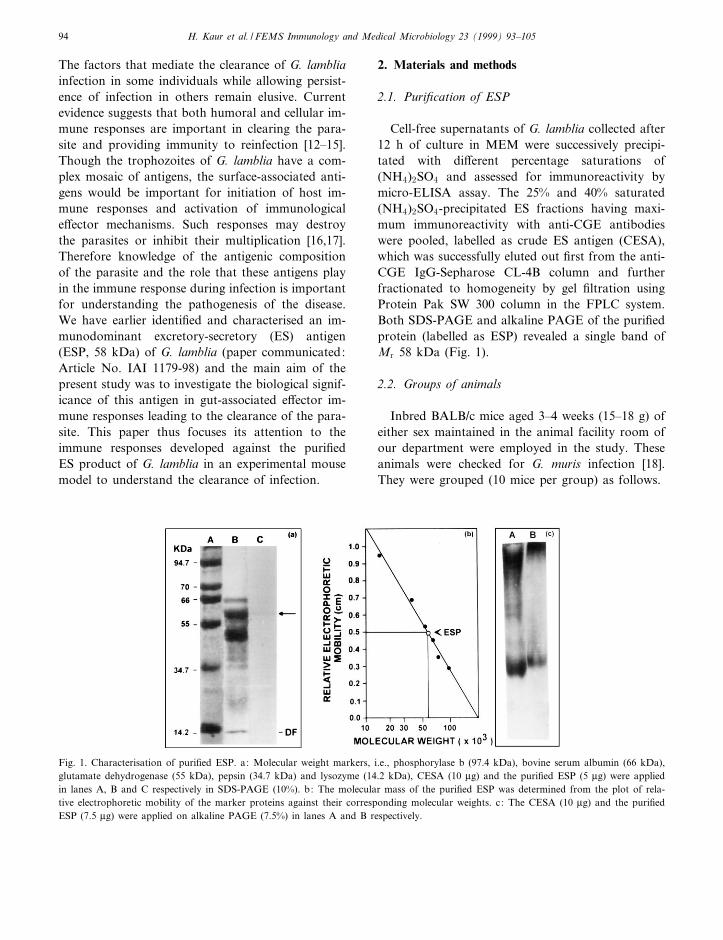
Cell-free supernatants of G. lamblia collected after12 h of culture in MEM were successively precipi-tated with di¡erent percentage saturations of(NH4)2SO4 and assessed for immunoreactivity bymicro-ELISA assay. The 25% and 40% saturated(NH4)2SO4-precipitated ES fractions having maxi-mum immunoreactivity with anti-CGE antibodieswere pooled, labelled as crude ES antigen (CESA),which was successfully eluted out ¢rst from the anti-CGE IgG-Sepharose CL-4B column and furtherfractionated to homogeneity by gel ¢ltration usingProtein Pak SW 300 column in the FPLC system.Both SDS-PAGE and alkaline PAGE of the puri¢edprotein (labelled as ESP) revealed a single band ofMr 58 kDa (Fig. 1).
2.2. Groups of animals
Inbred BALB/c mice aged 3^4 weeks (15^18 g) ofeither sex maintained in the animal facility room ofour department were employed in the study. Theseanimals were checked for G. muris infection [18].They were grouped (10 mice per group) as follows.
FEMSIM 960 8-2-99
Fig. 1. Characterisation of puri¢ed ESP. a: Molecular weight markers, i.e., phosphorylase b (97.4 kDa), bovine serum albumin (66 kDa),glutamate dehydrogenase (55 kDa), pepsin (34.7 kDa) and lysozyme (14.2 kDa), CESA (10 Wg) and the puri¢ed ESP (5 Wg) were appliedin lanes A, B and C respectively in SDS-PAGE (10%). b: The molecular mass of the puri¢ed ESP was determined from the plot of rela-tive electrophoretic mobility of the marker proteins against their corresponding molecular weights. c: The CESA (10 Wg) and the puri¢edESP (7.5 Wg) were applied on alkaline PAGE (7.5%) in lanes A and B respectively.
H. Kaur et al. / FEMS Immunology and Medical Microbiology 23 (1999) 93^10594
Group 1: immunised group. These mice receivedfour oral doses (50 Wg total) of puri¢ed ESP at in-tervals of 7 days each, after neutralising the stomachacidity with 0.25 ml NaHCO3 (100 mg ml31, pH8.5). The antiserum from these animals was testedby immunodi¡usion [19] using 1.5% agarose in 0.05M sodium barbital bu¡er (pH 8.3), to assess to thestatus of immunisation.
Group 2: immunised-challenged group. These an-imals were immunised as described for group 1.However, this group of animals was challengedwith G. lamblia trophozoites 7 days after the lastimmunising dose.
Group 3: infected group. The animals of thisgroup were infected with G. lamblia trophozoitesafter receiving NaHCO3 (as above).
Group 4: control group. These animals receivedthe same treatment of NaHCO3 (as above) prior toreceiving normal saline and served as control ani-mals. This is to control for the e¡ect of bicarbonatetreatment on trophozoite infectivity.
2.3. Infection/challenge of the experimental animals
Axenic G. lamblia trophozoites (Portland-1 strain)were harvested from 48^72-h culture and washedwith 0.15 M phosphate-bu¡ered saline, pH 7.2(PBS). The count was adjusted to 2U107 viabletrophozoites per millilitre of PBS and the animalswere challenged intraoesophageally with 0.5 ml ofinoculum with a catheter [20].
2.4. Follow-up of animals
The animals from each group were killed at vari-ous post immunisation/post challenge (PI/PC) orpost infection (p.i.) days, i.e., 3^5 days (establish-ment phase), 9^11 days (acute phase) and 17^21days (decline phase).
2.5. Trophozoite load in the intestine
The trophozoites were counted in the intestinalperfusate of infected as well as immunised-challengedanimals by the method of Vasudeva et al. [21]. Afterlaparotomy, the small intestine was excised and£ushed with a ¢xed volume of ice-cold normal saline.The perfusate was centrifuged at 50Ug for 2^3 min
to remove the tissue fragments and food particles.The supernatant was centrifuged at 600Ug for 10min. The pellet was washed with normal saline andthe total number of trophozoites was counted in ahaemocytometer.
2.6. Isolation of gut lymphocytes
Intraepithelial lymphocytes (IEL) and laminapropria lymphocytes (LPL) from control and exper-imental mice were isolated by the modi¢ed methodof Davies and Parrot [22] and Tagliabue et al. [23].
Brie£y, a 12^15 cm long segment of the small in-testine, cut longitudinally after surgical removal ofPeyer's patches, was fragmented into 1^2 cm longpieces. These pieces after washing in Hanks' bal-anced salt solution (HBSS) were incubated inHBSS with 5 mM dithiothreitol (DTT) (Sisco, India)for 5^10 min at room temperature to remove mucus.The gut pieces incubated at 37³C in Ca2�,Mg2�-freeHBSS with 1034 M EDTA for 20 min under con-stant stirring were washed 3^4 times in Ca2�,Mg2�-free HBSS containing 5% foetal calf serum (FCS), atroom temperature. The cells in the supernatant werewashed twice, suspended in RPMI 1640 mediumwith 5% FCS and passed through a loosely packedglass wool column (GWC). They were then subjectedto Ficoll Isopaque density gradient (Sigma ChemicalCo., USA) centrifugation. The cells at the interfacewere harvested, washed and resuspended in RPMI1640 medium with 10% FCS. These cells were la-belled `IEL'.
The gut pieces after the complete recovery ofIEL were suspended in RPMI 1640 medium con-taining 10% FCS and 20 U ml31 of collagenase(type IV, Sigma Chemical Co., USA). The cell sus-pension after incubation at 37³C for 10 min waspassed through GWC before being subjected toFicoll Isopaque density gradient centrifugation. Thecells recovered at the interface were labelled`LPL'.
2.7. Preparation of lymphocytes from Peyer's patches(PP) and spleen
The lymphocytes from spleen and PP were isolatedby the method of Lycke et al. [24]. Brie£y, the lym-phocytes from both tissue suspensions were collected
FEMSIM 960 8-2-99
H. Kaur et al. / FEMS Immunology and Medical Microbiology 23 (1999) 93^105 95

separately by teasing them in HBSS between theends of a frosted slide and passed through GWC.The resulting ¢ltrate was centrifuged at 250Ug for10 min at 4³C and the supernatant was discarded.The washed cells of both populations were then sep-arately subjected to Ficoll Isopaque gradient centri-fugation. The cells at the interface of the gradientwere collected and ¢nally washed and suspended inRPMI/FCS.
2.8. Cell viability
The viability of IEL, LPL as well as PP and spleenlymphocytes was checked by the 0.2% trypan blueexclusion procedure [25].
2.9. Enumeration of T cell subsets and assessment ofimmunoglobulin bearing cells by £ow cytometry[26]
Brie£y, the T/B cell markers on IEL, LPL, PP andspleen lymphocytes were labelled by incubating theindividual cell preparations with FITC-labelled anti-mouse Thy 1.2, L3T4 and Lyt 2.2 monoclonal anti-bodies (Becton Dickinson, USA) as well as FITC-labelled goat anti-mouse F(ab)2 IgG, IgA and IgM(Kirkegaard and Perry, USA) for 30 min at roomtemperature in the dark. After washing with PBS,the cells were ¢xed with 0.5% formaldehyde at 4³Cfor 30 min and acquired on FACScan (Becton Dick-inson, USA) to assess the percentage of £uorescentcells.
2.10. Humoral immune response
The levels of anti-ESP antibodies in the serumsamples collected after completion of immunisationas well as infection at various days after challenge inall group of animals were checked by standard mi-cro-ELISA assay [27].
2.11. Statistical analysis
The statistical analysis of data was done by em-ploying one-way analysis of variance (ANOVA) withmultiple comparisons and only values 6 0.01 wereconsidered as signi¢cant. All values are expressedas mean þ S.D.
3. Results and discussion
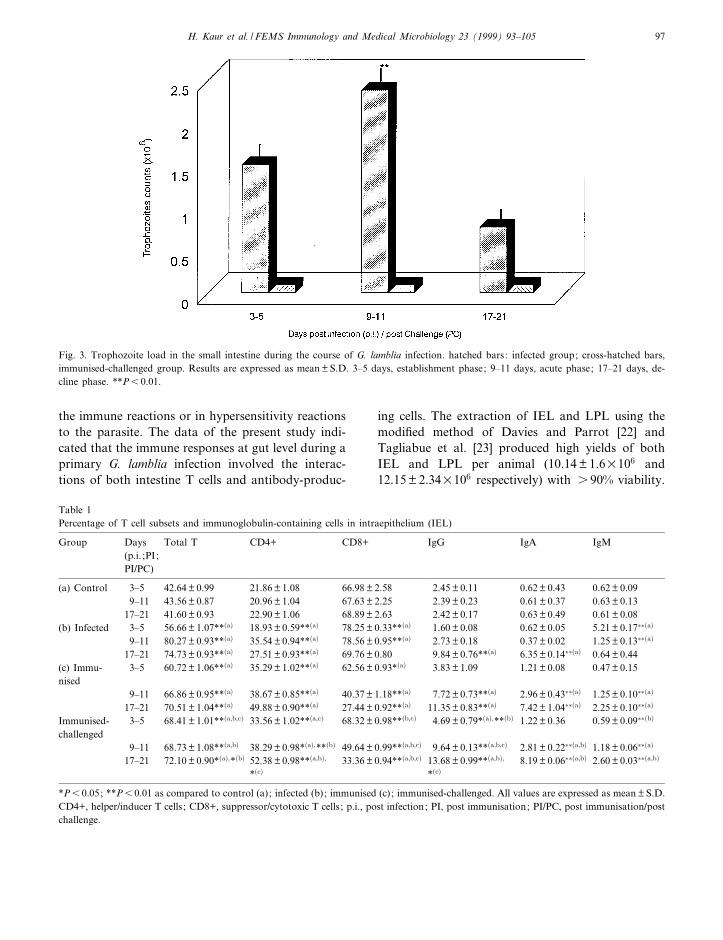
There has been much controversy in selecting asuitable mode of immunisation to stimulate an e¡ec-tive mucosal immunity. Parenteral immunisation hasbeen the main vaccination route for infectious dis-eases. This route has been e¡ective in preventing in-fectious diseases which have a systemic phase priorto or concomitant with their major pathological ef-fects. However, such a route of immunisation hasbeen at the best only marginally e¡ective in prevent-ing gut-dwelling infections [28^30]. The failure of theparenteral route of infection is obvious because theparenteral antigen administration fails to induce mu-cosal immunity [30]. However, the oral route hasbeen found to be very e¡ective at eliciting a speci¢cmucosal immune response although it requires pro-longed antigen administration in large amounts[20,30,31]. The oral immunisation of experimentalanimals with the puri¢ed ESP (as indicated by im-munodi¡usion, Fig. 2) was indeed found to stimulatethe immune system very e¤ciently. The introductionof the ESP into the intestinal lumen also preventedthe establishment of the G. lamblia trophozoites inthe immunised-challenged group, whereas the infec-tion established by 3^5 days p.i. in the intestine ofinfected control animals (group 3), after reachingpeak levels at 9^11 days p.i., was spontaneouslycleared by 17^21 days p.i. (Fig. 3).
Studies by Ferguson et al. [32] proposed that themucosal T lymphocytes are likely to be involved in
FEMSIM 960 8-2-99
Fig. 2. Immunodi¡usion pattern of the test sera of ESP-immu-nised mice. Wells 1, 2, 3 and 4 contain antisera from immunisedmice (group 1); well 5 contains preimmune sera; well 6 containsthe puri¢ed ESP.
H. Kaur et al. / FEMS Immunology and Medical Microbiology 23 (1999) 93^10596
the immune reactions or in hypersensitivity reactionsto the parasite. The data of the present study indi-cated that the immune responses at gut level during aprimary G. lamblia infection involved the interac-tions of both intestine T cells and antibody-produc-
ing cells. The extraction of IEL and LPL using themodi¢ed method of Davies and Parrot [22] andTagliabue et al. [23] produced high yields of bothIEL and LPL per animal (10.14 þ 1.6U106 and12.15 þ 2.34U106 respectively) with s 90% viability.
FEMSIM 960 8-2-99
Table 1Percentage of T cell subsets and immunoglobulin-containing cells in intraepithelium (IEL)
Group Days Total T CD4+ CD8+ IgG IgA IgM(p.i. ;PI;PI/PC)
(a) Control 3^5 42.64 þ 0.99 21.86 þ 1.08 66.98 þ 2.58 2.45 þ 0.11 0.62 þ 0.43 0.62 þ 0.099^11 43.56 þ 0.87 20.96 þ 1.04 67.63 þ 2.25 2.39 þ 0.23 0.61 þ 0.37 0.63 þ 0.13
17^21 41.60 þ 0.93 22.90 þ 1.06 68.89 þ 2.63 2.42 þ 0.17 0.63 þ 0.49 0.61 þ 0.08(b) Infected 3^5 56.66 þ 1.07**�a� 18.93 þ 0.59**�a� 78.25 þ 0.33**�a� 1.60 þ 0.08 0.62 þ 0.05 5.21 þ 0.17���a�
9^11 80.27 þ 0.93**�a� 35.54 þ 0.94**�a� 78.56 þ 0.95**�a� 2.73 þ 0.18 0.37 þ 0.02 1.25 þ 0.13���a�
17^21 74.73 þ 0.93**�a� 27.51 þ 0.93**�a� 69.76 þ 0.80 9.84 þ 0.76**�a� 6.35 þ 0.14���a� 0.64 þ 0.44(c) Immu-nised
3^5 60.72 þ 1.06**�a� 35.29 þ 1.02**�a� 62.56 þ 0.93*�a� 3.83 þ 1.09 1.21 þ 0.08 0.47 þ 0.15
9^11 66.86 þ 0.95**�a� 38.67 þ 0.85**�a� 40.37 þ 1.18**�a� 7.72 þ 0.73**�a� 2.96 þ 0.43���a� 1.25 þ 0.10���a�
17^21 70.51 þ 1.04**�a� 49.88 þ 0.90**�a� 27.44 þ 0.92**�a� 11.35 þ 0.83**�a� 7.42 þ 1.04���a� 2.25 þ 0.10���a�
Immunised-challenged
3^5 68.41 þ 1.01**�a;b;c� 33.56 þ 1.02**�a;c� 68.32 þ 0.98**�b;c� 4.69 þ 0.79*�a�;**�b� 1.22 þ 0.36 0.59 þ 0.09���b�
9^11 68.73 þ 1.08**�a;b� 38.29 þ 0.98*�a�;**�b� 49.64 þ 0.99**�a;b;c� 9.64 þ 0.13**�a;b;c� 2.81 þ 0.22���a;b� 1.18 þ 0.06���a�
17^21 72.10 þ 0.90*�a�;*�b� 52.38 þ 0.98**�a;b�;
*�c�33.36 þ 0.94**�a;b;c� 13.68 þ 0.99**�a;b�;
*�c�8.19 þ 0.06���a;b� 2.60 þ 0.03���a;b�
*P6 0.05; **P6 0.01 as compared to control (a); infected (b); immunised (c); immunised-challenged. All values are expressed as mean þ S.D.CD4+, helper/inducer T cells ; CD8+, suppressor/cytotoxic T cells ; p.i., post infection; PI, post immunisation; PI/PC, post immunisation/postchallenge.
Fig. 3. Trophozoite load in the small intestine during the course of G. lamblia infection. hatched bars: infected group; cross-hatched bars,immunised-challenged group. Results are expressed as mean þ S.D. 3^5 days, establishment phase; 9^11 days, acute phase; 17^21 days, de-cline phase. **P6 0.01.
H. Kaur et al. / FEMS Immunology and Medical Microbiology 23 (1999) 93^105 97

The separation of PP and splenic lymphocytes alsoprovided a pure population of viable (s 90%) cellsas assessed by 0.2% trypan blue exclusion. The cellcount was 5.6 þ 0.86U107 sp per animal and9.3 þ 0.34U106 pp per animal). The di¡erential re-
sponses of IEL and LPL observed during the colo-nisation, peak and decline phases of giardial infec-tion were considered speci¢c to G. lamblia infectionsince the observed alterations occurred following theinoculation of trophozoites. No alterations in the
FEMSIM 960 8-2-99
Table 3Percentage of T cell subsets and immunoglobulin-containing cells in Peyer's patches (PP)
Group Days Total T CD4+ CD8+ IgG IgA IgM(p.i. ;PI;PI/PC)
(a) Control 3^5 28.17 þ 0.84 17.44 þ 0.75 6.20 þ 0.54 29.03 þ 0.95 4.52 þ 0.26 25.55 þ 0.879^11 29.21 þ 0.86 17.99 þ 0.80 6.88 þ 0.57 29.98 þ 0.92 4.93 þ 0.70 26.39 þ 0.81
17^21 28.31 þ 0.85 17.53 þ 1.01 6.19 þ 0.63 29.79 þ 0.91 4.52 þ 0.26 25.55 þ 0.87(b) Infected 3^5 47.92 þ 0.94**�a� 30.72 þ 0.70**�a� 6.42 þ 0.79 29.88 þ 0.54 10.27 þ 0.66**�a� 33.64 þ 5.43**�a�
9^11 38.97 þ 0.93**�a� 17.74 þ 0.94 6.82 þ 0.69 29.25 þ 0.65 16.66 þ 0.94**�a� 47.60 þ 0.40**�a�
17^21 29.06 þ 0.92 17.53 þ 1.01 6.36 þ 0.84 29.28 þ 1.15 30.67 þ 0.97**�a� 32.18 þ 0.60**�a�
(c) Immunised 3^5 10.35 þ 0.85**�a� 9.07 þ 0.88**�a� 20.00 þ 0.93**�a� 28.01 þ 0.99 13.49 þ 1.17**�a� 12.34 þ 0.88**�a�
9^11 19.44 þ 0.88**�a� 17.39 þ 0.92 9.36 þ 0.89**�a� 53.73 þ 0.89**�a� 3.76 þ 0.85 40.35 þ 0.87**�a�
17^21 18.22 þ 0.62**�a� 12.07 þ 0.88 4.45 þ 0.95 81.65 þ 0.97**�a� 3.62 þ 0.89 48.30 þ 0.72**�a�
Immunised-challenged
3^5 16.16 þ 0.97**�a;b;c� 10.55 þ 0.80**�a;b�;*�c�
21.85 þ 0.95**�a�;*�c�
39.08 þ 0.69**�a�;*�b�
16.53 þ 0.06**�a;b;c� 19.19 þ 0.99*�a;c�;**�b�
9^11 23.18 þ 0.91**�a;b;c� 17.76 þ 0.88 10.47 þ 0.93**�a;b� 58.38 þ 1.00**�a;c�;*�b�
4.37 þ 0.10**�b� 44.62 þ 0.70**�a;b;c�
17^21 21.62 þ 0.92**�a;b;c� 9.46 þ 0.94**�a;b;c� 5.16 þ 0.96 83.33 þ 0.90**�a;b� 10.77 þ 0.97**�a;b� 54.39 þ 0.99**�a;b;c�
*P6 0.05; **P6 0.01 as compared to control (a); infected (b); immunised (c); immunised-challenged. All values are expressed as mean þ S.D.CD4+, helper/inducer T cells ; CD8+, suppressor/cytotoxic T cells ; p.i., post infection; PI, post immunisation; PI/PC, post immunisation/postchallenge.
Table 2Percentage of T cell subsets and immunoglobulin-containing cells in lamina propria (LPL)
Group Days Total T CD4+ CD8+ IgG IgA IgM(p.i. ;PI;PI/PC)
(a) Control 3^5 38.63 þ 0.91 25.42 þ 0.90 16.76 þ 0.93 22.19 þ 1.02 17.83 þ 0.88 3.08 þ 0.209^11 39.59 þ 0.89 26.51 þ 0.79 17.81 þ 0.10 23.40 þ 1.05 16.90 þ 0.98 3.02 þ 0.15
17^21 38.78 þ 0.98 24.91 þ 0.54 15.90 þ 0.88 21.35 þ 1.03 18.79 þ 0.77 3.05 þ 0.23(b) Infected 3^5 60.33 þ 1.11**�a� 26.51 þ 0.53 23.40 þ 1.04**�a� 21.07 þ 0.93 17.76 þ 0.98 8.25 þ 0.36**�a�
9^11 68.65 þ 0.85**�a� 25.49 þ 0.88 28.88 þ 1.03**�a� 23.48 þ 0.78 18.76 þ 0.35 3.53 þ 0.32*�a�
17^21 56.50 þ 0.88**�a� 33.83 þ 0.94**�a� 16.10 þ 0.20 32.94 þ 0.86**�a� 33.06 þ 0.54**�a� 2.16 þ 0.68**�a�
(c) Immunised 3^5 80.64 þ 0.86**�a� 33.90 þ 5.32*�a� 43.65 þ 1.18**�a� 7.46 þ 0.56**�a� 4.54 þ 0.23**�a� 3.08 þ 0.289^11 59.34 þ 0.69**�a� 25.84 þ 0.82 18.83 þ 0.82 10.76 þ 0.86**�a� 11.00 þ 0.89**�a� 3.03 þ 0.07
17^21 4.97 þ 0.65**�a� 2.89 þ 0.89**�a� 1.31 þ 0.09**�a� 58.57 þ 1.14**�a� 50.33 þ 0.98**�a� 3.06 þ 0.21Immunised-challenged
3^5 86.38 þ 0.02**�a;b;c� 37.26 þ 0.94**�a;b� 43.99 þ 1.26**�a;b� 7.96 þ 0.60**�a;b� 4.73 þ 0.70**�a;b� 3.09 þ 0.22
9^11 60.22 þ 0.90**�a� 25.84 þ 0.84 16.73 þ 0.90**�b� 17.89 þ 0.84**�a;b;c�
15.30 þ 0.03**�a;b;c�
3.06 þ 0.10**�b�
17^21 18.36 þ 0.89**�a;b;c� 2.97 þ 0.80**�a;b� 1.48 þ 0.41**�a;b� 53.34 þ 0.70**�a;b�;*�c�
52.55 þ 0.48**�a;b�;*�c�
3.08 þ 0.25
*P6 0.05; **P6 0.01 as compared to control (a); infected (b); immunised (c); immunised-challenged. All values are expressed as mean þ S.D.CD4+, helper/inducer T cells ; CD8+, suppressor/cytotoxic T cells ; p.i., post infection; PI, post immunisation; PI/PC, post immunisation/postchallenge.
H. Kaur et al. / FEMS Immunology and Medical Microbiology 23 (1999) 93^10598

lymphocyte T cell subsets or antibody-producingcells were noticed in uninfected control animalskept under identical conditions.
The establishment and acute phases of G. lambliainfection in the infected animals in the present studyaccompanied a signi¢cant in£ux of Lyt 2 (suppres-sor/cytotoxic lymphocytes) (Tables 1 and 2) and asigni¢cant decline in IgA-bearing plasma cells (Ta-bles 1 and 2) in the gut (IEL/LPL). It has earlierbeen shown that the intestinal lymphocytes isolatedduring the course of giardial infection from NMRImice were incapable of killing the G. lamblia troph-ozoites in vitro either in the presence or in the ab-sence of G. lamblia-speci¢c antibodies. Thus the Lyt2 cells in£uxed as a result of Giardia infection ap-peared not to be cytotoxic T cells in nature but weresuppressor T cells [33]. This in£ux of suppressor Tcells during establishment and peak phases of giar-dial infection has been suggested by these authors tobe due to the feedback suppressor pathway whichmight have been triggered very early or preferentiallyafter the immune induction following G. lamblia in-fection due to the release of some ES products by themultiplying trophozoites. An early selective localisa-tion of suppressor T cells observed in the infectedanimals may therefore explain the signi¢cantly lowlevel of IgA-bearing cells in the IEL and LPL ofthese animals as well as in the lamina propria ofimmunised animals during establishment and peakphases of giardial infection. The fact that ES prod-ucts signal the generation and localisation of Lyt 2cells in the gut accounts for a signi¢cant induction ofCD8+ T cells in the PP of immunised animals (Table3), which is the main site of antigen presentation.The present study, however, indicates a signi¢cantincrease in the percentage of IgA-bearing cells inthe PP of immunised as well as immunised-chal-lenged animals (Table 3), which might have beenresponsible for the inhibition of G. lamblia tropho-zoites to be colonised in the intestine.
Further, it was observed that the decline phase ofG. lamblia infection in the infected as well as theimmunised and immunised-challenged animals in-volved a selective induction of CD4+ T cells in theIEL (Table 1) and LPL (Table 2) along with a sig-ni¢cant reduction of CD8+ T cells (Tables 1 and 2).This phase of infection was also found to be accom-panied by an increased number of IgA- and IgG-
bearing cells (Tables 1 and 2 respectively). It thusappears that the inducer T cells may be responsiblefor the induction of antibody-dependent e¡ector re-sponses. It has earlier been noticed that s-IgA boundto the surface of Giardia trophozoites results in theclearance of the same from the gut of infected ani-mals [34,35] while parasites with negligible surface-bound s-IgA persisted during the decline phase ofinfection [36,37]. The anti-giardial IgA responseshave also been noticed to be altered in mice depletedof T helper lymphocytes [35]. Indeed it has earlierbeen observed that the clearance of G. muris infec-tion in mice is regulated by induction of CD4+T cells [35]. Based on the ¢ndings of others andourselves, we feel that a signi¢cant enhancement ofT helper/inducer activity along with a signi¢cant en-hancement of IgA-bearing cells results in the clear-ance of the trophozoites from the intestine. At thesame time, the role of IgG in the clearance of Giardiacannot be ignored, since a signi¢cant increase in theIgG-bearing cells during the decline phase of the ex-perimental animals was observed, although surface-bound parasite-speci¢c IgG antibodies on the troph-ozoites of Giardia isolated from the gut lumen ofmice reported by Heyworth et al. [38] suggested apossible role of IgG in the modulation of the infec-tion at gut level. However, the involvement of locallysynthesised IgG in providing long-lasting protectionagainst G. lamblia requires further investigation.
In contrast, the establishment phase of G. lambliainfection in the IEL, LPL as well as PP of the in-fected mice was accompanied by proliferation ofIgM-bearing cells (Tables 1 and 2 respectively) sug-gesting that during the early stage of G. lamblia in-fection the immunoglobulin production was predom-inantly of the IgM isotype. This observation agreeswell with the earlier reports in human giardiasis [39^41]. The proliferation of IgM-bearing cells during theacute phase of the disease is not a¡ected by the feed-back suppressor pathway as the initial di¡erentiationof pre-B cells to immune IgM-bearing cells is inde-pendent of T cells or lymphokines [42].
The present data demonstrate that the prior im-munisation of mice with ESP resulted in a signi¢cantstimulation of local immunity as evidenced by a sig-ni¢cant increase in IgA- and IgG-bearing cells in theLPL-immunised animals (Table 2) which probablyprevented the establishment of G. lamblia tropho-
FEMSIM 960 8-2-99
H. Kaur et al. / FEMS Immunology and Medical Microbiology 23 (1999) 93^105 99

zoites in the immunised-challenged animals. Further,it was observed that the IgM-bearing cells in theLPL of the immunised animals (Table 2) and controlanimals were statistically identical. These ¢ndingspoint to the fact that the genetic switching of IgM-bearing cells to those recognising the antigenthrough receptors of the IgG and IgA classes hasbeen completed during the immunisation scheduleand memory IgG and IgA clones which were formedgave a booster response upon challenge in immu-nised animals.
A signi¢cant decline in the percentage of total Tcells was observed within the PP of the immunisedand immunised-challenged animals during 3^5 daysPI/PC (Table 3). It appears that most of the acti-vated T cells including CD4+ T cells might havemigrated to the distal mucosal e¡ector sites, suchas the lamina propria, as the percentage of these cellswas found to be signi¢cantly increased in the LPLand IEL of these animals (Tables 2 and 1 respec-tively) where further stimulation and terminal di¡er-entiation into antibody-producing cells takes place[43^45]. Less is known about the induction and traf-¢cking of primed T cells after oral immunisation,particularly in man. However, immunisation of PPwith keyhole limpet haemocyanin in an animal mod-
el induced a population of primed T cells which sug-gested within the circulation before homing speci¢-cally to the gut [46]. In the gut, immunisation by theoral or enteral route has been found to e¡ectivelystimulate a speci¢c mucosal immune response[20,47]. The immunogen probably makes contactwith immunocompetent B and T cells in the PP,and these cells, while di¡erentiating, then migrateto the mesenteric lymph nodes and further via thethoracic duct, enter into the systemic circulation.Committed, migrating cells eventually return to theintestinal mucosa where further stimulation and ter-minal di¡erentiation into antibody-producing cellstakes place [43,48]. On repeated immunisation, theimmunogen may also, to a lesser extent, directlystimulate antigen-speci¢c memory cells in gut laminapropria [49,50]. It remains poorly de¢ned whetherthe IgA class predominance of the gut mucosal re-sponse is already initiated in the PP or results fromlater in£uences on the B cells, i.e., while en route toor during this ¢nal di¡erentiation in the laminapropria [51,52].
In the spleen (Table 4) of immunised animals, wefound a signi¢cant increase in the percentage ofCD4+ T cells. An increase in CD4+ T cells appearsto be bene¢cial as it related very well with elimina-
FEMSIM 960 8-2-99
Table 4Percentage of T cell subsets and immunoglobulin-containing cells in spleen (SP)
Group Days Total T CD4+ CD8+ IgG IgA IgM(p.i. ;PI;PI/PC)
(a) Control 3^5 56.15 þ 0.90 25.10 þ 0.99 27.26 þ 0.96 52.68 þ 0.98 8.22 þ 0.34 32.71 þ 0.929^11 57.17 þ 0.92 24.23 þ 0.81 28.31 þ 0.91 53.70 þ 0.91 8.99 þ 0.40 33.56 þ 0.90
17^21 58.14 þ 0.91 24.40 þ 0.87 26.46 þ 0.94 52.93 þ 0.94 9.03 þ 0.63 34.69 þ 0.91(b) Infected 3^5 26.38 þ 0.98**�a� 25.21 þ 0.91 6.49 þ 0.54**�a� 53.29 þ 0.98 1.96 þ 0.34**�a� 33.06 þ 0.93
9^11 24.32 þ 0.92**�a� 7.69 þ 0.91**�a� 3.33 þ 0.07**�a� 35.53 þ 0.95**�a� 11.39 þ 0.89**�a� 13.74 þ 0.96**�a�
17^21 50.28 þ 0.92**�a� 27.29 þ 0.92*�a� 11.49 þ 1.01**�a� 58.40 þ 0.90**�a� 2.63 þ 0.13**�a� 48.25 þ 0.95**�a�
(c) Immunised 3^5 43.56 þ 0.94**�a� 31.43 þ 0.91**�a� 5.64 þ 0.95**�a� 49.37 þ 1.01**�a� 0.89 þ 0.16**�a� 50.50 þ 0.90**�a�
9^11 39.31 þ 0.91**�a;b;c� 27.27 þ 0.28 4.21 þ 0.28**�a� 58.24 þ 0.94**�a� 0.31 þ 0.05**�a� 34.05 þ 0.9317^21 32.94 þ 0.96**�a� 24.96 þ 0.94 8.60 þ 1.46**�a� 62.67 þ 0.97**�a� 0.83 þ 0.13**�a� 34.33 þ 0.93
Immunised-challenged
3^5 55.84 þ 0.95**�b;c� 23.12 þ 0.94**�c� 11.18 þ 0.98**�a;b;c�
55.39 þ 0.99*�a�;**�c�
1.69 þ 0.43**�a�;��c� 39.53 þ 0.98**�a;c�
9^11 50.91 þ 0.98**�a;b;c� 28.76 þ 0.96**�a;b� 9.98 þ 0.97**�a;b;c�
22.90 þ 0.99**�a;b;c� 0.63 þ 0.35**�a;b� 13.05 þ 0.90**�a;c�
17^21 54.61 þ 0.99**�b;c� 32.69 þ 0.99**�a;b;c�
13.82 þ 0.96**�a;c� 2.48 þ 0.02**�a;b;c� 0.20 þ 0.10**�a;b;c� 5.66 þ 0.96**�a;b;c�
*P6 0.05; **P6 0.01 as compared to control (a); infected (b); immunised (c); immunised-challenged. All values are expressed as mean þ S.D.CD4+, helper/inducer T cells ; CD8+, suppressor/cytotoxic T cells ; p.i., post infection; PI, post immunisation; PI/PC, post immunisation/postchallenge.
H. Kaur et al. / FEMS Immunology and Medical Microbiology 23 (1999) 93^105100
tion of G. lamblia infection where it has been seenthat when the local immunologic response is at itspeak, G. lamblia is eliminated from the gut. We alsofound a sharp decline in IgA-bearing cells on the onehand and a signi¢cant increase in IgM-bearing cellson the other, in the spleen of the immunised animalsduring the establishment phase of the infection. It isquite likely from these ¢ndings that IgM may havetaken over the functions of IgA resulting in the clear-ance of G. lamblia trophozoites. An experimentalstudy of giardiasis utilising anti-IgM-treated micealso indicated the importance of IgM in regulationof giardial infection [14]. Farthing [53] has also re-ported that anti-giardial IgM potentiates the lytice¡ects of complement on G. lamblia. The role ofGiardia-speci¢c IgM response in regulating the dis-ease process especially in immunocompromised hostshas also been elucidated [54].
However, in-depth investigations are required es-pecially in view of the fact that the parasite remainscon¢ned to the intestinal lumen without invading theintestinal mucosa. It may thereby fail to elicit eithera systemic in£ammatory response or alterations inlymphocyte subsets in the peripheral blood [55].The small bowel morphological changes observedduring G. lamblia infection are believed to be deter-mined, at least in part, by T cell-dependent activity[13]. Indeed, it is the gut-associated lymphoid tissuethat plays a prominent role in the host defenseagainst intestinal pathogens. The cells express mainlythe CD4 antigen in the lamina propria whereas Tcells expressing the CD8 antigen and NK cells com-prise the majority of the IEL [56,57]. It remains to bedetermined to what degree the local intestinal im-mune response and changes in intestinal lymphocytesubsets are correlated by those detected in the pe-ripheral blood [55]. Further studies are thereforeneeded to elucidate the changes in intestinal mucosallymphocyte subsets occurring during G. lamblia in-fection and to correlate them with lymphocyte subsetdistribution in the circulation.
The present study further revealed a signi¢cantincrease in the population of total T as well asCD4+ T cells in the PP of infected animals (Table3) during the establishment phase of infection. Ear-lier reports on PP `switch' T cells which possess thesurface antigens and the known regulatory functionsof T cells led to the hypothesis that an increase in the
number of relative percentage of PP CD4+ T cellsinitiates subsequent events in the intestinal immuneresponse [58]. The relative de¢ciency of these cells, ascompared to CD8+ T cells in immunode¢cient nudemice, provided indirect support for this hypothesis[59].
The cellular events within PP that follow antigenpresentation are not well understood. `Switch' Tcells, which are T helper cells, have been isolatedfrom PP and in vitro have been demonstrated toparticipate in the immunoglobulin isotype switchingthat occurs with maturation of B cells into functionalplasma cells [60]. In vitro cloned PP `switch' T cellsproliferate when exposed to autologous B cells, Tcells and macrophages, suggesting that cell-cell inter-actions are important in the generation of these`switch' T cells [61].
Thus, from the present study, it appears that IgAantibodies act as a ¢rst line of defense for immuneexclusion of the parasite at the mucosal surface, i.e.,blocking the colonisation of mucosal surface by theparasite. On the other hand, the presence of circulat-ing anti-giardial antibodies [62^64] in clinical giar-diasis patients and speci¢c serum IgG and IgA anti-bodies in experimental giardiasis [65] in miceindicates stimulation of the host humoral immunesystem. The demonstration of anti-ESP antibodiesobserved in the serum of immunised animals (Fig.4) in the present study indicates that the antigenmust cross the intestinal mucosa to stimulate thesystemic lymphoid tissue. These antibodies mayalso be due to transepithelial translocation of immu-noglobulins from the intestinal lumen produced bylocal (mucosal) antibody-producing cells stimulatedby the parasite antigen [66].
The development of in vitro culture techniques forGiardia a decade ago has secured an adequate supplyof antigen such that serum antibody responses toGiardia are beginning to be well characterised,most commonly by ELISA. Experimental infectionsin mice con¢rm the appearance of speci¢c anti-Giar-dia IgA and IgG in intestinal secretions and clear-ance of the parasite relates closely to rising concen-trations of these antibodies in the intestinal £uid.
Although circulating anti-giardial antibodies havebeen demonstrated in individuals su¡ering from giar-diasis [61,64], their precise biological role has re-mained unclear. Vinayak et al. [33] have demon-
FEMSIM 960 8-2-99
H. Kaur et al. / FEMS Immunology and Medical Microbiology 23 (1999) 93^105 101
strated that a speci¢c antibody response to plasmamembrane (PM) and surface antigen GLSA 56 oc-curs in giardiasis. Comparison of groups of patientsrevealed low levels of antibodies against PM andGLSA 56 in persistent giardiasis, compared withnon-persistent and asymptomatic cases.
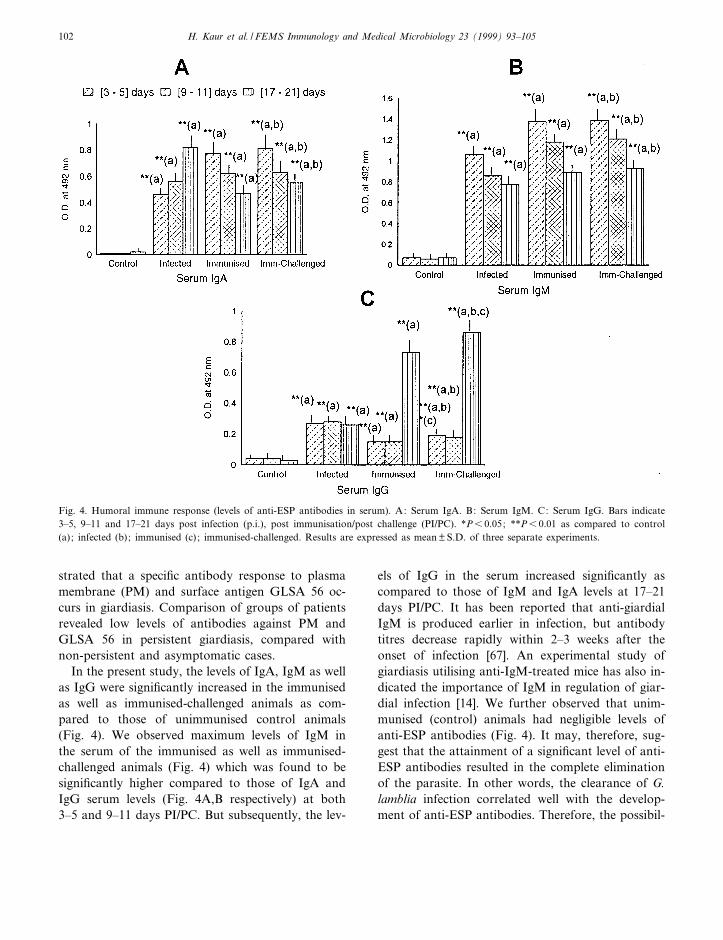
In the present study, the levels of IgA, IgM as wellas IgG were signi¢cantly increased in the immunisedas well as immunised-challenged animals as com-pared to those of unimmunised control animals(Fig. 4). We observed maximum levels of IgM inthe serum of the immunised as well as immunised-challenged animals (Fig. 4) which was found to besigni¢cantly higher compared to those of IgA andIgG serum levels (Fig. 4A,B respectively) at both3^5 and 9^11 days PI/PC. But subsequently, the lev-
els of IgG in the serum increased signi¢cantly ascompared to those of IgM and IgA levels at 17^21days PI/PC. It has been reported that anti-giardialIgM is produced earlier in infection, but antibodytitres decrease rapidly within 2^3 weeks after theonset of infection [67]. An experimental study ofgiardiasis utilising anti-IgM-treated mice has also in-dicated the importance of IgM in regulation of giar-dial infection [14]. We further observed that unim-munised (control) animals had negligible levels ofanti-ESP antibodies (Fig. 4). It may, therefore, sug-gest that the attainment of a signi¢cant level of anti-ESP antibodies resulted in the complete eliminationof the parasite. In other words, the clearance of G.lamblia infection correlated well with the develop-ment of anti-ESP antibodies. Therefore, the possibil-
FEMSIM 960 8-2-99
Fig. 4. Humoral immune response (levels of anti-ESP antibodies in serum). A: Serum IgA. B: Serum IgM. C: Serum IgG. Bars indicate3^5, 9^11 and 17^21 days post infection (p.i.), post immunisation/post challenge (PI/PC). *P6 0.05; **P6 0.01 as compared to control(a); infected (b); immunised (c) ; immunised-challenged. Results are expressed as mean þ S.D. of three separate experiments.
H. Kaur et al. / FEMS Immunology and Medical Microbiology 23 (1999) 93^105102
ity exists that the ES antigen from the live tropho-zoites was absorbed across the intestinal mucosa.Alternatively there may have been an interaction ofviable trophozoites with the surface epithelium re-sulting in activation and uptake by tissue macro-phages. In support of this hypothesis is the workby Owen et al. [68], who demonstrated phagocytosisof G. muris by macrophages present in mouse PP.The immunisation of animals with ES antigen re-sulted in a signi¢cant titre of anti-ESP antibodiesindicating that this protein is highly immunogenicand it seems that ESP is one of the major antigensof G. lamblia responsible for protection against thedisease.
It is therefore pertinent to conclude that ESP ofthe parasite can be considered a candidate for immu-noprophylactic studies. Such an agent may have auseful role in protecting vulnerable populations likechildren or persons from non-endemic areas travel-ling to endemic areas. Detailed investigations are es-sential to study the mechanisms by which the para-site evades the e¡ector immune mechanisms.Nevertheless, the investigations quoted above clearlyindicate that although gut-associated immune mech-anisms are involved in regulating giardial infection,other e¡ector immune mechanisms like mucosalmast cells, macrophages and other non-speci¢c intes-tinal factors cannot be ignored.
References
[1] Hartong, W.A., Gourley, W.K. and Arvanitakis, C. (1979)Giardiasis in clinical spectrum and functional structural ab-normalities of small intestine mucosa. Gastroenterology 77,61^69.
[2] Char, S., Shetty, N., Narasimha, M., Elliott, E., Macaden, R.and Farthing, M.J.G. (1991) Serum antibody response in chil-dren with Giardia lamblia infection and identi¢cation of animmunodominant 57 kDa antigen. Parasite Immunol. 13,329^337.
[3] Clark, J.T. and Holberton, D.V. (1986) Plasma membraneisolated from Giardia lamblia : identi¢cation of membrane pro-teins. Eur. J. Cell. Biol. 42, 200^206.
[4] Crossley, R. and Holberton, D.V. (1983) Characterisation ofproteins from the cytoskeleton of Giardia lamblia. J. Cell. Sci.59, 81^103.
[5] Edson, C.M., Farthing, M.J.G., Thorley-Lawson, D.A. andKeusch, G.T. (1986) An 88000 MW Giardia lamblia surfaceprotein which is immunogenic in humans. Infect. Immun. 54,621^665.
[6] Heyworth, M.F. (1990) Biological signi¢cance of Giardia spe-ci¢c antibodies. West. J. Med. 152, 293^295.
[7] Heyworth, M.F. and Vergara, J.A. (1994) Giardia muristrophozoite antigenic targets for mouse intestinal IgA anti-body. J. Infect. Dis. 169, 395^398.
[8] Kum, K., Khanna, R., Mehta, S., Khuller, M. and Vinayak,V.K. (1987) Plasma membrane associated antigen of tropho-zoites of axenic G. lamblia. Trans. R. Soc. Trop. Med. Hyg.82, 439^444.
[9] Rosof, J.D. and Stibbs, H.H. (1986) Isolation and identi¢ca-tion of a Giardia lamblia-speci¢c stool antigen (GSA 65) usefulin coprodiagnosis of giardiasis. J. Clin. Microbiol. 23, 905^910.
[10] Vinayak, V.K., Dutt, P. and Puri, M. (1991) An immunoen-zymatic dot-ELISA for the detection of Giardia lamblia anti-gen in stool eluates of clinical cases of giardiasis. J. Immunol.Methods 137, 245^251.
[11] Vinayak, V.K., Kum, K., Chandna, R., Venkateshwarlu, K.and Mehta, S. (1985) Detection of Giardia lamblia antigens inthe faeces by counterimmunoelectrophoresis. Pediat. Infect.Dis. J. 4, 383.
[12] Ament, M.E. and Rubin, C.E. (1972) Relation of giardiasis toabnormal intestinal structure and function in gastrointestinalimmunode¢ciency syndromes. Gastroenterology 62, 216^226.
[13] Roberts-Thomson, I.C. and Mitchell, G.F. (1978) Giardiasisin mice. I. Prolonged infections in certain mouse strains andhypothymic (nude) mice. Gastroenterology 75, 57^61.
[14] Snider, D.P., Gordon, J., McDermott, M.R. and Undertown,B.J. (1985) Chronic Giardia muris infection in anti-IgMtreated mice. Analysis of immunoglobulin and parasite speci¢cantibody in normal and immunoglobulin de¢cient animals.J. Immunol. 134, 4153^4162.
[15] Stevens, D.P., Frank, D.M. and Mahmoud, A.A.F. (1978)Thymus dependency of host resistance to Giardia muris infec-tion: Studies in nude mice. J. Immunol. 120, 680^682.
[16] Holberton, D.V. and Ward, A.P. (1981) Isolation of the cy-toskeleton from Giardia. Tubulin and a low molecular weightprotein associated with microribbon structures. J. Cell. Sci. 47,139^166.
[17] Scott, M.J. and Snary, D. (1979) Protective immunisation ofmice using cell surface glycoprotein from Trypanosoma cruzi.Nature 282, 73^74.
[18] Aggarwal, A., Sharma, G.L., Bhatia, A., Naik, S.R., Chakra-varti, R.N. and Vinayak, V.K. (1980) E¡ects of corticosteroidand irradiation on experimental G. lamblia infection in mice.Ann. Trop. Med. Parasitol. 74, 361^371.
[19] Ouchterlony, O. (1962) Di¡usion in gel methods for immuno-logical analysis II. In: Progress in Allergy (Kallos and Waks-man, B.H., Eds.), Vol. VI, pp. 30^154. Basel, Karger.
[20] Lange, S. and Holmgren, J. (1978) Protective antitoxic choleraimmunisation in mice: in£uence of route and number of im-munisations and mode of action of protective antibodies. ActaPathol. Microbiol. Scand. C Immunol. 86, 145^152.
[21] Vasudeva, V., Ganguly, N.K., Anand, B.S., Radhakrishnan,V.D. and Mahajan, R.C. (1982) A study of Giardia infectionin irradiated and thymectomised mice. J. Trop. Med. Hyg. 85,119^122.
FEMSIM 960 8-2-99
H. Kaur et al. / FEMS Immunology and Medical Microbiology 23 (1999) 93^105 103

[22] Davies, M.D.J. and Parrott, D.M.V. (1981) Preparation andpuri¢cation of lymphocytes from the epithelium and laminapropria of murine small intestine. Gut 22, 481^488.
[23] Tagliabue, A., Befus, A.D., Clark, D.A. and Bienenstock, J.(1982) Characteristics of natural killer cells in the murine in-testinal epithelium and lamina propria. J. Exp. Med. 155,1785^1796.
[24] Lycke, N., Lindholm, L. and Holmgren, J. (1983) IgA isotyperestriction in the mucosal but not in the extramucosal immuneresponse after oral immunisations with cholera toxin or chol-era B subunit. Int. Arch. Allergy Appl. Immunol. 72, 119^127.
[25] Jondal, M., Holm, G. and Wigzell, H. (1972) Surface markerson human lymphocytes. J. Exp. Med. 136, 207^215.
[26] Hornquist, E., Goldschmidt, J., Holmdahl, R. and Lycke, N.(1991) Host defense against cholera toxin is strongly CD4+ Tcell dependent. Infect. Immun. 59, 3630^3638.
[27] Voller, A., Bidwell, D. and Bartlett, A. (1976) Enzyme immu-noassay in diagnostic medicine. Theory and Practice. Bull.WHO, 53^55.
[28] Newcomb, R.W., Ischizaka, K. and Denald, B.L. (1969) Hu-man IgG and IgA diphtheria antitoxins in serum, nasal £uidsand saliva. J. Immunol. 103, 215^219.
[29] Ogra, P. (1971). The secretory immunoglobulin system of thegastrointestinal tract. In: The Secretory Immunological Sys-tem (Dayton, D.H. Jr., Small, P.A. Jr., Chanock, R.M. andTomasi, T.B. Jr., Eds.), p. 259. US Government Printing Of-¢ce, Washington, DC.
[30] Pierce, N.F. and Gowans, J. (1975) Cellular kinetics of theintestinal immune response to cholera toxoid in rats. J. Exp.Med. 142, 1550^1563.
[31] Dolezel, J. and Bienenstock, J. (1971) Gamma A and nongamma A immune response after oral and parenteral immu-nisation of hamster. Cell. Immunol. 2, 458^469.
[32] Ferguson, A., McClure, S.P. and Townley, R.R.W. (1976)Epithelial lymphocytes in¢ltration in biopsies from childrenwith diarrhoea. Acta Pediatr. Scand. 65, 541^546.
[33] Vinayak, V.K., Kum, K. and Khanna, R. (1989) Serum anti-bodies to giardial surface antigens: lower titres in persistentthan in non-persistent giardiasis. J. Med. Microbiol. 30, 207^212.
[34] Heyworth, M.F. (1989) Intestinal IgA response to Giardiamuris depleted of helper T lymphocytes in immunocompetentmice. J. Parasitol. 75, 246^251.
[35] Waight Sharma, A. and Mayrhofer, G. (1988) Biliary anti-body responses in rats infected with rodent Giardia duodenalisisolates. Parasite Immunol. 10, 181^191.
[36] Khanna, R., Joshi, K., Kum, K., Malik, A.K. and Vinayak,V.K. (1990) An ultrastructural analysis of changes in surfacearchitecture of intestinal mucosa following Giardia lambliainfection in mice. Gastroenterol. Japon. 25, 649^658.
[37] Vinayak, V.K., Khanna, R. and Kum, K. (1990) Gut associ-ated immune responses in clinical and experimental giardiasis :an overview. Trop. Gastroenterol. 11, 4^8.
[38] Heyworth, M.F., Kung, J.E. and Eriksson, E.C. (1986) Clear-ance of Giardia muris infection in mice de¢cient in naturalkiller cells. Infect. Immun. 54, 903^904.
[39] Goka, A.K.J., Rolston, D.D.K., Mathan, V.I. and Farthing,M.J.G. (1987) Human serum IgA response to Giardia lamblia.Gut 28, A1351.
[40] Ridley, M.J. and Ridley, D.S. (1976) Serum antibodies andjejunal histology in giardiasis associated with malabsorption.J. Clin. Pathol. 29, 30^34.
[41] Thompson, A., Rowland, R. and Hecker, R. (1977) Immuno-globulin bearing cells in giardiasis. J. Clin. Pathol. 30, 292^294.
[42] Abney, E.R., Cooper, M.D., Kearney, J.F., Lawton, A.R. andParkhouse, R.M.E. (1978) Sequential expression of immuno-globulin in developing mouse B lymphocytes: a systemic sur-vey that suggests a model for the generation of immuno-globulin class diversity. J. Immunol. 120, 2041^2049.
[43] Biewenga, J., VanRees, E.P. and Sminia, T. (1993) Inductionand regulation of IgA responses in microenvironment of gut.Clin. Immunol. Immunopathol. 67, 1^7.
[44] Czerkinsky, C., Svennerholm, A.M. and Holmgren, J.F.(1993) Induction and assessment of immunity at extramucosalsurfaces in humans: Implications for vaccine development.Clin. Infect. Dis. 16, 106^116.
[45] Miller, C.J., McGhee, J.R. and Gardner, M.B. (1992) Biologyof disease, mucosal immunity, HIV transmission and AIDS.Lab. Invest. 68, 129^145.
[46] Dunkley, M.L. and Husband, A.J. (1986) The induction andmigration of antigen-speci¢c helper cells for IgA responses inthe intestine. Immunology 15, 379^385.
[47] Crabbe, P.A., Nash, D.R., Bazin, H., Eyssen, H. and Here-mans, J.F. (1967) Antibodies of the IgA type in intestinalplasma cells of germfree mice after oral or parenteral immu-nisation with ferritin. J. Exp. Med. 130, 723^744.
[48] Husband, A.J. and Gowans, J.L. (1978) The origin and anti-gen dependent distribution of IgA containing cells in the in-testine. J. Exp. Med. 148, 1146^1160.
[49] Husband, A.J. (1982) Kinetics of extravasation and redistrib-ution of IgA-speci¢c antibody containing cells in the intestine.J. Immunol. 128, 1355^1359.
[50] Pierce, N.F. and Cray, W.C. (1982) Determinants of the local-isation, magnitude and duration of a speci¢c IgA plasma cellresponse in enterically immunised rats. J. Immunol. 128,1311^1315.
[51] Elson, C.O., Heck, J.A. and Strober, W. (1979) T cell regu-lation of murine IgA synthesis. J. Exp. Med. 149, 632^643.
[52] Tseng, J. (1981) IgA allotype suppression in mice: a cellularimplication for the IgA preponderance in the gut. Cell. Im-munol. 65, 247^257.
[53] Farthing, M.J.G. (1993) Diarrhoeal disease: Current conceptsand future challenges: Pathogenesis of giardiasis. Trans. Soc.Trop. Med. Hyg. 87, Suppl. 3, 17^21.
[54] Khanna, R., Kum, K. and Vinayak, V.K. (1990) Gut-associ-ated immune e¡ector responses in immunocompetent and im-munocompromised mice with Giardia lamblia. FEMS Micro-biol. Immunol. 2, 137^145.
[55] Schlesinger, M., Granot, E., Rabinowitz, R. and Deckelbaum,R.J. (1993) Peripheral blood lymphocyte subsets in infantswith diarrhoea with and without Giardia lamblia infection.Pediatr. Res. 33, 15^18.
FEMSIM 960 8-2-99
H. Kaur et al. / FEMS Immunology and Medical Microbiology 23 (1999) 93^105104

[56] Kelly, J., O'Farrelly, C., O'Mahoney, C., Weir, D.G. andFeighery, C. (1987) Immunoperoxidase demonstration of thecellular composition of the normal and coeliac small bowel.Clin. Exp. Immunol. 68, 177^188.
[57] Selby, W.S., Janossy, G., Bo¢ll, M. and Jewell, D.P. (1983)Lymphocyte subpopulations in the human small intestine. The¢ndings in normal mucosa and in the mucosa of patients withadult coeliac disease. Clin. Exp. Immunol. 52, 219^228.
[58] Strober, W. (1982) The regulation of mucosal immune system.J. Allergy Clin. Immunol. 70, 225^230.
[59] Carlson, J.R., Heyworth, M.F. and Owen, R.L. (1985) Re-sponse of Peyer's patch T cell subpopulation to Giardia murisinfection (abstr.) Clin. Res. 33, 32A.
[60] Kawanishi, H., Saltzman, L.E. and Strober, W. (1983) Mech-anisms regulating IgA class speci¢c immunoglobulin produc-tion in murine gut associated lymphoid tissues. I. T cells de-rived from Peyer's patches that switch sIgM B cells to sIgA Bcells in vitro. J. Exp. Med. 157, 433^450.
[61] Kawanishi, H., Ozato, K. and Strober, W. (1985) The prolif-erative response of cloned Peyer's patch switch T cells to syn-geneic and allogeneic stimuli. J. Immunol. 134, 3586^3591.
[62] Smith, P.D., Gillin, F.D., Brown, W.R. and Nash, T.E. (1981)IgG antibody to Giardia lamblia detected by enzyme linkedimmunosorbent assay. Gastroenterology 80, 1476^1480.
[63] Smith, P.D., Gillin, F.D., Spira, W.M. and Nash, T.E. (1982)Chronic giardiasis : Studies on drug sensitivity, toxin produc-tion and host immune response. Gastroenterology 83, 797^803.
[64] Vinayak, V.K., Jain, P. and Naik, S.R. (1978) Demonstrationof antibodies in Giardiasis using immunodi¡usion techniquewith Giardia cysts as antigen. Ann. Trop. Med. Parasitol. 72,581^582.
[65] Anders, R.F., Roberts-Thomson, I.C. and Mitchell, G.F.(1982) Giardiasis in mice. Analysis of humoral and cellularimmune response to Giardia muris. Parasite Immunol. 6, 45^57.
[66] Schlamowitz, M. (1976) Membrane receptors in the speci¢ctransfer of immunoglobulins from mother to young. Immu-nol. Commun. 5, 481^500.
[67] Goka, A.K.J., Mathan, V.I., Rolston, D.D.K. and Farthing,M.J.G. (1986) Diagnosis of giardiasis by speci¢c IgM anti-body by enzyme linked immunosorbent assay. Lancet ii 2,184^186.
[68] Owen, R.L., Allen, C.L. and Stevens, D.P. (1981) Phagocyto-sis of Giardia muris by macrophages in Peyer's patch epithe-lium in mice. Infect. Immun. 33, 591^601.
FEMSIM 960 8-2-99
H. Kaur et al. / FEMS Immunology and Medical Microbiology 23 (1999) 93^105 105