Sydney 1998: lessons from a Cryptosporidium and Giardia Crisis
Upload
khangminh22Category
view
0download
0
IN VITRO CULTURE AND ISOENZYME ANALYSIS OF GIARDIA LAMBLIA
ZILUNGILE L. KWITSHANA
December 1999

IN VITRO CULTURE AND ISOENZYME ANALYSIS OF GIARDIA LAMBLIA
by
ZILUNGILE L. KWITSHANA (NHD. -MED. TECH)
Submitted in partial fulfilment of the requirements for the degree of
MASTER OF SCIENCE (MEDICAL SCIENCE)
in the
Department of Medical Microbiology Faculty of Medicine University of Natal
Durban
1999

(j)edicated to my {ate parents
:No m usa et so6utfiongo

ABSTRACT
Giardia lamblia, an enteric protozoan parasite, infects a large number of
individuals worldwide. In South Africa prevalences ranging between 4 and 63%
are documented, however, the impact of giardiasis is underreseached in this
country. Giardia infections vary from asymptomatic carriage or a self-limiting
acute symptomatic illness to chronic, debilitating malabsorption syndrome. The
factors responsible for development of symptomatic versus asymptomatic
infection are poorly understood. It is believed by some that host factors
determine the clinical outcome of infection. On the other hand, the possibility of
the existence of pathogenic and non-pathogenic strains (a situation akin to
Entamoeba spp.) remains to be explored. One requirement for investigation of
the potential contribution of strain differences to pathogenecity of infection is
establishment of laboratory cultures of different strains isolated from
symptomatic and asymptomatic patients. The present study was undertaken to
develop and modify existing methods for: (i) establishment of laboratory cultures
of Giardia trophozoites from excystation of faecal cysts, (ii) long-term
maintenance and cryopreservation of the cultures and (iii) preliminary
characterisation methodology.
One thousand and twenty-three stool specimens were collected from day care
centres, hospital wards and Hlabisa hospital laboratory. A further 6246 were
retrieved from the Microbiology Laboratory at King Edward VIII Hospital and
screened by direct wet preparation. Giardia was detected by light microscopy
following formol-ether concentration (127 of 1023 samples) or direct
examination of wet preparations (78 of 6246 samples). Cysts were purified from
ii

the positive specimens by sucrose gradient separation. Viability was assessed
by a dye-exclusion method (eosin).
Three in vitro excystation techniques were employed in an attempt to obtain
trophozoites for initiation and establishment of viable cultures thereof. Culture
conditions were optimised using two reference strains of Giardia, WB & H7
(obtained from the National Institutes of Health, USA). The percentage
excystation ranged between 0-42% with all the in vitro methods of excystment.
Excysted trophozoites remained viable in TYI-S-33 culture medium for periods
ranging between 12-72 hours or up to 9 days, and gradually died, hence viable
trophozoite cultures could not be established. Some culture initiates (overall
65%) were lost through overwhelming bacterial and!or fungal contaminants.
An animal model was subsequently set up in which C57BLl6 and Praomys
(Mastomys) coucha mice were used for in vivo excystation experiments. 1-3
day old suckling mice were intragastrically injected with 105 -cysts! ml in 0,1 ml
distilled water. Trophozoites were retrieved from the stomachs of infected mice
7-10 days after inoculation and cultivated in TYI-S-33 medium. Six local isolates
were axenised using the in vivo excystation method. They have been
maintained for more than 15 months in culture after stabilates and Iysates of
confluent growths had been cryopreserved in Liquid Nitrogen. Successful
(100%) retrieval ofthe cryopreserved cultures has been achieved.
111

Seven isoenzyme electrophoresis systems have been set up and optimised.
Reproducible results were obtained in six of the enzymes. Some differences in
banding patterns of the enzymes were demonstrated.
iv

Declaration
This study represents original work by the author and has not been submitted in any form to another University. Where assistance was received and use of the work of others was made it was duly acknowledged in the text.
The work described in this dissertation was carried out in the South African Medical Research Council (Durban) under the supervision of Prof. TFHG Jackson and Prof A. W. Sturm as co-supervisor.
Ethical approval was obtained from the Faculty of Medicine Ethics Committee of the University of Natal.
v

Parts of this work were presented at the following conferences:
1. In Vitro Culture of Giardia lamblia.1996. Kwitshana ZL, Reddy SG, Sturm AW & Jackson TFHG. Presentation at the 10 th Annual Congress of the Parasitological Association of Southern Africa. 5-6 September 1996. Waterfront, Cape Town.
2. In vitro culture and isoenzyme analysis of Giardia lamblia.1998 Kwitshana ZL, Reddy SG, Sturm A.W & Jackson TFHG. 12 th Annual Congress of the Parasitological Association of Southern Africa. 9-11 September 1998. MontAuxources. Drakensburg.
3. Determination of viability of Giardia lamblia cysts: vital staining, in vitro and in vivo excystation. Kwitshana Z.L.; Reddy S.G.; Sturm A.W. & Jackson T.F.H.G.14 th Annual Congress of the Parasitological Association of Southern Africa. 19-23 October 1999. Augrabies Falls. Eastern Cape.
Publications.
In Vitro Culture of Giardia lamblia. (Abstract). 1996. Kwitshana Z.L. Jackson T.F.H.G. Journal of South African Veterinary Association 67(4): 175
In vitro culture and isoenzyme analysis of Giardia lambli8. 1998. (Abstract) Kwitshana Z.L.; Reddy S.G.; Sturm A.W. & Jackson T.F.H.G. Journal of South African Veterinary Association. 69:6
VI

ACKNOWLEDGEMENTS
The author wishes to express her sincere gratitude to the following individuals for their assistance in preparation of this thesis:
• Professor TFHG Jackson, (Supervisor) Regional Executive Director, S.A. Medical Research Council , Durban for supervising and affording me the opportunity to do this work. His constructive criticism, constant encouragement, expert advice, support and guidance are highly appreciated.
• Professor AW Sturm, (Co-supervisor} Head of Department, Microbiology, University of Natal, Medical School, for his expert Microbiology contribution, advice and encouragement.
• Mr. Selvan Reddy fQr his scientific input, patience, encouragement, support and professional criticism.
• The Superintendent of King Edward Hospital for giving permission to collect stool specimens from patients at the hospital.
• Professor J Coovadia Head of Paediatrics Dept. for allowing stool specimen collection from children's wards.
• Or. T.E.Nash and Mr. J.T.Conrad of the Laboratory of Parasitic Diseases, National Institute of Allergy and Infectious Diseases (NIH), Maryland, for their invaluable assistance in the establishment of the animal model and kind supply of Giardia reference strains.
• The Medical Research Council and the University of Natal for providing financial support for this project.
• Staff in Medical Microbiology at King Edward as well as Hlabisa hospital Laboratories for their assistance with retrieval of stool specimens.
• Mr Sathyanand Suparsad for his kind assistance with isoenzyme electrophoresis. Celia Anderson, Shirley Epstein, Annalies Gumede & Welcome Mkhasibe for their assistance during stool collections and all the staff at the MRC Amoebiasis Research Unit for their moral support.
• Ms. Charmine Bux & Mr James Wesley-Smith for their assistance with invertoscope and phase contrast photography respectively.
• Albert Hirashen and all the technical staff at the Medical Media Services for their assistance with photography.
• Staff at the MRC's Animal Facility: A. Saikoolal, Rould Cibane & Joseph Shozi for their assistance with animal care.
vu

• Personnel and children at the following day care centres: Othandweni Place of Safety, Kideo, & Amanzimtoti for coopertation and assistance during stool collection.
• To my late parents for gracing me with love and education, and my sisters Thobekile, Qhu, Carol, and brothers Sbongseni and Zama for their support and encouragement.
• Finally, my profound gratitude to my husband Sindile for his enduring patience, encouragement and support; my children Anele and Siyabulela who had to learn to cope on their own at a very tender age.
viii

Table of Contents
Page
CHAPTER 1 - PREAMBLE .............................................................. · .. · .............................................. 1
1.1 Taxonomy and Nomenclature ....................................................................................................... 2
1.2 Life-Cycle and Morphology .......................................................................... · .. ·· .. · .. ·· .. · ...... · ...... · .... · 7
1.3 Transmission ................................................................ · .. · ..... · .. ···· .. ··· .... ·· .. ···· .. · .. · .. · ...... ·· .. ··········· 10
1.3.1 Human hosts ................................... ......................................................................................... 1 0
1.3.2 The Zoonosis of giardiasis ............. .. ......................................................................................... 12
1.4 Distribution ......................................... ......................................................................................... 14
1.5 Host-parasite Relationship ................ .. ........................................................................................ 16
1.5.1 Pathogenesis ............................................................................................................................ 17
1.5.2 Do host factors play a role in increasing the susceptibility of certain hosts to infection? .......... 19
1.5.3 Are immune responses elicited by infection with Giardia responsible for parasite clearance and
t '1'" 't ? 19 s en Ismg ImmUnl y .......................................................................................................................... .
1.5.4 Are strain differences responsible for variations in the clinical course of infection? .................. 20
1.5.5 Identification of virulence determinants of the organism which would help in new drug and
vaccine design ................................................................................................................................... 21
1.6 Clinical Manifestations .................................................................................................................. 22
1.7 Risk Factors For Giardiasis ......................................................................................................... 27
1.7.1 Age and immune status of the individual ................................................................................. , 27
1.7.2 Genetic factors ......................................................................................................................... 30
1.7.2.1 Host factors ........................................................................................................................... 30
1.7.2.2 Parasite factors ...... .. .............................................................................................................. 31
1.7.3 Travel ................................................................................................................ \ ...................... 32
1.7.4 Nutritional status and gastric acidity ......................................................................................... 33
1.8 Summary ..................................................................................................................................... 34
1.9 Study objective ................................... ......................................................................................... 35
CHAPTER 2 - SAMPLE COLLECTION AND PREPARATION ........................................................ 37
2.1 INTRODUCTION ......................................................................................................................... 37
2.1.1 Sourcing And Harvesting of Cysts ............................................................................................ 37
2.1.2 Detection and isolation of cysts ................................................................................................ 38
2.2 MATERIALS AND METHODS ..................................................................................................... 40 ix

2.2.1 Specimen Collection ................................................................................................................. 40
2.2.2 Sample Preparation for Microscopic Detection ......................................................................... 41
2.2.2.1 Formol-ether Concentration of faeces for microscopic examination ..................................... .41
2.2.2.2 Examination of faecal wet preparations (modified Beemer's Stain) ....................................... 42
2.2.3 PurifICation of Cysts ................................................................. ·.······.········································ 43
2.2.3.1 The discontinuous gradient method (AI-Tukhi et al., 1991) .................................................. .43
2.2.3.2 The modified Zinc sulphate (ZnS04) floatation concentration method. (Faust et al. 1938) .. .44
2.2.3.3 The modified, continuous gradient method (Roberts-Thompson et al., 1976) ....................... 44
2.2.3.4 Comparison of the three purification methods ....................................................................... 45
2.2.4 Enumeration and sterilisation of Cyst preparations .................................................................. 46
2.2.5 Determination of Cyst Viability .................................................................................................. 47
2.3 RESULTS .................................................................................................................................... 48
2.3.1. Sample Collection and Concentration .................................................................................... 48
2.3.2 Cyst Purification ....................................................................................................................... 53
2.3.3 Total Cyst Counts ..................................................................................................................... 55
2.3.4 Cyst Viability ............................................................................................................................. 55
2.4 DiSCUSSiON .............................................................................................................................. 60
CHAPTER 3 - IN VITRO EXCYSTMENT ........................................................................................ 64
3.1 INTRODUCTION ......................................................................................................................... 64
3.2 MATERIALS AND METHODS ..................................................................................................... 67
3.2.1 The Acid Induction method (AI Tukhi et al., 1991) .................................................................... 67
3.2.2 The Modified Acid Induction Method of Rice and Schaefer (1981) (Hamilton and Jackson
1990) ................................................................................................................................................. 68
3.2.3 The modified Acid Pepsin method of Bingham and Meyer (1979} ............................................ 70
3.3 RESULTS .................................................................................................................................... 70
3.3.1 The AI-Tukhi Excystment Method (1991) ................................................................................. 70
3.3.2 Excystation with the modified Acid Induction method (Hamilton & Jackson, 1990) .................. 70
3.3.3 The modified Acid Pepsin excystment method (Bingham & Meyer, 1979) ............................... 76
3.4 DISCUSSION .............................................................................................................................. 80
CHAPTER 4 - IN VIVO EXCySTATION .......................................................................................... 82
4.1 INTRODUCTION ......................................................................................................................... 82-
4.2 MATERIALS AND METHODS ..................................................................................................... 85 x

4.2.1. Animal Sourcing and Care ........................................................... · ...... · ...... · .. · .......................... 85
4.2.2 Preliminary Experiments: .......................................................................................................... 85
4.2.2.1 Exclusion of Endogenous Giardia Infections ......................................................................... 85
4.2.2.2 Determination of a Reliable Infective Dose of Human Giardia Cysts in C57BU6 Suckling
mice ................................................................................................................................................... 86
4.2.2.3 Determination of Peak Trophozoite Growth ........................................................................... 86
4.2.3 In vivo Excystation .................................................................................................................... 87
4.3 RESULTS .................................................................................................................................... 90
4.3.1 Preliminary Experiments ........................................................................................................... 90
4.3.1.1 Endogenous infections .......................................................................................................... 90
4.3.1.2 Determination of a reliable infective dose of human-derived cysts in mice ............................ 90
4.3.1.3 Optimal period for maximal numbers of trophozoites ............................................................ 90
4.3.1.4 Duration of infections ............................................................................................................. 91
4.3.2 In Vivo Excystation Results ...................................................................................................... 92
4.4 Observations ............................................................................................................................... 94
4.5 DISCUSSION .............................................................................................................................. 96
CHAPTER 5 - IN VITRO CULTURE OF GIARDIA TROPHOZOITES ........................................... 101
5.1 INTRODUCTION ....................................................................................................................... 101
5.2 MATERIALS AND METHODS ................................................................................................... 106
5.2.1 Culture Medium ...................................................................................................................... 106
5.2.2 Optimisation of Culture System .............................................................................................. 106
5.2.1.1 Sera ..................................................................................................................................... 106
5.2.1.2 Biosate ................................................................................................................................ 1 07
5.2.1.3 Antibiotics ............................................................................................................................ 107
5.2.2 Propagation of Trophozoites In Vitro ...................................................................................... 109
5.2.2.1 Culture of in vitro excysted trophozoites .............................................................................. 1 09
5.2.2.2 Establishment of cultures from suckling mice ...................................................................... 11 0
5.2.2.3 Routine Maintenance of cultures ......................................................................................... 110
5.3 RESULTS .................................................................................................................................. 112
5.3.1 Optimisation Experiments ....................................................................................................... 112
5.3.2 Cultivation of In Vitro Excysted Trophozoites ......................................................................... 113
5.3.3 In vitro culture of in vivo-derived trophozoites ........................................... ~ ............................. 115
5.4 Observations ............................................................................................................................. 116 xi

5 5 DISCUSSION ............................................................................................................................ 120
CHAPTER 6 - CRYOPRESERVATION OF TROPHOZOITES ...................................................... 124
6 1 INTRODUCTION .................. ..... .......................................................... · .................................... · 124
6.2 MATERIALS AND METHODS ................................................................................................... 126
6.2.1 Cooling of Cultured Trophozoites ........................................................................................... 126
6.2.1.1 Preparation of samples ........................................................................................................ 126
6.2.2 Cooling Techniques .......... .. .................................................................................................... 126
6.2.2.1 Electronically controlled cryofreezing apparatus ................................................................. 127
6.2.2.2 Mechanically insulated freezing container ............................................... ............................ 127
6.2.3 Retrieval Of Frozen Cultures .................................................................................................. 127
6.3 RESULTS .................................................................................................................................. 129
6.4 DiSCUSSiON ............................................... .................................. .... .......................... ........... 132
CHAPTER 7 - APPLICATION OF ISOENZYME ELECTROPHORESIS TO GIARDIA L YSATES. 134
7.1 INTRODUCTION ....................................................................................................................... 134
7.2 MATERIALS & METHODS ........................................................................................................ 138
7.2.1 Preparation of Iysates ............................................................................................................. 138
7.2.2 Electrophoresis ......... .............. .. ....................................... ....................................................... 138
7.2.2.1 Enzyme systems ................................................................................................................. 138
7.2.2.2 Electrophoresis procedure ................................................................................................... 139
7.3 RESULTS ... .... ..................... ..... ........................................ .............................................. ........... 142
7.4 DISCUSSION ................................................ ............................................ ................................ 150
REFERENCES .................................................... .. ................. .. ....................................................... 154
APPENDICES ...... .. ......................................................................................................................... 167
APPENDIX 1 Information to participants and Consent Form .............................................. ........... 167
APPENDIX 2 Preparation of Beemer's stain ................................................................................ 170
APPENDIX 3 Tabulated results of excystation and culture (Hamilton & Jackson (1990) method) 171
APPENDIX 4 Tabulated results of excystation and culture (Bingham & Meyer(1979) method) ..... 173
APPENDIX 5 Tabulated results of in vitro and in vivo excystation ............................... '" ........ 175
APPENDIX 6 Preparation of excystation media (Hamilton & Jackson, 1990) ........................ ........ 178
APPENDIX 7 Preparation of Triptica se-Yeast- lron- Serum-33 culture medium ............................. 179 XlI

APPENDIX 8 List of enzyme systems used for isoenzyme electrophoresis ................................... 181
APPENDIX 9 Preparation of Agar and Buffer systems .................................................................. 183
APPENDIX 10 Quantitative description of substrates, cofactord and activators of enzymes .......... 185
X1l1

List of Figures Page
Fig.1.1 The two morphological forms of Giardia lamblia, the cyst and trophozoite .......... ...... ..... .. ......... ..................... ... .. ....... ...... ............ ....................... 7
Fig.1.2 Life-cycle of Giardia lamblia. . ........................................................... .. .... 9
Fig.1.3 Transmission Electron micrograph of the small intestine . . . d' . 25 In munne glar tasts ............. ................... ................... ....... ..... ....................... , .. .
Fig.2.1 Illustrates the percentage age distribution for 64 of 78 Patients whose faecal samples (submitted to KEH microbiology laboratory between January 1996 and December 1997) were harbouring Giardia ........................ .... .. .... .. .... ..... ... ......... ....... .. .. .................. .. ......... ..... .. ..... .. 51
Fig.2.2 A graphical illustration of the monthly percentage numbers of Giardia positive samples detected among all specimens submitted to the KEH laboratory for routine microbiological analysis from January 1996 to December 1997.. . ........ .. ......... ................................ , .......................... 53
Fig.3.7 A graphical representation of the summary of in vitro excystation using 3 methods ....... .. ................................................................... 79
Fig.4.1 A Summary of the outcome of 47 attempted excystations using animal inOCUlation (in vivo) and acid pepsin (in vitro) methods ............... 95
xiv

List of Tables Page
Table 1.1 Commonly used species names in the genus Giardia (Lymbery & Tibayrenc, 1994) ........ .......... .......... .... .. ........................ ................... 5
Table 2.1 Semi-quantitative assessment of formol-ether concentrated samples .. ....... ... ........... ... .... ... .... ... .... ....... ... ...... ............... .... ······ ···· .... .. .... .. ..... . 42
Table2.2 A summary of total stool samples screened after formol-ether concentration or direct stained wet preparation ................................................ .49
Table 2.3 Prevalence of Giardia in institutionalised subjects from January to December 1997 .............................................................. .. .............. 50
Table 2. 4 Monthly summary of stools screened (from the KEH Microbiology lab) and the percentages of Giardia positive samples over a two-year period. 52
Table 2.5 Results of total cyst counts obtained after purifying different stool specimens using the ZnS04; Discontinuous and Continuous (1 M) Sucrose gradient separation methods .............................................................. 55
Table 3.1 Summary data on all successfully excysted samples using the modified Acid Induction method (Hamilton and Jackson, 1990) . ................. 77
Table 3.2 Percentage excystation ranges for the Hamilton & Jackson and Acid pepsin methods . .. ............ ........ .......................... ........... .. ..... 78
Table 3.3 Summary results of the 3-excystation methods ............... ................. 79
Table 4.1 Results of infection of three different litters inoculated with different concentrations of cysts/m I ten days post inoculation ........................................ 91
Table 4.2. Summary of average numbers of trophozoites detected in the small intestine of infected mice 7-12 days after inoculation .......... ............................. 92
Table 4.3(a) Cyst excretion in Mastomys and C57BU6 mice 6-42 days after inoculation with G.lamblia cysts ................... ............................ 92
Table 4.3(b) Trophozoite recovery in intestines of Mastomys and C57BU6 mice sacrificed 8-42 days after inoculation with Giardia lamblia cysts ............. 93
Table 4.4. Summary of the inoculations attempted in Mastomys and C57BU6 mice with resultant findings . ... .. .............. ............................ .. ....... .... .. .... . 94
Table 5.1 Outlines a relative semi-quantitative assessment of growth (ranking) of Giardia trophozoites in culture which could be used to monitor growth rate over time by counting adherent trophozoites on the lower inner surface of a culture tube under a 40x microscope field ........................ ...... ......... ............... 110
xv

Table 5.2 Sensitivity to Ciprofloxacin of Giardia reference strains WB and H7:Summary of growth rank of the culture tubes containing different Ciprofloxacin concentrations after 20 and 48 hours incubation ........ ...... ... ...... 114
Table 5.3 Summary results of in vitro culture initiates for trophozoites obtained from excystation using different methods .... ... ..... ... .. .............. .. ... ............... .. ....... .. .. .. .. ... .. 116
Table 5.4 Culture results using in vivo excysted trophozoites as inoculum .... 117
Table 6.1 A longevity record of samples of two reference isolates (WB & H7) and 6 axenic South African isolates cryopreserved in liquid nitrogen and retrieved into culture ........................................................................................ 132
xvi

List of Plates Page
Plate 2.1 An eosin-stained preparation illustrating viable and non-viable cysts .................................................................................................................. 58
Plate 2.2 An eosin stain. Viable and non-viable cysts ..................................... 58
Plate 2.3 An eosin-stained preparation illustrating Morphologically non-viable cysts, non-viable trophozoites and a viable cyst. ............................. 59
Plate 2.4 A morphologically non-viable cyst, viable cysts and two stained trophozoites shown by eosin-stained preparation ............................................. 59
Plate 3.1 An excysting cyst and an induced cyst and an intact cyst as seen on microscopic examination ..................................................................... 73
Plate 3.2(a) A completely excysted trophozoite and a morphologically atypical cyst in the process of excysting ............................................................ 73
Plate 3.2(b) A completely excysted trophozoite ................................................ 74
Plates 3.3 (a) and (b) Excysted trophozoite dividing to form daughter trophozoites ....................................................................................................... 7 5
Plate 5.1 Trophozoites of G.lamblia that had been isolated in culture for 8 days .............................................................................................................. 119
Plate 5.2 Trophozoites after 15 days in culture .............................................. 119
Plates 5.3 and 5.4 Confluent growth of G. lamblia trophozoites in culture ..... 120
Plate 6.1 Trophozoites of Giardia lamblia that had been cryopreserved for 2 months17days,retrieved from cryopreservation and incubated for 30mins ........................................... .................................................................. 130
Plate 6.2 Confluent growth of trophozoites that were cryopreserved for 2 months 17days, retrieved from cryopreservation and incubated for 48hours ........................................................................................................... 131
Plate 6.3 A confluent culture of G.lamblia trophozoites 5 days after retrieval from cryopreservation in IN .............................................................. 131
Plate 7.1 Banding pattern of 8 serially cultivated trophozoites of Giardia we isolate in ME. ........... ................................................................... 144
Plate 7.2 GPI enzyme pattern of 8 serially cultivated trophozoites of Giardia WB isolate ....................................................................................................... 144
Plate 7.3 GPI bands for two reference strains and 6 local isolates ................. 145
xvii

Plate 7.4 PGM bands for two reference strains and 6 local isolates ....... ........ 146
Plate 7.5 ME bands for two reference strains and 6 local isolates .................. 147
Plate 7.6 HK bands for two reference strains and 6 local isolates .................. 148
Plate 7.7 G6PD bands for two reference strains and 6 local isolates ............. 149
Plate 7.8 6PDG bands for two reference strains and 6 local isolates ............. 150
xviii
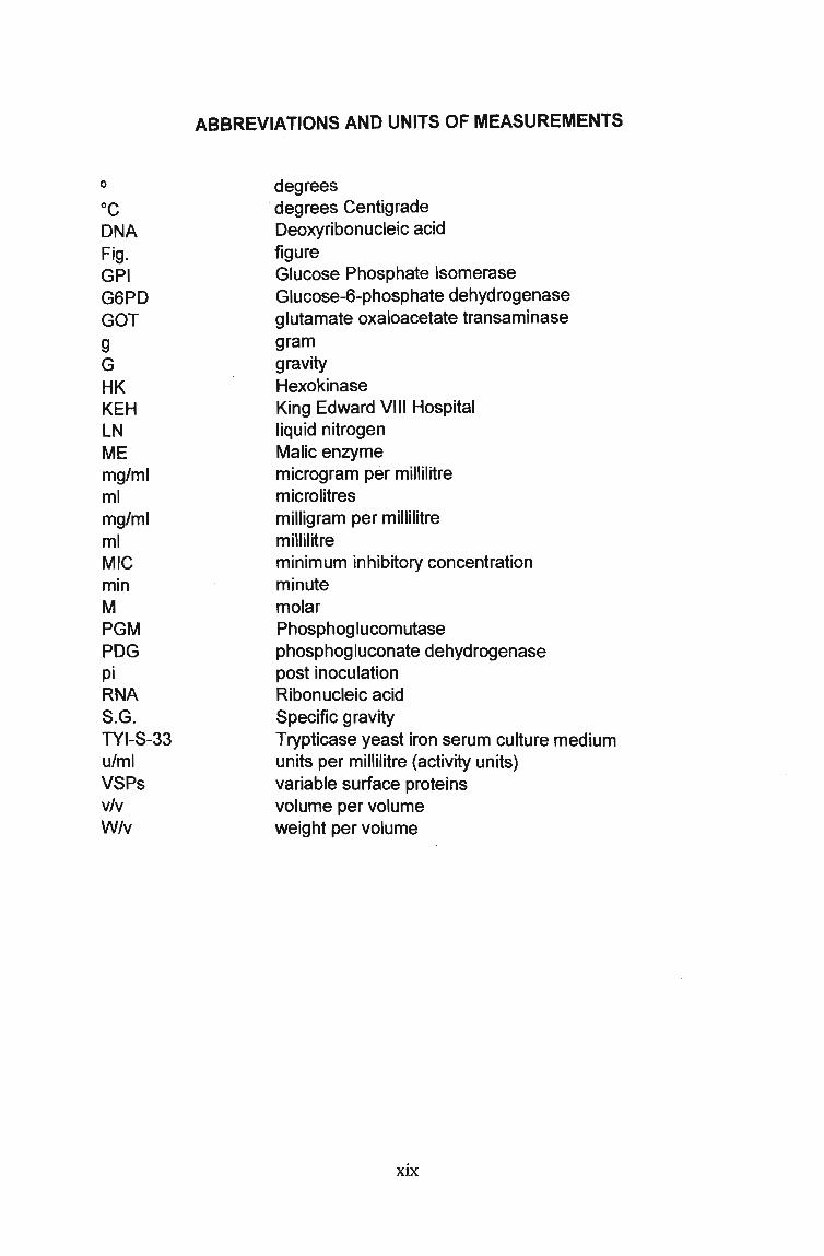
o
°C DNA Fig. GPI G6PD GOT g G HK KEH LN ME mg/ml ml mg/ml ml MIC min M PGM PDG pi RNA S.G. TYI-S-33 u/ml VSPs v/v W/v
ABBREVIATIONS AND UNITS OF MEASUREMENTS
degrees . degrees Centigrade Deoxyribonucleic acid figure Glucose Phosphate Isomerase Glucose-6-phosphate dehydrogenase glutamate oxaloacetate transaminase gram gravity Hexokinase King Edward VIII Hospital liquid nitrogen Malic enzyme microgram per millilitre microlitres milligram per millilitre millilitre minimum inhibitory concentralion minute molar Phosphoglucomutase phosphogluconate dehydrogenase post inoculation Ribonucleic acid Specific gravity Trypticase yeast iron serum culture medium units per millilitre (activity units) variable surface proteins volume per volume weight per volume
xix

CHAPTER 1
PRE-AMBLE
Giardia was one of the first protozoans to be described. In1681 van Leeuwenhoek
discovered the trophozoites of this genus. The Dutch microscopist made glass
lenses and set them into metal frames which he made into simple microscopes.
Among the many specimens he examined with these microscopes was his own
diarrhoeic stool. Clifford Dobell, an English scientist translated van
LeeuwenhoekDs findings of the stool examination thus:
"My excrement being so thin, I was ... . persuaded to examine it....wherein I have sometimes also seen
animacules a-moving very prettily; some of 'em a bit bigger, others a bit less, than a blood globule,
but all of the one and the same make; their bodies were somewhat longer than broad and the belly,
which was flatlike, furnished with sundry little paws, wherewith they made such a stir in the clear
medium and among the globules, that you might e'en fancy you saw a pissabed running up against
a wall; and albeit they made a quick motion with their paws, yet for al/ that they made but slow
progress"
(Dobell, 1920).
Willem Lambl (cited in Filice, 1952) redescribed the organisms in greater detail in
1859 and named them Cercomonas intestinalis. However, it was later found that
this name had been pre-empted by use of the term "intestinalis" for another
parasite in this genus by Diesing in 1850 (Filice, 1952).
In 1888 Blanchard proposed the genus name Lamblia in honour of W. Lambl, to
describe trophozoites from mammal hosts (Erlandsen & Feely, 1984) while
KUristler (Filice, 1952) had named trophozoites isolated from a tadpole Giardia
1

agi/is in 1882. In 1914, Alexeieff stated that the Giardia from a tadpole described
by Kunstler in 1882 and the Lamblia from mammals reported by Blanchard in 1888
are members of the same genus (Filice, 1952). However, the organisms became
known as Lamblia intestinalis and Giardia lamblia. Subsequently, the generic
name Giardia became more popular as it was recognised that the two were
synonymous. This name then took precedence and to-date is the definitive name
for the organisms in this genus.
The work of these early scientists led to the description of this protozoan parasite
that colonises and proliferates in the mucosal surface of the intestine of many
mammalian hosts.
1.1 Taxonomy and Nomenclature
Giardia belongs to the Phylum Sarcomastigophora, Class Zoomastigophora and
Order Diplomonadida. It is regarded as a most primitive eukaryote on the basis of
its small subunit ribosomal RNA (Sogin et al., 1989). However, L-arginine transport
and metabolic pathways characteristically vary from prokaryotes to eukaryotes.
Using these two pathways, Knolder et al. (1995), deduced that Giardia is in
transition between these two Kingdoms.
Early workers based their taxonomic groupings on host specificity (Hegner,
1926b). Filice (1952) proved this to be an inadequate system of classification as
more than forty species had been identified. In his report he illustrated that the
Giardia from some hosts could excyst and set up infections in the intestine of a
different species of host. For example, cysts from dogs effected infection in the
2

guinea-pig intestine; laboratory rats were infected by cysts isolated from human
faeces and cysts from man as well as those from the rat infected a domestic fowl.
He subsequently characterised constant morphological features such as cell
dimensions as well as the number and shape of median bodies, as specific
characteristics in addition to host occurrence. He recognised three morphologic
species, G.agilis, (club-shaped median body) which is infective to amphibians,
G.muris (two small rounded median bodies) from rodents and G.duodenalis (claw
hammer shaped median body) from mammals. G.duodenalis has a wide host
range.
In the ensuing years the taxonomic classification of Giardia remained in a state of
flux as it does today. Lack of agreement regarding the correct specific name for
the mammalian isolates, in particular the Giardia of human origin, greatly
contributes to the taxonomic confusion. The names G.lamblia and G.intestinalis
have been widely used to describe the human isolates. However, Thompson et al.
(1990), stated that the soundly structured, reproducible and logical morphometrics
scheme proposed by Filice (1952) had found widespread favour and was
advocated by many leading authorities in the field. They stated that according to
the Rules of Zoological Nomenclature, duodenalis has priority over intestinalis as a
specific name for the vertebrate isolates since there is no justification for using the
name intestinalis. Other authorities also express the view that the specific name
"duodena/is" is correct for isolates of human origin; other names ("intestinalis" and
"/amb/ia') are incorrect on taxonomic grounds. For example, Meyer (1985)
concluded of other names for the "duodenalis" group (i.e. G.intestinalis or
G.lamb/ia), that their use for Giardia in humans suggests that there is something
3

unique about Giardia in humans, which seemed, on evidence present then, not to
be the case. They therefore advocate use of the specific name "duodenalis" for
taxonomic correctness.
On the other hand, some authorities argue against use of the term G. duodenalis
to designate a species. Their argument is based on the fact that application of
biochemical and nucleic acid techniques has revealed marked genetic diversity
among Giardia isolates that belong to the duodenalis group (morphologically
indistinguishable). For example, a report by Erlandsen et al. (1990) indicated that
a variety of Giardia species possess claw hammer shaped median bodies (thus
belonging to the duodenalis group). However, comparative molecular typing
studies such as karyotype analysis, rDNA restriction enzyme pattern (Mahbubani
et al., 1992) and rRNA gene base pair sequencing (Weiss et al., 1992) show
marked distinction between G.lamblia, G.ardae and G.muri$ (Iamblia and ardae
expectedly belong to the same species (duodenalis) and are therefore presumably
similar). Therefore, according to Erlandsen (1994), Filice's description of the two
specific morphologic groups, G.muris (pair of small rounded median bodies) and
G.agi/is (club-shaped median body) appear to be acceptable. However use of
G.duodenalis (claw hammer shaped median body) to designatei:i species is
controversial. For these reasons, Erlandsen (1994) suggested that the term
G.duodenalis should in future be restricted to the description of the morphological
type of median body within trophozoites and it should not be used as a species
designation because as such it is a misnomer. He argues that although some
investigators indicate that G.duodenalis is synonymous with G.lamblia or
G.intestinalis, and that this species can infect humans, birds and reptiles, such
4

speciation is apparently inappropriate.
Use of the name Giardia lamblia has for a long time been familiar in diagnostic
laboratories. This concurs with the original recommendation made by Or Stiles in
1915, which reads thus: "If you look upon the form in the rabbit as identical with
that in man, duodenalis would be the correct specific [trivial] name. If you consider
the various forms in man, rabbits, rats, etc, as distinct, then in all probability a new
name should be suggested for the form that occurs in man .. .. I would be inclined to
suggest lamblia as specific [trivial] name for the form in man", (Filice, 1952).
Inexplicably, scientists subsequently regarded the duodenalis species name to be
synonymous with intestinalis and lamblia (Erlandsen et al., 1990; Hill, 1990; Adam,
1991; Flanagan 1992). Lymbery and Tibayrenc (1994), in their survey of past
publications identified seven commonly used species names for this genus, based
on host preference and morphology (Table 1.1).
Table 1.1 Commonly used species names in the genus Giardia (Lymbery & Tibayrenc, 1994)
G.duodenalis
G.agilis
G.muris
G.intestinalislG./amblia
G.psittaci
G.ardae
Vertebrates Median bodies claw hammer shaped
Amphibians Median bodies club shaped
Rodents Median bodies small, rounded
Humans As for G.duodenalis
Budgerigar Median bodies claw hammer shaped, trophozoites have incomplete ventrolateral flange
Great blue heron Median bodies small rounded or claw hammer shaped, nuclei tear drop shaped, single caudal f1agellum
5

To-date, use of the different specific names for human isolates (viz. G.Jamblia,
G.duodena/is and G.intestinalis) appears to be based on personal preference.
Although Mayrhofer et al. (1995) stated that those Giardia that infect humans
belong to the morphologic group G.duodenalis but have been assigned to a
separate species G.intestinalis (or G./amb/ia) on the basis of presumed host
specificity, attempts to obtain literature that documents some consensus regarding
the specific name for the human isolates were futile.
Lymbery and Tibayrenc (1994) quite correctly stated that the species level
systematics have not been resolved. Furthermore, there is increasing evidence
that the Giardia infecting humans comprise a species complex. For example,
Andrews et al. (1989) used isoenzyme electrophoresis to characterise 29
Australasian human isolates and 48 clones from these strains. Four distinct types
were identified based on the 26 enzyme patterns. Recently, Upcrofft et a/.,(1995)
characterised 40 stocks of Giardia using biochemical characteristics (karyotype,
RFLP and rDNA analyses) over eleven years (1982-1993). During this period at
least two major varieties had infected the population of Southeast Queensland.
This additional diversity among the human isolates may further complicate the
present classification problems currently faCing taxonomists. However, it is
anticipated that with the advent of advanced molecular typing techniques, the state
of flux existing in Giardia taxonomy will be resolved.
In the light of a lack of consensus on Giardia nomenclature at present, the name
Giardia lamb/ia will be used in the current study to describe all isolates obtained
from humans.
6

1.2 Life-Cycle and Morphology
Giardia has a simple life-cycle with two morphological forms, the trophozoite
(vegetative) and a robust cyst (Fig.1.1).
Cyst: 9 to 12 pm
Kinetosome
Median body
Axostyle
Trophozoite : 12 to 15 pm
Fig.1.1 The two morphological forms of Giardia lamblia. Cyst (transmisSible) and . trophozoite (vegetative).
The binucleated pear-shaped trophozoites measure 9-12 IJm by 5-151Jm (Hill,
1990). They have a convex dorsal surface and a flat ventral surface with an
anterior adhesive disk by which they adhere to the host's mucosa. Four pairs of
f1agella originate from the kinetosomal mass. This form attaches to and inhabits
the upper small intestine of the host. The parasites multiply rapidly by binary
fission, although there is evidence that at least some populations may be capable
of a form of sexual reproduction (Meloni et al., 1988; Lymbrey & Tibayrenc, 1994).
7

Differentiation into the cyst stage (8-12JJm by 7-10JJm) occurs in the relatively drier
environment in the lower intestine as trophozoites are flushed downstream by
peristalsis. The cyst is the transmissible form of the parasite. In diarrhoeic stools, a
shorter time span in the large intestine results in the passage of trophozoites with
the faeces. Although they are far more labile than cysts, results from experimental
infections suggest that trophozoites may survive passage through the stomach to
establish infections in the duodenum (Hegner, 1926a). However they have limited
survival outside the host and are therefore an unlikely agent of parasite
transmission.
Upon expulsion with the faeces, the cysts can survive for about two months in a
suitable environment such as cool water (Jakubowski et al., 1988; Adam, 1991).
Ingestion of cysts by the next host through faecal-oral contamination,
contaminated water or food results in excystation of cysts in the duodenum, which
initiates the next life-cycle (Fig.1.2).
Under a light microscope, cysts are highly refractile oval bodies with fragments of
unassembled disk structures and rod-like central axonemes (flagella remnants).
Viable cysts can be visually recognised by close adherence of protoplasm to the
highly refractile cyst wall. Increased granularity and a large peritrophic space
between the cyst wall and parasite indicates loss of viability (lsaac-Renton, 1991).
8

Fig. 1.2. Life-cycle of Giardia lamblia. Ingestion of cysts results in excystation in the duodenum and subsequent colonisation of upper small intestine.
9

1.3 Transmission
1.3.1 Human hosts
The cyst is spread through the faecal-oral route. Different mechanisms of
transmission have been documented such as direct person to person hand-mouth
transfer. This is more common in children (Isaac-Renton et al., 1993) owing to lack
of personal hygiene and faecal incontinence. Prevalence is higher in children than
in adults in endemic areas (Adam, 1991; Flanagan, 1992). There is suggestive
evidence that institutionalised persons and day care attendees are at higher risk of
infection, for example Cody and colleagues (1994) isolated cysts from chairs and
tables in day care centres. In such settings, transmission rates are expectedly very
high, and can subsequently enhance the overall transmission of the parasite. To
illustrate this, day care attenders were shown to spread Giardia infection to the
community in a controlled study by Polis and co-workers (1986). In this study, data
suggested that 47% of the children in the centre transmitted giardiasis to at least
one household contact. Although humans of all ages are susceptible to infection
with Giardia, the prevalence is higher in children (Farthing et al., 1986) and
infection frequently occurs in immunocompromised individuals (Webster, 1980).
Inadequate sanitation exacerbates dissemination of cysts to the environment.
Surface water supplies are also known to be contaminated with cysts and infected
humans replenish this reservoir. Furthermore, wild and domestic animals such as
beavers, muskrats, dogs and cats have also been implicated in transmission but
their potential as zoonotic reservoirs of giardiasis is still debatable (Erlandsen,
1994). Prolonged occurrence of viable cysts in fresh cool (4°C) water has resulted
in Giardia being reported as the most common aetiologic agent of epidemic
10

waterborne diarrhoeal disease in North America and Europe (Craun, 1984;
Jephcott et al., 1986). The organisms have been shown to resist the level of
chlorination used in many bulk water purification systems. Sole reliance on this
method can result in widespread transmission. Filtration and flocculation in
addition to chlorination is recommended in order to effectively eliminate Giardia
cysts from water supplies (Longsdon et al., 1979)
Travel (particularly to endemic areas) has also been documented to facilitate
transmission of Giardia. For example Brodsky et al. (1974) collected data on
surveillance of giardiasis in American travellers to the Soviet Union. This data
implicated Leningrad as the site of infection and tap water as the probable sburce.
Flies have been implicated in playing a role in transmission of intestinal protozoa
including Giardia, as cysts may remain alive in the fly intestine for a considerable
period (at least 24hours) and may be deposited in a viable state as early as 40
minutes after the fly has fed on contaminated material (Hegner, 1926b).
Furthermore, it is suggested that since the infective dose is very small (10-25
cysts), it is likely that flies may act as reservoirs and transmit Giardia cysts.
However Hall, (1994) noted that it would be difficult to establish whether cysts
isolated from flies are viable.
Other modes of transmission have been reported. Food-borne cases of giardiasis
have been reported with food-handlers being the most common source of
transmission of cysts to freshly prepared food (Adam, 1991) and sexual
transmission, particularly in homosexual males also occurs (Schmerin et al., 1978;
11

Owen, 1984)
1.3. 2 The Zoonosis of giardiasis
The role of wild and domestic animals in the transmission of giardiasis to humans
remains unclear. Many animals including dogs, cats, cattle, sheep and wild aquatic
mammals have been proposed as both potential reservoirs as well as amplification
hosts for spreading human Giardia infections (Isaac-Renton, 1991). In the USA
beavers have been incriminated as an animal reservoir (Wolfe, 1979; Isaac
Renton et al., 1993). Cross-species infections effected by several investigators in
animals such as rats (Filice, 1952), gerbils (8elosevic et al., 1983; Aggarwal &
Nash, 1987) and mice (Hill et al., 1983; Byrd et al., 1994) with Giardia from
humans provide evidence for lack of absolute host-specificity in these organisms.
Furthermore, Faubert (1988), proposed that giardiasis is a zoonosis since Giardia
species isolated from beavers and calves were indistinguishable from isolates of
human G.duodenalis at the light microscope level and, as with human G.lamblia
strains the trophozoites could be cultivated in vitro. Additionally, a report on the
occurrence of giardiasis in backpackers who drank water from areas with no
human inhabitants strongly suggested that beavers or other wild animals are
reservoirs (Barbour et al., 1976). Thompson et al. (1988) reported that all six feline
isolates (5 from Australia and 1 from the USA) were genetically identical (as
revealed by isoenzyme and DNA analyses). They were also very similar to 20 of
30 human isolates, thereby indicating that cats are a likely reservoir of infection for
humans. Meloni et al., (1988) also advocated that felines act as a reservoir of
infection to humans; therefore their potential in zoonotic transmission is important.
12

However, Erlandsen (1994) pointed out that most of the studies implicating animals
as potential reservoirs for human infection were flawed and/or biased. During
outbreaks, investigators overlooked the potential of birds and muskrats while
concentrating on beavers and other aquatic mammals only. Also, contamination by
raw sewage (particularly of human origin) was overlooked. Birds were implicated in
transmission of giardiasis based solely on the morphological similarity of Giardia from
Parakeets and Great Blue Herons to Giardia found in humans. He further stated that
"the evidence for the implication of the beaver or any animal in waterborne outbreaks
is very circumstantial". The fact that waterborne outbreaks of giardiasis are sporadic
whilst the aquatic animals are in semi-permanent residence in the water makes their
incrimination more questionable. More rigorously controlled studies are still required,
hence to-date, the zoonosis of giardiasis is still debatable.
An initial school of thought regarding host-specificity upheld by early scientists was
that the Giardia of mammals are rigidly host-specific and morphologically distinct
(Hegner, 1926b). Filice's breakthrough study (1952) in clarifying the species
definition led to the second school which holds that some Giardia are capable of
infecting more than one vertebrate host species, while others may be host specific
(Meyer, 1990) This has been reinforced by several cross-species experiments
wherein different animals such as rats, guinea-pigs, domestic fowls (Filice, 1952),
mice (Hill et al., 1983; Aggarwal et al., 1983; Mayrhofer et al., 1992, Byrd et al.,
1994) and gerbils (Belosevic et al., 1983; Wallis & Wallis,1986; Aggarwal &
Nash, 1988; Visvesvara, 1988) have been successfully infected with Giardia of
human origin. It would appear therefore that the second school of thought is the
view of the majority of the contemporary research community.
13

1.4 Distribution
The morbidity and misery of giardiasis is experienced world-wide because of the
ubiquity of Giardia. The global prevalence Was reported to be 200 million in 1993
(Crompton & Savioli, 1993). It is the most common protozoan cause of diarrhoea
in the United States (Wolfe, 1979; Isaac-Renton, 1991) and is the most frequently
reported intestinal parasite of humans in the United Kingdom, (a prevalence of
1,8% of 835 asymptomatic adults screened in a study in Manchester by
Kaczmarski & Jones was reported in 1989). In the United States 3,9% of more
than 300 000 submitted stool samples (with prevalence values as high as 16% in
some areas), were documented (Hill, 1990) and a mean annual rate of infection
per annum was reported to be 4,6 per 10 000 population (Flanagan, 1992). In
Australia 250 asymptomatic pre-school children in Sydney had a prevalence of
infection of 6,8% (Walker et al., 1986) and a rate of 20,2 per 100000 population
was reported (Kaczmarski & Jones, 1989). In Great Britain, an incidence of 0,9 per
10 000 population annually was reported (Flanagan, 1992).
Prevalence varies between <1 and 50% depending on the population being
sampled. It is more prevalent in underdeveloped than in developed countries. For
example, prevalence estimates vary between 8 and>40% in South America, the
Caribbean, the Middle East and South East Asia. In a study in Egypt, 100% of the
population was found to be infected over a six month study period (Flanagan,
1992). In contrast, in most developed countries of Western Europe, Australia, New
Zealand, and North America, prevalences of between 2 and 7% are more common
(Farthing et al., 1986; Flanagan, 1992). However, Giardia infections still have an
impact on public health problems in these countries.
14

Evidently Giardia differentially infects populations of differing socio-economic
backgrounds. It would not be unreasonable to expect rural communities with
sparse facilities to have a higher prevalence of giardiasis than the urban
population who have proper sanitation, piped and treated water and a relatively
higher level of personal hygiene. Contrary to expectations, two independent
studies in Bangladesh (Hossain et al., 1983) and Zimbabwe (Mason et al., 1986)
found that the prevalence of giardiasis was higher in urban children than in the
children in rural areas. It was speculated that this could be related to factors such
as high population density in urban areas complicated by overcrowding and poor
sanitation of urban slums in developing countries (Rabbani and Islam, 1994).
In rural areas in South Africa, there is often poor sanitation, restricted water
supplies and malnutrition, all of which promote Giardia infection and transmission.
Furthermore, urban areas are overcrowded and have slums with minimal sanitary
facilities. Therefore there is a need for diagnostic surveillance of this growth
retarding parasite in this country. Several local studies reflect significant infection
levels. For example, Millar and colleagues (1989) performed a survey of parasitic
infestation in Cape Town and found that of the 101 children screened, 8 had
Giardia cysts, and about 46% had multi-parasitosis. More recently, Evans et al.,
(1998) determined the prevalence of Giardia among five communities in the
Western Cape by multiple stool assessments. They reported a mean prevalence
of 18,1 % (with a range of 6-36%) after screening 3976 stools using the formol
ether methods.
In Kwa-Zulu Natal, a survey of intestinal parasitic infections in Black school
15

children by Schutte et al. in 1981 revealed Giardia prevalence levels ranging
between 2.8 and 4.3% within the four regions screened (all in the former northern
Zululand). In a recent study undertaken in the Amoebiasis Research Programme
laboratory (South African Medical Research Council-Kwa-Zulu Natal), 4% and
13% of 484 and 309 stool samples respectively, from local schools screened for
par?sites were found to have Giardia cysts (Unpublished data). Recently, Jackson
et al. (1998) reported a 63% prevalence in a cohort of 175 abandoned children in
two shelters in Durban (Kwa-Zulu Natal). Furthermore, a prospective study is
currently being undertaken by the Amoebiasis Research laboratory involving
multiple stool examinations (at least 3 examinations at 3 monthly intervals) of 984
adult subjects recruited from a disadvantaged community in Durban. Preliminary
results reveal a prevalence of 4,5% among this population. As most of the
reported surveys were based on single stool examinations and all were relying on
microscopy (both methods are documented to be insensitive -Ament & Rubin,
1972; Kamath & Murugasu, 1974) these data are an underestimation ofthe true
occurrence of Giardia infections locally. However, they provide an estimate of the
local prevalence.
1.5 Host .. parasite Relationship
Giardia organisms were once considered harmless commensals in the human gut
(Dobell, 1920; Rendtorff, 1954). Furthermore, Erlandsen and Chase (1974)
proposed that the rodent Giardia was an indigenous member of the enteric
microbiota. After years of debate on the pathogenic potential of this parasite, it
became evident that these organisms are associated with a cadre of symptoms
producing a disease known as giardiasis. The latter is illustrated by several studies
16

in which travellers (Brodsky et al., 1974), experimentally infected humans (Nash et
al., 1987) and animals (Roberts-Thompson et al., 1976; Aggarwal and Nash,
1987) developed symptoms after exposure. Infections give rise to varying clinical
signs and symptoms from asymptomatic cyst passage to acute and chronic
diarrhoea associated with villous atrophy, malabsorption and growth impairment in
children (Farthing et al., 1986; Hjelt et al., 1992). Consequently, renewed interest
has been shown in this genus. It has since been considered an important public
health problem and the most frequently identified intestinal protozoan parasite,
particularly in the United States (Wolfe, 1979; Smith et al., 1982; Hill, 1990).
Transmission is mainly via the faecal-oral route. For this reason, it is more likely to
be endemic in areas that are characterised by poverty, over-crowding, poor
personal hygiene, lack of proper sanitation facilities and inadequate purification of
water supplies. However, it also has a considerable impact in developed countries
such as the United States, Great Britain and Australia as discussed in the
preceding section.
Although establishment of axenic laboratory cultures was achieved two decades
ago thereby facilitating study of these organisms in vitro, many key areas still
require clarification such as:
1.5.1 Pathogenesis.
Many theories have been postulated. Early investigators (Erlandsen & Chase,
1974) suggested the possibility that numerous organisms in the small intestine act
as a mechanical barrier leading to malabsorption. This was disputed on the basis
17

of (a) the enormous absorptive capacity of the small bowel (Hill, 1990) and, (b)
fewer parasites were isolated in some patients with marked symptoms (Wolfe,
1979). However, Buret (1994) recently stated that the parasite burden contributes
to the pathology of giardiasis.
Production of diarrhoea has been ascribed to bacterial overgrowth and bile salt
deconjugation, which is common in giardiasis (Tandon et al., 1977). On the
contrary, Nash and colleagues (1987) found that bacterial overgrowth was not
associated with symptomatic giardiasis in studies of infected human volunteers.
Saha and Ghosh (1977) reported superficial invasion of intestinal mucosa in
human infection which was associated with steatorrhoea; however, contradictory
findings were reported by Owen and co-workers (1979) in their observations of
murine giardiasis. They reported that "in animals with intact epithelium, Giardia are
rare beneath the surface and that penetration probably occurs when trophozoites,
randomly moving forward, enter disruptions in the epithelium or cavities left by
desquamating cells." A recent finding by Nash and Mowatt (1993) indicated that
the variant-specific cysteine-rich surface proteins have a high affinity for certain
metals, including zinc. These proteins then can act as a "zinc sink" in which the
cations are trapped (on the surface of the trophozoites) and thus rendered
unavailable for digestion and absorption functions. They proposed that
malabsorption could be caused by zinc deficiency resulting in inhibition of all zinc
dependant enzymes.
Roberts-Thompson (1993) affirmed that the reasons for the development of
diarrhoea resulting from infection with Giardia species remain unclear. However,
18

two factors are associated with the development of symptoms, namely the degree
of inflammatory changes in the small bowel and damage to microvilli, which lead to
a deficiency of brush border enzymes, particularly lactase.
1.5.2 Do host factors play a role in increasing the susceptibility of certain
hosts to infection?
Many variables such as age, sex, immune and nutritional status, ABO blood
groups, Human Leukocyte Antigen (HLA) type, socio-economic factors and gastric
acidity have been implicated in predisposing hosts to giardiasis. The exact nature
of the relationship between 'the disease and these factors has not been
conclusively described. Brief discussions on these factors are outlined in 1.7
below.
1.5.3 Are immune responses elicited by infection with Giardia responsible
for parasite clearance and sterilising immunity?
In some instances these infections are cleared spontaneously. However re
infection of hosts frequently occurs (Farthing et al., 1986; Gilman et al., 1988). It is
not clear whether partial protective humoral immunity occurs in giardiasis. Re
infection has been documented in infants and children (Farthing et al., 1986) as
well as in adults (Nash et al., 1987; Gilman et al., 1988), thus the issue of
immunological maturation with age cannot be used to explain the recurrent
infections in some individuals. In experimental human infections, re-infection was
confirmed as the volunteers were successfully treated after initial infections and
they were successfully re-challenged (Nash et al., 1987). In stUdies of the Giardia
specific immune response in infected human volunteers, serum immunoglobulins
19

Ig M (100% of patients), IgG (70%), IgA (60%) and intestinal secretory IgA (50%)
were documented, but, in these patients the IgA response was not clearly
correlated with resolution of infection (Nash et al., 1987). On the other hand, in
one clinical study, the protective role of specific secretory IgA was apparently
passively acquired by breast-fed infants of G.lamblia-infected mothers with high
titres of the antibody in their milk (Nayak et al., 1987). The results of the latter
study suggest a protective humoral response, whereas in the former study, where
re-infection occurred despite the presence of immunoglobulins, such protection
was not demonstrated. However, it should be noted that human milk was shown to
have a non-immune lethal effect on trophozoites in vitro (Gillin, 1987). In the study
of Nayak et al. (1987) it was not indicated whether this effect was excluded or not.
1.5.4 Are strain differences responsible for variations in the clinical course
of infection?
In 1988, Nash and Aggarwal demonstrated that two distinct isolates of Giardia
gave rise to different clinical outcomes. In both humans and gerbils, one strain
appeared to induce resistance to re-infection on subsequent re-challenge with the
homologous organism, whilst infection with the other strain persisted for a longer
period and re-challenge resulted in infection. Further, the surface antigens of
Giardia show a large degree of diversity and undergo surface antigenic variation
(Adam et al., 1988; Nash et al., 1988; Nash and Mowatt, 1993). It is not clear
what role is played by this antigenic variation in clinical disease. However Nash
and colleagues (1991) demonstrated that different isolates expressing varying
surface antigens were variably susceptible to intestinal proteases (trypsin and
chymotrypsin). Such differences may explain some of the variability in the clinical
20

features noted in human infections.
1.5.5 Identification of virulence determinants of the organism which would
help in new drug and vaccine design.
IgA 1 protease activity (cleaving IgA 1 immunoglobulin and haemoglobin) has been
described in Giardia intestinalis trophozoites (Webster, 1980; Parenti 1989)
suggesting that Giardia species can survive in the intestine by degrading host IgA.
The protease activity may be a non-specific virulence factor that allows the
organism to evade host enteric defence mechanisms.
A trypsin-activated, mannose-binding lectin which mediates adherence of the
parasite to host epithelia has been demonstrated on the surface of Giardia
trophozoites (Lev et al., 1986). Whether absence or inhibition of these binding
molecules could prevent parasite colonisation and thus interfere with production of
clinical illness has yet to be investigated.
Giardia has been found to display a large degree of diversity in its surface
antigens (Adam et al., 1988; Aggarwalet al., 1989 Nash et al., 1988; 1990). These
antigens represent a distinct family of cysteine-rich proteins termed variant-specific
surface proteins (VSPs), which cover the entire surface of trophozoites including
the f1agella (Nash, 1992). The trophozoites can vary their surface antigens and
rates of change of VSPs vary markedly among isolates (Nash et al., 1988). These
surface antigens appear to play a role in pathogenecity (virulence) of different
isolates of Giardia:
• The documented differences in resistance to proteases (trypsin and
21

chymotrypsin) between different isolates expressing different VSPs (Na~h et
al., 1991) might suggest a possible mechanism for the virulence differences
among isolates by enhancing survival in the host intestine.
• Experimental infection of gerbils (Aggarwal & Nash, 1987) and humans (Nash
et al., 1987) with two different G. lamblia isolates showed a marked difference
in pathogenesis between the two isolates. All ten human volunteers inoculated
with the GS isolate were infected and 5 developed symptoms, whereas none of
5 volunteers inoculated with the ISR isolate were infected. These two isolates
had been shown to express different (Nash et al., 1988). In subsequent
studies, human volunteers were inoculated with two cloned GS isolates
expressing different VSPs (72 and 200 kDa) (Nash et al., 1990). AII4
inoculated with the GS clone expressing the 72 kDa antigen were infected,
whilst only 1 of 13 inoculated with the clone expressing the 200 kDa antigen
were infected. These observations also indicate differences in virulence among
surface antigen variants from the same isolate.
Further studies are required to find more virulence determinants in these
organisms and a possibility of manipulating the VSPs for new drug target sites.
1.6 Clinical Manifestations
In man, the infective dose of Giardia was documented to be 10-25 fresh cysts by
Rendtorff (1954) when imprisoned human volunteers were given encapsulated
Giardia cysts. After ingestion of the cysts, a broad clinical spectrum of disease
ensues. The most commonly noted feature world-wide is asymptomatic carriage.
However, whether these carriers have a transient symptomatic phase, which
22

passes unnoticed, is debatable. Although the parasite is frequently harboured by
apparently symptom less persons who transmit cysts to the environment, this may
give rise to symptomatic infections in other individuals.
The clinical course of acute giardiasis is easily recognised in travellers to endemic
areas as they present with sudden onset illness during or shortly after travel
(8rodsky et al., 1974). The predominant features of acute giardiasis are explosive
diarrhoea, abdominal discomfort, nausea, headache and bloating. Symptoms can
complicate steatorrhoea and malabsorption in up to 25% of patients (Hjelt et al.,
1992) and often cause weight loss (Gerhard, 1989; Heinz, 1988; Meloni et al.,
1988; Ukoli, 1984). Frequently, the infection is self-limiting; however, it is
estimated that 20 -50% of symptomatic patients will proceed to the chronic stage
of the disease. The symptoms can persist for weeks, months or several years.
Smith et al. (1982), described two and a half years of persistence of symptoms in
a patient with giardiasis, while Tandon and colleagues (1977) reported the
duration of diarrhoeal symptoms varying from two months to 12 years.
There is increasing evidence that G.lamblia infection is very debilitating particularly
in younger hosts. Although children apparently tolerate these infections, their
physical and mental development is severely retarded. Farthing et al. (1986)
studied the impact of Giardia infection on the growth of infants and children, and
found that it had had deleterious effects. The rate of weight gain was significantly
lower in the second year of life in Giardia infected children, when compared to that
of uninfected children. The duration of Giardia episodes and their association with
diarrhoea appeared to be the most important factors associated with growth
disturbance. Similarly, Hjelt et al. (1992) illustrated that giardiasis had a negative
23

impact on the physical growth of 29 children with chronic disease. According to
growth charts, the relative heights and weights of the subjects decreased
significantly during the course of the disease with severe villus atrophy also being
recorded. These workers also demonstrated that the degree of mucosal damage
correlated with D-xylose and lactose malabsorption. The younger patients were
also more severely affected than the older ones.
Upadhyay et al. (1985), showed that uptake of nutrients by infected, malnourished
animals was compromised. A similar phenomenon could be occurring in
malnourished children and be attributed mistakenly to the overall effects of
deprivation in poverty-stricken areas. Such an association between malnutrition
and giardiasis was reported by Gilman et al. (1985) where 51% of severely
malnourished children living in low socio-economic environments had giardiasis
compared to a prevalence of 21 % among age-matched control children. A
malnourished child has low gastric acidity, depressed intestinal immune functions,
retarded enzyme activity, poor intestinal motility and low levels of vitamins, trace
elements and minerals. All these factors make the child susceptible to enteric
infections including giardiasis (Rabbani and Islam, 1994).
It is suggested that trophozoites can restrict the absorptive area of the gut
epithelium, while competing with the host for nutrients, in addition to causing
mechanical irritation (Erlandson & Chase, 1974) leading to a malabsorption
syndrome (Heinz, 1988). During colonisation, Giardia trophozoites were shown to
leave imprints of their powerful adhesive disk on the mucosal surface (Owen et al.,
1979). This finding supports the theory of mechanical irritation playing a role in
24

pathogenesis. Malabsorption of electrolytes, solutes and water in the upper small
intestine appears to be the primary mechanism of diarrhoea production in
giardiasis (Buret, 1994).
As pathogenesis depends on successful establishment of infection, adherence has
been implicated as an indirect contributing factor. Trypsin-activated lectins have
been documented to play a role in parasite adherence to the mucosa (Lev et al.,
1986; Adam, 1991; Nash et al., 1991). Inge et al., 1988 showed that the parasite
lectins may be required for the selective establishment of Giardia infection in the
jejunal epithelia rather than colonic cells (a predilection for the host's upper small
intestine).
Vast reduction of the villus to crypt ratio (Fig.1.3) has been revealed by
transmission electron microscopy in animal giardiasis (Roberts-Thompson et al.
1976). Disruption of the brush border in the small intestine, crypt epithelial
hyperplasia and mononuclear inflammatory cell infiltration have been documented.
The latter is proposed to contribute both to the disease process as well as in
parasite eradication (Farthing, 1989; Owen et al. 1979). Sometimes the parasites
reach the gall bladder and bile ducts and cause jaundice.
25

Figure 1.3. Transmission Electron micrograph of the small intestine in murine giardiasis (right) compared to the normal jejunum of an uninfected control animal (left) (Roberts-Thompson, et al., 1976). (x 160) (Courtesy of A.Mahmoud)
Smith et al., (1982) tested four strains of Giardia from humans for toxin production
in rabbits and found that these organisms did not produce toxins. To date, no
reports showing evidence for toxin production have been published. However, it
was suggested that Giardia excretory-secretory products may play a role in the
pathogenesis of giardiasis, either as a trigger for the diffuse brush border injury, or
via direct cytopathogenetic effects during close contacts between host cells and
trophozoites, and this needs to be investigated (Buret, 1994). Lysosome-like
vacuoles that line the surface of trophozoites have been identified (Feely et al.,
1984). These organelles have been shown to release numerous hydrolases,
including a thiol-dependant proteinase (Parenti, 1989). A possibility of secretion of
the proteinase has been indicated, although there is only limited evidence in
support of this. It was further suggested that under selected conditions, the
proteinase might be released into the extracellular milieu and play a role in the
pathogenesis of diarrhoeal disease (Parenti, 1989).
The association of giardiasis and allergic reactions have been documented.
26

Balazs and Szatloczky (1978) reported a rare form of G./amblia infection in a
patient who presented with a clinical picture of nutritive allergy. An acute
hypersensitivity syndrome consisting of urticaria, polyarthropathy and eosinophilia
occurring in association with G./amblia was documented by Farthing et al. (1983).
Perez et al. (1994) have also reported the association of the above two clinical
conditions. Other extraintestinal manifestations of giardiasis have been
documented. For example, a more recent study by Corsi et al. (1998) has shown
that giardiasis can also cause ocular complications in the form of asymptomatic,
non-progressive retinal lesions. These retinal complications were particularly
common in younger children with the disease and were detected in both hosts with
active and/or past Giardia infections. The retinal changes have not been
associated with Giardia toxin. The authors suggested that they are more than
likely caused by immune mechanisms.
Although rare, fatal cases of giardiasis have been documented. For example,
Ryder et al. (1985) reported eight fatal cases of diarrhoea, one of which was
associated with G./amblia.
1.7 Risk Factors For Giardiasis
1.7.1 Age and immune status of the individual
Humans of all ages are susceptible to Giardia infections; however, some factors
appear to predispose certain individuals within some populations to increased risk
of infection. High prevalence levels have been reported in children in many
countries; this predisposition usually rapidly declines with increasing age.
Prevalence among adults is usually less than 10% and peak prevalence occurs
27

among children between the ages of 1 and 5 years (Roberts-Thompson, 1993).
Polis et a/., (1986) documented prevalences between 21-60% among infants and
pre-schoolers in a day care centre. Further, a 21 % prevalence in healthy children
between 5 and 10-years and up to 51% in malnourished, 1 to 5-year old children
was documented by Gilman et al. (1985). The results of a prospective, longitudinal
study of a cohort of 45 rural Guatemalan children from birth to 3 years showed that
the children had an average of 3,6 episodes of Giardia in their third year of life. All
children had at least one Giardia infection by this age (Farthing et a/.1986).
Prevalence is therefore apparently age-specific. It is speculated that both frequent
exposure combined with a low level of personal hygiene among small children
plays a predominant role. It is also postulated that malnutrition and chronic
diarrhoea (common in children in poorer communities) increases susceptibility
(although the true relationship in terms of cause and effect has yet to be
established). This view was supported by a study of 31 Gambian children with
chronic diarrhoea and malnutrition (Sullivan et al., 1991). Forty-five percent of
these children had giardiasis compared with only 12% of 33 healthy age and sex
matched control children recruited from the same geographical location. Similarly,
in the study of Bangladeshi malnourished children with chronic diarrhoea aged
between 1 and 5 years, a 51% Giardia prevalence was documented by Gilman et
al. (1985). It must be noted though that most of these studies were undertaken in
developing countries, therefore the age-specific prevalence must be considered in
conjunction with such factors as the socio-economic disposition of the population.
For example, in a first world country (Denmark), Hjelt et al. (1992) reported a low
incidence of 81 per 1 000000 children aged 0-7 years per annum over a 6-year
period.
28

Some investigators proposed that this age-related prevalence could also be a
function of an underdeveloped immune system. This was demonstrated in a
G.lamblia-suckling-mouse infection study (Hill et al., 1983). In this animal model of
Giardia infection, all mice, 3,7 and 14 days old became infected whereas older
mice (>14 days old), as well as the mothers of infected suckling mice, which
presumably were ingesting large numbers of cysts from their babies' stools, did not
become infected. All infected mice cleared the infection 17-21 days after
inoculation, and these workers have ascribed this to immunologic maturation,
which they reported to occur at one to four weeks of age in mice.
In humans, the possibility of a relationship between the humoral immune response
and the development of symptomatic Giardia infection was illustrated in a study by
Vinayak & Kumkum (1989). They showed that children with chronic symptomatic
giardiasis that were resistant to therapy had lower titres of antibody to two Giardia
antigens than children with acute or asymptomatic infection.
Immunodeficiency has been shown to predispose to Giardia infection and appears
to be a major contributor to persistence of symptoms as chronic giardiasis occurs
in individuals with immunoglobulin deficiency (Webster, 1980; Lo Galbo et al.,
1982). Some investigators have stated that patients with
hypogammaglobulinaemia are at increased risk of severe giardiasis (Lo Galbo et
al., 1982, Adam, 1991). Similarly, Ament & Rubin (1972) reported a strong
association between a defective immune system and giardiasis. Seven of 8
patients with hypogammaglobulinaemia and gastrointestinal symptoms were
infected with G.lamb/ia. Animal studies have also showed this phenomenon
29

where immunocompromised mice have presented with the chronic form of
giardiasis in comparison with their immunocompetent counterparts (Roberts
Thompson, 1993).
Other conditions suggestive of an association between immune dysfunction and
giardiasis are documented. For example, Cruz et al., 1991 and 8assoti et al.,
1991) both reported anassociation of Giardia with Whipples disease in
immunodeficient patients.
There is an apparent association of intestinal parasitosis (including giardiasis) and
HIV infection. A prevalence of 15,2% among 79 Brazilian, HIV-infected patients
was cited by Rabbani & Islam (1994). Connoly et al., (1990) stated that the
prevalence of giardiasis among AIDS patients may be underestimafed because of
incomplete investigation of suspected cases. They showed that extensive
microbiological and histological examinations of the intestine revealed 2 cases of
giardiasis, diagnoses previously missed among the 33 HIV positive patients.
1.7.2 Genetic factors
1.7.2.1 Host factors
Genetic profile is believed to predispose a host to infection with Giardia.
Association between certain ABO blood groups, HLA antigens and giardiasis has
been documented (Farthing, 1989). This author suggested that ABO blood groups
influence the duration or severity of symptoms because glycoproteins with blood
group activity can influence the adhesion of Giardia. Patients with giardiasis have
been documented to have a higher than expected frequency of blood group A
30

(Roberts-Thompson, 1993). Furthermore, it has been reported that individuals with
blood group A are prone to develop achlorhydria which increases their
susceptibility to Giardia infection (Paulsen, 1977). In a study in adults in Australia
(Roberts-Thompson et al., 1980), patients with prolonged giardiasis had a higher
than expected frequency of HLA-A1 and HLA-B12 and a lower than expected
frequency of HLA-A3 and HLA-B35. Roberts-Thompson et al. (1980) suggested
that HLA antigens or linked genes might be associated with a defective immune
response to 'host protective' antigens of Giardia, however this suggestion still
needs to be tested.
1.7.2.2 Parasite factors
Successful establishment of infection appears to depend on parasite factors as
well. For example variations in infectivity in experimental human infection by two
distinct G.lamblia isolates are documented (Nash et al., 1987). Furthermore, when
gerbils were infected with two well characterised Giardia isolates, different patterns
of infection and degrees of self-cure and varying abilities to induce resistance to
homologous and heterologous rechallenge were observed (Aggarwal & Nash,
1987). Other studies demonstrated variation in outcome of infection in gerbils
(virulence and infectivity) by Giardia organisms isolated from symptomatic and
asymptomatic humans (Visvesvara et al., 1988) and in mice (Aggarwal et al.,
1983). These stUdies indicate that intrinsic differences (which may be genetically
determined) exist among different isolates of Giardia. These differences may
confer variations in infectivity of the different isolates.
Clinical isolates of Giardia contain mixes of genotypes with a possibility of at least
31

up to 4 distinct genotypes within each clinical isolate (Andrews et al., 1992). These
authors illustrated that in vivo and in vitro culture methods select different
genotypes. Furthermore, it is speculated that in endemic areas where extensive
genetic heterogeneity exists within Giardia populations, mixed infections with more
than one genetic variant are likely to occur (Finn et al., 1994). These authors
demonstrated in vitro competitive interaction between 2 genetically distinct isolates
with different growth rates. In this study, the faster-growing isolate greatly out
competed the slower growing organism. It is likely that such competitive
interactions occur in vivo during colonisation of hosts. As discussed earlier,
Giardia genetic variants expressing different surface antigens were shown to be
differentially susceptible to the intestinal proteases trypsin and chymotrypsin (Nash
et al., 1991). It is therefore more likely that during natural Giardia infections, such
population dynamics of the parasite play a role in determining the outcome of
infections in humans.
1.7.3 Travel
Travel (particularly to endemic areas) has been implicated in increasing the risk of
contracting giardiasis, leading to the latter being widely known as travellers'
diarrhoea. The water supplies in Leningrad and towns in the Rocky Mountains of
North America have been strongly associated with outbreaks of Giardia in visitors
and local inhabitants. A questionnaire survey of 1 419 travellers to the Soviet
Union performed by the Center for Disease Control (COC) Bethesda, USA,
showed that 324 individuals (23%) had acquired giardiasis (Brodsky et al., 1974).
Furthermore, in a survey on intestinal parasites by the COC, the majority of the
670 cases of giardiasis were patients returning from travel abroad (COC, 1976).
32

Short-term residents of Colorado were documented to have higher attack rates of
giardiasis than did long-term residents (Schultz, 1975). This implies development
of acquired immunity among long term residents of communities where Giardia is
endemic.
1.7.4 Nutritional status and gastric acidity
There have been reports showing a strong association between malnutrition and
giardiasis prevalence. Upadhyay et al., (1985) showed that Giardia infected,
malnourished animals could not absorb nutrients. Similarly, Gilman et al. (1985),
reported that 51% of severely malnourished children at a Nutrition Unit in
Bangladesh acquired Giardia infections.
Giardiasis is more common and lasts longer in patients who are malnourished and
therefore immunocompromised. This was demonstrated by Sullivan et al.1991
when they determined the prevalence of giardiasis in 33 Gambian children with
chronic diarrhoea malnutrition. Fourteen of the 31 (45%) children with chronic
diarrhoea malnutrition syndrome had giardiasis compared with only 4 of 33 (12%)
healthy children matched for age and sex.
It is not clear whether giardiasis can contribute to malnutrition or whether
malnutrition predisposes to giardiasis (Rabbani & Islam, 1994).
A few findings regarding the role of gastric acid in giardiasis have been published.
The acid pH of the stomach was suggested to play a protective role against
Giardia infection (Giannella et al., 1973), However, the finding that the initial step
33

in initiation of infection (excystation) is induced by exposure to acid (Bingham &
Meyer, 1979) is contradictory. However, hypochlorhydria and achlorhydria have
also been documented to predispose to giardiasis (Hass & Bucken, 1967). It has
been reported that patients who had had previous gastrectomy had a higher
prevalence of giardiasis, probably because of reduced gastric acid production.
Although Giardia is primarily a duodenal parasite, it may sometimes colonise the
gastric mucosa if the stomach pH is low as in achlorhydria patients (Yardley &
Bayless, 1967). Feely et al., (1991) illustrated that excystation of G.muris cysts
can be successfully induced at pH 7,5. This might explain how giardiasis occurs in
persons with achlorhydria.
Further studies that will clarify the role of the pH in Giardia infections are
necessary.
1.8 Summary
Although Giardia infections are non-invasive and frequently asymptomatic, they
are still a major cause for concern worldwide as they are responsible for growth
faltering in infants and children and present large-scale waterborne epidemics of
diarrhoea. Moreover, since it is documented that severe forms of the disease are
seen in immunocompromised hosts, it is anticipated that the advent of HIV-AIDS
will increase the impact of giardiasis.
Owing to the nature of its life cycle, transmission and distribution and its relative
resistance to environmental desiccation, it is evident that complete eradication of
the parasite will be difficult. Intervention measures will therefore largely depend on
34

vaccines and chemotherapeutic agents. Control will also depend to a great extent
on education of children and adults about hygiene and sanitation. Equally
essential is the need to resolve the present taxonomic confusion in order to
facilitate effective epidemiological control when accuracy in species and strain
definition has been achieved. The extent of interspecies transmission (in order to
elucidate the role played by animals in transmission of giardiasis to humans) also
requires thorough investigation. These remarks emphasise the crucial need to fully
understand the pathogenesis/virulence factors of these organisms.
Finally, several studies have revealed marked genetic diversity among isolates.
Some of these studies illustrate that variability exists in many aspects of Giardia
infection, such as drug susceptibility, infectivity patterns, susceptibility to
enzymatic lysis and antigenicity. In view of the fact that many cell-cell interactions
occur at the surface (including host-parasite interactions), it remains to be shown
whether the surface antigenic variations have any association with virulence
determinants during the natural course of infections in vivo.
1.9 Study objective
Evidently, giardiasis still remains a poorly understood gastrointestinal infection.
Furthermore, Giardia is not a fully defined organism at present. The environment,
host factors and the parasite cyst interact to propagate giardiasis. A thorough
understanding of the organism requires more in-depth knowledge of its biology
and biochemistry. Epidemiological intervention to break the life-cycle, preventive
control measures and identification of prospective drug target sites and vaccine
design would be facilitated by a more comprehensive knowledge of the parasite.
35

In order to address these and many unanswered questions regarding Giardia and
giardiasis, both in vivo and in vitro models need to be established in the laboratory.
Characterisation of as many strains as possible and the establishment of cryo
banked stabilates would provide an invaluable research platform.
In South Africa, Giardia has been poorly researched and consequently many
aspects still require clarification in the local setting.
• It is not known to what extent giardiasis contributes to the morbidity of
diarrhoeal disease locally. The possibility that the effects of this parasite are
superimposed upon those of other entero-pathogens needs to be
investigated.
• The other effects of deprivation such as poverty and malnutrition could be
masking the subtle but deleterious effects of giardiasis, particularly in
infants and children.
• It is unclear whether different strains of Giardia give rise to variation in the
extent of disease in South Africa.
The work for this thesis was undertaken to establish a research platform for the
characterisation of South African strains of Giardia. This was accomplished by:
I. Initiation of cultures of different human strains of Giardia from locally
isolated, excysted cysts.
11. In vitro maintenance of cultures.
Ill. Establishment of a culture cryo-bank of Giardia isolates.
IV. Preliminary application of isoenzyme electrophoresis to the isolated strains.
36

Chapter 2
SAMPLE COLLECTION AND PREPARATION
2.1 INTRODUCTION
2.1.1 Sourcing And Harvesting of Cysts
In order to fulfil the aims of the present study, there was a need to obtain
trophozoites for in vitro culture initiation. There are two commonly employed
methods of obtaining trophozoites from infected persons. The first derives the
parasites by excystation of cysts that are excreted in faeces (Kasprzak and
Majewska, 1985; Isaac-Renton et al., 1986; Meloni & Thompson, 1987; AI-Tukhi
et al., 1991), while the second method involves the use of a gelatin string-capsule
(Enterotest) or intubation to obtain a duodenal aspirate containing the organisms
(Meyer, 1976, Gordts et al., 1984; Gordts et al., 1985). In vitro cultures of Giardia
trophozoites have been successfully initiated using both methods of recovery
(Gordts et al., 1985; Meloni & Thompson, 1987).
When 4 methods for detecting Giardia lamblia (namely examination of stools,
duodenal aspirates, intestinal biopsies and impression smears) were evaluated
(Kamath & Murugasu, 1974), parasites were detected by 1 or more of these
methods in 12 of 21 patients. The study showed that duodenal aspiration was
superior to faecal cyst detection. The former could detect the infection in 10 of the
12 Giardia infected patients (83% sensitivity), while triplicate stool examinations
detected infections in 6 of the 12 (50%) positive samples. (Kamath & Murugasu,
1974). Similarly, Ament & Rubin (1972) reported that Giardia was not detected by
37

multiple stool examinations in 4 of 7 infected patients but was correctly diagnosed
by small intestinal biopsy. However as biopsy collection is an invasive procedure
and duodenal intubation is unpleasant, collection of faeces became the method of
choice for this work, for both identification of infected persons as well as obtaining
cysts for experimental work.
Reliable sources of cysts had to be identified during the current study to enable
the development of the biochemical methods described in subsequent chapters.
Institutionalised persons are documented to be at increased risk of giardiasis
(Polis et al., 1986; Cody et al., 1994); furthermore, peak prevalence occurs at
ages 2-5 years. Day-care attendees, hospitalised toddlers and children of this age
group were therefore targeted as the source population.
2.1.2 Detection and isolation of cysts
It is recognised that direct examination of faecal smears for microscopic detection
of protozoan cysts is relatively insensitive. Hence to increase the frequency of
detection, concentration techniques are invaluable (Ritchie, 1948). Various
methods of concentrating and purifying cysts from faecal debris and bacteria are
available. Some of these use sedimentation principles, e.g. the formol-ether
technique (Ritchie, 1948) whilst others rely on floatation e.g. Zinc Sulphate (Faust
et al., 1938). In 1952 Ritchie et al. demonstrated that the formol-ether
concentration technique was superior to the Zinc Sulphate floatation method.
However, both methods use toxic reagents and the high concentrations of
formalin, iodine and ether employed for purification of cysts is unsuitable if they
are to be used for immunological, biological or other research purposes (Jyothi et
38

al., 1993). The use of these techniques should therefore be limited to diagnostic
detection of cysts and/or ova. Gradient solutions, using buoyant density, have
been widely used to purify viable parasite cysts and ova. Roberts-Thompson et
al., (1976) developed a method that uses a 1M solution of sucrose to separate
G.muris cysts. Since then this has been used by many workers to isolate viable
cysts that were processed for different experiments (Bingham and Meyer, 1979;
Schaefer et al. , 1984; Meloni and Thompson, 1987). Percoll (Pharmacia
Inc.,Sweden) is also widely used for obtaining purified cyst preparations. It causes
minimal osmolarity changes, has low viscosity and low toxicity to cells (Sauch,
1984). However, the use of this expensive product for large-scale separations has
cost implications. Sucrose, as it is inexpensive and less toxic, was more suited to
the current work.
The current chapter deals with specimen collection as well as the assessment of
efficacy of various methods in recovering optimum numbers of cysts from faeces.
Determination of cyst viability is also covered.
39

2.2 MATERIALS AND METHODS
2.2.1 Specimen Collection
Description of the aims and benefits of the study as well as the rights of the
participants to withdraw from the study at any time was discussed with the
mothers of the children recruited into the study. In case of the institutionalised
children, this was discussed with the caregivers. Following informed consent
(Appendix 1), faecal specimens were collected from two groups of subjects.
The first group consisted of patients at King Edward VIII hospital (KEH). It serves
Durban and its surrounding areas (mostly the disadvantaged community). This
symptomatic group had clinical giardiasis. Collection of samples was
accomplished mainly in the hospital's Paediatric wards and also from the
Gastroenterology Unit.
Sampling was also accomplished by retrieving all stools routinely submitted to the
Microbiology laboratories at King Edward VIII and Hlabisa (Northern Kwa-Zulu
Natal) hospitals. These were categorised as symptomatic or asymptomatic
according to availability of the following information:
(i) Clinical history
(ii) Microbiological culture results
(iii) Stool consistency (evidence of watery stool and presence of trophozoites)
In cases where any of the above was not available and where concomitant
bacterial andl or other enteropathogens were isolated, classification into
symptomatic/asymptomatic was not effected as this would obscure the
symptomatology of giardiasis.
40

The second group consisted of "apparently healthy" children from various
institutions: (i) Othandweni Home, (an institution that cares for abandoned
children) located 15 km south of Durban. Stools were collected mostly from
children aged from six months to ten years (ii) children (2-5 years of age)
attending pre-school and day care centres at the following residential areas for the
Black African community: (a) Clermont, (Kideo Pre-school), 30km west of Durban;
(b) Amanzimtoti (Siyakuthanda Creche) 45 km south of Durban.
2.2.2 Sample Preparation for Microscopic Detection
One thousand and twenty-three faecal specimens collected from the day care
centres and Hlabisa laboratory were concentrated by the formol-ether technique to
amplify cyst detection.
2.2.2.1 Formol-ether Concentration of faeces for microscopic examination The modified method of Ritchie (1948)
Approximately O,35g (pea-size) of stool was placed in a 15ml polypropylene
centrifuge tube followed by the addition of 7ml of 10% formalin. The faecal matter
was broken up with an applicator stick and the suspension was filtered through
two layers of wet gauze into a paper cup. The filtrate was returned to a clean
centrifuge tube and 3ml of diethyl ether added. The tube contents were thoroughly
mixed by shaking vigorously for 30 seconds prior to centrifugation at 400xg for
5min at 22°C. The supernatant formalin-ether layers were discarded and the
deposit was screened microscopically for the presence of Giardia forms. After
systematically scanning the entire area covered by a coverslip on the slide, all
Giardia positive samples were semi-quantitated as shown in Table 2.1.
41

Table 2.1. Semi-quantitative assessment of formol-ether concentrated samples.
Scanty <1
1+ 1-2
2+ 2-50
3+ 50 -200
4+ >200
2.2.2.2 Examination of faecal wet preparations (modified Beemer's Stain)
During the period of January 1996-December 1997, 6246 stool samples were
submitted to the Microbiology laboratory at King Edward Hospital for
microbiological investigation. This laboratory routinely analyses all submitted
stools for parasitosis by examining direct wet preparations stained with Beemer's
and Sheather's stains. These samples were only available for the current study in
the afternoon (after ro,utine microbiological analyses had been completed).
Therefore it was only feasible to screen by direct microscopy in order to allow for
timely detection of cysts. All these samples (which had been previously screened
by a laboratory technologist) were then re-examined for the presence of Giardia
by repeat stained wet preparations using the modified Beemer's stain (Appendix
2) as follows:
An aliquot of 10 IJL of Beemer's stain was transferred to a microscope slide with a
disposable Pasteur pipette. Approximately 10J!g of faeces was thoroughly
42

emulsified in the stain and a 22 x 40mm coverslip applied. The entire area was
systematically examined on the microscope at 100x magnification for the
presence of Giardia cysts. A higher magnification (400x) was also employed to
confirm suspicious objects identified using the 100x objective.
2.2.3 Purification of Cysts
It is essential to separate the cysts from the faecal matter as soon as possible to
minimise overwhelming fungal and bacterial multiplication, and to prevent
undesirable drying effects. If and when separation could not be undertaken
immediately, stools were diluted 1:3 in O,2M Phosphate Buffered Saline (PBS),. pH
7,0 and stored at 4°C until purification. Three methods were assessed for
optimum recovery of cysts from faecal material as outlined below:
2.2.3.1 The discontinuous gradient method (AI-Tukhi et al., 1991)
Approximately O,5g of stool samples (in duplicate) were emulsified in, and
vigorously shaken with, 10ml of distilled water and filtered through 2 layers of wet
gauze. The filtrate was layered onto a density gradient made with 5ml each of
O,4M and O,85M sucrose solutions and centrifuged at 400xg for 10min at 4°C in a
50ml polypropylene centrifuge tube. The material at the water/sucrose interface
was aspirated, resuspended in cold distilled water and washed by centrifuging
twice at 400xg for 5min. Lipid debris was removed by resuspending the cyst pellet
in a mixture of 5ml cold distilled water and 7ml ethyl ether, vortexing, and
centrifuging at 400xg for 10min at 4°C. The resultant cyst pellet was collected,
washed and resuspended in O,5ml of normal saline. Cysts were enumerated as
described in section 2.2.4.
43
2.2.3.2 The modified Zinc sulphate (ZnS04) floatation concentration method. (Faust et al. 1938)
Approximately 0,5g of faeces was introduced (in duplicate) into a 15ml
polypropylene centrifuge tube using a wooden applicator. Two millilitres of water
was added and the contents mixed by vortexing using the applicator as a shaft.
The tube was filled with water and applicator removed. The preparation was
centrifuged at 600xg for 1 min at 22°C and the supernatant decanted. One millilitre
of 33% ZnS04 was added and the tube tapped with a finger to mix the contents
thoroughly. More ZnS04 was added to half-fill the tube and the mixture was
filtered into a paper cup through 2 layers of wet gauze. The filtrate was returned to
the tube. Sufficient ZnS04 solution was added to fill the tube to within 3mm of its'
top and the sample was centrifuged at 600xg for 1min at 22°C. The uppermost
2mm layer (containing cysts) was collected with a Pasteur pipette into a clean 15
ml centrifuge tube which was filled with distilled water. The preparation was
centrifuged at 600xg for 5min at 22°C. This wash step was repeated twice and the
pelleted cysts were resuspended in 0,5ml of normal saline. Cysts were counted on
a haemocytometer.
44

2.2.3.3 The modified, continuous gradient method (Roberts-Thompson et al., 1976) Approximately 0,5g of fresh stools (in duplicates) were broken up in 10ml distilled
water in a 50ml specimen jar and gently emulsified using an applicator stick. The
slurry was filtered twice through two layers of wet gauze to remove large particles.
The filtered suspension was layered onto 5ml of 1 M sucrose (S. G.1, 11) in a 50ml
polypropylene centrifuge tube and centrifuged at 600xg for Smin at 4°C. The cyst
concentrate was removed at the water-sucrose interface with a Pasteur pipette
and washed three times by resuspension in SOml of distilled water followed by
centrifugation at 600xg for 10min. The supernatant was aspirated, discarded and
the cyst pellet resuspended in 0,5ml normal saline for counting and further
processing.
2.2.3.4 Comparison of the three purification methods
Thirteen samples containing Giardia cysts were purified by the three methods (in
duplicate) on different days. After collecting cysts from the respective gradient
layers, the resultant deposits were examined to assess cyst losses for each
separation method. Following these trials, the modified method of Robert
Thompson et al. (1976) using the 1 M sucrose gradient was selected for this work
with the following modification also being effected:
45

Preliminary work with five samples having different cyst loads revealed that for
stools with mild to moderate (1+ to 2+) cyst loads, a larger volume of stool
(approximately 20 to 30g) had to be used. Aliquots of 5g each were filtered
through gauze and centrifuged in a 50ml polypropylene centrifuge tube. Cysts
were then purified from the pelletted specimens in at least 6 replicates. The
replicated cyst concentrates were then pooled in order to obtain an improved
yield. For specimens with 3+ to 4+ cyst loads, about 19 was sufficient to produce
sufficient numbers of cysts. All subsequent purifications were undertaken with this
modification.
2.2.4 Enumeration and sterilisation of Cyst preparations.
The purified cyst suspension was diluted 1: 1 0 in normal saline. A 10JlI aliquot of
the dilution was transferred to a Neubauer counting chamber and the cysts were
enumerated. Thereafter the following antibiotics were added to the suspensions:
Penicillin (Biowhittaker, Walkersville, MD)
Streptomycin (Biowhittaker, Walkersville, MD)
Amphotericin B (Biowhittaker, Walkersville,MD)
10000ufml
10000l-lgfml
25Jlg fml
Amikacin
Vancomycin
(Bristol Myers Squibb, New Jersey, USA) 20l-lgfml
(Mast Diagnostics, Merseyside, UK)
The cyst suspensions were then aliquoted in 0,1 ml volumes in 1 ml microfuge
tubes. The suspensions were washed twice by centrifugation in sterile distilled
water before experiments were initiated. If sufficient numbers were available, an
aliquot was processed on the same day of purification and the remainder stored at
46

40C for further processing on subsequent days to replicate experiments. At the
early stages of this work, cyst numbers that were used ranged between 100 and
10 000 cysts/ml, depending on the number of cysts obtained after purification.
Later, cyst concentrations were adjusted to have at least 100 000 cysts/m I in
0,1 ml of distilled water and stored at 4°C in microfuge tubes (Nash, personal
communication). Where low numbers were obtained, (as determined in 2.2.4) the
gradient purification method was repeated and a larger volume of stool was used
for replicate purifications and the replicate cyst concentrates were pooled to obtain
larger numbers of cysts.
2.2.5 Determination of Cyst Viability
To ascertain whether cysts would be capable of excysting, their viability was
assessed by the eosin exclusion method (Bingham et aI, 1979). Viable cysts
inhibit dye penetration while non-viable ones absorb the dye and appear pink
internally.
Equal volumes of the washed cyst suspension and 0,0025% aqueous eosin
solution were mixed in a 5ml-polycarbonate tube. Ten microlitres of the
suspension was transferred to a haemocytometer with a coverslip in position and
the preparation incubated in a moist chamber at 22°C for 15 min. Cysts were then
examined under a light microscope at 400x magnification for dye uptake and
enumerated. Percentage viability was determined by enumerating unstained and
stained cysts in the five large squares of the counting chamber and expressing
these as percentage viability as follows:
47

% Viability = unstained cysts/total cysts x 100
Viability was determined for every batch of cysts prior to any excystment being
attempted.
48

2.3 RESULTS
2.3.1. Sample Collection and Concentration
One thousand and twenty three stool samples were collected and screened
following formol-ether concentration. Giardia forms were detected in 127 (12,4%)
of these specimens (Table 2.2).
Table2.2 A summary of total stool samples screened after formol-ether concentration or direct stained wet preparation.
SampleScreened 1023 6246 7269
Giardia positive 127 (12,4%) 78 (1,25%) 205
One hundred and fifty-three of 205 samples containing Giardia could be used in
the experimental work. The rest could not be utilised as they contained very
scanty and/or morphologically abnormal cysts.
Of the 1023 stools, 591 were collected from the 3 child-care centres and 100
(16,9%) contained Giardia cystsl trophozoites. In one institution (Othandweni),
prevalence was as high as 75% and 59% in February and June 1997 respectively.
The overall prevalences varied between 12,5% and 75 % among the
institutionalised subjects (Table 2. 3).
49

Table 2.3. Prevalence of Giardia in institutionalised subjects from January to December 1997.
January Ne Ne
February Ne Ne
March Ne Ne
April Ne Ne
May 0 17 28,6 30
June 16,7 18 Ne
July 12 25 Ne
August 33,3 18 Ne
September 0 12 Ne
October 3,9 52 Ne
November 2,2 46 Ne
December 2,8 36 Ne
Total NC t
=Total number of stools screened =No collection for the specified period =Laboratory
:1: #
=Day care centers =Children's home
Ne Ne 26
28,6 14 23,5 17 75
Ne 10,5 19 Ne
Ne 0 8 0
20 15 4,5 22 12,5
Ne 0 8 59
Ne 0 20 22,7
Ne 8,3 12 25,9
Ne 0 22 36,8
36,4 33 4,7 43 36,3
Ne 4,8 21 20
Ne 0 16 0
84
16
14
18
83
22
32
49
69
75
70·
* =5tools were collected two weeks after a mass Mebendazole treatment in the institution.
During the study period, 6246 stool samples were submitted to KEH Microbiology
laboratory and Giardia was isolated in 78 (1.25%) of these using direct microscopy
(Table 2.2). Sixty-four of the 78 samples were obtained from children aged 0-12
50

years. Figure 2.1 illustrates age distribution for these samples. The majority of the
children (68%) were between the ages 2-5 years. Five of the 78 were obtained from
adults while no ages were indicated in the hospital records in 9 samples.
Age distribution for Giardia positive sam pies (from children) retrieved
from the KEH laboratory (n=64)
8 0 7 0
60
~ 5 0 .. . ;;; 40 o ~ 30
2 0
1 0
o
1_ % po s itiv e 1
0-2 3 m 2 - 5 Y 6 - 1 2 Y
age
Fig.2.1 Illustrates the percentage age distribution for 64 of 78 patients whose faecal samples (submitted to KEH microbiology laboratory between January 1996 and December 1997) were harbouring Giardia. Approximately 69% of the patients were children aged between 2 and 5 years.
The percentage of Giardia positive samples ranged between 0,36 (July 1996) and
4,11 (May 1997) as shown in Table 2. 4 and Giardia was not detected from samples
submitted during the months of May and October 1996. It should be noted, that
many confounding factors in this sample population do not allow for determination
of real prevalence: (i) the submitted samples were obtained from patients who came
from different geographical areas because KEH is a referral hospital for many far-
outlying areas (ii) the sample population comprised of a mixture of adults and
children (iii) direct microscopic screening is known to be less sensitive than the
concentration methods, therefore real prevalence would be underestimated.
51

Table 2. 4. Monthly summary of stools screened (from the KEH Micr?biology lab) and the percentages of Giardia positive samples over a two-year penod.
January 2,83 460 0,82 363
February 0,62 325 0,99 302
March 1,08 278 0,83 240
April 0,8 251 0,91 222
May ° 291 4,11 219
June 0,37 273 2,21 226
July 0,36 276 0,95 211
August 0,48 210 0,61 163
September 0,89 226 2,8 179
October ° 248 0,39 258
November 0,67 298 1,65 242
December 0,72 278 0,9 207
Figure 2.2 indicates the monthly pattern of Giardia positive samples from the KEH
laboratory. There were more positive samples in 1997 than in 1996, however this
was not statistically significant (p=O.67). This increase was inexplicable because
all the technical parameters were constant during the 2-year collection period.
Seasonal variation (in terms of rainfall patterns) would be difficult to measure
because of the diversity of geographical location of the source patients.
52

Pattern of Giardia-positive samples submitted over 2 years
~ 4.5~----------------------------------------~ ?fl. - 4 --------------- - ----
= 3.5 Q. 3
E 2.5 ca tn 2
~ 1.5
E 1
~ 0.5 ~ O+-~~~--~--~~r_~--,_--r__,~~--,_ _____
months
Fig.2.2 A graphical illustration of the monthly percentages of Giardia positive samples detected among all specimens submitted to the KEH laboratory for routine microbiological analysis from January 1996 to December 1997. The values do not represent prevalences because of sample bias. Detected by direct microscopic screening.
General Observations:
• Approximately 25% of the Giardia positive stools, particularly from the
hospitalised individuals, were found to contain moderate to high numbers of
yeasts.
• Many other parasite ova and cysts were detected microscopically from stools
that were collected from the day care centers and KEH wards. The predominant
ones were the ova of Ascaris lumbricoides (overall 62%) and Trichuris trichuria
(57%) which, more often than not, were found in association with each other.
The second most commonly detected were the Entamoeba
53

spp (59%). Other parasites less frequently isolated from KEH inpatients
included oocysts of Cryptosporidium (0,9%) and Isospora spp (0,5%). While a
predominance of Ancylostoma duodenalis (12,2%) was noted in the stools
collected from Hlabisa laboratory, Schistosoma mansoni (1 %) was also
detected from these stool samples.
• Giardia was commonly found (26%) in association with Hymenolepis nana,
particularly in the stools collected from the childcare centres. The association
of Giardia with other parasites for the entire sample was as follows:
Entamoeba spp 26,8%; Ascaris 14,2%; Trichuris trichuria 13,1%; Blastocysts
3,1%; Ancylostoma duodenalis 1,58%; Isospora, Chilomastix and Trichomonas
hominis 0,5%.
• Of the 78 stool samples harbouring Giardia (obtained from KEH), 13 (16,7%)
were sourced from asthmatic children attending the allergy lasthma clinic. Four
of the 78 (5,1%) children harbouring Giardia presented with eosinophilia. Nine
of the 78 (11.5%) were obtained from anaemic patients, predominantly
children. Giardia cysts were also isolated from a 9-day old premature neonate.
• In the child-care centres, the same individuals were more often found to be
infected with Giardia over a 2-year period.
2.3.2 Cyst Purification
Comparison of the three methods tested for cyst purification
Thirteen different stool specimens containing Giardia cysts were purified (in
duplicate) by each of the three methods. A total cyst count was subsequently
performed on each resultant purified sample. This was done on different days.
The results of each averaged count are listed in Table 2. 5.
54

Table 2.5. Results of total cyst counts obtained after purifying different stool specimens using the ZnS04; Discontinuous and Continuous (1 M) Sucrose gradient separation methods. (Each result represents an average of two counts). Count =N x 103 /ml
1 15 21 19
2 13 17 25
3 12 22 18
4 15 24 28
5 143 104 139
6 83 58 62
7 27 24 10
8 227 183 216
9 21 19 15
10 32 13 28
11 47 69 75
12 71 59 67
13 36 39 57
*F 54% 15% 31% *frequency of maximal cyst detection = total maximum counts observed for each method per total number of samples
Frequencies of maximal cyst detection by the three methods were calculated and
were found to be 54%,15% and 31% for ZnS04, Discontinuous and 1M sucrose
methods respectively. It is clear that the sucrose 1 M-gradient and ZnS04
floatation methods gave better recovery of cysts from stools than the O,85M and
O,4M gradients technique.
55

Observations
• A large proportion of cysts separated using ZnS04 floatation appeared granular
intracellularly, even though attempts were made to remove the ZnS04 solution
by washing as soon as possible.
• Fluid and semi-formed stool specimens were much easier to separate and
produced cleaner cysts than hard-formed stools, which required repeated
gradient separations.
• Upon examination of the deposits obtained after retrieving cysts from the
respective gradient material, more cyst losses were observed on the two
sucrose gradient techniques compared to that obtained from the ZnS04
sediment after cysts were retrieved from the float.
2.3.3 Total Cyst Counts
After purification, all cyst suspensions were enumerated on a Neubauer counting
chamber and all results appear in Appendices 3-5. Fifty-two samples contained
very scanty Giardia cysts (detected on the formol-ether concentrated samples). In
addition, many of these were non-viable and were therefore considered unsuitable
for experimental work.
2.3.4 Cyst Viability
Direct observation of cysts under a light microscope allowed for a qualitative
differentiation of viable and non-viable cysts. The former were recognised by their
highly refractile cyst wall and clearly defined internal features which are closely
adherent to the cyst wall. Similarly, in eosin stained preparations, viable cysts
56
==~_.,_, __ , ---------------------------

exhibited these features (Plate2.1). Non-viable cysts were highly gran~lar, with
internal fragments contracted from the less/or non-refractile cyst wall. (Plate 2.3)
The eosin-exclusion viability assay was performed on all purified cyst preparations
to facilitate quantitative assessment of viability and the results are tabulated in
Appendices 3- 5. The wall of non-viable cysts allowed the dye to be absorbed and
the internal features of the cyst appeared pink (Plate 2.2), whilst viable ones
remained pale grey as their walls prevented penetration of the dye (Plate 2.1 ).
However, some cysts failed to absorb the dye despite the fact that their qualitative
appearance suggested that they were non-viable. (Plates 2.3 & 2.4)
57

Plate 2.1. An eosin-stained preparation. Viable cysts remain unstained and appear pale-grey. (Background consists of bacteria and faecal debris, two non viable trophozoites are also seen). (Magnification 400x)
Plate 2.2 Eosin stain. Viable cysts as in Plate 2.1 with two non-viable cysts that takeup stain (arrows). Background: faecal debris. (400x)
58

Plate 2.3 Morphologically non-viable cysts (arrows). The cell contents are partially detached from the cyst wall. The cysts remained unstained. Non-viable trophozoites and a viable cyst are also seen. Faecal debris in the background. (400x)
Plate 2. 4 A morphologically non-viable (unstained) cyst (arrow outline) and viable cysts (arrowheads). Two stained trophozoites and a stained,nonviable cyst are also shown. (400x)
59

2.4 DISCUSSION
In terms of yield, it is recognised that retrieval of trophozoites from duodenal
aspirates is superior to attempting recovery of these stages through excystation of
cysts. In this study, however, the organisms were preferentially retrieved from
stools for various reasons. The intubation procedure or use of the string test
should be performed by a clinician I physician whose presence would then be
required during all collection times. Moreover, in random mass collections where
infection is not verified, duodenal aspirates would not be practically feasible and
thus collection of faecal specimens would be appropriate. Finally, donors, (who in
most cases were children and toddlers), might not have consented to repeated
intubation which is an unpleasant procedure.
The selected method did, nevertheless, present problems as stool collection and
screening were found to be time consuming. Obtaining a steady, reliable source of
Giardia lamblia cysts proved difficult. In many instances, stools were not readily
available from children when required and this resulted in irregular collections.
Since Giardia lamblia cyst production is intermittent, obtaining a faecal specimen
even from an infected individual is no guarantee of cyst recovery. Only a small
fraction of all screened stools were found to harbour Giardia cysts: 12,4% of 1023
were detected following formol-ether concentration and 1,2% by two examinations
of wet preparations. Some, even if positive on screening, were not suitable for use
in experiments due to very low cyst numbers and/or abnormal morphology (non
viable).
Seasonal patterns of Giardia occurrence have been documented in some
60
countries with peak infections in late summer/early autumn (Flanagan, 1992; Hall ,
1994). In the present study, such a pattern of transmission could not be
established from the samples submitted to KEH Micribiology laboratory. This
group comprised of a mixture of outpatient and hospitalised subjects coming from
different geographical areas. Owing to (1) sample bias (2) the periodic deworming
and irregular collection periods from day care centres and institutions, conclusions
regarding seasonal trends could not be drawn for this population subset. However,
data from this study indicate that the prevalence of Giardia in childcare centres
ranged between 12,5% and 75% at anyone-collection time. Among the faecal
specimens submitted to the laboratory and KEH hospitalised patients, the number
of positive samples ranged between 0,36 and 23,5%. Predominantly higher
infection rates occurred in children aged 2-5 years. This is consistent with the
reported peak prevalence at this age interval (Sullivan et al. 1991; Hall, 1994).
However, the values obtained in this study are most probably an underestimation
of the true prevalence of this organism in the local setting for two reasons:
• In most cases, single stool examinations were undertaken. It has been shown
that single stool examinations will miss many pathogenic protozoan infections.
Hiatt et al. (1995), compared the sensitivity of one stool examination with that
of three examinations and found that with the additional examinations, the
yield increased by 22,7% for E.histolytica, 11,3% for G.lamblia and 31,1 % for
D.fragi/is
• Second1y, the low sensitivity of stool microscopy for Giardia results in high
rates of false negatives in their detection (Ament & Rubin, 1972; Kamath &
Murugasu, 1974).
• All the samples retrieved from the KEH laboratory were screened by direct
61

examination of wet preparations. This method is documented to be far less
sensitive than using a concentration method to enhance detection of cysts
(Ritchie, 1948).
Nonetheless, these data reflect a high prevalence of Giardia among
institutionalised, young subjects. In the one institution where repeated stool
collections were made, the same children were consistently found to be excreting
cysts at different collection times (time intervals in between collections varied
between two weeks to three months). It is not clear whether these were repeated
reinfections or persistence of the same infections because at Othandweni Home,
regular mass treatments with Mebendazole were undertaken by a clinician
attached to the MRC Amoebiasis Research Unit during the study period. (Previous
experience with a treatment regimen comprising of Mebendazole and
Metronidazole could not eradicate Giardia infections at this Home (Jackson TFHG,
personal communication). Finally, institutionalised individuals, particularly pre
school children, proved to be a reliable source of Giardia cysts. Work requiring a
constant supply of fresh cysts, should be based in such a setting.
Early investigators speculated on probable symbiont existence between Giardia
and yeasts in the small intestine (Karapetyan, 1962; Meyer, 1979). Association of
Giardia with yeasts was found in 25% of stools from the hospital subjects and was
not prominent in stools obtained from the day-care subjects (4,5%). It would
appear from this observation that antibiotic-induced disruption of the gut microbial
flora (in the hospitalised individuals who in all likelihood were on antimicrobial
therapy) resulted in the preponderance of opportunistic yeasts.
62

Association of Giardia with allergy has previously been reported by others,
(Farthing et al., 1983; Perez et al. 1994). In the present study, a general
observation wherein 5 and 17% of the 78 hospital patients harbouring this
protozoan presented with eosinophilia and asthma respectively was noted.
However, no strict statistical analyses were undertaken because there were no
matching controls against which to measure this variable (outside the scope of this
dissertation) .
In preliminary experiments, the ZnS04 floatation and the 1 M sucrose step gradient
purification methods were shown to yield better retrieval of cysts from stools than
the sucrose discontinuous gradient method. Isaac-Renton et al. (1986) reported
that the ZnS04 method generally yielded cleaner samples and produced larger
numbers of organisms than the sucrose gradient technique. We found this to be
true in the sense that the cysts were indeed cleaner, and the numbers were higher
as revealed by the highest frequency. However, cysts separated by the former
method appeared morphologically abnormal. This finding is in keeping with results
obtained by Truant and colleagues (1981), who reported that the morphology of
Giardia cysts was better preserved with sedimentation procedures than with the
ZnS04 floatation technique. However, increased cyst losses were noted when
using the sucrose gradient separation method, a finding similar to that of Jyothi et
al. (1993).
It was essential to determine the viability of purified cysts, as it would impact on
the interpretation of all subsequent excystation experiments (described in chapters
3 and 4) in the present work. Isaac Renton (1993) reported that viable cysts are
63

recognised (on unstained preparations) by, inter alia, their high refractility. In the
present study, this feature was observed while the cysts were enumerated
(section 2.3.3). In our findings, a proportion of morphologically non-viable cysts
retained their refractility even though they were non-viable (Plates 2.3 & 2.4).
Very old (>2months old) cysts evidently became non-refractile. The eosin-exlusion
method was employed in order to quantify the levels of viability. Here viable cysts
maintain an intact dy~-excluding wall, while dead ones allow dye penetration. On
the contrary, some stained preparations revealed cysts that did not allow the eosin
to penetrate through the cyst-wall although morphologically they were apparently
non-viable. It is likely that the chitin wall of the cysts retained their ability to act as
a dye-excluding barrier although the trophozoites inside were non-viable. The
eosin exclusion assay consistently suggested high levels of viability. This and the
fact that even morphologically nonviable cysts excluded the dye prove that the
eosin method is not reliable for determination of viability. Similar findings were
reported by other investigators (Bingham et al., 1979; Schaefer, 1990).
Contractility and disintegrated morphology were found to be relatively more
reliable visual measures of reduced viability than the dye exclusion method. A
quantitative value of cyst viability cannot however be derived from the latter. More
work is required to develop and evaluate more reliable methods of assessing
viability.
64

Chapter 3
IN VITRO EXCYSTMENT
3.1 INTRODUCTION
Research on excystation of Giardia began as early as 1925 (Hegner, 1925). In his
later report on in vitro excystation of human intestinal protozoa, he recognised
moisture and a temperature of about 37°C as the only factors necessary for
excystation (Hegner, 1927). It was only in 1979 that all conditions responsible for
induction of this process were defined by Bingham and Meyer (1979). These
authors demonstrated that Giardia excystation is pH dependant, being initiated by
acidic conditions. The process is only completed successfully if the acid exposure
is promptly followed by incubation in an alkaline medium. This process was
described as analogous to passage of cysts through an acidic stomach followed
by the alkaline environment encountered in the duodenum. They illustrated that
the hydrogeh ion was responsiQle for induction of excystation. In this experiment,
cysts were successfully excysted after being exposed to synthetic gastric juice
containing inorganic salts and pepsin at acid pH.
Most excystation methods subsequently published have been based on these
workers' findings, some with slight modifications. Various combinations of salts,
bicarbonate and pepsin or trypsin were mainly used in the modified versions
(Bingham et al., 1979; Rice & Schaeffer, 1981; Coggins & Schaefer, 1986; Buchel
et al., 1987; Hamilton & Jackson, 1990). In recent years, several workers have
reported successful excystation of Giardia from humans and subsequent
65

cultivation in vitro (Kasprzak & Majewska, 1985; Isaac-Renton et al., 1986; Meloni
& Thompson, 1987; Hautus et al., 1988; AI-Tukhi et al., 1991; Feely et al. , 1991;
Korman et al., 1992).
Bingham et al. (1979) investigated the effects of several factors on Giardia spp.
excystation in vitro and noted that temperature, pH, time and incubation medium
affect the levels of excystation achieved. They deduced that in general, those
conditions most closely approximating the organism's in vivo environment induced
the highest levels of excystation. Similarly, Schaeffer et al. (1984) studied the
factors promoting in vitro excystation of Giardia muris cysts. They reported similar
conditions to those noted by Bingham et al. (1979) and further deduced that in
addition, the presence of C02 and positive oxidation-reduction potentia Is were
equally essential. Various combinations of solutions were used to induce
excystation, some containing pepsin digest and others with trypsin. They reported
that trypsin accentuated excystation as it enhanced the escape of the trophozoites
from cysts. There was a significant increase to over 90% excystation when a
solution containing trypsin was used in place of a pepsin digest. However, Buchel
et al. (1987) studied in vitro excystation of G.lamblia in detail using a scanning
electron microscope and indicated that for G./amb/ia, best excystation is obtained
when pepsin is used.
The effector mechanisms of the excystation process are not completely
understood, however, many enlightening findings have been reported. Feely et al.
(1984) described contractile systems that are responsible for excystation. Coggins
and Schaefer (1986) proposed the possibility that enzymatic agents are
66
synthesised internally and then secreted by the vacuoles to digest the cyst wall.
Small dense vesicles were seen on the surface of the trophozoites as they
emerge from the cyst wall. In 1991, Feely et al. induced excystation of G.muris
cysts in a phosphate-bicarbonate solution. In their experiment, histochemical
analysis demonstrated acid phosphatase activity in the lysosome-like peripheral
vacuoles in induced cysts. This report tallied with the earlier suggestions of an
enzymatic process being responsible for the excystation process. Buchel and co
workers, (1987) employing scanning electron microscopy of the process, observed
that fJagella play a mechanical role during excystation. It is thus evident that
excystation is an active process on the part of the parasite.
One of the aims of the present study was to initiate viable laboratory cultures of
Giardia from in vitro excysted trophozoites. It was therefore necessary to identify a
suitable method that would facilitate optimal retrieval of maximum numbers of
trophozoites from faeces-derived cysts. Several published methods using different
combinations of solutions were then explored in order to identify the ideal
technique. This chapter details these methods and the results obtained.
67
3.2 MATERIALS AND METHODS
Three excystment techniques were assessed as detailed below:
3.2.1 The Acid Induction method (AI Tukhi et al., 1991).
Cysts purified as described in Chapter 2 (2.2.3.3) were aliquoted in duplicate
(where sufficient numbers were available, triplicate aliquots were made). A 0,1 ml
aliquot of the purified cyst suspension, containing between 1 x1 02
and 18,3 x 10⁴
cysts/m I was suspended in 0,9 ml of 1 M Hydrochloric acid (Hel) (Polychem,
Durban, SA.) at pH 2 in a microfuge tube. The tubes were incubated upright at 37°
C for one hour and the mixture then centrifuged at 600xg for 10 minutes at 22°C.
Supernatant acid was gently decanted and the preparation was washed free of
acid by resuspending in an alkaline TYI-S-33 medium (Appendix 7) and
centrifuging as described above. The pellet was aseptically harves
ted and inoculated into an 8ml Kimax (Kimble Glass Inc., USA) glass tube
containing 7ml of pre-warmed, modified TYI-S-33 medium with bile and antibiotics.
The tubes were incubated at 35,5°C at a 5-degree angle (to allow emerging
trophozoites, if any, to adhere to glass) for at least 7 days to allow complete
excystation. At daily intervals, the tubes were examined on an inverted
microscope, and an aliquot was aseptically transferred to a slide and examined for
the presence of excysted trophozoites.
68

3.2.2 The Modified Acid Induction Method of Rice and Schaefer (1981) (Hamilton and Jackson 1990).
Washed cyst pellets as obtained in Chapter 2 (section 2.2.3.3) containing at least
105 cysts were transferred to a 5ml polypropylene tube. The tubes were filled with
freshly prepared pre-warmed acid induction medium (Appendix 6a) and tightly
capped to keep the contents anaerobic. The mixture was incubated at 37°C for
30min and thereafter centrifuged at 400xg for 1 min at 22°C. The acidic
supernatant was gently aspirated and discarded. The pellet was resuspended in
5ml excystment medium (HSP-1, Appendix 6b) and centrifuged at 400xg for 1 min
at 22°C and the supernatant medium was discarded. The resultant sediment was
incubated at 37°C in 3ml of prewarmed excystment medium into which 1 ml of
0,3M Glutathione (SDH, Poole, England) was added. A liquid paraffin overlay (0,5
ml) was added. After one hour, the tubes were gently centrifuged at 200xg for
2min at 22°C and the pellet transferred to Bml Kimax glass tubes (in duplicate)
containing 7ml TVI-S-33 medium supplemented with serum, bile and antibiotics
(Appendix 7). The tubes were then incubated at 35,5°C at a five-degree angle for
at least 7 days. Following examination for excysted trophozoites by inverted
microscopy; fresh medium was added to the tubes at 4Bh intervals. If excysted
trophozoites were detected, one of the duplicate tubes was retrieved, chilled on
ice for 20min and centrifuged at 400xg at 4°C for 5mins. The deposit was
resuspended in 0,1 ml of sterile TVI medium, diluted 1 in 10 in 1 % formalin (to fix
motile trophozoites) and transferred to a haemocytometer. Intact cysts and
excysted trophozoites were enumerated and the percentage excystation was
determined.
69

3.2.3 The modified Acid Pepsin method of Bingham and Meyer (1979)
One percent Pepsin (Sigma, St. Louis, USA.) was made in 0, 15M NaCI solution
and the pH adjusted to 2,0 with 10 M HCI (Polychem, Durban, SA). A purified cyst
suspension (as described in Chapter 2) containing at least 1x 10⁵ cysts/m I in 0,1ml
normal saline was incubated with 0,9ml of 1 % Acid Pepsin at 37°C for 30 minutes
in a microfuge tube. The tube contents were centrifuged at 200xg for 2min at
22°C. The supernatant was discarded and pelleted cysts aseptically added to
Kimax tubes containing TYI-S-33 medium with antibiotics. The culture tubes were
incubated at a 5° angle at 35,5°C for up to 7 days. All experiments were
performed in duplicate. One hour after incubation, the tubes were examined with
an inverted microscope for the presence of excysted trophozoites. Thereafter, one
tube was retrieved from the preparation, chilled for 20 minutes and centrifuged at
400xg at 4°C for 5mins. A 1 O~I aliquot of the pelleted trophozoites (resuspended
in 0,1 ml of TYI-S-33) was fixed in 0,09ml of 1 % formalin and the number of
excysted cysts was determined using a haemocytometer under a light microscope.
70

3.3 RESULTS
Successfully excysted trophozoites are readily identifiable by light microscopy
because of their characteristic motility. Where successful excystation was
achieved, percentage excystation was determined.
3.3.1 The AI-Tukhi Excystment Method (1991)
Excystation experiments were undertaken on sixteen samples (5 from
symptomatic and 11 from asymptomatic subjects) using the method described in
3.2.1. Mostly, cysts were used while fresh, however, whenever possible a
proportion was stored for up to 8 days at 4°C and replicate excystation attempts
were undertaken. In 9 samples, duplicated trials were repeated three times on
different days while in the remaining 7 of 16 samples, excystation was undertaken
in duplicate once only owing to limited cyst numbers. A total of 34 duplicated
excystment trials were performed (in 21 trials the modified method was used) and
all were unsuccessful in our hands. The total cyst counts and percentage viability
ranged from 1 X 10 2 to 183 X 10 4 and 64 to 97% respectively. Varying the period
of acid exposure did not alter the results.
3.3.2 Excystation with the modified Acid Induction method (Hamilton & Jackson, 1990)
Thirty-four faecally derived cyst samples were processed for excystment by the
above method. The details on the source of cysts, the total count, viability and
excystation results appear in Appendix 3. The excystation process could be
observed under a light microscope when at timed intervals, an aJiquot of the
excystment preparation was transferred to a microscope slide and examined.
71

• After 15 minutes incubation:
Many forms of the cyst were seen at different stages, though the majority
were intact cysts. In some, the internal structure was visible; granular,
retracted from the cyst wall on one side (cystozoites) (Plate 3.1). Within
some of these, there was contractile movement within the intema. Some
forms appeared as budding cells, showing balloon-like extrusions, which
grew larger. There were few partially excysted trophozoites.
• After 30 minutes incubation:
There were more cystozoites with contractile movement within the cyst, and
the extrusions in the budding forms grew much larger until a small round
trophozoite-like structure appeared (thirty minutes after initiation of
excystment). There were also motile forms (atypical in shape) which
narrowed at one end as they moved in their typical falling leaf-like motion.
• After 45 minutes incubation:
Although there were still some budding cystozoite forms, they were fewer in
numbers and appeared larger in size. There were completely excysted
trophozoites and morphologically atypical cysts (Plate 3.2. a and b). The
completely excysted trophozoites were joined in pairs at the posterior end;
they subsequently divided into two trophozoites (Plate 3.3 a and b).
• 60 minutes after incubation:
Motile excysted trophozoites with their typical appearance and some non
motile, adhering ones were detected. Some cysts were still intact.
72

Plate 3.1 An excysting cyst with the internal contents partially detached (cystozoite) (small arrow) and an induced cyst (wherein contractile movement was observed) (big arrow). An intact cyst is also seen. Background includes faecal debris. (400x).
Plate 3.2 (a) A completely excysted trophozoite (arrow-head) and a morphologically atypical cyst in the process of excysting (arrow). (400x)
73

Plate 3.2 (b) A completely excysted trophozoite. (400x)
74

Plates 3.3.(a) and (b) Excysted trophozoite dividing to form daughter trophozoites joined at the posterior end. Bacteria are seen in the background. (400x)
75

Twenty-four batches of cysts (71 % of 34 excystation trials) excysted successfully
with this method. The overall percent excystation ranged between 2 and 41% and
viability ranged between 30 and 100% by the eosin dye exclusion method.
Addition of Glutathione to the induction medium did not improve the level of
excystation (which remained below 40% in all but one of the excysted samples).
The results of successfully excysted samples (expressed in percentages), their
viabilities as determined by dye-exclusion and total counts are summarised in
Table 3.1. This data was extracted from the full list of all attempted excystations in
Appendix 3. The eosin exclusion method showed higher viability levels (with a
mean percent viability of 75) whilst the mean excystation value was 8,9%.
In most experiments, low levels of excystation were attained. Only in 3 of the 25
successful excystations were the levels of excystation above 30%, the rest were
below 20%.
The method described by AI Tukhi et al. (1991) (3.2.1) was attempted on seven
samples that had been successfully excysted previously using the Hamilton &
Jackson (1990) method. These cyst samples did not excyst using the former
method.
76

Table 3.1 Summary data on all successfully excysted samples using the modified Acid Induction method (Hamilton and Jackson, 1990)
12 56 5
10 70 10
79 44 *8
11 82 5
110 27 *2
18 75 8
10 88.2 10
8 87.5 2
103 90.9 *36
14 63 3
11 71 12
3 94 5
21 64 33
7 70 10
11 55 2
23 94 20
17 33 2
23 86 *41
5 41 7
? 7 79 10
11 87 20
13 56 20
2 81 10
14 96 22
Sympt 13 75.4 5 22,24 70,64 8,84
*Result represents average of two duplicated attempts.
77

3.3.3 The modified Acid Pepsin excystment method (Bingham & Meyer, 1979)
One hundred and three samples were used for excystation experiments using the
modified Acid-pepsin method. Of these, forty-three (42%) excysted successfully.
The full details of cyst sources, viability, excystation and culture results appear in
Appendices 4 & 5. The individual approximate levels of excystation ranged
between 1 % and 42% while percentage viability as determined by eosin exclusion
ranged between 20% and 100%. With the majority of the excysted samples, low
levels of excystation (below 50%) were attained. Table 3.2 summarises the
percentage ranges for the two methods. The results obtained with each of the 3 in
vitro excystation methods are summarised in Table 3.3 and the graphical
representation thereof appears in Fig. 3.1
Table 3.2 Percentage excystation ranges for the Hamilton & Jackson and Acid pepsin methods
1-5% 9 (36%) 8 (19%)
6-10% 8 (32%) 12 (28%)
11-20% 4 (16%) 16 (37%)
21-30% 1(4%) 5 (12%)
31-40% 3 (12%) 2 (5%)
*Hamilton & Jackson, 1990 tBingham & Meyer, 1979
78

Table 3.3 Summary results of the 3-excystation methods. At least two excystation attempts were made on each sample.
AI Tukhi et al
Acid Induction*
Acid Pepsint
*Hamilton & Jackson, 1990 tBingham & Meyer, 1979
Hamilton/Jackson
34
34 24 71%
103 43 42%
Summary of Excystation
(Bingham/Meyer) AI Tukhi
• Total excystment experiments • Successful excystments
Fig.3.1 A graphical representation of the summary of in vitro excystation using 3 methods. The total number of trials as well as the number of successful attempts are indicated for each method.
It is evident that that the modified Acid Induction method of Hamilton & Jackson
resulted in higher levels of successful excystation (overall 71 %) than the Acid
Pepsin method (overall 42%).
79

3.4 DISCUSSION
Since our source of material for culture initiation was faecally-derived cysts,
progress of this work was largely dependent on successful excystation of
trophozoites. Several excystment procedures were therefore explored in search of
optimal results. It has been acknowledged that none of the available excystment
methods work all the time (Nash, 1988). Furthermore, Schaefer (1990) reported
large variations in excystation results (2-80%) in his review of available techniques
and he concluded that, " Giardia duodenalis do not excyst routinely at high levels
with available methods". There are also other investigators of the same opinion
(Gordts et al., 1985, Mayrhofer et al., 1992). Similarly, in the current study, varying
results were obtained using different excystment methods.
In the classic in vitro excystation work of Bingham and Meyer (1979), it was shown
that hydrochloric acid alone was capable of inducing excystation and that salts
and digestive enzymes did not significantly alter the level of excystation. In our
hands, however, 68 replicated attempts at inducing excystation of cysts obtained
from both symptomatic and asymptomatic donors by use of Hel only and
subsequently incubating in the culture medium (TYI-S-33) as described by AI
Tukhi et al. (1991) were unsuccessful. To exclude the possibility of variation in
ability of cysts to excyst in vitro, the method was repeated (with no success) on
seven samples which had been previously excysted with success using the
method of Hamilton and Jackson, (1990). Therefore failure to successfully repeat
the method described by AI-Tukhi et al. (1991) could not be explained in terms of
an inherent inability of cysts to excyst in vitro.
When salts and trypsin were incorporated into the induction medium (modified
80

Rice & Schaefer method by Hamilton and Jackson, 1990), a high success rate
(71 %) was attained in the current work. The method however, involved a post
induction incubation step in modified HSP medium. The excysted tropholoites
had to be subsequently transferred to TYI-S-33 medium for propagation and long
term in vitro maintenance. Later it became apparent that this was not suited for
the aim of propagation of viable cultures (as detailed in Chapter 5). therefore the
modified method of Hamilton & Jackson (1990) was abandoned.
The acid-pepsin method was better suited for fulfilment of the aims of the present
work. Furthermore, in their experiments, Kasprzak and Majewska (1985) showed
that although the percentage excystation of G.intestinalis and G.muris cysts
induced by using the TYI-S-33 as the excystment solution is reduced, the resulting
yields of tropholoites are greater. The two media (Acid Pepsin induction and TYI
S-33 excystment) were subsequently employed in the current study. Using this
method, successful excystation was also achieved, however a comparatively
lower success rate was attained.
Excystation is dependent, among other factors upon viability of the cysts. In the
current work, viability was assessed by the eosin exclusion method. Although
excystation is in itself a measure of viability, the dye exclusion method consistently
revealed higher levels of viability than that resulting from any of the excystation
experiments (Appendices 3,4 & 5). The two processes therefore are not
comparable. However it must be noted that various factors such as the ability of in
vitro methods to induce successful excystation must be considered in interpreting
these results. On the other hand, the ability of cysts to exclude dye appears to be
81

an unreliable indicator of viability as shown by some cysts which appeared
morphologically non-viable but still retained their ability to exclude dye (Plate 2.3)
p59.
It is noteworthy that low levels «45%) of excystation were obtained with all in vitro
techniques attempted in the current work and efforts to initiate viable laboratory
cultures thereof were unsuccessful (Chapter 5). Since establishment of cultures
from excysted trophozoites requires high cyst numbers, the ideal concentration
with which to attempt in vitro excystation is said to be 100000 cysts/m I (Nash,
1988). However, in the present study, low levels of excystation were still obtained
with all in vitro techniques in spite of the high numbers of cysts used for
excystation.
Interestingly, in a study for assessing the efficacy of a water treatment agent,
Hamilton & Jackson (1990) used G. muris cysts in vitro excystation to determine
the disinfection efficiency. They obtained high levels of excystment (92,2-97,3%).
Their experiments were undertaken in the same laboratory where this work was
carried out. This finding corresponds with earlier reports that "G. muris cysts can
be routinely excysted with efficiencies above the 90th percentile while G.
duodenalis excystation efficiencies are erratic and usually much less than 90%"
(Schaefer, 1990).
82

CHAPTER 4
IN VIVO EXCYSTATION
4.1 INTRODUCTION
Animal models of giardiasis using mice and G.muris have been used for various
studies since the 1920's. Hegner (1927) reported excystation of cysts from human
faeces in laboratory rats. Similar experiments by Filice (1952) illustrated that
Giardia cysts from humans can excyst and establish infections in the rodent small
intestine. Roberts-Thompson et al., (1976) described a novel animal model of
murine giardiasis which was later adopted by many workers. They induced
reproducible infections by intra-oesophageal inoculation of G.muris cysts into
Swiss albino mice and investigated the effects of varying doses of inoculated cysts
(100,1000 and 10000 cysts). Twenty-two of 24 mice receiving inoculations of 100
cysts became infected while all mice receiving 1000 or 10 000 cysts were infected.
Maximal cyst output did not differ significantly in the three groups, however the
mice inoculated with higher dosages excreted maximum numbers of cysts earlier
than those receiving the lower dose. A similar pattern of trophozoite counts was
observed; maximum counts occurred on days 5,7, and 14 post inoculation with 10
000, 1000, and 100 cysts respectively.
Hill and co-workers (1983) pioneered the use of suckling mice for infection with
the Giardia sourced from humans. They inoculated the CF-1 strain of mice with
axenically cultured G.lamblia trophozoites to assess the susceptibility of mice to
infection. All of the 65 mice that were challenged became infected. Subsequently,
Nash et al. (1985) first reported the use of neonatal mice as a means of
83

propagating Giardia for subsequent in vitro isolation. They axenised five strains
using this technique. Recently Mayrhofer and colleagues (1992) also used the
suckling mice model for primary isolation of Giardia from faecal specimens and for
the subsequent growth of the organisms to obtain sufficient material for genetic
analysis. Their work revealed that natural infections can be comprised of mixed
genotypes. Isolation and purification can lead to selection of distinct genotypes by
both in vivo and in vitro techniques.
Gerbils have also been used as excystation hosts in a study to characterise
isolates retrieved from humans and animals in a waterborne outbreak of giardiasis
(Isaac-Renton et al., 1993). In another study, gerbils were inoculated with Giardia
lamblia cysts and infections were effected (8elosevic et al., 1983). In this model it
was established that the pattern of cyst release and the number of trophozoites in
the intestines of orally and duodenally inoculated gerbils were similar. Visvesvara
et al. (1988) stated that of all the various animals used as hosts for G./amblia,
consistently good results have been obtained only with suckling mice and gerbils.
These workers studied the infectivity patterns of Giardia cysts obtained from 10
subjects (7 symptomatic and 3 asymptomatic) in gerbils. A strikingly variable
infection pattern was reported. One of the ten human isolates (obtained from an
asymptomatic person) tested for infectivity in gerbils infected all 12 animals
inoculated. Of the nine other isolates inoculated into gerbils, 4 isolates (2 from
asymptomatic carriers and 2 from symptomatic patients) failed to establish
infections in the animals while cysts from the other five patients produced
infections in 11 to 75% of the animals. These results clearly indicated differences
between human isolates of Giardia in their innate ability to infect gerbils.
84
The mouse inoculation method using Giardia from humans is favoured by
renowned Giardia authorities like Nash. This method proved to be the most
successful for obtaining trophozoites from cysts in his laboratory with a
consequent 70% success rate ofaxenisation of Giardia (Nash, 1988).
Although in vitro excystation was successfully accomplished (Chapter 3) by
various methods, axenic cultures (Chapter5) could never be established with
trophozoites from this source. An alternative method for sourcing trophozoites was
needed. Or T.E. Nash and J. Conrad of the National Institutes for Health (N.I.H)
Laboratory for Parasitic Diseases in Bethesda in U.S.A were consulted. They
recommended the in vivo model and assisted with establishing this method in our
laboratory in Durban. The current chapter describes the adaptation of the method
to our local environment and the results obtained.
85

-
4.2 MATERIALS AND METHODS
4.2.1. Animal Sourcing and Care
C57BU6 inbred mice were recommended by Nash and Conrad (personal
communication) and these (eight weeks old) were obtained from the Biomedical
Resource Centre at the University of Durban Westville. In addition, an indigenous
species, readily available in our laboratory, Praomys (Mastomys) coucha (outbred)
was used. The mice were housed in cages cushioned with sawdust and the
temperature maintained at 25°C. A standard pellet diet and fresh water were
supplied to the animals ad lib. Soiled sawdust was replaced daily.
4.2.2 Preliminary Experiments:
To ensure that the animals were well suited for the in vivo experiments, some pilot
studies were necessary to:
~ eliminate any endogenous infections
~ *establish appropriate cyst dosages with which to effectively inoculate the mice
~ *establish the appropriate time to retrieve the trophozoites from infected mice
in order to ensure that maximum numbers of organisms are obtained.
*Although these have been published previously elsewhere, this type of work was being done for the first time in SA and in our laboratory, furthermore the Mastomys strain used here differs from those used by the other investigators.
4.2.2.1 Exclusion of Endogenous Giardia Infections
During the acclimatisation period, mouse faecal samples were collected on wetted
absorbent cotton-wool pads daily from each of the cages and were screened for
the presence of Giardia cysts using the formal-ether technique (2.2.2.1) for ten
days. The mice were deemed free of parasite infection if all faecal screens were
86

negative. Metronidazole, 20~g/ml (Flagyl; Rhone-Poulenc, Montreal) was
administered orally with a blunted feeding needle for three consecutive days to all
the mice. The animals were then mated.
4.2.2.2 Determination of a Reliable Infective Dose of Human Giardia Cysts in C57BU6 Suckling mice.
Pelleted cysts (prepared as previously described in 2.2.3.3) were diluted to contain
different concentrations varying from 100, 1000 to 10 000 cysts/m I in 0,1 ml
distilled water. Three different litters of C57BLl6 mice (each consisting of 5, 6 and
9 three-day old neonatal mice born of mothers who were pre-treated with
Metronidazole) were intragastrically inoculated with the doses of cysts as follows:
Five neonatal mice inoculated with 1 OD-cysts/ ml; nine neonatal mice inoculated
with 1000 cysts/ml; and a dose of 10000 cysts/m I injected onto the stomachs of
six suckling mice. The mice were sacrificed ten days post inoculation (pi). The
entire small intestines were finely chopped with a fine pair of scissors in 1 ml of
cold TYI-S-33 in Petri dishes. A 0,01 ml aliquot was transferred to a glass slide
and a 20 x 40mm coverslip was applied. The area of the slide covered by the
coverslip was scanned for the presence of trophozoites by light microscopy. The
rest of the chopped intestines were transferred to an 8ml Kimax glass tube
containing 7ml TYI-S-33 medium and maintained as described in 4.2.3 below.
4.2.2.3 Determination of Peak Trophozoite Growth.
The optimum period (days after inoculation) during which maximum intestinal
trophozoite numbers are obtained had to be determined before the in vivo
experiments were initiated.
87

A litter comprising of 7 C57BU6 mice was inoculated with 1000 cysts/pup by intra
gastric injection. Two to three mice from the litter were sacrificed seven, ten and
twelve days post-inoculation. To retrieve at least 80% of trophozoites from their
small intestine, a vigorous shaking method (Olveda et al., 1982) was utilised and
modified as follows: After killing the mice by cervical dislocation, the entire small
intestine (from the gastroduodenal junction) was dissected longitudinally in a Petri
dish. The macerated intestines were then transferred to 5 ml of cold, sterile
phosphate-buffered saline (PBS) in a sterile 15-ml polypropylene centrifuge tube.
The contents of the tube were shaken vigorously for 10 seconds using a vortex
mixer. The suspension was thoroughly mixed by sucking in and out with a sterile
Pasteur pipette. A 0,001ml aliquot was fixed in 1% formalin, transferred to a
counting chamber and the number of trophozoites was counted microscopically.
The suspension was then filtered through sterile gauze and the filtrate was
centrifuged at 400g at 4 QC for 5 mins. The supernatant PBS was decanted
aseptically and the pelleted trophozoites were transferred to culture tubes with TVI
-S-33 medium containing antibiotics (Appendix 7).
In a separate experiment, to determine the duration of infection in the mice
One litter (9 pups) of Mastomys was inoculated orally with a feeding needle with
1000 cysts/mllmouse. Another litter of the C57BU6 (7 pups) was inoculated
similarly with 1000 cysts/mllmouse. Cyst excretion was monitored on days 6, 7, 8
10,15,20,24 and 42 days by retrieving stools from the cages. The mice were
sacrificed at intervals on days 8,12,24 and 42 days post inoculation.
88

4.2.3 In vivo Excystation
This method was based on that of Dr TE Nash & JT Conrad of the N.I.H
(Bethesda, Maryland) (personal communication) and modified for local
experimentation.
Suspensions containing 1000 cysts/m I in 0,1 ml aliquots of distilled water were
injected into each of the stomachs of 1-2 day old suckling mice in a litter (a litter
consisted of at least 4 up to 9 pups). A total of 53 litters were inoculated. Depending
on the availability of neonatal mice and the number of cysts in a sample, the
inoculations were replicated either within the same strain of mice or between the two
strains. Seven to ten days after inoculation, the infected mice were sacrificed by
cervical dislocation. The proximal 8-cm of small intestine was removed and
macerated with scissors in 1 ml of cold TYI-S-33 medium in a Petri dish. The mixture
was transferred to a sterile 9' 0 x 10mm Kimax glass tube filled to 80% capacity with
the TYI-S-33 culture medium containing antibiotics and its cap was immediately
screwed on. Inoculated tubes were chilled for 30 minutes to detach trophozoites from
the macerated intestines and then incubated at 35,5° C for 1 hour at a five-degree
angle to allow the trophozoites to adhere to glass. Medium containing intestine
fragments was filtered through a double layer of gauze to eliminate the toxic
intestines. The filtrate, which contained the unattached trophozoites, was transferred
to a sterile 8ml Kimax tube and the latter filled to 80% capacity with fresh TYI-S-33
medium with antibiotics. All the tubes were incubated at 35,5°C for 1 hour and
supernatant medium gently decanted and replaced with an equal volume of fresh
TYI-S-33 with antibiotics. All tubes were then incubated for 1 hour and the medium
was changed one more time. The tubes were examined on an inverted microscope
89

for attached trophozoites and maintained at 35,5°C for at least seven days even if no
trophozoites were detected on the first day.
Note:
Aliquots of all batches of cysts inoculated into mice were concurrently subjected to
attempted excystation in vitro by the modified acid pepsin method described in
3.2.3. Where cysts numbers were sufficient, (and depending on the availability of
neonatal mice when cysts were isolated) repeated inoculations of the same
mouse strains and different mice strains were attempted using the same cyst
batches which had been aliquoted and maintained at 4°C.
90

4.3 RESULTS
4.3.1 Preliminary Experiments
4.3.1.1 Endogenous infections
None of the mice were found to be excreting Giardia cysts when their stools were
screened for 10 days. The mice were nevertheless treated with Metronidazole to
eliminate any occult infections that might have been present.
4.3.1.2 Determination of a reliable infective dose of human-derived cysts in mice.
Three different concentrations of cysts 100,1000 and 10000 inoculated into mice
produced infections in 60, 100 and 83% of the mice respectively (Table 4.1).
Consequently, standardised inoculations of 1000 cysts/m I were used for the
subsequent experiments.
Table 4.1. Results of infection of three different litters inoculated with 100,1000 or 10000 cysts /mllmouse ten days post inoculation
100 3 (5)
1000 9 (9)
10000 5 (6)
4.3.1.3 Optimal period for maximal numbers of trophozoites
Suckling mice inoculated with 1000 cysts/mllmouse and sacrificed for
quantification of trophozoite production on days 7,10 and 12 pi were found to
harbour a greater number of trophozoites at 10 and 12 days in comparison to 7
91

days pi. However, accurate quantification was difficult. The results are listed in
Table 4.2.
Table 4.2. Summary of average numbers of trophozoites detected in the small intestine of infected mice 7-12 days after inoculation.
7 4
10 10.5
12 9.5
pi = post inoculation
4.3.1.4 Duration of infections
Cyst excretion (stool microscopy) and trophozoite recovery (sacrificed mice
3-5
8-13
9-11
intestines) were monitored on different days from the 2 mice strains inoculated as
described in 4.2.2.3 above. Tables 4.3(a) and (b) list the results.
Table 4.3(a) Cyst excretion in Mastomys and C57BU6 mice 6-42 days after inoculation with G.lamblia cysts.
7
8 +
10 + +
15 + + 20
24
42
+ Cysts detected - no cysts detected
92

Table 4.3(b) Trophozoite recovery from intestines of Mastomys and C57BLl6 mice sacrificed 8-42 days after inoculation with Giardia lamblia cysts.
8 + +
12 + +
24 +
42
+ trophozoites detected - no trophozoites detected
4.3.2 In Vivo Excystation Results
Successful in vivo excystation was demonstrated by the presence of trophozoites
in the small intestine of inoculated mice. Inoculation with at least 1000 Giardia
cysts/mllmouse in distilled water was attempted with fifty-three litters of Mastomys
and C57BU6 mice. The results of these in vivo excystation experiments are
summarised in Table 4.4 and the full details of cyst sources, counts as well as
their in vitro (acid pepsin) excystation results are listed in full in Appendix 5.
93
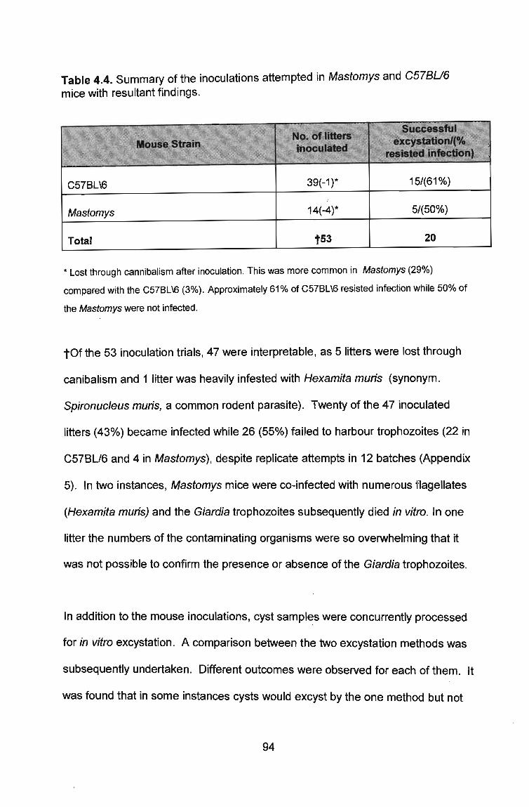
Table 4.4. Summary of the inoculations attempted in Mastomys and C57BU6 mice with resultant findings.
C57BL\6 39(-1)* 15/(61%)
Mastomys 14(-4)* 5/(50%)
Total t53 20
* Lost through cannibalism after inoculation. This was more common in Mastomys (29%)
compared with the C57BL\6 (3%). Approximately 61% of C57BL\6 resisted infection while 50% of
the Mastomys were not infected.
tOf the 53 inoculation trials, 47 were interpretable, as 5 litters were lost through
canibalism and 1 litter was heavily infested with Hexamita muris (synonym.
Spironuc/eus muris, a common rodent parasite). Twenty of the 47 inoculated
litters (43%) became infected while 26 (55%) failed to harbour trophozoites (22 in
C57BU6 and 4 in Mastomys), despite replicate attempts in 12 batches (Appendix
5). In two instances, Mastomys mice were co-infected with numerous flagellates
(Hexamita muris) and the Giardia trophozoites subsequently died in vitro. In one
litter the numbers of the contaminating organisms were so overwhelming that it
was not possible to confirm the presence or absence of the Giardia trophozoites.
In addition to the mouse inoculations, cyst samples were concurrently processed
for in vitro excystation. A comparison between the two excystation methods was
subsequently undertaken. Different outcomes were observed for each of them. It
was found that in some instances cysts would excyst by the one method but not
94

the other while in others excystment occurred with both methods. A proportion of
the samples could not be excysted with either method. The summary of
comparative results of excystation using both in vitro (acid pepsin) and in vivo
(mouse inoculation) methods is presented in Fig.4.1 below.
in vivo in vitro both no excyst
Excystment Type
11 Samples Excysted
Fig.4.1 Summarises the outcome of 47 attempted excystations using animal inoculation (in vivo) and acid pepsin (in vitro) methods. A total of 53 excystation attempts were made but only 47 were interpretable. Of those, 10% excysted in vitro only and 9% excysted in vitro only. Ten percent excysted by both methods while 18% did not excyst at all.
4.4 Observations
Infectivity patterns to mice
Variations in infection patterns of the 2 mice strains were observed:
• The Mastomys appeared to be more susceptible to infection than the C57BU6
mice. Firstly, repeated inoculations were required to effect infections in the
C57BU6 mice (personal observations) whereas Mastomys were often infected
on the first attempt. Secondly, 23 of 38 (61 %) inoculated C57BU6 litters could
95

not be infected in comparison with 5 of 10 (50%) Mastomys litters. (Appendix 5
& Table 4.4). Thirdly, repeated attempts to inoculate different litters of C57BU6
mice with the same isolate of Giardia failed to effect infections in 12 instances.
Finally, three separate repeated inoculations of the 2 mouse strains
consistently produced infections in Mastomys but not the C57BU6 mice.
Different batches of cysts varied in their ability to infect mice:
• In some instances, both Mastomys and C57BU6 mice were found to be
equally susceptible. Four of the six axenised Giardia strains (described in
Chapter 5 and arbitrarily designated SA 18, 24, 29 and 305) established
infections in both mouse strains. Attempts were not made to infect both strains
with the other two isolates SA6 and SA7. On the other hand, repeated
inoculations of both mouse strains failed to establish infections with 9 batches
of cysts (from symptomatic patients).
96
4.5 DISCUSSION
In the current work, two mice strains have been successfully infected with Giardia
cysts of human origin. These results strongly suggest that the parasite displays
host-preference rather than strict host-specificity and this confirms the findings of
earlier workers (Filice, 1952; Hill et al., 1983; Mayrhofer et al., 1992, Mayrhofer &
Andrews, 1994) who reported cross-species infections with Giardia organisms.
Furthermore, in the current study, mice were infected with Giardia of human origin;
this may have some implications in terms of mice acting as reservoir hosts.
However, in the current study the infections were of short term duration in
neonatal mice therefore further research is still required to clarify the role played
by adult mice in transmission of Giardia to humans and vice versa. As Mastomys
is an indigenous species there could be serious health implications in the South
African context if these were indeed acting as reservoir animals.
Animal infectivity was compared with excystation in vitro with the same sets of
cysts. Of the 53 excystation trials by both animal inoculation and in vitro methods,
varying results were obtained. Although some could be excysted by both methods,
a proportion of the isolated cysts could not excyst in vitro but produced infections
in mice while some failed to produce animal infections whilst they excysted in
vitro. The fact that some cysts could excyst in one model but not the other
suggests that the two methods provide different environments for factors that
initiate the excystation process. Alternately, it is possible that some stimulatory
factors within the cysts are favoured by one of the two excystment conditions but
not the other. These excystment methods therefore provide a marker that implies
strain differences in these morphologically identical Giardia of human origin.
97

A proportionately higher percentage of apparently viable cysts could not be
excysted by either of the two methods. Possibly, the factors responsible for
excystment were suboptimal or depleted, or the cysts were non-viable. These
results indicate wide variations in behaviour of Giardia in different conditions. It is
however noteworthy that there may be important inconsistencies in both the
mouse inoculation method and in vitro excystment, therefore the results should be
interpreted with caution. For example, it is likely that numerous host factors
determine its susceptibility to successful infection by a parasite. Likewise, parasite
factors also play a role in determining it's ability to establish an infection in the
host.
Judging from the inconsistent pattern of excystation in the two models that were
set up in the present study, it is evident that successful in vitro excystation does
not necessarily imply animal infectivity and vice versa. However, using G. muris
cysts for comparison of animal infectivity and excystation, Hoft et al. (1985)
indicated that in vitro excystation is an adequate indicator of G. muris cyst
infectivity. This report does not correlate with our experience. This disparity of
results may be explained by reports that G. duodenal is cysts require greater
stimulation to excyst in vitro than those of G.muris (Schaefer, 1990). Therefore, for
G.lamblia, as demonstrated in the present work, it would not be appropriate to
infer negative animal infectivity from a negative in vitro excystation result.
Andrews et al. (1989) provided electrophoretic evidence that the Giardia of
human origin is a species complex. Also, it has been shown that infections can be
composed of mixed genotypes and that isolation and purification techniques can
be selective (Mayrhofer et al., 1992). Our results may be an indirect reflection of
98

these findings, i.e. different genotypes responding differently to the different
excystment conditions. Mayrhofer et al. (1992) suggested that some strains of G.
intestinalis do not grow in suckling mice. Differences in the ability of cysts obtained
from infected humans to infect neonatal mice were also reported by Nash &
Aggarwal (1988). Similarly, Visvesvara and colleagues (1988), using gerbils as
hosts, concluded that only certain strains of Giardia from humans could infect
gerbils in their study. In the present work, twelve repeated inoculations of Giardia
cysts into litters of the two mice strains failed to effect infections. This observation
suggests that cysts vary in their intrinsic ability to cause infection in mice.
Between-mouse strain variations in infectivity patterns were also observed in the
current study. For example, occasionally, when the same Giardia organisms that
failed to produce infections in the C57BU6 mice were injected to the Mastomys,
infection was established. The Praomys (Mastomys) caucha (known to have a
defective immune system) appeared to be more readily infectable when compared
with the C57BU6 mouse strain (observations). Because the C57BU6 mice are
inbred and were maintained as such in our facility (therefore genetically identical),
a consistent pattern of infection would be expected from this strain. However,
some litters completely resisted infection whilst others became infected. Further
unexpected were within-litter variations in which some mice within a litter became
infected while others resisted infection. During inoculations, meticulous effort was
taken to maintain consistency in technique, therefore it would be unlikely that this
variation resulted from technical discrepancy. POSSibly, factors such as the gastric
pH of the individual mice at the time of inoculation may have been responsible for
the irregular infection patterns.
99

These findings strongly suggest that host factors also play an important role in
determining successful establishment of infection. Furthermore, evidence
suggesting that host dynamic factors play a role in establishment of and type of
infection is documented. For example, Tsuchiya (1931) showed that the diet of the
host affects the size of Giardia cysts excreted while a study by Leitch et al. (1989)
illustrated that dietary fibre reduces the rate of intestinal infection by G.lamblia in
gerbils.
Use of surrogate animals has proved to be superior to use of in vitro excystation
techniques (Chapter 3) in the current study. This excystation technique offers
added value for the aims of the study, as a larger number of trophozoites is
available for in vitro culture, which in turn increases the likelihood ofaxenisation.
Furthermore, the contaminating bacterial/fungal flora encountered with the in vitro
excystation methods are minimised or eliminated in the animal excystation
method.
Schaefer (1990) reviewed all available excystation techniques and concluded that:
(1) great variability is to be expected with the currently developed procedures for
Giardia duodenalis (2) The optimal conditions for repeatable, maximal excystation
of G. duodena lis cysts are not yet known (3) G.duodenalis cysts do not excyst
routinely at high levels. Furthermore, it is also documented that none of the
methods of excystation work all of the time for all strains of the parasite (Nash,
1988). Our excystation results agree with these conclusions as the present study
illustrated that some Giardia isolates can readily infect animals but fail to excyst in
vitro and vice versa. Giardia seems to display inherent variation in it's ability to
100

establish infection; interestingly this was demonstrated in human experimental
infections by Nash et al. (1987). It is not established whether this variation occurs
during the natural course of Giardia infections.
101

CHAPTERS
IN VITRO CULTURE OF GIARDIA TROPHOZOITES
5.1 INTRODUCTION
Of all the common intestinal protozoa, organisms of the genus Giardia have
reportedly been among the most difficult to establish in culture (Meyer, 1979). To
date, of the three recognised morphologic types of Giardia namely G.agilis,
G.muris and G.duodena/is (Filice, 1952), only the latter has been successfully
cultured in vitro. However, G.duodenalis strains of canine origin were documented
to be refractory to culture (Meloni and Thompson, 1987).
Reports on in vitro cultivation of these organisms appeared in the early twentieth
century when Chatterjee (1927) first reported maintaining Giardia trophozoites
alive in vitro for up to 5 weeks. Karapetyan then reported in 1960 that he had
cultured Giardia intestinalis xenically with chick fibroblasts and Candida
guilliermondi for seven months. The Giardia trophozoites continued to grow even
after the chick fibroblasts had died. In 1962, Karapetyan successfully cultured G.
duodena/is isolated from a rabbit monoxenically with Saccharomyces cerevisiae
without fibroblasts. However, his attempts to axenise G. duodenalis were
unsuccessful. His complex culture medium included serum, chick embryo extract,
chick amniotic fluid or a tryptic digest of meat with Hank's or Earle's solutions. The
medium had to be replaced daily.
In 1970, Meyer first axenised G. duodenalis from the rabbit, chinchilla and cat. His
method entailed use of a U tube, wherein the Giardia trophozoites were inoculated
102

together with yeasts on the right arm of the tube. The motile trophozoites migrated
across its base and were isolated, free of yeast, on the left arm of the tube.
In 1976, Meyer developed a culture medium (Hanks, Serum, Phytone-1) (HSP-1)
for axenic cultivation of human isolates. One of the major components of Meyer's
medium was human serum (15-20%). Visvesvara (1980) highlighted the limitations
of using this product as batches of human serum varied in their ability to support
good growth of G.lamblia; some even inhibited growth and caused cultures to be
lost. He also recognised that G.lamblia grown in media containing human serum
(HSP-1) were unsuitable for use as antigens in serological tests because of non
specific staining reactions. To circumvent this, he gradually adapted the human
Giardia to grow in Diamond's Trypticase-Panmede-Serum (TPS-1) medium, which
is supplemented with bovine or rabbit sera.
A new medium, Trypticase-Yeast-lron-Serum-33 (TYI-S-33) was developed for
axenic culture of Entamoeba spp by Diamond et al. (1978) to replace the TPS-1
medium, which was at that time the most widely used for these organisms. One of
the essential ingredients of TPS-1 medium was Panmede (Paines and Byrne,
Ltd.,Greenford, Middlesex, England), a papain digest of ox liver. In time it became
increasingly difficult to obtain batches which supported good growth, hence TYI-S-
33, which is based on Trypticase, a casein digest (BBL, Cockeysville, USA) and
yeast, was developed.
Keister (1983) modified TYI-S-33 for cultivation of Giardia organisms by adding
bile and increasing the concentration of L-cysteine to the original formulation of
103

Diamond et al.'s (1978) medium. He successfully cultured four strains, two of
which had been grown and maintained in TPS-1. This was a significant
contribution to G.lamblia cultivation methodologies as this modified version of TYI
S-33 is currently the most widely used medium for axenic cultivation of Giardia.
This strategy obviates the need to resort to monoxenic culture first, thus
eliminating all problems related with associated organisms.
Although no reports on a clearly defined medium for Giardia are presently
available, scientists have identified some of the essential ingredients of TYI-S-33
(the medium which supports luxuriant growth of this parasite). Gillin and Diamond
(1981 a) evaluated the responses of growth of Entamoeba histolytica and
G.lamblia to reducing agents. They found that the requirement for L-cysteine was
very specific for growth of Giardia, and could not be substituted by other reducing
agents. They also discovered that attachment in complex growth media, growth
and survival in culture required cysteine (Gillin and Diamond, 1981a, b). In 1994,
Lujan and Nash demonstrated that L-cysteine is essential for growth of Giardia
trophozoites in vitro (as it protects the trophozoites against the lethal effects of
Oxygen) and that it's uptake depends on the presence of serum: thus serum is
one of the key components for successful culture. Lujan et al. (1994) also showed
that bovine serum Cohn fraction Cf-IV-1 enhances the uptake of L-cysteine by
trophozoites. In addition to it's ability to enhance cysteine uptake, serum also
provides growth factors and nutrients. Lipids were reported by Adam (1991) to be
necessary for growth.
Keister (1983) showed that the vitamin-Tween mixture added to TYI-S-33 by
104

Diamond et al. (1978) does not enhance the growth of G.lamblia in bile
supplemented medium. Bile, therefore, among other factors apparently obviates
the need for vitamin supplementation. Giardia are unable to synthesise their own
membrane phospholipids, thus the mechanism by which bile promotes parasite
growth is related to the requirement of pre-formed phospholipid which is abundant
in bile and whose uptake is facilitated by presence of conjugated bile salts
(Farthing, 1989).
Certain factors can affect in vitro Giardia cultures.
• Although many antibiotics such as the penicillins, gentamycin, streptomycin,
clindamycin and amphotericin B do not affect the growth of Giardia
trophozoites in vitro (Jakubowski et al., 1988), some however. can impede it's
propagation and growth, e.g. Amikacin, an aminoglycoside, can be toxic to the
trophozoites at high concentrations.
• Bacterial and viral endosymbionts have been found in Giardia of human and
animal origin (Feely et al., 1990). The presence of bacteria in trophozoite
cultures was reported to cause the rate of Giardia multiplication to decrease
markedly (Meyer, 1976) while viral endosymbionts in Giardia trophozoites are
associated with decreased adherence and growth rate (Adam, 1991).
• Low cysteine concentration, high oxygen tension and low temperature cause
detachment of trophozoites from the culture vessel.
Although it is difficult to culture Giaqrdia lamblia, many workers from various parts
of the world have successfully axenised these organisms: Portland (Meyer, 1976);
Atlanta (Visvesvara, 1980); Maryland (Gillin & Diamond, 1981a,b; Keister, 1983);
Australia (Phillips et al., 1984); Belgium (Gordts et al., 1985); Poland (Kasprzak &
105

Majewska, 1985) United States (Nash et al., 1985); Western Australia (Meloni &
Thompson, 1987; Meloni et al., 1988); Mexico (Cedillo-Rivera et al., 1989); Saudi
Arabia (AI Tukhi et al., 1991); Israel (Korman et al., 1992). No reports of similar
work have been documented from South Africa. This chapter describes
procedures to initiate, expand and maintain viable cultures of Giardia trophozoites
derived from excystation of locally isolated cysts. Since the trophozoites used for
initiation of the cultures were derived from in vitro (Chapter 3) and in vivo
(Chapter4) excystation experiments, reference to these chapters will be made
constantly in the current chapter.
106
5.2 MATERIALS AND METHODS
5.2.1 Culture Medium
TYI-S-33 was utilised for initiation and maintenance of Giardia cultures.
Essentially it was prepared as described by Diamond et al. (1978) and modified by
Keister in 1983. The main ingredients are Biosate (a pancreatic digest of casein
and yeast extract), bile, serum, salts, glucose and iron. Details of it's preparation
appear in Appendix 7.
5.2.2 Optimisation of Culture System
Since biological products vary in their ability to support growth of cells in vitro
(Diamond et al., 1978), several trials using different brands of reagents were
necessary to identify suitable lots of the different products.
Two strains of Giardia, (WB, isolated from a patient infected in Afghanistan; and
the H7 clone of the GS isolate obtained from a scientist from the National
Institutes of Health (NIH) who had camped in Alaska) were obtained from Or Nash
at the NIH, USA. These have been well established as axenic cultures and hence
were used as control reference strains to assess good growth-supporting
reagents.
5.2.1.1 Sera
Several batches of bovine sera (lots: CN 1872; CN 1399 and CN2290) from
Highveld Biological, Gauteng, (SA); fetal bovine serum (lots: 35-603-240 -1419;
35-603-240-1410 & 34-511-248-75) from Delta Bioproducts, Gauteng, (SA); fetal
bovine serum from Sigma St. Louis USA, lots 25H4602, 46H4648 & 65H4664
107

were evaluated.
5,2,1,2 Biosate
Biosate from BBL- Becton Dickinson and Company, USA, lots 1000L 7DESR and
1000E9DGNP were evaluated.
5,2,1,3 Antibiotics
Because of expected widespread penicillin/streptomycin resistance among local
bacterial strains, various antimicrobial agents (such as Gentamycin, Vancomycin,
and low dose Amikacin) were used over and above the penicillin/streptomycin
originally incorporated into the culture medium.
While the optimisation experiments were undertaken, an aerophilic laboratory
contaminant, Xanthomonas maltophilia (identified by biochemical reactions in the
Api 20E system (BioMerieux) was detected on some culture tubes of the WB
reference strain (the trophozoites continued to grow and multiply in the presence
of these microbes). A disk-diffusion susceptibility assay revealed that the
organism was resistant to the antibiotics incorporated in the medium, including
Amikacin but sensitive to Ciprofloxacin. Subsequently, this antibiotic was required
to eliminate the bacterial contaminants without inhibiting the growth of Giardia
trophozoites. An assay to determine the Ciprofloxacin concentration tolerated by
trophozoites was therefore performed, as literature citing use of this quionolone on
Giardia cultures was not found.
108

Assay for Susceptibility of Giardia to Ciprofloxacin
A 100!-lg/ml Ciprofloxacin (Bayer) stock solution was prepared (from a 2 mg/ml
intravenous fluid concentrate) in sterile TYI-S-33 medium. Various dilutions of the
stock solution were prepared to give final concentrations of 5; 2,5; 1,25; 0,625 and
0,3125 !-Ig/ml in 7ml of TYI-S-33 in the culture tubes. The two reference strains
(WB and H7) were retrieved from liquid nitrogen (as described in Chapter 6) and
re-cultured in TYI-S-33 medium. Following 48 hours of incubation, the cultures
were ice- chilled for 15min, centrifuged at 400xg for 10min at 4°C and the
supernatant medium decanted. The pelleted cells were resuspended in 1ml of
cold sterile medium and counted using a Nuebauer counting chamber. Aliquots of
0,1 ml containing 1000 cells/ml of each isolate were distributed to 6 Kimax glass
tubes containing the TYI-S-33 culture medium. Each of the twelve cultures was
incubated with each of the concentrations of Ciprofloxacin. Two tubes (one of
each strain) incubated without the antibiotic served as control tubes. The tubes
were incubated and maintained at 35,5°C at a 5° angle. Growth rate and
attachment to glass were monitored in the twelve tubes for 20 and 48 hours by
modifying the adhesion assay (Meyer, 1976; Farbey et al. 1995) as follows: After
20 and 48 hours incubation, all tubes were observed for adherent cells under a
40x microscope field in the entire inner surface of the culture tubes. A number ·
(growth rank) was assigned to each culture tube (including control tubes). This
number represented the average number of adherent trophozoites present per
40x-microsope field in the lower inner surface of a culture tube using the guide
outlined in Table 5.1.
109

Table 5.1 Outlines a relative semi-quantitative assessment of growth (ranking) of Giardia trophozoites in culture which could be used to monitor growth rate over time by counting adherent trophozoites on the lower inner surface of a culture tube under a 40x microscope field. (Adapted from Meyer, 1976).
1 1-5
2 5-50
3 50-250
4 250-500
The assay was repeated with higher concentrations of the drug ranging from 10-
50 J..Ig fml of Ciprofloxacin.
Data obtained from the 8ayer electronic publication (1997) indicated a minimum
inhibitory concentration (MIC) of 4pg fml for X. maltophilia. This concentration of
Ciprofloxacin was subsequently incorporated into the contaminated cultures until
no bacterial growth was detected on subcultures.
5.2.2 Propagation of Trophozoites In Vitro.
5.2.2.1 Culture of in vitro excysted trophozoites
Cultures were initiated with 67 trophozoite batches derived from in vitro
excystation (Chapter 3). These were aseptically transferred to sterile glass tubes
containing TYI-S-33 medium with antibiotics. The tubes were incubated at 35,50C
at a 5° angle to facilitate adherence of excysted trophozoites. After 48-72 hours,
old medium was decanted (without dislodging the trophozoites from the tube
walls) and replaced with fresh TYI-S-33. Culture tubes were maintained at 35,50C
110

for up to seven days even when no trophozoites were detected by microscopy. To
monitor bacterial and fungal contamination, subcultures of medium, aseptically
aspirated from the tubes, were inoculated onto aerobic and anaerobic blood
plates; the latter were incubated in a Gas-Pak MD 21030 (BBL Microbiological
Systems; Becton Dickinson & Co; Cockeysville, Maryland, USA) anaerobic jar.
The plates were incubated at 37°C for 24 (aerobic) and 48hrs (anaerobic).
5.2.2.2 Establishment of cultures from suckling mice
Twenty batches of trophozoites (a batch consisted of 4-9 tubes) obtained from
mice intestines as described in Section 4.2.3 were incubated in TYI-S-33 medium
with antibiotics at 35,5°C at a 5° angle. Cultures were examined daily using an
inverted microscope at 100x and 400x magnification. At 48 or 72hour intervals, old
culture medium containing non-adherent and dead trophozoites was carefully
decanted (without dislodging the attached trophozoites) and 7ml of fresh medium
was added. Aseptic conditions were maintained at all times. When cultures
formed a confluent adherent monolayer, antibiotics were gradually withdrawn from
the medium. Cultures were deemed axenic when they continued to multiply in
antibiotic-free medium in the absence of other organisms. Under aseptic
conditions, subcultures of TYI-S-33 medium from the culture tubes were streaked
onto aerobic and anaerobic blood agar plates to assess for the presence of
bacterial/fungal contaminants.
5.2.2.3 Routine Maintenance of cultures
For routine maintenance and expansion of the cultures, trophozoites that grew
well and formed a dense monolayer on the surface of the tube walls were
111
dislodged by chilling the culture tubes in an ice bath for 20 minutes. Thereafter the
tube contents were mixed by gentle inversion and centrifuged at 400xg for 5min at
4°C. Old supernatant medium was gently decanted and pelletted trophozoites
resuspended in 1 ml of fresh TYI-S-33. A 1: 1 0 dilution of trophozoites was
prepared in 1 % formalin in a clean tube to fix and enumerate the trophozoites.
Aliquots of 0,1 ml of the remaining trophozoite suspensions containing 1 x1 04 cells
fml were distributed to two or more tubes containing 7ml TYI-S-33 medium. The
expanded cultures were maintained as above until confluent monolayers were
obtained for cryopreservation (Chapter 6) and preparation of Iysates for
isoenzyme electrophoresis (Chapter 7).
112
5.3 RESULTS
5.3.1 Optimisation Experiments
Using the two reference strains, good viable growth was feasible with fetal bovine
serum from Sigma St. Louis USA, lots 25H4602, 46H4648 & 65H4664; and
Biosate from BBL- Becton Dickinson and Company, USA, lot 1000E9DGNP. None
of the sera from Highveld Biological and Delta Bioproducts respectively, could
support good growth and caused loss of cultures.
The Ciprofloxacin assays showed that concentrations between O,3125-50J.Jg/ml
were well tolerated by the trophozoites. The rate of growth and adhesion of
trophozoites to glass was comparable to that of the control tubes (antibiotic-free)
20 and 48 hrs after incubation at all these antibiotic concentrations (Table 5.2).
Culture tubes containing the WB strain showed evidence of cell division after 8
hours incubation. By day 3, a confluent monolayer was formed in all tubes.
The H7 strains showed signs of division after 12 hours incubation in the bottom
most part of the tube. A confluent adherent monolayer was detected 5 days after
incubation. A similar pattern of results was observed when the experiment was
repeated with the high concentrations (5-50ug/ml) of Ciproflxacin.
113

Table 5.2. Sensitivity to Ciprofloxacin of Giardia reference strains WB and H7: ~ummary of growth rank of the culture tubes containing different Ciprofloxacin concentrations after 20 and 48 hours incubation.
5 2 1 3 2
2,5 2 1 3 2
1,25 2 1 4 2
0,625 2 1 3 2
0,3125 2 1 3 2
° (control) 2 1 3 2
5.3.2 Cultivation of In Vitro Excysted Trophozoites.
Table 5.3 summarises the results of all culture initiates derived from in vitro
excystations.
The behaviour of in vitro excysted trophozoites was not consistent in culture.
Some survived for up to six hours, rounded up and died. Others survived for 12,
24 or 72 hours or as long as 9 days in culture but gradually died. The majority of
these never adhered to the culture tube surface but swam around vigorously and
later became sluggish. A few attached to the glass but remained as single cells at
114

the bottom of the culture tubes and never showed signs of division. Most of these
gradually began to die until, by day 7, no living trophozoites were seen in the
culture tubes. In 36% of them (Table 5.3), no evidence of contaminants (aerobic
and anaerobic) could be demonstrated.
Sixty-four percent of the cultures were overwhelmed by bacterial contaminants
and died, despite the incorporation of antibiotics in the culture media (Table 5.3).
Some contaminating organisms were identified. Most frequently gas-forming
anaerobes (foul smelling gas evident in the TYI-S-33 culture tubes and growth
only occurred on the c;inaerobic blood plate) contaminated the cultures.
Occasionally, yeasts and Pseudomonas spp were identified as the contaminating
organisms. In some cases Klebsiella and Aerobacter spp were isolated.
No viable cultures were established using the trophozoites obtained from in vitro
excystations.
115
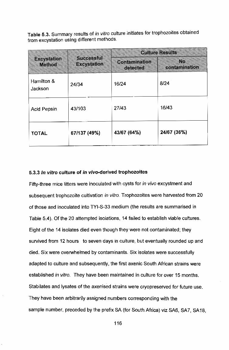
Table 5.3. Summary results of in vitro culture initiates for trophozoites obtained from excystation using different methods.
Hamilton &
Jackson
Acid Pepsin
TOTAL
24/34
43/103
67/137 (49%)
16/24
27/43
43/67 (64%)
5.3.3 In vitro culture of in vivo-derived trophozoites
8124
16/43
24/67 (36%)
Fifty-three mice litters were inoculated with cysts for in vivo excystment and
subsequent trophozoite cultivation in vitro. Trophozoites were harvested from 20
of those and inoculated into TYI-S-33 medium (the results are summarised in
Table 5.4). Of the 20 attempted isolations, 14 failed to establish viable cultures.
Eight of the 14 isolates died even though they were not contaminated; they
survived from 12 hours to seven days in culture, but eventually rounded up and
died. Six were overwhelmed by contaminants. Six isolates were successfully
adapted to culture and subsequently, the first axenic South African strains were
established in vitro. They have been maintained in culture for over 15 months.
Stabilates and Iysates of the axenised strains were cryopreserved for future use.
They have been arbitrarily assigned numbers corresponding with the
sample number, preceded by the prefix SA (for South Africa) viz SA6, SA7, SA18,
116

SA24 , SA29 and SA305.
Table 5.4. Culture results using in vivo excysted trophozoites as inoculum. Thirty percent of the culture initiates were lost through contamination, while 40% failed to establish cultures.
ExcystationlT otal inoculations
20/53 (38%)
Contaminated
6/20 (30%)
*6 isolates axenised (30% success).
5.4 Observations
Behaviour of trophozoites in vitro
No contamination but died out
8/20(40%)
*No contaminationsurvived
6/20 (30%)
For fresh isolates, during the initial period of culture, the trophozoites adhered to
the bottom of the culture tube mostly as individual cells without signs of division
(Plate 5.1). This inert period varied with different isolates (from 10 to 15days) and
could be as long as twenty days in some instances after which they showed signs
of division (Plate 5.2). However, as the organisms became adapted to the culture
environment, this period became shorter. When the trophozoites are continuously
passaged they can be seen dividing by day three after subculture. A confluent
monolayer of cells was seen by day 18 in some isolates (Plates 5.3 and 5.4)
However one isolate took 32 days to start dividing; the concentration of fetal
bovine serum was increased from 10 to 15% on day 24 and confluent growth was
reached after 55 days. Interestingly, some trophozoites continually swam around,
never adhering to the surface of the tube.
117

Once established in culture for a long time, the trophozoites became highly
resilient. Some trophozoites survived de&pite long periods without medium
replacement. Two tubes of the reference strain (W8) were kept in the 35,5°C
incubator for 10 and 45 days respectively without changing the medium and later
took to culture. The local strains have also survived up to 96 hours without
medium changes. In these cases, although many were rounded up and detached
from the glass surface, plenty were still adhering and they continued to grow well
after replenishment with fresh medium.
118

Plate 5.1 Trophozoites of G.lamblia that had been isolated in culture for 8 days. All exist as individual cells (400x).
Plate 5.2. Trophozoites after 15 days in culture. Signs of division were noted (400x).
119

Plates 5.3 and 5.4 Confluent growth of G. lamblia trophozoites in Culture. Magnification x400.
120

5.5 DISCUSSION
In the present work, cultures were initiated from excystation of faecally derived
cysts, therefore parts of this discussion ~onsider the results in accordance with the
in vitro and in vivo culture findings.
Initiation of viable long-term cultures from in vitro excystation proved to be
impossible. Excystment and trophozoite survival were the two major factors that
governed successful attempts at in vitro cultivation of the organisms. Although
successful excystment was achieved in some cases, the individual levels of
excystation were low, and if successfully excysted, trophozoites survived for
several hours and subsequently died. Therefore, only short-term cultures were
established. While some authors reported successful establishment of viable
cultures from in vitro excystation of cysts (Bingham & Meyer, 1979; Isaac-Renton
et al., 1986; Meloni & Thompson, 1987; Meloni et al., 1988; Cedillo-Rivera et al.,
1989; AI-Tukhi et al., 1991), others, as in the present study experienced difficulty
in establishing cultures from in vitro excysted trophozoites (Isaac-Renton et al.,
1986; Kaur et al., 1986).
In the current study, apart from the scanty numbers of trophozoites that resulted
from low levels of excystation, contaminating faecal microrganisms played a
negative role in the failed attempts to establish cultures from in vitro excysted
cysts of faecal origin. Although a variety of antibiotics were used, a large
proportion of cultures was overwhelmed by faecal contaminants. This might be a
reflection of the wide distribution of resistant organisms in the local population
from which cysts were obtained. The combined effect of these factors made it
121

difficult to produce viable cultures.
Use of surrogate animals as excystation hosts proved to be superior to in vitro
excystment. Viable cultures of Giardia have been established using this model.
Subsequently six local strains have been axenised for the first time in South
Africa. The greater numbers of trophozoites available for culture initiation as well
as the reduction of contaminating faecal organisms are considered to be key
factors in the efficacy of this method.
In the present study only a limited number of in vivo excysted trophozoites
resulted in the establishment of a successful culture. Therefore the establishment
of viable cultures of Giardia through mice inoculations can be considered to be
dependant on many complex factors such as: Giardia isolate variations, mouse
strain variations, selection pressures placed by both in vivo and in vitro conditions
and the fact that Giardia is fastidious with no defined medium being available for
these organisms. Little is known about the growth dynamics of populations of
Giardia. Furthermore, previous studies reflect that multiple attempts are required
to successfully initiate viable cultures of a single isolate. For example, Kasprzak &
Majewska (1985) reported that up to 107 consecutive attempts were required to
isolate strains (from faecal cysts or duodenal aspirates) in vitro. Owing to the
limited numbers of cysts available for simultaneous in vitro and in vivo
experiments, few faecal samples allowed for repeated attempts in the current
work.
While working with Giardia it became clear that establishment of these organisms
122

in culture is complex, time consuming and requires patience. In the culture
system, different batches of excysted trophozoites showed marked variation.
Some organisms swam around in the medium for several hours or up to 7 days in
some cases. These never adhered to the glass surface. Other trophozoites
adhered within minutes or hours to the glass inner surface. They remained
attached to the bottom-most inner surface of the tube as single cells for different
periods. This was assumed to be an adaptation period that was then followed by
cell division and multiplication. Some isolates took 3-4 weeks to produce an
adherent, confluent monolayer of cells while one isolate was successfully
harvested after approximately two months. Some were easily axenised while
others grow axenically with difficulty or not at all. Differences in growth rates
between the different strains including the two reference isolates were also
observed. This phenomenon strongly suggests there are differences in growth
characteristics among the different strains.
The observation that only those trophozoites that adhered and adapted to the
culture conditions produced established cultures indicate that selection occurred.
The non-adherent trophozoites eventually died or were lost when old medium was
decanted and replaced with fresh medium. Upcroft et al. (1994) noted that not all
in vitro cultivated samples will generate viable laboratory cultures and that it has
been shown through DNA fingerprinting that what finally establishes as a viable
laboratory culture is effectively a clone, but it is not known at what stage this
occurs. Mayrhofer et al. (1994) and Andrews et al.(1992) suggested that
adherence is a potential point at which selection can occur. Furthermore, it is
equally possible that differences in adherence capability can lead to the
123

dominance of certain genotypes under in vitro conditions.
The differences in behavioural characteristics of Giardia isolates that have been
highlighted above suggest that not all members of this group have identical growth
requirements. Because of this, it is likely that only isolates that are able to adapt to
the conditions of the methods of in vitro propagation can be cultured. If this is the
case, it is possible that several different media will eventually be required to
propagate the range of species (and strains) within the genus. It is clear that
presently, culture methods inadvertently select for certain genotypes.
124
CHAPTER 6
CRYOPRESERVATION OF TROPHOZOITES
6.1 INTRODUCTION
Diamond et al. (1961) reported the first successful attempt at preservation of
protozoa in liquid nitrogen. The first attempt to culture Giardia from cryopreserved
material was made by Meyer & Chadd (1967). Subsequently, Giardia cultures
have been cryo-preserved apd retrieved with success by other workers (Warhurst
& Wright, 1979; Lyman & Marchin, 1984; Phillips et al., 1984; Diamond, 1995).
In his report on cryopreservation and storage of parasitic protozoa, Diamond
(1995) advises that the best time to harvest many cell types, including protozoa, is
during the transition phase between logarithmic and stationary growth as rapidly
multiplying cells are more vulnerable. Other parameters such as the cryoprotective
agent are also equally important in successfully cryopreserving cells. Various
cryoprotective agents have been used to preserve cooled Giardia trophozoites.
They include glycerol, dimethylsulphoxide (DMSO) (Meyer & Chadd, 1967; Lyman
& Marchin, 1984; Phillips et al., 1984), either glycerol or DMSO supplemented with
glucose (Diamond, 1995) and DMSO supplemented with salts (Warhurst & Wright
1979). Lyman & Marchin, (1984) used a protocol which includes an ethanol bath.
They recommended an ethanol cooling rate of minus 1, 19°C/min for
cryopreserving G. lamblia trophozoites.
125

Cryopreservation and successful retrieval of viable cultures has facilitated the
establishment of cryobanks of axenic cultures. This helps to avoid the selection
and variation of strains created by serial subcultures and minimises the cost of
maintaining cultures indefinitely.
One of the aims of the present study was long term storage at cryogenic
temperatures and establishment of a cryobank of locally isolated strains.
This section describes procedures undertaken to cryopreserve, store and retrieve
into culture trophozoites that had been axenised.
126

6.2 MATERIALS AND METHODS
6.2.1 Cooling of Cultured Trophozoites
Two techniques of cooling were assessed for their efficacy in achieving long term
preservation of trophozoites. Subsequent success in retrieval of viable cultures
was also evaluated. In both, the same cryopreservant was used. A 9% (v/v)
solution of Dimethyl sulphoxide (DMSO) (Saarchem, Durban, and SA.) was made
in sterile TYI-S-33.
6.2.1.1 Preparation of samples
Tubes of cultures at early stationary phase were chilled on ice for 20 minutes to
detach the trophozoites from the glass. After mixing by gentle inversion they were
centrifuged at 400xg for 5 minutes at 4°C and supernatant medium was
discarded. Pelletted trophozoites were resuspended in 1 ml of fresh TYI-S-33
medium. The organisms in a 1 ml aliquot were enumerated in a haemocytometer.
One ml of suspensions containing 1 x1 06 trophozoites fml were then transferred
with a sterile Pasteur pipette to a Nunc cryotube (Nalge Nunc International, USA).
Immediately before cooling, 1 ml of 18% DMSO was gradually introduced (by
dropwise addition) to the cryogenic vial to give a final concentration of 9% DMSO.
6.2.2 Cooling Techniques
6.2.2.1 Electronically controlled cryofreezing apparatus (modification of Phillips et al., 1984 method)
Vials containing cryopreservant and cells were promptly (within 5 minutes)
transferred to a Liquid Nitrogen, programmable controlled freezer, Planer model
Xb634 (Planer Products, Sanbury-On-Thames, England). The cooling rate was
127

adjusted to 5°C per minute from ambient temperature to OOC; 1°C per minute from
o to -25°C and then 5°C per minute from -25 to -135°C. At completion of the
cooling cycle, the cryotubes were transferred to a liquid nitrogen (LN) storage
Dewar flask (Taylor-Wharton, British Oxygen, UK) until required for retrieval.
6.2.2.2 Mechanically insulated freezing container
The procedure was carried out according to the manufacturer's instructions as
follows. Isopropanol (250ml) (Merck, Darmstadt, Germany) was added into the
Nalgene Cryo 1°C Freezing Polycarbonate Container (Nalge Nunc International,
U.S.A, Cat.No.51 00-0001). An insulating foam insert was immersed into the
alcohol. Samples prepared as described in section 6. 2.1.1 were placed into the
vial holder, the unit was promptly placed into a -70°C mechanical freezer and left
undisturbed for at least 4 hours. This system allows a gradual rate of cooling at
1°C/min from room temperature to -70°C. The frozen vials were transferred for
long-term storage into a liquid nitrogen Dewar flask until retrieval was necessary.
6.2.3 Retrieval Of Frozen Cultures.
Cryopreserved cultures were retrieved from liquid nitrogen and thawed quickly at
37°C for 2 minutes without agitating the tubes. Using a sterile Pasteur pipette, the
contents were gently mixed and transferred to a sterile 8ml glass Kimax tube
containing 7ml of TYI-S- 33 medium. The cultures were examined on an inverted
microscope and the percentage of motile trophozoites (indicating viability) was
128

estimated. The tubes were then incubated at a 5° angle for 30 minutes at 35,5°C.
Medium with cryopreservant and dead cells was gently poured off and replaced
with fresh TYI-S-33.
The cultures were incubated and maintained as in 5.2.2.3 until adequate confluent
growth was obtained.
129

6.3 RESULTS
Trophozoites have been successfully preserved for periods varying from 23 days
to approximately 2 years. Qualitative assessment upon retrieval revealed 70-80%
viability of trophozoites. Within 30 minutes, more than 50% of the trophozoites
were adhering to the inner surface of the glass culture tubes (Plate 6.1). Confluent
cells were seen within 48h of culture (Plates 6.2 & 6.3). All retrieved samples
have been re-established in culture: a 100% success rate has been achieved
using both electronically controlled (6 times) and mechanical (15 times) cooling
techniques. Table 6.1 presents the records of duration of preservation of the 6
local isolates and the two reference strains.
Viable cultures of the retrieved trophozoites have been maintained in culture for
several months and cryopreserved again with success. A reserve supply
(cryobank) has been established for the 6-axenised local strains.
Plate 6.1 Trophozoites of Giardia lamblia that had been cryopreserved for 2 ~onths 17 days. The organisms were retrieved from cryopreservation and Incubated at 35,5°C for 30mins in TYI-S33 medium and residual DMSO. More than 50% of the organisms are adhering to the tube inner surface. (400x)
130

Plate 6.2 Confluent growth of trophozoites that were cryopreserved for 2 months17days, retrieved from cryopreservation and incubated at 35,5°C for 48hours in TYI-S33 medium. (400x magnification).
Plate 6.3 A confluent culture of G.lamblia trophozoites 5 days after retrieval from cryopreservation in liquid nitrogen. (400x)
131

Table 6.1. A longevity record of samples of two reference isolates (WB & H7) and 6 axenic South African isolates cryopreserved in liquid nitrogen and retrieved into culture.
WB 03-01-97 17-3-97 2mnths 14d
WB 03-01-97 18-2-97 1mnth 17d*
WB 03-01-97 27-01-97 24d*
H7 03-01-97 08-04-97 2mnths 5d*
H7 03-01-97 27-01-97 24d*
SA6 03-01-97 05-11-97 10mnths 2d
SA6 03-01-97 19-08-97 7mnths 18d
WB 16-04-97 05-11-97 6mnths 19d
H7 30-04-97 23-05-97 23d
H7 11-05-97 17-02-98 9mnths 6d
WB 11-05-97 17-02-98 9mnths 6d
SA18 05-09-97 06-02-98 5mths 1d
SA29 05-09-97 17-02-98 5mnths 12d
WB 14-04-97 05-11-97 6mnth 6d
H7 03-01-97 23-05-97 4mths 20d *
SA24 26-02-98 11-06-98 3mnths 15d
SA29 25-09-97 22-09-98 11mnths 26d
SA305 24-04-98 25-09-98 5mnths 1d
SA7 03-01-97 28-08-98 1y 7mnths*
WB 25-08-98 26-10-98 1mnth 1d
SA6 25-08-98 26-10-98 1mnth 1d y = year mnth/s = month/s d = days * = electronically controlled cooling method
132

6.4 DISCUSSION
In the current work, effective cryopreservation (for approximately two years) has
been achieved with the two different cooling techniques. These results concur with
Diamond's report that "once a specimen is cryopreserved in liquid nitrogen or its'
vapours it can be expected to remain viable for many years regardless of the
technique employed to achieve preservation" (Diamond, 1995).
Although the two methods of cooling were found equally effective, the electronic
cooling device is more expensive, requires the use of liquid nitrogen and is more
time-consuming, whilst the Nunc cryopreservation system is simple in addition to
being cost-and-time effective. The Nunc cryopreservation system therefore
became the method of choice.
In our experience, cryopreserved Giardia cultures are more resilient than
Entamoeba, which are documented to have increased fragility after
cryopreservation. Giardia trophozoites remained viable even after exposure to
cryopreservant (9% DMSO) for 30 minutes. Diamond (1995) stated that frozen
E.histolytica lyse after recovery when kept longer than 10 minutes in the
cryopreservant. Furthermore, Phillips et al., (1984) stated that a washing step to
remove cryopreservant, after thawing retrieved Giardia trophozoites, is essential
for successful re-establishment of cultures. In the present work, trophozoites that
were thawed and incubated in the presence of DMSO for 30 minutes could still be
re-established as viable cultures.
133

Diamond (1995) stated that an important parameter in cryopreservation is the
temperature at which the cryopreservant is added to the cells as osmotic
,equilibration takes place before freezing is initiated. He studied the effects of two
temperatures (O°C and 24°C) on a monoxenic culture of Ehistolytica using 5%
DMSO as cryopreservant with 30 minutes equilibration. None of the amoebae
survived cryopreservation when equilibrated at OOC. Phillips et al., (1984),
maintained the cryopreservant and the cultures on ice prior to initiation of the
freezing procedures, and reported more than 70% motile organisms after thawing.
Warhurst & Wright (1979), added the 7,5% DMSO, equilibrated at room
temperature, and reported more than 50% motility on thawing. In the current
study, addition of DMSO and equilibration was allowed to take place at room
temperature (22-25°C) and 70 -80% of the trophozoites were motile after thawing.
The protocol described in the present chapter has been found to be highly
reproducible, as all cultures retrieved after cryopreservation have been
successfully re-established in culture and repeatedly cryopreserved. It is has also
allowed us to establish the first cryobank for Giardia isolates in South Africa.
134

CHAPTER 7
APPLICATION OF ISOENZYME ELECTROPHORESIS TO GIARDIA L YSATES
7.1 INTRODUCTION
Isoenzyme electrophoresis is a technique whereby separation of the multiple
forms of a given enzyme in a single organism I individual, or in different members
of the species, is achieved by electrophoresis. The characteristic banding patterns
of enzymes then allow grouping into zymodemes (strains determined by
isoenzyme patterns). Isoenzyme (and DNA) analyses have been proven to be
excellent tools in determining the extent of inter- and intra-specific variation in
protozoan, helminth and arthropod parasites (Heinz-1988). Isoenzyme
electrophoresis distinguishes the distinct forms of enzymes that are genetically (or
post-translationally) determined. Thus an isoenzyme profile is a direct reflection of
the genetic make up of an organism.
The technique of isoenzyme electrophoresis has been successfully employed in
characterisation of zymodemes of pathogenic and non-pathogenic strains of
Entamoeba histolytica (Jackson et al., 1982; Sargeaunt et al., 1982; Matthews et
aI, 1983). The technique has also been applied in an attempt to characterise
strains of Giardia duodenalis, to address different aspects of giardiasis such as:
• Epidemiological distribution
Meloni, et al. (1988) compared thirty isolates from humans and felines by
isoenzyme electrophoresis using ten of 25 enzyme systems that were
assayed. They identified 13 different zymodemes, which could be grouped
135

into two categories. One group contained human isolates restricted to one
geographic location (Western Australia), the other group comprised human
and feline strains with world-wide distribution.
• Determination of the role played by animals in transmission of Giardia
To determine if mammals such as beavers serve as reservoirs in
waterborne outbreaks of giardiasis, Isaac-Renton et al., (1993) employed
isoenzyme electrophoresis to characterise outbreak and non-outbreak
associated isolates. In this study they could group all outbreak-associated
strains in one of eight zymodemes. Similarly, Meloni and colleagues (1988)
characterised thirty isolates of Giardia (from humans and felines); they
produced suggestive evidence for felines serving as a reservoir of infection
to humans.
• Characterisation of strains in relation to biological characteristics
Cedillo-Rivera et al. (1989) performed isoenzyme electrophoresis of 19
Giardia isolates from symptomatic and asymptomatic patients from a single
geographic locality. The study aimed at correlating the isoenzyme banding
patterns to the behavioural characteristics (pathogenesis). In their findings,
although the strains were genetically homogenous, there were no
consistent zymodeme differences between isolates from symptomatic and
asymptomatic groups. Similarly, Moss et al., (1992) studied enzyme profiles
of eleven Giardia strains from symptomatic (6) and asymptomatic (3)
humans as well as animals (2), from different geographic locations.
Although these strains were genetically different, they displayed some
136

common characteristics.
Several studies have showed that the isoenzyme characterisation technique has
highly reputable performance characteristics. In these experiments the same sets
of isolates were analysed by isoenzyme electrophoresis in conjunction with other
molecular biological typing methods. Results of both pulsed field gel
electrophoresis and isoenzyme analyses were similar when isolates retrieved from
drinking water and from animal and human sources associated with a waterborne
outbreak were subjected to both analytic methods (Isaac-Renton et al., 1993).
Morgan and colleagues (1993) analysed random amplified polymorphic
deoxyribonucleic acid (RAPD) to characterise fourteen isolates of Giardia and
grouped them into rapdenes(isolates with the same banding pattern). A
comparison of the enzyme profiles of the same strains, which were previously
obtained by isoenzyme electrophoresis, was also made. Both methods were
Significantly correlated, each resulting in 10 zymodemes (isoenzymes) and 10
rapdenes (RAPD).
The studies indicate that data obtained by other sensitive molecular techniques
correlate with those obtained by . isoenzyme electrophoresis. The feasibility of the
latter method, compared to the other molecular typing methods that require
specialised expertise, expensive and sophisticated equipment, particularly in the
developing world makes isoenzyme analysis a practical choice.
Isoenzyme electrophoresis has been previously employed to characterise strains
that were isolated from other countries. This section describes application of the
137

technique to the first local (South African) isolates of Giardia, in an attempt to
investigate genetic differences among them. Modification of existing methods was
undertaken to suit our local environment.
138

7.2 MATERIALS & METHODS
7.2.1 Preparation of Iysates
Seventy two-hour confluent cultures were ice-chilled for 20 minutes and
centrifuged at 400xg for 10 min at 4°C. Supernatant medium was gently
discarded and the pelletted cells were transferred to Eppendorf microtubes using
a sterile Pasteur pipette. The cells were treated with equal volumes of 1 M each of:
ethylene diamine tetra acetic acid di-sodium salt, (Polychem, Durban, SA); 6-
caprionic acid, (BDH, Poole, England); Dithiothreitol (Sigma, St. Louis, USA) to
stabilise the target enzymes and inhibit proteolytic activity.
The mixture was centrifuged in a microfuge (Beckman Instruments) at 15000xg for
2.5 min and frozen at -20°C for 24hours. The samples were subsequently thawed
at 22°C and re-centrifuged as before. Using a sterile Pasteur pipette, supernatant
was collected and formed into beads by adding it dropwise into liquid nitrogen in a
plastic beaker. The settled beads of lysate were collected and placed in Nunc
cryogenic vials and stored in liquid nitrogen until electrophoresis was undertaken.
7.2.2 Electrophoresis
7.2.2.1 Enzyme systems
The enzyme systems that were used are based on methods modified from Harris
and Hopkinson (1976). Seven different enzymes were employed to determine the
banding patterns of the local isolates. Their buffer systems are described in
Appendix 8 and the preparation of buffers and quantities of substrates, co-factors
and enzymes added are outlined in Appendices 9(b) & 10 respectively. Several
preliminary runs were performed to determine optimum electrophoresis conditions
139

for Giardia Iysates. These included variations in: duration of electrophoresis,
buffer pH and concentration of some substrates and cofactors. The seven
enzyme systems employed were:
(i) Glucose phosphate isomerase (GPI) *E.C.5.3.1.9
(ii) Malic enzyme (ME) E.C.1.1.1.40
(iii) Phosphoglucomutase (PG M) E.C 2.7.5.1
(iv) Hexokinase (HK) E.C 2.7.1.1
(v) Glucose-6- phosphate dehydrogenase (G6PD) E.C 1.1.1.49
(vi) 6-Phosphogluconate dehydrogenase (PDG) E.C. 1.1.1.44
(vii) Glutamate oxaloacetate transaminase (GOT) E.C 2.6.1.1
*Note: The numbers represent the Enzyme Commission's (EC) numbering according to the recommendations of the Commissioo on Biological Nomenclature(Harris & Hopkinson, 1976)
7.2.2.2 Electrophoresis procedure
Twelve-percent starch (w/v) (Connaught Laboratories Ltd.) was dissolved in the
appropriate gel buffers for each of the different enzymes as described in
Appendix7.
The mixture was melted by boiling gently in a round bottom flask over a flame,
degassed and poured onto framed glass plates (230x5x3 mm) and allowed to cool
and gel. Using a metal edged cutter, transverse wells were cut onto the surface of
the gelled starch in each plate. A paper template was used to ensure accurate
alignment of the inoculation slots in a straight line. Beaded Iysates (as prepared in
7.2.1) were retrieved from storage, thawed and absorbed onto crotchet cotton
(5mm long and 0.5mm thick) and embedded into the slots in each gel. Appropriate
140

bridge buffers for each enzyme (Appendix 7) were introduced (in 75ml volumes)
into each chamber of the electrophoresis apparatus (LKB, Biochrom, Model
2103). Inoculated plates were placed in their respective tanks and saturated wicks
adjoining the anodal and cathodal compartments were laid onto each end of the
inoculated plate. A cooling platform was incorporated into the system to maintain
the temperature at 11°C during electrophoresis. The surface of the cooling plate
was insulated with plastic sheeting and inoculated gels were electrophoresed for 2
hours in the electrophoresis apparatus at 240V. Following electrophoresis the gels
were laid on a flat surface and a Perspex frame covering the whole migration zone
was placed around the edges of the gel to retain the staining solution. Agar (as
prepared in Appendix 8a) was melted by heating over a Bunsen burner and mixed
with a staining solution specific for each enzyme. The agar/stain mixture was
overlaid onto the gel and the plates were incubated at 37°C for 1 hour in the dark.
The formazan reaction was used to visualise the banding patterns of the enzymes.
CONTROLS:
1. Entamoeba histolytica zymodeme II (strain HM1) was included as a control in
every run. The H7 and WB reference strains were used as standard Giardia
controls.
2. Uninoculated TYI-S-33 medium was included as a control in every
electrophoresis run.
3. Lysates were prepared from different subcultures of the WB reference strain to
assess the stability of isoenzymes during serial subculture of trophozoites.
Lysates made from serial subcultures of the local isolates were also inoculated
within runs.
141

4. To exclude the presence of bacterial bands, supernatant medium from (i) a
WB culture that had been contaminated with Xanthomonas spp. and
subsequently axenised through use of antibiotics, and (ii) a local isolate that
was obtained from mice intestines and axenised, were also assessed by
isoenzyme electrophoresis.
142

7.3 RESULTS
Although isoenzyme electrophoresis (ME, GPI, PGM and HK) of Entamoeba spp.
is routinely performed in the Amoebiasis Research Lab (where this work was
done) a series of preliminary runs were performed to establish the optimal
conditions for Giardia isolates for these and 3 other additional enzyme systems
set up for analysis of six local strains and two reference strains. The optimised
electrophoretic conditions for Giardia resulted in atypical bands for the E.
histo/ytica control sample; for example the shorter run time resulted in poorly
separated bands for GPI, and a shorter than usual migration distance in HK.
Lysates of serial subcultures of the WB isolate consistently gave similar banding
patterns in ME and GPI (Plates 7.1 and 7.2). Uninoculated TYI-S-33 medium
demonstrated no bands for all the enzymes analysed. Differences in enzyme
migration of the two protozoa (E. histo/ytica and Giardia) were distinguishable in
the gels. Electrophoresis was replicated several times; of the seven enzyme
systems, 6 gave reproducible results in terms of their banding patterns. These
were GPI, PGM, ME, HK, G6PD, and 6PGD. Representative examples of their
electrophoretic mobility patterns are shown in Plates 7.3-7.8.
143

Plate 7.1 Banding pattern of 8 serially cultivated trophozoites of Giardia WB isolate in Malic enzyme. Consistent bands were observed for all 8 different subcultures. Lane1.E. histolityca control. Lane 2. Uninoculated TVI -S-33 medium. Lanes 3-10. Serially subcultured WB isolate (12 -57 subcultures).
Plate 7.2. Glucosephosphate isomerase enzyme pattern of 8 serially cultivated trophozoites of Giardia WB isolate. Consistency of bands for all subcultures was noted.
Lane1. E. histolityca control. Lane 2. Uninoculated TVI -S-33 medium. Lanes 3-10. WB subcultures. The number of subcultures ranged between 12 & 57).
144

For GPI activity, different subcultures of the WB isolate showed consistent bands
(Plate7.2). Poor separation of E. histolytica (resulting from a shorter run time) was
observed. The Giardia enzymes migrated cathodally at pH 7,0. E. histolytica
enzyme migrated anodally. Attempts to alter the direction of migration by
changing the pH of the bridge buffer to 8.6 were unsuccessful as no bands were
obtained at this pH. Single bands were displayed by all isolates. There were 2
patterns wherein the reference strain-WB and local isolates SA6, SA24 and
SA305 showed a shorter migration distance than the reference strain H7 and local
isolates SA7, SA18 and SA29 (Plate 7.3)
Plate 7.3. Glucosephosphate isomerase bands. Lane1. Uninoculated TVI medium. Lane 2. E. histolityca control. Lanes 3-11 G.lamblia trophozoites H7, WB, SA6, SA7, SA18, SA24, SA29a, SA29b and SA305. (SA29a =27 subcultures; SA29b =54 subcultures) Two patterns are noted: Similar migration was obtained for reference strain WB SA~ and SA24 (arrows) while the ret. strain H7 showed similar migration with lo~al strains SA7, SA18, SA29 and SA305. Consistent bands were seen for the serial subcultures of the same isolate (SA29).
145

PGM activity was displayed by Giardia isolates and two banding patterns were
seen. Five local isolates showed similar migration with both reference strains but
one isolate (SA6) showed a different, slightly slower migration. Two isolates, WB
and SA29 exhibited intense bands while SA7 and SA305 consistently showed
very diffuse bands (Plate7.4)
Plate 7.4. Phosphoglucomutase. Lane1. E. histolityca control. Lane 2, Uninoculated TVI medium. Lanes 3-10. SA7, SA305, H7, WB, SA6, SA 18, SA24 and SA29. Two patterns are seen in which both reference strains WB and H7 had similar migration with local isolates SA 18, SA24, SA29 while local isolate SA6 showed a different, slightly shorter migration zone (arrow). WB and SA29 exhibited intense bands while SA7and SA305 consistently showed very diffuse bands.
In Malic enzyme, a single band with similar electrophoretic pattern common to all
isolates was observed in all the runs (Plate 7,5). This is also a typical pattern
seen in Entamoeba ME activity. Two isolates, the reference strain WB and SA29
displayed intense bands while the rest of the other isolates exhibited diffuse
bands.
146

Plate 7.5. Malic enzyme Lane1 . Uninoculated TVI. Lane 2. SA305. Lanes 3-4. E. histo/ytica group ii and xi Lanes 5-11. H7, WB, SAS, 7, 18, 24 and 29. Similar Giardia enzymes (arrowhead) migrated slower than those of the Entamoeba controls (arrow).
Entamoeba histo/ytica demonstrated typical HK activity. Marked heterogeneity
was observed in Giardia isolates in which 3 isolates (WB, H7 and SAS) showed
double bands of different mobilities for each isolate and 3 isolates displayed
indistinct single bands of at least 2 different mobilities (i) (SA7, SA24 and SA305);
(ii) (SA18 and SA29). The isoenzymes of all Giardia isolates migrated farther
than those of E. histo/ytica (Plate 7.S).
147

Plate 7.6. Hexokinase Lane 1. Uninoculated TVI . Lane 2. E. histolytica control. Lanes 3-11. WB, H?, SA6, SA?, SA18, SA24, SA29 SA305a and SA305b, SA305c. 305a =54 x subcultured; 306b =111 subcultures, SA305c=216 subcultures. Consistent bands in all 3 lanes. Distinct double bands(3 different patterns) were consistently exhibited by 3 Giardia isolates WB, H? and SA6 (arrow-heads), indistinct single bands were seen in isolates SA18, 24 and 29 (big arrows) while very poor band resolution was seen for isolates SA? and 305 (small arrows). All 3 isolates with single bands had a different pattern. Shorter migration for E. histolytica resulted from the shorter run time and Giardia enzymes migrated farther.
InG6PD 4 electrophoretic patterns were observed; in the fist pattern, WB isolate
migrated farther than the rest of the isolates, in the second, SA6SA 18, SA24 and
SA305 showed similar migration while in the third, H? and SA? migrated slowest.
One isolate (SA29) showed double banding in the fourth pattern. Intense bands
were observed in all but one isolate (SA305), seen in Plate??
148

Plate 7.7. Glucose-6-phosphate dehydrogenase Lane1. Uninoculated TVI. Lane 2. E. histo/ytica control. Lanes 3-11. WB, H7, SA6, SA7a, SA7b, SA18, SA24, SA29, SA305. SA7a=33 and b=108 subcultures. Similar bands in both lanes. In G6PD, Single bands were observed for all Giardia isolates while isolate SA29 had double bands (arrow).
No bands were detected in 6PDG for E. histo/ytica. 2 different patterns were
displayed by Giardia isolates; WB migrated slower while H7 &SA6, SA7, SA 18,
SA24, SA29 and SA305 showed a similar migration distance. However, distinct
bands were seen in WB, H7, SA18 and SA305 while the other isolates
consistently displayed diffuse bands Plate 7.8.
149

Plate 7.8. Phosphogluconate dehydrogenase Lane1. Uninoculated TYI-S-33. Lane 2. E. histo/ytica control. Lanes 3-11 WB, H?, SA6, SA?, SA18, SA24, SA 29a,SA29b, SA305. Distinct bands were exhibited by the WB, H?, SA?, S18, and SA305 isolates. Double bands were also detected from the latter isolate (lane 11). Poor band resolution was obtained for the rest of the other isolates. WB migrated slower than the all the isolates. No bands were detected for E. histolytica in this enzyme.
No bands were detected in GOT on repeated attempts.
150

7.4 DISCUSSION
As Giardia lamblia infections may have an asymptomatic course or they may
produce acute or chronic diarrhoea, a mechanism of establishing if the different
clinical outcome of these infections could be due to genetic strain differences
would be useful. Isoenzyme analysis has proved to be an invaluable biochemical
tool in differentiating pathogenic and non-pathogenic strains of Entamoeba
histolytica (Jackson et al., 1982; Sargeaunt et al., 1982). Genetic variation in
Giardia has peen shown by isoenzyme analysis (Cedillo-Rivera et al., 1989; Isaac
Renton et al., 1993) however none of the investigations have demonstrated
genetic variations that are correlated to pathogenecity. In the present study, it was
not possible to characterise and group strains from symptomatic and
asymptomatic subjects owing to limited numbers of isolates. Five isolates were
obtained from "presumably" healthy children and 1 from a patient with diarrhoea.
However, in the latter concomitant infection with Salmonella spp was present;
therefore, symptomatology could not be unequivocally assigned to giardiasis.
Furthermore, there was marked heterogeneity among the few isolates that were
analysed by the enzyme systems described in the current work. An exhaustive
genetic interpretation of the banding patterns of these isolates was therefore not
made. Nonetheless, some polymorphism was demonstrated in this limited study.
This suggests that the different isolates analysed in the present study posses
enzyme variants, and therefore heterogeneous.
In this study, it is shown that serial cultivation of trophozoites does not alter the
isoenzymes as shown by consistent banding patterns of serially cultivated
isolates. This could indicate that the genes encoding the respective enzymes are
151

stably expressed between different subcultures; or that the culture conditions
induce expression of stable genotypes. Therefore isoenzyme analysis of cultured
trophozoites seems to be a reliable method for characterisation of different
strains.
The findings reported in this work are comparable to results obtained by other
investigators. For example, in ME, a homogenous pattern of single bands were
exhibited by all isolates analysed in this study and Cedillo-Rivera et al., (1989)
reported strong homogeneity of electrophoretic patterns (all comprising of a single
band) for ME when they analysed 19 strains isolated from symptomatic and
asymptomatic patients in Mexico. However, other investigators reported different
isoenzyme patterns when they analysed strains obtained from (i) widely
distributed geographic locations and (ii) both human and animal strains. Proctor
et al., (1989) found single ME bands with 3 different patterns when they analysed
32 G. duodena/is isolates from humans and animals of various geographic
locations. Similar findings (2 different patterns of Single bands) were reported by
Moss et al., (1992) for 11 G.lamblia strains from various geographic locations;
Isaac-Renton et al., 1993, reported similar findings for this enzyme. Meloni et al.,
(1988) obtained single and multiple banding of various patterns for 30 isolates
from humans and felines with worldwide distribution. From these studies it would
appear therefore that the ME homogeneity is restricted to human isolates from a
defined locality.
In the present study, no bands were obtained in the GOT enzyme system despite
replicated attempts. Similarly, Proctor et al., 1989 reported unsatisfactory results
152

for GOT while Moss et al., 1992 reported that GOT did not develop bands when
they analysed the isoenzymes of 11 axenic G. lamblia strains. However, Meloni et
al., 1988 were able to detect some activity with this enzyme.
In the current work, for the other enzymes, the number of different banding
patterns demonstrated by each enzyme system ranged from 2 (GPI, PGM &
6PDG) or 4 (G6PD) to 6 (HK). These results indicate heterogeneity among the
few isolates analysed in this work. This finding is contrary to a previous report by
Cedillo-Rivera et al., (1989) which suggested that isolates of G. lamblia from a
common geographical locality show little heterogeneity. In the present work,
homogeneity was demonstrated in ME only. It would appear that this suggestion
holds for Malic enzyme (as discussed above).
Although Giardia display marked heterogeneity among different isolates, some
similarities exist in the banding patterns of the various enzyme systems exist, for
example the commonly poor or undetectable GOT bands and the common single
bands in ME reported in this discussion.
Isoenzyme electrophoresis requires a relatively large number of trophozoites to
ensure that the lysate has adequate enzyme activity. This in turn necessitates
good growth of the organisms in culture. This is labour-intensive, time-consuming
and unreliable, as it depends on successful excystation of cysts or invasive
procedures of retrieving trophozoites from the gut. Another limiting factor is that
establishment of viable cultures is not always guaranteed. It would appear
therefore that other methods of differentiation should be sought. Molecular typing
153

techniques, which would allow differentiation of strains directly from cysts, and that
require small amounts of material, would be ideal. The polymerase chain reaction
could potentially meetthese specifications.
154

REFERENCES
Adam RD. 1991. The Biology of Giardia spp. Microbiological Reviews. 55, (4) 706-732.
Adam RD, Aggarwal A, Lol AA, de la Cruz VF, MucCutchan T, Nash TE. 1988. Antigenic variation of a cysteine- rich protein in Giardia lamblia. Journal of Experimental Medicine. 167,109-118.
Aggarwal A, Bhatia A, Naik SR, Vinayak VK. 1983. Variable virulence of isolates of Giardia lamblia in mice. Annals Of Tropical Medicine And Parasitology. 77, (2):163-167.
Aggarwal A, Merrit JW Jnr., Nash TE. 1989. Cysteine- rich variant surface proteins of Giardia lamblia. Molecular Biochemimcal Parasitology. 32, 39-48.
Aggarwal A, Nash TE. 1987. Comparison of two antigenically distinct Giardia lamblia isolates in gerbils. American Journal of Tropical Medicine And Hygiene. 36, 325-332.
Aggarwal A, Nash TE. 1988. Antigenic variation of Giardia lamblia in vivo. Infection and Immunity. 56,(6) 1420-1423.
AI-Tukhi MH, AI-Ahdal MN, Peters W. 1991. A simple method for excystation of Giardia lamblia cysts. Annals Of Tropical Medicine And Parasitology. 85, (4) 427-431.
Ament JE, Rubin CE. 1972. Relation of giardiasis to abnormal intestinal structure and function in gastrointestinal immunodeficiency syndrome. Gastroenterology. 62,216-226.-
Andrews RH, Adams M, Boreham PFL, Mayrhofer G, Meloni BP. 1989. Giardia intestinalls: electrophoretic evidence for a species compex. International Journal For Parasitology. 19, 183-190.
Andrews RH, Chilton NB, Mayrhofer G. 1992. Selection of specific genotypes of Giardia intestinalis by growth in vitro and in vivo. Parasitology. 105, 375-386.
Balazs M, Szatloczky E. 1978. Electron microscopic examination of the mucosa of the small intestine in infection due to Giardia lamblia. Path. Res. Pract. 163,251-260.
Barbour AG, Nichols CR, Fukushima T.1976. An outbreak of giardiasis in a group of campers. American Journal of Tropical Medicine And Hygiene. 25" 384-389.
Bassoti G, Pelli MA, Ribacchi R. 1991. Giardia lamblia infestation reveals underlying Whipple's disease in a patient with long satnding constipation. American Journal of Gastroenterology. 86, 371-374.
155
Belosevic M, Faubert GM, MacLean JD, Law C, Croll NA. 1983. Giardia lamblia infections in Mongolian gerbils: An animal model. The Journal of Infectious Diseases. 147, (2) 222-226.
Bingham AK, Jarroll EL ,jr. Meyer EA, Radulescu S. 1979. Giardia Sp.: Physical factors of excystation in vitro, and excystation vs eosin exclusion as determinants of viability. Experimental Parasitology. 47,284 -291
Bingham AK, Meyer EA. 1979. Giardia excystation can be induced in vitro in acidic solutions. Nature. 277, 301-302.
Brodsky RE, Spencer HC Jr. Schultz MG. 1974. Giardiasis in American travellers to the Soviet Union. The Journal Of Infectious Diseases. 130, (3) 319-323.
Buchel LA, Gorenflot A, Chochillon C, Savel J, Gorbet JG. 1987. In vitro excystaion of Giardia from humans: A scanning electron microscopy study. Journal Of Parasitology. 73, (3) 487-493.
Buret A. 1994. Pathogenesis- How does Giardia cause disease? !n Thompson RCA, Reynoldson JA, Lymbery AJ. (eds). Giardia: From molecules to disease. Wallingford, UK; CAB International.
Cedillo-Rivera R E, Enciso-Morena JA, Martinez-Palomo A, Ortega-Pirres G. 1989. Giardia lamblia: Isoenzyme analysis of 19 axenic strains isolated from symptomatic and asymptomatic patients in Mexico. Transactions of The Royal Society Of Tropical Medicine And Hygiene. 83, 644-646.
Center for Disease Control: Intestinal Parasite Surveillance, Annual Summary, 1976.
Chatterjee GC. 1927. On cultivation of Giardia intestinalis. Indian Medical Record.47, 33.
Cody MM, Sottnek HM, O'Leary VS. 1994. Recovery of Giardia lamblia cysts from chairs and tables in child day- care centers. Paediatrics. 94 (6Pt2 : 1006-1008.
Coggins JR, Schaefer FW (iii). 1986. Giardia muris: Ultrastructural analysis of in vitro excystation. Experimental Parasitology. 61, 219-228. Connoly GM, Forbes A, Gazzard BG. 1990. Investigation of seemingly pathogennegative diarrhoea in patients infected with HIV1. Gut. 31. 886-889.
Corsi A, Nucci C, Knafelz 0, Bulgarini 0 , Diiorio L, Polito A, Derisi F, Morini FA, Paone FM. 1998. Ocular changes associated with Giardia lamblia infection in children. British Journal OfOpthalmology. 82, (1) 59-62.
Craun GF. 1984. Waterborne outbreaks of giardiasis. Current status. In: Erlandsen EL, Meyer EA. (eds.) Giardia and giardiasis. New York: Plenum Press. 243-261.
156

Crompton DWT, Savioli L. 1993. Intestinal parasitic infections and urbanization. Bulletin ofthe World Health Organization. 71, (1) 1-7.
Cruz I, Ricardo JL, Moura Nunes JF, Chaves Serras A, Porto Mt, Lopes JMG, Veloso FT, Freitas J. 1991. Giardia and immune deficiency. American Journal of Gastroenterology. 86, (10) 1554-1555.
Diamond LS. 1995. Cryopreservation and storage of parasitic protozoa in liquid Nitrogen. Journal of Eukaryotic Microbiology. 42, (5) 585-590.
Diamond LS, Harlow DR, Cunnic CC. 1978. A new medium for the axenic cultivation of Entamoeba histolytica and other Entamoeba. Transactions of The Royal Society of Tropical Medicine and Hygiene. 72, (4) 431-432.
Diamond LS, Meryman HT, Kafig E. 1961 . Storage of frozen Entamoeba histolytica in liquid nitrogen . Journal of Parasitology. 47, (ii) 28-29.
Dobell C. 1920. The discovery of the intestinal protozoa of man. Proceedings Of The Royal Society of Medicine. 13, 1-15.
Erlandsen SL. 1994. Biotic Transmission-Is Giardiasis a Zoonosis? 10.: Thompson RCA, Reynoldson JA, Lymbery AJ . (eds) GIARDIA: From Molecules To Diease. Cab International. UK.pp 83-97.
Erlandsen SL, Bemrick WJ, Wells CL, Feely DE, Knudson L, Campbell SR, van Keulen H, Jarrol EL. 1990. Axenic culture and characterisation of Giardia ardae from the great blue heron (Ardae herodias) . Journal Of Parasitology. 76, 267-271 .
Erlandsen SL, Chase DG. 1974. Morphological alterations in the microvillous border of villous epithelial cells produced by intestinal microorganisms. American Journal of Clinical Nutrition. 27, 1277- 1286.
Erlandsen SL, Feely DE. 1984. Trophozoite motility and the mechanism of attachment. 10.: Erlandsen SL. Meyer EA. (eds) Giardia and giardiasis. New York: Plenum Press.pp 33-60.
Evans AC, Fincham JE, Markus MB, Thompson RCA, Arendse VJ, Mwamba J. 1999. Prevalence of Giardia infection in children of five communities in South Africa. Journal of South African Veterinary Association .70 (1):43-49.
Farbey MD, Reynoldson JA, Thompson RCA. 1995. In vitro drug susceptibility of 29 isolates of Giardia duodenalis from humans as assessed by an adhesion assay. International Journal For Parasitology. 25, (5) 593-599.
Farthing MJG. 1989. Host-parasite interactions in giardiasis. Quartely Journal of Medicine, New Series 70: (263) 191-204.
Farthing MJG, Chong S, Walker-Smith JA. 1983. Acute allergic phenomena in giardiasis. Lancet 2 (8364): 1428.
157

Farthing MJG, Mata L, Urrutia JJ, Kronman RA. 1986. Natural history of Giardia infection of infant and children in rural Gautemala and its impact on physical growth. American Journal Of Clinical Nutrition. 43, (3) 395-405.
Faubert GM. 1988. Evidence that giardiasis is a zoonosis . Parasitology Today. 4 66-68.
Faust EC, D'A ntoni JS, Odom V, Miller MJ, Peres C, Sawitz W, Thomen LF, Tobie J, and Walker JH. 1938. A critical study of clinical laboratory technics for the diagnosis of protozoan cysts and helminth eggs in feces. American Journal of Tropical Medicine. 18, 169-183.
Feely DE, Erlandsen SL. Chase DG. 1984. Structure of the trophozoite and cyst. !!l. Erlandsen SL, Meyer EA. (eds) Giardia and Giardiasis. Plenum Press. New York & London. pp3-31.
Feely DE, Gardner MD, Hardin EL. 1991. Excystation of Giardia muris induced by a phosphate-bicarbonate medium: localisation of acid phosphatase. Journal Of Parasitology.77, (3): 441-448.
Feely DE, Holberton DV, Erlandsen SL. 1990. The biology of Giardia. m: Meyer EA (ed). Giardiasis. Elsevier, Amsterdam pp 15-30.
Filice FP. 1952. Studies on the cytology and life history of a Giardia from the lab~ratory rat. Univ.Of California Public. Zoology. 57, 53-146.
Finn KC, Melorii BP, Lymbery AJ Mendis A, Thompson RCA. 1994. Competitive interaction between isolates of Giardia duodenalis !n: Thompson RCA, Reynoldson JA, Lymbery AJ. (eds).Giardia: From molecules to disease. Wallingford, UK; CAB International.pp 59 -61 ..
Flanagan PA, 1992. Giardia-diagnosis, clinical course and epidemiology. A review. Epidemiology and Infection. 109: 1-22.
Gerhard P.1989. Medical Parasitology. Springer Verlag , New York. pp. 40-42.
Giannella RA, Briotman SA, Zamcheck N. 1973. Influence of gastric acidity on bacterial and parasitic enteric infections. A perspective. Annals of Internal Medicine 78,271-276.
Gillin FD. 1987. Giardia lamblia : the role of conjugated and unconjugated bile salts in killing by human milk. Experimental Parasitology .63: 74-83.
Gillin FD, Diamond LS. 1981 a. Entamoeba histolytica and Giardia.lamblia: Growth responses to reducing agents. Experimental Parasitology. 51, 382-391.
Gillin FD, Diamond LS. 1981b. Entamoeba histolytica and Giardia lamblia: Effects of cysteine and oxygen tension on trophozoite attachment to glass and survival in culture media. Experimental Parasitology. 52,9-17.
158

Gilman RH, Brown KH, Visvesvara GS, Mondal G, Greenberg, Sack RB, Brandt F, Khan MU. 1985. Epidemiology and serology of Giardia lamblia in a developing country: Bangladesh . Transactions of the Royal Society of Tropical Medicine and Hygiene. 79,469-473.
Gilman RH, Marquis GS, Miranda E, Vestegui M, Martinez H. 1988. Rapid reinfection by Giardia lamblia after treatment in a hyperendemic third world community. Lancet. i, 343-345.
Gordts B, Hemelhof W, van Tilborgh K, Retore P, Cadranel S, Butzler JP. 1985. Evaluation of a new method for routine in vitro cultivation of Giardia lamblia from human dodenal fluid. Journal of Clinical Microbiology. 22, (5) 702-704.
Gordts B, Retore P, Cadranel S, Hemelhof W, Rahman M, Butzler JP. 1984. Routine culture of Giardia lamblia trophozoites from human duodenal aspirates. The Lancet 2:137-138 .
Hall A. 1994. Giardia infections: Epidemiology and nutritional consequences. in Thompson RCA, Reynoldson JA, Lymbery AJ. (eds).Giardia: From molecules to disease. Wallingford, UK; CAB International.pp 251- 2291.
Hamilton HO and Jackson TFHG. 1990. Efficacy of a small water treatment method for inactivating Giardia cysts in raw water. Water SA. 16, (3) 201-204 .
Ha'rris H, Hopkinson OA. 1976. Handbook Of Enzyme Electrophoresis in Human Genetics. North Holland.
Hass J, Bucken EW. 1967. Zum Krnakheeiitwert der Lambien Infektion. Deutshe Medizzinissche Wochensssscrift 92, 1869-1871. (Abstract) .
Hautus MA, Kortbeek LM, Vetter JC, Laarman JJ. 1988. In vitro excystation and subsequent axenic growth of Giardia lamblia. Transactions of The Royal Society Of Tropical Medicine And Hygiene. 823, (6): 858-861 .
Hegner RW. 1925. Excystation in Giardia lamblia. American Journal of Hygiene. 5, 250-257.
Hegner, RW. 1926 (a). Animal infections with the trophozoites of intestinal protozoa and their bearing on the functions of cysts. American Journal of Hygiene 6, 593-601.
Hegner RW.1926(b) . The biology of host-parasite relationships among protozoa living in man. Quartely Reviews of Biology. 1 , 393-418.
Hegner R. 1927. Excystation in vitro of human intestinal protozoa. Science. 65, 577-578.
Heinz M. 1988. Parasitology In Focus. Facts and Trends. Springler-Verlag; Berlin,
159

Heidelberg pp.397-398.
Hiatt RA, Markell EK, Ng E. 1995. How many stool examinations are necessary to detect pathogenic intestinal protozoa? American Journal of Tropical Medicine and Hygiene. 53, (1): 36-39.
Hill R. 1990. Giardia lamblia. ill: Mandell GL,Douglas RG, Bennet JE. (eds). 3rd ed. Principles and Practise Of Infectiuos Diseases. Churchill Livingstone New YORK. pp. 2110-2115.
Hill OR, Guerrant RL, Pearson RD, Hewlett EL. 1983. Giardia larT!blia infection of suckling mice. Journal oflnfectious Oiseses. 147, (2) 217-221 . 1
Hjelt K, Paerregaard A, Krasilnikoff PA. 1992. Giardiasis causing chronic diarrhoea in suburban Copenhagen : incidence, physical growth, clinical symptoms and small intestinal abnormality. Acta Paedriatrica. 81, 881-6.
Hoff JC, Rice E W, Schaefer FW Ill. 1985. Comparison of animal infectivity and excystation as measures of Giardia muris cyst inactivation by Chlorine. Applied and Environmental Microbiology. 50, (4) 1115-1117.
Hossain MM, Ljungstrom I, Glass Rl,Lundin L, Stoll BJ,Huldt G. 1983. Amoebiasis and giardiasis in Bangladesh: parasitological and serological studies. Transactions ofthe Royal Society of Tropical Medicine and Hygiene.77, (4) 552-554.
Inge PGM, Edson CM, Farthing MJG. 1988. Attachment of Giardia lamblia to rat intestinal epithelial cells. Gut 29, 795-801.
Isaac-Renton JL. 1991 . Laboratory diagnosis of Giardiasis. Clinics In Laboratory Medicine 11, (4) 811-827.
Isaac-Renton JL, Cordeiro C, Sarafis K, Shahriari H. 1993. Characterization of Giardia duodenalis isolates from a waterborne outbreak. The Journal of Infectious Diseases. 167,431-440.
Isaac-Renton J, Proctor EM, Prameya R, Wong Q. 1986. A method of excystation and culture of Giardia lamblia. Transactions of The Royal Society Of Medicine And Hygiene. 80: 989
Jackson TFHG, Epstein SR, Gouws E, Cheetham RF. 1998. A comparison of Mebendazole and Albendazole in treating children with Trichuris trichiura infection in Durban, South Africa. South African Medical Journal. 88: (7) 880-883.
Jackson TFHG, Sargeaunt PG, Williams JE, Simjee AE. 1982. Observations on zymodeme studies of Entamoeba histolytica in Durban, South Africa. Archives Of Investigation In Medicine. 13, 83-88.
Jakubowski W, Meyer EA, Nash TE, Hibbler CP. 1988. Panel discussion on methods of handling Giardia in the laboratory, !rr. PM Wallis and BR Hammond
160

(eds) . Advances In Giardia Research. University of Calgary Press, Calgary, Alberta, Canada. pp. 291 -294.
Jephcott AE, Begg NT, Baker lA. 1986. Outbreak of giardiasis associated with mains water in the United Kingdom. Lancet. 1 , 730-732.
Jyothi R, Foerster B, Hamelmann C, Shetty NP. 1993. Improved method for the concentration and purification of faecal cysts of Entamoeba histolytica for use as antigen Journal of Tropical Medicine and Hygiene. 96,249-250.
Kaczmarski EB, Jones OM, 1989. Let sleeping Giardia lie. Lancet. 2: 872. \
Kamath KR, Murugaso R. 1974. A comparative study of four methods for detecting Giardia lamblia in children with diarrhoeal disease and malabsorption. Gastroenterology. 66: 16-21.
Karapetyan AE. 1960. A method of cultivation of Giardia. TSITOLOGIIA 2: 379-84. (Abstract). Karapetyan AE. 1962. In vitro cultivation of Giardia duodenalis. Journal, of Parasitology. 48: 337-340.
Kasprzak W, Majewska AC. 1985. Improvement in isolation and axenic growth of Giardia intestinalis strains. Transactions of The Royal Society of Tropical Medicine and Hygiene. 79: (4}551-557.
Kaur M, Bajeva Uk, Jyothi AS, Anand BS. 1986. Axenic cultures from excysted trophozoites of Giardia lamblia and their viability. Indian Journal of Medical Research. 83:265-267.
Keister DB. 1983. Axenic culture of Giardia lamblia in TYI-S-33 medium supplemented with bile. Transactions of The Royal Society of Tropical Medicine and Hygiene. 77: (4) 487-488.
Knolder LA, Schoefield PJ, Edwards MR. 1995. L-Arginine transport and metabolism in Giardia intestinalis support its position as a transition between the prokaryotic and eukaryotic kingdoms. Microbiology. 141 : 2063-2070.
Korman SH, Le Bancq SM, Deckelbaun RJ, Van der Ploeg LHT. 1992. Investigation of human giardiasis by Karyotype analysis. Clinical Investigation. 89: 1725-1733. .
Leitch GJ, Visvesvara GS, Wahlquist SP. Harmon CT.1989. Dietary fiber and giardiasis: Dietary fiber reduces rate of intestinal infection by Giardia lamblia in the gerbil. American Journal of Tropical Medicine and Hygiene. 41: (5) 512-520.
Lev B, Ward H, Keusch TG, Pereira MEA. 1986. Lectin activation in Giardia lamblia by host protease: A novel host-parasite interaction . Science. 232:71-73.
LoGalbo PR, Sampson HA, Buckley RH. 1982. Symptomatic giardiasis in three
161

patients with X-linked agammaglobulinaemia. Journal of Pediatrics. 101 :78-80.
Longsdon GS, Symons JM, Hoye RL. 1979. Water filtration techniques for removal of cysts and cyst models In: Jakubowski W, Hoff JC (eds). Waterborne transmission of gardiasis. Us Environmental Protection Agency, Office of Research and Development, Environmental Research Center, Cincinnati.
Lujan HD, Byrd LG, Mowatt MR, Nash TE. 1994. Serum Cohn Fraction 1V-1 supports the growth of Giardia lamblia in vitro. Infection and Immunity. 62 (10): 4664-4666.
Lujan HD, Nash TE. 1994. The uptake and metabolism of L-cysteine by Giardia lamblia trophozoites. Journal of Eukaryotic Microbiology.41: 169-175.
Lyman JR, Marchin GL. 1984. Cryopreservation of Giardia lamblia with Dimethyl Sulfoxide using a Dewarflask. Cryobiology. 21:170-176.
Lymbery AJ, Tibayrenc M. 1994. Discussant report: Population Genetics and Systematics: How many species of Giardia are there? ill Thompson RCA, Reynoldson JA , Lymbery AJ.(eds) Giardia: From Molecules To Disease .pp 71-79. Cab international, Wallingford, UK.
Mahbubani MH, Bej AK, Perlin MH, Schaefer FW 111, Jakubowski W, Atlas RM . 1992. Differentiation of Giardia duodenalis from other Giardia spp. by using Po!ymerase chain reaction and gene probes. Journal of Clinical Microbiology. 60: (1)74-78.
Mason PR, Patterson BA, Loewenson R. 1986. Piped water supply and intesinal parasitism in Zimbabwean schoolchildren. Transactions of the Royal Society of Tropical Medicine And Hygiene. 80: 88-93.
Matthews HM, Moss DM, Healy GR, Visvesvara GS. 1983. Polyacrylamide gel electrophoresis of isoenzymes from Entamoeba species. Journal of Clinical Microbiology. 17: (6) 1009-1012.
Mayrhofer G, Andrews RH. 1994. Selection of genetically distinct Giardia by growth in vitro and in vivo. ill: Thompson RCA, Reynoldson JA, Lymbery AJ. (eds) GIARDIA: From Molecules To Disease. Cab International, Wallingford, UK. p 52.
Mayrhofer G, Andrews RH, Ey PL, Albert MJ, Grimmond TR, Merry DJ. 1992. The use of suckling mice to isolate and grow Giardia from mammalian faecal specimens for genetic analysis. Parasitology. 105: 255-263.
Mayrhofer G, Andrews RH, Ey PL, Chilton NB. 1995. Division of Giardia isolates from humans into two genetically distinct assemblages by electrophoretic analysis of enzymes encoded at 27 loci and comparison with Giardia muris. Parasitology. 111: 11-17.
Meloni BP, Lymbery AJ, Thompson RCA. 1988. Isoenzyme electrophoresis of 30
162

isolates of Giardia from humans and felines. American Journal of Tropical Medicine and Hygiene. 38:(1) 65-73.
Meloni BP, Thompson RCA. 1987. Comparative studies on the axenic in vitro cultivation of Giardia of human and canine origin: evidence for intraspecific variation. Transactions of the Royal Society of Tropical Medicine And Hygiene. 81:637-640.
Meyer EA. 1970. Isolation and axenic cultivation of Giardia triphozoites from the rabbit, chinchilla and cat. Experimental Parasitology. 27: 179-183.
Meyer EA. 1976. Giardia lamblia: isolation and axenic cultivation. 'Experimental Parasitology. 39:101-105.
Meyer EA. 1979. The propagation of Giardia trophozoites in vitro. in Jakubowski W, Hoff JC. (eds) . Water transmission of giardiasis. US Environmental Protection Agency.Cincinati. 211-216.
Meyer EA. 1985. The epidemiology of giardiasis. Parasitology Today. 1: 101-105.
Meyer EA. 1990. Giardiasis. Elsevier, Amsterdam. p1
Meyer EA, Chadd JA. (1967) Preservation of Giardia trophozoites by freezing. Journal of Parasitology. 1108-1109.
Meyer EA, Radulescu S. 1979. Giardia and giardiasis. Advances In Parasitology. 17: 1-47.
Millar AJ, Bass OH, van der Merwe P. 1989. Parasitic infestation in Cape Town children. A random study of 101 patients. South African Medical Journal. 76: (5) 197-198.
Morgan UM, Constatine CC, Greene WK, Thompson RCA. 1993. RAPO (random amplified polymorphic DNA) analysis of Giardia DNA and correlation with isoenzyme data. Transactions of The Royal Society of Tropical Medicine and Hygiene. 87: 702-705.
Moss OM, Visvesvara GS, Matthews HM, Ware OA. 1992. Isoenzyme comparison of axenic Giardia lamblia strains. Journal of Protozoology. 39: (5) 559-564.
Nash TE. 1988. Panel discussion on methods of handling Giardia in the laboratory. in Jakubowski W, Meyer EA, Nash TE, Hibler CP (eds). Advances in Giardia Research. pp.291-294.
Nash TE. 1992. Surface antigen variability and variation in Giardia lamblia. Parasitology Today. 8:229-234.
Nash TE, Aggarwal A. 1988. Biological differences in Giardia lamblia. In: Wallis PM, Hammond BR (eds). Advances In Giardia Research.University of Calgary
163

Press, Calgary. pp 57-58.
Nash TE, Aggarwal A, Adam RD, Conrad JT, Merritt JW jr. 1988 Antigenic variation in Giardia lamblia. The Journal of Immunology. 141 : (2) 636-641.
Nash TE, Herrington DA, Levine MM, Conrad JT, Merritt JW, jr. 1990. Antigenic variation of Giardia lamblia in experimental human infections. The Journal of Immunology. 144: (11) 4362-3469.
Nash TE, Herrington DA, Losonsky GA, Levine MM. 1987. Experimental human infections with Giardia lamblia. The Journal of Infectious Disease$. 156:(6) 974-
\
984.
Nash TE, Mc-Cutchan T, Keister 0, Dame JB,Conrad JT, Gillin FD. 1985. Restriction endonuclease analysis of DNA from 15 Giardia isolates obtained from humans and animals. Journal of Infectious Disease. 152. 64-73.
Nash TE, Mowatt MR. 1993. Variant-specific surface proteins of Giardia lamblia are zinc-binding proteins. Proceedings of The National Academy of Sciences of the USA.90:(12) 5489-5493.
Nash TE, Merritt JW jr., Conrad JT. 1991. Isolate and epitope variability in susceptibility of Giardia lamblia to intestinal proteases. Infection and Immunity 59:(4) 1334-1340.
Nayak N, Ganguly NK, Walia BNS, Vanita W, Kanwar SS, Mahajan RC. 1987. Specific secretory IgA in the milk of Giardia lablia-infected and uninfected women. The Journal of Infectious Diseases. 155: (4) 724-727.
Olveda RK, Andrews JS jr., Hewlett EL. 1982. Murine giardiasis: localisation of trophozoites and small bowel histopathology during the course of infection. American Journal of Tropical Medicine and Hygiene. 31: (1) 60-66.
Owen RL. 1984. Direct fecal-oral transmission of giardiasis. In :Erlandsen SL, Meyer EA, (eds.) Giardia and Giardiasis. New York: Plenum Press. pp. 329-339.
Owen RL, Nemanic PC, Stevens DP. 1979. Ultrastructural Observations on giardiasis in a murine model.1 . Intestinal distribution, attachment, and relationship to the immune system of Giardia muris. Gastroenterology. 76:757-769 .
Parenti OM. 1989. Characterisation of a thiol proteinase in Giardia lamblia. Journal of Infectious Diseases. 160: 1076-1080.
Paulsen O. 1977. Blood-group A and giardiasis . Lancet. 2(8045) : 984.
Perez 0, Lastre M, Bandera F, Diaz M, Domenech I, Fagundo R, Torres D, Finlay C, Campa C, Sierra G. 1994. Evaluation of the immune respone in symptomatic and asymptomatic human giardiasis. Archives of Medical Research. 25: (2) 171-177.
164

Phillips RE, Boreham PFL, Shepherd RW. 1984. Cryopreservation of viable Giardia intestinalis trophozoites. Transactions of the Royal Society of Tropical Medicine and Hygiene. 78: 604-606.
Polis MA, Tuazon CU, Ailing OW, Talmanis E. 1986. Transmission of Giardia lamblia from a day care center to the community. American Journal of Public Health. 76: (9) 1142 -1144.
Proctor EM, Isaac-Renton JL, Boyd J, Wong 0, Bowie WR. 1989. Isoenzyme analysis of human and animal isolates of Giardia duodenalis from British Columbia, Canada. American Journal ofTropical Medicine and Hygiene.41:(4) 411-415. I
Rabbani GH, Islam A. 1994. Giardiasis in humans: Populations most at risk and prospects for control for control.lrL Thompson RCA, Reynoldson JA, Lymbery AJ. (eds). Giardia: From Molecules to Disease. Wallingford, UK; CAB International. pp 217-249.
Rendtorff RC.1954. The experimental transmission of human intestinal protozoan parasites. 11. Giardia lamblia cysts given in capsules. American Journal of Hygiene. 59: 209-220.
Rice EW and Schaefer FW111. 1981. Improved in vitro excystation procedure for Giardia lamblia cysts. Journal of Clinical Microbiology. 14: (6) 709-710.
RitChie LS. 1948. Ether sedimentation technique for routine stool examination. Bulletin of the Us Army Medical Department. 8: 326.
Ritchie LS, Pan C, Hunter GW.1952. A comparison of the zinc sulfate and the MGL (formalin-ether) technics. Journal of Parasitology 38:(4) Suppl. 16.
Roberts-Thompson IC. 1993. Genetic studies of human and murine giardiasis. Clinical Infectious Diseases. 16 (SuppI.2): S98-104.
Roberts-Thompson IC, Mitchel GF, Anders RF, Tait BD, Kerlin P, Kerr-Grant A, Cavanagh P. 1980. Genetic studies in human and murine giardiasis. Gut. 21: 3970-401.
Roberts-Thompson IC, Stevens OP, Mahmoud AF, Warren KS. 1976. Giardiasis in the mouse: An animal model. Gastroenterology. 71 :57 -61.
Ryder RW, Reeves WC, Sack RB. 1985. Risk factors for fatal childhood 'diarrhoea: a case control study from two remote Panamanian islands. American Journal of Epidemiology. 121: (4) 605-610.
Saha TK, Ghosh TK.1977. Invasion of small intestinal mucosa by Giardia lamblia in man. Gastroenterology. 72: 402-405.
Sargeaunt PG, Wi"iams JE, Jackson TFHG, Simjee AE. 1982. A zymodeme study
165

of Entamoeba histolytica in a group of South African school children . Transactions of The Royal Society Of Tropical Medicine And Hygiene. 76: 401-402 .
Sauch JF. 1984. Purification of Giardia muris cysts by velocity sedimentation . Applied and Environmental Microbiology. 48:(2) 454-455.
Schaefer FW. 1990. Methods for excystation of Giardia. ill: Meyer EA. (ed) Giardiasis, Elsevier, Amsterdam, pp. 111-136.
Schaefer FW 111, Rice EW, Hoft JC. 1984. Factors promoting in vitro excystation of Giardia muris cysts. Transactions of The Royal Society Of Tropical Medicine And Hygiene. 78:795-800.
Schmerin MJ, Jones TC, Kleins H. 1978. Giardiasis: association with homosexuality. Annals oflnternal Medicine. 88: 801-803 .
Schultz MG. 1975. Giardiasis. JAMA. 233: (13) 1383-1384
Schutte CHJ, Eriksson IM, Anderson CB, Lamprecht T. 1981. Intestinal parasitic infections in Black scholars in northern KwaZulu. South African Medical Journal. 60: 137.
Smith PO, Gillin FO, Spira WM, Nash TE. 1982. Chronic giardiasis: studies on drug sensitivity, toxin production and host immune response. Gastroenterology. 83: 797-803. .
Sogin ML, Gunderson J H, Elwood HJ, Alonso RA, Paettie OA.1989. Phylogenetic meaning of the Kingdom concept: an unusual ribosomal RNA from Giardia lamblia. Science. 243: 75-77.
Sullivan PB, Marsh MN, Phillips MB, Oewit 0, Neale G,Cevallos AM, Yamson P, Farthing MJG. 1991. Prevalence and treatment of giardiasis in chronic diarrhoea and malnutrition. Archives Of Diseases In Children. 60:304-306.
Tandon BN, Tandon RK, Satpathy BK, Shriniwas. 1977. Mechanism of malabsorption in giardiasis: a study of bacterial flora and bile salt deconjugation in upper jejunum. Gut. 18:176-181.
Thompson RCA, Lymbery AJ, Meloni BP. 1990. Genetic variation in Giardia Kunstler, 1882:taxonomic and epidemiological significance. Protozoological Abstracts. 14:(1) 1-28.
Thompson RCA, Meloni BP, Lymbery AJ. 1988. Humans and cats have genetically identical forms of Giardia: evidence of a zoonotic relationship. Medical Journal Of Australia. 148: 207-209.
Truant AL, Elliott SH, Kelly MT, Smith JH. 1981. Comparison of Formalin-Ethyl Ether Sedimentation, Formalin-Ethyl AcetateSedimentation, and Zinc Sulfate Floatation Techniqures for detection of intestinal parasites. Journal of Clinical
166

Microbiology. 13: 882-884.
Tsuchiya H. 1931 . Changes in morphology of Giardia canis as affected by diet. Proceedings of The Society of Experimental Biology and Medicine. 28:708-709.
Ukoli FMA. 1984. Introduction to Parasitology in Tropical Africa. John Wiley & Sons Ltd., Chichester. pp. 395-397.
Upaydhay P,Ganguly NK, Mahajan RC, Walia BN.1985. Intestinal uptake of nutrients in normal and malnourished animals infected with Giardia lamblia. Digestion. 32: (4) 243-248.
Upcroft JA, Boreham PFL, Campbell RW, Shepherd RW, Upcroft P. 1995. Biological and genetic analysis of a longitudinal collection of Giardia samples derived from humans. Acta Tropica. 60 :335-46.
Upcroft J, Mitchell R, Upcroft P. 1994. Heterogeneity of Giardia duodenalis. In Thompson RCA, Reynoldson JA, Lymbery AJ.(eds) Giardia: From molecules to disease.Cab International, Wallingford, UK. pp. 58-59.
Vinayak VK, Kumkum KR. 1989. Serum antibodies to giardial surface antigens: lower titers in persistent than in non-persistent giardiasis. Journal of Medical Microbiology. 30: 207-212.
Visvesvara GS. 1980. Axenic growth of Giardia lamblia in Diamonds TPS-1 medium. Transactions of The Royal Society of Tropical Medicine and Hygiene. 74 (2) 213-214.
Visvesvara GS, Dickerson JW, Healy GR .1988. Variable infectivity of humanderived Giardia lamblia cysts for Mongolian gerbils (Meriones unguiculatus). Journal of Clinical Microbiology. 26: (5)837-841.
Wallis PM, Wallis HM. 1986. Excystation and culturing of human and animal Giardia spp. By using gerbils and TYI-S-33 medium. Applied and Environmental Microbiology.51 :(3) 647-651.
Walker JC, Conner G, Christopher PJ. et al. 1986. A presumed epidemic of Giardiasis. Medical Journal of Australia. 145: 548-549.
Warhurst DC, Wright S.G. 1979. Cryopreservation of Giardia intestinalis. Transactions of The Royal Society of Tropical Medicine and Hygiene. 73: (5) 601.
Webster ADB. 1980. Giardiasis and immunodeficiency diseases. Transactions of The Royal Society of Tropical Medicine and Hygiene. 74: 440-448.
Weiss JB, van Keulen H, Nash TE. 1992. Classification of subgroups of Giardia lamblia based upon ribosomal RNA gene sequence using the polymerase chain reaction. Molecular and Biochemical ParasitologY. 54:73-86.
167

Wolfe M. S.1979. Giardiasis. Symposium on unusual infections. Paediatric Clinics of North America. 26: (2) 295-303.
Yardley JH, 8ayless OM. 1967. Giardiasis. Gastroenterology. 52: (2) 301-304.
168

APPENDICES
APPENDIX 1
Information to participants and Consent Form
The aim of the study is to establish if strain variation that correlates with
pathogenecity occurs within the species of the parasite, Giardia lamblia.
The parasite will be isolated from faeces. If you/your child participate in the study,
faeces will be collected from you/your child and screened for the presence of
Giardia lamblia cysts in the laboratory. If cysts are found in your specimen, you or
your doctor will be notified so that you may seek treatment, or if already
hospitalised, it is availed to you from the hospital.
The results from this study will be used solely for research purposes and your
identity will not be released to any person for any other reason.
Participation in this study will, in no way interfere with all your hospital needs as
deemed necessary by your doctor. You/your child may not participate in, or
withdraw from, the study at will without suffering any prejudice as far as your
medical care is concerned.
Knowledge gained from this study will help in future management of G. lamblia
infections and hence the health of the community appropriately cared for.
I. . . . . . . . . . . . . . . . . . . . . . . . . . . . . . . . . . . . understand the S\ims of the study and what is
expected of me should I agree to participate.
170

APPENDICES
APPENDIX 1
Information to participants and Consent Form
The aim of the study is to establish if strain variation that correlates with
pathogenecity occurs within the species of the parasite, Giardia lamblia.
The parasite will be isolated from faeces. If you/your child participate in the study,
faeces will be collected from you/your child and screened for the presence of
Giardia lamblia cysts in the laboratory. If cysts are found in your specimen, you or
your doctor will be notified so that you may seek treatment, or if already
hospitalised, it is availed to you from the hospital.
The results from this study will be used solely for research purposes and your
identity will not be released to any person for any other reason.
Participation in this study will, in no way interfere with all your hospital needs as
deemed necessary by your qoctor. You/your child may not participate in, or
withdraw from, the study at will without suffering any prejudice as far as your
medical care is concerned.
Knowledge gained from this study will help in future management of G. lamblia
infections and hence the health of the community appropriately cared for.
I. . . . . . . . . . . . . . . . . . . . . . . . . . . . .. . . . . .. understand the ~ims of the study and what is
expected of me should I agree to participate.
170

I agree to participate in the study and will donate my! my child's faecal sample
when requested to.
Signature of participant/parent: ........ . ........................... . Date ............... .. .
Witness: ... .. .... .................... . ... Date ...... ..... ... ... . ... .
Zulu version
Inhloso yalolucwaningo ukuthola ukuba isilwayana esibizwa ngokuthi yi- Giardia
lamblia esitholakala emathunjini abanye abantu sihlukahlukene yini. Ingabe
kukhona uhlobo olugulisayo kanye nolungagulisi na.
Lesisilwanyana sizokhishwa emakakeni. Uma wena (nomaingane yakho) uvuma
ukungenela lolucwaningo, azothathwaamakaka ayocwaningwa eLabhorethiukuthi
awanawo amaqanda alesisilwanyana. Uma lamaqanda etholakala emakakeni
akholomntwana wakho, wena noma udokotela wakho uyokwaziswa khona
uzofuna umuthi wokuzelapha, noma uma vele ususesibhedlela, lomuthi uzonikwa
khona.
171

Unayo imvume yokunqaba ukungenela lolucwaningo. Uma wenqaba, ukwlashwa
kwakho esibhedlela angeke kuphazamiseke noma ngaluphi uhlobo.
Imiphumela iyosetshenziselwa kuphela ucwaningo, namagama akho ngeke
lidalulwe kunoma imuphi umuntu.Ulwazi oluyotholakala kulolucwaningo luzosiza
ekwnzeni ngcono ukwelapha izifo ezibangwa yilesisilwanyana, ngalokhom impilo
yompohakathi inakekelwe kangcono.
Mina ......................... Ngiyaziqonda izinjongo zalolucwaningo. Ngiyaqonda futhi
ukuthi uma ngivuma ukulungenela ucwaningo yini ekumele ngiyenze.
Ngiyavuma ukungenela ucwaningo. Ngiyonikela ngamakaka ami/omntwana wami
uma ngicelwa.
Isandla songelela
ucwaningo/umzali: ......................................... Date ..................... .
Ufakazi. ............................................................. Date ." .................... .
172

Appendix 2
Preparation of 8eemer's stain
Solution A
Methylene blue (Polychem, Durban South Africa)
Sodium citrate (Polychem, Durban, SA)
Sodium chloride (Polychem, Durban, SA)
Saturated mercuric chloride (Merck, Darmstadt, Germany)
Distilled water
Solution B
Eosin (BOH)
0.15 M Sodium Chloride (normal saline)
0.2g
1.1g
0.55g
0.1ml
100ml
19
100ml
The working solution is prepared by mixing equal volumes of solutions A and B.
173

Appendix 3
Excystation and culture of cysts from symptomatic and asymptomatic patients stools using the modified Rice and Schaefer method (Hamilton & Jackson, 1990). Total cyst counts and percent viabilities are indicated. Some cultures were contaminated while others died without evident contamination (indicated as sterile)
12 56 5 24hrs contaminated
34 30 Nil
10 70 10 24hrs contaminated
79 44 *8 24hrs contaminated
11 82 5 72hrs contaminated
8 100 nil
110 27 *2 6hrs contaminated
18 75 8 6hrs contaminated
10 88.2 10 24hrs Sterile
5 97 nil
8 87.5 2 12hrs contaminated
103 90.9 *36 72hrs contaminated
14 63 3 < 12hrs contaminated
11 71 12 < 12hrs contaminated
3 94 5 24hrs Sterile
13 87 nil
21 64 33 72hrs Sterile
2 41 nil
7 70 10 24hrs contaminated
11 55 2 < 12hrs contaminated
23 94 20 72hrs Sterile
9 43 nil
8 36 nil
17 33 2
174

14 83 nil
23 86 *41 24hrs contaminated
5 41 7 48hrs sterile
7 79 10 24hrs sterile
11 87 20 < 12hrs contaminated
13 56 20 < 12hrs contaminated
2 81 10 72hrs sterile
14 96 22 < 12hrs contaminated
10 81.3 Nil
13 75.4 5 24hrs sterile
*Result represents average of duplicated attempts to excyst.
175

Appendix 4
Results of excystation and culture using the modified Bingham & Meyer (1979) acid pepsin method. Total cyst counts and percent viabilities are indicated. Some cultures were contaminated while others died without evident contamination (indicated as sterile)
33 96 20 96hrs Contaminated
39 85 20 72hrs Contaminated
49 90 20 96hrs Contaminated
15 45 2 <12hrs Contaminated
8 55 nil
25 70 6 <12hrs contaminated
11 84 nil
2 79 2 72hrs sterile
16 95 15 24hrs contaminated
4 100 8 <12hrs contaminated
2 33 nil
2 87.5 nil
4 22.5 nil
9 77.7 nil
154 52 15 <12hrs Contaminated
10 40 20 24hrs sterile
23 85.7 nil
14 25 nil
2 73 nil
3 66 nil
13 87 nil
13 50 nil
67 58.2 22 72hrs sterile
? 15 84 nil
176

55 16 10 96hrs contaminated
12 33 nil
46 79 nil
23 57 nil
54 33 nil
12 26 nil
4 18 nil
6 66 nil
87 82.8 10 72hrs contaminated
31 67.6 15 72hrs contaminated
66 66 5 72hrs contaminated
43 81.4 10 sterile
25 60 nil
4 50 nil
32 65.6 10 24hrs contaminated
144 88.9 25 <12hrs contaminated
11 81.8 20 48hrs sterile
55 78.2 20 sterile
185 68.6 30 48hrs contaminated
13 100 nil
52 46 5 48hrs sterile
40 58 nil
13 49 nil
10 32 nil
87 16.4 10 48hrs contaminated
Sympt 12 33.3 Nil
177

Appendix 5
Results of in vitro (acid-pepsin) and in vivo (mice inoculations) excystation and subsequent cultivation in vitro. Cysts were obtained from symptomatic and asymptomatic subjects. Percent viability was determined by the eosin exclusion method. Litters of two mouse strains (C57BLl6 and Mastomys) were inoculated and the percentage of mice infected per litter is indicated. Survival time for each culture (derived from both excystation methods) is indicated.
33 96 20 C57BU6
39 85 20 C57BU6 Axenised
49 90 20 C57BI Axenised
3 33 Nil Nil C57BU6
53 64 30 Nil C57BU6
282* 52 nil Nil C57BU6
13 53.8 nil Nil C57BU6
282* 52 nil Nil C57BU6
5 45 nil Nil C57BU6
3 52 nil Nil C57BU6
144* 88.9 20 Nil C57BU6
43 81.4 42 Nil C57BU6
55 78.2 20 Axenised
43 81.4 42 Axenised
144* 88.9 20 Axenised
Nil
185* 68.6 30 Nil
22 100 nil Nil
27 49 30t3 C57BU6 48hrs
87* 82.8 10 Nil C57BU6
11 32 nil 20t8 C57BU6
31 67.7 10 Nil C57BU6
20 42 nil Nil C57BU6
178

6 66 nil C57BU6
31 67.7 cont
6 66 nil
25 60 nil nil C57BU6
4 20 nil 1of4 C57BU6
32 65.6 10 Nil Mast
11 81.8 20 30f6 Mast
355* 66.2 nil Nil C57BU6
14 75 nil eaten
123* 78.9 4of8
86 25.7 Nil
18 30 nil Nil C57BU6
123* 66 10 4of8 C57BI
16 59 nil Nil C57BU6
16 59 nil Nil C57BI
123* 78.9 5 4days (s) ?± H. muris
8 37 nil Nil
20 50 nil nil Mastomys
203* 39 nil Nil C57BU6
18 65 nil 30f6 C57BU6
16 58 nil 3 of C57BU6
11 36 nil 30f5 C57BU6
18 65 nil 4 of C57BU6
Sympt 11 40 nil Pups eaten Mastomys
Sympt 11 36 nil 2 of 7(29%) C57BU6 5 days (s)
Sympt 59 97 nil 50f7t(71%) C57BU6 3days (c)
Sympt 2836* 33 nil 6 of 6(100%)) C57BU6 Axenised Sympt
2836* 33 nil 9 of 9(100%) Mastomys H. muris
12 nil nil
179

Key to Appendix 5 *Replicated inoculation of mice. ± The mice were heavily infested with Hexamita muris such that the presence or absence of Giardia trophozoites could not be confirmed. There was no difference in infectivity of cysts from symptomatic (40%) and asymptomatic (39%) hosts.
180

APPENDIX 6
Preparation Of Excystation Media (Hamilton & Jackson, 1990)
Appendix 6a
Induction Medium
Twenty-five ml of Hanks salt solution was made up to 100 ml with distilled water,
autoclaved for 10 min at 103 kPa, cooled to 37°C and 0.40g L-cysteine
hydrochloride (Polychem, Durban, South Africa) and 0.42 g sodium bicarbonate
(Associated Chemicals Enterprise cc, Gauteng, South Africa) added. The pH was
adjusted to 2,0 with 10M HCI (Polychem, Durban, and SA).
Appendix 6b
Excystment medium
Forty millilitres of Hanks solution was made up to 100 ml with distilled water. One
gram of Trypticase (Merck, Darmstadt, Germany) and 0,5g glucose (Polychem, ~
Durban, SA) were added. The solution was autoclaved at 103 kPa for 10 min.
Following cooling to 37°C, the following were added: 0,4 g L-cysteine
hydrochloride (Polychem, Durban, SA.); 0,25 g Trypsin (Merck, Darmstadt,
Germany) and 15 ml decomplemented horse serum *(Highveld Biological, JHB,
SA.) The pH was adjusted to 7,5 with 1M NaOH (Polychem. Durban, South
Africa) and the medium distributed in 3ml aliquots in glass tubes, overlaid with
0.5mlliquid paraffin and incubated at 37°C. Excess medium was used for
washing cysts following the induction step.
*Later, fetal calf serum (Sigma, St Louis, USA) was used in place 0/ the Highveld brand
0/ horse serum.
181

Appendix 7
Preparation of culture medium - Trypticase-Yeast-Iron-Serum -33 (TYI-S-33)
modified from Keister (1983). Bile and an increased amount of L-cysteine were the
essential modifications added by Keister to the original preparation.
Biosate Peptone (BBL, Cockeysville, USA) 30g/L
Sodium chloride (Polychem, Durban, SA) 2g/L
Di-potassium hydrogen phosphate (Polychem, Durban, SA) 19/L
Potassium dihydrogen phosphate (BDH Chemicals) 0.6g/L
Ascorbic acid (Polychem, SA) 0.2g/L
Ferric ammonium citrate, brown granules (Merck, Germany) 0.023g/L
D-Glucose (Polychem, SA) 10g/L
L-cysteine monohydrochloride (Polychem, SA) 2g/L
Decomplemented fetal bovine serum (Sigma, St.Louis, USA) 100ml
Bovine bile (Sigma, St Louis, USA) 0.5g/L
The pH is adjusted to 7.0-7.2 with 10M NaOH (Polychem, Durban, SA).
The following antibiotics were added to the medium:
Penicillin (Biowhittaker,Walkersville, MD) 100000 u/ml
Streptomycin (Biowhittakker, Walkersville, MD) 1000 ug/ml
Amphotericin B (Biowhittaker, Walkersville, MD) 5 ug/ml
Amikacin 10ug/ml (Bristol Myers, Squibb, New Jersey, USA)
Occassionally,Gehtamycin (Bayer) 40 ug/ml was also used.
The medium is sterilised by filtration through a 0,22u filter (Millipore, Bedford, MA)
and stored at 4°C.
Modification (Diamond, 1995; Nash TE and Conrad JT, personal communication)
182

A broth concentrate (five times concentrated by increasing the concentration of the
ingredients listed on p182 five times) containing Biosate, glucose and sodium
chloride was prepared and filter sterilised. This medium can be stored at 4°C for
up to 1 year. A buffer containing 5x the concentration of the buffer salts was also
made. A 6,5% bile solution and 2,28mg/ml Ferric ammonium citrate were
prepared separately. All stock solutions were stored at 4°C. The complete medium
was made by diluting the broth concentrate to 500 millilitres of distilled water, 10%
serum, antibiotics, and a 5x concentrated, freshly prepared reducing solution of L
cysteine and Ascorbic acid. The complete medium was used within 1 week of
preparation.
Our modification involved use of the other aminoglycosides because of expected
widespread penicillin/streptomycin resistance among local bacterial strains.
183

Appendix 8
List of enzyme systems used for isoenzyme electrophoresis. The numbering recommended by the Commission on Biological Nomenclature is used. This number (preceeded by the letters E.C. for Enzyme Commission) is indicated after each enzyme.
(i) Glucose Phosphate Isomerase (GP!) E.C.5.3.1.9
Buffers: bridge 0.2M P04 pH 7.0
Gel 1 in 10 dilution of bridge buffer
(ii) Malic Enzyme (ME) E.C.1.1.1.40
Buffers: bridge 0.2M P04 I 0.2M citrate buffer pH 7.0
Gel 1 in 5 dilution of bridge buffer
(iii) Phosphoglucomutase (PGM) E.C 2.7.5.1
Bridge buffer: 0.1M Tris maleate pH 7.4
Gel buffer is 1 in 10 dilution of bridge buffer
(iv) Hexokinase (HK) E.C 2.7.1.1
Bridge buffer 0.2 M Tris maleate pH 7.4
Gel buffer is 1 in 10 dilution of Tris maleate
(v) Glucose- 6- Phosphate dehydrogenase (G6PD) E.C 1.1.1. 49
Buffers:
Bridge: 0.1 M NaH2P04, pH 7.0.
NADP (0.22ml of 5mg/ml) was added to 75ml of the cathodal end compartment.
Gel: 1 in 40 dilution' of bridge buffer. NADP (0.48 ml of 5mg/ml) was added to
heated gel mixture just before degassing.
184

(vi) 6-Phosphogluconate Dehydrogenase (PDG) E.C. 1.1.1.44
Buffers:
Bridge: 0.1 M P04 buffer at pH 7.0 and 1.5mg of NADP was added to 75 ml of the
cathodal compartment buffer.
Gel: A 1 in 10 dilution of bridge buffer and starch to which 2.4ml of NADP was
added just before degassing.
(vii) Glutamate Oxaloacetate (GOT) E.C 2.6.1.1 Buffers:
Bridge: 0.2M di-Sodium Hydrogen Phosphate! 0.03M Boric acid, pH 7,5
Gel: 1 in 10 dilution of bridge buffer
185

Appendix 9
Appendix (9a)
Preparation of Agar for Overlay
Agar Noble (Difco Labs., Detroit, USA) was prepared by dissolving 12g in one liter
of distilled water, melted and allowed to cool to 37°C. Ten millilitre volumes were
aliquoted in 50 ml bijou bottles and stored at -20°C until required.
Appendix (9b)
List of Electrophoresis Buffers and Their Preparation
(1) 0.2 M Phosphate (P04) Buffer.
17.4g/L di-Sodium Hydrogen orthophosphate (Polychem. Durban, SA.)
11.5g/L Sodium dihydrogen orthophosphate (BD H)
pH 7.0
(2) 0.1 M Phosphate buffer was prepared similarly with the proportionate
adjustments of quantities.
(3) 0.1 M Tris maleate Buffer
12.14 g/L Tris (Polychem. Durban, SA.)
11.16g/L Maleic acid (Saarchem. Krugersdorp, SA)
2.03 g/L Magnesium Chloride (Saarchem. SA.)
3.72g/L EDTA (Polychem, Durban, SA)
The pH was adjusted to7, 4 with 10 M Sodium Hydroxide.
186

(4) 0.2 and 0.3M Tris maleate buffers were prepared similarly, with the appropriate
adjustments in quantities.
(5) 0.3 M Tris Buffer
37.34g/L Tris buffer (Associated Chemical Enterprises cc., Reuven, SA.)
(6) O,3M Tris-Hydrochloride buffer, pH adjusted to 8,0 with 10M NaOH
(7) O,3M Tris-Hydrochloride buffer, pH 7,4
(8) 0,1 M Sodium di-Hydrogen phosphate, pH adjusted to 7,0 with di-Sodium
Hydrogen phosphate
(9) O,2M Sodium hydrogen phosphate / O,03M Boric Acid
(10) O,5M Tris Hydrochloride pH adjusted to 8.0 with 10M NaOH.
(11) L-Aspartic acid (30mM) is dissolved in 0.1 M Tris/HCI buffer and pH adjusted
to 8.0 with KOH
(12) 2-0xoglutaric acid (15mM) is dissolved in 0.1M Tris/Hel buffer and pH
adjusted to 8.0 with KOH
(13) 0, 2M Citrate buffer, pH 7,0
187

Appendix 10.
Quantitative description of substrates, cofactors and activatiors of each enzyme system.
0.3M Tris-Hydrochloride pH7.4 14mls 14mls
0.3M Tris Hydrochloride pHS.O 14mls 14mls 14mll 14mls
NADP (10 m/gml) 1ml 1ml 1.Sml 1ml 1ml 1ml
1 M Magnesium Chloride O.4ml O.4ml O.Sml O.4ml O.Sml O.Sml
G6PD (140 u/ml) O.Sml 0.2ml 0.2ml
Fructose6-Phosphate(Na3 salt) 0.2ml
1M Maleate 0.4ml
Glucose-1 Phoshate(Na2 salt) 2Smg
Glucose 20mg
ATP (Na2 salt) 40mg
G6P(0.6mM )Na2 salt 10mg
6PhosphogluconateO,SmM (Na3 ) 10mg
L-Aspartic aCid(30mM)
2-0xoglutaric acid (1SmM)
Fast-blue
*MTT( O.OSg/ml) 1ml 1ml 1ml 1ml 1ml 1ml
tPMS (0.01g/ml) 1ml 1ml 1ml 1ml 1ml
Agar (1.2%) 10ml 10ml 10ml 10ml 10ml 10ml
*3-( 4,5Dimethylthiazolyl-2)-2,5 · diphenyltetrazolium Bromide (MTT) t Phenazinemethosulphate (PMS)
188
3Smls
7,Sml
7.Sml
2S0mg
10ml
Copyright © 2022 FDOKUMEN