Nundkumar_Nirasha_2017.pdf - ResearchSpace@UKZN
163
A Comparative Investigation of Transgene Expression and Gene Silencing of Non-functionalized Layered Double Hydroxides versus Amino-acid Functionalized Hydrotalcites by NIRASHA NUNDKUMAR Submitted in fulfilment of the academic requirements for the degree of DOCTOR OF PHILOSOPHY In the School of Life Sciences, University of KwaZulu-Natal, Durban December 2017
-
Upload
khangminh22 -
Category
Documents
-
view
4 -
download
0
Transcript of Nundkumar_Nirasha_2017.pdf - ResearchSpace@UKZN

A Comparative Investigation of Transgene Expression and Gene Silencing of Non-functionalized Layered Double
Hydroxides versus Amino-acid Functionalized Hydrotalcites
by
NIRASHA NUNDKUMAR
Submitted in fulfilment of the academic requirements for the degree of
DOCTOR OF PHILOSOPHY
In the School of Life Sciences, University of KwaZulu-Natal, Durban
December 2017

i
As the candidate’s supervisor I have approved this thesis/dissertation for submission.
Supervisor: Prof. M. Singh Signed: ……………………………. Date: …………
Co-Supervisor: Dr S. Singh

ii
ABSTRACT
The highly tunable properties of anionic clays make them potential candidates as gene delivery
vehicles. The initial momentum created by LDH-mediated gene delivery has subsided somewhat
due to recent reports indicating lower LDH-mediated transfection efficiency as a result of
degradation of the genetic material within the endosome. Functionalization of the vector to
bring about quicker endosomal escape appears to be a suitable strategy to overcome this hurdle.
In this study, the hydrotalcites (HTs) were functionalized with positively charged amino acids in
an attempt to achieve higher transfection efficiency by exploitation of the proton-sponge effect.
Hydrotalcites (HTs), amino acid functionalized hydrotalcites (aa-HTs) and layered double
hydroxides (LDHs) were synthesized using the co-precipitation method. These compounds were
characterized using X-Ray diffraction (XRD), inductively coupled plasma-optical emission
spectroscopy (ICP-OES), Fourier Transform Infrared Spectroscopy (FTIR), transmission electron
microscopy (TEM), scanning electron microscopy (SEM) and nanoparticle tracking analysis (NTA).
The characterization assays confirmed that the compounds prepared were LDH in nature, and of
the desired M2+:M3+ ratio. TEM and NTA was successfully utilized to assess morphology, size
and zeta potential of the nanomaterials and their nucleic acid:LDH nanocomplexes. The optimal
nucleic acid binding capacity was determined using gel retardation assays.
The nucleic acid:LDH complexes were found to be relatively non-cytotoxic to four mammalian
cell lines (HEK293, Caco-2, HepG2 and HeLa tat luc) using the MTT assay. The ability of the
prepared LDHs to effectively transfect pCMV-luc DNA into mammalian cell lines was investigated
by the luciferase reporter gene assay. It was found that the degree of transfection was largely
cell line dependent, with the HEK293 cells exhibiting the greatest degree of transfection, followed
by the Caco-2 cell line. The transfection efficiency in these two cell lines were approximately 800
fold greater than that in the HepG2 cells. Overall, the HTs prepared in the M2+:M3+ ratio of 2:1
displayed significantly higher transfection than those prepared in the 3:1 ratio. The histidine
functionalized HTs complexes appeared to be better transfection agents than their arginine
functionalized counterparts. These aa-HTs also showed significantly better transfection than
MgAl 0.25. However, the non-functionalized HT, MgAl 0.33 showed the greatest degree of

iii
transfection in all cell lines. MgFe 0.33, and the ZnAl LDHs showed promising results with MgFe
0.33 showing up to 50 fold greater transfection than several of the other compounds. Gene
silencing studies conducted in the Hela tat luc cell line, showed that the aa-HTs elicited
significantly higher levels of gene knockdown than the non-functionalized HTs, with MgAl 0.25
performing better than MgAl 0.33. The gene knockdown efficiency of the MgFe LDHs were
comparable to that observed for the non-functionalized HTs, whilst the ZnAl LDHs showed a
marginal but measurable degree of gene silencing. Overall, these LDHs have shown the potential
to bind, protect and efficiently deliver both pDNA and siRNA, in vitro, necessitating further
studies in an in vivo system.
Keywords: Hydrotalcite, Layered double hydroxide, gene transfection, gene silencing

iv
PREFACE
The experimental work described in this thesis was carried out in the School of Life Sciences,
University of KwaZulu-Natal, Durban, from July 2012 to December 2017, under the supervision
of Professor Moganavelli Singh and co-supervision of Dr Sooboo Singh.
These studies represent original work by the author and have not otherwise been submitted in
any form for any degree or diploma to any tertiary institution. Where use has been made of the
work of others it is duly acknowledged in the text.

v
DECLARATION – PLAGIARISM
I, Nirasha Nundkumar declare that
1. The research reported in this thesis, except where otherwise indicated, is my original
research.
2. This thesis has not been submitted for any degree or examination at any other university.
3. This thesis does not contain other persons’ data, pictures, graphs or other information,
unless specifically acknowledged as being sourced from other persons.
4. This thesis does not contain other persons’ writing unless specifically acknowledged as
being sourced from other researchers. Where other written sources have been quoted,
then:
• Their words have been re-written but the general information attributed to them
has been referenced.
• Where their exact words have been used, then their writing has been placed in
italics and inside quotation marks, and referenced.
5. This thesis does not contain text, graphics or tables copied and pasted from the internet,
unless specifically acknowledged, and the source being detailed in the thesis and in the
Reference section.
Signed:

vi
TABLE OF CONTENTS
ABSTRACT ii
PREFACE iv
DECLARATION - PLAGIARISM v
TABLE OF CONTENTS vi
LIST OF FIGURES x
LIST OF TABLES xvi
ABBREVIATIONS xvii
ACKNOWLEDGEMENTS xix
DEDICATION xx
CHAPTER ONE
1 INTRODUCTION 1
1.1 Background of this study 1
1.2 Aims and objectives of study 3
1.3 Significance of the study 3
1.4 Novelty of the study 4
1.5 Overview of the dissertation 4
vii
CHAPTER TWO
2. LITERATURE REVIEW 6
2.1 Historical background of layered double hydroxides 6
2.2 Layered double hydroxide structure and composition 10
2.2.1 Anion exchange property of layered double hydroxides 12
2.2.2 Anion preference 12
2.2.3 Cations in layered double hydroxides 16
2.3 Applications of layered double hydroxides 17
2.3.1 Gene therapy 18
2.3.1.1 Gene therapy using plasmid DNA 19
2.3.1.2 Gene silencing using siRNA 20
2.3.2 Layered double hydroxides in gene therapy 23
2.3.3 Functionalization of hydrotalcite with amino acids 25
CHAPTER THREE
3. PREPARATION AND CHARACTERIZATION OF HYDROTALCITES AND HYDROTALCITE-LIKE COMPOUNDS 28
3.1 Preparation of hydrotalcites and hydrotalcite-like compounds 28
3.1.1 Introduction 28
3.1.2 Materials and methods 30
3.2 Characterization of hydrotalcite and hydrotalcite-like compounds 31
3.2.1 X-Ray diffraction 31
3.2.2 Inductively coupled plasma-optical emission spectroscopy 32
3.2.3 Fourier-Transform infrared spectroscopy 32
3.2.4 Scanning electron microscopy and transmission electron microscopy 33
3.2.5 Nanoparticle tracking analysis 33
3.2.6 Identification of amino acids 34
3.3 Results and Discussion 36

viii
3.3.1 Physical properties of synthesized layered double hydroxides 36
3.3.2 Characterization of layered double hydroxides 37
3.3.2.1 Powder X-ray diffraction 37
3.3.2.2 Inductively coupled plasma-optical emission spectroscopy 42
3.3.2.3 Fourier-Transform infrared spectroscopy 43
3.3.2.4 Scanning and transmission electron microscopy 46
3.3.2.5 Nanoparticle tracking analysis 54
3.3.2.6 Identification of amino acids 56
3.4 Conclusion 57
CHAPTER FOUR
4. NUCLEIC ACID BINDING AND PROTECTION STUDIES 58
4.1 Introduction 58
4.2 Materials and methods 59
4.2.1 Preparation of nucleic acid:LDH complexes 60
4.2.2 Agarose gel electrophoresis 60
4.2.3 Characterization of nucleic acid:LDH complexes 61
4.2.4 Nuclease digestion and nucleic acid release assay 61
4.3 Results and discussion 62
4.3.1 Nucleic acid binding studies 62
4.3.2 Nuclease digestion and nucleic acid release assay 76
4.4 Conclusion 79
CHAPTER FIVE
5. CYTOTOXICITY GENE EXPRESSION AND SILENCING STUDIES 80
5.1 Introduction 80
5.1.1 Cytotoxicity studies 84
5.1.2 Gene expression and silencing studies 85
5.2 Materials and methods 86
ix
5.2.1 Materials 86
5.2.2 MTT Assay 86
5.2.3 Luciferase gene expression studies 87
5.2.4 Luciferase gene silencing studies 88
5.3 Statistical Analysis 88
5.4 Results and discussion 89
5.4.1 Cytotoxicity studies 89
5.4.1.1 MTT Assay of DNA:LDH complexes 89
5.4.1.2 MTT assay ofsiRNA:LDH complexes 92
5.4.2 Luciferase gene expression studies 94
5.4.3 Luciferase gene silencing studies 100
5.5 Conclusion 104
CHAPTER SIX
6. CONCLUSION 105
REFERENCES 107
APPENDIX 132
x
LIST OF FIGURES
Figure 2.1 Structure of (a) Brucite and (b) LDH. 11
Figure 2.2 The RNAi mechanism. 22
Figure 2.3 Diagrammatic representation of clathrin-mediated endocytosis. 25
Figure 2.4 Amino Acids Arginine and Histidine at physiological pH. 27
Figure 3.1 Diagrammatic representation of zeta potential. 34
Figure 3.2 Solubility of HTs and LDHs in distilled water (1) MgAl 0.25, (2) MgAl 0.33, (3) MgAlArg 0.25, (4) MgAlArg 0.33, (5) MgAlHist 0.25, (6) MgAlHist 0.33, (7) MgFe 0.25, (8) MgFe 0.33, (9) ZnAl 0.25, (10) ZnAl 0.33. 36
Figure 3.3 XRD pattern of MgAl 0.33. 37
Figure 3.4 XRD patterns of (a) MgAlArg 0.25, (b) MgAlHist 0.25 and (c) MgAl 0.25. 39
Figure 3.5 XRD patterns of (a) MgAl 0.33, (b) MgAlArg 0.33 and (c) MgAlHist 0.33. 39
Figure 3.6 FTIR patterns of MgAl 0.25, MgAlArg 0.25 and MgAlHist 0.25. 44
Figure 3.7 FTIR patterns of MgAl 0.33, MgAlArg 0.33 and MgAlHist 0.33. 44
Figure 3.8 FTIR patterns of MgFe 0.25, MgFe 0.33, ZnAl 0.25 and ZnAl 0.33. 46
Figure 3.9 Electron microscopy images of MgAl 0.25 (a) TEM at 150K magnification, (b) TEM at 300K magnification and (c) SEM at 42.61K magnification. The bars represent (a) 200 nm, (b) 100 nm and (c) 200 nm respectively. 47
Figure 3.10 Electron microscopy images of MgAl 0.33 (a) TEM at 150K magnification, (b) TEM at 300K magnification and (c) SEM at 42.40K magnification. The bars represent (a) 200 nm, (b) 100 nm and (c) 200 nm respectively. 48
Figure 3.11 Electron micrographs showing TEM images of arginine functionalized
LDHs (a) MgAlArg 0.25 and (b) MgAlArg 0.33 at 300K magnification, and SEM images of (c) MgAlArg 0.25 and (d) MgAlArg 0.33 at 50.01K and 37.82K magnification respectively. The bars represent (a) 100 nm, (b) 200 nm, (c) 100 and (d) 200 nm respectively. 49
Figure 3.12 Electron micrographs showing TEM images of histidine functionalized LDHs (a) MgAlHist 0.25 and (b) MgAlHist 0.33 at 300K magnification, and SEM images of (c) MgAlHist 0.25 and (d) MgAlHist 0.33 at 42.71K and 45.70K magnification respectively. The bars represent (a) 100 nm, (b) 200 nm, (c) 100 and (d) 200 nm respectively. 50
xi
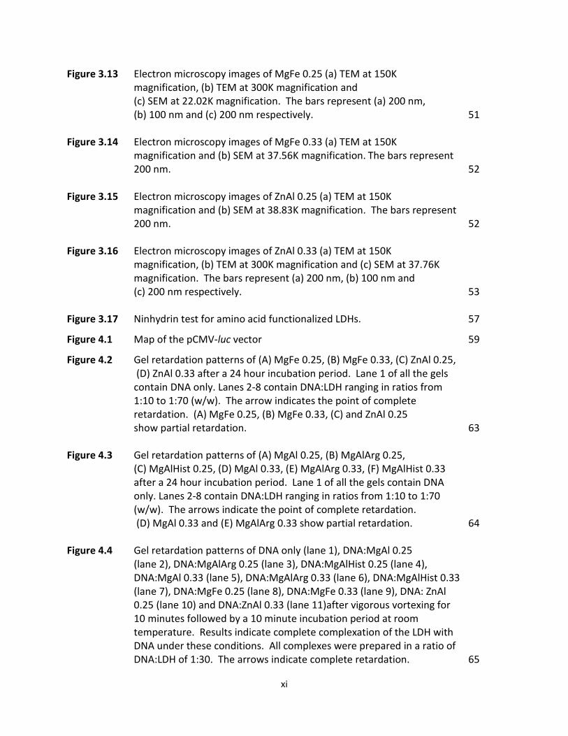
Figure 3.13 Electron microscopy images of MgFe 0.25 (a) TEM at 150K magnification, (b) TEM at 300K magnification and (c) SEM at 22.02K magnification. The bars represent (a) 200 nm, (b) 100 nm and (c) 200 nm respectively. 51
Figure 3.14 Electron microscopy images of MgFe 0.33 (a) TEM at 150K
magnification and (b) SEM at 37.56K magnification. The bars represent 200 nm. 52
Figure 3.15 Electron microscopy images of ZnAl 0.25 (a) TEM at 150K
magnification and (b) SEM at 38.83K magnification. The bars represent 200 nm. 52
Figure 3.16 Electron microscopy images of ZnAl 0.33 (a) TEM at 150K
magnification, (b) TEM at 300K magnification and (c) SEM at 37.76K magnification. The bars represent (a) 200 nm, (b) 100 nm and (c) 200 nm respectively. 53
Figure 3.17 Ninhydrin test for amino acid functionalized LDHs. 57
Figure 4.1 Map of the pCMV-luc vector 59
Figure 4.2 Gel retardation patterns of (A) MgFe 0.25, (B) MgFe 0.33, (C) ZnAl 0.25, (D) ZnAl 0.33 after a 24 hour incubation period. Lane 1 of all the gels contain DNA only. Lanes 2-8 contain DNA:LDH ranging in ratios from 1:10 to 1:70 (w/w). The arrow indicates the point of complete retardation. (A) MgFe 0.25, (B) MgFe 0.33, (C) and ZnAl 0.25 show partial retardation. 63
Figure 4.3 Gel retardation patterns of (A) MgAl 0.25, (B) MgAlArg 0.25,
(C) MgAlHist 0.25, (D) MgAl 0.33, (E) MgAlArg 0.33, (F) MgAlHist 0.33 after a 24 hour incubation period. Lane 1 of all the gels contain DNA only. Lanes 2-8 contain DNA:LDH ranging in ratios from 1:10 to 1:70 (w/w). The arrows indicate the point of complete retardation. (D) MgAl 0.33 and (E) MgAlArg 0.33 show partial retardation. 64
Figure 4.4 Gel retardation patterns of DNA only (lane 1), DNA:MgAl 0.25
(lane 2), DNA:MgAlArg 0.25 (lane 3), DNA:MgAlHist 0.25 (lane 4), DNA:MgAl 0.33 (lane 5), DNA:MgAlArg 0.33 (lane 6), DNA:MgAlHist 0.33 (lane 7), DNA:MgFe 0.25 (lane 8), DNA:MgFe 0.33 (lane 9), DNA: ZnAl 0.25 (lane 10) and DNA:ZnAl 0.33 (lane 11)after vigorous vortexing for 10 minutes followed by a 10 minute incubation period at room temperature. Results indicate complete complexation of the LDH with DNA under these conditions. All complexes were prepared in a ratio of DNA:LDH of 1:30. The arrows indicate complete retardation. 65

xii
Figure 4.5 Co-ordination between DNA phosphate backbone and LDH outer surface. 66
Figure 4.6 TEM images of DNA:LDH complexes (a) DNA-MgFe 0.25 at 300K magnification, (b) DNA-MgFe 0.33 at 150K magnification, (c) DNA-ZnAl 0.25 at 200 K magnification and (d) DNA-ZnAl 0.33 at 200K magnification. The bar represents 100 nm in (a) and 200 nm in (b), (c) and (d) respectively. 68
Figure 4.7 TEM images of DNA:LDH complexes (a) DNA-MgAl 0.25 at 60K
magnification, (b) DNA-MgAl 0.33 at 200K magnification , (c) DNA-MgAlArg 0.25 at 100K magnification, (d) DNA-MgAlArg 0.33 at 150K magnification, (e) DNA-MgAlHist 0.25 at 200K magnification and (f) DNA-MgAlHist 0.33 at 200K magnification. The bar represents 200 nm in (a) – (f). 69
Figure 4.8 XRD pattern of (a) MgAl 0.33 (b) DNA:MgAl 0.33 complex. 71
Figure 4.9 Gel retardation patterns of (A) MgAl 0.25, (B) MgAlArg 0.25, (C) MgAlHist 0.25, (D) MgAl 0.33, (E) MgAlArg 0.33, (F) MgAlHist 0.33 (G) MgFe 0.25, (H) MgFe 0.33, (I) ZnAl 0.25, (J) ZnAl 0.33 after incubation with siRNA at 60 oC overnight. 72
Figure 4.10 TEM images of complexes (a) siRNA-MgAl 0.25, (b) siRNA-MgAl 0.33, (c)
siRNA-MgAlArg 0.33 and (d) siRNA-MgAlHist 0.33 at 300 K magnification. The bar represents 100 nm in (a) – (d). 73
Figure 4.11 TEM images of complexes (a) siRNA-MgFe 0.25, (b) siRNA-MgFe 0.33,
(c) siRNA- ZnAl 0.25 and (d) siRNA-ZnAl 0.33 at 300K magnification. The bar represents 100 nm in (a) – (d). 74
Figure 4.12 TEM images of complexes (a) siRNA-MgAlArg 0.25 and
(b) siRNA-MgAlHist 0.25 at 300K magnification. The bar represents 100 nm in (a) and (b). 75
Figure 4.13 Results of nuclease digestion assay of DNA:MgAl 0.33. Lane 1 : naked DNA, Lane 2: DNA exposed to nuclease digestion, Lane 3 :MgAl 0.25 DNA-LDH complex, and Lane 4: MgAl 0.25 DNA-LDH complex exposed to Nuclease digestion. 78

xiii
Figure 4.14 Results of nuclease digestion assay of siRNA:MgAl 0.25. Well 1 was loaded with naked siRNA, well 2 contained siRNA exposed to nuclease digestion, well 3 was loaded with the MgAl 0.25 siRNA-LDH complex, and well 4 contained the MgAl 0.25 siRNA-LDH complex exposed to nuclease digestion. 79
Figure 5.1 Image of HEK293 cells (x100) captured using an Olympus inverted
microscope. 81
Figure 5.2 Image of HepG2 cells (x100) captured using an Olympus inverted microscope. 82
Figure 5.3 Image of Caco-2 cells (x100) captured using an Olympus inverted
microscope. 83 Figure 5.4 Image of HeLa-tat-luc cells (x100) captured using an Olympus
inverted microscope. 83 Figure 5.5 MTT assay of Caco-2, HEK293 and HepG2 cells exposed to
(a) pCMV-luc:MgAl 0.25 complexes, (b) pCMV-luc:MgAl 0.33 complexes, (c) pCMV-luc:MgAlArg 0.25 complexes,(d) pCMV-luc:MgAlArg 0.33 complexes, (e) pCMV-luc:MgAlHist 0.25 complexes and (f) pCMV-luc:MgAlHist 0.33 complexes. Data is represented as means ±SD (n=3). *p<0.05 was considered statistically significant. 90
Figure 5.6 MTT assay of Caco-2, HEK293 and HepG2 cells exposed to
(a) pCMV-luc:MgFe 0.25 complexes, (b) pCMV-luc:MgFe 0.33 complexes, (c) pCMV-luc:ZnAl 0.25 complexes and (d) pCMV-luc:ZnAl 0.33 complexes. No statistically significant change in percent cell survival was observed. 91
Figure 5.7 MTT assay of Hela-tat-luc cells exposed to anti-luc siRNA:MgAl 0.25
complexes, anti-luc siRNA:MgAlArg 0.25 complexes and anti-luc siRNA:MgAlHist 0.25 complexes. *p<0.05 was considered statistically significant. 92
Figure 5.8 MTT assay of Hela-tat-luc cells exposed to anti-luc siRNA:MgAl 0.33,
anti-luc siRNA:MgAlArg 0.33 complexes and anti-luc siRNA:MgAlHist 0.33 complexes. *p<0.05 was considered statistically significant. 93
xiv
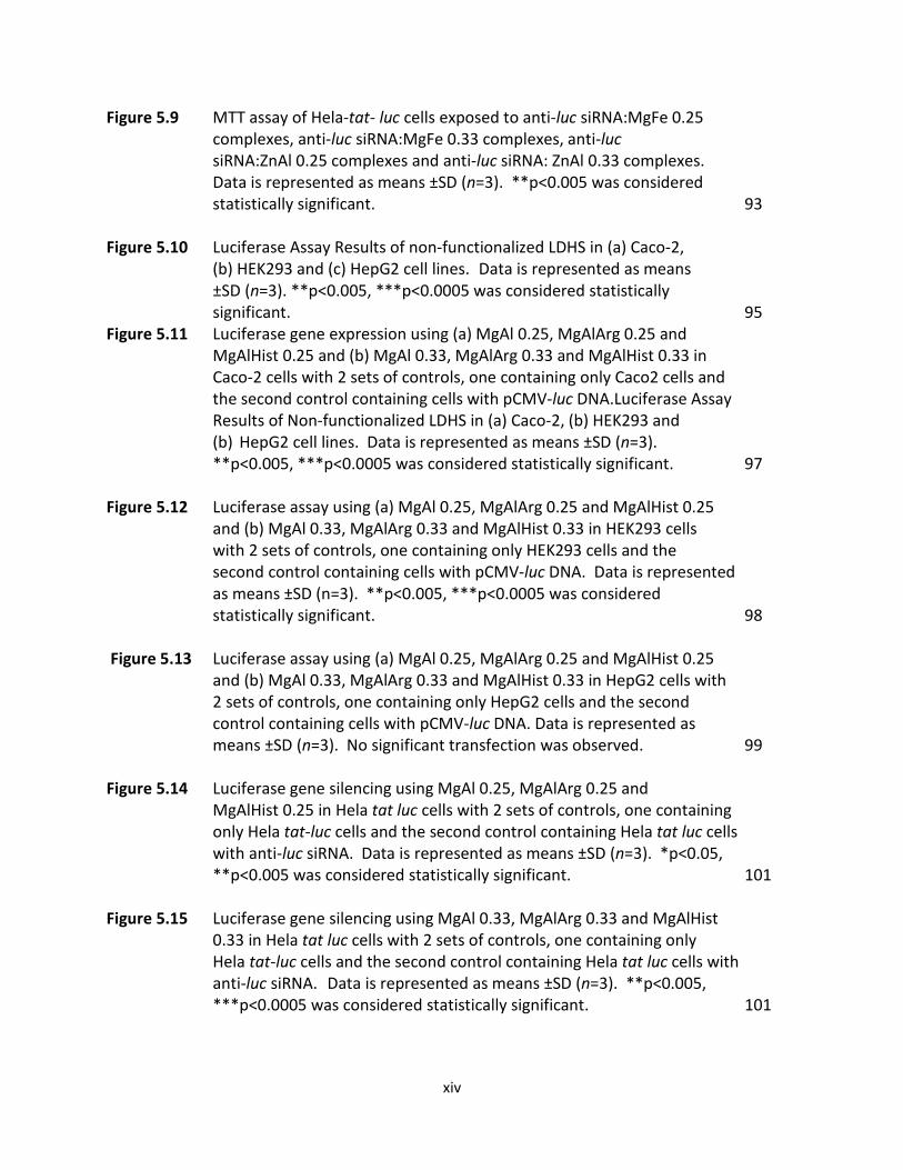
Figure 5.9 MTT assay of Hela-tat- luc cells exposed to anti-luc siRNA:MgFe 0.25 complexes, anti-luc siRNA:MgFe 0.33 complexes, anti-luc siRNA:ZnAl 0.25 complexes and anti-luc siRNA: ZnAl 0.33 complexes. Data is represented as means ±SD (n=3). **p<0.005 was considered statistically significant. 93
Figure 5.10 Luciferase Assay Results of non-functionalized LDHS in (a) Caco-2,
(b) HEK293 and (c) HepG2 cell lines. Data is represented as means ±SD (n=3). **p<0.005, ***p<0.0005 was considered statistically significant. 95
Figure 5.11 Luciferase gene expression using (a) MgAl 0.25, MgAlArg 0.25 and MgAlHist 0.25 and (b) MgAl 0.33, MgAlArg 0.33 and MgAlHist 0.33 in
Caco-2 cells with 2 sets of controls, one containing only Caco2 cells and the second control containing cells with pCMV-luc DNA.Luciferase Assay Results of Non-functionalized LDHS in (a) Caco-2, (b) HEK293 and (b) HepG2 cell lines. Data is represented as means ±SD (n=3). **p<0.005, ***p<0.0005 was considered statistically significant. 97
Figure 5.12 Luciferase assay using (a) MgAl 0.25, MgAlArg 0.25 and MgAlHist 0.25 and (b) MgAl 0.33, MgAlArg 0.33 and MgAlHist 0.33 in HEK293 cells with 2 sets of controls, one containing only HEK293 cells and the second control containing cells with pCMV-luc DNA. Data is represented as means ±SD (n=3). **p<0.005, ***p<0.0005 was considered statistically significant. 98
Figure 5.13 Luciferase assay using (a) MgAl 0.25, MgAlArg 0.25 and MgAlHist 0.25 and (b) MgAl 0.33, MgAlArg 0.33 and MgAlHist 0.33 in HepG2 cells with 2 sets of controls, one containing only HepG2 cells and the second control containing cells with pCMV-luc DNA. Data is represented as means ±SD (n=3). No significant transfection was observed. 99
Figure 5.14 Luciferase gene silencing using MgAl 0.25, MgAlArg 0.25 and
MgAlHist 0.25 in Hela tat luc cells with 2 sets of controls, one containing only Hela tat-luc cells and the second control containing Hela tat luc cells with anti-luc siRNA. Data is represented as means ±SD (n=3). *p<0.05, **p<0.005 was considered statistically significant. 101
Figure 5.15 Luciferase gene silencing using MgAl 0.33, MgAlArg 0.33 and MgAlHist
0.33 in Hela tat luc cells with 2 sets of controls, one containing only Hela tat-luc cells and the second control containing Hela tat luc cells with anti-luc siRNA. Data is represented as means ±SD (n=3). **p<0.005, ***p<0.0005 was considered statistically significant. 101
xv
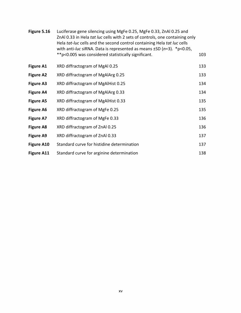
Figure 5.16 Luciferase gene silencing using MgFe 0.25, MgFe 0.33, ZnAl 0.25 and ZnAl 0.33 in Hela tat luc cells with 2 sets of controls, one containing only Hela tat-luc cells and the second control containing Hela tat luc cells with anti-luc siRNA. Data is represented as means ±SD (n=3). *p<0.05, **p<0.005 was considered statistically significant. 103
Figure A1 XRD diffractogram of MgAl 0.25 133
Figure A2 XRD diffractogram of MgAlArg 0.25 133
Figure A3 XRD diffractogram of MgAlHist 0.25 134
Figure A4 XRD diffractogram of MgAlArg 0.33 134
Figure A5 XRD diffractogram of MgAlHist 0.33 135
Figure A6 XRD diffractogram of MgFe 0.25 135
Figure A7 XRD diffractogram of MgFe 0.33 136
Figure A8 XRD diffractogram of ZnAl 0.25 136
Figure A9 XRD diffractogram of ZnAl 0.33 137
Figure A10 Standard curve for histidine determination 137
Figure A11 Standard curve for arginine determination 138
xvi
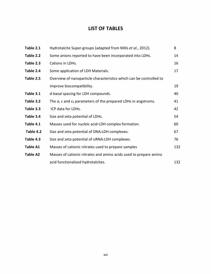
LIST OF TABLES
Table 2.1 Hydrotalcite Super-groups (adapted from Mills et al., 2012). 8
Table 2.2 Some anions reported to have been incorporated into LDHs. 14
Table 2.3 Cations in LDHs. 16
Table 2.4 Some application of LDH Materials. 17
Table 2.5 Overview of nanoparticle characteristics which can be controlled to
improve biocompatibility. 19
Table 3.1 d-basal spacing for LDH compounds. 40
Table 3.2 The a, c and c0 parameters of the prepared LDHs in angstroms. 41
Table 3.3 ICP data for LDHs. 42
Table 3.4 Size and zeta potential of LDHs. 54
Table 4.1 Masses used for nucleic acid-LDH complex formation. 60
Table 4.2 Size and zeta potential of DNA:LDH complexes. 67
Table 4.3 Size and zeta potential of siRNA:LDH complexes. 76
Table A1 Masses of cationic nitrates used to prepare samples 132
Table A2 Masses of cationic nitrates and amino acids used to prepare amino
acid functionalized hydrotalcites. 132
xvii
ABBREVIATIONS
aa : amino acid
AONS : Antisense oligonucleotides
ATP : Adenosine triphosphate
BCA : Bicinchoninic acid
Caco-2 : Colorectal adenocarcinoma cells
DMSO : Dimethyl sulphoxide
DNA : Deoxyribonucleic acid
EDTA : Ethylenediaminetetraacetic acid
EMEM : Eagle’s Minimum Essential Medium
EMSA : Electrophoretic mobility shift assay
FBS : Foetal bovine serum
FTIR : Fourier transform infrared spectroscopy
HBS : HEPES-buffered saline
HEK293 : Human embryonic kidney cells
Hela tat-luc : cervical carcinoma cells transfected with tat protein and stably expressing the luciferase gene
HEPES : 2-[-(2-hydroxyethyl)-piperazinyl]-ethanesulphonic acid
HepG2 : Liver hepatocellular carcinoma cells
HT : Hydrotalcite
HT-like : Hydrotalcite-like
ICP-OES : Inductively coupled plasma-optical emission spectroscopy
LDH : Layered double hydroxide
miRNA : Micro ribonucleic acid
xviii
MTT : 3-(4,5-dimethylthiazol-2-yl)-2,5-diphenyltetrazolium
nm : Nanometres
NTA : Nanoparticle tracking analysis
PBS : Phosphate-buffered saline
pCMV : Plasmid containing cytomegalovirus promoter
pDNA : Plasmid DNA
RISC : RNA-induced-silencing-complex
RLU : Relative light units
RNAi : RNA interference
SEM : Scanning electron microscopy
shRNA : Short hairpin ribonucleic acid
siRNA : Small interfering ribonucleic acid
TEM : Transmission electron microscopy
Tris : Tris(hydroxymethyl)aminomethane
v/v : volume to volume ratio
w/w : weight to weight ratio
XRD : X-Ray diffraction
μg : micrograms
μL : microlitres

xix
ACKNOWLEDGEMENTS
I would like to express my sincere gratitude to:
• Prof. M. Singh for her invaluable supervision and guidance. The unselfish
sharing of her immense knowledge, words of encouragement, constructive
criticism and insightful comments will always be greatly appreciated.
• Dr. S. Singh for his expert guidance in the chemistry aspects of this study.
• Drushan Padayachee for assistance with the chemical analyses.
• My colleagues in the non-viral gene delivery for always being supportive and
helpful.
• My mum and dad Nirmala and Devjith Bhagwandin, my sister Nerushka,
brothers Navesh and Nikesh as well as my mother-in-law Shanthi Nundkumar
for their constant love and blessings.
• My husband Prashant Nundkumar and my son Ashmil, for always believing in
me. I am most grateful for your love, support and encouragement.

xx
DEDICATION
This thesis is dedicated to
Miler and Ashmil

1
CHAPTER ONE
1. INTRODUCTION
1.1 BACKGROUND OF STUDY
Gene therapy encompasses a range of methods aimed at regulating gene expression by insertion
of exogenous nucleic acids, such as deoxyribonucleic acid (DNA), small interfering ribonucleic
acid (siRNA), micro RNA (miRNA), short hairpin RNA (shRNA) and antisense oligonucleotides
(AONS), to either replace or repair a defective gene. Unlike conventional therapeutic approaches
which tend to treat the symptoms of a disease, gene therapy offers a strategy to treat the
underlying cause of a disorder or disease.
Naked nucleic acids cannot easily traverse the cell membrane, partly due to their negative charge,
and even when they do enter the cell, they are prone to degradation by nucleases. A suitable
“carrier” or “vector” is therefore required to efficiently transport the nucleic acids into the cell.
Thus far, the most successful vectors employed have been of viral origin, but they suffer from a
number of major drawbacks such as immunogenic responses, low DNA carrying capacity,
insertional mutagenesis, lack of specific cell targeting and high cost [Ragusa et al., 2007]. Over
the years a number of non-viral vectors have been explored; such as liposomes [Templeton,
2002; Daniels et al., 2013; Habib et al., 2013; Naicker et al., 2014; Lee et al., 2017], gold
nanoparticles [Riley and Vermerris, 2017], dendrimers [Pan et al., 2007; Pan et al., 2009],
quantum dots [Kralj and Pavelic, 2003], carbon nanotubes [Singh et al., 2005; Gao et al., 2006;
Lacerda et al., 2007; Ahmed et al., 2009; Ahmed1 et al., 2009] magnetic nanoparticles [Mulens et
al., 2013; Riley and Vermerris, 2017] and functionalized polymers [Perez et al., 2001; Nimesh et
al., 2006]. These non-viral carriers also suffer from a number of disadvantages, the most common
being cytotoxicity and immunogenic responses. Thus, there remains a need for the development
of safe and effective gene delivery vehicles.
Anionic clays such as layered double hydroxides (LDH) or hydrotalcite-like (HT-like) materials,
have recently attracted much attention as carriers of bioactive molecules and genes [Ladewig1 et

2
al., 2010; Choy et al., 2000; Nalawade et al., 2009; Balcomb et al., 2015]. Hydrotalcites (HTs)
refer specifically to magnesium aluminium LDHs. LDHs can complex with various types of
molecules such as drugs [Choy et al., 2004; Costantino et al., 2008], sugar phosphates [Jellicoe
and Fogg, 2012], fullerenes [Fortner et al., 2012], and biomolecules such as oligomers, DNA and
nucleotides [Kwak et al., 2002; Choy et al., 2007].
Unlike most inorganic nanoparticles that require chemical and/or biological pre-modification to
obtain the desirable properties for cellular delivery, anionic clays allow for direct loading of
anionic drugs or biomolecules due to their anion-exchange properties [Ladewig et al., 2009; Xu
et al., 2006]. Mg-Al LDH’s are less toxic than most inorganic nanoparticles [Kriven et al., 2004;
Choi et al., 2008]. LDH nanoparticles upon disintegration in the cytoplasm results in cyto-friendly
ions such as Mg2+, Al3+ and NO3- [Xu et al., 2006; Kura et al., 2014]. Research into the use of LDHs
as gene delivery vehicles has thus far, shown promising results.
Reports indicate that functionalization of other non-viral gene delivery vectors has resulted in
enhanced gene delivery, with up to a 1000 fold increase in transfection [Smith et al., 1997]. It
has been suggested that better transfection occurs upon functionalization as a result of enhanced
uptake through receptors on the cell surface, which may retain complexes at the cell surface
rather than merely relying on non-specific electrostatic attraction between the nanoparticles and
the cell surface [Wu and Wu, 1987; Choy et al., 2000]. The possibility thus exists that
functionalization of the LDHs could result in greater, more effective transfection. Del Hoyo and
co-workers (2007), observed that LDHs displayed lower transfection efficiency due to pH related
degradation of DNA in the endosome, a hurdle that could possibly be overcome by
functionalization of the LDH. Functionalization of the LDHs with amino acids containing cationic
side-chains could result in enhancement of the ‘proton-sponge’ effect, leading to quicker and
more efficient endosomal escape and improved transfection, when compared to non-
functionalized LDHs.

3
1.2 AIMS AND OBJECTIVES OF STUDY
The aim of the study was to prepare LDHs (both non-functionalized and amino acid
functionalized) and compare their efficiencies as vectors for transfection, to achieve gene
expression and gene silencing.
In order to accomplish this aim, the objectives of the study were to:
1. Prepare amino acid functionalized and non-functionalized LDHs using the co-precipitation
method.
2. Characterize the prepared LDHs.
3. Evaluate the DNA and siRNA binding capacity of the prepared LDHs.
4. Investigate the cytotoxicity of the prepared LDHs.
5. Investigate and compare the transfection and gene silencing efficiency of the amino acid
functionalized versus the non-functionalized LDHs.
1.3 SIGNIFICANCE OF THE STUDY
Amino acid functionalized LDHs offers a novel and promising concept for enhanced gene delivery
in vitro. The potential benefits of this study may include the following:
• Although there have been many reports of the use of LDHs (both functionalized and non-
functionalized) as gene delivery vehicles in literature, there has as yet not been any study
involving amino acid functionalized LDHs as carriers of genes. Hence, we undertook to
prepare amino acid functionalized LDHs, which will exhibit enhanced cellular uptake and
rapid endosomal escape, leading to a greater degree of gene expression or knockdown.
• It is anticipated that this project could possibly lead to the development of a potential
gene delivery vector to aid in realizing the dream of using gene therapy to overcome
human genetic diseases.

4
1.4 NOVELTY OF THE STUDY
There have been several published reports on the use of LDHs as DNA delivery vehicles in vitro
[Choy et al., 2000; Choy et al., 2004; Del Hoyo, 2007; Xu et al., 2007; Ladewig1 et al., 2010] and
in the delivery of siRNA [Ladewig2 et al., 2010; Chen et al., 2013; Li et al., 2014]. It has been
further shown that arginine functionalization of hydroxyapatites [Wang et al., 2015], and amino
acid modification of chitosan [Zheng et al., 2015], improved transfection efficiency. The
intercalation of amino acids into layered double hydroxides [Aisawa et al., 2000; Aisawa et al.,
2001; Nakayama et al., 2004; Aisawa et al., 2006], have been attempted but was not exploited
as gene delivery vehicles. This study reports for the first time on DNA transfection and gene
silencing using amino acid functionalized LDHs. The transfection efficiency of amino acid
functionalized versus non-functionalized LDHs is also investigated.
1.5 OVERVIEW OF THE DISSERTATION
Chapter 1 briefly introduces and summarizes the background to the study. It outlines the
challenges encountered with current gene therapy vectors and explores the rationale for and
novelty of the study. The aim and objectives of the study are also described.
Chapter 2 is a detailed literature review covering the historical background of LDHs, the chemical
compositions of various LDHs, the applications of LDHs and their use in gene therapy.
Chapter 3 describes the preparation and characterization of LDHs. Methods of preparation of
LDHs are briefly discussed and the characterization of the prepared LDHs using X-ray diffraction
(XRD), Inductively coupled plasma-optical emission spectroscopy (ICP-OES), Fourier transform
infrared spectroscopy (FTIR), Transmission electron microscopy (TEM), scanning electron
microscopy (SEM), and nanoparticle tracking analysis (NTA) are described and discussed.

5
Chapter 4 includes the nucleic acid binding studies using gel retardation assays. The nucleic
acid:LDH complexes are characterized using TEM, XRD and NTA. The susceptibility of the nucleic
acid:LDH complex to serum nucleases is also investigated.
Chapter 5 describes the in vitro cell culture assays such as the cytotoxicity studies, gene
transfection and gene knockdown studies. Cytotoxicity was evaluated in the HepG2, HEK293,
Caco-2 and Hela tat luc cells, while pCMV-luc DNA transfection was assessed in the HepG2,
HEK293 and Caco-2 only. The knockdown of the luciferase gene (stably expressed in Hela tat luc
cells) by anti-luc-siRNA, employing the synthesized LDHs as carriers is also investigated.
Chapter 6 provides the conclusions derived from this study and includes recommendations for
future studies.

6
CHAPTER TWO
2. LITERATURE REVIEW
2.1 HISTORICAL BACKGROUND OF LAYERED DOUBLE HYDROXIDES
There are two main classes of clays, viz. cationic clays which are made up of negatively charged
aluminosilicate or magnesiosilicate layers with positive ions in the interlayer region [Rajamathi
et al., 2001] and anionic clays which consist of lamellar metal hydroxides composed of two (or
more) kinds of metals in the main layers and anions in the interlayer region [He et al., 2005; de
Roy et al., 2006; Boulet et al., 2006]. Anionic clays are generally referred to as layered double
hydroxides (LDHs).
Hydrotalcite (HT) is an anionic clay mineral that was first reported by Hochstetter in Sweden in
1842 [Bravo-Suarez, 2004]. The formula for HT was proposed by an Italian professor of
mineralogy, Professor E. Manasse, in Florence Italy in 1915 and confirmed by Foshag in 1920
[Mills et al., 2012]. Investigations by Aminoff and Broome in 1930 revealed the existence of two
polytypes, viz. hydrotalcite and manasseite, the latter named in honour of Manasse [Cavani et
al., 1991]. The two polytypes differ in their symmetry with the HT being rhombohedral and the
manasseite, hexagonal [Belloto et al., 1996]. Since HT is one of the most representative minerals
of the anionic clays, other layered double hydroxides are often referred to as hydrotalcite-like
compounds (HT-like). The general LDH formula, based on a combination of divalent and trivalent
metal cations can be written as:
[MII1-x MIIIx(OH)2]Aq-x/q.nH2O
Where MII and MIII represent the divalent and trivalent cations respectively, Aq- represents an
exchangeable anion, x is the MIII/MII + MIII molar ratio and n is the number of moles of water
[Posati et al., 2012; Ladewig et al., 2009; Luan et al., 2009]. The divalent and trivalent cations, the interlayer anions as well as the value of the stoichiometric
co-efficient (x) may be widely varied, giving rise to a large class of isostructural materials [Kuang
7
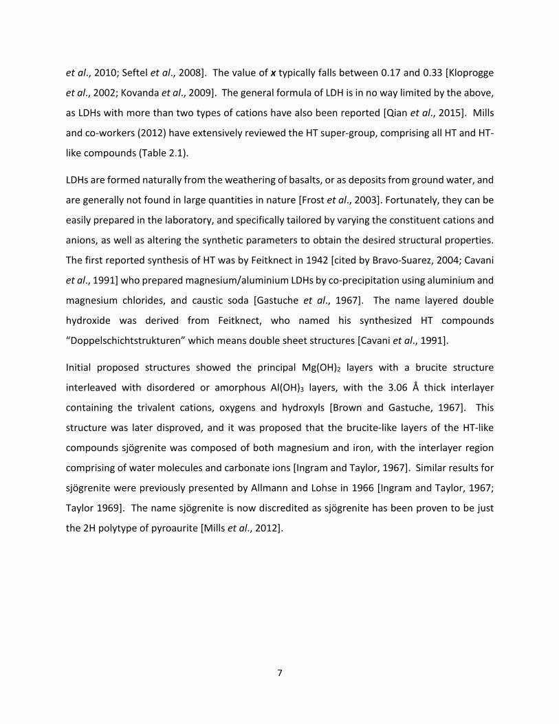
et al., 2010; Seftel et al., 2008]. The value of x typically falls between 0.17 and 0.33 [Kloprogge
et al., 2002; Kovanda et al., 2009]. The general formula of LDH is in no way limited by the above,
as LDHs with more than two types of cations have also been reported [Qian et al., 2015]. Mills
and co-workers (2012) have extensively reviewed the HT super-group, comprising all HT and HT-
like compounds (Table 2.1). LDHs are formed naturally from the weathering of basalts, or as deposits from ground water, and
are generally not found in large quantities in nature [Frost et al., 2003]. Fortunately, they can be
easily prepared in the laboratory, and specifically tailored by varying the constituent cations and
anions, as well as altering the synthetic parameters to obtain the desired structural properties.
The first reported synthesis of HT was by Feitknect in 1942 [cited by Bravo-Suarez, 2004; Cavani
et al., 1991] who prepared magnesium/aluminium LDHs by co-precipitation using aluminium and
magnesium chlorides, and caustic soda [Gastuche et al., 1967]. The name layered double
hydroxide was derived from Feitknect, who named his synthesized HT compounds
“Doppelschichtstrukturen” which means double sheet structures [Cavani et al., 1991]. Initial proposed structures showed the principal Mg(OH)2 layers with a brucite structure
interleaved with disordered or amorphous Al(OH)3 layers, with the 3.06 Å thick interlayer
containing the trivalent cations, oxygens and hydroxyls [Brown and Gastuche, 1967]. This
structure was later disproved, and it was proposed that the brucite-like layers of the HT-like
compounds sjögrenite was composed of both magnesium and iron, with the interlayer region
comprising of water molecules and carbonate ions [Ingram and Taylor, 1967]. Similar results for
sjögrenite were previously presented by Allmann and Lohse in 1966 [Ingram and Taylor, 1967;
Taylor 1969]. The name sjögrenite is now discredited as sjögrenite has been proven to be just
the 2H polytype of pyroaurite [Mills et al., 2012].

8
Table 2.1 Hydrotalcite super-groups [adapted from Mills et al., 2012].
Name Cell Parameters (Å) Polytype Formula
a c
1 Hydrotalcite 3.065 23.07 3R Mg6Al2(OH)16[CO3].4H2O
2 Manasseite 3.06 15.34 2H Mg6Al2(OH)16[CO3].4H2O 3 Pyroaurite 3.089 23.23 3R Mg6Fe23+(OH)16[CO3].4H2O 4 Sjögrenite 3.097 15.56 2H Mg6Fe23+(OH)16[CO3].4H2O 5 Stichtite 3.09 23.19 3R Mg6Cr2(OH)16[CO3].4H2O 6 Barbertonite 3.085 15.52 2H Mg6Cr23+(OH)16[CO3].4H2O 7 Meixnerite 3.046 22.93 3R Mg6Al2(OH)18.4H2O 8 Iowaite 3.1183 24.113 3R Mg6Fe23+(OH)16Cl2.4H2O 9 Droninoite 6.206 46.184 6R Ni6Fe23+(OH)16Cl2.4H2O 10 Woodallite 3.102 24.111 3R Mg6Cr2(OH)16Cl2.4H2O 11 Desautelsite 3.114 24.39 3R Mg6Mn23+(OH)16[CO3].4H2O 12 Takovite 3.0250 22.595 3R Ni6Al2(OH)16[CO3].4H2O 13 Reevesite 3.082 22.770 3R1 Ni6Fe23+ (OH)16[CO3].4 H2O 14 Jamborite 3.07 23.3 3R Ni62+Ni23+(OH)]16S.4H2O 15 Quintinite 10.751 22.71/15.171 3T/2H Mg4Al2(OH)12[CO3].3H2O 16 Charmarite 10.985 15.10/22.63 2H/3T Mn4Al2(OH)12[CO3].3H2O 17 Caresite 10.805 22.48 3T Fe42+Al2(OH)12[CO3].3H2O 18 Zaccagnaite 3.0662 22.6164 3R Zn4Al2(OH)12[CO3].3H2O 19 Chlormagaluminite 5.29 15.46 2H Mg4Al2(OH)12Cl2. 3H2O 20 Comblainite 3.038 22.79 3R Ni4Co23+(OH)12(CO3).3H2O 21 Fougerite 3.17-3.18 22.7-22.9 3R Fe42+Fe23+(OH)12[CO3].3H2O 22 Mossbauerite 3.079 22.253 3R Fe63+O4(OH)8[CO3].3H2O 23 Woodwardite 3.00 27.3 3R Cu1-xAlx(OH)2[SO4]x/2.6H2O 24 Zincowoodwardite 3.063 8.91/25.42 1T/3R Zn1-xAlx(OH)2[SO4]x/2.nH2O 25 Honessite 3.083 25.8 3R (Ni1-xFe3+x)(OH)2[SO4]x/2.nH2O n ˂ 3x/2
HYDROTALCITE SUPERGROUP
QUINTINITE SUPERGROUP
FOUGЀRITE SUPERGROUP
WOODWARDITE SUPERGROUP

9
Table 2.1 (continued)…
Name Cell Parameters (Å) Polytype Formula
a c
26 Glaucocerinite 3.057-3.070 32.52-32.65 3R1 (Zn1-xAlx)(OH)2[SO4]x/2.nH2O 27 Hydrowoodwardite 3.066 32.80 3R Cu1-xAlx(OH)2[SO4]x/2.nH2O 28 Carrboydite 3.022 32.45 3R1 (Ni1-xAlx)(OH)2[SO4]x/2.nH2O 29 Hydrohonessite 3.09 33.4 3R2 (Ni1-xFe3+x)(OH)2[SO4]x/2.nH2O n ˃ 3x/2 30 Mountkeithite 10.698 22.545 2H (Mg1-xFe3+x)(OH)2[SO4]x/2.nH2O 31 Zincaluminite No structural data available (Zn1-xAlx)(OH)2[SO4]x/2.nH2O 32 Wermlandite 9.303 22.57 1T Mg7Al2(OH)18[Ca(H2O)6] [SO4]2.6H2O 33 Shigaite 9.512 33.074 3R Mn6Al3(OH)18[Na(H2O)6] [SO4]2.6H2O 34 Nikischerite 9.347 33.000 3R Fe62+Al3(OH)18[Na(H2O)6] [SO4]2.6H2O 35 Motukoreaite 9.172 33.51 3R Mg6Al3(OH)18[Na(H2O)6] [SO4]2.6H2O 36 Natroglaucocerinite No structural data available Zn6Al3(OH)18[Na(H2O)6] [SO4]2.6H2O 37 Karchevskyite 16.0556 25.66 1T Mg18Al9(OH)54Sr2(CO3)9(H2O)6(H3O)5
38 Cualstibite 9.15 9.745 1T Cu2Al(OH)6[Sb(OH)6] 39 Cyanophyllite 9.941 5.498 1M Cu2Al(OH)6[Sb(OH)6] 40 Zincalstibite 5.327 9.792 1T Zn2Al(OH)6[Sb(OH)6] 41 Omsite 5.351 19.5802 2T Ni2Fe3+(OH)6[Sb(OH)6] 42 Hydrocalumite 5.765 46.978 3R Ca4Al2(OH)12Cl2.4H2O 43 Kuzelite 5.76 53.66 3R Ca4Al2(OH)12[SO4].6H2O 44 Coalingite 3.12 37.4 3R Mg10Fe23+(OH)24 [CO3].2H2O 45 Brugnatellite 5.47 15.97 1T Mg6Fe3+ CO3(OH)13.4H2O 46 Muskoxite 3.07 4.6 Further
investigation required
Mg7(Fe3+) (OH)26.H2O
GLAUCOCERINITE SUPERGROUP
CAULSTIBITE SUPERGROUP
HYDROCALUMITE SUPERGROUP
WERMLANDITE SUPERGROUP
UNCLASSIFIED
10
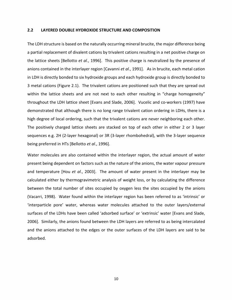
2.2 LAYERED DOUBLE HYDROXIDE STRUCTURE AND COMPOSITION
The LDH structure is based on the naturally occurring mineral brucite, the major difference being
a partial replacement of divalent cations by trivalent cations resulting in a net positive charge on
the lattice sheets [Bellotto et al., 1996]. This positive charge is neutralized by the presence of
anions contained in the interlayer region [Cavanni et al., 1991]. As in brucite, each metal cation
in LDH is directly bonded to six hydroxide groups and each hydroxide group is directly bonded to
3 metal cations (Figure 2.1). The trivalent cations are positioned such that they are spread out
within the lattice sheets and are not next to each other resulting in “charge homogeneity”
throughout the LDH lattice sheet [Evans and Slade, 2006]. Vucelic and co-workers (1997) have
demonstrated that although there is no long range trivalent cation ordering in LDHs, there is a
high degree of local ordering, such that the trivalent cations are never neighboring each other.
The positively charged lattice sheets are stacked on top of each other in either 2 or 3 layer
sequences e.g. 2H (2-layer hexagonal) or 3R (3-layer rhombohedral), with the 3-layer sequence
being preferred in HTs [Bellotto et al., 1996]. Water molecules are also contained within the interlayer region, the actual amount of water
present being dependent on factors such as the nature of the anions, the water vapour pressure
and temperature [Hou et al., 2003]. The amount of water present in the interlayer may be
calculated either by thermogravimetric analysis of weight loss, or by calculating the difference
between the total number of sites occupied by oxygen less the sites occupied by the anions
(Vacarri, 1998). Water found within the interlayer region has been referred to as ‘intrinsic’ or
‘interparticle pore’ water, whereas water molecules attached to the outer layers/external
surfaces of the LDHs have been called ‘adsorbed surface’ or ‘extrinsic’ water [Evans and Slade,
2006]. Similarly, the anions found between the LDH layers are referred to as being intercalated
and the anions attached to the edges or the outer surfaces of the LDH layers are said to be
adsorbed.

11
Figure 2.1 Structures of (a) brucite and (b) LDH [Redrawn and adapted from Benicio et al.,
2015].
(a)
(b)

12
The nature of the interlayer anions and presence of water in the interlayer regions results in an
increase in basal spacing from 4.78 Å in brucite to 7.8 Å in HT [Brindley and Kikkawa, 1980;
Wypych, 2004]. Generally, the thickness of the interlayer depends on the number, size,
orientation and strength of the bonds between the anions and the hydroxyl groups of the brucite-
like layers [Seftel et al., 2008].
2.2.1 ANION EXHANGE PROPERTY OF LAYERED DOUBLE HYDROXIDES
The anions in the LDH interlayer are relatively weakly bound and can be readily exchanged [Meyn
et al., 1990]. The potential anion exchange capability for an LDH depends on its charge density
(or unit charge per area) which is based on the different trivalent to divalent cation ratios. The
formula for charge density is,
Charge density = xe/a2sin60o
where, x is the ratio of trivalent to total amount of metal, a is the distance between adjacent
metal ions in the layer, e is the electronic charge, and sin60° is the angle between the a and b
axes [Richardson, 2007]. Thus, a HT with Mg:Al ratio of 1:2 would have a higher charge density than one with a Mg:Al ratio
of 1:3. The higher the trivalent cation content, the higher the positive charge density, and the
higher the amount of anions that can be potentially adsorbed [Beavers, 1999]. A major advantage of LDHs over other anion exchangers is the “large space” between the brucite
layers. This allows for a diverse array of anionic compounds to be exchanged using LDHs. Bish
(1980) showed that anion exchange in the LDH interlayer can cause displacement of the brucite
layers, resulting in a possible change in polytype.
2.2.2 ANION PREFERENCE
Miyata (1983) showed that selectivity of monovalent anions are in the order of OH- ˃ Cl- ˃ Br- ˃
NO3- ˃ I-, in slight contradiction to Bontchev and co-workers (2003), who showed that
monovalent anion exchange preference was Br- ˃ Cl- ˃ NO3- ˃ I-. Generally, carbonate (CO32-) is

13
the easiest to intercalate and the most difficult to exchange [Crosby et al., 2014], whereas halides
and nitrates are easy to intercalate and exchange. Therefore, most LDH materials are prepared
using either nitrate or chloride as the initial anion. Divalent anions are more strongly adsorbed
than monovalent anions [Forano et al., 2006], with the anion preference for divalent anions being
CO32- > HPO42- > CrO42- >SO42- > MoO42- [Auerbach et al., 2004]. Multivalent anions are preferred
by LDHs, due to stronger electrostatic interaction [Choy et al., 2007]. Naturally occurring LDHs usually contain carbonate as the interlayer anion, but synthetic LDHs
may have a variety of anions of different geometries, sizes and charges incorporated within the
layers, resulting in a large class of isostructural materials [Evans and Duan, 2006], with diverse
physico-chemical properties [Miyata, 1975; Miyata and Okada, 1977]. Some of the reported
anions incorporated within the interlayers of LDHs can be seen in Table 2.2. For biological studies, the preferred anion for the synthesis of LDHs is the nitrate anion, which
creates a non-toxic, biologically compatible form of LDH [Costantino et al., 2008]. The nitrate
anion, being monovalent, is also a good precursor for exchange reactions [Choy et al., 2007].

14
Table 2.2 Some anions reported to have been incorporated into LDHs.
Class of Anion Anion Reference Organic anions Carboxylic acids
Alkoxides, naphthalene, β-napthol, phenol Acetate, stearate, oleate Tartarate, succinate Oxalate Citrate Sugar phosphates Cyclodextrins Unsaturated carboxylates
Carlino, 1997 Butenko et al., 2014 Kandare and Hossenlopp, 2006 Prevot et al., 1998 Prevot et al., 1999 Kumar et al., 2006 Jellicoe and Fogg, 2012 Mohanambe and Vasudevan, 2006 Takagi et al., 1993
Inorganic anions
Carbonate Nitrate, chloride, iodide, bromide Chromate, dichromate, chlorate Chloride, percholate, sulphate chlorate Hydroxide Acetate, terephthalate, benzoate Metals and oxometalates Hexacyanocobalt (III) Mo7O24
6-, Cr2O72-, SiO3
2-
Oxalate, [Fe(CN)6]-, bromide, chloride, carbonate, hydroxide, nitrate
Trujillano, 2006 Bontchev et al., 2003; Miyata and Okada, 1977 Malherbe and Besse, 2000 Brindley and Kikkawa,1980 Miyata, 1975 Newman et al., 2002 Newman and Jones, 1999 Rives and Ullibarri, 1999 Suzuki et al., 1989 Misra and Perrotta, 1992 Khan and O’Hare, 2002
15
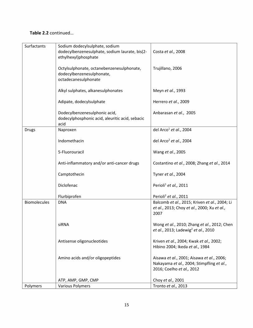
Table 2.2 continued…
Surfactants Sodium dodecylsulphate, sodium dodecylbenzenesulphate, sodium laurate, bis(2-ethylhexyl)phosphate Octylsulphonate, octanebenzenesulphonate, dodecylbenzenesulphonate, octadecanesulphonate Alkyl sulphates, alkanesulphonates Adipate, dodecylsulphate Dodecylbenzenesulphonic acid, dodecylphosphonic acid, aleuritic acid, sebacic acid
Costa et al., 2008 Trujillano, 2006 Meyn et al., 1993 Herrero et al., 2009 Anbarasan et al., 2005
Drugs
Naproxen Indomethacin 5-Fluorouracil Anti-inflammatory and/or anti-cancer drugs Camptothecin Diclofenac Flurbiprofen
del Arco1 et al., 2004 del Arco2 et al., 2004
Wang et al., 2005 Costantino et al., 2008; Zhang et al., 2014 Tyner et al., 2004 Perioli1 et al., 2011 Perioli2 et al., 2011
Biomolecules
DNA siRNA Antisense oligonucleotides Amino acids and/or oligopeptides ATP, AMP, GMP, CMP
Balcomb et al., 2015; Kriven et al., 2004; Li et al., 2013; Choy et al., 2000; Xu et al., 2007 Wong et al., 2010; Zhang et al., 2012; Chen et al., 2013; Ladewig2 et al., 2010 Kriven et al., 2004; Kwak et al., 2002; Hibino 2004; Ikeda et al., 1984 Aisawa et al., 2001; Aisawa et al., 2006; Nakayama et al., 2004; Stimpfling et al., 2016; Coelho et al., 2012 Choy et al., 2001
Polymers Various Polymers Tronto et al., 2013
16
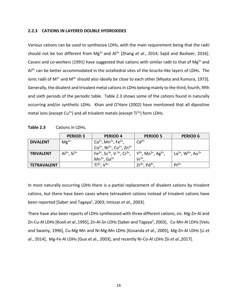
2.2.3 CATIONS IN LAYERED DOUBLE HYDROXIDES
Various cations can be used to synthesize LDHs, with the main requirement being that the radii
should not be too different from Mg2+ and Al3+ [Zhang et al., 2014; Sajid and Basheer, 2016].
Cavani and co-workers (1991) have suggested that cations with similar radii to that of Mg2+ and
Al3+ can be better accommodated in the octahedral sites of the brucite-like layers of LDHs. The
ionic radii of M2+ and M3+ should also ideally be close to each other [Miyata and Kumura, 1973].
Generally, the divalent and trivalent metal cations in LDHs belong mainly to the third, fourth, fifth
and sixth periods of the periodic table. Table 2.3 shows some of the cations found in naturally
occurring and/or synthetic LDHs. Khan and O’Hare (2002) have mentioned that all dipositive
metal ions (except Cu2+) and all trivalent metals (except Ti3+) form LDHs.
Table 2.3 Cations in LDHs.
PERIOD 3 PERIOD 4 PERIOD 5 PERIOD 6 DIVALENT Mg2+ Ca2+, Mn2+, Fe2+,
Co2+, Ni2+, Cu2+, Zn2+ Cd2+
TRIVALENT Al3+, Si3+ Fe3+, Sc3+, V 3+, Cr3+, Mn3+, Ga3+
Y3+, Mo3+, Ag3+, In3+,
La3+, W3+, Au3+
TETRAVALENT Ti4+, V4+ Zr4+, Pd4+, Pt4+
In most naturally occurring LDHs there is a partial replacement of divalent cations by trivalent
cations, but there have been cases where tetravalent cations instead of trivalent cations have
been reported [Saber and Tagaya1, 2003; Intissar et al., 2003]. There have also been reports of LDHs synthesized with three different cations, viz. Mg-Zn-Al and
Zn-Cu-Al LDHs [Kooli et al.,1995], Zn-Al-Sn LDHs [Saber and Tagaya2, 2003], Cu-Mn-Al LDHs [Velu
and Swamy, 1996], Cu-Mg-Mn and Ni-Mg-Mn LDHs [Kovanda et al., 2005], Mg-Zn-Al LDHs [Li et
al., 2014], Mg-Fe-Al LDHs [Guo et al., 2003], and recently Ni-Co-Al LDHs [Si et al.,2017].
17
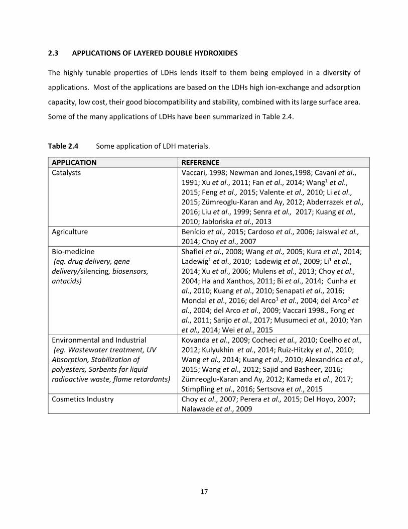
2.3 APPLICATIONS OF LAYERED DOUBLE HYDROXIDES The highly tunable properties of LDHs lends itself to them being employed in a diversity of
applications. Most of the applications are based on the LDHs high ion-exchange and adsorption
capacity, low cost, their good biocompatibility and stability, combined with its large surface area.
Some of the many applications of LDHs have been summarized in Table 2.4.
Table 2.4 Some application of LDH materials.
APPLICATION REFERENCE Catalysts Vaccari, 1998; Newman and Jones,1998; Cavani et al.,
1991; Xu et al., 2011; Fan et al., 2014; Wang1 et al., 2015; Feng et al., 2015; Valente et al., 2010; Li et al., 2015; Zümreoglu-Karan and Ay, 2012; Abderrazek et al., 2016; Liu et al., 1999; Senra et al., 2017; Kuang et al., 2010; Jabłońska et al., 2013
Agriculture Benício et al., 2015; Cardoso et al., 2006; Jaiswal et al., 2014; Choy et al., 2007
Bio-medicine (eg. drug delivery, gene delivery/silencing, biosensors, antacids)
Shafiei et al., 2008; Wang et al., 2005; Kura et al., 2014; Ladewig1 et al., 2010; Ladewig et al., 2009; Li1 et al., 2014; Xu et al., 2006; Mulens et al., 2013; Choy et al., 2004; Ha and Xanthos, 2011; Bi et al., 2014; Cunha et al., 2010; Kuang et al., 2010; Senapati et al., 2016; Mondal et al., 2016; del Arco1 et al., 2004; del Arco2 et al., 2004; del Arco et al., 2009; Vaccari 1998., Fong et al., 2011; Sarijo et al., 2017; Musumeci et al., 2010; Yan et al., 2014; Wei et al., 2015
Environmental and Industrial (eg. Wastewater treatment, UV Absorption, Stabilization of polyesters, Sorbents for liquid radioactive waste, flame retardants)
Kovanda et al., 2009; Cocheci et al., 2010; Coelho et al., 2012; Kulyukhin et al., 2014; Ruiz-Hitzky et al., 2010; Wang et al., 2014; Kuang et al., 2010; Alexandrica et al., 2015; Wang et al., 2012; Sajid and Basheer, 2016; Zümreoglu-Karan and Ay, 2012; Kameda et al., 2017; Stimpfling et al., 2016; Sertsova et al., 2015
Cosmetics Industry Choy et al., 2007; Perera et al., 2015; Del Hoyo, 2007; Nalawade et al., 2009
18
2.3.1 GENE THERAPY Gene therapy generally refers to the transfer of nucleic acids into a cell to either repair or correct
defective genes. This is achieved by introducing either plasmid DNA (pDNA) to replace a defective
gene, or small interfering RNA (siRNA) to silence an overexpressed gene. Despite much research in the area of gene therapy in recent years, efficient gene delivery remains
the greatest challenge for its practical application. Systemic delivery of naked nucleotides is
ineffective due to their sensitivity to nuclease degradation, as well as the difficulty of crossing the
cell membrane due to their negative charge. Hence, there is a need to develop carrier vectors.
An ideal gene delivery vector should have the ability to deliver a therapeutic gene safely into a
cell, have high loading capacity, exhibit little or no toxicity, be non-immunogenic, biodegradable,
be stable during storage and after administration, have the ability to target specific cells and
afford relatively efficient gene expression [Xu et al., 2007; Huang et al., 1999].
Carriers thus far have included viral vectors as well as a number of non-viral vectors such as
cationic lipids, liposomes, cationic polymers, gold nanoparticles, magnetic nanoparticles,
quantum dots, silica nanoparticles, fullerenes, carbon nanotubes and supramolecular systems
[Smith et al., 1997; Lv et al., 2006; Ragusa et al., 2007; Ahmed et al., 2009; Pan et al., 2007; Gao
et al., 2006; Pan et al., 2009]. Of these, viral vectors have been the most efficient, but they suffer
the disadvantage of possible immunogenic responses, low DNA-loading capability, insertional
mutagenesis as well as a lack of specific cell targeting. Cationic lipids and polymers may avoid
such problems, but are often highly toxic to cells [Xu and Lu, 2006]. Inorganic nanoparticles
appear more suitable for cellular delivery due to their wide availability, rich surface functionality,
good biocompatibility, potential capability of targeted delivery and the ability for controlled
release of genes/drugs [Xu et al., 2007].
Biocompatibility of the carrier is also of the utmost importance and is dependent on a number of
factors (Table 2.5).

19
Table 2.5 Overview of nanoparticle characteristics which can be controlled to improve biocompatibility [adapted from Soenen et al., 2011].
Parameter Advantage Disadvantage Size Small (< 10 nm): greater cellular
uptake
Small: higher cytotoxicity Large (> 200 nm): Too little cellular uptake
Shape Spherical: highest uptake Rods: Reduced cytotoxicity Flat Platelets (eg. LDH): increased surface area resulting in increased carrying capacity
Spherical: highest cytotoxicity Rods: Diminished uptake
Purity The presence of biologically compatible metals in suitable concentrations will not induce cytotoxic effects.
Certain metals or organic impurities present on the carrier may induce cytotoxic effects
Surface charge
Positively charged: better interaction with cell membrane
Positively charged: could be more cytotoxic as they have a tendency to interact with many biological components. Negatively charged: poor interaction with cell membrane
Ultimately an ideal gene delivery vector should be able to bind or condense nucleic acids forming
nanocomplexes, protect nucleic acids from enzymatic degradation [Imami et al., 2015], promote
cellular uptake, promote endosomal escape, and also enhance entry into the nucleus for
strategies involving pDNA delivery [Dong et al., 2013]. 2.3.1.1 GENE THERAPY USING PLASMID DNA
In gene therapy strategies involving DNA transfection, functional or therapeutic DNA is delivered
into cells to replace a missing or mutated gene [Morachis et al., 2012]. pDNA is generally used in
gene therapy strategies since it is an inherently stable molecule, and is capable of withstanding
changes in pH and ionic strength [Smith et al., 1997]. For successful gene therapy involving DNA
transfection, the DNA needs to be effectively transported into the nucleus of the cell, where the
exogenous gene must be integrated into the chromosome to obtain the desired level of
expression to produce therapeutic effects [Liang and Lam, 2012; Dong et al., 2013].
20
There are a number of major obstacles to efficient pDNA delivery, including naked DNA’s inability
to cross the cell membrane, endosomal escape, nuclear uptake and de-complexation. pDNA is
negatively charged and hydrophilic in nature and thus requires assistance to cross the plasma
membrane. Non-viral DNA delivery into the cell proceeds via an endocytic pathway upon which
the DNA-carrier complex would be trapped within an endosome. It is important that the complex
be released from the endosome at an early stage to prevent lysosomal destruction of both the
DNA and the carrier. Elouahabi and co-workers (1997) have reported that endosomal escape is
a crucial and possibly the rate-limiting step for efficient transfection.
The process of nuclear uptake is not very clearly understood. It has been suggested that
complexes may be internalized into cells during cell division, but non-dividing cells would
probably require a receptor-mediated process for entry [Smith et al., 1997]. Smaller complexes
appear to be able to diffuse through nuclear pores to gain entry into the nucleus [Elaesser and
Howard, 2012]. Another possible mode of nuclear entry involves the endoplasmic reticulum,
where the nanocomplex containing endosome fuses with the endoplasmic reticulum, releasing
the DNA into the lumen of the reticulum, from where the DNA enters the nucleus through the
nuclear membrane-reticulum network [Elouahabi and Ruysschaert, 2005].
De-complexation is also crucial for efficient gene expression. Partial de-complexation probably
occurs in the lysosome, leaving the DNA in a partially condensed form. This allows for entry of
the smaller, partially condensed DNA into the nucleus through basal leakage of the nuclear
envelope [Smith et al., 1997]. Zhang and co-workers (2012) have reported that de-complexation
is pH dependent, where an acidic pH disrupts the complex to release the nucleic acid.
2.3.1.2 GENE SILENCING USING siRNA Antisense therapy involves siRNA, a class of small 20-26 bp double-stranded (ds) RNA molecules
[Valencia-Sanchez et al., 2006], that play an important role in RNA interference (RNAi). RNAi
involves the silencing of expression of specific genes with nucleotide sequences complementary
to that of the siRNA [Zhang et al., 2007]. This ultimately results in suppression of expression of
the target (disease-causing) gene.

21
The RNAi pathway is triggered by the entry of a ds RNA molecule into the cell (Figure 2.2). The
endonuclease DICER cuts the ds RNA molecule into smaller fragments (siRNAs) [Zhang et al.,
2007]. These siRNA fragments integrate into a multi-subunit protein called the RNA-induced-
silencing-complex (RISC) [Matranga et al., 2005]. The RISC complex contains Argonaute proteins
that have a PAZ domain for miRNA/siRNA binding and a PIWI domain that functions in slicer
activity [Valencia –Sanchez et al., 2006]. The Argonaute proteins cleaves the ds RNA into 2
strands: a guide or anti-sense strand (which remains bound to RISC), and the passenger or sense
strand which is degraded. The anti-sense strand directs the sequence specific silencing of the
target mRNA by cleaving the target mRNA with Argonaute 2. This leads ultimately to the
inhibition of translation of the target mRNA [Ladewig2 et al., 2010]. The DICER step may be
bypassed by the addition of chemically synthesized siRNAs of the appropriate size. As with DNA delivery, there are also a number of hurdles to successful siRNA delivery. Firstly,
the net negative charge on siRNA molecules prevents entry into the cell [Chen et al., 2013]. Upon
entry into the cell, the siRNAs are targeted by RNases and macrophages [Nguyen et al., 2008],
and like DNA, the siRNA also needs to be released before it reaches the degradative environment
of the lysosome. LDHs show great promise as siRNA carriers due to their high buffering capacity
which not only protects the siRNA from the effects of the acidic environment but also contributes
to bursting of the late endosome, and subsequent release of the siRNA, by the proton sponge
effect.

22
Figure 2.2 The RNAi mechanism.
Ladewig2 and co-workers (2010) successfully employed LDHs as a siRNA carrier and reported fast
and effective target gene knockdown. Gene knockdown was also successfully carried out in
cortical neurons using LDH mediated siRNA delivery [Wong et al., 2010]. RNAi is an extremely
promising technique and may prove to be the intervention method of choice for treatment of
diseases in the future.

23
2.3.2 LAYERED DOUBLE HYDROXIDES IN GENE THERAPY An ideal gene delivery vehicle needs to be able to adequately bind and protect the biomolecule
being carried (DNA or siRNA), traverse the cell membrane, release its load in the appropriate
region of the cell, achieve good transfection, and in doing so, not cause any harm (toxicity) to the
cell [Imani et al., 2015].
The unique and highly tunable properties of LDHs lends itself to making them ideal gene delivery
vehicles. Firstly, DNA or siRNA can be easily incorporated into/onto LDHs by either ion exchange,
intercalation or adsorption [Rojas et al., 2015]. The nanocomplex thus formed gains extra
stabilization energy due to the electrostatic interaction between the cationic brucite layers and
the DNA/siRNA [Choy et al., 2000]. The positively charged atoms on the LDH surface interacts
with the negatively charged cell membrane leading to the internalization of the LDH
nanocomplex. Studies by Nagaraj and co-workers (2015) have shown that higher nanoparticle
concentrations result in increased energy-dependent cellular uptake mechanisms.
This internalization occurs via clathrin-mediated endocytosis [Ladewig2 et al., 2010; Wong et al.,
2010; Chen et al., 2013; Rojas et al., 2015]. A diagrammatic representation of this process is
shown in Figure 2.3. Clathrin-mediated endocytosis involves the entrapment of the nanocomplex
within an endosome, followed by either an exocytic pathway (endosome → Golgi apparatus) or
an endocytic route (early endosome → late endosome → lysosome) [Chung et al., 2012; Rojas et
al., 2015]. The trafficking pathway followed is nanoparticle size dependent, with smaller particles
generally following the endocytic route and larger particles taking the exocytic pathway [Chung
et al., 2012]. The bursting of the endosome to release the nanocomplexes is facilitated initially
by the ATPase mediated influx of H+ ions into the late endosome, which partially dissolves the
nanocomplexes, thus maintaining the pH at 5-6, and preventing formation of the lysosome
[Wong et al., 2010]. This leads to an increase in the number of ions within the endosome which
results in increased osmosis of water into the endosome. This culminates in the endosome
bursting and releasing the free DNA/siRNA from the partially dissolved nanocomplexes and as
well as the residual nanocomplexes into the cytoplasm [Rojas et al., 2015]. Another possible

24
release pathway of DNA/siRNA from the nanocomplex could be through ion exchange with
cytoplasmic anions [Gu et al., 2008].
Release of the nucleic acid:LDH complex from the endosome, before the endosome can fuse
with the lysosome is key to successful nucleic acid delivery. There are a number of endosomal
escape mechanisms such as the ‘proton sponge’ hypothesis, the flip flop mechanism, the
endosomal membrane fusion or destabilization mechanism, pore formation and photochemical
internalization [Liang and Lam, 2012]. The ‘proton sponge effect’ causes endosomal bursting due
to the influx of positive ions into the endosome, and this appears to be the mechanism of
endosomal escape employed by LDHs.
The susceptibility of LDHs to changes in pH contributes to the controlled release properties
exhibited by LDHs [Kriven et al., 2004; Zhang et al., 2012]. The pH dependent degradation of
LDHs also prevents nanocarrier accumulation which could result in toxicity. Toxicity is also
reduced due to the breakdown products of LDHs being biocompatible [Nagaraj et al., 2015]. Xu
and Lu (2006) have indicated that LDH nanocomplexes undergo a reaction similar to the one
shown below under physiological conditions:
MgAl-Cl-DNA + H+ → Mg2+ + Al3+ + Cl- + DNA + H2O
The release of these cyto-friendly by-products aids in endosomal escape of the nanoparticles.
The degradation products of LDHs, being small ions, may also leave the cell through “ion tunnels”
and not accumulate in the cell thereby decreasing the possibility of cytotoxicity [Xu et al., 2006].
Overall the low cytotoxicity, high biocompatibility, controllable particle size, ease of availability,
low cost, high loading capacity, effective load protection and controlled release properties, make
LDHs an attractive gene therapy vehicle [Ladewig2 et al., 2010; Chen et al., 2013].
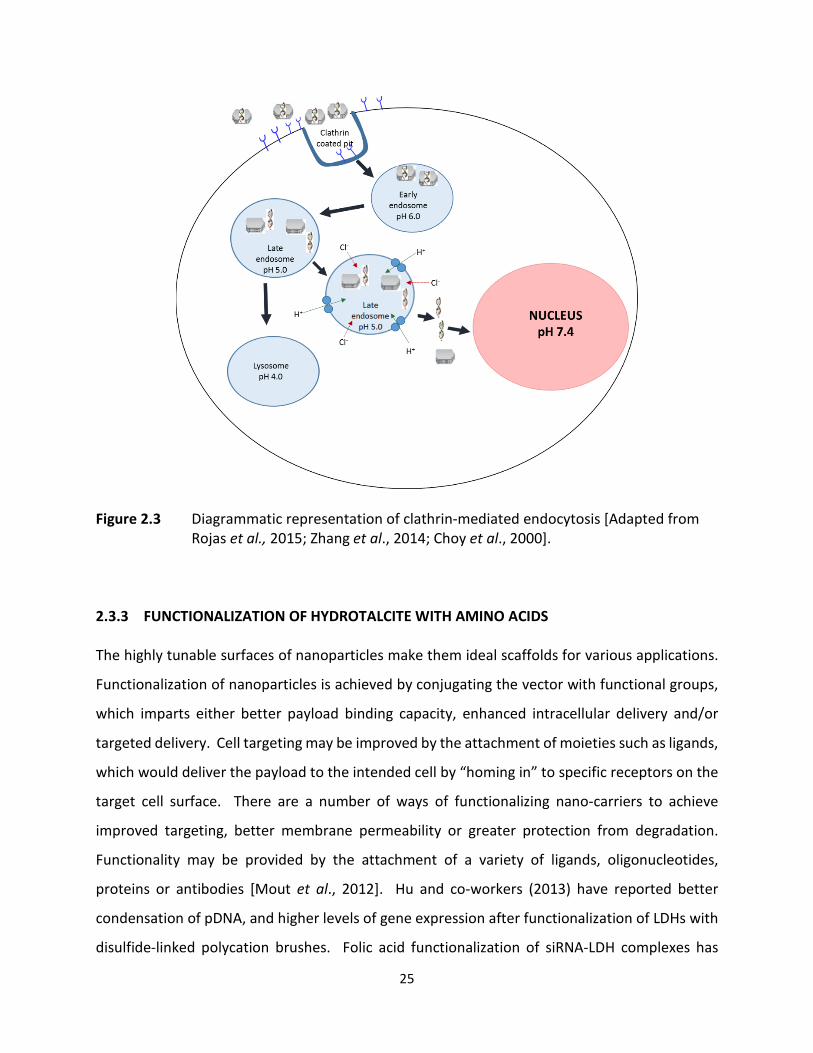
25
Figure 2.3 Diagrammatic representation of clathrin-mediated endocytosis [Adapted from Rojas et al., 2015; Zhang et al., 2014; Choy et al., 2000].
2.3.3 FUNCTIONALIZATION OF HYDROTALCITE WITH AMINO ACIDS The highly tunable surfaces of nanoparticles make them ideal scaffolds for various applications.
Functionalization of nanoparticles is achieved by conjugating the vector with functional groups,
which imparts either better payload binding capacity, enhanced intracellular delivery and/or
targeted delivery. Cell targeting may be improved by the attachment of moieties such as ligands,
which would deliver the payload to the intended cell by “homing in” to specific receptors on the
target cell surface. There are a number of ways of functionalizing nano-carriers to achieve
improved targeting, better membrane permeability or greater protection from degradation.
Functionality may be provided by the attachment of a variety of ligands, oligonucleotides,
proteins or antibodies [Mout et al., 2012]. Hu and co-workers (2013) have reported better
condensation of pDNA, and higher levels of gene expression after functionalization of LDHs with
disulfide-linked polycation brushes. Folic acid functionalization of siRNA-LDH complexes has

26
resulted in potent gene silencing in vitro and significantly high levels of tumor suppression in vivo
[Park et al., 2016]. The preferential in vivo bio-distribution of chitosan functionalized LDHs to the
lung paved the way for the development of organ-specific delivery systems [Kura et al., 2014].
Kuo et al., (2015) have recently investigated LDHs as organ specific carriers with promising
results.
Early endosomal escape appears to be the success-limiting step in gene therapy strategies. It
may be possible to achieve better transfection by manipulation of the vector to cause faster
endosomal escape, and one way of doing this is to functionalize the vector. Functionalization of
vectors with arginine may have begun as a result of attempting to improve cellular internalization
by bio-mimicking arginine-rich cell penetrating peptides [Zheng et al., 2015]. Arginine
functionalized hydroxyapatites have displayed better transfection efficiency than non-
functionalized hydroxyapatites [Wang et al., 2015]. Other arginine functionalized non-viral gene
delivery systems such as chitosan and lipidosomes have also shown increased transfection
efficiency and lower cytotoxicity when compared to non-functionalized chitosan [Gao et al.,
2008; Wang et al., 2015]. Arginine is positively charged at physiological pH (Figure 2.4), which
could enhance transfection is two ways. Firstly, the increased positive charge of the
functionalized nanocomplex could lead to greater internalization of the complexes due to
improved interaction between the complex and the negatively charged cell membrane.
Secondly, the greater positive charge of the arginine-functionalized complex could also result in
faster endosomal escape due to enhancement of the ‘proton sponge’ effect. This manipulation
of the proton-sponge effect may also be applied to histidine, which is also positively charged at
physiological pH. The incorporation of amino acids into LDHs is also relatively simple as amino
acids act as anionic species above their isoelectric points, allowing for easy intercalation via ion
exchange [Coelhoe et al., 2012]. Structures of arginine and histidine are shown in Figure 2.4. Xu
and co-workers (2006) have also suggested that the addition of amino groups to a vector result
in stronger interaction with the DNA chains. This indicates that the presence of amino acids on
the LDH should lead to stronger interaction with DNA during complex formation, which could
possibly result in better DNA protection and ultimately transfection.

27
Figure 2.4 Amino acids arginine and histidine at physiological pH.
There have been a number of reports describing the functionalization of LDHs with amino acids
[Aisawa et al., 2000; Aisawa et al., 2001; Nakayama et al., 2004; Aisawa et al., 2006]. Despite
varying degrees of success in intercalating amino acids into LDHs, there has, as yet, not been any
reports involving gene delivery using amino acid functionalized LDHs. This study therefore
includes a comparison between the transfection efficiency of arginine or histidine functionalized
HTs and non-functionalized HTs, in the delivery of DNA and siRNA.

28
CHAPTER THREE
3. PREPARATION AND CHARACTERISATION OF HYDROTALCITES AND HYDROTALCITE-LIKE COMPOUNDS
3.1 PREPARATION OF HYDROTALCITES AND HYDROTALCITE-LIKE COMPOUNDS
3.1.1 INTRODUCTION
Hydrotalcites (HT) and hydrotalcite-like (HT-like) compounds, also referred to as layered double
hydroxides (LDHs) can be very easily synthesized in the laboratory by a number of methods.
Synthetic LDHs may be composed of many different MII/MIII cation combinations with different
anions in the interlayer region. The particle shape and size can be controlled by adjusting
synthesis conditions [Benito et al., 2010; Herrero et al., 2009; Oh et al., 2006; Xu et al., 2008;
Zhao et al 2002].
Co-precipitation is the most commonly used method for the preparation of LDHs. In this method,
aqueous solutions of M2+ and M3+ are slowly added to a solution containing the anion that is to
be incorporated into the LDH, whilst maintaining a pH that is higher or equal to the pH at which
the most soluble hydroxide is precipitated. Co-precipitation is also used in the preparation of
organic anion-containing LDHs. Co-precipitation is followed by thermal or hydrothermal
treatment to enhance yield and crystallinity of the prepared LDH.
There are two methods of co-precipitation, viz. precipitation at low supersaturation which
involves addition of alkali to the reaction vessel (containing a solution of the anion to be
intercalated) simultaneously to the addition of the metal salt solution, thus maintaining the pH
at a selected value and precipitation at high supersaturation, where the metal salt solution is
added to an alkaline solution containing the desired anion. Co-precipitation at low
supersaturation has the advantage of forming more uniform LDHs with a higher crystallinity than
that produced by co-precipitation at high supersaturation [He et al., 2005].
A co-precipitation method involving separate nucleation and aging steps reported by Zhao and
co-workers in 2002 is a very rapid mixing and nucleation process in a colloid mill followed by a
29
separate aging process resulting in the production of small, uniform and highly crystalline
product. Dong and co-workers (2014) prepared LDH nanoparticles by non-aqueous co-
precipitation. Another modified co-precipitation method for the preparation of LDHs is referred
to as the urea hydrolysis method. Costantino and co-workers (1998) prepared MgAl-carbonate,
ZnAl-carbonate and NiAl-carbonate LDHs using the urea hydrolysis method. This method
involves dissolving solid urea in a solution of the chosen metal chlorides and then maintaining
this solution at 100 °C for 36 hours. The LDHs produced by this method have a larger particle
sizes and higher charge density than those prepared by the other co-precipitation methods. The
intercalated carbonate anions can be replaced with chloride by treating the LDH with gaseous or
liquid HCl. Thereafter, chloride can be replaced quite easily by organic anions [Costantino et al.,
1998]. Pérez-Bernal and co-workers (2009) prepared layered double hydroxides via the co-
precipitation method in the presence of a surfactant, which controlled aggregation of particles
through the formation of mixed micelles. The resultant crystals were larger and more ordered
than those prepared by normal co-precipitation.
The interlayer anions of preformed LDHs can be replaced with guest anions by ion exchange. Ion-
exchange may be carried out in one of two ways. Firstly, the precursor LDH would contain anions
such as chloride, nitrate or perchlorate which would have a weak interaction with the LDH layers.
The second method involves LDHs containing anions which are susceptible to acid attack such as
carbonate or carboxylates [He et al., 2005]. The extent of ion-exchange depends on the affinity
for the incoming anion, the nature of the exchange medium, the pH of the reaction solution and
the chemical composition of the layers. Bontchev et al., (2003) showed that monovalent anion
exchange preference is Br- ˃ Cl- ˃ NO3- ˃ I- and was independent of the method of synthesis.
Upon calcination, i.e. heating LDHs to about 500 °C, the interlayer water, interlayer anions and
the hydroxyl groups are removed. Calcined LDHs are able to regenerate its layered structure
through the “memory effect” when exposed to water and anions [Bontchev et al., 2003;
Klemkaite et al., 2011; Tao et al., 2006]. The anion does not have to be the same as the one that
was present in the original sample, thus this is a method which can be used to introduce various
other types of organic or inorganic anions into the interlayer. The reconstruction process is

30
carried out under nitrogen for incorporation of a non-carbonate anion since the carbonate LDH
is preferentially formed in the presence of atmospheric carbon dioxide [He et al., 2005]. Wang
and co-workers (2005) preferentially intercalated 5-fluorouracil into an MgAl LDH using this
restructure method.
Other methods for the preparation of LDHs include hydrothermal methods [Cavani et al., 1991;
Wang et al., 2013], secondary intercalation, salt-oxide method, non-equilibrium aging method,
surface synthesis, templated synthesis, non-conventional aging methods, the sol-gel method and
electrosynthesis [He et al., 2005]. Xu and co-workers (2010) have devised an atom-economic
method of preparing LDHs, where the reactants are completely incorporated into the target
product and there is no need for washing or filtering of the LDHs as there are no by-products.
Modifications in the synthesis procedure has led to the production of LDHs with sheet-like
morphology [Kelkar and Schutz, 1997], needle-shaped morphology [Kelkar and Schutz, 1997],
LDH nanoscrolls [Ren et al., 2007; Lv et al., 2015], LDH films [Kuang et al., 2010] and LDH
nanorattles [Liu et al., 2011]. Another recent development involved the synthesis of LDHs using
a mechano-chemical method to prepare highly dispersed LDH nanoparticles [Qu et al., 2016].
This mechano-chemical method is reported to produce less aqueous waste and requires less
energy than conventional LDH preparation methods.
3.1.2 MATERIALS AND METHODS
Mg(NO3)2.6H2O, Al(NO3)3.9H2O, Fe(NO3)3.9H2O, Zn(NO3)2.6H2O, NaOH and Na2CO3 were all of
analytical grade purchased from Merck (Darmstadt, Germany). L-Arginine and L-Histidine were
obtained from Sigma-Aldrich (St Louis, USA).
The HT and HT-like compounds were prepared using the co-precipitation technique at low
supersaturation. HT and HT-like compounds were prepared using MII:MIII ratios of 2:1 and 3:1.
To prepare a Mg-Al HT with a MII:MIII ratio of 2:1 (denoted MgAl 0.33), 25.6 g (0.1 mol)
Mg(NO3)2.6H2O and 18.75 g (0.05 mol) Al(NO3)3.9H2O were dissolved in 200 mL deionized water.
The mixed metal solution was added dropwise to 1 L of a 0.5 M solution of Na2CO3, with vigorous
stirring. The pH was maintained at 11 by addition of a 1 M NaOH solution as required. After

31
complete addition of the mixed cation solution, the suspension was transferred to a flat-
bottomed flask, and heated to 80 °C under reflux for 18-20 hours. Thereafter, the precipitate
was filtered and washed thoroughly until the filtrate had a pH of 7. The precipitate was then
dried in an oven at 110 °C overnight. The resulting dry precipitate was ground to a fine powder
(talc-like consistency) using a mortar and pestle.
Similarly, the Mg-Al HT with MII:MIII = 3:1 (denoted MgAl 0.25), was prepared using a mass of 25.6
g (0.1 mol) Mg(NO3)2.6H2O and 12.38 g (0.033 mol) Al(NO3)3.9H2O. The arginine and histidine
functionalized HTs were also prepared using the co-precipitation method. To prepare the
arginine functionalized HT in the molar ratio Mg:Al:Arg of 2:1:1 (denoted MgAlArg 0.33), a mass
of 5.12 g (0.02 mol) Mg(NO3)2.6H2O, 3.76 g (0.01 mol) Al(NO3)3.9H2O and 2.11 g (0.01 mol) L-
arginine hydrochloride was used.
The masses of all the precursors used in the preparation of the HTs and HT-like compounds (LDHs)
in this study is shown in Table A1 (Appendix). The masses of the metal precursors and amino
acids that were used to prepare amino acid-functionalized HT are shown in Table A2 (Appendix).
3.2 CHARACTERIZATION OF HYDROTALCITE AND HYDROTALCITE-LIKE COMPOUNDS
The prepared HT and HT-like compounds (LDHs) were characterized using powder X-ray
diffraction (XRD), inductively coupled plasma-optical emission spectroscopy (ICP-OES), Fourier
transform infrared spectroscopy (FTIR), transmission electron microscopy (TEM), scanning
electron microscopy (SEM) and nanoparticle tracking analysis (NTA).
3.2.1 X-RAY DIFFRACTION
Single crystal X-ray diffraction (XRD) is usually the technique of choice for crystalline samples, but
obtaining a single crystal sample from synthesized LDH is almost impossible due to the
polycrystalline nature of the sample [Ennadi et al., 2000]. Powder XRD is therefore used to
elucidate much of the structural information for LDHs.

32
Powder X-ray diffraction (PXRD) patterns were obtained using a Bruker D8 Advance
diffractometer equipped with a graphite monochromator operating at 40 kV and 40 mA. The
radiation source was a CuKα X-ray source with λ = 1.5406 Å. Data was collected at a step of
0.021 ° and at a scanning speed of 0.454 °s-1.
3.2.2 INDUCTIVELY COUPLED PLASMA-OPTICAL EMISSION SPECTROSCOPY
Inductively coupled plasma- optical emission spectroscopy (ICP-OES) is used to detect and
quantify elements in a solution.
The samples were analyzed on a Perkin Elmer Optical Emission Spectrometer Optima 5300 DV.
The instrument was calibrated using a mixed metal standard solution. The standard solutions
with concentrations of 20 ppm, 40 ppm, 60 ppm, 80 ppm and 100 ppm were prepared using ICP
and atomic absorption spectroscopy standards of 1000 ppm (1000 Magnesium 1000, 1000
Aluminium 1000, 1000 Iron 1000 and 1000 Zinc 1000), which were obtained from Polychem
Supplies cc (South Africa). Samples were prepared by dissolving 10 mg of the powdered
compound in 10 mL of 0.1 M HNO3. This solution was then diluted to 100 mL with double distilled
water. Approximately 14 mL of each sample was transferred to ICP tubes and analyzed.
3.2.3 FOURIER-TRANSFORM INFRA-RED SPECTROSCOPY
Fourier-transform infrared spectroscopy (FTIR) is one of the principal tools for LDH
characterization, used primarily for functional group identification. Each functional group
absorbs specific IR radiation at different frequencies, making FT-IR a useful tool for indicating the
presence or absence of a specific functional group in a compound. FT-IR is usually carried out in
the infrared region (4000-250 cm-1).
The samples were analyzed on a Perkin Elmer Spectrum Universal ATR 100 FT-IR Spectrometer.
The crystal area was cleaned with methanol prior to analysis of each sample. Approximately,
0.5 g of each LDH sample was placed on the crystal area, the pressure arm was locked into place
and the sample was then analysed.
33
3.2.4 SCANNING AND TRANSMISSION ELECTRON MICROSCOPY
Transmission electron microscopy (TEM) is an extremely useful nanoparticle characterization tool
for the determination of particle distribution and ultrastructural morphology. Scanning electron
microscopy (SEM) was carried out to determine the surface topography of the LDH samples.
For TEM, the samples were prepared by suspending 1 mg of the powdered sample in about 1 mL
of double distilled water and sonicating the mixture for 15-20 minutes. About 1 µL of the
suspended sample was placed on a copper grid, air-dried and then viewed under a JEOL 1010
Megaview 3 Soft Imaging TEM system. For SEM, a strip of double sided carbon tape (Nisshin EM,
Co Ltd) was placed onto an SEM stub. The LDH sample was placed onto the stub and coated with
gold particles using a Polaron SC500 Sputter Coater. The stub was placed in a LEO 1450 SEM, and
analyzed using SmartSEM software version 5.03.05.
3.2.5 NANOPARTICLE TRACKING ANALYSIS
Particle size and zeta potentials are accurately determined using nanoparticle tracking analysis
(NTA). Zeta (ζ) potential refers to the electric potential present in the double layer between the
surface of a particle and the solvent/fluid in which the particle is suspended (Figure 3.1).
NTA was used to obtain both particle size and ζ-potential. A 1:1000 dilution of the LDH sample
in double distilled water was analyzed in a NanoSight NS500 system (Malvern Instruments Ltd.,
Worcestershire, UK), using NanoSight nanoparticle tracking analysis software v3.2.

34
Figure 3.1 Diagrammatic representation of zeta potential [adapted from http://www.pharmainfo.net/book/emerging-trends-nanotechnology-pharmacy-physicochemical-characterization-nanoparticles/zeta Date accessed: 31st August 2017).
3.2.6 IDENTIFICATION OF AMINO ACIDS
The ninhydrin test is a very simple and inexpensive test to confirm the presence of amino groups
in a sample. Ninhydrin reacts with the amines of amino acids to produce a blue or purple color.
The ninhydrin test for this study was conducted using a chromatographic method. Standard
samples of arginine and histidine as well as solutions of the amino acid-functionalized LDHs were
spotted onto cellulose paper and then developed in a solvent system composed of n-
butanol:acetic acid:water (5:2:2) containing 0.2 g of ninhydrin. Qiu et al (2010) have reported
35
that adding ninhydrin to the solvent system resulted in a cleaner and more rapid method for
amino acid detection than the conventional post-staining method.
The actual amount of arginine present in the prepared samples (MgAlArg 0.25 and MgAlArg 0.33)
was determined using a spectrophotometric method developed by Wang and co-workers (2008).
This method involved the use of solutions of 8-hydroxyquinoline (0.2 % (w/v) in 95 % ethanol), 2
M sodium hydroxide and sodium hypobromite (2 % bromine (v/v) in 5 % sodium hydroxide). An
arginine stock solution (100 µg/mL) was made up in 1 M sodium hydroxide. The arginine-
functionalized LDH samples (0.01 g) were each dissolved in 1 mL of 10 M hydrochloric acid and
this was made up to 5 mL in deionized water. The test samples contained 100 µL of the arginine-
functionalized LDH samples in 2.4 mL of deionized water. Standard arginine samples (containing
10 µg – 60 µg) were prepared to a volume of 2.5 mL with deionized water. To a tube containing
2.5 mL of arginine solution, was added 0.5 mL of 8-hydroxyquinoline solution and 0.5 mL of 2 M
sodium hydroxide. The solutions were thoroughly mixed and placed on ice for 10 minutes.
Thereafter, 0.25 mL of cold sodium hypobromite and 0.5 mL of 40 % urea was added, thoroughly
mixed and the optical density was measured at 500 nm using an Irmeco V1200
spectrophotometer. A deionized water blank solution was prepared in a similar manner.
Quantification of histidine in the prepared samples (MgAlHist 0.25 and MgAlHist 0.33) was
conducted by adapting a method described by Patel and co-workers (2009). The color reagent
was prepared by dissolving 200 mg sodium nitrite in 60 mL deionized water, followed by the
addition of 1 mL concentrated hydrochloric acid. This solution was allowed to stand for 1 hour,
after which 500 mg of sulphanilic acid was added and the final volume made up to 100 mL. The
solution was allowed to stand for a further 30 minutes prior to use. A stock solution of standard
histidine was prepared (100 µg/mL) in 1 M sodium hydroxide. Standard histidine samples
(containing 10 µg – 50 µg) were prepared from the stock solution to a volume of 2.5 mL with
deionized water. The histidine functionalized LDH samples (0.01 g) were each dissolved in 1 mL
of 10 M hydrochloric acid and then made up to 5 mL with deionized water. The test samples
comprised of 100 µl of the histidine-functionalized LDH samples added to 2.4 mL of deionized
water and 100 µL 1 M sodium hydroxide. To a tube containing 2.5 mL of histidine solution, 5 mL

36
of colour reagent was added and allowed to stand for 5 minutes. The absorbance was measured
at 405 nm using an Irmeco V1200 spectrophotometer with 1 M sodium hydroxide used as a blank.
3.3 RESULTS AND DISCUSSION
3.3.1 PHYSICAL PROPERTIES OF SYNTHESIZED LAYERED DOUBLE HYDROXIDES
All the LDHs synthesized were obtained in the form of smooth, finely textured powders. The
prepared compounds denoted with “0.25” indicate a mole ratio of 3:1 (M2+:M3+) and those
denoted with “0.33” indicate a mole ratio of 2:1 (M2+:M3+). The MgAl 0.25, MgAl 0.33, MgAlArg
0.25, MgAlArg 0.33, MgAlHist 0.25, MgAlHist 0.33, ZnAl 0.25 and ZnAl 0.33 samples were all
pristine white, talc-like powders. The MgFe 0.25 sample appeared as a smooth, fine cream-
coloured powder while the MgFe 0.33 sample had a similar texture, but appeared reddish-brown
in color. The LDH samples were insoluble in water, but formed suspensions when vortexed or
sonicated. Only the MgAl 0.33, MgAlArg 0.33 and ZnAl 0.33 samples remained in suspension
indefinitely, whilst all the other samples eventually settled out in the water solution within 24
hours (Figure 3.2). This observation suggests partial dissolution of these LDH nanoparticles, a
phenomenon confirmed to occur with HTs prepared in a Mg:Al ratio of 2:1 by aqueous stability
studies [Nagaraj et al., 2015].
Figure 3.2 Solubility of HTs and LDHs in distilled water (1) MgAl 0.25, (2) MgAl 0.33, (3) MgAlArg 0.25, (4) MgAlArg 0.33, (5) MgAlHist 0.25, (6) MgAlHist 0.33, (7) MgFe 0.25, (8) MgFe 0.33, (9) ZnAl 0.25, (10) ZnAl 0.33.
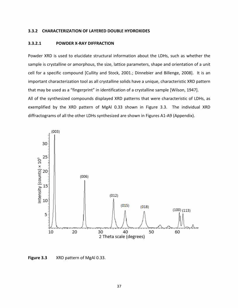
37
3.3.2 CHARACTERIZATION OF LAYERED DOUBLE HYDROXIDES
3.3.2.1 POWDER X-RAY DIFFRACTION
Powder XRD is used to elucidate structural information about the LDHs, such as whether the
sample is crystalline or amorphous, the size, lattice parameters, shape and orientation of a unit
cell for a specific compound [Cullity and Stock, 2001.; Dinnebier and Billenge, 2008]. It is an
important characterization tool as all crystalline solids have a unique, characteristic XRD pattern
that may be used as a “fingerprint” in identification of a crystalline sample [Wilson, 1947].
All of the synthesized compounds displayed XRD patterns that were characteristic of LDHs, as
exemplified by the XRD pattern of MgAl 0.33 shown in Figure 3.3. The individual XRD
diffractograms of all the other LDHs synthesized are shown in Figures A1-A9 (Appendix).
Figure 3.3 XRD pattern of MgAl 0.33.
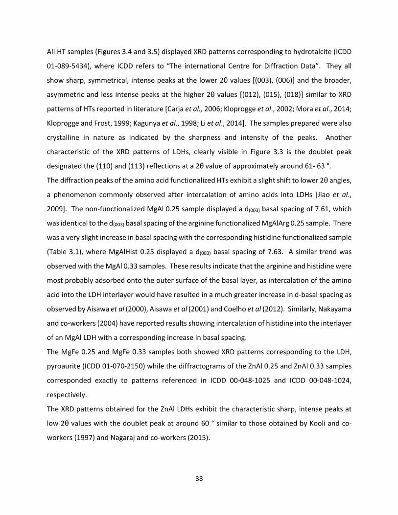
38
All HT samples (Figures 3.4 and 3.5) displayed XRD patterns corresponding to hydrotalcite (ICDD
01-089-5434), where ICDD refers to “The international Centre for Diffraction Data”. They all
show sharp, symmetrical, intense peaks at the lower 2θ values [(003), (006)] and the broader,
asymmetric and less intense peaks at the higher 2θ values [(012), (015), (018)] similar to XRD
patterns of HTs reported in literature [Carja et al., 2006; Kloprogge et al., 2002; Mora et al., 2014;
Kloprogge and Frost, 1999; Kagunya et al., 1998; Li et al., 2014]. The samples prepared were also
crystalline in nature as indicated by the sharpness and intensity of the peaks. Another
characteristic of the XRD patterns of LDHs, clearly visible in Figure 3.3 is the doublet peak
designated the (110) and (113) reflections at a 2θ value of approximately around 61- 63 °.
The diffraction peaks of the amino acid functionalized HTs exhibit a slight shift to lower 2θ angles,
a phenomenon commonly observed after intercalation of amino acids into LDHs [Jiao et al.,
2009]. The non-functionalized MgAl 0.25 sample displayed a d(003) basal spacing of 7.61, which
was identical to the d(003) basal spacing of the arginine functionalized MgAlArg 0.25 sample. There
was a very slight increase in basal spacing with the corresponding histidine functionalized sample
(Table 3.1), where MgAlHist 0.25 displayed a d(003) basal spacing of 7.63. A similar trend was
observed with the MgAl 0.33 samples. These results indicate that the arginine and histidine were
most probably adsorbed onto the outer surface of the basal layer, as intercalation of the amino
acid into the LDH interlayer would have resulted in a much greater increase in d-basal spacing as
observed by Aisawa et al (2000), Aisawa et al (2001) and Coelho et al (2012). Similarly, Nakayama
and co-workers (2004) have reported results showing intercalation of histidine into the interlayer
of an MgAl LDH with a corresponding increase in basal spacing.
The MgFe 0.25 and MgFe 0.33 samples both showed XRD patterns corresponding to the LDH,
pyroaurite (ICDD 01-070-2150) while the diffractograms of the ZnAl 0.25 and ZnAl 0.33 samples
corresponded exactly to patterns referenced in ICDD 00-048-1025 and ICDD 00-048-1024,
respectively.
The XRD patterns obtained for the ZnAl LDHs exhibit the characteristic sharp, intense peaks at
low 2θ values with the doublet peak at around 60 ° similar to those obtained by Kooli and co-
workers (1997) and Nagaraj and co-workers (2015).
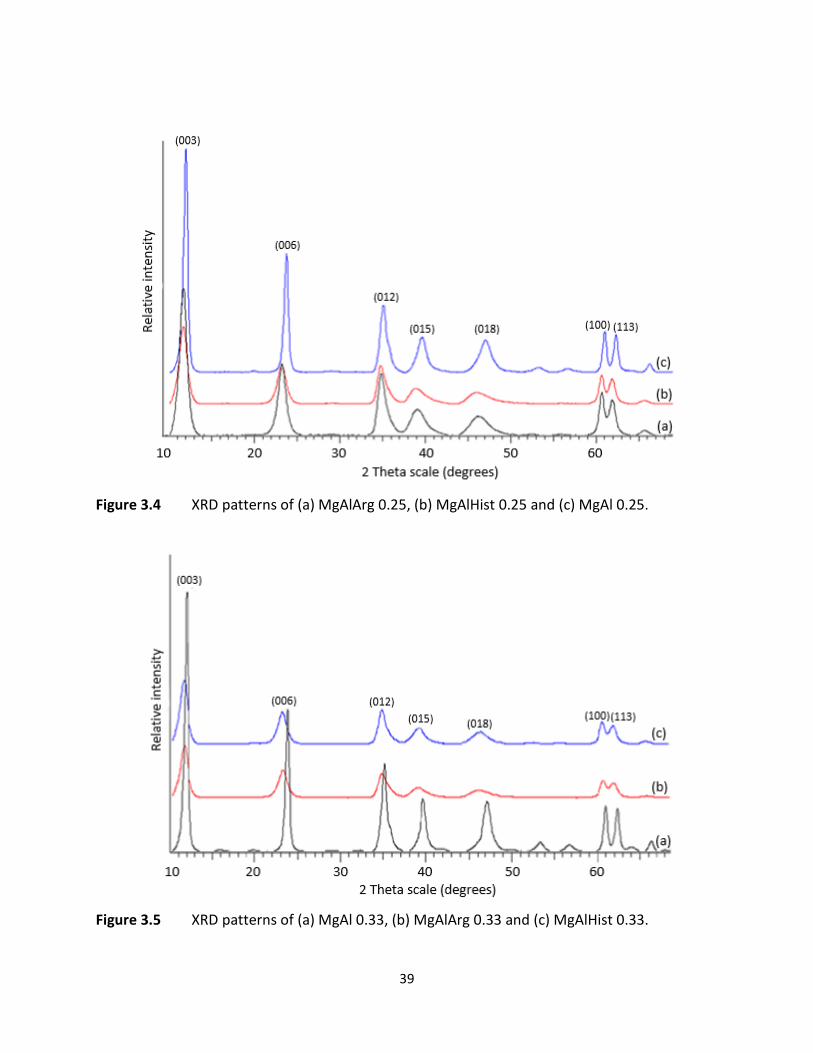
39
Figure 3.4 XRD patterns of (a) MgAlArg 0.25, (b) MgAlHist 0.25 and (c) MgAl 0.25.
Figure 3.5 XRD patterns of (a) MgAl 0.33, (b) MgAlArg 0.33 and (c) MgAlHist 0.33.

40
Table 3.1 d-basal spacing for LDH compounds.
Basal Spacing / Å
Compound MII:MIII d(003) d(006) d(012) d(015) d(018) d(110) d(113)
MgAl 0.25 0.25 7.61 3.83 2.57 2.30 1.96 1.52 1.49
MgAl 0.33 0.33 7.42 3.75 2.55 2.26 1.92 1.51 1.48
MgAlArg 0.25 0.25 7.61 3.83 2.57 2.30 1.97 1.52 1.50
MgAlArg 0.33 0.33 7.61 3.84 2.58 2.31 1.97 1.52 1.50
MgAlHist 0.25 0.25 7.63 3.84 2.57 2.34 1.96 1.52 1.50
MgAlHist 0.33 0.33 7.42 3.75 2.55 2.27 1.93 1.51 1.49
MgFe 0.25 0.25 7.61 3.84 2.60 2.32 1.97 1.54 1.52
MgFe 0.33 0.33 7.49 3.78 2.59 2.30 1.95 1.54 1.51
ZnAl 0.25 0.25 7.46 3.75 2.57 2.28 1.93 1.53 1.50
ZnAl 0.33 0.33 7.36 3.72 2.56 2.27 1.93 1.53 1.50
As previously mentioned, the prepared compounds denoted with “0.25” indicate a mole ratio of
3:1 (M2+:M3+) and those denoted with “0.33” indicate a mole ratio of 2:1 (M2+:M3+), the latter
having more trivalent metals present. Brindley and Kikkawa (1979) reported that an increase in
the amount of trivalent metal results in a decrease in both lattice parameters, a and c. Lattice
parameter c decreases because as M3+ ions increase, the electrostatic attraction between the
positive and negative interlayers also increases. Lattice parameter a, decreases with increasing
M3+ ions because M3+ are generally smaller in size than M2+ ions and the higher charge of the
trivalent ion combined with the smaller ionic radii leads to shorter bond lengths. This trend can
be clearly seen in Table 3.2.

41
Table 3.2 The a, c and c0 parameters of the prepared LDHs in angstroms.
Compound d(003) c d(110) a
MgAl 0.25 7.61 22.83 1.52 3.04
MgAl 0.33 7.42 22.26 1.51 3.02
MgAlArg 0.25 7.61 22.83 1.52 3.04
MgAlArg 0.33 7.61 22.83 1.52 3.04
MgAlHist 0.25 7.63 22.89 1.52 3.04
MgAlHist 0.33 7.42 22.26 1.51 3.02
MgFe 0.25 7.61 22.83 1.54 3.08
MgFe 0.33 7.49 22.47 1.54 3.08
ZnAl 0.25 7.46 22.38 1.53 3.06
ZnAl 0.33 7.36 22.08 1.53 3.06
The interlayer spacing of LDHs containing carbonate is 7.8 Å [Kloprogge and Frost, 1999], the
same as the interlayer spacing for chloride containing LDHs [Rossi et al., 2005]. The replacement
of carbonate by nitrate anions should result in a slight increase of interlayer space to between
8.1 Å [Kloprogge et al., 2002] to 8.8 Å [Nagaraj et al., 2015], thus the lower interlayer spacing of
prepared samples suggests that some carbonate may have been incorporated into the interlayers
during LDH preparation. This was expected as the LDHs were not prepared under nitrogen, which
would have aided in decreasing the level of contamination by atmospheric carbonate anions.
The results in Tables 3.1 and 3.2 shows a decreased interlayer spacing closer to 7.6 Å for the
Mg/Al LDHs which seems to agree more with the values reported by Bookin and Drits (1993) and
Li and co-workers (2014), indicating the presence of carbonate anions in the interlayer region of
the prepared LDHs. This may have an effect on the ion exchange capacity of the LDHs prepared
as carbonate anions have a high affinity for the hydroxide layers in LDHs [Kloprogge et al., 2002].
The basal spacings for ZnAl 0.25 and ZnAl 0.33 was 7.46 and 7.36 Å respectively which was quite
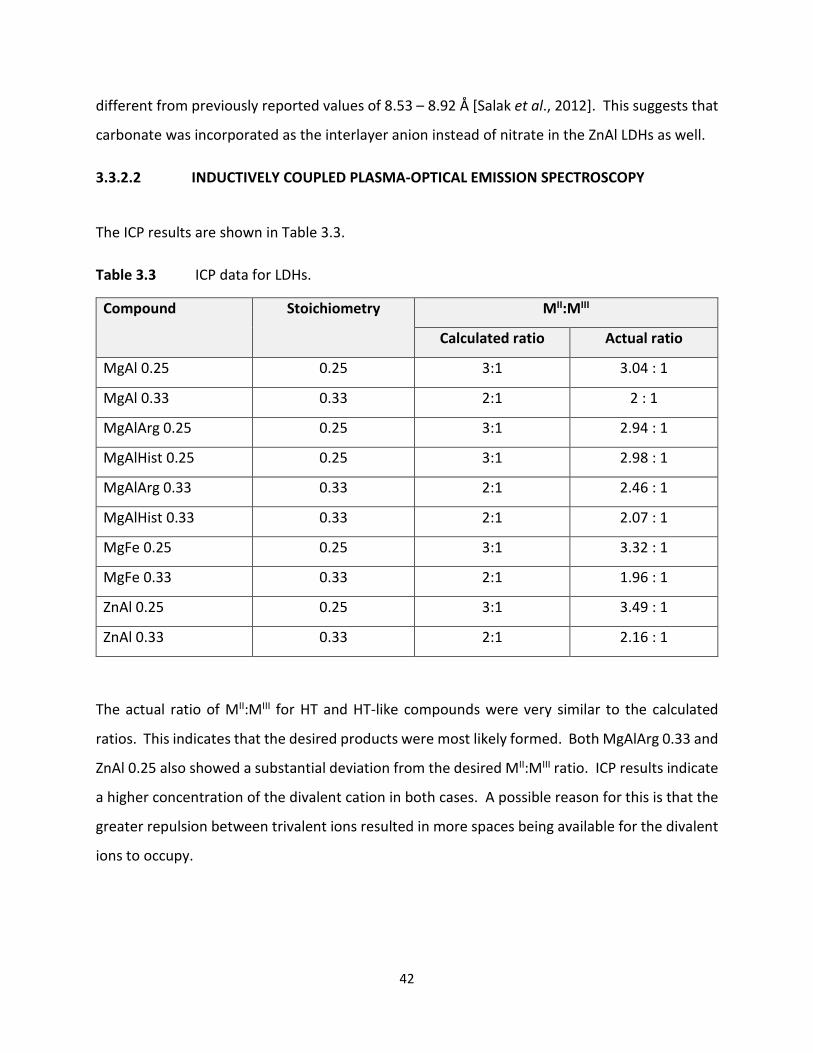
42
different from previously reported values of 8.53 – 8.92 Å [Salak et al., 2012]. This suggests that
carbonate was incorporated as the interlayer anion instead of nitrate in the ZnAl LDHs as well.
3.3.2.2 INDUCTIVELY COUPLED PLASMA-OPTICAL EMISSION SPECTROSCOPY
The ICP results are shown in Table 3.3.
Table 3.3 ICP data for LDHs.
Compound Stoichiometry MII:MIII
Calculated ratio Actual ratio
MgAl 0.25 0.25 3:1 3.04 : 1
MgAl 0.33 0.33 2:1 2 : 1
MgAlArg 0.25 0.25 3:1 2.94 : 1
MgAlHist 0.25 0.25 3:1 2.98 : 1
MgAlArg 0.33 0.33 2:1 2.46 : 1
MgAlHist 0.33 0.33 2:1 2.07 : 1
MgFe 0.25 0.25 3:1 3.32 : 1
MgFe 0.33 0.33 2:1 1.96 : 1
ZnAl 0.25 0.25 3:1 3.49 : 1
ZnAl 0.33 0.33 2:1 2.16 : 1
The actual ratio of MII:MIII for HT and HT-like compounds were very similar to the calculated
ratios. This indicates that the desired products were most likely formed. Both MgAlArg 0.33 and
ZnAl 0.25 also showed a substantial deviation from the desired MII:MIII ratio. ICP results indicate
a higher concentration of the divalent cation in both cases. A possible reason for this is that the
greater repulsion between trivalent ions resulted in more spaces being available for the divalent
ions to occupy.
43
3.3.2.3 FOURIER-TRANSFORM INFRARED SPECTROSCOPY
Analysis of the infrared spectra of the LDHs revealed three major types of vibrations, viz.
vibrations as a result of the interlayer anions, vibrations due to the hydroxyl groups (molecular
vibrations) and vibrations due to the octahedral layers (lattice vibrations) [Hernandez-Moreno et
al., 1985].
The FTIR spectra of the HTs are shown in Figures 3.6 and 3.7. The ѵ(OH) stretching modes can be
seen at around 3440 cm-1, presenting at slightly lower wavenumbers than free hydroxyl groups
which have an average ѵ(OH) value of 3620 cm-1 [Kagunya et al., 1998]. This may be attributed
to the hydroxyl groups in the samples being hydrogen bonded to lattice water. The band
occurring at around 1650 is due to interlayer water bending modes [Kloprogge and Frost, 1999].
Water is strongly bonded to both the anions and the hydroxyl surfaces of the LDHs and the
presence of the water bending modes indicate that water plays an important role in the structure
of the LDHs [Palmer et al., 2009].
Vibrations occurring below 800 cm-1 are assigned ѵ(M-O) or metal-oxygen absorption bands
[Kooli et al., 1997., Wu et al., 2014., Mallakpour and Dinari, 2016]. The bands appearing around
500 cm-1 can be assigned to the translational modes of hydroxyl groups influenced by trivalent
aluminium, while the bands around 600 cm-1 are due to translational modes of hydroxyl groups
influenced by divalent magnesium [Millange et al., 2000].
Bands due to intercalated and adsorbed anions are observed between 1800-1000 cm-1 [Medina-
Juárez et al., 2016]. The strong reflection between 1300 and 1400 cm-1 is associated with the
asymmetric stretching modes of the interlayer carbonate ions (ѵ3 vibrational mode of CO32-)
[Kooli et al., 1997; Millange et al., 2000; Frost et al., 2003]. A band associated with the CO32- -
H2O bridging mode in the interlayer at around 3000 cm-1 [Kloprogge et al., 2002; Kloprogge and
Frost, 1999; Millange et al., 2000], would have confirmed incorporation of carbonate within the
interlayer. The absence of this band suggests that perhaps some nitrate and amino acid residues
(in the case of the amino acid functionalized HTs) may have also been incorporated into the
interlayer.

44
Figure 3.6 FTIR patterns of MgAl 0.25, MgAlArg 0.25 and MgAlHist 0.25.
Figure 3.7 FTIR patterns of MgAl 0.33, MgAlArg 0.33 and MgAlHist 0.33.
Weak shoulder band
Weak shoulder band
45
The weak shoulder at around 3100 cm-1 (shown in figures 3.6 – 3.8) is ascribed as the stretching
modes of hydroxyl groups linked to carbonate ions [Kagunya et al., 1998]. Weak CO32- vibrational
modes are also present between 700 and 1100 cm-1, with ѵ4 vibrational modes of CO32- generally
occurring at around 730 cm-1, ѵ2 at around 850 cm-1 and ѵ1 at around 1000 cm-1 [Kloprogge et
al., 2001].
FTIR spectra of the aa-functionalized hydrotalcites all exhibit O-H stretching modes between
3400 cm-1 and 3600 cm-1, strong reflections between 1300 cm-1 and 1400 cm-1 due to asymmetric
stretching of carbonate ions and low frequency vibrations between 400 cm-1 and 800 cm-1
attributed to M-O and M-O-M vibrations of the LDH lattice [Kooli et al., 1997; Kagunya et al.,
1998; Millange et al., 2000; Li1 et al., 2014; Choi et al., 2015].
Bands attributed to symmetric and asymmetric stretching of the COO- groups of the amino acids
usually occur between 1400 cm-1 and 1600 cm-1, but these bands are not clearly visible in the
spectra shown possibly due to the low levels of incorporation of the amino acids into the HTS
[Aisawa et al., 2000; Aisawa et al., 2001; Kumar and Rai, 2010; Choi et al., 2015]. The band at
2943 cm-1 for MgAlArg 0.25 and at 2933 cm-1 for MgAlArg 0.33 are characteristic for arginine FTIR
spectra and correspond to asymmetric stretching of the methyl group [Kumar and Rai, 2010].
Bands due to a mixed vibration of COO- asymmetric stretching and NH bending modes of the
histidine moiety usually occur at around 1630 cm-1 [Kumar et al., 2010] and the band due to
guanidine C=N stretching and C=O carbonyl stretching which would normally be seen at around
1700 cm-1 [Imani et al., 2015; Barth, 2000] but these appear to be masked here by the interlayer
water bending modes.
The infrared spectra of of HT-like compounds (MgFe 0.35, MgFe 0.33, ZnAl 0.25 and ZnAl 0.33)
show similar patterns to the spectra of the HTs (see Figure 3.8). The spectra are characteristic of
LDHs. The O-H stretching of water molecules appear at around 3440 cm-1, the H-OH bands occur
at around 1650 cm-1, the peak at around 1300 corresponds to the intercalated carbonate anions
and the M-O (metal-oxygen) absorption bands occur below 800 cm-1 [Frost1 et al., 2003; Choi et
al., 2015; Komarala et al., 2016].

46
Figure 3.8 FTIR patterns of MgFe 0.25, MgFe 0.33, ZnAl 0.25 and ZnAl 0.33.
3.3.2.4 SCANNING AND TRANSMISSION ELECTRON MICROSCOPY The TEM and SEM images of the hydrotalcite, MgAl 0.25 are shown in Figure 3.9. The crystals
generally appear as hexagonal platelets, tending to agglomerate, resulting in the platelets being
piled on top of each other. TEM analysis at 150 000X magnification shows irregularly shaped
crystals with most being in the size range of less than 50 nm. It has been suggested that
irregularity in shape occurs due to crystal defects [Wang et al., 2012]. SEM analysis at a much
lower magnification (42.61 K X), also shows some larger particles with a lateral width of about
100 nm. This indicates a large size distribution of the LDH nanoparticles. The morphology
observed for MgAl 0.25 was similar to that reported by Mondal and co-workers (2016).
Weak shoulder band

47
Figure 3.9 Electron microscopy images of MgAl 0.25 (a) TEM at 150K magnification, (b) TEM at 300K magnification and (c) SEM at 42.61K magnification. The bars represent (a) 200 nm, (b) 100 nm and (c) 200 nm respectively.
The MgAl 0.33 hydrotalcite sample shows a similar flat platelet type structure, typical of layered
double hydroxides (Figure 3.10). The particles appear more uniform in shape when compared to
the MgAl 0.25 sample. Both TEM and SEM indicate a more even particle size distribution with
most of the particles within the 20-70 nm range. The rod-like structures visible on the TEM
micrographs are most likely particles lying on either sides or in a position not in the direct plane
of view.
(a)
(c)
(b)

48
Figure 3.10 Electron microscopy images of MgAl 0.33 (a) TEM at 150K magnification, (b) TEM at 300K magnification and (c) SEM at 42.40K magnification. The bars represent (a) 200 nm, (b) 100 nm and (c) 200 nm respectively.
TEM and SEM analysis of the aa-functionalized LDHs are shown in Figures 3.11 and 3.12. These
LDHs exhibit the characteristic flat platelet like structure of the non-functionalized LDHs.
Although XRD analysis of the aa-functionalized LDHs suggest that the amino acid groups are most
probably attached to the external surfaces of the LDH molecules, it is not possible for the amino
acid moieties to be viewed at the magnifications used here for TEM and SEM, due to their very
small size. The sizes of the arginine functionalized LDHs ranged between 20 and 50 nm. The size
of the histidine functionalized LDH MgAlHist 0.25 appear to average around 70 nm, while the
MgAlHist 0.33 particles appear much smaller, and range between 15 and 50 nm.
(b)
(c)
(a)
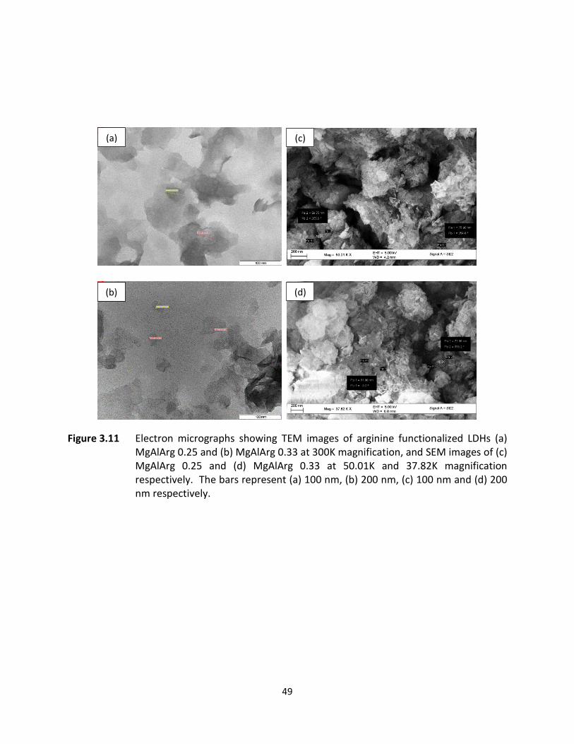
49
Figure 3.11 Electron micrographs showing TEM images of arginine functionalized LDHs (a) MgAlArg 0.25 and (b) MgAlArg 0.33 at 300K magnification, and SEM images of (c) MgAlArg 0.25 and (d) MgAlArg 0.33 at 50.01K and 37.82K magnification respectively. The bars represent (a) 100 nm, (b) 200 nm, (c) 100 nm and (d) 200 nm respectively.
(c)
(d) (b)
(a)

50
Figure 3.12 Electron micrographs showing TEM images of histidine functionalized LDHs (a) MgAlHist 0.25 and (b) MgAlHist 0.33 at 300K magnification, and SEM images of (c) MgAlHist 0.25 and (d) MgAlHist 0.33 at 42.71K and 45.70K magnification respectively. The bars represent (a) 100 nm, (b) 200 nm, (c) 100 nm and (d) 200 nm respectively.
The particle morphology of the MgFe 0.25 and MgFe 0.33 LDHs appear very similar to each other
(Figures 3.13 and 3.14). Typical hexagonal flat platelets are observed with a wide size
distribution. The lateral particle size range for MgFe 0.25 was 40-80 nm, whilst that of MgFe 0.33
ranged from 70-200 nm. SEM at low magnification also revealed the presence of some larger
particles in the 150-300 nm range, once again emphasizing the wide particle size distribution of
the samples.
(a
(b)
(c) (d)

51
Figure 3.13 Electron microscopy images of MgFe 0.25 (a) TEM at 150K magnification, (b) TEM at 300K magnification and (c) SEM at 22.02K magnification. The bars represent (a) 200 nm, (b) 100 nm and (c) 200 nm respectively.
(a)
(c)
(b)
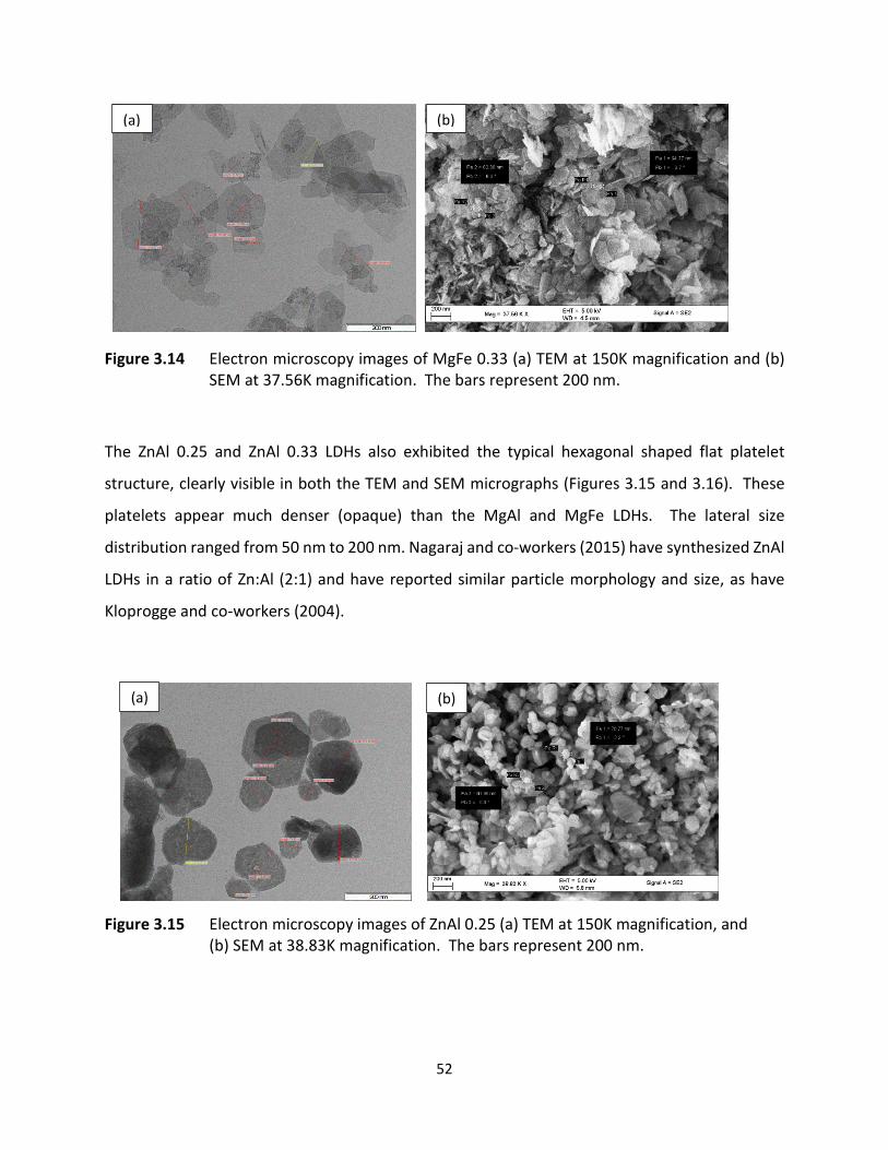
52
Figure 3.14 Electron microscopy images of MgFe 0.33 (a) TEM at 150K magnification and (b) SEM at 37.56K magnification. The bars represent 200 nm.
The ZnAl 0.25 and ZnAl 0.33 LDHs also exhibited the typical hexagonal shaped flat platelet
structure, clearly visible in both the TEM and SEM micrographs (Figures 3.15 and 3.16). These
platelets appear much denser (opaque) than the MgAl and MgFe LDHs. The lateral size
distribution ranged from 50 nm to 200 nm. Nagaraj and co-workers (2015) have synthesized ZnAl
LDHs in a ratio of Zn:Al (2:1) and have reported similar particle morphology and size, as have
Kloprogge and co-workers (2004).
Figure 3.15 Electron microscopy images of ZnAl 0.25 (a) TEM at 150K magnification, and (b) SEM at 38.83K magnification. The bars represent 200 nm.
(a) (b)
(a) (b)
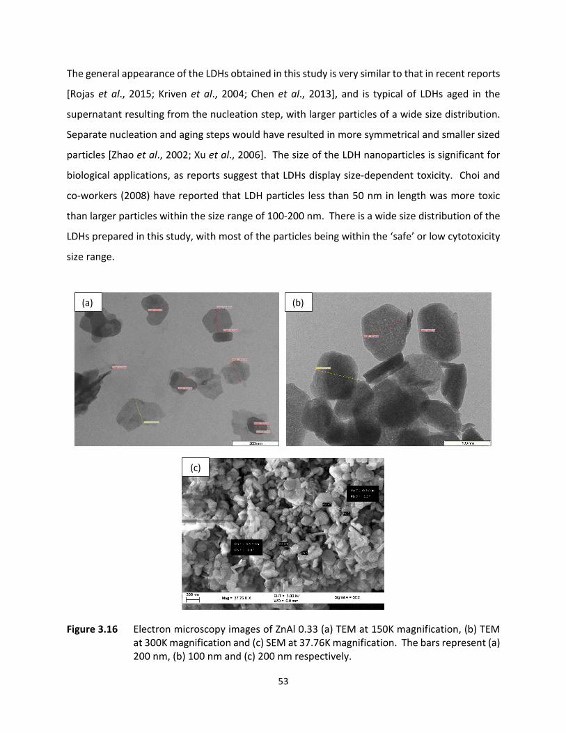
53
The general appearance of the LDHs obtained in this study is very similar to that in recent reports
[Rojas et al., 2015; Kriven et al., 2004; Chen et al., 2013], and is typical of LDHs aged in the
supernatant resulting from the nucleation step, with larger particles of a wide size distribution.
Separate nucleation and aging steps would have resulted in more symmetrical and smaller sized
particles [Zhao et al., 2002; Xu et al., 2006]. The size of the LDH nanoparticles is significant for
biological applications, as reports suggest that LDHs display size-dependent toxicity. Choi and
co-workers (2008) have reported that LDH particles less than 50 nm in length was more toxic
than larger particles within the size range of 100-200 nm. There is a wide size distribution of the
LDHs prepared in this study, with most of the particles being within the ‘safe’ or low cytotoxicity
size range.
Figure 3.16 Electron microscopy images of ZnAl 0.33 (a) TEM at 150K magnification, (b) TEM at 300K magnification and (c) SEM at 37.76K magnification. The bars represent (a) 200 nm, (b) 100 nm and (c) 200 nm respectively.
(c)
(b) (a)

54
3.3.2.5 NANOPARTICLE TRACKING ANALYSIS (NTA)
The efficacy of nanoparticle mediated delivery is affected by physicochemical properties such as
size, shape and surface charge [Dong et al., 2015]. The size of nanoparticles greatly affects its
interaction with cells, and influences cellular uptake efficiency, route of internalization,
localization within the cell as well as cytotoxicity [Shang et al., 2014; Rojas et al., 2015].
Zeta potential provides an indication of the stability of a colloidal dispersion. Zeta potential
depends not only on the properties of the nanoparticle but also on its environment and is thus
affected by both pH and ionic strength of the suspension fluid [Xu, 2008]. It is extremely
important to assess the ζ-potential of nanoparticles to be used for biological studies, as it greatly
affects interactions between the nanoparticles themselves, the interactions between cells and
nanoparticles, and also affects the cell targeting ability of the nanoparticles.
The modal size and ζ-potential values are recorded in Table 3.4. The large particle size
distribution observed by TEM and SEM was confirmed by NTA, which revealed size variations of
up to ± 100 nm of the modal size value.
Table 3.4 Size and zeta potential of LDHs.
LDH SIZE (nm) ZETA POTENTIAL (mV)
MgAl 0.25 148.4 -11.6
MgAl 0.33 72.4 15.0
MgAlArg 0.25 193.1 -19.8
MgAlArg 0.33 123.7 6.7
MgAlHist 0.25 60.8 -24.6
MgAlHist 0.33 44.5 22.7
MgFe 0.25 307.4 1.2
MgFe 0.33 27.9 -12.8
ZnAl 0.25 30.0 -7.2
ZnAl 0.33 67.9 29.7
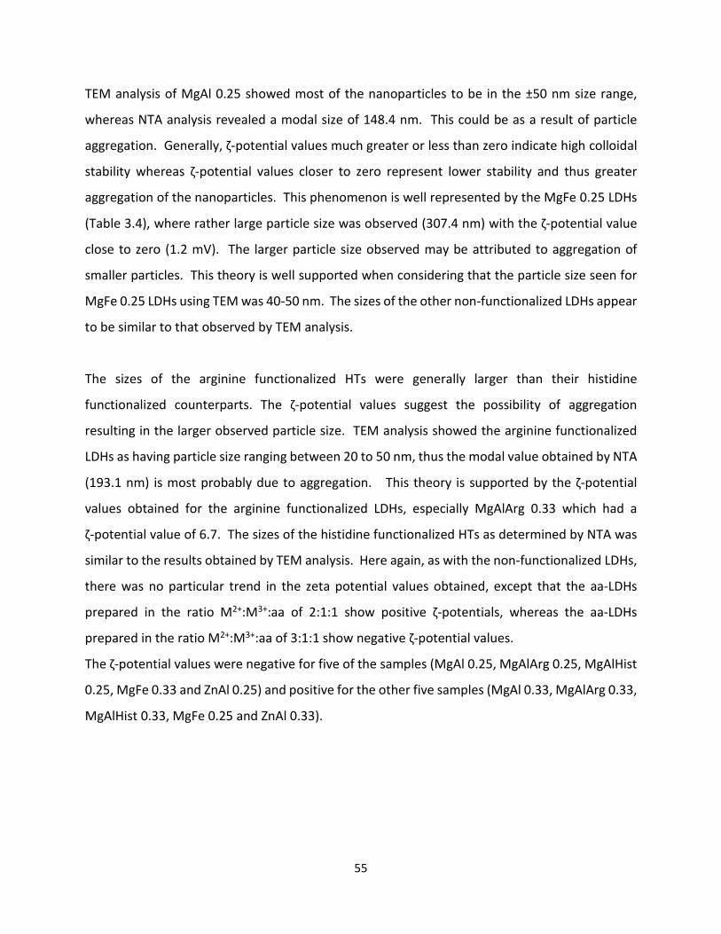
55
TEM analysis of MgAl 0.25 showed most of the nanoparticles to be in the ±50 nm size range,
whereas NTA analysis revealed a modal size of 148.4 nm. This could be as a result of particle
aggregation. Generally, ζ-potential values much greater or less than zero indicate high colloidal
stability whereas ζ-potential values closer to zero represent lower stability and thus greater
aggregation of the nanoparticles. This phenomenon is well represented by the MgFe 0.25 LDHs
(Table 3.4), where rather large particle size was observed (307.4 nm) with the ζ-potential value
close to zero (1.2 mV). The larger particle size observed may be attributed to aggregation of
smaller particles. This theory is well supported when considering that the particle size seen for
MgFe 0.25 LDHs using TEM was 40-50 nm. The sizes of the other non-functionalized LDHs appear
to be similar to that observed by TEM analysis.
The sizes of the arginine functionalized HTs were generally larger than their histidine
functionalized counterparts. The ζ-potential values suggest the possibility of aggregation
resulting in the larger observed particle size. TEM analysis showed the arginine functionalized
LDHs as having particle size ranging between 20 to 50 nm, thus the modal value obtained by NTA
(193.1 nm) is most probably due to aggregation. This theory is supported by the ζ-potential
values obtained for the arginine functionalized LDHs, especially MgAlArg 0.33 which had a
ζ-potential value of 6.7. The sizes of the histidine functionalized HTs as determined by NTA was
similar to the results obtained by TEM analysis. Here again, as with the non-functionalized LDHs,
there was no particular trend in the zeta potential values obtained, except that the aa-LDHs
prepared in the ratio M2+:M3+:aa of 2:1:1 show positive ζ-potentials, whereas the aa-LDHs
prepared in the ratio M2+:M3+:aa of 3:1:1 show negative ζ-potential values.
The ζ-potential values were negative for five of the samples (MgAl 0.25, MgAlArg 0.25, MgAlHist
0.25, MgFe 0.33 and ZnAl 0.25) and positive for the other five samples (MgAl 0.33, MgAlArg 0.33,
MgAlHist 0.33, MgFe 0.25 and ZnAl 0.33).
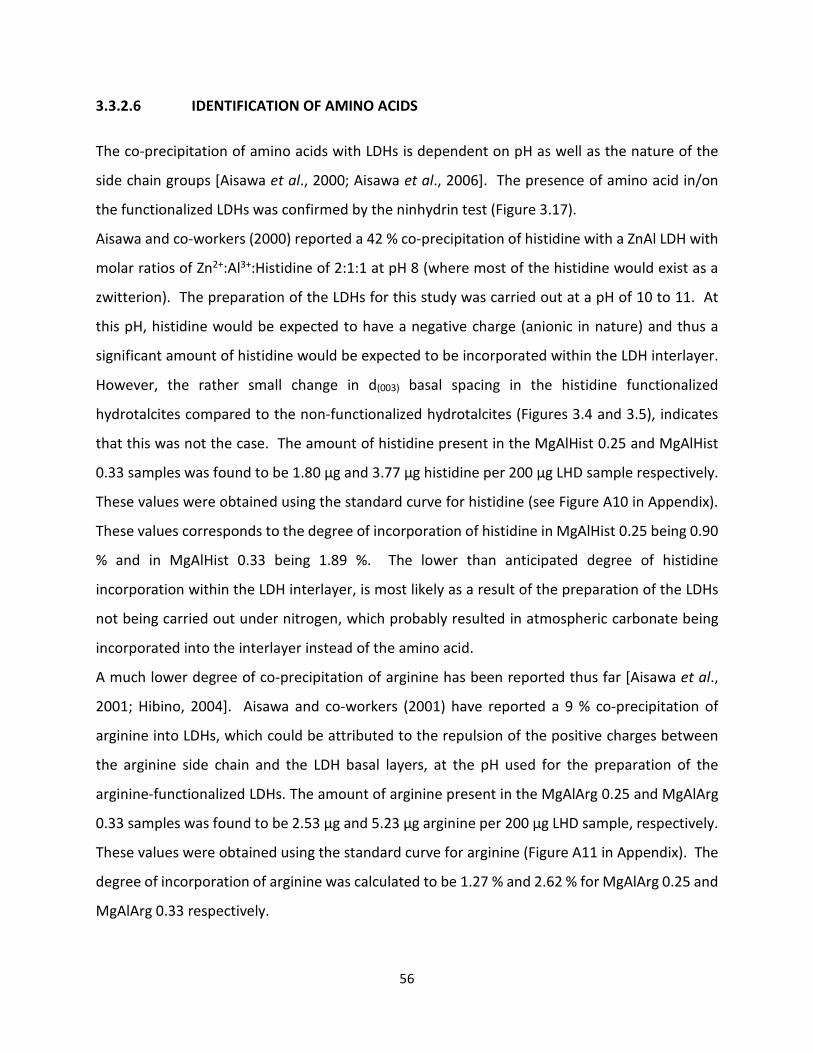
56
3.3.2.6 IDENTIFICATION OF AMINO ACIDS
The co-precipitation of amino acids with LDHs is dependent on pH as well as the nature of the
side chain groups [Aisawa et al., 2000; Aisawa et al., 2006]. The presence of amino acid in/on
the functionalized LDHs was confirmed by the ninhydrin test (Figure 3.17).
Aisawa and co-workers (2000) reported a 42 % co-precipitation of histidine with a ZnAl LDH with
molar ratios of Zn2+:Al3+:Histidine of 2:1:1 at pH 8 (where most of the histidine would exist as a
zwitterion). The preparation of the LDHs for this study was carried out at a pH of 10 to 11. At
this pH, histidine would be expected to have a negative charge (anionic in nature) and thus a
significant amount of histidine would be expected to be incorporated within the LDH interlayer.
However, the rather small change in d(003) basal spacing in the histidine functionalized
hydrotalcites compared to the non-functionalized hydrotalcites (Figures 3.4 and 3.5), indicates
that this was not the case. The amount of histidine present in the MgAlHist 0.25 and MgAlHist
0.33 samples was found to be 1.80 µg and 3.77 µg histidine per 200 µg LHD sample respectively.
These values were obtained using the standard curve for histidine (see Figure A10 in Appendix).
These values corresponds to the degree of incorporation of histidine in MgAlHist 0.25 being 0.90
% and in MgAlHist 0.33 being 1.89 %. The lower than anticipated degree of histidine
incorporation within the LDH interlayer, is most likely as a result of the preparation of the LDHs
not being carried out under nitrogen, which probably resulted in atmospheric carbonate being
incorporated into the interlayer instead of the amino acid.
A much lower degree of co-precipitation of arginine has been reported thus far [Aisawa et al.,
2001; Hibino, 2004]. Aisawa and co-workers (2001) have reported a 9 % co-precipitation of
arginine into LDHs, which could be attributed to the repulsion of the positive charges between
the arginine side chain and the LDH basal layers, at the pH used for the preparation of the
arginine-functionalized LDHs. The amount of arginine present in the MgAlArg 0.25 and MgAlArg
0.33 samples was found to be 2.53 µg and 5.23 µg arginine per 200 µg LHD sample, respectively.
These values were obtained using the standard curve for arginine (Figure A11 in Appendix). The
degree of incorporation of arginine was calculated to be 1.27 % and 2.62 % for MgAlArg 0.25 and
MgAlArg 0.33 respectively.
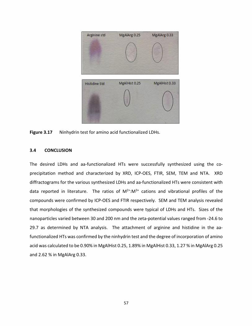
57
Figure 3.17 Ninhydrin test for amino acid functionalized LDHs.
3.4 CONCLUSION The desired LDHs and aa-functionalized HTs were successfully synthesized using the co-
precipitation method and characterized by XRD, ICP-OES, FTIR, SEM, TEM and NTA. XRD
diffractograms for the various synthesized LDHs and aa-functionalized HTs were consistent with
data reported in literature. The ratios of M2+:M3+ cations and vibrational profiles of the
compounds were confirmed by ICP-OES and FTIR respectively. SEM and TEM analysis revealed
that morphologies of the synthesized compounds were typical of LDHs and HTs. Sizes of the
nanoparticles varied between 30 and 200 nm and the zeta-potential values ranged from -24.6 to
29.7 as determined by NTA analysis. The attachment of arginine and histidine in the aa-
functionalized HTs was confirmed by the ninhydrin test and the degree of incorporation of amino
acid was calculated to be 0.90% in MgAlHist 0.25, 1.89% in MgAlHist 0.33, 1.27 % in MgAlArg 0.25
and 2.62 % in MgAlArg 0.33.

58
CHAPTER FOUR
4. NUCLEIC ACID BINDING AND PROTECTION STUDIES
4.1. INTRODUCTION
Nucleic acids are negatively charged and would therefore bind to LDHs as any other anion would.
The degree of binding can be assessed using the electrophoretic mobility shift assay (EMSA), also
known as the gel retardation assay. It was first described in the 80’s by Garner and Revzin (1981)
and Fried and Crothers (1981), two groups working independently on DNA-protein interactions.
EMSA is a simple, efficient and widely used method for detection of binary changes such as the
presence or absence of binding of nucleic acids [Holden and Tacon, 2011]. The method has since
been adapted to study the interactions of nucleic acids with various compounds such as drugs
and carrier vectors in gene delivery studies. The rationale behind EMSA is that free/unbound
nucleic acids would have a different electrophoretic mobility in a gel matrix than nucleic acid that
is bound to a carrier. In this study, the migration of DNA:LDH or siRNA:LDH complexes would be
‘retarded’ when compared to the migration of free/unbound DNA or siRNA.
This assay is used to determine the optimal binding ratio, which refers to the lowest ratio of
nucleic acid:LDH, where complete binding of the nucleic acid occurs. The optimal binding ratio
is very important, as it plays a key role in determining whether a vector would be suitable for
gene therapeutic applications in vivo.
When unbound ‘naked’ DNA is exposed to agarose gel electrophoresis, a typical band pattern is
observed. Depending on the forms of DNA present in the sample, three bands may be observed.
The supercoiled form of DNA is more compact and migrates the furthest of the three forms of
DNA. The circular form of DNA migrates much slower than the other two forms, most probably
due to stearic hindrance, whilst the linear form of DNA migrates at a rate intermediate to the
supercoiled and circular forms. When DNA is bound to a vector such as LDH, the mobility of the
DNA:LDH is retarded and the complex would remain in the well.
The 6.2 kilobase pair pCMV-luc DNA plasmid was used for this study (Figure 4.1). In addition to
the luciferase gene, this plasmid contains a cytomegalovirus (CMV) promoter from pcDNA3, a

59
simian virus promoter, enhancer and late polyadenylation site, as well as an ampicillin resistance
gene and bacterial origin of replication.
Figure 4.1 Map of the pCMV-luc vector [Plasmid Factory, Bielefeld, Germany].
Small interfering RNA (siRNA) are short (20-25 base pairs long) double stranded RNA molecules
which generally migrate as a single band when exposed to gel electrophoresis. The anti-luc siRNA
targeting the luciferase gene was used for the gene knockdown/silencing studies.
4.2. MATERIALS AND METHODS
DNA grade agarose was obtained from Bio-Rad Laboratories, Richmond, USA. pCMV-luc plasmid
DNA was supplied by Plasmid Factory (Bielefeld, Germany). Anti-luciferase siRNA [5’-
GAUUAUGUCCGGUUAUGUA-3’] was purchased from Dharmacon (Lafayette CO, USA). Ethidium
bromide and ethylenediaminetetraacetic acid (EDTA) (disodium salt, dihydrate) were acquired
from Merck, Darmstadt, Germany. All other reagents were of analytical grade.

60
4.2.1. PREPARATION OF NUCLEIC ACID-LDH COMPLEXES
To find the optimal binding ratios, 0.5 µg of pCMV-luc DNA was added to the respective LDH
samples at varying weight ratios in a total volume of 10 µl HEPES buffered saline [HBS (20 mM
HEPES and 150 mM NaCl, pH 7.5)] (Table 4.1). This mixture was vigorously vortexed for 10
minutes and then allowed to stand at room temperature for 30 minutes.
Table 4.1 Ratios used for nucleic acid:LDH complex formation.
Lane Ratio nucleic acid :LDH Nucleic Acid (µg) LDH (µg) 1 1:0 0.5 0 2 1:10 0.5 5 3 1:20 0.5 10 4 1:30 0.5 15 5 1:40 0.5 20 6 1:50 0.5 25 7 1:60 0.5 30 8 1:70 0.5 35
The siRNA-LDH complexes were prepared in a similar manner as the DNA-LDH complexes using
anti-luciferase siRNA. The siRNA-LDH-HBS mixtures were briefly vortexed and then incubated
overnight at 60 °C to allow for optimal binding to occur.
4.2.2 AGAROSE GEL ELECTROPHORESIS
A 1 % agarose gel was prepared by dissolving (with heating) 0.2 g of agarose in 18 ml distilled
water. The solution was cooled to approximately 75 °C, after which 2 ml of 10x electrophoresis
buffer (0.36 M Tris-HCl; 0.3 M Na2HPO4 and 0.1 M EDTA in 1 litre 18 Mohm water, pH 7.5) and 3
µl of ethidium bromide (10 mg/ml) were added to it. The solution was cooled further to
approximately 60 °C, and then poured onto an electrophoresis gel tray fitted with an 8-well comb.
The agarose gel was allowed to set for ± 1 hour, after which the comb was removed and the gel
placed in an electrophoresis tank (Bio-Rad Mini-Sub®Cell GT) containing 1x electrophoresis
buffer. A volume of 3 μL of gel loading buffer [glycerol 50 %, bromophenol blue 0.05 % and xylene

61
cyanol (0.05 %)] was added to the complexes, and 10 μL of each sample was loaded onto the
agarose gel and exposed to electrophoresis at 50 volts for 90 minutes. The gel was then viewed
under UV light using a Syngene G-Box documentation system (Syngene, Cambridge, UK).
4.2.3 CHARACTERIZATION OF NUCLEIC ACID:LDH COMPLEXES
The ultrastructural morphology and size and ζ-potential of the DNA:LDH and the siRNA:LDH
complexes was determined using TEM and NTA, respectively. The mechanism of DNA binding
was evaluated by XRD analysis of the DNA:LDH complex. For this aspect of the study MgAl 0.33
was selected as an ideal representative of all the LDHs prepared. XRD analysis required a
substantial amount of sample and budgetary constraints did not allow for the preparation of such
large quantities of the siRNA:LDH complexes. This analysis was therefore not carried out on the
siRNA:LDH complex. The methods employed are as described in Chapter 3, sections 3.2.1, 3.2.4
and 3.2.5.
4.2.4 NUCLEASE DIGESTION AND NUCLEIC ACID RELEASE ASSAY
This assay was performed using only the DNA:MgAl 0.33 complex as all the nucleic acid:LDH
complexes would be expected to react similarly when exposed to serum nucleases. A DNA:LDH
(10 µl) complex was prepared at the optimal binding ratio determined from the electrophoretic
mobility shift assay. Two control samples were also prepared; a positive control containing only
the DNA and a negative control containing DNA and 10 % v/v foetal bovine serum (FBS). The 10
% v/v FBS was also added to the DNA:LDH complex, and all samples were incubated at 37 °C for
4 hours. The nuclease digestion reaction was stopped by the addition of ethylenediamine
tetraacetic acid (EDTA), to a final concentration of 1 mM. The sample was then centrifuged, the
supernatant discarded, and 0.1 M HCl (1 µl) was added, and sample incubated at 37 °C for 1 hour.
Thereafter, 2 µl of gel loading buffer was added to each sample and they were subjected to
electrophoresis at 50 V for 90 minutes. The gels were viewed under UV light using a Syngene G-
Box documentation system (Syngene, Cambridge, UK).
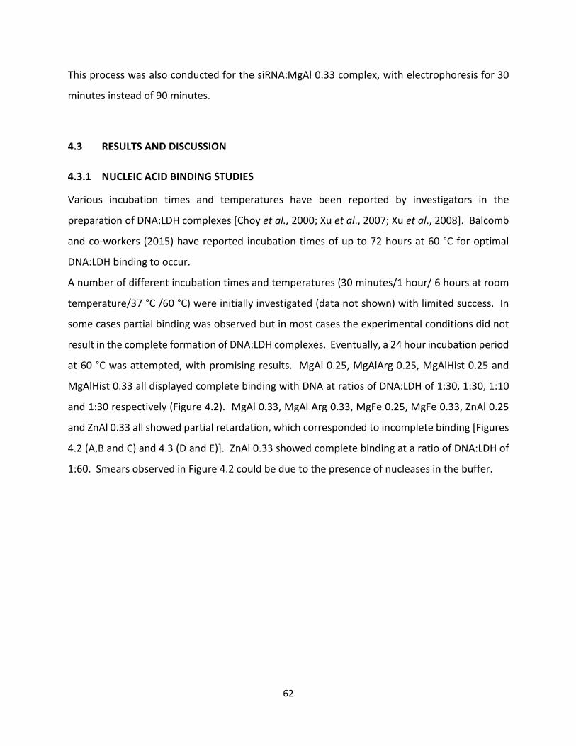
62
This process was also conducted for the siRNA:MgAl 0.33 complex, with electrophoresis for 30
minutes instead of 90 minutes.
4.3 RESULTS AND DISCUSSION
4.3.1 NUCLEIC ACID BINDING STUDIES
Various incubation times and temperatures have been reported by investigators in the
preparation of DNA:LDH complexes [Choy et al., 2000; Xu et al., 2007; Xu et al., 2008]. Balcomb
and co-workers (2015) have reported incubation times of up to 72 hours at 60 °C for optimal
DNA:LDH binding to occur.
A number of different incubation times and temperatures (30 minutes/1 hour/ 6 hours at room
temperature/37 °C /60 °C) were initially investigated (data not shown) with limited success. In
some cases partial binding was observed but in most cases the experimental conditions did not
result in the complete formation of DNA:LDH complexes. Eventually, a 24 hour incubation period
at 60 °C was attempted, with promising results. MgAl 0.25, MgAlArg 0.25, MgAlHist 0.25 and
MgAlHist 0.33 all displayed complete binding with DNA at ratios of DNA:LDH of 1:30, 1:30, 1:10
and 1:30 respectively (Figure 4.2). MgAl 0.33, MgAl Arg 0.33, MgFe 0.25, MgFe 0.33, ZnAl 0.25
and ZnAl 0.33 all showed partial retardation, which corresponded to incomplete binding [Figures
4.2 (A,B and C) and 4.3 (D and E)]. ZnAl 0.33 showed complete binding at a ratio of DNA:LDH of
1:60. Smears observed in Figure 4.2 could be due to the presence of nucleases in the buffer.
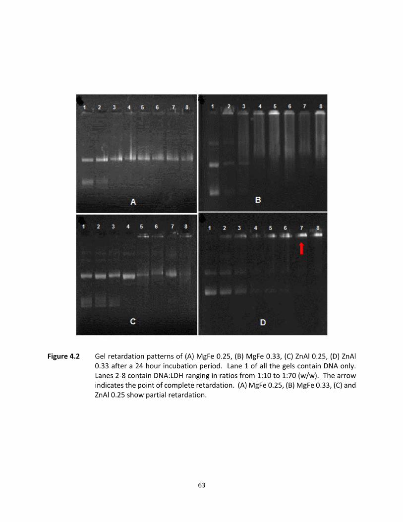
63
Figure 4.2 Gel retardation patterns of (A) MgFe 0.25, (B) MgFe 0.33, (C) ZnAl 0.25, (D) ZnAl 0.33 after a 24 hour incubation period. Lane 1 of all the gels contain DNA only. Lanes 2-8 contain DNA:LDH ranging in ratios from 1:10 to 1:70 (w/w). The arrow indicates the point of complete retardation. (A) MgFe 0.25, (B) MgFe 0.33, (C) and ZnAl 0.25 show partial retardation.

64
Figure 4.3 Gel retardation patterns of (A) MgAl 0.25, (B) MgAlArg 0.25, (C) MgAlHist 0.25, (D) MgAl 0.33, (E) MgAlArg 0.33, (F) MgAlHist 0.33 after a 24 hour incubation period. Lane 1 of all the gels contain DNA only. Lanes 2-8 contain DNA:LDH ranging in ratios from 1:10 to 1:70 (w/w). The arrows indicate the point of complete retardation. (D) MgAl 0.33 and (E) MgAlArg 0.33 show partial retardation.
Barichello et al (2012) reported that agitation by vortex resulted in enhanced binding between
siRNA and cationic liposomes, resulting in superior gene knockdown. This methodology was then
applied to the formation of the DNA:LDH complexes. The components required to make up the
DNA:LDH complexes were vortexed at high speed for 10 minutes followed by a 10 minute
incubation at room temperature. This method resulted in complete binding of the DNA to LDH,
as confirmed by the gel retardation (Figure 4.4). It has been further suggested that the high
energy transmitted during vortexing harmonizes the interaction between the nucleic acid and
the carrier vector [Barichello et al., 2012].
65
Figure 4.4 Gel retardation patterns of DNA only (lane 1), DNA:MgAl 0.25 (lane 2), DNA:MgAlArg 0.25 (lane 3), DNA:MgAlHist 0.25 (lane 4), DNA:MgAl 0.33 (lane 5), DNA:MgAlArg 0.33 (lane 6), DNA:MgAlHist 0.33 (lane 7), DNA:MgFe 0.25 (lane 8), DNA:MgFe 0.33 (lane 9), DNA: ZnAl 0.25 (lane 10) and DNA:ZnAl 0.33 (lane 11)after vigorous vortexing for 10 minutes followed by a 10 minute incubation period at room temperature. Results indicate complete complexation of the LDH with DNA under these conditions. All complexes were prepared in a ratio of DNA:LDH of 1:30 (w/w). The arrows indicate complete retardation.
Zhang and co-workers (2003) have reported a reduction in the number of water molecules in a
lipoplex after complexation of liposomes with nucleic acids, resulting in a closer and more
harmonious interaction between the two components. It is possible that a similar reaction occurs
during complex formation between the DNA and LDHs, where loss of water molecules from the
interlayer could lead to a slight decrease in interlayer spacing consequently “trapping” the DNA
or at least part of the DNA within the interlayer region and enhancing complex formation.
Lu and co-workers (2016) have recently hypothesized a new, more likely mechanism of DNA
interaction with LDHs. They have suggested that LDHs are electrostatically attracted to and bind
to the LDH surfaces via the phosphate backbone of the DNA molecules. The phosphate oxygen
thus forms co-ordination metal-oxygen-phosphate bonds resulting in the adsorption of DNA onto
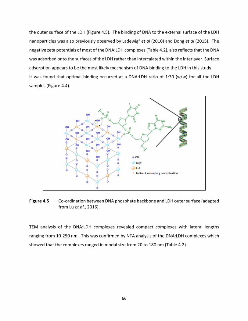
66
the outer surface of the LDH (Figure 4.5). The binding of DNA to the external surface of the LDH
nanoparticles was also previously observed by Ladewig1 et al (2010) and Dong et al (2015). The
negative zeta potentials of most of the DNA:LDH complexes (Table 4.2), also reflects that the DNA
was adsorbed onto the surfaces of the LDH rather than intercalated within the interlayer. Surface
adsorption appears to be the most likely mechanism of DNA binding to the LDH in this study.
It was found that optimal binding occurred at a DNA:LDH ratio of 1:30 (w/w) for all the LDH
samples (Figure 4.4).
Figure 4.5 Co-ordination between DNA phosphate backbone and LDH outer surface (adapted from Lu et al., 2016).
TEM analysis of the DNA:LDH complexes revealed compact complexes with lateral lengths
ranging from 10-250 nm. This was confirmed by NTA analysis of the DNA:LDH complexes which
showed that the complexes ranged in modal size from 20 to 180 nm (Table 4.2).
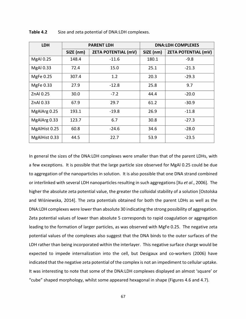
67
Table 4.2 Size and zeta potential of DNA:LDH complexes.
In general the sizes of the DNA:LDH complexes were smaller than that of the parent LDHs, with
a few exceptions. It is possible that the large particle size observed for MgAl 0.25 could be due
to aggregation of the nanoparticles in solution. It is also possible that one DNA strand combined
or interlinked with several LDH nanoparticles resulting in such aggregations [Xu et al., 2006]. The
higher the absolute zeta potential value, the greater the colloidal stability of a solution [Ostolska
and Wiśniewska, 2014]. The zeta potentials obtained for both the parent LDHs as well as the
DNA:LDH complexes were lower than absolute 30 indicating the strong possibility of aggregation.
Zeta potential values of lower than absolute 5 corresponds to rapid coagulation or aggregation
leading to the formation of larger particles, as was observed with MgFe 0.25. The negative zeta
potential values of the complexes also suggest that the DNA binds to the outer surfaces of the
LDH rather than being incorporated within the interlayer. This negative surface charge would be
expected to impede internalization into the cell, but Desigaux and co-workers (2006) have
indicated that the negative zeta potential of the complex is not an impediment to cellular uptake.
It was interesting to note that some of the DNA:LDH complexes displayed an almost ‘square’ or
“cube” shaped morphology, whilst some appeared hexagonal in shape (Figures 4.6 and 4.7).
LDH PARENT LDH DNA:LDH COMPLEXES SIZE (nm) ZETA POTENTIAL (mV) SIZE (nm) ZETA POTENTIAL (mV)
MgAl 0.25 148.4 -11.6 180.1 -9.8
MgAl 0.33 72.4 15.0 25.1 -21.3
MgFe 0.25 307.4 1.2 20.3 -29.3
MgFe 0.33 27.9 -12.8 25.8 9.7
ZnAl 0.25 30.0 -7.2 44.4 -20.0
ZnAl 0.33 67.9 29.7 61.2 -30.9
MgAlArg 0.25 193.1 -19.8 26.9 -11.8
MgAlArg 0.33 123.7 6.7 30.8 -27.3
MgAlHist 0.25 60.8 -24.6 34.6 -28.0
MgAlHist 0.33 44.5 22.7 53.9 -23.5
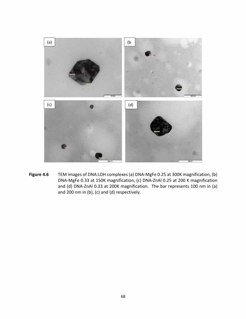
68
Figure 4.6 TEM images of DNA:LDH complexes (a) DNA-MgFe 0.25 at 300K magnification, (b) DNA-MgFe 0.33 at 150K magnification, (c) DNA-ZnAl 0.25 at 200 K magnification and (d) DNA-ZnAl 0.33 at 200K magnification. The bar represents 100 nm in (a) and 200 nm in (b), (c) and (d) respectively.
(a) (b
(c) (d)
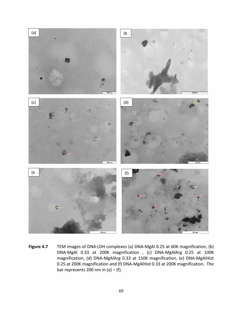
69
Figure 4.7 TEM images of DNA:LDH complexes (a) DNA-MgAl 0.25 at 60K magnification, (b)
DNA-MgAl 0.33 at 200K magnification , (c) DNA-MgAlArg 0.25 at 100K magnification, (d) DNA-MgAlArg 0.33 at 150K magnification, (e) DNA-MgAlHist 0.25 at 200K magnification and (f) DNA-MgAlHist 0.33 at 200K magnification. The bar represents 200 nm in (a) – (f).
(a) (b
(c) (d)
(e
(f)
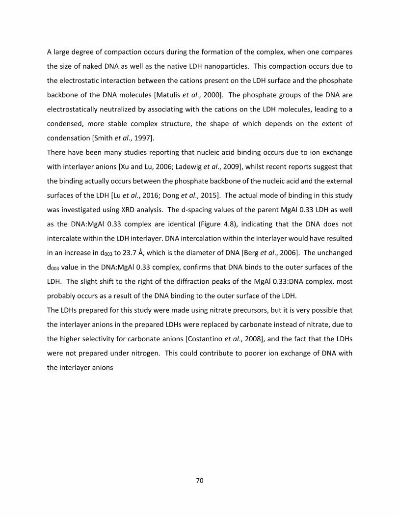
70
A large degree of compaction occurs during the formation of the complex, when one compares
the size of naked DNA as well as the native LDH nanoparticles. This compaction occurs due to
the electrostatic interaction between the cations present on the LDH surface and the phosphate
backbone of the DNA molecules [Matulis et al., 2000]. The phosphate groups of the DNA are
electrostatically neutralized by associating with the cations on the LDH molecules, leading to a
condensed, more stable complex structure, the shape of which depends on the extent of
condensation [Smith et al., 1997].
There have been many studies reporting that nucleic acid binding occurs due to ion exchange
with interlayer anions [Xu and Lu, 2006; Ladewig et al., 2009], whilst recent reports suggest that
the binding actually occurs between the phosphate backbone of the nucleic acid and the external
surfaces of the LDH [Lu et al., 2016; Dong et al., 2015]. The actual mode of binding in this study
was investigated using XRD analysis. The d-spacing values of the parent MgAl 0.33 LDH as well
as the DNA:MgAl 0.33 complex are identical (Figure 4.8), indicating that the DNA does not
intercalate within the LDH interlayer. DNA intercalation within the interlayer would have resulted
in an increase in d003 to 23.7 Å, which is the diameter of DNA [Berg et al., 2006]. The unchanged
d003 value in the DNA:MgAl 0.33 complex, confirms that DNA binds to the outer surfaces of the
LDH. The slight shift to the right of the diffraction peaks of the MgAl 0.33:DNA complex, most
probably occurs as a result of the DNA binding to the outer surface of the LDH.
The LDHs prepared for this study were made using nitrate precursors, but it is very possible that
the interlayer anions in the prepared LDHs were replaced by carbonate instead of nitrate, due to
the higher selectivity for carbonate anions [Costantino et al., 2008], and the fact that the LDHs
were not prepared under nitrogen. This could contribute to poorer ion exchange of DNA with
the interlayer anions
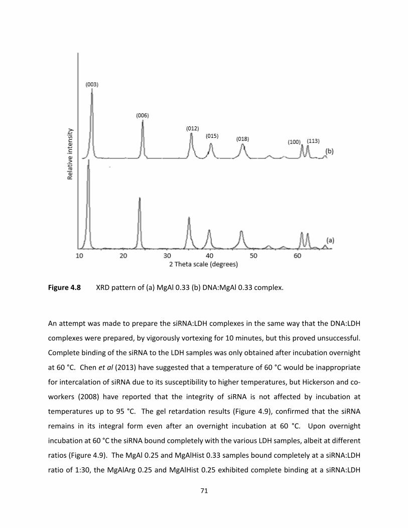
71
Figure 4.8 XRD pattern of (a) MgAl 0.33 (b) DNA:MgAl 0.33 complex.
An attempt was made to prepare the siRNA:LDH complexes in the same way that the DNA:LDH
complexes were prepared, by vigorously vortexing for 10 minutes, but this proved unsuccessful.
Complete binding of the siRNA to the LDH samples was only obtained after incubation overnight
at 60 °C. Chen et al (2013) have suggested that a temperature of 60 °C would be inappropriate
for intercalation of siRNA due to its susceptibility to higher temperatures, but Hickerson and co-
workers (2008) have reported that the integrity of siRNA is not affected by incubation at
temperatures up to 95 °C. The gel retardation results (Figure 4.9), confirmed that the siRNA
remains in its integral form even after an overnight incubation at 60 °C. Upon overnight
incubation at 60 °C the siRNA bound completely with the various LDH samples, albeit at different
ratios (Figure 4.9). The MgAl 0.25 and MgAlHist 0.33 samples bound completely at a siRNA:LDH
ratio of 1:30, the MgAlArg 0.25 and MgAlHist 0.25 exhibited complete binding at a siRNA:LDH
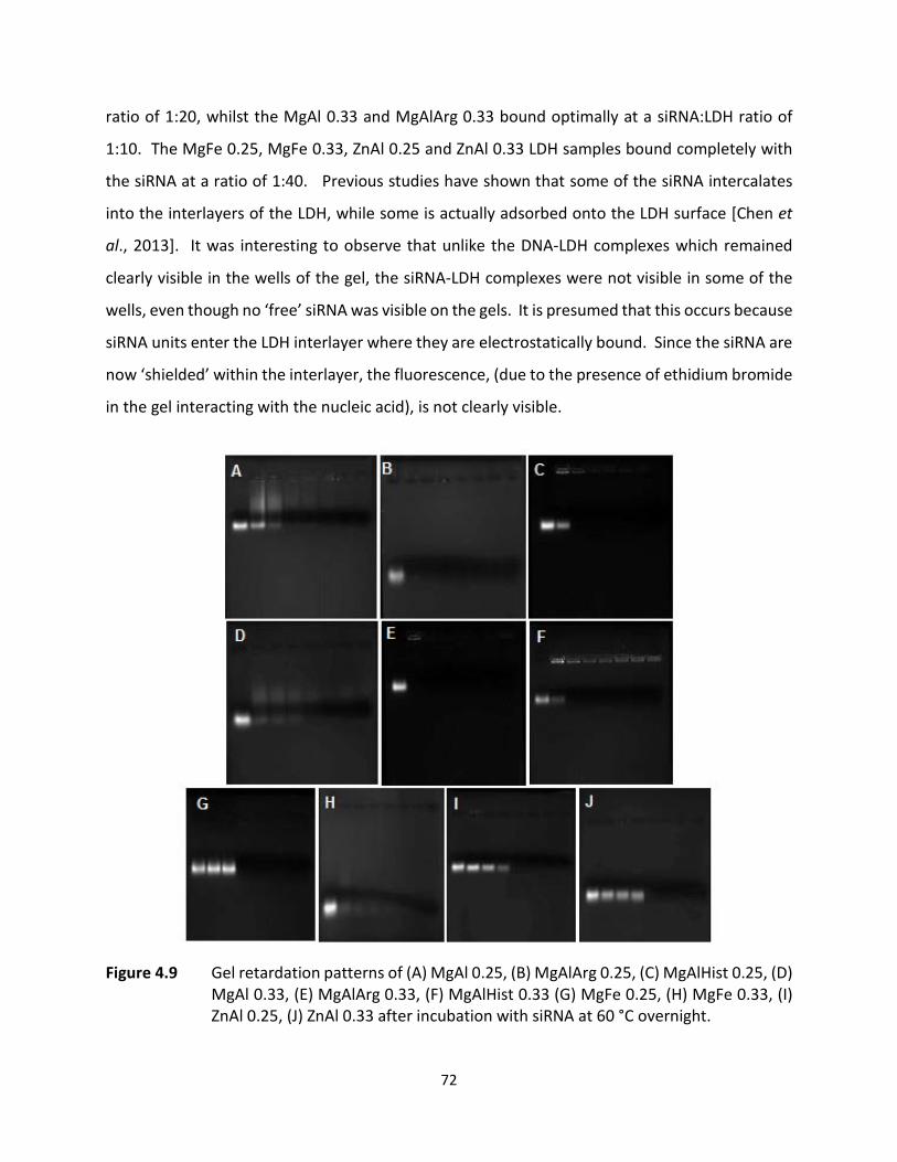
72
ratio of 1:20, whilst the MgAl 0.33 and MgAlArg 0.33 bound optimally at a siRNA:LDH ratio of
1:10. The MgFe 0.25, MgFe 0.33, ZnAl 0.25 and ZnAl 0.33 LDH samples bound completely with
the siRNA at a ratio of 1:40. Previous studies have shown that some of the siRNA intercalates
into the interlayers of the LDH, while some is actually adsorbed onto the LDH surface [Chen et
al., 2013]. It was interesting to observe that unlike the DNA-LDH complexes which remained
clearly visible in the wells of the gel, the siRNA-LDH complexes were not visible in some of the
wells, even though no ‘free’ siRNA was visible on the gels. It is presumed that this occurs because
siRNA units enter the LDH interlayer where they are electrostatically bound. Since the siRNA are
now ‘shielded’ within the interlayer, the fluorescence, (due to the presence of ethidium bromide
in the gel interacting with the nucleic acid), is not clearly visible.
Figure 4.9 Gel retardation patterns of (A) MgAl 0.25, (B) MgAlArg 0.25, (C) MgAlHist 0.25, (D) MgAl 0.33, (E) MgAlArg 0.33, (F) MgAlHist 0.33 (G) MgFe 0.25, (H) MgFe 0.33, (I) ZnAl 0.25, (J) ZnAl 0.33 after incubation with siRNA at 60 °C overnight.

73
TEM analysis of the siRNA:hydrotalcite complexes shows a large degree of condensation of the
LDH structure upon binding with siRNA. The nanoparticles, which before binding ranged in size
from 20-200 nm, condensed down to a size range of 4-50 nm in lateral diameter. The complexes
appear somewhat spherical but not quite uniform in size or morphology (Figure 4.10).
Figure 4.10 TEM images of complexes (a) siRNA-MgAl 0.25, (b) siRNA-MgAl 0.33, (c) siRNA-MgAlArg 0.33 and (d) siRNA-MgAlHist 0.33 at 300 K magnification. The bar represents 100 nm in (a) – (d).
The HT-like compounds complexed with siRNA showed a similar condensation formation to those
observed for the siRNA-MgAl 0.25, siRNA-MgAl 0.33, siRNA-MgAlArg 0.33 and siRNA-MgAlHist
0.33 (Figure 4.11). Lateral sizes of the complexes ranged between 5 and 30 nm.
(a) (b)
(c) (d)

74
Figure 4.11 TEM images of complexes (a) siRNA-MgFe 0.25, (b) siRNA-MgFe 0.33, (c) siRNA- ZnAl 0.25 and (d) siRNA-ZnAl 0.33 at 300K magnification. The bar represents 100 nm in (a) – (d).
The condensation in structure seen for all the other LDH-siRNA complexes is different from that
observed for the siRNA-MgAlArg 0.25 and siRNA-MgAlHist 0.25 complexes. Instead of the
compact ‘spherical shaped’ complexes, nanoflakes were observed (Figure 4.12). It appears as if
the amino acid functionalized hydrotalcites prepared in the ratio of Mg:Al:aa of 3:1:1 have a more
rigid structure, that is unable to condense down completely upon complexation with the siRNA,
and the nanoparticles merely folds over around the siRNA.
(a) (b)
(c) (d)

75
Figure 4.12 TEM images of complexes (a) siRNA-MgAlArg 0.25 and (b) siRNA-MgAlHist 0.25 at 300K magnification. The bar represents 100 nm in (a) and (b).
Unlike the DNA:LDH complexes which have an almost ‘square/cube’ shape, the siRNA:LDH
complexes are more spherical in shape. The difference in shape suggests that the mechanism of
structural condensation of the siRNA:LDH complexes is most probably quite different from the
mechanism involved in DNA:LDH structural condensation. siRNAs are much smaller molecules
than DNA, and are thus probably easily intercalated within the interlayer region of the LDHs.
Unlike the electrostatic attraction occurring between the cations on the outer surface of the LDH
molecules and DNA, the electrostatic attraction most probably occurs between the cations on
the inner surfaces of the LDH molecules and siRNA, resulting in the distinct difference in shape
of the DNA:LDH complexes compared to the siRNA:LDH complexes.
Computational simulation studies by Zhang et al (2012) suggest that RNA molecules intercalate
within the LDH interlayers, where the RNA molecule is oriented parallel to the hydroxide layers.
The strong electrostatic interactions between the siRNA molecules and the positively charged
LDH sheets keep the siRNA molecules ‘bound’ within the interlayer.
The small sizes of the siRNA:LDH complexes was confirmed by NTA analysis (Table 4.3). The zeta
potential values obtained for the siRNA:LDH complexes suggest greater colloidal stability than
the DNA:LDH complexes, which could be a contributing factor to the generally smaller size of
particles observed. The small siRNA:LDH complex size could also be attributed to the initial high
speed vortexing during complex preparation, which is reported to result in better interaction
(a) (b)

76
between the siRNA and the carrier vector [Barichello et al., 2012]. There is also a greater change
in zeta potential of the siRNA:LDH complexes compared to the parent LDHs, which shows that
the LDHs have a high affinity for siRNA [Rojas et al., 2015]. The negative charge of the siRNA:LDH
complexes indicates that siRNA similar to the DNA binds predominantly to the outer surfaces of
the LDH [Wong et al., 2010; Chen et al., 2013].
Table 4.3 Size and zeta potential of siRNA:LDH complexes.
4.3.2 NUCLEASE DIGESTION AND NUCLEIC ACID RELEASE ASSAY
The ultimate role of a gene delivery vector is to eventually deliver its load into the cell in vivo.
Here the complexes would be exposed to degradative enzymes such as nucleases. It is therefore
essential to investigate whether the vector is able to transport the nucleic acids into the cell in
its integral form. This may be determined by performing a nuclease digestion assay, where the
nucleic acid-vector complex is exposed to serum nucleases in an environment mimicking in vivo
conditions. The intercalation reaction between DNA and LDH is reversible and the DNA may be
released either by addition of K2CO3 [Wu et al., 2014] or by dropping the pH of the solution. In
LDH PARENT LDH siRNA:LDH COMPLEXES
SIZE (nm)
ZETA POTENTIAL (mV) SIZE (nm)
ZETA POTENTIAL (mV)
MgAl 0.25 148.4 -11.6 28.4 -34.7
MgAl 0.33 72.4 15.0 47.0 -30.3
MgFe 0.25 307.4 1.2 45.1 -28.2
MgFe 0.33 27.9 -12.8 55.4 -21.8
ZnAl 0.25 30.0 -7.2 27.0 -29.7
ZnAl 0.33 67.9 29.7 25.7 -46.1
MgAlArg 0.25 193.1 -19.8 34.3 -28.1
MgAlArg 0.33 123.7 6.7 36.0 -13.5
MgAlHist 0.25 60.8 -24.6 41.0 -22.6
MgAlHist 0.33 44.5 22.7 48.8 -24.0

77
this study, the nucleic acid was released from the vector by dropping the pH. An acidic pH
disrupts and dissolves the hydroxide layers of the LDH, resulting in release of the nucleic acid
[Zhang et al., 2012]. The sample is then exposed to gel electrophoresis to visualize the integrity
of the nucleic acid and/or extent of nuclease digestion. Integral DNA would appear as three
distinct bands (depending on the forms of DNA initially present in the sample) and integral siRNA
would appear as a single band. Nucleic acid that had been degraded by the serum enzymes would
appear as smears on the gel. If the nucleic acid remains in its integral form after exposure to the
nuclease digestion assay, then this suggests that the vector would be suitable for in vivo
applications.
From the results obtained (Figure 4.13), it can be clearly seen that the DNA:LDH complex does
offer some degree of protection to the DNA from serum nucleases. The naked, unprotected DNA
(lane 2) was disintegrated after exposure to serum nucleases as the DNA appeared as a smudged
band on the gel. For the DNA:LDH complexes, the DNA retained its integrity with the bands of
the linear, circular and supercoiled forms of the DNA still being quite clearly visible (lane 4). Some
of the DNA:LDH complex still remained in the well, as was also reported by Kriven et al (2004)
who suggested that DNA can only be completely released from the complex at a pH of 2 or less.
The mechanism of release of the DNA from the DNA:LDH complex has been proposed to be via a
dissolution process that occurs in the endosome due to low pH [Ladewig et al., 2009; Choy et al.,
2000]. LDHs begin to dissolve in a pH range of 4-5, resulting in the release of their cargo in a pH-
dependent manner [Wong et al., 2010].
The buffering capacity of LDHs have been well documented and this property has been exploited
in the production of antacids from LDHs for many years [Cunha et al., 2010]. Bejoy (2001)
reported that hydrotalcites are capable of buffering the contents of the stomach at pH 4 for
extended periods of time. It is this property that has also made LDHs increasingly attractive for
medical applications, where it can be used for controlled release of medicines. Thus the
incomplete degradation of the complex, indicated by the presence of some of the complexes
remaining in the well, as can be seen in lane 4 (Figure 4.13), was as expected after an overnight
78
incubation period. The slow dissolution process of the DNA:LDH complex indicates that the DNA
transfection rate via LDH nanoparticles would be a slow process. This is the reason for the
extended incubation period of 72 hours for the transfection studies (Chapter 6).
Figure 4.13 Results of nuclease digestion assay of DNA:MgAl 0.33. Lane 1 : naked DNA, Lane
2: DNA exposed to nuclease digestion, Lane 3 :MgAl 0.25 DNA-LDH complex, and Lane 4: MgAl 0.25 DNA-LDH complex exposed to nuclease digestion.
There does appear to be a small amount of degradation of the DNA from the DNA:LDH complex.
This confirms reports by Xu et al (2007), Ladewig1 et al (2010) and Lu et al (2016) that some of
the DNA is bound to the surface of the LDH, leaving the DNA partially exposed to nuclease
digestion. Similar results were obtained for the siRNA:LDH complex (Figure 4.14). The naked
siRNA is almost totally digested by the nuclease present in the serum (lane 2), whilst there
appears to be minimal degradation of the siRNA:LDH complex after exposure to nuclease
digestion. Most of the siRNA from the siRNA:LDH complex exposed to nuclease digestion still
appears to be present in the well of the gel (lane 4), indicating that the siRNA is still bound to the
LDH. This implies that some degree of protection has been afforded to the siRNA by the LDHs.

79
Figure 4.14 Results of nuclease digestion assay of siRNA:MgAl 0.25. Well 1 was loaded with naked siRNA, well 2 contained siRNA exposed to nuclease digestion, well 3 was loaded with the MgAl 0.25 siRNA-LDH complex, and well 4 contained the MgAl 0.25 siRNA-LDH complex exposed to nuclease digestion.
4.4 CONCLUSION The optimal binding ratio of DNA:LDH was found to be 1:30 (w/w). The optimal binding ratios of
siRNA:LDH complexes varied between 1:10 to 1:40 (w/w) depending on the LDH used. The MgAl
0.25 and MgAlHist 0.33 samples bound completely at a siRNA:LDH ratio of 1:30 (w/w), the
MgAlArg 0.25 and MgAlHist 0.25 exhibited complete binding at a siRNA:LDH ratio of 1:20 (w/w),
whilst the MgAl 0.33 and MgAlArg 0.33 bound optimally at a siRNA:LDH ratio of 1:10 (w/w). The
MgFe 0.25, MgFe 0.33, ZnAl 0.25 and ZnAl 0.33 LDH samples bound completely with the siRNA
at a ratio of 1:40 (w/w).
Results of the nuclease digestion assay show that most of the nucleic acid (DNA/RNA), appear to
retain its integrity after being exposed to the serum nucleases, and afforded reasonable
protection to the respective nucleic acid cargo. Hence, these LDHs show potential as suitable
gene carriers for further transfection studies.
1 2 3 4
80
CHAPTER 5
5. IN VITRO ASSAYS: CYTOTOXICITY, GENE EXPRESSION AND GENE SILENCING STUDIES
5.1. INTRODUCTION The use of cell-based assays is important for testing the various parameters with regards to their
physiological processes and their response to external factors and compounds. Hence, they have
become useful tools to predict the possible outcome of in vivo studies. Their use in preclinical
applications of gene and drug therapy is without doubt. The cell viability, MTT assay and
luciferase gene expression studies were conducted on the human cell lines: embryonic kidney
(HEK293), hepatocellular carcinoma (HepG2), and colon adenocarcinoma (Caco-2). The human
cervical carcinoma cells stably transformed with the luciferase gene (Hela-tat-luc), was also used
in the MTT assay, but solely employed in the gene silencing assay. A brief description of the cell
lines is provided below.
HEK293
In 1973, Frank Graham in the Netherlands, transformed cultures of normal human embryonic
kidney cells (from an apparently healthy, legally aborted fetus) with sheared adenovirus 5 DNA to
form HEK293 cells. The cell line was so named because it was Grahams 293rd experiment
[www.hek293.com (Date accessed: 15th September 2017)]. The transformation was due to the
incorporation of a 4.5 kb insert from the viral genome into human chromosome 19
[https://en.wikipedia.org/wiki/HEK_293_cells (Date accessed: 15th September 2017)]. The exact
cell type of HEK293 is unknown as adenovirus 5 significantly changes cell morphology and
expression post-transformation, but it is speculated to be neuronal in origin.
HEK293 cells have a modal chromosome number of 64, and are referred to as being hypotriploid,
because they contain less than three times the number of chromosomes of a normal diploid
human cell. HEK293 expresses the corticotrophin releasing factor type 1 receptor, sphingosine-
1-phosphate receptors EDG1, EDG3 and EDG5, muscarinic acetylcholine receptor M3 and the

81
transient receptor potential TRPC1, TRPC3, TRPC4, TRPC6 [www.hek293.com (Date accessed:
15th September 2017)]. HEK293 cells (Figure 5.1) are extremely popular in cell culture due to
their ease of growth and transfectibility, which makes them ideal for gene expression studies.
Figure 5.1 Image of HEK293 cells (x100) captured using an Olympus inverted microscope.
HepG2
The HepG2 cell line (Figure 5.2) was derived from a well differentiated liver hepatocellular
carcinoma of a 15-year old Caucasian male [www.hepG2.com (Date accessed: 15th September
2017)]. The cells have a modal chromosome number of 55. HepG2 cells are relatively easy to
culture and grow as adherent monolayers and/or small aggregates with an epithelial
morphology. When properly cultured, HepG2 cells differentiate to form apical and basolateral
cell surfaces, which bear resemblance to liver structures in vivo. This makes HepG2 cells useful
in the study of intracellular transport between different liver cell types. The high degree of
morphological and functional differentiation of HepG2 cells in vitro, as well as the absence of
viral infection (no hepatitis B surface antigens), makes them ideal models for hepatocellular
carcinoma studies [Costantini et al., 2013]. HepG2 cells are regularly used when examining
toxicity of substances to the liver, liver metabolism and drug metabolism studies.
HepG2 cells contain unique asialoglycoprotein receptors that bind and internalize galactose
terminal asialoglycoproteins [Wu and Wu, 1987; Wu and Wu, 1988], making HepG2 a useful cell

82
line for receptor mediated endocytosis studies. These cells have even been used in trials with
bio-artificial liver devices [www.hepG2.com (Date accessed: 15th September 2017)].
HepG2 cells secrete a number of plasma proteins such as albumin, transferrin, fibrinogen, alpha-
2-macroglobulin, alpha-1-antitrypsin, alpha-1-antichymotrypsin, plasminogen, haptoglobulin,
ceruloplasmin, alpha-1-acid glycoprotein, alpha-2-HS-glycoprotein, beta-lipoprotein and retinol
binding protein [https://en.wikipedia.org/wiki/Hep_G2 (Date accessed: 15th September 2017)],
and has 3-hydroxy-3-methylglutaryl-CoA reductase and hepatic triglyceride lipase activities.
Figure 5.2 Image of HepG2 cells (x100) captured using an Olympus inverted microscope.
Caco-2
Caco-2 cells (Figure 5.3) have a modal chromosome number of 96, and were first isolated from a
primary colonic tumour, from a 72 year old Caucasian male in the 1970s. It is a continuous,
heterogeneous cell line that undergoes spontaneous differentiation and expresses the
morphological and biochemical characteristics of intestinal enterocytes, making them ideal
models of the intestinal barrier for in vitro cytotoxicity studies [Natoli et al., 2012; Sambuy et al.,
2005]. Caco-2 cells are adherent cells that display an epithelial morphology and grow in a
monolayer. Caco-2 cells express a number of enzymes such as sucrase-isomaltase, peptidases
and esterases, and nutrient transporters of sugars, amino acids, bile acids, carboxylic acids, as
well as transporters of micronutrients such as heavy metals and nucleosides [Sambuy et al., 2005;
https://en.wikipedia.org/wiki/Caco-2 (Date accessed: 15th September 2017)].

83
Figure 5.3 Image of Caco-2 cells (x100) captured using an Olympus inverted microscope.
Hela-tat-luc
The original Hela cell line was derived from a cervical carcinoma of a 31-year old African American
female in 1951 [https://en.wikipedia.org/wiki/HeLa (Date accessed: 15th September 2017)].
These were the first human immortal cell line used for medical research. The Hela-tat-luc cells
(Figure 5.4) used in this study have been transfected with the Tat protein from the human
immunodeficiency type 1 virus (HIV-1), and also stably transformed using the firefly luciferase
gene.
Figure 5.4 Image of HeLa-tat-luc cells (x100) captured using an Olympus inverted microscope.

84
5.1.1 CYTOTOXICITY STUDIES
Before undertaking gene expression and silencing studies, it is essential to determine the
cytotoxicity of the LDHs in cell culture, since high levels of toxicity, even in the presence of good
transfection, would render the nanoparticles unsuitable for in vivo applications. There are
currently many convenient and cost-effective colorimetric and fluorometric based cytotoxicity
assays available, where the samples can be assessed directly on the culture plate using a plate
reader [Weyermann et al., 2005]. These methods include lactate dehydrogenase release, neutral
red uptake, crystal violet staining, ATP and the 3-(4,5-dimethylthiazol-2-yl)-2,5-
diphenyltetrazolium (MTT) assays [Fotakis and Timbrell, 2006; Weyermann et al., 2005], among
others.
For this study, the MTT assay was used to assess cell viability after exposure to the nucleic
acid:LDH complexes. The MTT assay was first proposed by Mosmann (1983) and has been
extensively used despite disadvantages such as decreased formazan production due to the
actions of reducing agents and respiratory chain inhibitors of mitochondrial MTT reduction. It
has also been reported that the MTT assay could be influenced by factors such as medium pH, D-
glucose concentration of the growth medium and cellular concentration of pyridine nucleotides
[Chiba et al., 1998]. Advantages of the MTT assay include the simplicity of the method, high
sensitivity and the use of equipment easily found in most laboratories.
MTT is a water-soluble salt containing a tetrazolium ring, which can be cleaved by mitochondrial
succinate dehydrogenases, leading to the formation of an insoluble, purple-colored formazan
product which is impermeable to the cell membrane and therefore accumulates within healthy
cells [Fotakis and Timbrell, 2006]. The amount of formazan produced is proportional to the
number of viable cells present in the culture. Hence, the ability of the cells to reduce MTT
correlates to the cells viability, since only viable cells will have an active mitochondrion [Chiba et
al., 1998]. There have been many modifications to Mosmann’s method over the years [Tada et
al., 1986; Denizot and Lang, 1986; Carmichael et al., 1987; Twentyman and Luscombe, 1987], and
the method employed for this study is similar to that reported by Carmicheal and co-workers
(1987), with DMSO being used to solubilize the formazan instead of mineral oil. This is because
85
DMSO does not cause precipitation of proteins and results in a clearer formazan solution, with
stable optical density [Twentyman and Luscombe, 1987].
5.1.2 GENE EXPRESSION AND SILENCING STUDIES
A transfection study is carried out to determine whether a vector is able to efficiently deliver the
nucleic acid into a cell resulting in either sustained expression of the transgene (DNA) or silencing
of the target gene (siRNA).
The plasmid DNA utilised in this study is the pCMV-luc DNA, described previously in chapter 4
(section 4.1). The luciferase assay is based on the fact that mammalian cells do not naturally
express the luciferase gene, which is only expressed upon successful transfection of the pCMV-
luc DNA into eukaryotic cells. This reaction requires the addition of the substrates luciferin, ATP
and oxygen, to catalyze the production of light or luminescence, which can be measured using a
luminometer. The luciferase gene isolated from the firefly P. pyralis is composed of seven exons
separated by six very short introns, all less than 60 bases in length [de Wet et al., 1987]. The reactions catalyzed by firefly luciferase are:
Mg2+ Luciferase + luciferin + ATP ↔ luciferase.luciferyl AMP + PPi
luciferase.luciferyl AMP + O2 → luciferase + oxyluciferin + AMP + CO2 + hv (light)
The amount of light produced is directly proportional to the quantity of luciferase present in the
reaction mixture.
The luciferase enzyme activity can also be used as an indicator of gene silencing. The Hela tat-
luc cell line stably expresses the luciferase gene and the anti-luc-siRNA specifically targets the
luciferase gene. Efficient delivery of the anti-luc-siRNA results in a decrease in luciferase activity,
which can be similarly measured using a luminometer.

86
5.2. MATERIALS AND METHODS
5.2.1 MATERIALS
Eagle’s Minimum Essential Medium (EMEM) containing Earle’s salts and L-glutamine, trypsin-
versene and penicillin-streptomycin mixtures was purchased from Lonza BioWhittaker
(Walkersville, USA). Foetal bovine serum (FBS) was purchased from Hyclone GE Healthcare (Utah,
USA). Phosphate-buffered saline (PBS) tablets were purchased from Calbiochem (Canada). The
MTT salt (3-(4,5-dimethylthiazol-2-yl)- 2,5-diphenyltetrazolium bromide) was purchased from
Merck (Darmstadt, Germany). All sterile tissue culture plastic ware obtained from Corning Inc.
(NY, USA). HepG2 and Caco-2 cells were sourced from Highveld Biologicals (PTY) LTD (Lyndhurst,
South Africa). HEK293 (human embryonic kidney) cells were supplied by the University of
Witwatersrand, Medical School, South Africa. HeLa tat luc cell line was provided by the
Department of Physiology, University of KwaZulu-Natal, Westville Campus. The pCMV-luc DNA
was purchased from Plasmid Factory (Bielefeld, Germany), while the anti-luc-siRNA was obtained
from Thermo-Scientific Dharmacon Products (Lafayette, CO). The Luciferase Assay kit was
purchased from the Promega Corporation, Madison, USA. The bicinchoninic acid (BCA) assay
reagents were purchased from the Sigma-Aldrich Co., St. Louis, USA. All other reagents were of
analytical grade.
5.2.2 MTT ASSAY
Cells were maintained in EMEM supplemented with 10 % (v/v) gamma-irradiated FBS and 1 %
antibiotics (100 U/mL penicillin, 100 μg/mL streptomycin) at 37 °C and 5% CO2, in a HEPA Class
100 Steri-Cult CO2 incubator (Thermo-Electron Corporation, Waltham, Massachusetts, USA). The
MTT assay was carried out using Caco-2, HEK293 and HepG2 cells for the DNA:LDH complexes
and using the HeLa- tat-luc cell line for the siRNA:LDH complexes.
For the MTT assay, cells were trypsinized and seeded into 48-well plates at a seeding density of
approximately 3 X 10⁴ cells/well, and incubated at 37 °C for 24 hours to allow for attachment.
The medium was then replaced with fresh medium, and the respective DNA:LDH complexes were
added to the wells and allowed to incubate for 48 hours at 37 °C. Complexes prepared were at
87
DNA:LDH ratios (sub-optimal, optimal and supra-optimal), as determined from the gel
retardation assays (chapter 4, section 4.3). A positive control containing only cells was set as 100
% cell survival/viability. All assays were performed in triplicate. After the 48-hour incubation
period the spent medium was removed and 200 μL fresh medium and 200 μL MTT reagent (5
mg/mL in PBS) was added to each well.
The cells were incubated at 37 °C for a further 4 hours, after which the MTT-medium solution
was removed and 200 μL DMSO was added to solubilize the formazan crystals. Absorbance was
read at 540 nm using a Mindray MR-96A microplate reader (Vacutec, Hamburg, Germany).
5.2.3 LUCIFERASE GENE EXPRESSION STUDIES DNA transfection studies were carried out in the HEK293, HepG2 and Caco-2 cell lines, using the
pCMV-luc plasmid DNA. The cells were seeded in 48 well plates at a cell density of 3.0 × 10⁴ cells
per well and incubated at 37 °C for 24 hours. The medium was then replaced with fresh medium
and the respective DNA:LDH complexes were added to the wells and allowed to incubate for 72
hours at 37 °C. Complexes were prepared as in 5.2.2. Each assay was done in triplicate. Two
controls were used, cells only and cells with naked plasmid DNA. After the 72-hour incubation
period, the luciferase activity of the cells was determined using the Promega luciferase assay
system.
The cells were prepared by removing the growth medium and washing twice with PBS.
Thereafter, 80 µl of 1× cell lysis reagent (25 mM Tris-phosphate, pH 7.8; 2 mM dithiothreiotol, 2
mM 1,2- diaminocyclohexane – N, N, N’N’- tetra-acetic acid, 10 % (v/v) glycerol, 1 % (v/v) triton
X-100) was added to the cells, which were then gently rocked on a Stuart Scientific Platform
Shaker for 15 minutes at 30 rev/ min. The lysed cells were dislodged from the multiwell plates
by gently scraping. The cell suspension was then pelleted by centrifugation at 12000 x g for 5
seconds. The cell free extract was retained for the determination of luciferase activity. A volume
of 20 µl of each of the cell free extracts was transferred to a white multiwell plate and placed in
a GloMax Multi+ detection system (Promega). Luciferase assay reagent (100 µL; 20 mM Tricine,
1.1 mM Magnesium carbonate hydroxide pentahydrate, 2.7 mM Magnesium sulphate, 0.1 mM

88
EDTA, 33.3 mM dithiothreitol, 270 µM coenzyme A, 470 µM luciferin, 530 µM ATP) was then
automatically injected into the samples and the luminescence was read immediately. The
protein content of the cell free extracts was determined using the BCA assay, at 540 nm. All
luminescence readings were normalized against the protein content and expressed as relative
light units per milligram protein (RLU/mg protein). The results were presented as means ± SD.
5.2.4 LUCIFERASE GENE SILENCING STUDIES
The overall procedure for the gene silencing studies was similar to that for the DNA transfection
studies (5.2.3). The Hela tat-luc cell line, stably expressing the luciferase gene was used and the
complexes comprised of anti-luc-siRNA and LDH prepared at ratios as determined by the binding
assays (chapter 4, section 4.3). The only difference in methodology for the knockdown studies
was that the cells were allowed to incubate with the complexes for 48 hours instead of the 72-
hour incubation period used for the DNA transfection studies. This was because previous reports
have shown that optimal gene knockdown occurs between a 36 and 48-hour incubation period
[Ladewig2 et al., 2010]. Wong and co-workers (2010) have also reported effective gene silencing
following a 24-hour incubation period. To validate transfection, the luciferase assay was carried
out as described above (5.2.3).
5.3 STATISTICAL ANALYSIS
All graphs were plotted in Microsoft Excel™. All data are presented as mean ± standard deviation
(± SD n=3). Graphpad Instat Prism 4.0.3 software was used for statistical analysis. Multi group
comparisons of means were carried out using one-way analysis of variance (ANOVA) followed by
the Tukey-Kramer post hoc test. Statistical significance for all tests was set at ***p<0.0005 (high
significance), **p<0.005 (medium significance) and *p<0.05 (low significance). Each experimental
value was compared to its corresponding control.

89
5.4 RESULTS AND DISCUSSION
5.4.1 CYTOTOXICITY STUDIES
5.4.1.1 MTT ASSAY OF DNA:LDH COMPLEXES
Cytotoxicity of nanoparticles is influenced by a number of factors such as the cell line used, cell
culture conditions, nanoparticle size, nanoparticle concentration and duration of exposure [Choi
and Wang, 2011]. The six DNA:hydrotalcite complexes were very well tolerated by the all cell
lines at the ratios and conditions tested (Figure 5.5). The cell viabilities, after a 48 hour incubation
period with complexes prepared at optimal ratio were, for Caco-2 cells -MgAl 0.25 (83.50 %),
MgAl 0.33 (91.16 %), MgAlArg 0.25 (105.2 %), MgAl Arg 0.33 (102.9 %), MgAlHist 0.25 (103.3 %)
and MgAlHist 0.33 (102.8 %); for the HEK293 cells -MgAl 0.25 (103.7 %), MgAl 0.33 (99.69 %),
MgAlArg 0.25 (98.58 %), MgAl Arg 0.33 (102.1 %), MgAlHist 0.25 (105.3 %) and MgAlHist 0.33
(109.8 %); and for the HepG2 cells - MgAl 0.25 (97.57 %), MgAl 0.33 (101.5 %), MgAlArg 0.25
(108.3 %), MgAl Arg 0.33 (103.5 %), MgAlHist 0.25 (109.1 %) and MgAlHist 0.33 (106.3 %)
respectively.
A similar trend was observed for all cell lines after exposure to the other DNA:LDH complexes as
shown in Figure 5.6. The cell viabilities, after a 48-hour incubation period with complexes
prepared at optimal ratio were, for Caco-2 cells -MgFe 0.25 (91.38 %), MgFe 0.33 (96.83 %), ZnAl
0.25 (95.90 %) and ZnAl 0.33 (94.16 %); for the HEK293 cells - MgFe 0.25 (106.8 %), MgFe 0.33
(100.5 %), ZnAl 0.25 (110.6 %) and ZnAl 0.33 (98.15 %); and for HepG2 cells - MgFe 0.25 (99.02
%), MgFe 0.33 (100.6 %), ZnAl 0.25 (109.3 %) and ZnAl 0.33 (108.9 %) respectively. The Tukeys
comparison test revealed no statistical significance, except in the case of MgAl 0.25 at the supra-
optimal binding ratios. Minimal cell death was observed with the Caco-2 cell line after treatment
with all the complexes, but the presence of the complexes appeared to be growth-promoting in
the case of the HEK293 and the HepG2 cell lines. The complexes appeared to ‘shroud’ the cells,
protecting them, and also promoting growth of the cells. Balcomb et al (2015) reported similar
results and suggested that the increased cell viabilities, especially with iron containing LDHs was
on account of the increased redox potential and cell oxidative processes brought about within
the cell due to the presence of increased intracellular iron.
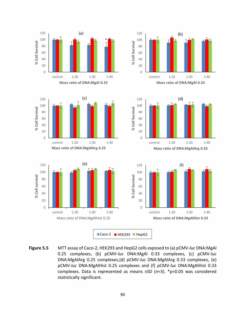
90
Figure 5.5 MTT assay of Caco-2, HEK293 and HepG2 cells exposed to (a) pCMV-luc DNA:MgAl 0.25 complexes, (b) pCMV-luc DNA:MgAl 0.33 complexes, (c) pCMV-luc DNA:MgAlArg 0.25 complexes,(d) pCMV-luc DNA:MgAlArg 0.33 complexes, (e) pCMV-luc DNA:MgAlHist 0.25 complexes and (f) pCMV-luc DNA:MgAlHist 0.33 complexes. Data is represented as means ±SD (n=3). *p<0.05 was considered statistically significant.
0
20
40
60
80
100
120
control 1:20 1:30 1:40
% C
ell S
urvi
val
Mass ratio of DNA:MgAl 0.25
*
0
20
40
60
80
100
120
control 1:20 1:30 1:40
% C
ell S
urvi
val
Mass ratio of DNA:MgAl 0.33
(b)
0
20
40
60
80
100
120
control 1:20 1:30 1:40
% C
ell S
urvi
val
Mass ratio of DNA:MgAlArg 0.25
(c)
0
20
40
60
80
100
120
control 1:20 1:30 1:40
% C
ell S
urvi
val
Mass ratio of DNA:MgAlArg 0.33
0
20
40
60
80
100
120
control 1:20 1:30 1:40
% C
ell s
urvi
val
Mass ratio of DNA:MgAlHist 0.25
0
20
40
60
80
100
120
control 1:20 1:30 1:40
% C
ell S
urvi
val
Mass ratio of DNA:MgAlHist 0.33
(a)
(d)
(e) (f)
Caco-2 HEK293 HepG2
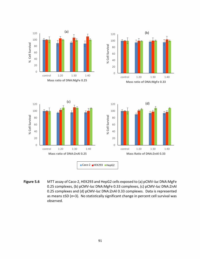
91
Figure 5.6 MTT assay of Caco-2, HEK293 and HepG2 cells exposed to (a) pCMV-luc DNA:MgFe 0.25 complexes, (b) pCMV-luc DNA:MgFe 0.33 complexes, (c) pCMV-luc DNA:ZnAl 0.25 complexes and (d) pCMV-luc DNA:ZnAl 0.33 complexes. Data is represented as means ±SD (n=3). No statistically significant change in percent cell survival was observed.
0
20
40
60
80
100
120
control 1:20 1:30 1:40
% C
ell S
urvi
val
Mass ratio of DNA:MgFe 0.25
0
20
40
60
80
100
120
control 1:20 1:30 1:40
% C
ell S
urvi
val
Mass ratio of DNA:MgFe 0.33
0
20
40
60
80
100
120
control 1:20 1:30 1:40
% C
ell S
urvi
val
Mass ratio of DNA:ZnAl 0.25
0
20
40
60
80
100
120
control 1:20 1:30 1:40
% C
ell S
urvi
val
Mass Ratio of DNA:ZnAl 0.33
(a) (b)
(c) (d)
Caco-2 HEK293 HepG2
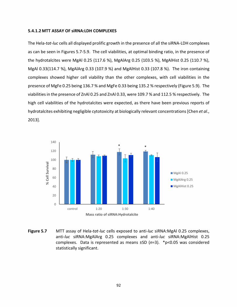
92
5.4.1.2 MTT ASSAY OF siRNA:LDH COMPLEXES
The Hela-tat-luc cells all displayed prolific growth in the presence of all the siRNA-LDH complexes
as can be seen in Figures 5.7-5.9. The cell viabilities, at optimal binding ratio, in the presence of
the hydrotalcites were MgAl 0.25 (117.6 %), MgAlArg 0.25 (103.5 %), MgAlHist 0.25 (110.7 %),
MgAl 0.33(114.7 %), MgAlArg 0.33 (107.9 %) and MgAlHist 0.33 (107.8 %). The iron containing
complexes showed higher cell viability than the other complexes, with cell viabilities in the
presence of MgFe 0.25 being 136.7 % and MgFe 0.33 being 135.2 % respectively (Figure 5.9). The
viabilities in the presence of ZnAl 0.25 and ZnAl 0.33, were 109.7 % and 112.5 % respectively. The
high cell viabilities of the hydrotalcites were expected, as there have been previous reports of
hydrotalcites exhibiting negligible cytotoxicity at biologically relevant concentrations [Chen et al.,
2013].
Figure 5.7 MTT assay of Hela-tat-luc cells exposed to anti-luc siRNA:MgAl 0.25 complexes, anti-luc siRNA:MgAlArg 0.25 complexes and anti-luc siRNA:MgAlHist 0.25 complexes. Data is represented as means ±SD (n=3). *p<0.05 was considered statistically significant.
0
20
40
60
80
100
120
140
control 1:20 1:30 1:40
% C
ell S
urvi
val
Mass ratio of siRNA:Hydrotalcite
MgAl 0.25
MgAlArg 0.25
MgAlHist 0.25
**
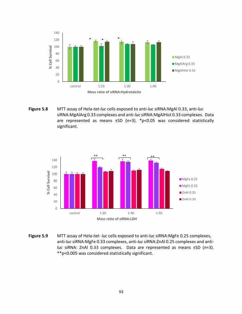
93
Figure 5.8 MTT assay of Hela-tat-luc cells exposed to anti-luc siRNA:MgAl 0.33, anti-luc siRNA:MgAlArg 0.33 complexes and anti-luc siRNA:MgAlHist 0.33 complexes. Data are represented as means ±SD (n=3). *p<0.05 was considered statistically significant.
Figure 5.9 MTT assay of Hela-tat- luc cells exposed to anti-luc siRNA:MgFe 0.25 complexes, anti-luc siRNA:MgFe 0.33 complexes, anti-luc siRNA:ZnAl 0.25 complexes and anti-luc siRNA: ZnAl 0.33 complexes. Data are represented as means ±SD (n=3). **p<0.005 was considered statistically significant.
0
20
40
60
80
100
120
140
control 1:20 1:30 1:40
% C
ell S
urvi
val
Mass ratio of siRNA:Hydrotalcite
MgAl 0.33
MgAlArg 0.33
MgAlHist 0.33
* * *
0
20
40
60
80
100
120
140
control 1:30 1:40 1:50
% C
ell S
urvi
val
Mass ratio of siRNA:LDH
MgFe 0.25
MgFe 0.33
ZnAl 0.25
ZnAl 0.33
**
94
There have been many reports on the low toxicity of LDHs [Yan et al., 2014], with a drug delivery
study showing that LDHs not only decreased the toxicity of the parent drug, but also increased
the desired effect of the drug both in vitro and in vivo [Kura et al., 2014]. The results of this study
confirm the low/almost negligible toxicity of LDHs. Results obtained in this study affirms reports
by Xu and Lu (2006), that LDHs are about 10 times less cytotoxic than commonly used polymer
transfection agents.
5.4.2 LUCIFERASE GENE EXPRESSION STUDIES
The level of transgene expression of the luciferase gene in all cell lines tested indicated successful
transfection of the pCMV-luc DNA.
Results obtained indicate that the degree of transfection is largely cell line dependent. The
HEK293 cell line displayed the highest luciferase gene expression, with the Caco-2 cell line
showing lower but promising gene expression. The HepG2 cell line showed minimal gene
expression, which was approximately 800 fold less than that displayed by the HEK293 and Caco-
2 cell lines. Possible reasons for the difference in expression observed for the three cell lines
investigated in this study, may be due to the different cell types having a preference for different
endocytic pathways [Elouahabi and Ruysschaert, 2005], and also due to differences in cell
membrane properties of the different cell lines [Spector and Yorek, 1985], that affect cellular
uptake.
The level of luciferase activity of the non-functionalized LDHs (Figure 5.10), in the HEK293 was as
follows: MgAl 0.25 < MgFe 0.25 < ZnAl 0.33 < ZnAl 0.25 < MgAl 0.33 < MgFe 0.33. MgFe 0.33
showed 15% greater transfection than MgAl 0.25. In the Caco-2 cell line, luciferase activity was
as follows: MgFe 0.25 < ZnAl 0.25 < MgAl 0.25 < ZnAl 0.33 < MgAl 0.33 < MgFe 0.33. The HepG2
cell line showed a minimal but measurable degree of luciferase activity for all the non-
functionalized LDHs. This was expected as HepG2 cells have a reputation of being a difficult to
transfect cell line [Cemazar et al., 2009]. Although MgAl 0.33 showed best transfection of all the
LDHs tested, with the HepG2 cell line, the level of transfection observed was not as significant,
as their functionalized counterparts.

95
Figure 5.10 Luciferase Assay Results of non-functionalized LDHS in (a) Caco-2, (b) HEK293 and (c) HepG2 cell lines. Data is represented as means ±SD (n=3). **p<0.005, ***p<0.0005 was considered statistically significant.
0
5
10
15
20
25
30
35
cells only cells +DNA 1:20 1:30 1:40
RLU
/mg
Prot
ein
(x10
5 )
Ratio of DNA:LDH
MgAl 0.25
MgAl 0.33
MgFe 0.25
MgFe 0.33
ZnAl 0.25
ZnAl 0.33
0
20
40
60
80
100
120
cells only cells +DNA 1:20 1:30 1:40
RLU
/mg
Prot
ein
(x10
5 )
Ratio of DNA:LDH
MgAl0.25
MgAl 0.33
MgFe 0.25
MgFe 0.33
ZnAl 0.25
ZnAl 0.33
0
5
10
15
20
25
30
35
cells only cells +DNA 1:20 1:30 1:40
RLU
/mg
Prot
ein
(x10
4 )
Ratio of DNA:LDH
MgAl 0.25
MgAl 0.33
MgFe 0.25
MgFe 0.33
ZnAl 0.25
ZnAl 0.33
(a)
(b)
(c)
***
***
**
***
**
***
***
***
***
**
***
***

96
A generally low level of transfection was observed with MgAl 0.25 in all cell lines tested, which
was expected considering the large size of the DNA:MgAl 0.25 complex, probably due to
aggregation caused by the DNA complexing to several LDH nanoparticles. This would have
resulted in poor cellular uptake and internalization and consequently lower levels of transfection.
Overall the non-functionalized LDHs prepared in the M2+:M3+ ratio of 2:1 displayed significantly
higher transfection than those prepared in the 3:1 ratio. The use of the MgAl 0.33 LDH carrier
resulted in the highest level of transfection from all the HTs, in all cell lines tested. Li and co-
workers (2014) have reported that LDHs prepared at M2+:M3+ ratio of 2:1 show better adsorption
of nucleic acids than those prepared in a 3:1 ratio. It seems that this increased adsorption of
nucleic acid corresponded to increased transfection.
The zeta potential and size of a nanoparticle plays a key role in the cellular uptake of the particles
[Nagaraj et al., 2015; Dong et al., 2015]. It also provides a good indication of the colloidal stability
of the nanoparticles for in vivo application. Negative zeta potentials are expected to lead to poor
cellular uptake and consequently poor transfection. Zeta potential is not necessarily an
indication of the actual surface charge of the nanoparticle, but rather it is a measure of the
potential difference between the particle surface and the solvent, and is dependent on other
factors such as the pH of the solvent [Honary and Zahir, 2013]. Desigaux and coworkers (2006)
have previously hypothesized that a negative zeta potential would not impede cellular
internalization. Thus, even though the nanocomplexes displayed a negative zeta potential at pH
7, the zeta potential may be altered at physiological pH resulting in reduced effects on the
transfection efficacy.
Positively charged nanoparticles are usually associated with enhanced cellular uptake (Kim et al
2014). This was exemplified by MgFe 0.33 (the only DNA:LDH complex with a positive zeta
potential), which showed the greatest transfection of all the samples investigated. Rojas and co-
workers (2015), have reported that transfection efficiency decreases as particle size increases
beyond 200 nm, and that smaller particles (< 100 nm) may release their load faster, but may lead
to toxicity, suggesting an ideal size range of 100-200 nm. On the contrary, the cytotoxicity assay
results discussed earlier in this chapter show that although most of the complexes ranged in size
from 20 to 60 nm, they do not exhibit much cytotoxicity.

97
Targeted drug delivery studies by Nagaraj and co-workers (2015) suggest that ZnAl LDHs may be
better nano-carriers than MgAl LDHs. Here again, the current results contradict this, as MgAl
0.33 clearly displays significantly better transfection than the ZnAl 0.33 LDHs as shown in Figure
5.10.
The transfection results of the amino acid functionalized HTs in the Caco-2, HEK293 and HepG2
cell lines are shown in Figures 5.11, 5.12 and 5.13 respectively. In the Caco-2 cell line the
luciferase activity was as follows: MgAlArg 0.25 < MgAlHist 0.25 < MgAlArg 0.33 < MgAl 0.25 <
MgAlHist 0.33 < MgAl 0.33 (Figure 5.11).
Figure 5.11 Luciferase gene expression using (a) MgAl 0.25, MgAlArg 0.25 and MgAlHist 0.25
and (b) MgAl 0.33, MgAlArg 0.33 and MgAlHist 0.33 in Caco-2 cells with 2 sets of controls, one containing only Caco2 cells and the second control containing cells with pCMV-luc DNA. Data is represented as means ±SD (n=3). **p<0.005, ***p<0.0005 was considered statistically significant.
02468
10121416
cells only cells +DNA 1:20 1:30 1:40
RLU
/mg
Prot
ein
(x10
5 )
Ratio of DNA:LDH
MgAl 0.25
MgAlArg 0.25
MgAlHist 0.25
0
5
10
15
20
25
30
cells only cells +DNA 1:20 1:30 1:40RLU
/mg
Prot
ein
(x10
5 )
Ratio of DNA:LDH
MgAl 0.33
MgAlArg 0.33
MgAlHist 0.33
**
***
**
***
(a)
(b)
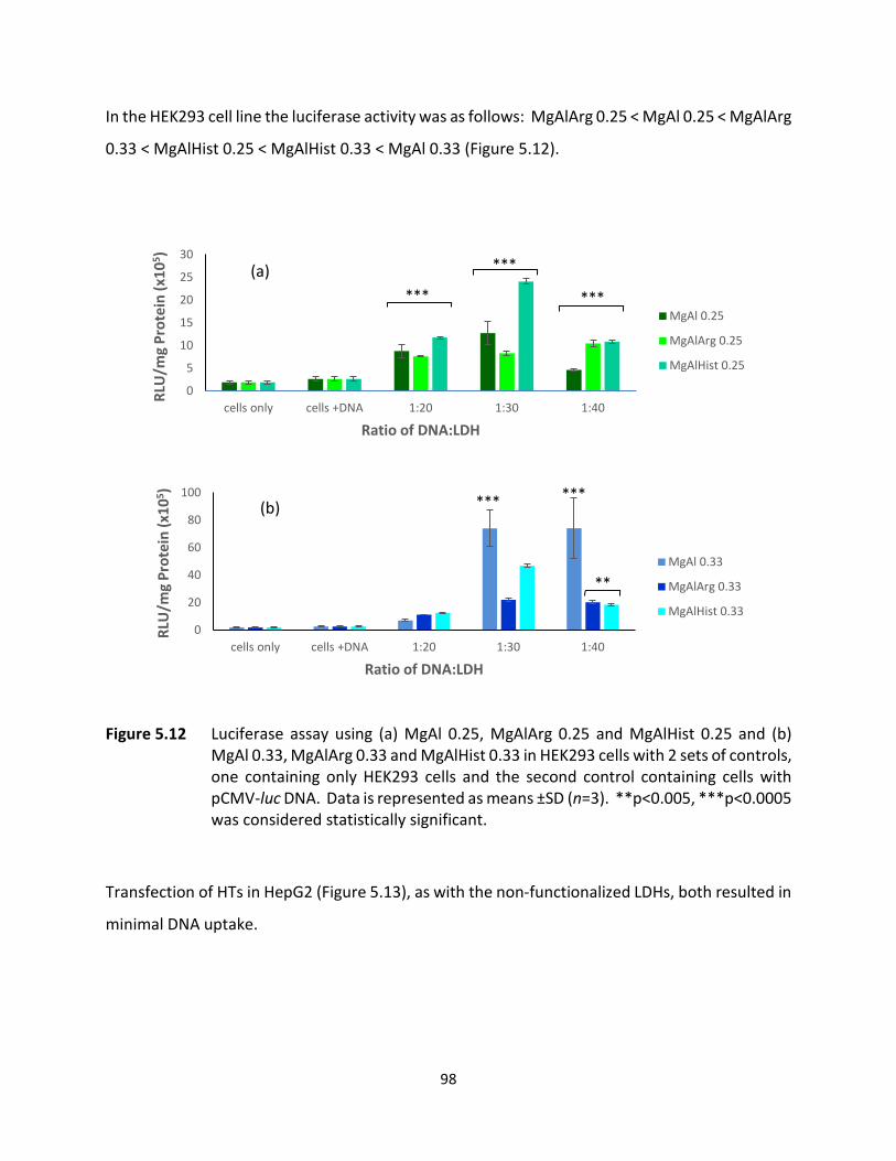
98
In the HEK293 cell line the luciferase activity was as follows: MgAlArg 0.25 < MgAl 0.25 < MgAlArg
0.33 < MgAlHist 0.25 < MgAlHist 0.33 < MgAl 0.33 (Figure 5.12).
Figure 5.12 Luciferase assay using (a) MgAl 0.25, MgAlArg 0.25 and MgAlHist 0.25 and (b) MgAl 0.33, MgAlArg 0.33 and MgAlHist 0.33 in HEK293 cells with 2 sets of controls, one containing only HEK293 cells and the second control containing cells with pCMV-luc DNA. Data is represented as means ±SD (n=3). **p<0.005, ***p<0.0005 was considered statistically significant.
Transfection of HTs in HepG2 (Figure 5.13), as with the non-functionalized LDHs, both resulted in
minimal DNA uptake.
0
5
10
15
20
25
30
cells only cells +DNA 1:20 1:30 1:40
RLU
/mg
Prot
ein
(x10
5 )
Ratio of DNA:LDH
MgAl 0.25
MgAlArg 0.25
MgAlHist 0.25
***
***
***
0
20
40
60
80
100
cells only cells +DNA 1:20 1:30 1:40
RLU
/mg
Prot
ein
(x10
5 )
Ratio of DNA:LDH
MgAl 0.33
MgAlArg 0.33
MgAlHist 0.33
*** ***
**
(a)
(b)

99
Figure 5.13 Luciferase assay using (a) MgAl 0.25, MgAlArg 0.25 and MgAlHist 0.25 and (b) MgAl 0.33, MgAlArg 0.33 and MgAlHist 0.33 in HepG2 cells with 2 sets of controls, one containing only HepG2 cells and the second control containing cells with pCMV-luc DNA. Data is represented as means ±SD (n=3). No significant transfection was observed.
Similar to the non-functionalized LDHs, aa-HTs prepared in the M2+:M3+ ratio of 2:1 showed
higher transfection than those prepared in the 3:1 ratio. Characterization of the aa-HTs indicated
a higher degree of incorporation of the amino acid in MgAlArg 0.33 (2.62%, ζ-potential +6.7) and
MgAlHist 0.33 (1.89%, ζ-potential +22.7) when compared to MgAlArg 0.25 1.27%, ζ-potential -
19.8) and MgAlHist 0.25 (0.90%, ζ-potential -24.6), as indicated by the quantitative assays in
chapter 3, and confirmed by the zeta potentials of the aa-functionalized HTs (chapter 4, section
4.3). The higher degree of incorporation of amino acid in the aa-HTs prepared in the M2+:M3+
ratio of 2:1, appears to have resulted in higher transfection efficiency. Histidine functionalized
HTs displayed better transfection efficiency than arginine functionalized HTs in the Caco-2 and
HepG2 cells lines, as shown in Figures 5.11 and 5.13. There have been reports indicating that the
0
5
10
15
20
25
30
cells only cells +DNA 1:20 1:30 1:40RLU
/mg
Prot
ein
(x10
4 )
Ratio of DNA:LDH
MgAl 0.25
MgAlArg 0.25
MgAlHist 0.25
05
101520253035
cells only cells +DNA 1:20 1:30 1:40
RLU
/mg
Prot
ein
(x10
4 )
Ratio of DNA:LDH
MgAl 0.33
MgAlArg 0.33
MgAlHist 0.33
(a)
(b)

100
incorporation of histidine into gene delivery vehicles does improve cellular uptake, as well as
enhance endosomal escape through the proton sponge effect [Shi et al., 2013; Meng et al., 2017],
as does the incorporation of arginine [Gao et al., 2008; Wang et al., 2015]. The results of this
study suggest that histidine functionalization of the gene delivery vehicle results in better
transfection than arginine functionalization.
5.4.3 LUCIFERASE GENE SILENCING STUDIES
The results of the gene silencing studies are shown in Figures 5.14-5.16. Control 1 was untreated
Hela tat-luc cells, and this was represented as 100%, as luciferase activity can be assumed to be
highest in this control. Control 2 comprised of Hela tat-luc cells treated with naked anti-luc-
siRNA, which produced gene silencing of less than 4%. This can be expected, as naked RNA can
be easily degraded by nucleases before it reaches the cell, as was seen in the nuclease protection
assay (Chapter 4, Section 4.2.4). The much greater percentage of gene silencing observed with
the siRNA:LDH complexes confirms the requirement and significance of a gene delivery vehicle
for efficient delivery and gene silencing. The siRNA:LDH complexes, all had negative zeta
potentials (Chapter 4, Section 4.3) which was probably due to the siRNA binding to the outer
surfaces of the LDHs, but this did not appear to be a hindrance to cellular uptake, internalization
and the subsequent gene silencing.
The HT complexes prepared in a MgAl ratio of 3:1 ie. MgAl 0.25 showed a higher percentage of
gene silencing than the HTs prepared in a MgAl ratio of 2:1 ie. MgAl 0.33. The highest degree of
silencing for the non-functionalized HTs was 44.63%, which was observed for the siRNA:MgAl
0.25 complex prepared at the supra-optimal binding ratio. The amino acid functionalized HTs
showed a higher degree of gene silencing (Figures 5.14 and 5.15), than the non-functionalized
HTs. The arginine functionalized HTs (MgAlArg 0.25 and MgAlArg 0.33) showed almost identical
gene silencing profiles, with the sub-optimal binding ratios of siRNA:HT exhibiting the greatest
degree of silencing (approximately 56.9% gene silencing). The histidine functionalized HTs
(MgAlHist 0.25 and MgAlHist 0.33) showed slightly improved gene silencing on average when

101
compared to the arginine functionalized HTs. This was a similar trend to the gene expression
observed using the pCMV-luc DNA.
Figure 5.14 Luciferase gene silencing using MgAl 0.25, MgAlArg 0.25 and MgAlHist 0.25 in Hela
tat luc cells with 2 sets of controls, one containing only Hela tat-luc cells and the second control containing Hela tat luc cells with anti-luc siRNA. Data is represented as means ±SD (n=3). *p<0.05, **p<0.005 was considered statistically significant.
Figure 5.15 Luciferase gene silencing using MgAl 0.33, MgAlArg 0.33 and MgAlHist 0.33 in Hela
tat luc cells with 2 sets of controls, one containing only Hela tat-luc cells and the second control containing Hela tat luc cells with anti-luc siRNA. Data is represented as means ±SD (n=3). **p<0.005, ***p<0.0005 was considered statistically significant.
0
20
40
60
80
100
120
140
cells only cells +siRNA sub-optimalratio
optimalbinding ratio
super-optimalratio
RLU
/mg
Prot
ein
(x10
6 )
Ratio of DNA:LDH
MgAl 0.25
MgAlArg 0.25
MgAlHist 0.25
**** *
0
20
40
60
80
100
120
140
cells only cells +siRNA sub-optimalratio
optimalbinding ratio
super-optimalratio
RLU
/mg
Prot
ein
(x10
6 )
Ratio of DNA:LDH
MgAl 0.33
MgAlArg 0.33
MgAlHist 0.33
** *****
*****
***
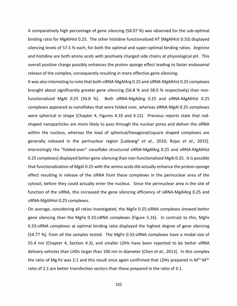
102
A comparatively high percentage of gene silencing (58.07 %) was observed for the sub-optimal
binding ratio for MgAlHist 0.25. The other histidine functionalized HT (MgAlHist 0.33) displayed
silencing levels of 57.5 % each, for both the optimal and super-optimal binding ratios. Arginine
and histidine are both amino acids with positively charged side chains at physiological pH. This
overall positive charge possibly enhances the proton sponge effect leading to faster endosomal
release of the complex, consequently resulting in more effective gene silencing.
It was also interesting to note that both siRNA-MgAlArg 0.25 and siRNA-MgAlHist 0.25 complexes
brought about significantly greater gene silencing (56.8 % and 58.0 % respectively) than non-
functionalized MgAl 0.25 (34.8 %). Both siRNA-MgAlArg 0.25 and siRNA-MgAlHist 0.25
complexes appeared as nanoflakes that were folded over, whereas siRNA-MgAl 0.25 complexes
were spherical in shape (Chapter 4, Figures 4.10 and 4.12). Previous reports state that rod-
shaped nanoparticles are more likely to pass through the nuclear pores and deliver the siRNA
within the nucleus, whereas the load of spherical/hexagonal/square shaped complexes are
generally released in the perinuclear region [Ladewig2 et al., 2010; Rojas et al., 2015].
Interestingly the “folded-over” nanoflake structured siRNA-MgAlArg 0.25 and siRNA-MgAlHist
0.25 complexes) displayed better gene silencing than non-functionalized MgAl 0.25. It is possible
that functionalization of MgAl 0.25 with the amino acids did actually enhance the proton-sponge
effect resulting in release of the siRNA from these complexes in the perinuclear area of the
cytosol, before they could actually enter the nucleus. Since the perinuclear area is the site of
function of the siRNA, this increased the gene silencing efficiency of siRNA-MgAlArg 0.25 and
siRNA-MgAlHist 0.25 complexes.
On average, considering all ratios investigated, the MgFe 0.25:siRNA complexes showed better
gene silencing than the MgFe 0.33:siRNA complexes (Figure 5.16). In contrast to this, MgFe
0.33:siRNA complexes at optimal binding ratio displayed the highest degree of gene silencing
(54.77 %), from all the samples tested. The MgFe 0.33:siRNA complexes have a modal size of
55.4 nm (Chapter 4, Section 4.3), and smaller LDHs have been reported to be better siRNA
delivery vehicles than LHDs larger than 100 nm in diameter [Chen et al., 2013]. In this complex
the ratio of Mg:Fe was 2:1 and this result once again confirmed that LDHs prepared in M2+:M3+
ratio of 2:1 are better transfection vectors than those prepared in the ratio of 3:1.

103
Figure 5.16 Luciferase gene silencing using MgFe 0.25, MgFe 0.33, ZnAl 0.25 and ZnAl 0.33 in
Hela tat luc cells with 2 sets of controls, one containing only Hela tat-luc cells and the second control containing Hela tat luc cells with anti-luc siRNA. Data is represented as means ±SD (n=3). *p<0.05, **p<0.005 was considered statistically significant.
The lowest levels of gene silencing were observed with the complexes of siRNA and ZnAl LDHs
(Figure 5.16). The percentage silencing for ZnAl 0.25 complexes and ZnAl 0.33 complexes was
16.11 and 15.93 % respectively. Posati and co-workers (2012) have previously reported
significant uptake of ZnAl LDH nanoparticles in Hela cells, but the reasons for the low level of
gene silencing observed in this study remains unclear. The Hela-tat-luc cells have displayed
prolific growth in the presence of the siRNA:ZnAl complexes (Section 5.4.1.2), but unfortunately,
this has not significantly affected gene silencing.
All the nanocomplexes prepared for this study showed some degree of gene silencing despite
them all having a negative surface charge which was most probably as a result of the siRNA
binding to the outer surfaces of the LDH. This negative zeta potential has not appeared to hinder
cellular internalization, and may have actually resulted in a faster silencing owing to the quick
release of siRNA from the surface of the LDH, when the LDH nanocomplexes start to dissolve in
the endosome.
0
20
40
60
80
100
120
140
cells only cells +siRNA sub-optimalratio
optimal bindingratio
super-optimalratio
RLU
/mg
Prot
ein
(x10
6 )
Ratio of DNA:LDH
MgFe 0.25
MgFe 0.33
ZnAl 0.25
ZnAl 0.33
***

104
5.5 CONCLUSION
The results of the cytotoxicity assay indicate that all the LDHs prepared are non-toxic to the cell
lines (at the concentrations tested) making them suitable candidates for future in vivo studies.
Gene expression studies revealed that HTs prepared in the M2+:M3+ ratio of 2:1 (w/w) (both
functionalized and non-functionalized) are better transfection agents than those prepared in the
3:1 (w/w) ratio. The results also indicate that functionalizing HTs with positively charged amino
acids does not necessarily improve DNA transfection efficiency, since the non-functionalized
LDHs MgAl 0.33 and MgFe 0.25 displayed the highest levels of transfection of all the samples
investigated.
Results of the gene silencing studies clearly indicate that the complexes prepared from amino
acid functionalized HTs exhibit greater gene silencing than the non-functionalized HTs. The
presence of the positively charged amino acids probably resulted in exploitation of the proton
sponge effect for quicker endosomal escape and subsequently increased levels of gene silencing.
105
CHAPTER SIX
6. CONCLUSION
LDHs have recently garnered much attention in the quest for safer and more effective gene
delivery systems. Recent studies suggest that functionalization of inorganic gene delivery
vehicles leads to enhanced transfection. In this study, the effect of functionalizing HTs with
positively charged amino acids as compared to non-functionalized LDHs, with respect to gene
delivery, was investigated.
LDHs and aa-functionalized HTs were successfully synthesized using the co-precipitation method,
as confirmed by various analytical techniques. Characteristic XRD patterns with sharp,
symmetrical, intense peaks at the lower 2θ values; broader, asymmetric, less intense peaks at
the higher 2θ values, and the distinctive doublet peak at 2θ value of around 60 were observed
for all prepared HTs/LDHs. The nanoparticles, which displayed flat, plate-like morphology, varied
in size between 30 nm and 200 nm, with zeta potentials ranging from -24.7 to 29.7. The degree
of incorporation of amino acid in the aa-functionalized HTs were found to be 0.90 % in MgAlHist
0.25, 1.89 % in MgAlHist 0.33, 1.20 % in MgAlArg 0.25 and 2.62 % in MgAlArg 0.33.
All the synthesized HTs and LDHs bound both DNA and siRNA, producing stable complexes and
concurrently providing adequate protection against serum nuclease digestion. Vigorous
agitation of DNA:HT/LDH complexes at room temperature resulted in complete binding within
10 minutes, without the need for overnight incubation at higher temperatures, as reported in
other studies. The negative zeta potentials of most of the nucleic acid: LDH nanocomplexes
combined with results of the XRD analysis of the DNA:MgAl 0.33 complex suggests that the
nucleic acids bound to the outer surfaces of the LDHs, and does not actually intercalate within
the interlayers.
Results of the MTT assays indicate that the nucleic acid:LDH nanocomplexes were well tolerated
by all the cell lines tested. Minimal cell death was observed in the Caco-2 cell line whilst the
presence of the nucleic acid:LDH nanocomplexes actually had a growth promoting effect on the
HEK 293, HepG2 and Hela-tat-luc cell lines.
106
Transfection levels of the pCMV-luc plasmid as measured by the luciferase reporter gene assay
was found to be cell line dependent. Minimal transfection was observed in the HepG2 cell line,
while transgene expression in the HEK293 and Caco-2 cell lines was up to 800-fold higher than
that measured in the HepG2 cell line. The highest levels of transgene expression were obtained
with the non-functionalized LDHs MgAl 0.33 and MgFe 0.25. It can therefore be concluded that
functionalization of LDHs with positively charged amino acids does not lead to enhanced
transfection of plasmid DNA. This was in stark contrast to results obtained for the gene silencing
studies, which revealed that the employment of aa-functionalized HTs as siRNA vectors lead to
higher levels of gene knockdown when compared to the non-functionalized LDHs. Amino acid
functionalized LDHs appear to be able to bypass one of the major hurdles faced in the formulation
of a successful siRNA vector, which is the release of uncomplexed siRNA from the endocytic
pathway before it reaches the lysosome. This has been achieved in the current study by
exploitation of the proton-sponge effect, which is enhanced by the presence of the positively
charged amino acids, arginine and histidine.
Results from this study suggests that aa-functionalized HTs are more effective siRNA vectors than
pDNA carriers and would therefore be more applicable to gene silencing strategies. Possible
future endeavors include the study of aa-functionalized HTs in in vivo gene silencing strategies
using a suitable mouse model, as well as further modification of the vectors for targeted delivery.

107
REFERENCES
• Abderrazek K, Najoua FS, Srasra E, Synthesis and characterization of [Zn–Al] LDH: Study of the
effect of calcination on the photocatalytic activity, Applied Clay Science 119 (2016) 229-235.
• Ahmed M, Jiang X, Deng Z and Narain R, Cationic Glyco-Functionalized Single-walled carbon
nanotubes as efficient gene delivery vehicles, Bioconjugate Chemistry 20 (2009) 2017-2022.
• Ahmed1 M, Deng Z, Liu S, Lafrenie R, Kumar A and Narain R, Cationic Glyconanoparticles:
Their complexation with DNA, Cellular uptake and transfection efficiencies, Bioconjugate
Chemistry 20 (2009) 2169-2176.
• Aisawa S, Takahashi S, Ogasawara W, Umetsu Y, Narita E, Coprecipitation behaviour of amino
acids with Zn-Al layered double hydroxide precipitates, Clay Science 11 (2000) 317-328.
• Aisawa S, Takahashi S, Ogasawara W, Umetsu Y, Narita E, Direct intercalation of amino acids
into layered double hydroxides by coprecipitation, Journal of Solid State Chemistry 162 (2001)
52-62.
• Aisawa S, Sasaki S, Takahashi S, Hirahara H, Nakayama H, Narita E, Intercalation of amino
acids and oligopeptide into Zn-Al layered double hydroxide by coprecipitation reaction,
Journal of Physics and Chemistry of solids 67 (2006) 920-925.
• Alexandrica C, Silion M, Hritcu D, Popa MI, Layered double hydroxides as adsorbents for
anionic dye removal from aqueous solutions, Environmental Engineering and Management
Journal 14 (2) (2015) 381-388.
• Anbarasan R, Lee WD, Im SS, Adsorption and intercalation of anionic surfactants onto layered
double hydroxides - XRD study. Bulletin of Materials Science 28(2) (2005) 145–149.
• Auerbach SM, Carrado KA and Dutta PK, Handbook of Layered Materials, Marcel Dekker Inc.,
(2004) New York.
• Balcomb B, Singh M, Singh S, Synthesis and characterization of layered double hydroxides and
their potential as nonviral gene delivery vehicles, Chemistry Open 4 (2015) 137-145.
• Barichello JM, Kizuki S, Tagami T, Soares LAL, Ishida T, Kikuchi H, Kiwada H, Agitation during
lipoplex formation harmonizes the interaction of siRNA to cationic liposomes, International
Journal of Pharmaceutics 430 (2012) 359-365.

108
• Barth A, The infrared absorption of amino acid side chains, Progress in Biophysics & Molecular
Biology 74 (2000) 141–173.
• Beavers K (1999) A crystal chemical study on Layered Double Hydroxides (LDHs): An approach
to develop an anion exchanger, Ph.D. Thesis, (1999) University of Hanover, Germany.
• Bejoy N, Hydrotalcite: The clay that cures, Resonance (2001) 57-61.
• Bellotto M, Rebours B, Clause O and Lynch J, A re-examination of hydrotalcite crystal
chemistry, Journal of Physical Chemistry 100 (1996) 8527-8534.
• Benício LPF, Silva RA, Lopes JA, Eulálio D, dos Santos RMM, de Aquino LA, Vergütz L, Novais
RF, da Costa LM, Pinto FG, Tronto J, Layered double hydroxides: Nanomaterials for
applications in agriculture, Revista Brasileira de Ciêcia do Solo 39 (1) (2015) 1-13.
• Benito P, Herrero M, Labajos FM, Rives V, Effect of post-synthesis microwave hydrothermal
treatment on the properties of layered double hydroxides and related materials, Applied Clay
Science 48 (2010) 218-227.
• Berg JM, Tymoczko JL, Stryer L, Biochemistry, Sixth Edition, WH Freeman and Company, New
York (2006) pp 787.
• Bi X, Zhang H, Dou L, Layered double hydroxide based nanocarriers for drug delivery,
Pharmaceutics 6 (2014) 298-332.
• Bish DL, Anion exchange in Takovite: Application to other hydroxide minerals, Bulletin de
Mineralogie 103 (1980) 170-175.
• Bookin AS, Drits VA, Polytype diversity of the hydrotalcite-like minerals I. Possible polytypes
and their diffraction features, Clays and Clay Minerals 41 (5) (1993) 551-557.
• Bontchev RP, Liu S, Krumhansl JL, Voigt J, Nenoff TM, Synthesis, characterization, and ion
exchange properties of hydrotalcite Mg6Al2(OH)16(A)x(A’)2-x 4H2O (A, A’ = Cl-, Br-, I-, and NO3-,
2 ≥ x ≥ 0) derivatives, Chemistry of Materials 15(19) (2003) 3669-3675.
• Boulet P, Greenwell, HC, Stackhouse S, Coveney PV, Recent advances in understanding the
structure and reactivity of clays using electronic structure calculations, Journal of Molecular
Structure THEOCHEM 762 (2006) 33-48.
• Bravo-Suarez, Review of the Synthesis of Layered Double Hydroxides: A Thermodynamic
Approach, Quim. Nova, 27 (4) (2004) 601-614.
109
• Brindley WG and Kikkawa S, Thermal behavior of hydrotalcite and of anion-exchanged forms
of hydrotalcite, Clays and Clay Minerals 28 (2) (1980) 87-91.
• Brindley WG and Kikkawa S, A crystal chemical study of Mg,Al and Ni,Al hydroxyl-perchlorates
and hydroxyl-carbonates, American Mineralogist 64 (1979) 836-843.
• Brown G and Gastuche MC, Mixed Magnesium-Aluminium Hydroxides II. Structure and
Structural Chemistry of Synthetic Hydroxycarbonates and Related Mineral Compounds, Clay
Minerals 7 (1967) 193-201.
• Butenko E, Malyshev A, Kapustin A, Influence of Hydrocarbon Radicals on the Structure of
Layered Double Hydroxides, American Journal of Materials Science and Engineering 2(1)
(2014) 1-6.
• Cardoso LP, Celis R, Cornejo J, Valim JB, Layered double hydroxides as supports for the slow
release of acid herbicides, Journal of Agricultural and Food Chemistry 54 (16) (2006) 5968-
5975.
• Carja G, Ciobanu G, Toyota M, The role of the organic solvent in obtaining hydrotalcite-like
anionic clay nanopowders with specific textural and porous properties, Scientific Study and
Research VII (1) (2006) 157-162.
• Carlino S, The intercalation of carboxylic acids into layered double hydroxides: A critical
evaluation and review of different methods, Solid State Ionics 98(1) (1997) 73-84.
• Carmichael, J DeGraff WG, Gazdar AF, Minna JD, Mitchell JB, Evaluation of a tetrazolium-
based semiautomated colorimetric assay. Assessment of chemosensitivity testing, Cancer
Research 47 (1987) 936-946.
• Cavani F, Trifiro F, Vaccari A, Hydrotalcite type Anionic Clays: Preparation, Properties and
Applications, Catalysis Today 11 (1991) 173-301.
• Cemazar M, Hreljac I, Sersa G, Filipic M, Construction of EGFP expressing HepG2 cell line using
electroporation, World Congress on Medical Physics and Biomedical Engineering (2009) 128-
131.
• Chen M, Cooper HM, Zhou JZ, Bartlett PF, Xu ZP, Reduction in the size of layered double
hydroxide nanoparticles enhances the efficiency of siRNA delivery, Journal of Colloid and
Interface Science 390 (2013) 275-281.
110
• Chiba K, Kawakami K, Tohyama K, Simultaneous evaluation of cell viability by neutral red, MTT
and crystal violet staining assays of the same cells, Toxicology in Vitro 12 (1998) 251-258.
• Choi J and Wang NS, Nanoparticles in Biomedical Applications and Their Safety Concerns,
(2011) Reza Fazel-Rezai (Ed.), ISBN: 978-953-307-637-9, InTech, Available from:
http://www.intechopen.com/articles/show/title/nanoparticles-in-biomedical-applications-
and-their-safety-concerns.
• Choi S-J, Oh J-M, Choy J-H, Safety aspect of layered nanoparticles: size dependency in vitro
and in vivo, Journal of Nanoscience and Nanotechnology 8 (2008) 5297-5301.
• Choi G, Yang J-H, Park G-Y, Vinu A, Elzatahry A, Yo CH, Choy J-H, Intercalative ion-exchange
route to amino acid layered double hydroxide nanohybrids and their sorption properties,
European Journal of Inorganic Chemistry (2015) 925-930 DOI:10.1002/ejic.201403115.
• Choy J-H, Kwak S-Y, Jeong Y-J, Park J-S, Inorganic layered double hydroxides as nonviral
vectors, Angewandte Chemie International Edition 39(22) (2000) 4042-4045.
• Choy J-H, Kwak S-Y, Park J-S, Jeong Y-J, Cellular uptake behavior of [γ-32P] labeled ATP-LDH
nanohybrids, Journal of Materials Chemistry 11 (2001) 1671-1674.
• Choy J-H, Jung J-S, Oh J-M, Park M, Jeong J, Kang Y-K, Han O-J, Layered double hydroxide as
an efficient reservoir for folate derivatives, Biomaterials 25 (2004) 3059-3064.
• Choy J-H, Choi S-J, Oh J-M, Park T, Clay minerals and layered double hydroxides for novel
biological applications, Applied Clay Science 36 (2007) 122-132.
• Chung H-E, Park D-H, Choy J-H, Choi S-J, Intracellular trafficking pathway of layered double
hydroxide nanoparticles in human cells: Size-dependent cellular delivery, Applied Clay Science
65-66 (2012) 24-30.
• Cocheci L, Barvinschi P, Pode R, Popovici E, Seftel EM, Structural characterization of some
Mg/Zn-Al type hydrotalcites prepared for chromate sorption from wastewater, Chemical
Bulletin of “Politehnica” University of Timisoara, Romania 55 (69) (2010) 40-45.
• Coelhoe C, Stimpfling T, Leroux F, Verney V, Inorganic-Organic hybrid materials based on
amino acid modified hyrotalcites used as UV-absorber fillers for polybutylene succinate,
European Journal of Inorganic Chemistry (2012) 5252-5258.

111
• Costa FR, Leuteritz A, Wagenknecht U, Jehnichen D, Häußler L, Heinrich G, Intercalation of
Mg–Al layered double hydroxide by anionic surfactants: Preparation and characterization,
Applied Clay Science 38 (2008) 153-164.
• Costantini S, Di Bernado G, Cammorata M, Castello G, Colonna G, Gene expression signature
of human HepG2 cell line, Gene 518 (2) (2013) 335-345.
• Costantino U, Ambrogi V, Nocchetti M, Perioli L, Hydrotalcite-like compounds: Versatile
layered hosts of molecular anions with biological activity, Microporous and Mesoporous
Materials 107 (2008) 149-160.
• Costantino U, Marmottini F, Nocchetti M, Vivani R, New synthetic routes to hydrotalcite-like
compounds – characterization and properties of the obtained materials, European Journal of
Inorganic Chemistry 10 (1998) 1439-1446.
• Crosby S, Tran D, Cocke D, Duraia EM, Beall GW, Effect of isomorphous substitution on the
thermal decomposition mechanism of hydrotalcites, Materials 7 (2014) 7048-7058.
• Cullity BD, Stock SR, Elements of X-Ray Diffraction, 3rd edition, Prentice Hall: Upper Saddle
River, NJ, (2001).
• Cunha VRR, Ferreira AMC, Constantino VRL, Tronto J, Valim JB, Layered double hydroxides:
inorganic nanoparticles for storage and release of species of biological and therapeutic
interest, Quimica Nova 33 (1) (2010) 159-171.
• Daniels A, Singh M, Ariatti M, Pegylated and non-pegylated siRNA lipoplexes formulated with
cholesteryl cytofectins promote efficient luciferase knockdown in Hela tat luc cells,
Nucleosides Nucleotides Nucleic Acids 32 (4) (2013) 206-220.
• de Roy A, Forano C, Besse JP, Layered double hydroxides: Synthesis and post-synthesis
Modification, In: Layered Double Hydroxides: Present and Future, Edited by Vincente Rives
(2006) 1-39.
• Del Arco M, Fernández A, Martín C, Rives V, Release studies of different NSAIDs encapsulated
in Mg,Al,Fe-hydrotalcites, Applied Clay Science 42 (2009) 538-544.
• Del Arco1 M, Gutiérrez S, Martín C, Rives V, Rocha J, Synthesis and characterization of layered
double hydroxides (LDH) intercalated with non-steroidal anti-inflammatory drugs (NSAID),
Journal of Solid State Chemistry 177 (2004) 3954-3962.
112
• Del Arco2 M, Cebadera E, Gutiérrez S, Martín C, Montero MJ, Rives V, Rocha J and Sevilla MA,
Mg, Al layered double hydroxides with interalated indomethacin: Synthesis, characterization,
and pharmacological study, Journal of Pharmaceutical Sciences 93 (6) (2004) 1649-1658.
• Del Hoyo C, Layered double hydroxides and human health: an overview, Applied Clay Science
36 (2007) 103-121.
• Denizot F, Lang R, Rapid colorimetric assay for cell growth and survival: Modifications to the
tetrazolium dye procedure giving improved sensitivity and reliability, Journal of
Immunological Methods 89 (1986) 271-277.
• Desigaux L, Belkacem BM, Richard P, Cellier J, Leone P, Cario L, Leroux F, Taviot-Guého C,
Pitard B, Self-assembly and characterization of layered double hydroxide/LDH hybrids, Nano
Letters 6 (2) (2006) 199-204.
• de Wet JR, Wood KV, DeLuca M, Helinski DR, Subramani S, Firefly luciferase gene: Structure
and expression in mammalian cells, Molecular and Cellular Biology 7 (2) (1987) 725-737.
• Dinnebier RE, Billenge SJL, Powder Diffraction, Theory and Practice, RSC Publishing,
Cambridge, UK (2008).
• Dong H, Chen M, Rahman S, Parekh HS, Cooper HM, Xu ZP, Engineering small MgAl-layered
double hydroxide nanoparticles for enhanced gene delivery, Applied Clay Science 100 (2014)
66-75.
• Dong H, Parekh HS, Xu ZP, Particle size and number-dependent delivery to cells by layered
double hydroxide nanoparticles, Journal of Colloid and Interface Science 431 (2015) 10-16.
• Dong L, Gou G, Huang J, Sun Y, Xue B, A review on gene delivery system of layered double
hydroxide, Applied Mechanics and Materials 320 (2013) 533-539.
• Elsaesser A, Howard CV, Toxicology of nanoparticles, Advanced drug delivery reviews 64
(2012) 129-137.
• Elouahabi A, Thiry M, Pector V, Fuks R, Ruysschaert JM, Vandenbranden M, The role of
endosome destabilizing activity in the gene transfer process mediated by cationic lipids, FEBS
letters 414 (2) (1997) 187-192.
• Elouahabi A, Ruysschaert JM, Formation and intracellular trafficking of lipoplexes and
polyplexes, Molecular Therapy 11 (3) (2005) 336-347.
113
• Ennadi A, Legrouri A, De Roy A, Besse JP, X-Ray diffraction pattern simulation for thermally
treated [An-Al-Cl] layered double hydroxide, Journal of Solid State Chemistry 152 (2000) 568-
572.
• Evans DG and Duan X, Preparation of layered double hydroxides and their applications as
additives in polymers, as precursors to magnetic materials and in biology and medicine,
Chemical Communications 5 (2006) 485-496.
• Evans DG and Slade RCT, Structural Aspects of Layered Double Hydroxides, Structure and
Bonding 119 (2006) 1-87.
• Fan G, Li F, Evans DG, Duan X, Catalytic applications of layered double hydroxides: recent
advances and perspectives, Chemical Society reviews 43 (2014) 7040-7066.
• Feng J, He Y, Liu Y, Du Y, Li D, Supported catalysts based on layered double hydroxides for
catalytic oxidation and hydrogenation: general functionality and promising application
prospects, Chemical Society Reviews 44 (2015) 5291-5319.
• Fong N, Guagliardo P, Williams J, Musemeci A, Martin D, Smith SV, Clay particles – potential
of positron annihilation lifetime spectroscopy (PALS) for studying interlayer spacing, Journal
of Physics: Conference Series 262 (2011) 012022.
• Forano C, Hibino T, Leroux F, Taviot-Gucho C, Layered Double Hydroxides, In: Handbook of
Clay Science, Edited by F. Bergaya, BKG Thong and G Lagaly (2006) 1021-1096.
• Fortner JD, Solenthaler C, Hughes JB, Puzrin AM, Plötze M, Interactions of clay minerals and
a double layered hydroxide with water stable, nano scale fullerene aggregates (nC60), Applied
Clay Science 55 (2012) 36-43.
• Fotakis G, Timbrell JA, In vitro cytotoxicity assays: Comparison of LDH, neutral red, MTT and
protein assay in hepatoma cell lines following exposure to cadmium chloride, Toxicology
Letters 160 (2006) 171-177.
• Fried M and Crothers DM, Equilibria and kinetics of repressor-operator interactions by
polyacrylamide gel electrophoresis, Nucleic Acids Research 9 (23) (1981) 6505-6525.
• Frost RL, Weier ML, and Kloprogge JT, Raman spectroscopy of some natural hydrotalcites with
sulphate and carbonate in the interlayer, Journal of Raman Spectroscopy 34 (10) (2003) 760-
768.
114
• Frost1 RL, Weier ML, Clissold ME, Williams PA, Infrared spectroscopic study of natural
hydrotalcites carrboydite and hydrohonessite, Spectrochimica Acta Part A 59 (2003) 3313-
3319.
• Gao L, Nie L, Wang T, Qin Y, Guo Z, Yang D, Yan X, Carbon nanotube delivery of the GFP gene
into mammalian cells, ChemBiochem 7 (2006) 239-242.
• Gao Y, Xu Z, Chen S, Gu W, Chen L, Li Y, Arginine-chitosan/DNA self-assemble nanoparticles
for gene delivery: In vitro characteristics and transfection efficiency, International Journal of
Pharmaceutics 359 (1-2) (2008) 241-246.
• Garner MM and Revzin A, A gel electrophoresis method for quantifying the binding of proteins
to specific DNA regions: application to components of the Escherichia coli lactose operon
regulatory system, Nucleic Acids Research 9 (13) (1981) 3047-3059.
• Gasuche MC, Brown G, Moerland MM, Mixed Magnesium-Aluminium Hydroxides I.
Preparation and Characterization of Compunds formed in Dialysed Systems-, Clay Minerals 7
(1967) 177-192.
• Gu Z, Thomas AC, Xu ZP, Campbell JH, Lu GQ, In vitro sustained release of LMWH from MgAl-
layered double hydroxide nanohybrids, Chemistry of Materials 20 (11) (2008) 3715-3722.
• Guo X, Li S, Hou W, Han S, Hu J, Li D, Layered double hydroxides with hydrotalcite type
structure containing Fe3+, Al3+ and Mg2+, Chemical Research in Chinese Universities 19 (2)
(2003) 211-215.
• Ha JU and Xanthos M, Drug release characteristics from nanoclay hybrids and their dispersions
in organic polymers, International Journal of Pharmaceutics 414 (2011) 321-331.
• Habib S, Singh M, Ariatti M, Glycosylated liposomes with proton sponge capacity: Novel
hepatocyte specific gene carriers, Current Drug Delivery 10 (2013) 685-695
• He J, Wei M, Li B, Kang Y, Evans DG, Duan X, Preparation of Layered Double Hydroxides,
Structure and Bonding 119 (2005) 89-119.
• Herrero M, Labajos FM, Rives V, Size control and optimization of intercalated layered double
hydroxides, Applied Clay Science 42 (2009) 510-518.
• Hernandez-Moreno MJ, Ulibarri MA, Rendon JL, Serna CJ, IR Characteristics of hydrotalcite-
like compounds, Physics and Chemistry of Minerals 12 (1985) 34-38.

115
• Hibino T, Delamination of layered double hydroxides containing amino acids, Chemistry of
Materials 16 (2004) 5482-5488.
• Hickerson RP, Vlassov AV, Wang Q, Leak D, Ilves H, Gonzalez-Gonzalez E, Contag CH, Johnston
BH, Kaspar RL, Stability of unmodified siRNA and relevance to clinical use, Oligonucleotides
18 (4) (2008) 345-354.
• Holden NS and Tacon CE, Principles and problems of the electrophoretic mobility shift assay,
Journal of Pharmacological and Toxicological Methods 63 (2011) 7-14.
• Honary S, Zahir F, Effect of zeta potential on the properties of nano-drug delivery systems – A
Review (Part 2), Tropical Journal of Pharmaceutical Research 12 (2) (2013) 265-273.
• Hou X, Bish D, Wang S, Johnstone T, Kirkpatrick R, Hydration, expansion, structure and
dynamics of layered double hydroxides, American Mineralogist 88 (2003) 167-179.
• Hu H, Xiu KM, Xu SL, Yang WT, Xu FJ, Functionalized layered double hydroxide nanoparticles
conjugated with disulfide-linked polycation brushes for advanced gene delivery, Bioconjugate
Chemistry 24 (6) (2013) 968-978.
• Huang L, Hung M-C, Wagner E (Editors), Nonviral vectors for gene therapy, Academic Press.
San Diego, California, USA (1999).
• Ikeda T, Amoh H, Yasunaga T, Stereoselective exchange kinetics of L- and D-Histidines for Cl-
in the interlayer of a hydrotalcite-like compound by chemical relaxation method, Journal of
the American Chemical Society 106 (1984) 5772-5775.
• Imani R, Emami SH, Faghihi S, Synthesis and characterization of an octaarginine functionalized
graphed oxide nanocarrier for gene delivery applications, Physical Chemistry Chemical Physics
17 (2015) 6328-6339.
• Ingram L and Taylor HFW, The Crystal Structures of Sjögrenite and Pyroaurite, Mineralogical
Magazine 36 (280) (1967) 465-479.
• Intissar M, Jumas J-C, Besse J-P, Leroux F, Reinvestigation of the layered double hydroxide
containing tetravalent cations: Unambiguous response provided by XAS and Mössbauer
spectoscopies, Chemistry of Materials 15 (24) (2003) 4625-4632.
• Jabłońska M, Chmielarz L, Węgrzyn A, Hydrotalcite-like materials containing manganese: A
short review, Copernican Letters 4 (2013) 59-65.
116
• Jaiswal A, Gautam RK, Chattopadhyaya MC, Layered double hydroxides and the environment:
An overview, In: Advanced Materials for Agriculture, Food and Environmental Safety (Eds. A
Tiwari and M Syvӓjӓrvi), (2014) John Wiley and Sons, Inc., Hoboken, NJ, USA.
• Jellicoe TC, Fogg AM, Synthesis and characterization of layered double hydroxides intercalated
with sugar phosphates, Journal of Physics and Chemistry of Solids 73 (12) (2012) 1496-1499.
• Jiao FP, Chen XQ, Fu ZD, Hu YH, Wang YH, Synthesis and structural characterization of L-(-)-
malic acid pillared layered double hydroxides, Latin American Applied Research 39 (2009) 127-
130.
• Kagunya W, Baddour-Hadjean R, Kooli F, Jones W, Vibrational Modes in layered double
hydroxides and their calcined derivatives, Chemical Physics 236 (1998) 225-234.
• Kameda T, Oba J, Yoshioka T, Removal of boron and fluoride in wastewater using Mg-Al
layered double hydroxide and Mg-Al oxide, Journal of Environmental Management 188 (2017)
58-63.
• Kandare E and Hossenlopp JM, Thermal degradation of acetate-intercalated hydroxyl double
and layered hydroxyl salts, Inorganic Chemistry 45(9) (2006) 3766-3773.
• Kelkar CP and Schutz AA, Ni-, Mg- and Co-containing hydrotalcite-like materials with a sheet-
like morphology: synthesis and characterization, Microporous Materials 10 (1997) 163-172.
• Khan AI and O’Hare D, Intercalation chemistry of layered double hydroxides: recent
developments and applications, Journal of Materials Chemistry 12 (2002) 3191-3198.
• Klemkaite K, Prosycevas I, Taraskevicius R, Khinsky A, Kareiva A, Synthesis and
Characterization of layered double hydroxides with different cations [Mg, Co, Ni, Al],
decomposition and reformation of mixed metal oxides to layered structures, Central European
Journal of Chemistry 9(2) (2011) 275-282.
• Kloprogge JT and Frost RL, Fourier Transform Infrared and Raman Spectroscopic Study of the
Local Structure of Mg-, Ni- and Co-Hydrotalcites, Journal of Solid State Chemistry 146 (1999)
506-515.
• Kloprogge JT, Hickey L, Frost RL, Heating stage Raman and Infrared emission spectroscopic
study of the dehydroxylation of synthetic Mg-hydrotalcite, Applied Clay Science 18 (2001) 37-
49.
117
• Kloprogge JT, Wharton D, Hickey L, Frost RL, Infrared and Raman study of the interlayer anions
CO32-, NO3-, SO42- and ClO42- in Mg/Al-hydrotalcite, American Mineralogist 87 (2002) 623-629.
• Kloprogge JT, Hickey L, Frost RL, The effects of Synthesis pH and Hydrothermal treatment on
the Formation of Zinc Aluminum Hydrotalcites, Journal of Solid State Chemistry 177 (2004)
4047-4057.
• Komarala EP, Doshi S, Mohammed A, Bahadur D, Efficient antibacterial activity via protein
degradation of a 3D layered double hydroxide-reduced graphene oxide nanohybrid, Royal
Society of Chemistry Advances 6 (2016) 40389-40398.
• Kooli F, Kosuge K, Tsunashima A, Mg-Zn-Al-CO3 and Zn-Cu-Al-CO3 hydrotalcite-like
compounds: preparation and characterization, Journal of Materials Science 30 (1995) 4591-
4597.
• Kooli F, Depége C, Ennaqadi A, de Roy A, Besse JP, Rehydration of Zn-Al layered double
hydroxides, Clays and Clay Minerals 45 (1) (1997) 92-98.
• Kovanda F, Grygar T, Dorničák V, Rojka T, Bezdička P, Jirátová K, Thermal behavior of Cu-Mg-
Mn and Ni-Mg-Mn layered double hydroxides and characterization of formed oxides, Applied
Clay Science 28(2005) 121-136.
• Kovanda F, Jindová E, Doušová B, Koloušek D, Pleštil J, Sedláková Z, Layered double
hydroxides intercalated with organic anions and their application in preparation of
LDH/polymer nanocomposites, Acta Geodynamica et Geomateriali 6 (153) (2009) 111-119.
• Kralj M and Pavelic K, Medicine on a small scale, EMBO reports 4 (11) (2003) 1008-1012.
• Kriven WM, Kwak S-Y, Wallig MA, Choy J-H, Bio-resorbable nanoceramics for gene and drug
delivery, Materials Research Society Bulletin 29 (1) (2004) 33-37.
• Kuang Y, Zhao L, Zhang S, Zhang F, Dong M, Xu S, Morphologies, preparations and applications
of double layered hydroxide Micro-/Nanastructures, Materials 3 (2010) 5220-5235.
• Kulyukhin SA, Krasavina EP, Rumer IA, Klimovich IV, Sorption of Strontium and Yttrium
radionuclides from aqueous solutions onto layered double hydroxides of various compositions,
Radiochemistry 56 (6) (2014) 593-606.
• Kumar PP, Kalinichev AG, Kirkpatrick RJ, Hydration, swelling, interlayer structure, and
hydrogen bonding in organolayered double hydroxides: Insights from molecular dynamics
118
simulation of citrate-intercalated hydrotalcite, Journal of Physical Chemistry B 110 (9) (2006)
3841-3844.
• Kumar S and Rai SB, Spectroscopic studies of L-arginine molecule, Indian Journal of Pure and
Applied Physics 48 (2010) 251-255.
• Kumar S, Rai AK, Rai SB, Rai DK, Infrared and Raman spectra of Histidine: an ab initio DFT
calculations of histidine molecule and its different protonated forms, Indian Journal of Physics
84 (5) (2010) 563-573.
• Kuo Y, Kuthati Y, Kankala RK, Wei P, Wang C, Liu C, Sung P, Mou C, Lee C, Layered double
hydroxide nanoparticles to enhance organ-specific targeting and anti-proliferative effect of
cisplatin, Journal of Materials Chemistry B 3 (2015) 3447-3458.
• Kura AU, Hussein MZ, Fakurazi S, Arulselvan P, Layered double hydroxide nanocomposite for
drug delivery systems; bio-distribution, toxicity and drug activity enhancement, Chemistry
Central Journal 8 (47) (2014) 1-8.
• Kwak S-Y, Jeong Y-J, Park J-S, Choy J-H, Bio-LDH nanohybrid for gene therapy, Solid State Ionics
151 (2002) 229-234.
• Lacerda L, Raffa S, Prato M, Bianco A, Kostarelos K, Cell penetrating CNTs for delivery of
therapeutics, Nanotoday 2 (6) (2007) 38-43.
• Ladewig K, Xu ZP, Lu GQ, Layered double hydroxide nanoparticles in gene and drug delivery,
Expert Opinion Drug Delivery 6 (9) (2009) 907-922.
• Ladewig1 K, Niebert M, Xu ZP, Gray PP, Lu GQ, Controlled preparation of layered double
hydroxide nanoparticles and their application as gene delivery vehicles, Applied Clay Science
48 (2010) 280-289.
• Ladewig2 K, Niebert M, Xu ZP, Gray PP, Lu GQM, Efficient siRNA delivery to mammalian cells
using layered double hydroxide nanoparticles, Biomaterials 31 (2010) 1821-1829.
• Lee D-J, Kessel E, Lehto T, Liu X, Yoshinaga N, Padari K, Chen Y-C, Kempter S, Uchida S, Radler
JO, Pooga M, Sheu M-T, Kataoka K, Wagner E, Systemic delivery of folate-PEG siRNA lipoplexes
with enhanced intracellular stability for in vivo gene silencing in leukemia, Bioconjugate
Chemistry 28(9) (2017) 2393-2409, DOI: 10.1021/acs.bioconjchem.7b00383.

119
• Li1 B, Wu P, Ruan B, Liu P, Zhu N, Study on the adsorption of DNA on the layered double
hydroxides, Spectrochimica Acta Part 1: Molecular and Biomolecular Spectroscopy, 121(2014)
387-393.
• Li C, Wei M, Evans DG, Duan X, Recent advances for layered double hydroxides (LDHs)
materials as catalysts applied in green aqueous media, Catalysis Today 247 (2015) 163-169.
• Li L, Gu W, Chen W, Xu ZP, Co-delivery of siRNAs and anti-cancer drugs using layered double
hydroxide nanoparticles, Biomaterials 35 (2014) 3331-3339.
• Li S, Li J, Wang CJ, Wang Q, Cader MZ, Lu J, Evans DG, Duan X, ‘O Hare D, Cellular uptake and
gene delivery using layered double hydroxide nanoparticles, Journal of Materials Chemistry B
1 (2013) 61-68.
• Liang W and Lam JKW, Endosomal Escape Pathways for Non-Viral Nucleic Acid Delivery
Systems, Molecular Regulation of Endocytosis, Dr. Brian Ceresa (Ed.) (2012), InTech, DOI:
10.5772/46006. Available from: http://www.intechopen.com/books/molecular-regulation-
of-endocytosis/endosomal-escape-pathways-for-non-viral-nucleic-acid-delivery-systems.
• Liu J, Harrison R, Zhou JZ, Liu TT, Yu C, Lu GQ, Qiao SZ, Xu ZP, Synthesis of nanorattles with
layered double hydroxide core and mesoporous silica shell as delivery vehicles, Journal of
Materials Chemistry 21 (2011) 10641-10644.
• Liu H-C, Yang X-Y, Ran G-P, Min E-Z, Liu J-K, Ying P-L, Xin Q, Li C, Structure and base properties
of calcined hydrotalcites, Chinese Journal of Chemistry 17 (4) (1999) 319-330.
• Lu M, Shan Z, Andrea K, Macdonald B, Beale S, Curry DE, Wang L, Wang S, Oakes KD, Bennett
C, Wu W, Zhang X, Chemisorption mechanism of DNA on Mg/Fe layered double hydroxides
nanoparticles: Insights into engineering effective siRNA delivery systems, Langmuir 32 (11)
(2016) 2659-2667.
• Luan L, Liu S, Sun D, Dispersion control and nematic ordering of Ni/Al layered double hydroxide
suspensions, Journal of solid State Chemistry 182 (2009) 1462-1467.
• Lv H, Zhang S, Wang B, Cui S, Yan J, Toxicity of cationic lipids and cationic polymers in gene
delivery, Journal of Controlled Release 114 (2006) 100-109.

120
• Lv W, Du M, Ye W, Zheng Q, The formation mechanism of layered double hydroxide
nanoscrolls by facile trinal-phase hydrothermal treatment and their adsorption properties,
Journal of Materials Chemistry A 3 (2015) 23395-23402.
• Malherbe F, Besse J-P, Investigating the effects of guest-host interactions on the properties of
anion-exchanged Mg-Al hydrotalcites, Journal of Solid State Chemistry 155 (2) (2000) 332-
341.
• Mallakpour S and Dinari M, Using Mg-Al-Layered double hydroxide intercalated with chiral
dicarboxylic acid for the reinforcement of isoleucine amino acid containing poly(amide-imide),
Polymer Composites 37 (11) (2016) 3288-3295.
• Matranga C, Tomari Y, Shin C, Bartel DP, Zamore PD, Passenger-Strand Cleavage Facilitates
Assembly of siRNA into Ago2-Containing RNAi Enzyme Complexes, Cell 123 (4) (2005) 607-
620.
• Matulis D, Rouzina I, Bloomfield VA, Thermodynamics of DNA binding and condensation:
isothermal titration calorimetry and electrostatic mechanism, Journal of Molecular Biology
296 (2000) 1053-1063.
• Medina-Juárez O, García-Sánchez MA, Arellano-Sánchez U, Kornhauser-Straus I and Rojas-
González F, Optimal surface amino-functionalization following thermo-alkaline treatment of
nanostructured silica adsorbents for enhanced CO2 adsorption, Materials 9 (11) (2016) 898
doi:10.3390/ma9110898.
• Meng Z, Luan L, Kang Z, Feng S, Meng Q, Liu K, Histidine-enriched multifunctional peptide
vectors with enhanced cellular uptake and endosomal escape for gene delivery, Journal of
Materials Chemistry B 5 (2017) 74-84.
• Meyn M, Beneke K, Lagaly G, Anion exchange reactions of layered double hydroxides,
Inorganic Chemistry 29 (1990) 5201-5207.
• Meyn M, Beneke K, Lagaly G, Anion exchange reactions of hydroxyl double salts, Inorganic
Chemistry 32 (1993) 1209-1215.
• Millange F, Walton RI, O’ Hare D, Time resolved in situ X-Ray diffraction study of the liquid-
phase reconstruction of Mg-Al-carbonate hydrotalcite-like compounds, Journal of Materials
Chemistry 10 (2000) 1713-1720.

121
• Mills SJ, Christy AG, Genin JMR, Kameda T, Colombo F, Nomenclature of the hydrotalcite
supergroup: natural layered double hydroxides, Mineralogical Magazine 76 (2012) 1289-1336.
• Misra C and Perrotta AJ, Composition and properties of synthetic hydrotalcites, Clays and Clay
Minerals 40 (2) (1992) 145-150.
• Miyata S and Kumura T, Synthesis of new hydrotalcite-like compounds and their physio-
chemical properties, Chemistry Letters (1973) 843-848.
• Miyata S, Synthesis of hydrotalcite-like compounds and their structures and physico-chemical
properties - The systems Mg2+ -Al3+ -NO3- and Mg2+ -Al3+ -Cl-, Mg2+ -Al3+ -ClO4-, Ni2+ -Al3+ -Cl- and
Zn2+ -Al3+ -Cl-, Clays and Clay Minerals 23 (1975) 369-375.
• Miyata S and Okada A, Synthesis of hydrotalcite-like compounds and their physico-chemical
properties - The systems Mg2+-Al3+ -SO42- and Mg2+ -Al3+ -CrO42-, Clays and Clay Minerals 25
(1977) 14-18.
• Miyata S, Anion exchange properties of Hydrotalcite-like compounds, Clay and Clay Minerals
31 (4) (1983) 305-311.
• Mohanambe L and Vasudevan S, Inclusion of poly-aromatic hydrocarbon (PAH) molecules in
a functionalized layered double hydroxide, Journal of Chemical Sciences 118 (1) (2006) 105-
115.
• Mondal S, Dasgupta S, Maji K, MgAl- Layered double hydroxide nanoparticles for controlled
release of salicyclate, Materials Science and Engineering C 68 (2016) 557-564.
• Mora M, Jiménez-Sanchidrián C, Ruiz JR, Raman Spectroscopy study of Layered-double
hydroxides containing magnesium and trivalent metals, Materials Letters 120 (2014) 193-195
• Morachis JM, Mahmoud EA, Sankaranarayanan J, Almutairi A, Triggered Rapid Degradation
of Nanoparticles for gene delivery, Journal of Drug Delivery (2012) doi:
10.1155/2012/291219.
• Mosmann T, Rapid colorimetric assay for cellular growth and survivals: application to
proliferation and cytotoxicity assays, Journal of Immunological Methods 65 (1983) 55-63.
• Mout R, Moyano DF, Rana S, Rotello VM, Surface functionalization of nanoparticles for
nanomedicine, Chemical Society Reviews 41 (2012) 2539-2544.
122
• Mulens V, Morales MP, Barber DF, Development of magnetic nanoparticles for cancer gene
therapy: A comprehensive review, ISRN Nanomaterials (2013) 1-14.
• Musumeci AW, Mortimer GM, Butler MK, Xu ZP, Minchin RF, Martin DJ, Fluorescent layered
double hydroxide nanoparticles for biological studies, Applied Clay Science 48 (2010) 271-279.
• Nagaraj VJ, Sun X, Mehta J, Martin M, Ngo T, Dey SK, Synthesis, characterization and in vitro
drug delivery capabilities of (Zn, Al)-based layered double hydroxide nanoparticles, Journal of
Nanotechnology (2015) Article ID 350370, 10 pages.
• Naicker K, Ariatti M, Singh M, PEGylated galactosylated cationic liposomes for hepatocytic
gene delivery, Colloids and Surfaces B: Biointerfaces 122 (2014) 482-490.
• Nakayama H, Wada N, Tsuhako M, Intercalation of amino acids and peptides into MG-Al
layered double hydroxide by re-construction method, International Journal of Pharmaceutics
269 (2004) 469-478.
• Nalawade P, Aware B, Kadam VJ, Hirlekar RS, Layered double hydroxides: A review, Journal of
Scientific and Industrial Research 68 (2009) 267-272.
• Natoli M, Leoni BD, D’Agnano I, Zucco F, Felsani A, Good CACO-2 cell culture practices,
Toxicology in Vitro 26 (2012) 1243-1246.
• Newman SP and Jones W, Synthesis, characterization and applications of layered double
hydroxides containing organic guests, New Journal of Chemistry (1998) 105-115.
• Newman SP and Jones W, Comparative study of some layered hydroxide salts containing
exchangeable interlayer anions, Journal of Solid State Chemistry 148 (1999) 26-40.
• Newman SP, Jones W, O’ Conner P, Stamires DM, Synthesis of the 3R2 polytypeof a
hydrotalcite-like mineral, Journal of Materials Chemistry 12 (2002) 153-155.
• Nimesh S, Kumar R, Chandra R, Novel polyallylamine-dextran sulfate-DNA nanoplexes: Highly
efficient non-viral vector for gene delivery, International Journal of Pharmaceutics 320 (2006)
143-149.
• Nguyen J, Steele TWJ, Merkel O, Reul R, Kissel T, Fast degrading polyesters as siRNA nano-
carriers for pulmonary gene therapy, Journal of Controlled Release 132 (2008) 243-251.
• Oh JM, Hwang SH, Choy JH, The effect of synthetic conditions on tailoring the size of
hydrotalcite particles, Solid State Ionics 151 (2006) 285-291.

123
• Ostolska I, Wiśniewska M, Application of the zeta potential measurements to explanation of
colloidal Cr2O3 stability mechanism in the presence of the ionic polyamino acids, Colloid and
Polymer Science 292(10) (2014) 2453-2464 doi:10.1007/s00396-014-3276-y.
• Palmer SJ, Frost RL, Spratt HJ, Synthesis and Raman spectroscopic study of Mg/Al, Fe
hydrotalcites with variable cationic ratios, Journal of Raman Spectroscopy 40 (9) (2009) 1138-
1143.
• Pan B, Cui D, Xu P, Chen H, Liu F, Li Q, Huang T, You X, Shao J, Bao C, Gao F, He R, Shu M, Ma
Y, Design of dendrimer modified carbon nanotubes for gene delivery, Chinese Journal of
Cancer Research 19(1) (2007) 1-6.
• Pan B, Cui D, Xu P, Ozkan C, Feng G, Ozkan M, Huang T, Che B, Li Q, He R, Hu G, Synthesis and
characterization of polyamidoamine dendrimer-coated multi-walled carbon nanotubes and
their application in gene delivery systems, Nanotechnology 20 (2009) 1-9.
• Park D-H, Jaeyong C, Kwon O-J, Yun C-O, Choy J-H, Biodegradable inorganic nanovector:
passive versus active tumor targeting, Angewandte Chemie International Edition 55 (14)
(2016) 4582-4586.
• Patel VB, Patel KN, Shah MM, Mayank B, Spectrophotometric determination of histidine
hydrochloride monohydrate in pharmaceutical formulations, International Journal of
PharmTech Research 1 (3) (2009) 852-856.
• Perera J, Weerasekera M, Kottegoda N, Slow release anti-fungal skin formulations based on
citric acid intercalated layered double hydroxides nanohybrids, Chemistry Central Journal 9:27
(2015) doi:10.1186/s13065-015-0106-3.
• Perez C, Sanchez A, Putnam D, Ting D, Langer R, Alonso MJ, Poly (lactic acid)-poly (ethylene
glycol) nanoparticles as new carriers for the delivery of plasmid DNA, Journal of Controlled
Release 75 (2001) 211-224.
• Pérez-Bernal ME, Ruano-Casero RJ, Benito F, Rives V, Nickel-Aluminium layered double
hydroxides prepared via inverse micelles formation, Journal of Solid State Chemistry 182
(2009) 1593-1601.

124
• Perioli1 L, Posati T, Noccetti M, Bellezza F, Costantino U, Cipiciani A, Intercalation and release
of anti-inflammatory drug diclofenac into nanosized ZnAl hydrotalcite-like compound, Applied
Clay Science 53 (2011) 374-378.
• Perioli2 L, Ambrogi V, di Nauta L, Noccetti M and Rossi C, Effects of hydrotalcite-like
nanostructured compounds on biopharmaceutical properties and release of BCS class II drugs:
The case of flurbiprofen, Applied Clay Science 51 (2011) 407-413.
• Posati T, Bellezza F, Tarpani L, Perni S, Latterini L, Marsili V, Cipiciani A, Selective
internalization of ZnAl-HTlc nanoparticles in normal and tumor cells. A study of their potential
use in cellular delivery, Applied Clay Science 55 (2012) 62-69.
• Prevot V, Forano C, Besse JP, Synthesis and thermal and chemical behaviors of tartrate and
succinate intercalated Zn3Al and Zn2Cr layered double hydroxides, Inorganic Chemistry 37(17)
(1998) 4293-4301.
• Prevot V, Forano C, Besse JP, Reactivity of oxalate with ZnAl layered double hydroxides
through new materials, Journal of Materials Chemistry 9 (1999) 155-160.
• Qian L, Lu Z, Xu T, Wu X, Tian Y, Li Y, Huo Z, Sun X, Duan X, Trinary Layered Double Hydroxides
as High-Performance Bifunctional Materials for Oxygen Electrocatalysis, Advanced Energy
Materials 5 (2015) 1500245. doi: 10.1002/aenm.201500245.
• Qiu T, Li H, Cao Y, Pre-staining thin layer chromatography method for amino acid detection,
African Journal of Biotechnology 9 (50) (2010) 8679-8681.
• Qu J, Zhang Q, Li X, He X, Song S, Mechanochemical approaches to synthesize layered double
hydroxides: a review, Applied Clay Science 19 (2016)185-192.
• Ragusa A, Garcia I, Penades S, Nanoparticles as nonviral gene delivery vectors, IEEE
Transactions on Nanobioscience 6(4) (2007) 319-330.
• Rajamathi M, Thomas GS, Kamath PV, The many ways of making anionic clays, Proceedings
of the Indian Academy of Science 13 (5-6) (2001) 671-680.
• Ren L, Hu J-S, Wan L-J, Bai C-L, A simple method to synthesize double hydroxide nanoscrolls,
Materials Research Bulletin 42 (2007) 571-575.
• Richardson MS, Layered Double Hydroxides as Anion and Cation Exchanging Materials, Ph.D.
Thesis (2007) University of North Texas, USA.
125
• Riley MK and Vermerris W, Recent advances in nanomaterials for gene delivery – A Review,
Nanomaterials 7 (94) (2017) doi:10.3390/nano7050094.
• Rives V and Ulibarri MA, Layered double hydroxides (LDH) intercalated with metal
coordination compounds and oxometalates, Coordination Chemistry Reviews 181 (1999) 61-
120.
• Rojas R, Bedoya DA, Vasti C, Giacomelli CE, LDH nanoparticles: synthesis, size control and
applications in nanomedicine, in Layered Double Hydroxides (LDHs) Synthesis,
Characterization and Application, Edited by Sherman IT (2015) ISBN:978-1-63482-040-0.
• Rossi C, Schoubben A, Ricci M, Perioli L, Ambrogi V, Latterini L, Aloisi GG, Rossi A, Intercalation
of the radical scavenger ferulic acid in hydrotalcite-like anionic clays, International Journal of
Pharmaceutics 295 (2005) 47-55.
• Ruiz-Hitzky E, Aranda P, Darder M, Rytwo G, Hybrid materials based on clays for
environmental and biomedical applications, Journal of Materials Chemistry 20 (2010) 9306-
9321.
• Saber O and Tagaya1 H, New layered double hydroxide, Zn-Ti LDH: Preparation and
Intercalation Reactions, Journal of Inclusion Phenomena and Macrocyclic Chemistry 45 (2003)
109-116.
• Saber O and Tagaya2 H, Preparation and intercalation reactions of Zn-Sn LDH and Zn-Al-Sn
LDH, Journal of Porous Materials 10 (2003) 83-91.
• Sajid M and Basheer C, Layered double hydroxides: Emerging sorbent materials for analytical
extractions, Trends in Analytical Chemistry 75 (2016) 174-182.
• Salak AN, Tedim J, Kuznetsova AI, Ribeiro JL, Vieira LG, Zheudkevich ML, Ferreira MGS,
Comparative X-ray Diffraction and infrared spectroscopy study of Zn-Al layered double
hydroxides: Vanadate vs nitrate, Chemical Physics 397 (2012) 102-108.
• Sambuy Y, De Angelis I, Ranaldi G, Scarino ML, Stammati A, Zucco F, The CACO-2 cell line as a
model of the intestinal barrier: influence of cell culture-related factors on CACO-2 cell
functional characteristics, Cell Biology and Toxicology 21 (2005) 1-26.

126
• Sarijo SH, Jadam ML, Saleh FM, Intercalation of Fenoprofen into Layered Double Hydroxide for
the Formation of Controlled Release Anti-Inflammatory Drug, Advanced Materials Research
1142 (2017) 230-233.
• Seftel EM, Popovici E, Mertens M, De Witte K, Van Tendeloo G, Cool P, Vansant EF, Zn-Al
layered double hydroxides: Synthesis, characterization and photocatalytic application,
Microporous and Mesoporous Materials 113 (2008) 296-304.
• Senapati S, Thakur R, Verma SP, Duggal S, Mishra DP, Das P, Shripati T, Kumar M, Rana D,
Maiti P, Layered double hydroxides as effective anticancer drugs and tailoring release rate
through interlayer anions, Journal of Controlled Release 224 (2016) 186-198.
• Senra JD, Silva AC, Santos RV, Malta LFB, and Simas ABC, Palladium on Layered Double
Hydroxide: A Heterogeneous System for the Enol Phosphate Carbon-Oxygen Bond Activation
in Aqueous Media, Journal of Chemistry, vol. 2017 (2017) Article ID 8418939, 10 pages,
doi:10.1155/2017/8418939.
• Sertsova AA, Subcheva EN, Yurtov EV, Synthesis and study of structure formation of layered
double hydroxides based on Mg, Zn, Cu and Al, Russian Journal of Inorganic Chemistry 60 (1)
(2015) 23-32.
• Shafiei S, Birgani ZT, Darvish A, Azimi MS, Solati-Hashjin M, Layered double hydroxides for
diagnostic applications, International Congress of Medical Diagnosis Modern Technologies
(2008) 1-16.
• Shang L, Nienhaus K, Nienhaus GU, Engineered nanoparticles interacting with cells: size
matters, Journal of Nanobiotechnology 12 (5) (2014) doi: 10.1186/1477-3155-12-5.
• Shi J, Schellinger JG, Johnson RN, Choi JL, Chou B, Anghel El, Pun SH, Influence of histidine
incorporation on buffer capacity and gene transfection efficiency of HPMA-co-oligolysine
brush polymers, Biomacromolecules 14 (6) (2013) 1961-1970.
• Si J, Ping P, Xie H, Yang W and Lu H, The influence of multiwalled carbon nanotubes-NiCoAl
layered double hydroxide hybrids on fire safety performance of poly (ethylene-co-vinyl
acetate) composites, Polymer Composites (2017) doi:10.1002/pc.24281.
• Singh R, Pantarotto D, McCarthy D, Chaloin O, Hoebeke J, Partidos CD, Briand JP, Prato M,
Bianco A, Kostarelos K, Binding and condensation of plasmid DNA onto functionalized carbon

127
nanotubes: toward the construction of nanotube-based gene delivery vectors, Journal of the
American Chemical Society 127 (12) (2005) 4388-4396.
• Smith J, Zhang Y, Niven R, Toward development of a non-viral gene therapeutic, Advanced
Drug Delivery Reviews 26 (1997) 135-150.
• Soenen SJ, Rivera-Gil P, Montenegro J-M, Parak WJ, De Smedt SC, Braeckmans K, Cellular
toxicity of inorganic nanoparticles: Common aspects and guidelines for improved nanotoxicity
evaluation, Nano Today 6 (2011) 446-465.
• Spector AA, Yorek MA, Membrane lipid composition and cellular function, The Journal of Lipid
Research 26 (1985) 1015-1035.
• Stimpfling T, Vialat P, Hintze-Bruening H, Keil P, Shkirskiy V, Volovitch P, Ogle K, Leroux F,
Amino acid interleaved layered double hydroxides as promising hybrid materials for AA2024
corrosion inhibition, European Journal of Inorganic Chemistry (2016) 2006-2016.
• Suzuki E, Idemura S, Ono Y, Properties of hexacyanocobaltate(III)-exchanged hydrotalcite-like
minerals, Clays and Clay Minerals 37(2) (1989) 173-178.
• Tada H, Shiho O, Kuroshima K-I, Koyama M, Tsukamoto K, An improved colorimetric assay for
interleukin 2, Journal of Immunological Methods 93(2) (1986) 157-165.
• Takagi K, Shichi T, Usami H, Sawaki Y, Controlled photocycloaddition of unsaturated
carboxylates intercalated in hydrotalcite clay interlayers, Journal of the American Chemical
Society 115 (1993) 4339-4344.
• Tao Q, Zhang Y, Zhang X, Yuan P, He H, Synthesis and Characterization of Layered Double
Hydroxides with a High Aspect Ratio, Journal of Solid State Chemistry 179 (2006) 708-715
• Taylor HFW, Segregation and Cation-Ordering in Sjögrenite and Pyroaurite, Mineralogical
Magazine 37 (287) (1969) 338-342.
• Templeton NS, Cationic liposome-mediated gene delivery in vivo, Bioscience Reports 22
(2002) 283-295.
• Tronto J, Bordonal AC, Naal Z, Valim JB, Conducting Polymers / Layered Double Hydroxides
Intercalated Nanocomposites, In: Materials Science - Advanced Topics, Y. Mastai, (Ed.),
InTech, (2013), 2-30.

128
• Trujillano R, Holgado MJ, Pigazo F, Rives V, Preparation, physicochemical characterisation and
magnetic properties of Cu–Al layered double hydroxides with CO32- and anionic surfactants
with different alkyl chains in the interlayer, Physica B 373 (2006) 267–273.
• Twentyman PR, Luscombe M, A study of some variables in a tetrazolium dye (MTT) based
assay for cell growth and chemosensitivity, British Journal of Cancer 56 (3) (1987) 279-285.
• Tyner KM, Schiffman SR, Giannelis EP, Nanobiohybrids as delivery vehicles for camptothecin,
Journal of Controlled Release 95 (2004) 501-514.
• Vacarri A, Preparation and catalytic properties of cationic and anionic clays, Catalysis Today
41 (1998) 53-71.
• Valente JS, Hernandez-Cortez J, Cantu MS, Ferrat G, López-Salinas E, Calcined layered double
hydroxides Mg-Me-Al (Me: Cu, Fe, Ni, Zn) as bifunctional catalysts, Catalysis Today 150 (2010)
340-345.
• Valencia-Sanchez MA, Liu J, Hannon GJ, Parker R, Control of translation and mRNA
degradation by miRNAs and siRNAs, Genes & Development 20 (2006) 515-524.
• Velu S and Swamy CS, Synthesis and physicochemical properties of new copper-manganese-
aluminium ternary hydrotalcite-like compound, Journal of Materials Science Letters 15 (19)
(1996) 1674-1677.
• Vucelic M, Jones W, Moggridge GD, Cation ordering in synthetic layered double hydroxides,
Clays and Clay Minerals 45 (6) (1997) 803-813.
• Wang G-H, Zhao YZ, Tan J, Zhu S-H, Zhou K-C, Arginine functionalized hydroxyapatite
nanoparticles and its bioactivity for gene delivery, Transactions of Nonferrous Metals Society
of China 25 (2015) 490-496.
• Wang G, Rao D, Li K, Lin Y, UV Blocking by Mg-Zn-Al layered double hydroxides for the
protection of asphalt road surfaces, Industrial and Engineering Chemistry Research 53 (11)
(2014) 4165-4172.
• Wang1 L, Wang F, Xu L, Layered double hydroxides based on different cations as catalysts for
synthesis of poly(1,5-pentadiol) carbonate diols, Indian Journal of Chemistry 54A (2015) 607-
612.
129
• Wang H, Liang X-H, Zhao R-X, Feng L-D, Li H, Spectrophotometric determination of arginine in
grape juice using 8-hydroxyquinoline, Agricultural Sciences in China 7 (10) (2008) 1210-1215.
• Wang Q, Tang SVT, Lester E, O’Hare D, Synthesis of ultrafine layered double hydroxide (LDHs)
nanoplates using continuous-flow hydrothermal reactor, Nanoscale 5 (2013) 114-117.
• Wang S, Huang J, Chen F, Study on Mg-Al hydrotalcites in flame-retardant paper preparation,
Bioresources 7 (1) (2012) 997-1007.
• Wang Z, Wang E, Gao L, Xu L, Synthesis and properties of Mg2Al layered double hydroxides
containing 5-fluorouracil, Journal of Solid State Chemistry 178 (2005) 736-741.
• Wei P-R, Kuthati Y, Kankala RK, Lee C-H, Synthesis and Characterization of Chitosan-Coated
Near-Infrared (NIR) Layered Double Hydroxide-Indocyanine Green Nanocomposites for
Potential Applications in Photodynamic Therapy, International Journal of Molecular Science
16 (2015) 20943-20968.
• Weyermann J, Lochmann D, Zimmer A, A practical note on the use of cytotoxicity assays,
International Journal of Pharmaceutics 288 (2005) 369-376.
• Wilson, AJC, X-ray diffraction index for the identification of materials: an appeal for new data,
Journal of Scientific Instruments, 24(4) (1947) 110.
• Wong Y, Markham K, Xu ZP, Chen M, Lu GQ, Bartlett PF, Cooper HM, Efficient delivery of siRNA
to cortical neurons using layered double hydroxide nanoparticles, Biomaterials 31 (2010)
8770-8779.
• Wu GY, Wu CH, Receptor-mediated in vitro gene transformation by a soluble DNA carrier
system, The Journal of Biological Chemistry 262 (10) (1987) 4429-4432.
• Wu GY, Wu CH, Receptor-mediated gene delivery and expression in vivo, The Journal of
Biological Chemistry 263 (29) (1988) 14621-14624.
• Wu P, Li W, Zhu Y, Tang Y, Zhu N, Guo C, The protective effect of layered double hydroxide
against damage to DNA induced by heavy metals, Applied Clay Science 100 (2014) 76-83.
• Wypych F, Chemical Modifications of Clay Surfaces, In: Clay Surfaces, Fundamentals and
Applications, Edited by F. Wypych and KG Satyanarayana, (2004) Elsevier Academic Press,
London.
130
• Xu X, Li D, Song J, Lin Y, Lv Z, Wei M, Duan X, Synthesis of Mg-Al-carbonate layered double
hydroxide by an atom-economic reaction, Particuology 8 (3) (2010) 198–201.
• Xu ZP, Zhang J, Adebajo MO, Zhang H, Zhou C, Catalytic applications of layered double
hydroxides and derivatives, Applied Clay Science 53 (2011) 139-150.
• Xu ZP, Niebert M, Porazik K, Walker TL, Cooper HM, Middelberg APJ, Gray PP, Bartlett PF, Lu
GQ, Subcellular compartment targeting of layered double hydroxide nanoparticles, Journal of
Controlled Release 130 (2008) 86-94.
• Xu R, Progress in nanoparticle characterization: sizing and zeta potential measurement,
Particuology 6 (2008) 112-115.
• Xu ZP and Lu GQ, Layered double hydroxide nanomaterials as potential cellular drug delivery
agents, Pure Applied Chemistry 78 (9) (2006) 1771-1779.
• Xu ZP, Zeng QH, Lu GQ, Yu AB, Inorganic nanoparticles as carriers for efficient cellular delivery,
Chemical Engineering Science 61 (2006) 1027-1040.
• Xu ZP, Walker TL, Liu KL, Cooper HM, Lu GQ, Bartlett PF, Layered double hydroxide
nanoparticles as cellular delivery vectors of supercoiled plasmid DNA, International Journal of
Nanomedicine, 2 (2) (2007) 163-174.
• Yan M, Zhang Z, Cui S, Lei M, Zeng K, Liao Y, Chu W, Deng Y, Zhao C, Improvement of
pharmacokinetic and antitumor activity of layered double hydroxide nanoparticles by coating
with PEGylated phospholipid membrane, International Journal of Nanomedicine 9 (1) (2014)
4867-4878.
• Zhang S, Zhao B, Jiang H, Wang B and Ma B, Cationic lipids and polymers mediated vectors for
delivery of siRNA, Journal of Controlled Release 123 (2007) 1-10.
• Zhang Y, Garzon-Rodriguez W, Manning MC, Anchordoquy TJ, The use of fluorescence
resonance energy transfer to monitor dynamic changes of lipid-DNA interactions during
lipoplex formation, Biochimica et Biophysica Acta 1614 (2003) 182-192.
• Zhang H, Ouyang D, Murthy V, Wong Y, Xu Z, Smith SC, Hydrotalcite intercalated siRNA:
Computational characterization of the interlayer environment, Pharmaceutics 4 (2012) 296-
313.

131
• Zhang K, Xu ZP, Lu J, Tang ZY, Zhao HJ, Good DA, Wein MQ, Potential for layered double
hydroxide-based, innovative drug delivery systems, International Journal of Molecular
Sciences 15 (2014) 7409-7428.
• Zhao Y, Li F, Zhang R, Evans DG, Duan X, Preparation of layered double hydroxide
nanomaterials with a uniform crystallite size using a new method involving separate
nucleation and aging steps, Chemistry of Materials 14 (2002) 4286-4291.
• Zheng H, Tang C, Yin C, Exploring advantages/disadvantages and improvements in
overcoming gene delivery barriers of amino acid modified trimethylated chitosan,
Pharmaceutical Research 32 (6) (2015) 2038-2050.
• Zümreoglu-Karan B and Ay AN, Layered double hydroxides – multifunctional nanomaterials,
Chemical Papers 66(1) (2012) 1-10.
• https://en.wikipedia.org/wiki/HEK_293_cells (Date accessed: 15th September 2017).
• https://en.wikipedia.org/wiki/Hep_G2 (Date accessed: 15th September 2017).
• https://en.wikipedia.org/wiki/Caco-2 (Date accessed: 15th September 2017).
• https://en.wikipedia.org/wiki/HeLa (Date accessed: 15th September 2017).
• http://www.pharmainfo.net/book/emerging-trends-nanotechnology-pharmacy-
physicochemical-characterization-nanoparticles/zeta (Date accessed: 31st August 2017).
• www.hepG2.com (Date accessed: 15th September 2017).
• www.hek293.com (Date accessed: 15th September 2017).

132
APPENDIX
Table A1: Masses of cationic nitrates used to prepare samples.
Mass (g) of precursor salt
Compound Ratio M2+:M3+ M2+ M3+
MgAl 0.25 3:1 25.6 18.75
MgAl 0.33 2:1 25.6 12.38
MgFe 0.25 3:1 25.6 20.02
MgFe 0.33 2:1 25.6 13.33
ZnAl 0.25 3:1 29.75 18.75
ZnAl 0.33 2:1 29.75 12.38
Table A2: Masses of cationic nitrates and amino acids used to prepare amino acid functionalized hydrotalcites.
Mass (g)
Compound Ratio M2+:M3+:amino acid M2+ M3+ Amino Acid
MgAlArg 0.25 3:1:1 5.12 3.76 2.11 (L-arginine hydrochloride)
MgAlArg 0.33 2:1:1 5.12 2.46 1.38 (L-arginine hydrochloride)
MgAlHist 0.25 3:1:1 5.12 3.76 1.92 (L-histidine hydrochloride)
MgAlHist 0.33 2:1:1 5.12 2.46 1.26 (L-histidine hydrochloride)

133
Figure A1 XRD diffractogram of MgAl 0.25.
Figure A2 XRD diffractogram of MgAlArg 0.25.
(003)
(006)
(012)
(015) (018)
(100) (113)
(003)
(100) (113)
(006) (012)
(015) (018)

134
Figure A3 XRD diffractogram of MgAlHist 0.25.
Figure A4 XRD diffractogram of MgAlArg 0.33.
(006) (012)
(015) (018)
(100)
(113)
(003)
(003)
(006) (012)
(015) (018)
(113) (100)

135
Figure A5 XRD diffractogram of MgAlHist 0.33.
Figure A6 XRD diffractogram of MgFe 0.25.
(006)
(012)
(015) (018)
(003)
(100) (113)
(003)
(006)
(012)
(015) (018)
(100) (113)

136
Figure A7 XRD diffractogram of MgFe 0.33.
Figure A8 XRD diffractogram of ZnAl 0.25.
(003)
(006) (012)
(015) (018) (100) (113)
(003)
(006) (012)
(015)
(018)
(100) (113)

137
Figure A9 XRD diffractogram of ZnAl 0.33.
Figure A10 Standard curve for histidine determination.
y = 0,0061x
0
0,05
0,1
0,15
0,2
0,25
0,3
0,35
0,4
0 10 20 30 40 50 60
Abso
rban
ce a
t 405
nm
Amount of histidine (µg)
(003)
(006)
(015)
(018)
(012)
(100) (113)

138
Figure A11 Standard curve for arginine determination.
y = 0,015x
0
0,1
0,2
0,3
0,4
0,5
0,6
0,7
0,8
0,9
1
0 10 20 30 40 50 60 70
Abso
rban
ce a
t 500
nm
Amount of Arginine (µg)

139
TURNITIN REPORT
2
1
1
ORIGINALITY REPORT
14% SIMILARITY INDEX
6% INTERNET SOURCES
13% PUBLICATIONS
5% STUDENT PAPERS
PRIMARY SOURCES
www.mindat.org Int ernet Source %
2 Chakraborty, Jui, Manjusha Chakraborty, Swapankumar Ghosh, and Manoj Kumar Mitra. "Drug Delivery Using Nanosized Layered Double Hydroxide, an Anionic Clay", Key Engineering Materials, 2013. Publication
edepot.wur.nl Int ernet Source %
4 Goh, K.-H.. "Application of layered double hydroxides for removal of oxyanions: A review", Water Research, 200803 Publication
5 Submitted to Institute of Graduate Studies, UiTM Student Paper
Submitted to Bilkent University Student Paper %
1
3
6
2%
1%
1%

140
7 8
9
10
11
12
Arne Burzlaff. "Flow cytometry: Interesting tool for studying binding behavior of DNA on inorganic layered double hydroxide (LDH)", Cytometry, 11/2004 Publication
Wen-Fu Lee. "Effect of hydrotalcite on the swelling and mechanical behaviors for the hybrid nanocomposite hydrogels based on gelatin and hydrotalcite", Journal of Applied Polymer Science, 04/05/2006 Publication
Li, Cang, Lianying Wang, David G. Evans, and Xue Duan. "Thermal Evolution and Luminescence Properties of Zn−Al-Layered Double Hydroxides Containing Europium(III) Complexes of Ethylenediaminetetraacetate and Nitrilotriacetate", Industrial & Engineering Chemistry Research, 2009. Publication
Submitted to Universiti Tenaga Nasional Student Paper
etheses.bham.ac.uk Internet Source
Sergey V. Krivovichev. "Natural Double Layered Hydroxides: Structure, Chemistry, and Information Storage Capacity", Minerals as
1%
1%
1%
<1%
<1%
<1%

141
13
14
15
16
17
18
Advanced Materials II, 2011 Publication
Zhitova, E.S., S.V. Krivovichev, I.V. Pekov, V.N. Yakovenchuk, and Ya.A. Pakhomovsky. "Correlation between the d-value and the M2+:M3+ cation ratio in Mg–Al–CO3 layered double hydroxides", Applied Clay Science, 2016. Publication
a-c-s.confex.com Internet Source
Nanotechnology and Plant Sciences, 2015. Publication
Khan, Aamir I., and Dermot O???Hare. "Intercalation chemistry of layered double hydroxides: recent developments and applicationsBasis of a presentation given at Materials Discussion No. 5, 22���25 September 2002, Madrid, Spain.", Journal of Materials Chemistry, 2002. Publication
Submitted to Durban University of Technology Student Paper
Stoica, Georgiana, Iván Castelló Serrano, Albert Figuerola, Irati Ugarte, Roberto Pacios, and Emilio Palomares. "Layered double
<1%
<1%
<1%
<1%
<1%
<1%
142
19
20 21
22
23 24
25
hydroxides as carriers for quantum dots@silica nanospheres", Nanoscale, 2012. Publication
Khalaf Alghamdi. "Base Catalysis with Metal Oxides", Metal Oxide Catalysis, 10/15/2008 Publication
T. Hongo. "Synthesis and adsorption properties of nanosized Mg-Al layered double hydroxides with Cl−, NO 3 − or SO 4 2− as interlayer anion", Materials Science-Poland, 06/2011 Publication
minerva-access.unimelb.edu.au Internet Source
Jing He. "Preparation of Layered Double Hydroxides", Structure and Bonding, 2006 Publication
nvlpubs.nist.gov Internet Source
www.scielo.br Internet Source
repositorio.uam.es Internet Source
<1%
<1%
<1%
<1%
<1%
<1%
<1%
Exclude quotes On Exclude bibliography On