Giardia lamblia:Incorporation of Free and Conjugated Fatty Acids into Glycerol-Based Phospholipids
11
Experimental Parasitology 92, 1–11 (1999) Article ID expr.1999.4389, available online at http://www.idealibrary.com on Giardia lamblia: Incorporation of Free and Conjugated Fatty Acids into Glycerol-Based Phospholipids George R. Gibson, David Ramirez, Julie Maier, Cynthia Castillo, and Siddhartha Das 1 Department of Biological Sciences, University of Texas, El Paso, Texas 79968-0519, USA Gibson, G. R., Ramirez, D., Maier, J., Castillo, C., and Das, S. 1999. involved in esterification reactions, (iii) in G. lamblia, PG is localized in perinuclear membranes, as well as intracellularly, but not in the Giardia lamblia: Incorporation of free and conjugated fatty acids into glycerol-based phospholipids. Experimental Parasitology 92, 1–11. plasma membrane, and (iv) various synthetic analogs of PG inhibit the growth of the parasite in vitro. These studies suggest that PG is Giardia lamblia trophozoites are flagellated protozoa that inhabit the human small intestine, where they are exposed to various dietary lipids an important phospholipid of Giardia and a potential target for lipid- based chemotherapy against giardiasis. q 1999 Academic Press and fatty acids. It is believed that G. lamblia, which colonizes a lipid- rich environment of the human small intestine, is unable to synthesize Index Descriptors and Abbreviations: Giardia lamblia; phospholip- ids; fatty acids; phospholipases; aristolochic acid; NBD, N-7-nitrobenz- phospholipids, long-chain fatty acids, and sterols de novo. Therefore, it is possible that this protozoan has developed a special process for 2-oxa-1,3-diazole; AA, arachidonic acid; PA, palmitic acid; MA, myris- tic acid; OA, oleic acid; PG, phosphatidylgycerol; PC, phosphatidyl- acquiring lipids from its host. We have previously shown that G. lamblia can take up saturated fatty acids and incorporate them into choline; SM, sphingomyelin; PE, phosphatidylethanolamine; FFA, free fatty acid; TLC, thin-layer chromatography; PBS, phosphate-buffered phosphatidylglycerol (PG) and other glycerol-based phospholipids (Stevens et al., Experimental Parasitology , 86, 133–143, 1997). In the saline; LPC, lipoprotein–cholesterol mixture; POPG, 1-palmitoyl-2- oleoyl-sn-glycero-3-phosphorylglycerol; DMPG, 1,2-dimyristoyl-sn- present study, an attempt has been made to investigate the underlying mechanisms of transesterification and interesterification reactions of glycero-3-phosphorylglycerol; DPPG, 1,2-dipalmitoyl-sn-glycero-3- phosphorylglycerol; DSPG, 1,2-distearoyl-sn-glycero-3-phospho- giardial phospholipids by free and conjugated fatty acids. Results show that exogenously supplied, unsaturated, fatty acids were taken up by rylglycerol; PLA 1 , phospholipase A 1 ; PLA 2 , phospholipase A 2 . Giardia and incorporated into various phosphoglycerides, including PG. To test whether this intestinal pathogen can utilize conjugated fatty acids, live trophozoites were exposed to either [ 3 H]phosphatidylcholine (PC), where the fatty acid was 3 H-labeled at its sn 2 position, or to [ 14 C]lyso-PC (fatty acid was 14 C-labeled at the sn 1 position) for 90 INTRODUCTION min, followed by phospholipid analysis using thin-layer chromatogra- phy. The results suggest that conjugated fatty acids, like free fatty acids, were incorporated into PG. It was also observed that aristolochic acid, an inhibitor of Ca 21 -ionophore-stimulated phospholipase A 2 , de- Giardiasis is a commonly diagnosed waterborne intestinal creased the transfer of fatty acids from [ 3 H]PC to PG, indicating that disease worldwide (Adam 1991). Infection with Giardia giardial phospholipases were involved in these esterification reactions. Additional experiments, which include culturing trophozoites in serum- lamblia can be asymptomatic or symptomatic with severe supplemented and serum-deprived medium, along with numerous bio- diarrhea and fat malabsorption (Hartong et al. 1979). It has chemical analyses suggest that (i) PG is a major transesterified and been proposed that the growth of Giardia is sustained by interesterified product, (ii) it is likely that giardial phospholipases are lipids and lipid-related components present in the small in- testine, with conjugated bile acids playing a major role (Jar- roll et al. 1981; Farthing et al. 1985; Gillin et al. 1986; 1 To whom correspondence should be addressed at Department of Mohareb et al. 1991; Das et al. 1997). Fatty acids and bile Biological Sciences, University of Texas, 500 W. University Avenue, El Paso, TX 79968-0519. Fax: (915) 747-5808. E-mail: [email protected]. salts are also important for encystation (Gillin et al. 1987). 0014-4894/99 $30.00 1 Copyright q 1999 by Academic Press All rights of reproduction in any form reserved.
-
Upload
independent -
Category
Documents
-
view
2 -
download
0
Transcript of Giardia lamblia:Incorporation of Free and Conjugated Fatty Acids into Glycerol-Based Phospholipids

Experimental Parasitology 92, 1–11 (1999)
Article ID expr.1999.4389, available online at http://www.idealibrary.com on
Giardia lamblia: Incorporation of Free and Conjugated Fatty Acids intoGlycerol-Based Phospholipids
George R. Gibson, David Ramirez, Julie Maier, Cynthia Castillo, and Siddhartha Das1
Department of Biological Sciences, University of Texas, El Paso, Texas 79968-0519, USA
Gibson, G. R., Ramirez, D., Maier, J., Castillo, C., and Das, S. 1999.Giardia lamblia: Incorporation of free and conjugated fatty acids intoglycerol-based phospholipids. Experimental Parasitology 92, 1–11.Giardia lamblia trophozoites are flagellated protozoa that inhabit thehuman small intestine, where they are exposed to various dietary lipidsand fatty acids. It is believed that G. lamblia, which colonizes a lipid-rich environment of the human small intestine, is unable to synthesizephospholipids, long-chain fatty acids, and sterols de novo. Therefore,it is possible that this protozoan has developed a special process foracquiring lipids from its host. We have previously shown that G.lamblia can take up saturated fatty acids and incorporate them intophosphatidylglycerol (PG) and other glycerol-based phospholipids(Stevens et al., Experimental Parasitology, 86, 133–143, 1997). In thepresent study, an attempt has been made to investigate the underlyingmechanisms of transesterification and interesterification reactions ofgiardial phospholipids by free and conjugated fatty acids. Results showthat exogenously supplied, unsaturated, fatty acids were taken up byGiardia and incorporated into various phosphoglycerides, includingPG. To test whether this intestinal pathogen can utilize conjugated fattyacids, live trophozoites were exposed to either [3H]phosphatidylcholine(PC), where the fatty acid was 3H-labeled at its sn2 position, or to[14C]lyso-PC (fatty acid was 14C-labeled at the sn1 position) for 90min, followed by phospholipid analysis using thin-layer chromatogra-phy. The results suggest that conjugated fatty acids, like free fattyacids, were incorporated into PG. It was also observed that aristolochicacid, an inhibitor of Ca21-ionophore-stimulated phospholipase A2, de-creased the transfer of fatty acids from [3H]PC to PG, indicating thatgiardial phospholipases were involved in these esterification reactions.Additional experiments, which include culturing trophozoites in serum-supplemented and serum-deprived medium, along with numerous bio-chemical analyses suggest that (i) PG is a major transesterified andinteresterified product, (ii) it is likely that giardial phospholipases are
1To whom correspondence should be addressed at Department ofBiological Sciences, University of Texas, 500 W. University Avenue,El Paso, TX 79968-0519. Fax: (915) 747-5808. E-mail: [email protected].
0014-4894/99 $30.00 1Copyright q 1999 by Academic PressAll rights of reproduction in any form reserved.
involved in esterification reactions, (iii) in G. lamblia, PG is localizedin perinuclear membranes, as well as intracellularly, but not in theplasma membrane, and (iv) various synthetic analogs of PG inhibitthe growth of the parasite in vitro. These studies suggest that PG isan important phospholipid of Giardia and a potential target for lipid-based chemotherapy against giardiasis. q 1999 Academic Press
Index Descriptors and Abbreviations: Giardia lamblia; phospholip-ids; fatty acids; phospholipases; aristolochic acid; NBD, N-7-nitrobenz-2-oxa-1,3-diazole; AA, arachidonic acid; PA, palmitic acid; MA, myris-tic acid; OA, oleic acid; PG, phosphatidylgycerol; PC, phosphatidyl-choline; SM, sphingomyelin; PE, phosphatidylethanolamine; FFA, freefatty acid; TLC, thin-layer chromatography; PBS, phosphate-bufferedsaline; LPC, lipoprotein–cholesterol mixture; POPG, 1-palmitoyl-2-oleoyl-sn-glycero-3-phosphorylglycerol; DMPG, 1,2-dimyristoyl-sn-glycero-3-phosphorylglycerol; DPPG, 1,2-dipalmitoyl-sn-glycero-3-phosphorylglycerol; DSPG, 1,2-distearoyl-sn-glycero-3-phospho-rylglycerol; PLA1, phospholipase A1; PLA2, phospholipase A2.
INTRODUCTION
Giardiasis is a commonly diagnosed waterborne intestinaldisease worldwide (Adam 1991). Infection with Giardialamblia can be asymptomatic or symptomatic with severediarrhea and fat malabsorption (Hartong et al. 1979). It hasbeen proposed that the growth of Giardia is sustained bylipids and lipid-related components present in the small in-testine, with conjugated bile acids playing a major role (Jar-roll et al. 1981; Farthing et al. 1985; Gillin et al. 1986;Mohareb et al. 1991; Das et al. 1997). Fatty acids and bilesalts are also important for encystation (Gillin et al. 1987).

in lipid-free labeling buffer, which was maximal around 60 min of
2
Reports suggest that exogenously obtained lipids and fattyacids may participate in various metabolic events in Giardia.For example, [3H]arachidonic acid is incorporated into awide variety of neutral lipids and phospholipids, while[3H]palmitic acid is incorporated mainly into glycerides andphosphatidylinositol (PI) (Blair and Weller 1987). Exoge-nously supplied [3H]palmitic acid is incorporated into GP49,an invariant surface antigen of G. lamblia (Das et al. 1991),as well as into a 90-kDa variant-specific surface protein ofG. duodenalis (Papanastasiou et al. 1997). Lujan et al. (1995)reported that various proteins in Giardia undergo isoprenyla-tion. More recently, Ellis et al. (1996) reported the expressionof fatty acid desaturase activity in G. lamblia during encysta-tion.
In higher eukaryotes, extra- and intracellular sterols, fattyacids, and phospholipids are important for maintaining mem-brane architecture and assembly (Vial and Ancelin 1992).The concentration of free and esterified cholesterol (as wellas related sterols) determines membrane fluidity in yeast(Yang et al. 1996). Similarly, fatty acid composition influ-ences the phospholipid esterification in mouse macrophagecells. Enrichment of phospholipids with saturated or unsatu-rated fatty acids also regulates macrophage adhesion (Calderet al. 1990). Using fluorescent and radiolabeled lipid probes,we have recently shown (Stevens et al. 1997) that G. lambliais able to carry out selective uptake, compartmentalization,and stage- or analog-specific localization of exogenous lip-ids. Radiolabeled fatty acids are esterified into cellular phos-pholipids of G. lamblia. Detailed analysis revealed that phos-phatidylglycerol (PG) is a major esterified product, followedby phosphatidylcholine (PC), phosphatidylethanolamine(PE), and PI, indicating the ability of Giardia to carry outtransesterification reactions (Stevens et al. 1997).
The present investigation was undertaken to study thetransesterification (e.g., exchange of acyl groups between afatty acid and an ester) and interesterification (i.e., exchangeof fatty acyl moieties between two esters) of phospholipidsby fatty acids. We found that PG was a major phospholipid,transesterified and interesterified by free and conjugatedfatty acids. Moreover, we have observed that various syn-
thetic analogs of PG were toxic to Giardia and inhibited thegrowth of trophozoites in vitro. These studies along withprevious reports (Jarroll et al. 1981; Blair and Weller 1987;Stevens et al. 1997) suggest that G. lamblia, due to its limitedlipid synthesis capability, may rely more on exogenoussources of fatty acids and phospholipids to supply precursors,which ultimately comprise an integral structural componentof the trophozoite’s plasma and endomembranes.GIBSON ET AL.
MATERIALS AND METHODS
Materials. [9,10-3H]Palmitic acid (185 MBq), [9,10-3H]myristicacid (185 MBq), [9,103H(N)]oleic acid (37 MBq), [5,6,8,9,11,12,14,15-3H(N)]arachidonic acid (1. 85 MBq), [2-palmitoyl-9,10-3H(N)]phos-phatidylcholine (9.25 MBq), lyso-palmitoyl phosphatidylcholine-L-1-[palmitoyl-1-14C] (370 MBq), and autoradiographic films were ob-tained from Dupont–New England Nuclear (Boston, MA). Aristolochicacid was purchased from Biomol (Plymouth Meetings, PA). Phosphati-dylcholine, phospholipids, and natural and synthetic phosphatidylglyc-erols were obtained from Matreya Inc. (Pleasant Gap, PA). Other finechemicals were purchased from Sigma Chemicals (St. Louis, MO). Thelipoprotein–cholesterol mixture was obtained from ICN Biochemicals(Costa Mesa, CA). NBD-phosphatidylglycerol was purchased fromAvanti Polar Lipids, Inc. (Alabaster, AL). NBD-sphingomyelin wasobtained from Molecular Probes (Eugene, OR).
Parasite. Giardia lamblia. WB (ATCC 30597) clone C6 trophozo-ites were grown in tissue culture flasks to late log phase in Diamond’sTYI-S-33 medium (Diamond et al. 1978) with bovine bile (500 mg/ml) and serum (10%) as previously described (Keister 1983; Gillinet al. 1988). Attached trophozoites were separated from nonattachedtrophozoites by discarding the growth medium and refilling with label-ing buffer (PBS 1 5 mM cysteine 1 2 mM ascorbic acid 1 5 mMglucose, pH 7.1). Chilled cells were harvested by centrifugation at1500g for 10 min at 48C, washed twice, and resuspended in freshlabeling buffer. Trophozoites were also grown in serum-deprivedgrowth medium, with bovine serum replaced with either PC (42 mM)or a lipoprotein–cholesterol–BSA mixture (LPC, 0.4% v/v, and 0.1%BSA) following the protocols previously described by Gillin et al.(1986) and Reiner et al. (1995).
Radiolabeling experiments. Chilled G. lamblia trophozoites wereharvested by centrifugation (1500g for 10 min at 48C), washed, andresuspended in labeling buffer as described above. Radiolabeled fattyacids (stock solutions) were dried under a N2 stream and resuspendedin a minimum volume of absolute ethanol. Approximately 1 3 108
trophozoites, in duplicates, were incubated with radioactive fatty acids(1-ml final volume). Uptake was initiated by addition of 25 mM (,13 106 dpm) of 3H-labeled-fatty acids (saturated and unsaturated) andincubated for 0, 10, 20, 30, 60, and 90 min at 378C with shaking(Balsinde et al. 1995). The radiolabeled cells were isolated by centrifu-gation (5000g, for 10 min at 48C) in a microfuge and washed threetimes with cold PBS containing 0.5% BSA (fat-free). The cell pelletswere resuspended in the same labeling buffer (100 ml), and the radioac-tivity was measured.
For labeling with phospholipids, trophozoites (,1 3 108) werepreincubated for 1 h at 378C and the viability was monitored by trypanblue-exclusion procedure and/or by attachment assay (Das et al. 1988).In preliminary experiments we found that efficient labeling of trophozo-ites by phospholipids was dependent on the preincubation of cells
incubation. We hypothesize that preincubation in lipid-free bufferallows the parasites to use endogenous lipids and thus increases theinternalization of radioactive phospholipids. At the end of the preincu-bation, trophozoites were washed, resuspended in the same labelingbuffer (0.5-ml final volume), checked for viability, and incubated witheither 2-palmitoyl [3H]phosphatidylcholine (10 mM, 20 mCi/ml) or 1-palmitoyl [14C]lysophosphatidylcholine (10 mM, 5 mCi/ml) for 1 h at378C with shaking. After labeling, cells were centrifuged (5000g for5 min at 48C), washed, and stored at 2208C until further use.

for 30 min at 378C and fixed with methanol-free formaldehyde (4%in PBS) for 15 min. Slides were rinsed three times in PBS, and cov-erslips were mounted with mounting medium (DAKO Corp., Carpen-
PHOSPHOLIPID ESTERIFICATION BY Giardia
Pulse–chase experiment. Approximately 3 3 108 trophozoiteswere pulse-labeled with [3H]palmitic acid (30 mCi) for 1 h at 378C (3 ml,final volume). After pulse-labeling, cells were washed by centrifugation(5000g for 10 min at 48C), resuspended in cold labeling buffer (48C),and divided into six separate microfuge tubes (0.5 ml, ,5 3 107 cells/tube), prior to chase for various time points, i.e., 0, 1, 2, 3, 5, and6 h. After the chase period, cells were separated by centrifugation,washed, and stored at 2208C until further use.
Extraction of phospholipids. After radiolabeling with fatty acidsor phospholipids, trophozoites were resuspended in 800 ml of 0.1 MKCl and subjected to three freeze–thaw cycles (Jarroll et al. 1981).Lipids were extracted by the addition of 2 ml absolute methanol and1 ml chloroform to the sample, which was then incubated at 48C for2 h. The extraction mixture was centrifuged at 2500g for 20 min(0–48C), and the supernatant was saved. The pellets were resuspendedin 800 ml of 0.1 M KCl, and the extraction was repeated twice. Superna-tants (from three extractions) were pooled and mixed with enoughchloroform and 0.1 M KCl to achieve a final methanol:chloroform:KClratio of 1:1:0.9 (v/v/v). The chloroform layer was separated by centrifu-gation (1000g for 2 min), dried under N2, and stored in 250 ml of achloroform:methanol mixture (19:1; v/v) at 2208C until further use.
Separation and identification of phospholipids by thin-layer chroma-tography. Giardial lipid extracts (amount specified in the text) wereused to analyze phospholipids by either one- or two-dimensional thin-layer chromatography (TLC). For two-dimensional chromatography,samples were separated in the first dimension by chloroform:metha-nol:ammonium hydroxide:water (60:50:1:4; v/v/v/v) and in thesecond dimension by chloroform:acetone:methanol:acetic acid:water(80:30:26:24:14; v/v/v/v/v), as described by Traynor-Kaplan et al.(1989). For the one-dimensional analysis, samples were separated usingchloroform:acetone:methanol:acetic acid:water (80:30:26:24:14; v/v/v/v/v). Phospholipids were identified by staining with iodine vaporand by comparison to respective standards. Quantitative analyses of3H-labeled phospholipids were performed by scraping the identifiedspots from TLC plates and counting the radioactivity using liquidscintillation spectrophotometry or by densitometric scanning of theautoradiogram.
Identification of phospholipids by head group staining. Phospho-lipids were separated in one or two dimensions on TLC plates andsprayed with stains specific for various head groups. Phospholipidscontaining free amino groups (i.e., PE) were detected using ninhydrinreagent; choline-containing lipids (i.e., PC) were identified by Dragend-orff’s stain, and PG was identified by Schiff’s base reaction (Christie1982; Higgins 1990).
HPLC analysis. Phospholipids were first identified by one-dimen-sional TLC and isolated by scraping the spot. The isolated PG wasextracted in chloroform:methanol:H2O (65:25:4; v/v/v) and injected(20 ml) into HPLC (Hewlett-Packard) with a normal phase diol columnfor separation. Fractions were isolated by triple-gradient elution(80:19.5:0.5 of CHCl3:methanol:acetone to 60:39.5:0.5 of CHCl3:meth-
anol:acetone to 60:34:5.5:0.5 of CHCl3:methanol:water:acetone byvolume) (flow 1 ml/min) and detected by an evaporative light-scattering detector.Treatment with aristolochic acid. Giardia trophozoites were resus-pended in labeling buffer preincubated for 1 h at 378C. Preincubatedcells were harvested by centrifugation (1500g for 5 min at 48C), resus-pended in the same labeling buffer (5 3 107 cells/2ml), mixed with250 mM aristolochic acid (final volume 2.1 ml), and incubated for 30min at 378C with shaking. The concentration of aristolochic acid wasdetermined from a separate dose-dependent experiment. Radiolabeled
3
PC or lyso-PC was added (as specified in the text) to the incubationmixture, and the incubation was continued for 1 h. Inhibitor-treated(as well as control) trophozoites were separated by centrifugation,washed, tested for viability (described below), and stored at 2208Cuntil further use.
The viability of trophozoites after preincubation, radiolabeling, andinhibitor teatment was monitored by attachment assay (Das et al. 1988)and/or by the trypan blue exclusion method.
Treatment with phospholipid analogs. Four synthetic phosphati-dylglycerol analogs, 1-palmitoyl-2-oleoyl-sn-glycero-3-phospho-rylglycerol (POPG), 1,2-dimyristoyl-sn-glycero-3-phosphorylglycerol(DMPG), 1, 2-dipalmitoyl-sn-glycero-3-phosphorylglycerol (DPPG),and 1, 2-distearoyl-sn-glycero-3-phosphorylglycerol (DSPG), weretested on the in vitro growth of G. lamblia. Analogs were solubilizedin absolute methanol. Various concentrations (0–350 nM) of analogswere transferred to microfuge tubes, dried under a N2 stream, andresuspended in absolute ethanol (10 ml) before the treatment. Trophozo-ites were harvested, washed, and resuspended in labeling buffer beforethe preincubation (1 h at 378C). Preincubated trophozoites were isolatedby centrifugation (1500g for 5 min at 48C), resuspended in labelingbuffer (1.5 3 105 cells/0.5 ml), and treated with various concentrations(0, 70, 210, and 350 nM) of analogs in 10 ml of absolute ethanol for1 h at 378C with shaking. At the end of the incubation the inhibitor-treated cells were mixed with Diamond’s TYI-S-33 medium, supple-mented with bovine serum and bile (5 ml, final volume), and incubatedovernight at 378C. Tubes were chilled in ice-cold water for 45 min todetach the trophozoites before counting.
Labeling with fluorescent conjugated phosphatidylglycerol. G.lamblia trophozoites (1 3 107 cells/ml) were harvested, washed, andresuspended in labeling buffer prior to incubation with NBD-PG andNBD-sphingomyelin (SM). NBD (fluorophore 4-nitrobenz-2-oxa-1, 3-diazole) conjugated PG and SM were dissolved in PBS, added to thecell suspension (1 mM final concentration), and incubated for 30 minat 378C. Labeled trophozoites were allowed to attach to glass slides
teria, CA). Slides were allowed to dry and examined by epifluorescenceand confocal microscopy.
Identification of phospholipids by autoradiography. Radiolabeledphospholipids (esterified by radiolabeled fatty acids) were separatedby TLC as described above. After air drying, TLC plates were exposedon X-ray film (Du Pont) for 10–12 weeks (at 2208C) prior to devel-opment.
RESULTS
Uptake/Transport of Free Fatty Acids by GiardiaTrophozoites
Long-chain, nonesterified, free fatty acids are precursorsof triglycerides and other lipid components of cell mem-branes. They are also involved in transducing intracellularsignals for gene expression. The process of exogenous fattyacid movement across the plasma membranes is, however,

4
poorly understood (Berk 1996). Since trophozoites are ex-posed to high concentrations of fatty acids in the humansmall intestine (Gillin et al. 1987), we measured the uptake/transport of saturated and unsaturated fatty acids by G. lam-blia following the protocol described by Balsinde et al.(1995). Results show that fatty acids are taken up by G.lamblia (Fig. 1); however, the rate of AA uptake/transportwas three- to fivefold higher than that in the other fatty acidstested. The uptake of AA reached a maximum (,50–55nmol/108 trophozoites) at around 15–20 min of incubationand then declined. The rate of palmitic acid uptake/transportincreased slightly faster than that of myristic and oleic acidsand reached a maximum (,15 nmol/108 trophozoites) at
20–30 min of incubation of live trophozoites with the radio-FIG. 1. Relative uptake/transport of saturated and unsaturated fattyacids by G. lamblia. Trophozoites were harvested, washed, and resus-pended in labeling buffer prior to preincubation as described underMaterials and Methods; Preincubated trophozoites (,1 3 108) werethen mixed with 3H-labeled fatty acids (25 mM; ,1 3 106 dpm), andreactions were carried out for various time points (0–90 min) at 378Cbefore the radioactivity was counted. v indicates arachidonic acid, m
denotes palmitic acid, . shows myristic acid, and m indicates oleicacid. The data presented are means of four individual experiments.The standard error bars (1–11%) have been omitted for clarity.
GIBSON ET AL.
saturated and unsaturated fatty acids is important for main-taining membrane architecture, we asked whether exogenousfatty acids, once taken up by Giardia, are incorporated intomembrane phospholipids. In this experiment trophozoiteswere metabolically labeled with radioactive AA, PA, MA,and OA, and phospholipids were extracted and analyzedby one-dimensional TLC following the protocol describedunder Materials and Methods. Figure 2A shows giardialphospholipids after staining with iodine vapor. The relativemigration of individual phospholipids on TLC was identifiedby comparing with authentic standards, head group staining,and treatment with phospholipases (Stevens et al. 1997).Figure 2B is the autoradiogram of the same TLC shown inFig. 2A, which demonstrates that both saturated and unsatu-rated fatty acids are transesterified into various phospholip-
ids. Relatively larger portions of these fatty acids wereactive fatty acids.
Incorporation of Free Fatty Acids into CellularPhospholipids
Since phospholipids are major components of plasma andendomembranes and transesterification of phospholipids by
transesterified into PG, indicating that PG is a major transes-terified product in trophozoites. Figure 2B also shows thatthe extent of fatty acid incorporation into giardial phospho-lipids is higher for MA, PA, and OA than AA. In a separateexperiment, we observed (not shown) that PG is also a majoresterified product when trophozoites are cultured in serum-deprived medium supplemented with PC or LPC–albumin
FIG. 2. Transesterification of giardial phospholipids by free fattyacids. Cells were grown in bovine serum and bile-supplemented TYI-S-33 medium. Trophozoites (,108 cells) were harvested and incubatedwith [3H]arachidonic acid (25 mM; ,1 3 106 dpm), [3H] palmitic acid
6 3 6
(25 mM; ,1 3 10 dpm), [ H]oleic acid (25 mM; ,1 3 10 dpm),and [3H]myristic acid (25 mM; ,1 3 106 dpm). Phospholipids wereextracted, applied (,20 ml) on TLC, and separated using chloroform:acetone: methanol: acetic acid: water (80:30:26:24:14), as describedpreviously by Traynor-Kaplan et al. (1989). (A) Giardial phospholipidsafter staining with iodine-vapor. (B) The autoradiogram of the sameTLC that was exposed to X-ray film for 12 weeks before development.AA; arachidonic acid; PA, palmitic acid; MA, myristic acid; OA, oleicacid. Individual phospholipids were identified by calculating Rf valuesand head group staining.
PHOSPHOLIPID ESTERIFICATION BY Giardia
mixture (Gillin et al. 1986; Reiner et al. 1995). This suggeststhat esterification of PG by fatty acids is independent ofserum. Pulse–chase experiments suggest that PG is a stabletransesterified product and was not metabolized within 6 hof chase at 378C (not shown).
Cardiolipin, a PG dimer, often comigrates with PG onTLC. Therefore, we asked whether giardial PG is cardiolipin.To test this, PG was isolated from a TLC plate by scrapingthe spot, extracting in chloroform:methanol:H2O (65:25:4;v/v/v), and separating by HPLC, using a standard normalphase diol column. Fractions were collected by a triple-gradient elution procedure as discussed under Materials andMethods. Reference standards (and retention times) includedPG (11.787 min), cardiolipin (12.727 min), and PE (15.656min). Giardial PG was eluted at 11.877 min; however, nocardiolipin could be detected (not shown).
Uptake and Cellular Localization of NBD-PG by Giardia
PG is a major phospholipid (12–22%) in bacterial mem-branes (Yeagle 1993). In mammalian cells, PG is found inmany intracellular locations as a minor component of cellularphospholipids, representing less than 1% of total lipid phos-phorous, except in the lamellar body fraction of the lung,where it represents about 10% of the total phospholipids(Ohtsuka et al. 1993). Schlame et al. (1993) reported thatcardiolipin is found primarily in mitochondrial membranesand is synthesized in mitochondria of higher eukaryotes.Since G. lamblia is considered a primitive eukaryote lackingmitochondria and typical eukaryotic organelles (Hashimotoet al. 1994), we investigated the uptake and cellular localiza-tion of PG by trophozoites. To test this, G. lamblia trophozo-ites were incubated with NBD-PG (1 mM) for 30 min at 378Cand fixed before microscopic examinations. The labeling ofNBD-PG was also compared with NBD-SM. Figure 3 showsthat both lipids are taken up by Giardia and localized inperinuclear/nuclear regions as well as intracellularly (Figs.3A and 3C). Figures 3B and 3D show corresponding DIC
images. Confocal microscopy (Fig. 4) demonstrates that amajor portion of NBD-PG is localized in the perinuclear/nuclear regions of nonencysting trophozoites. Interestingly,NBD-SM is concentrated in the plasma membranes (innerleaflet), in nuclear membranes, and in the cytoplasm (Fig.4B). Figure 4 suggests that the labeling patterns of PG andSM are specific, and lipid moieties (not fluorophores) arecrucial for their cellular localization (Stevens et al. 1997).5
Incorporation of Conjugated Fatty Acids into CellularPhospholipids
Next, we asked whether G. lamblia trophozoites couldutilize conjugated fatty acids to interesterify some of itsphospholipids. To test this, live trophozoites were incubatedwith [3H]PC (where palmitic acid at the sn2 position wasradiolabeled) and/or [14C]lyso-PC (where the fatty acid moi-ety at its sn1 position was 14C-labeled) for 90 min, followedby extraction and separation of phospholipids as describedunder Materials and Methods. Results (Fig. 5) show thatboth sn2-palmitic acid of [3H]PC (lane A) and sn1-palmiticacid of [14C]lyso-PC (lane B) are hydrolyzed from originalmolecules and incorporated into PG. Figure 5 (lane C) alsodemonstrates that when trophozoites are incubated with a[3H]PC and [14C]lyso-PC mixture, both 3H- and 14C-labeledfatty acids (from sn1 and sn2 positions, respectively) areincorporated into PG.
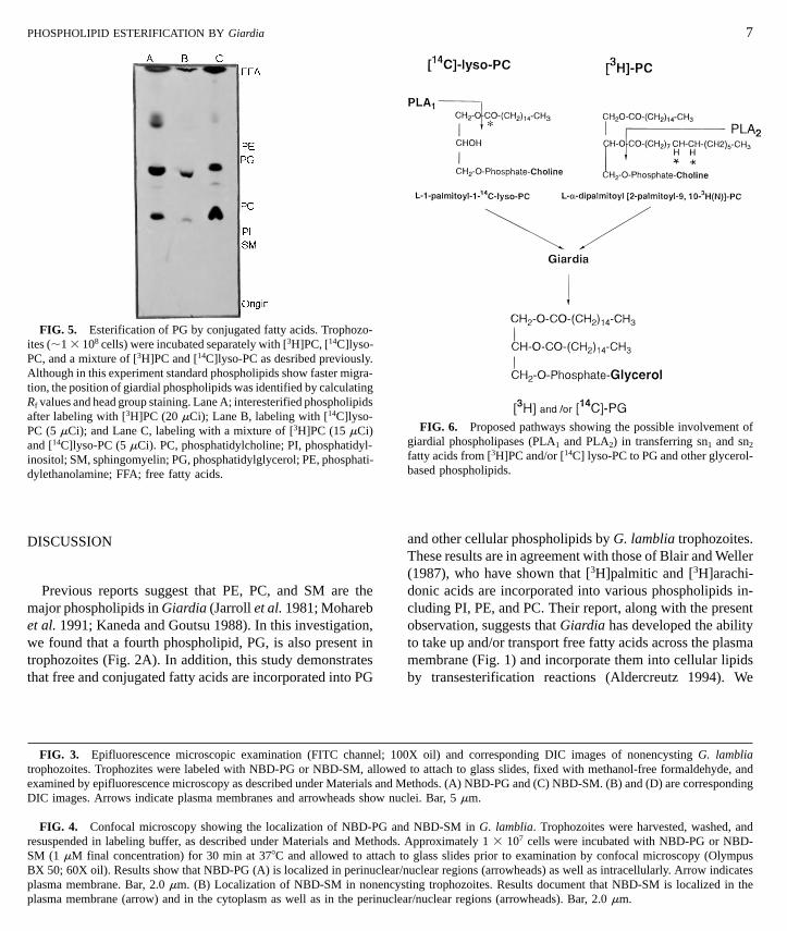
Since phospholipases are involved in modifying the fattyacid compositions in phospholipids and sn2-palmitic acid of[3H]PC is hydrolyzed off and transferred to giardial PG(shown in Fig. 6), we asked whether aristolochic acid (8-methoxy-6-nitrophenanthro [3, 4-d]-1, 3-dioxole-5-carbox-ylic acid) could affect this transfer. In this experiment, tro-phozoites were incubated with 250 mM aristolochic acid for30 min at 378C (which did not affect the viability—pleasesee Materials and Methods) with shaking. The dose andduration of aristolochic acid treatment were determined fromseparate experiments (data not shown). [3H]PC (10 mM, 20mCi/ml) or [14C]lyso-PC (10 mM, 5 mCi/ml) was added tothe incubation mixture, and incubation was continued for60 min. Inhibitor-treated (as well as control) trophozoiteswere separated by centrifugation before the extraction andanalysis of phospholipids. It is clear from Fig. 7 that aristo-lochic acid inhibits (,55%) the transfer (interesterificationreaction) of sn2-palmitic acid from [3H]PC to giardial PG.
Synthetic PG Analogs
The final question we asked was whether synthetic PGanalogs were toxic to trophozoites. The preincubated tropho-zoites were treated with PG analogs as described under
Materials and Methods. Results show that DPPG, DSPG,and DMPG were toxic to Giardia (LD50 ,200 nM) andinhibited the growth of the parasite in vitro (Fig. 8). At alower concentration (i.e., 70 nM), DSPG was more effective(,40% inhibition) than DMPG and DPPG. However, athigher doses (i.e., 210 and 350 nM) these inhibitors wereequally effective. Approximately 80% inhibition occurredat 350 nM. Interestingly, POPG was least effective (Fig. 8).
6 GIBSON ET AL.
PHOSPHOLIPID ESTERIFICATION BY Giardia
FIG. 5. Esterification of PG by conjugated fatty acids. Trophozo-ites (,1 3 108 cells) were incubated separately with [3H]PC, [14C]lyso-
3 14
PC, and a mixture of [ H]PC and [ C]lyso-PC as desribed previously.Although in this experiment standard phospholipids show faster migra-tion, the position of giardial phospholipids was identified by calculatingRf values and head group staining. Lane A; interesterified phospholipidsafter labeling with [3H]PC (20 mCi); Lane B, labeling with [14C]lyso-PC (5 mCi); and Lane C, labeling with a mixture of [3H]PC (15 mCi)and [14C]lyso-PC (5 mCi). PC, phosphatidylcholine; PI, phosphatidyl-inositol; SM, sphingomyelin; PG, phosphatidylglycerol; PE, phosphati-dylethanolamine; FFA; free fatty acids.DISCUSSION
FIG. 3. Epifluorescence microscopic examination (FITC channel; 10trophozoites. Trophozites were labeled with NBD-PG or NBD-SM, allowedexamined by epifluorescence microscopy as described under Materials and MDIC images. Arrows indicate plasma membranes and arrowheads show nu
FIG. 4. Confocal microscopy showing the localization of NBD-PG anresuspended in labeling buffer, as described under Materials and Methods.SM (1 mM final concentration) for 30 min at 378C and allowed to attach tBX 50; 60X oil). Results show that NBD-PG (A) is localized in perinuclear/plasma membrane. Bar, 2.0 mm. (B) Localization of NBD-SM in nonencyplasma membrane (arrow) and in the cytoplasm as well as in the perinucle
FIG. 6. Proposed pathways showing the possible involvement ofgiardial phospholipases (PLA1 and PLA2) in transferring sn1 and sn2
fatty acids from [3H]PC and/or [14C] lyso-PC to PG and other glycerol-based phospholipids.
7
and other cellular phospholipids by G. lamblia trophozoites.These results are in agreement with those of Blair and Weller
3 3
(1987), who have shown that [ H]palmitic and [ H]arachi-donic acids are incorporated into various phospholipids in-Previous reports suggest that PE, PC, and SM are themajor phospholipids in Giardia (Jarroll et al. 1981; Mohareb cluding PI, PE, and PC. Their report, along with the presentobservation, suggests that Giardia has developed the abilityet al. 1991; Kaneda and Goutsu 1988). In this investigation,
we found that a fourth phospholipid, PG, is also present in to take up and/or transport free fatty acids across the plasmamembrane (Fig. 1) and incorporate them into cellular lipidstrophozoites (Fig. 2A). In addition, this study demonstrates
that free and conjugated fatty acids are incorporated into PG by transesterification reactions (Aldercreutz 1994). We
0X oil) and corresponding DIC images of nonencysting G. lambliato attach to glass slides, fixed with methanol-free formaldehyde, and
ethods. (A) NBD-PG and (C) NBD-SM. (B) and (D) are correspondingclei. Bar, 5 mm.
d NBD-SM in G. lamblia. Trophozoites were harvested, washed, andApproximately 1 3 107 cells were incubated with NBD-PG or NBD-o glass slides prior to examination by confocal microscopy (Olympusnuclear regions (arrowheads) as well as intracellularly. Arrow indicatessting trophozoites. Results document that NBD-SM is localized in thear/nuclear regions (arrowheads). Bar, 2.0 mm.

acid. Trophozoites were initially treated with aristolochic acid (250mM) and then incubated with [3H]PC (10 mM) described under Materi-
Preincubated trophozoites were treated with various concentrations ofanalogs for 1 h at 378C as described under Materials and Methods.
als and Methods. Phospholipids were extracted from control and treatedsamples and analyzed by TLC. For quantitation, radiolabeled phospho-lipids were isolated from TLC by scraping “hot spots” and measuringradioactivity. PC, phosphatidylcholine; PG; phosphatidylglycerol; PE,phosphatidylethanolamine; FA, fatty acid. Although data shown werefrom one experiment, similar results were obtained in two separateexperiments. V indicates untreated and v indicates aristolochic acid-treated cells.
found that both saturated and unsaturated fatty acids werelargely incorporated into PG (Fig. 2B). Interestingly, AA(20:4), which was taken up by Giardia rapidly (Fig. 1),showed the lowest level of incorporation into PG and otherphospholipids (Fig. 2 B). This can be explained by the factthat AA is incorporated not only into phospholipids but intoa wide range of cellular lipids as well. On the other hand,PA (16:0), MA (14:0), and OA (18:1) are transesterifiedmainly into phospholipids (Blair and Weller 1987).
PG was also found to be a major transesterified productin trophozoites cultured in serum-deprived (PC or LPC-albumin supplemented) medium (not shown), suggestingthat this glycerol-based phospholipid can be derived fromother phospholipids present in the growth medium. Thishypothesis can further be supported by our recent findings
that PG is not present in bovine serum or bile (not shown).It is possible that PG in Giardia can also be synthesized denovo. Using fluorescence conjugated lipid probes, we haveshown previously (Stevens et al. 1997) that encysting andnonencysting trophozoites are able to carry out selectiveuptake, compartmentalization, and stage- or analog-specificlocalization of exogenous lipids. For example, in nonen-cysting cells PC was localized in the plasma membranes,Inhibitor-treated and control cells were inoculated into 5-ml culturetubes containing TYI-S-33 medium, supplemented with bovine bileand serum, and incubated overnight at 378C. Tubes were chilled inice-cold water for 45 min to detach trophozoites before counting. vindicates POPG; m denotes DMPG; m and . show the inhibition ofgrowth by DPPG and DSPG, respectively. The data presented aremeans of three separate experiments with standard errors between 5and 12%.
whereas ceramide was localized intracellularly. Like otherlipids, exogenously supplied PG is taken up by Giardiatrophozoites and is concentrated mostly in perinuclear/nu-clear regions. A substantial amount of NBD-PG is also local-ized intracellularly (Figs. 3 and 4A). In contrast, NBD-SMis localized in the inner leaflet of plasma membranes andnuclear/perinuclear regions, as well as intracellularly (Figs. 3and 4B). The localization of NBD-PG and SM in perinuclear/nuclear regions, which have recently been identified as theendoplasmic reticulum (ER) of Giardia (Soltys et al. 1996),suggests that the ER may be responsible for carrying outintracellular trafficking/metabolism of exogenous phospho-
8
FIG. 7. Decreased interesterification of [3H]PG by aristolochic
GIBSON ET AL.
FIG. 8. Effects of PG analogs on in vitro growth of trophozoites.
lipids in nonencysting trophozoites. In a separate experimentwe found that Brefeldin A, which induces the rapid redistri-bution of Golgi into the ER in eukaryotic cells (Shah andKlausner 1993), alters the localization of fluorescent lipidsin encysting cells but not in nonencysting trophozoites (notshown), indicating that the mechanism of lipid movementin encysting and nonencysting trophozoites may be different.It is possible that Golgi or Golgi-like organelles induced

long since been lost by this obligate parasite (Roger et al.1998)? Investigating the synthesis and function of PG in
This work was supported by Grants AI 136597 and GM 08012 fromthe National Institutes of Health. We are grateful to Dr. J. Moore
PHOSPHOLIPID ESTERIFICATION BY Giardia
during encystation (Reiner et al. 1990) may be involved inlipid trafficking in encysting Giardia. We also observed thatPG, unlike PC, does not support the growth of the parasitein serum-deprived medium (not shown). One possible expla-nation is that PC is a major phospholipid of the giardialplasma membranes (Stevens et al. 1997), whereas PG isfound mainly in perinuclear and other endomembranes (Figs.3 and 4).
Figure 5 shows that G. lamblia can utilize conjugatedfatty acids to esterify some of its phospholipids, includingPG, most likely by interesterification reactions (Aldercreutz1994). It is clear from Fig. 5 that when a mixture of [14C]lyso-PC and [3H]PC was used, both 14C- and 3H-labeled fattyacids could be cleaved and transferred to giardial PG, indicat-ing both sn1 and sn2 fatty acids from lyso-PC and PC canbe hydrolyzed off and transferred to PG as demonstrated inFig. 6. Gillin et al. (1986) found that lecithin with 1-palmitic-2-linoleic or 1-palmitic-2-oleic (present in human bile) inculture medium is potentially useful to trophozoites andsupports the growth of G. lamblia in serum-deprived me-dium. It is likely that PC and other exogenous phospholipidsare used directly as an energy source, as well as for interester-ification of cellular phospholipids.
The importance of an ongoing deacylation/reacylation cy-cle of membrane phospholipids in mammalian cells has re-cently been demonstrated by Balsinde et al. (1995) and byBalsinde and Dennis (1997). In this cycle, a preexistingphospholipid is cleaved by an intracellular PLA2 to generatea 2-lysophospholipid, which in turn generates a new phos-pholipid (Lands and Crawford 1976). Therefore, we askedwhether, in Giardia, PLA2 is also involved in esterificationreactions. To investigate this, we have used aristolochic acid,an inhibitor of calcium ionophore-stimulated PLA2 activityin human neutrophils (Rosenthal et al. 1989). Results showthat this inhibitor was able to inhibit the incorporation ofsn2 fatty acids from PC to PG (Fig. 7), indicating that giardialPLA2 is involved in hydrolyzing the sn2 fatty acid from[3H]PC. A similar observation was also reported by Balsindeet al. (1995), who found that a Ca21-independent PLA2 inP388D1 macrophages plays a major role in regulating theincorporation of AA into membrane phospholipids, whichcould be inhibited by bromo-enol-lactone (an inhibitor of
Ca21-independent PLA2 activity).Next, we asked whether synthetic analogs of PG wereeffective against trophozoites. DMPG, DPPG, and DSPGwere found to be toxic, inhibiting growth (LD50 5 ,200nM) of trophozoites in vitro (Fig. 8). Interestingly, POPGwas not a potent inhibitor, suggesting that the type and chainlength of fatty acids in PG analogs are important for theseeffects. Although, at present, we are unable to explain the
9
mechanism of inhibitor action, it is possible that PG analogsbind (competitively or noncompetitively) to phosphatidyl-glycerolphosphate (PGP) synthase, one of the major en-zymes of the PG biosynthetic pathways (Ohtsuka et al.1993), as well as other enzymes that are involved in phospho-lipid remodeling and/or de novo synthesis. Alternatively,fatty acids released from PG analogs by phospholipasesmay also be responsible for these toxic effects. However, itremains to be seen whether G. lamblia expresses PGP syn-thase or other enzymes of lipid metabolic pathways.
While PG is a major phospholipid in bacterial membranes(Yeagle 1993), in higher eukaryotes its dimer, i.e., cardio-lipin, is only found associated with mitochondrial mem-branes (Schlame et al. 1993). Thus, the finding of PG inGiardia, an amitochondriate eukaryote (Hashimoto et al.1994), raises an interesting evolutionary question. Does thePG in Giardia derive from a common ancestor betweenbacteria and this early diverging eukaryote (Sogin et al.1989) or is it a remnant of an early mitochondrion that has
Giardia trophozoites, therefore, may be helpful to answerthese questions. This will also allow us to understandwhether G. lamblia has some ability to carry out de novophospholipid synthesis or whether it is dependent entirelyon a remodeling process that chemically alters phospholipidsobtained by this parasite from its host.
ACKNOWLEDGMENTS
(Avanti Polar lipids Inc., Alabaster, AL) for performing HPLC analysisof phospholipids and Laura Dader (UTEP) for technical support. DICand confocal microscopy experiments were carried out at the AnalyticalCytology and Confocal Microscopy Core Facilities at the Universityof Texas at El Paso, funded by RR 08124 (NIH) and MRI-NSF grants.Ms. C. Castillo was supported by the MARC (NIH/GM 08048-13)program.
REFERENCES
Adam, R. 1991. The biology of Giardia spp. Microbiological Reviews55, 706–732.
Aldercreutz, P. 1994. Enzyme-catalyzed lipid modification. Biotechnol-ogy and Genetic Engineering Review 12, 231–254.

10
Balsinde, J., Bianco, I. D., Ackerman, E. J., Conde-Frieboes, K., andDennis, E. A. (1995). Inhibition of calcium-independent phospholi-pase A2 prevents arachidonic acid incorporation and phospholipidremodeling in P338D1 macrophages. Proceedings of the NationalAcademy of Sciences of the USA 92, 8527–8531.
Balsinde, J., and Dennis, E. (1997). Function and inhibition of intracel-lular calcium-dependent phospholipase A2. Journal of BiologicalChemistry 272, 16069–16072.
Berk, P. D. 1996. How do long-chain free fatty acids cross cell mem-branes? Proceedings of Society of Experimental and Biological Medi-cine 212, 1–4.
Blair, R., and Weller, P. F. 1987. Uptake and esterification of arachidonicacid by trophozoites of Giardia lamblia. Molecular and BiochemicalParasitology 25, 11–18.
Calder, P. C., Bond, J. A., Harvey, D. J., Gordon, S., and Newsholme,E. A. 1990. Uptake and incorporation of saturated and unsaturatedfatty acids into macrophage lipids and their effect upon macrophageadhesion and phagocytosis. Biochemical Journal 269, 807–814.
Christie, W. W. 1982. “Lipid Analysis.” Pergamon, Oxford, England.
Das, S., Reiner, D. S., Zenian, J., Hogan, D. L., Koss, M. A., Wang,C. S., and Gillin, F. D. 1988. Killing of Giardia trophozoites byhuman intestinal fluid in vitro. Journal of Infectious Diseases157, 1257–1260.
Das, S., Traynor-Kaplan, A., Reiner, D. S., Meng, T. C., and Gillin,F. D. 1991. A surface antigen of Giardia lamblia with a glycosylphos-phatidylinositol anchor. Journal of Biological Chemistry 266,21318–21325.
Das, S., Schteingart, C. D., Hofmann, A. F., Reiner, D. S., Aley, S.B., and Gillin, F. D. 1997. Giardia lamblia: Evidence for carrier-mediated uptake and release of conjugated bile acids. ExperimentalParasitology 87, 133–141.
Diamond, L. S., Harlow, D., and Cunnick, C. C. 1978. A new mediumfor the axenic cultivation of Entamoeba histolytica and other Enta-moeba. Transactions of the Royal Society of Tropical Medicine andHygiene 27, 487–488.
Ellis, J. E., Wyder, M. A., Jarroll, E. L., and Kaneshiro, E. S. 1996.Changes in lipid composition during in vitro encystation and fattyacid desaturase activity of Giardia lamblia. Molecular and Biochemi-cal Parasitology 81, 13–25.
Farthing, M. J. G., Keush, G. T., and Carey, M. C. 1985. Effects ofbile and bile salts on growth and membrane lipid uptake by Giardialamblia. Journal of Clinical Investigation 76, 1727–1732.
Gillin, F. D., Gault, M. J., Hofmann, A. F., Gurantz, D., and Sauch,J. F. 1986. Biliary lipids support serum-free growth of Giardialamblia. Infection and Immunity 53, 641–645.
Gillin, F. D., Reiner, D. S., Gault, M. J., Douglas, H., Das, S., Wunder-lich, A., and Sauch, J. F. 1987. Encystation and expression of cystantigens by Giardia lamblia in vitro. Science 235, 1040–1043
Gillin, F. D., Reiner, D. S., and Boucher, S. E. 1988. Small intestinalfactors promote encystation of Giardia lamblia in vitro. Infectionand Immunity 56, 705–707.
Hartong, W. A., Gourley, W. K., and Arvanitakis, C. 1979. Giardiasis:Clinical spectrum and functional–structural abnormalities of thesmall intestinal mucosa. Gastroenterology 77, 61–69.
GIBSON ET AL.
Hashimoto, T., Nakamura, Y., Nakamura, F., Shirakura, T., Adachi, J.,Goto, N., Okamoto, K., and Hasegawa, M. 1994. Protein phylogenygives a robust estimation for early divergences of eukaryotes: Phylo-genetic place of a mitochondria-lacking protozoan, Giardia lamblia.Molecular Biology of Evolution 11, 65–71.
Higgins, J. A. 1990. Separation and analysis of membrane lipid compo-nents. In “Biological Membranes: A Practical Approach” (J. B. C.Findlay and W. H. Evans, Eds.), pp. 103–137. IRL Press, Oxford,UK.
Jarroll, E. L., Muller, P. J., Meyer, E. A., and Morse, S. A. 1981. Lipidand carbohydrate metabolism of Giardia lamblia. Molecular andBiochemical Parasitology 2, 187–196.
Kaneda, Y., and Goutsu, T. 1988. Lipid analysis of Giardia lambliaand its culture medium. Annals of Tropical Medicine and Parasitol-ogy 82, 83–90.
Keister, D. B. 1983. Axenic culture of Giardia lamblia in TYI-S-33medium supplemented with bile. Transactions of Royal Society ofTropical Medicine and Hygiene 77, 487–488.
Lands, W. E. M., and Crawford, C. G. 1976. “The Enzyme of BiologicalMembranes” (A Mortonosi, Ed.), vol. 2, pp. 3–85. Plenum, NewYork.
Lujan, H. D., Mowatt, M. R., Chen, G. Z., and Nash, T. E. 1995.Isoprenylation of proteins in the protozoan Giardia lamblia. Molecu-lar and Biochemical Parasitology 72, 121–127.
Mohareb, E. W., Rogers, E. J., Weiner, E. J. and Bruce, J. L. 1991.Giardia lamblia: Phospholipid analysis of human isolates. Annalsof Tropical Medicine and Parasitology 85, 591–597.
Ohtsuka, T., Nishijima, M., and Akamatsu, Y. 1993. A somatic cellmutant defective in phosphatidylglyceolphosphate synthase, withimpaired phosphatidylglycerol and cardiolipin biosynthesis. Journalof Biological Chemistry 268, 22908–22913.
Papanastasiou, P., McConville, M., Ralton, J., and Kohler, P. 1997.The variant-surface protein of Giardia, VSP41, is glycosylated andpalmitoylated protein. Biochemical Journal 322, 49–56.
Reiner, D. S., McCaffery, M., and Gillin, F. D. 1990. Sorting of cystwall proteins to a regulated secretory pathway during differentiationof the primitive eukaryote Giardia lamblia. European Journal ofCell Biology, 53, 963–968.
Reiner, D. S., Hetsko, M. L., and Gillin, F. D. 1995. A lipoprotein-cholesterol albumin substitute stimulates Giardia lamblia encysta-tion vesicle formation. Journal of Eukaryotic Microbiology 42,622–627.
Roger, A. J., Svard, S. G., Tovar, J., Clark, C. G., Smith, M. W., Gillin,F. D., and Sogin, M. L. 1998. A mitochondrial-like chaperon 60gene in Giardia lamblia: Evidence that diplomonad once harbored
an endosymbiont related to the progenitor of mitochondria. Proceed-ings of the National Academy of Sciences of the USA 95, 229–334.Rosenthal, M.D., Vishwanath, B. S., and Franson, R. C. 1989. Effectsof aristolochic acid on phospholipase A2 activity and arachidonatemetabolism of human neutrophils. Biochimica et Biophysica Acta1001, 1–8.
Schlame, M., Brody, S., and Hostetler, K. Y. 1993. Mitochondrialcardiolipin in diverse eukaryotes. European Journal of Biochemistry212, 727–735.

PHOSPHOLIPID ESTERIFICATION BY Giardia
Shah, N., and Klausner, R. D. 1993. Brefeldin A reversibly inhibitssecretion in Saccharomyces cerevisiae. Journal of Biological Chem-istry 268, 5345–5348.
Sogin, M. L., Gunderson, J. H., Elwood, H. J., Alonso, R. A., andPeattie, D. A. 1989. Phylogenetic meaning of the kingdom concept:An unusual ribosomal RNA from Giardia lamblia. Science 243,75–77.
Soltys, B. J., Falah, M., and Gupta, R. S. 1996. Identification ofendoplasmic reticulum in the primitive eukaryote Giardia lambliausing cryoelectron microscopy and antibody to BiP. Journal of CellScience 109, 1909–1917.
Stevens, T. L., Gibson, G. R., Adam, R., Maier, J., Allison-Ennis, M.,and Das, S. 1997. Uptake and cellular localization of exogenouslipids by Giardia lamblia, a primitive eukaryote. Experimental Para-sitology 86, 133–143.
11
Traynor-Kaplan, A. E., Thompson, B. L., Harris, A. L., Taylor, P.,Oman, G. M., and Sklar, L. A. 1989. Transient increase in phosphati-dyl 3, 4-bisphosphate and phosphatidylinositol triphosphate duringactivation of human neutrophils. Journal of Biological Chemistry265, 15668–15673.
Vial, H. J., and Ancelin, M. L. 1992. Malerial lipids—An overview.“Subcellular Biochemistry” (J. L. Avila and J. R. Harris, Eds.), vol.18, pp. 259–356. Plenum, New York.
Yang, H., Bard, M., Bruner, D. A., Gleeson, A., Deckelbaum, R. J.,Aljinovic, G., Phol, T. M., Rothstein, R., and Sturley, S. L. 1996.Sterol esterification in yeast: A two-gene process. Science 272,1353–1356.
Yeagle, P. L. 1993. “The Membane of Cells,” 2nd ed., Academic Press,San Diego.
Received 9 June 1998; accepted with revision 21 December 1998