Inter-kingdom conservation of mechanism of nonsense-mediated mRNA decay
Upload
independentCategory
view
3download
0
Nonsense-Mediated mRNA Decay Impacts MSI-DrivenCarcinogenesis and Anti-Tumor Immunity in ColorectalCancersJamila El-Bchiri1,2, Agathe Guilloux1,2, Peggy Dartigues1,2, Etienne Loire3,4, Dominique Mercier1,2, Olivier
Buhard1,2, Iradj Sobhani5, Pierre de la Grange6, Didier Auboeuf6, Francoise Praz1,2, Jean-Francois
Flejou1,2, Alex Duval1,2*
1 INSERM, UMR S893, Team 13 ‘‘Microsatellite Instability and Cancers’’, Paris, France, 2 UPMC Univ Paris 06, UMR S893, Paris, France, 3 UPMC Univ Paris 06, Atelier de
Bioinformatique, Paris, France, 4 UPMC Univ Paris 06, UMR 7592, Institut Jacques Monod, Paris, France, 5 Departement de Gastro-Enterologie, CHU Henri Mondor, Creteil,
France, 6 INSERM, U685, Hopital Saint-Louis, Paris, France
Abstract
Nonsense-mediated mRNA Decay (NMD) degrades mutant mRNAs containing premature termination codon (PTC-mRNAs).Here we evaluate the consequence of NMD activity in colorectal cancers (CRCs) showing microsatellite instability (MSI)whose progression is associated with the accumulation of PTC-mRNAs encoding immunogenic proteins due to frameshiftmutations in coding repeat sequences. Inhibition of UPF1, one of the major NMD factors, was achieved by siRNA in theHCT116 MSI CRC cell line and the resulting changes in gene expression were studied using expression microarrays. Theimpact of NMD activity was also investigated in primary MSI CRCs by quantifying the expression of several mRNAs relativeto their mutational status and to endogenous UPF1 and UPF2 expression. Host immunity developed against MSI cancer cellswas appreciated by quantifying the number of CD3e-positive tumor-infiltrating lymphocytes (TILs). UPF1 silencing led to theup-regulation of 1251 genes in HCT116, among which a proportion of them (i.e. 38%) significantly higher than expected bychance contained a coding microsatellite (P,2610216). In MSI primary CRCs, UPF1 was significantly over-expressedcompared to normal adjacent mucosa (P,0.002). Our data provided evidence for differential decay of PTC-mRNAscompared to wild-type that was positively correlated to UPF1 endogenous expression level (P = 0.02). A negative effect ofUPF1 and UPF2 expression on the host’s anti-tumor response was observed (P,0.01). Overall, our results show that NMDdeeply influences MSI-driven tumorigenesis at the molecular level and indicate a functional negative impact of this systemon anti-tumor immunity whose intensity has been recurrently shown to be an independent factor of favorable outcome inCRCs.
Citation: El-Bchiri J, Guilloux A, Dartigues P, Loire E, Mercier D, et al. (2008) Nonsense-Mediated mRNA Decay Impacts MSI-Driven Carcinogenesis and Anti-TumorImmunity in Colorectal Cancers. PLoS ONE 3(7): e2583. doi:10.1371/journal.pone.0002583
Editor: Cathal Seoighe, University of Cape Town, South Africa
Received April 2, 2008; Accepted May 28, 2008; Published July 9, 2008
Copyright: � 2008 El-Bchiri et al. This is an open-access article distributed under the terms of the Creative Commons Attribution License, which permitsunrestricted use, distribution, and reproduction in any medium, provided the original author and source are credited.
Funding: This work was partly supported by grants from the Association pour la Recherche contre le Cancer (credit number 3946). JEB, EL and PDLG wererecipients of a fellowship from the Ministere Francais de la Recherche (MRT).
Competing Interests: The authors have declared that no competing interests exist.
* E-mail: [email protected]
Introduction
Nonsense-mediated mRNA decay (NMD) is an evolutionarily
conserved mRNA surveillance mechanism that recognizes and
eliminates aberrant mRNAs harboring premature termination
codons (PTC), thereby preventing the accumulation of potentially
deleterious truncated proteins in eukaryotic cells [1]. Following pre-
mRNA splicing, NMD targets are recognized via a multiprotein
exon-junction complex (EJC) that is deposited 24 nucleotides
upstream of each exon-exon junction. As a general rule, it has been
established that aberrant mRNAs containing PTC located either
less than 50–55 nucleotides upstream of the last exon-exon junction
or in the last exon are not degraded by NMD (NMD-irrelevant) [2].
The core of NMD effectors comprises the evolutionary-conserved
UPF proteins, UPF1/RENT1, UPF2/RENT2, and two paralogs
of UPF3, UPF3 (also called UPF3a) and UPF3X (also called
UPF3b). UPF1 is an RNA helicase whose activity is regulated by
cycles of phosphorylation/dephosphorylation. Phosphorylation of
UPF1 requires UPF2 and UPF3 and is catalyzed by SMG1, a
protein kinase related to the phosphoinositide-3-kinase family.
UPF1 phosphorylation by SMG1 has been shown to be the rate-
limiting step in NMD [3,4]. It is noteworthy that SMG1 activity is
not only devoted to NMD but also plays a role in DNA damage
signaling and repair, notably by phosphorylating p53 [5].
Dephosphorylation of UPF1 is mediated by SMG5, SMG6 and
SMG7, three proteins that act as adaptors between phosphorylated
UPF1 and protein phosphatase 2A [3]. UPF1 function is crucial in
NMD, while UPF3 and UPF3X are partially redundant [6] and
UPF2 is dispensable in some cases suggesting the existence of a
UPF2-independent NMD pathway [7]. UPF1 acts not only in
NMD-mediated degradation of aberrant transcripts but also in the
physiological decay of various mRNAs, regulating the expression of
3–10% of the transcriptome [8,9]. Silencing of UPF1 and to a lesser
extent UPF2 has been reported to modulate the expression level of
a number of physiological substrates of NMD, including transcripts
with upstream open reading frames in the 59-untranslated region,
PLoS ONE | www.plosone.org 1 July 2008 | Volume 3 | Issue 7 | e2583

transcripts containing an intron within their 39-untranslated region
as well as transcripts derived from transposon or endogenous
retrovirus [8,10]. NMD has also been proposed to play a role in the
regulation of alternative splicing events, since sequence-based
analyses predict that about 35% of mammalian alternative splicing
events produce PTC-containing spliced variants. Nevertheless, it
has recently been reported that most PTC-containing splice
variants are produced at low levels in human cells, independently
of the action of NMD, suggesting that the majority of such PTC-
introducing events are not under positive selection pressure and
therefore are not expected to contribute important functional
roles [11].
To date, the consequences of NMD activity have been mostly
investigated in monogenic hereditary diseases, including heredi-
tary tumors [12]. NMD has been reported to protect heterozygous
carriers from the deleterious effects of some aberrant PTC-
mRNAs encoding truncated proteins with dominant-negative
activity [12]. Conversely, NMD inability in degrading NMD-
irrelevant transcripts has been proposed to favor the phenotypic
expression of dominant hereditary diseases [12]. Because cancer
cells are genetically unstable and accumulate numerous somatic
frameshift mutations in genes with an expected role in cell
transformation, the NMD system has been proposed to play a role
in tumor development [12]. However, the assessment of its overall
impact on this process has been poorly described and remains
hard to define since it depends on the nature and the number of
mutants accumulated in cancer cells during tumor progression. To
date, NMD inhibition has been successfully used as an in vitro
strategy to discover new cancer-related genes harboring truncating
mutations, suggesting that it may indeed have a role in favoring
the selection of some frequent mutational events in various tumor
types [13]. It is noteworthy that NMD activity has been shown to
be highly variable, leading to incomplete and differential decay of
putatively NMD-relevant mRNAs containing a PTC (referred to
as NMD-escape) [14,15,16].
MSI tumors harboring mismatch repair (MMR) deficiency are
frequent in humans. They represent about 15% of sporadic
colorectal, gastric and endometrial tumors and include neoplasms
arising in the Hereditary Non Polyposis Colorectal Cancer
(HNPCC) syndrome. Dozens of mutations affecting genes
containing coding repeat sequences have been reported in these
tumors, defining the so-called mutator pathway whose role is
suspected to be crucial in MSI-driven tumorigenesis [17]. To date,
a large number of publications have reported frameshift mutations
in genes involved in various biological pathways such as cell cycle
regulation (e.g. TGFBR2, IGF2R, TCF4, AXIN2, PTEN, RIZ),
apoptosis (e.g. BAX, CASP5, BCL10, APAF1, FAS), DNA damage
repair (e.g. ATR, DNA-PKcs, RAD50, MSH3, MSH6, MBD4,
MLH3, BLM, CHK1) and others [17]. We recently reported that
the decay of frameshift mutation-derived mRNAs following in vitro
silencing of UPF1 and/or UPF2 in a panel of MSI CRC cell lines
was differential and incomplete [14]. If not fully degraded,
frameshift mutant mRNAs encode proteins containing aberrant C-
terminal tails, among which some have been shown to display
immunogenic properties [18,19]. Several studies have emphasized
the fact that, in keeping with this process, MSI tumors were
markedly infiltrated by cytotoxic intra-epithelial tumor-infiltrating
T lymphocytes (TILs) and that such a cellular immune response
was predictive of a relatively favorable outcome independently of
the initial tumor stage and other clinical factors [20]. Therefore,
NMD activity may interfere with anti-tumor immunity by limiting
the expression of some of these aberrant proteins. Using MSI
colorectal cancer as a model, we aimed here at further
investigating the role of NMD in oncogenesis.
Results
Transcriptome changes secondary to UPF1 silencing inHCT116 CRC cells and their relationship to microsatelliteinstability
Using Affymetrix GeneChipH Human Exon 1.0 ST gene
expression arrays, the expression of 1363 genes was found to be
significantly deregulated upon UPF1 silencing in the HCT116 (MSI)
CRC cell line, with a fold change $1.5 compared to untreated cells
(Table S1). With 111 others, UPF1 was, as expected, one of the genes
to be significantly down-regulated. The level of inhibition was
significant (.75%) and agreed well with our data obtained by real-
time quantitative RT-PCR (data not shown). Overall, 1251 genes
were up-regulated upon UPF1 silencing (1251/1363, i.e. 92% of all
deregulated genes under these conditions), amongst which 472 (38%)
contained a mononucleotide repeat sequence of at least 7 base pairs
in the coding region (Table S2). Of interest, the total number of genes
in the human genome with a coding repeat tract $7N is lower (4470/
22218 ; 20%. Data not shown), making significantly higher than
expected by chance the overall number of such target genes up-
regulated in HCT116 upon UPF1 silencing (P,2610216; Chi2 test).
Amongst the aforementioned 472 genes in HCT116 cells, 22
genes containing a coding mononucleotide repeat of 7 to 10
nucleotides were chosen because of their putative role in colorectal
carcinogenesis and screened for insertion/deletion mutations in
these cells (Table 1). All these genes but 3 (MBD4, MSH3,
TGFBR2) had never been reported to be mutated in MSI CRCs
(Table 1). Using this approach, we detected or confirmed
homozygous mutations in the SLC35F5, TGFBR2, ARV1, MSH3,
SMAP1 genes and heterozygous mutations in the MBD4, EFHC1,
TTC3, and WDR19 genes (Figure 1a). All the corresponding
mutant mRNAs harbored a PTC located in coding regions that
may be prone to NMD. In addition, we observed that 4 other
target genes with previously described heterozygous frameshift
mutations in HCT116 (BAX, RECQL, RAD50 and MSH6) were not
significantly re-expressed following UPF1 silencing (Figure 1a).
The re-expression rates for PTC-mRNAs induced by UPF1
silencing in HCT116 are shown in Figure 1a and highlight the fact
that, as described [14], UPF1-mediated mRNA decay is highly
variable within a series of endogenously mutated PTC-mRNAs
although allele specific expression assays were not used to
differentiate between mutated and wild-type mRNAs in the case
of heterozygous frameshift alterations.
Frameshift mutations in MSI primary CRCs according totheir NMD status
All the target genes mutated in HCT116 for which the impact
of NMD had previously been determined were screened for
frameshift mutations in coding microsatellite sequences in a series
of primary MSI CRCs (n = 44). This includes genes whose
mutated mRNAs are more or less sensitive to NMD (TGFBR2,
SLC35F5, ARV1, MSH3, TTC3, EFHC1, MBD4, SMAP1, WDR19,
BAX, RECQL, RAD50, MSH6) (Figure 1a). The mutation
frequencies of these 13 genes representing possible targets for
MSI-driven instability were highly variable in these tumors
(Figure 1b). Based on their high mutation frequency, SLC35F5,
ARV1, TTC3, and SMAP1 represent new target genes in which
frameshift mutations have not previously been reported in MSI
CRC. They were mutated in 48% (21/44), 23% (10/44), 32%
(14/44) and 73% (32/44) of tumors, respectively (Figure 1b).
Over-expression of UPF1 mRNA in MSI primary CRCsBy measuring the levels of UPF1 and UPF2 mRNAs by real-
time quantitative RT-PCR in another independent series of MSI
NMD Role in Colorectal Cancers
PLoS ONE | www.plosone.org 2 July 2008 | Volume 3 | Issue 7 | e2583

primary CRCs for which we also obtained the samples
corresponding to matching normal mucosa, we observed an
approximately 7-fold over-expression of UPF1 in tumors (n = 25)
compared to normal mucosa (P = 0.0015; paired t-test) (Figure 2).
Since the quantification (7-fold) of the over-expression has to be
taken with cautiousness due to the small amount of data, we
verified that UPF1 mRNA was indeed over-expressed in MSI
primary CRCs by performing a qualitative chi-square test, which
is robust to extreme values. Using this approach, the number of
cases in which UPF1 expression was higher in tumors compared to
matching normal mucosa (N = 20/25) was significantly different
than expected by chance (P = 0.009; chi-square test). These data
are illustrated on Figure 2, in which 20 and 5 open circles
representing MSI tumor samples over-expressing UPF1 or not,
respectively, are represented above a doted line symbolizing the
expression of this NMD factor in normal mucosa. Of interest, a
trend for over-expression of this factor was also observed in non-
MSI (MSS) CRCs (n = 31) in the same conditions (P = 0.08; paired
t-test) (Figure 2). In contrast, UPF2 was not differentially expressed
in either MSI or MSS CRCs compared to matching normal
mucosa (P = 0.56 and 0.83, respectively; paired t-test) (Figure 2).
Significant decay of frameshift mutation-derived mRNAsthat depend on UPF1 expression in MSI primary CRCs
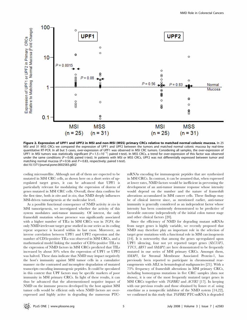
In our series of 44 primary MSI CRCs, we further determined
the relative in vivo expression of 18 MSI target genes by real-time
quantitative RT-PCR using experimental conditions that we
previously determined in a series of CRC cell lines (ATR, BAX,
BLM, CBF2, CDX2, GRB14, GRK4, IGF2R, MBD4, MSH3, MSH6,
RAD50, RBBP8, RECQL, RIZ, TCF4, TFDP2, TGFBR2) (Figure 3
and also Table S3 for the mutational status of these 18 genes in
MSI CRCs) [14]. For all genes except 3 (CDX2, GRB14, TFDP2), a
trend for decay of mutant compared to wild-type mRNAs was
observed. The decay of mutant mRNAs compared to wild-types
was significant for MSH6 (P = 0.02; Student’s t-test) and nearly
significant for a few others only, e.g. BAX (P = 0.08), CDX2
(P = 0.10), MSH3 (P = 0.10), RIZ (P = 0.10) and TFDP2 (P = 0.10),
providing evidence for differential decay of PTC-mRNAs
compared to wild-type in our series of primary CRCs, as already
described in a series of MSI CRC cell lines [14] (Figure 3).
Overall, there was a highly significant decay of PTC-mRNAs in
the MSI primary CRCs (P,1024, Student’s t-test; see the
‘‘Materials and Methods’’ section for Statistical analyses). As
indirect proof of the contribution of NMD, the overall intensity of
this process in MSI primary CRCs was positively correlated to the
endogenous UPF1 expression level (P = 0.02, Student’s t-test; see
the ‘‘Materials and Methods’’ section for statistical analyses),
whereas the impact of UPF2 was not significant (P = 0.72,
Student’s t-test) (data not shown).
UPF1 and UPF2 are negative predictive factors of thehost’s immune response against MSI CRCs
With the exception of TCF4, no significant positive correlations
were found between the endogenous expression of PTC-mRNAs
Table 1. List of 22 Target genes up-regulated following UPF1 silencing in HCT116 and their mutational status.
Gene Symbol Ensembl Human Gene Fold Change Repeat Repeat Position in cDNA Type of Mutation in HCT116
ARV1 ENSG00000148926 2.08 9A 539 hmz
CDC23 ENSG00000094880 1.99 7A 411 wt
CSPG2 ENSG00000038427 2.17 7A 7874 wt
EFHC1 ENSG00000096093 1.94 7A 1316 htz
MBD4* ENSG00000129071 1.89 10A 930 htz
MSH3* ENSG00000113318 1.6 8A 1394 hmz
NR3C1 ENSG00000113580 1.87 7A 1974 wt
ORC6L ENSG00000091651 2.24 7A 561 wt
PIGB ENSG00000069943 1.85 8T 1086 wt
PSD3 ENSG00000156011 1.6 8A, 8A 737, 2333 wt
PSEN1 ENSG00000080815 2.02 7T 774 wt
REV3L ENSG00000009413 1.91 8A 1481 wt
RIF1 ENSG00000080345 1.75 7T, 8A 1538, 4637 wt
SHPRH ENSG00000146414 1.69 8A 887 wt
SLC35F5 ENSG00000115084 2.2 10T 1157 hmz
SMAP1 ENSG00000112305 1.6 10A 550 hmz
TBC1D23 ENSG00000036054 1.66 9A 1901 wt
TFE3 ENSG00000068323 2.39 8G 1676 wt
TGFBR2* ENSG00000163513 2.45 10A 831 hmz
TMEM161B ENSG00000164180 1.57 8T 651 wt
TTC3 ENSG00000182670 2.42 8A, 7A 1867, 2432 htz
WDR19 ENSG00000157796 1.54 8T 788 htz
The threshold for re-expression was considered significant when the fold change was .1.5 (see the Materials and Methods section). Newly described target genes forinstability in MSI CRCs are indicated in bold characters. Hmz: homozygously mutated at the coding repeat tract in HCT116. Htz: heterozygously mutated at the codingrepeat tract in HCT116. wt: not mutated at the coding repeat tract in HCT116. Target genes that have been already described to be mutated in MSI CRCs are indicatedwith an *.doi:10.1371/journal.pone.0002583.t001
NMD Role in Colorectal Cancers
PLoS ONE | www.plosone.org 3 July 2008 | Volume 3 | Issue 7 | e2583

and the presence of CD3e-positive TILs in tumors (PTCF4 = 0.06;
Bootstrapped t-test) (data not shown). The link between the
number of TILs and the expression of UPF1 and UPF2 was not
linear but log-linear. Thus, the influence of UPF1 and UPF2
expression on the numbers of TILs was investigated via a Wald test
and the coefficients in the Poisson regression model were estimated
via iteratively re-weighted least squares. Based on these observa-
tions, a negative effect of UPF1 and UPF2 expression on the
overall number of CD3e-positive TILs was demonstrated (P,0.01
for UPF1 and UPF2; Wald test). A mathematical model
correlating the number of CD3e-positive TILs with the expression
of these NMD factors in MSI CRCs was established, predicting
that TILs increased by about 30% when the expression of UPF1
was halved.
These data are illustrated for UPF1 and UPF2 in Figure 4; tumor
samples in which UPF1 and UPF2 expression are low (below the
average) presented with variable rates of CD3e positive-TILs while
tumor samples in which UPF1 and UPF2 expression are high
(above the average) were found to be nearly always characterized by
a low CD3 count, making UPF1 and UPF2 possible markers of poor
anti-tumor immunity in MSI primary CRCs.
Discussion
By evaluating a large number of target genes that were mutated
at variable rates in their coding repeat sequences, we show a
significant decay of the corresponding mutant mRNAs compared
to wild-type in MSI primary CRCs with a significant impact of
endogenous expression of UPF1 in this process, with UPF2 playing
a minor role in the process, as recently described by our group in a
series of MSI CRC cell lines [14]. Taken one by one, we observed
a significant or nearly significant decay of only some mutant
mRNAs compared to wild-types and this is not surprising since: (i)
as recently published by our group, NMD impact on the
expression of frameshift mutation-derived mRNAs is highly
variable from one mutant to another [14]; (ii) frequencies of
target gene alterations were highly variable and sometimes very
low in our tumor series; (iii) some mutants in particular (TCF4) are
NMD-irrelevant. In this study, we also investigate large scale gene
expression changes in MSI CRC cells in the context of NMD
inhibition using a specific method, showing that it led to the up-
regulation of 1251 genes in HCT116, among which a proportion
of them significantly higher than expected by chance contained a
Figure 1. a. Impact of UPF1 on the decay of PTC-mRNAs in HCT116 cells. TGFBR2, SLC35F5, ARV1, MSH3, SMAP1, TTC3, EFHC1, MBD4, WDR19,BAX, RECQL, RAD50 and MSH6 harbor homozygous or heterozygous frameshift mutations in the HCT116 MSI CRC cell line. Using expression array, foldchanges in the expression of the corresponding mRNAs relative to UPF1 silencing were determined (fold change = EmRNA in cells treated with a UPF1siRNA / EmRNA in cells treated with control siRNA). Genes that are underlined are those whose re-expression was significant in these conditions (1.5fold-change up with a p-value #0.005). Evidence for variable sensitivity to UPF1-mediated decay of such PTC-mRNAs could therefore be obtained,although allele specific expression assays were not used to differentiate between mutated and wild-type mRNAs in the case of heterozygousframeshift alterations. b. Coding frameshift mutations in target genes relative to NMD-status in MSI primary CRCs. Frequencies of frameshiftmutations of the same series of 13 target genes for instability in 44 MSI primary CRCs are represented. Target genes whose frameshift mutations arefrequently selected for during MSI CRC progression harbor different sensitivities to UPF1-mediated decay.doi:10.1371/journal.pone.0002583.g001
NMD Role in Colorectal Cancers
PLoS ONE | www.plosone.org 4 July 2008 | Volume 3 | Issue 7 | e2583
coding microsatellite. Although not all of them are expected to be
mutated in MSI CRC cells, as shown here on a short series of up-
regulated target genes, it can be advanced that UPF1 is
particularly relevant for modulating the expression of dozens of
genes mutated in MSI CRC cells. Overall, these data confirm for
the first time, both in vitro and in vivo, that NMD deeply influences
MSI-driven tumorigenesis at the molecular level.
As a possible functional consequence of NMD activity in vivo in
MSI tumorigenesis, we investigated whether the activity of this
system modulates anti-tumor immunity. Of interest, the only
frameshift mutation whose presence was significantly associated
with a higher number of TILs in MSI CRCs was in TCF4, the
only NMD-irrelevant target gene studied in our series as its coding
repeat sequence is located within its last exon. Moreover, an
inverse correlation between UPF1 and UPF2 expression and the
number of CD3e-positive TILs was observed in MSI CRCs, and a
mathematical model linking the number of CD3e-positive TILs to
the expression of NMD factors in MSI CRCs predicted that TILs
increased by about 30% when the expression of UPF1 or UPF2
was halved. These data indicate that NMD may impact negatively
the host’s immunity against MSI tumor cells in a cumulative
manner via the contrasted and incomplete degradation of mutated
transcripts encoding immunogenic peptides. It could be speculated
in this context that UPF factors may be specific markers of poor
immunity in MSI primary CRCs. In light of these results, it can
thus be advanced that the aforementioned negative impact of
NMD on the immune process developed by the host against MSI
tumor cells would be efficient only when NMD factors are over-
expressed and highly active in degrading the numerous PTC-
mRNAs encoding for immunogenic peptides that are synthesized
in MSI CRCs. In contrast, it can be assumed that, when expressed
at lower rates, NMD factors would be inefficient in preventing the
development of an anti-tumor immune response whose intensity
would depend on the number and the nature of frameshift
alterations accumulated in MSI cancer cells. These findings may
be of clinical interest since, as mentioned earlier, anti-tumor
immunity is generally considered as an independent factor whose
intensity has been consistently demonstrated to be predictive of
favorable outcome independently of the initial colon tumor stage
and other clinical factors [20].
Since the efficiency of NMD for degrading mutant mRNAs
from target genes is highly variable, we recently proposed that
NMD may therefore play an important role in the selection of
target gene mutations with a functional role in MSI carcinogenesis
[14]. It is noteworthy that among the genes up-regulated upon
UPF1 silencing, four not yet reported target genes (SLC35F5,
TTC3, ARV1 and SMAP1) are here demonstrated to be frequently
mutated in our series of MSI primary CRCs. Amongst them,
SMAP1, for Stromal Membrane Associated Protein-1, has
previously been reported to participate in chromosomal rear-
rangements with MLL in hematological malignancies [21]. With a
73% frequency of frameshift alterations in MSI primary CRCs,
including homozygous mutations in five CRC samples (data not
shown), it is one of the most frequently mutated target genes in
MSI CRCs together with TGFBR2 and ACVR2 [17]. In keeping
with our previous results and those obtained by Ionov et al. using
emethine as a nonspecific inhibitor of the NMD system [14,22],
we confirmed in this study that TGFBR2 PTC-mRNA is degraded
Figure 2. Expression of UPF1 and UPF2 in MSI and non-MSI (MSS) primary CRCs relative to matched normal colonic mucosa. In 25MSI and 31 MSS CRCs we compared the expression of UPF1 and UPF2 between the tumors and matched normal colonic mucosa by real-timequantitative RT-PCR. In all but 5 cases, over-expression of UPF1 was observed in MSI CRC tumors. Considering all samples, the over-expression ofUPF1 in MSI tumors was statistically significant (P = 1.561023; paired t-test). In MSS CRCs, a trend for over-expression of this factor was observedunder the same conditions (P = 0.08; paired t-test). In patients with MSI or MSS CRCs, UPF2 was not differentially expressed between tumor andmatching normal mucosa (P = 0.56 and P = 0.83, respectively; paired t-test).doi:10.1371/journal.pone.0002583.g002
NMD Role in Colorectal Cancers
PLoS ONE | www.plosone.org 5 July 2008 | Volume 3 | Issue 7 | e2583
through NMD in MSI CRC cells. In a recent paper, You et al.
[23] reported contradictory data, claiming that this target gene as
well as MSH3 escape NMD in the HCT116 cell line. They
presented results on a series of PTC-mRNAs showing these
transcripts were either completely sensitive or completely resistant
to NMD in MMR-deficient cells. Their data were obtained by
performing expression assays that were neither quantitative nor
allele-specific. In the same study, these authors also suggested a
systematic translational repression of truncated proteins from
frameshift mutation-derived mRNAs that escaped NMD (NMTR
for ‘‘Nonsense Mediated Translational Repression’’) [23]. We
verified by western blotting that expression of the corresponding
proteins for both of the homozygously mutated target genes
(TGFBR2, MSH3) was not detectable in HCT116 cells (data not
shown). Contrary to You et al, we propose that loss of TGFBR2
and MSH3 expression is mainly due to NMD rather than to an
hypothetical NMTR pathway. It is noteworthy that the existence
of an NMTR pathway does not suit well with the immunogenic
properties of MSI cancer cells that depend directly on the
accumulation of immunogenic peptides derived from numerous
frameshift-related proteins whose PTC-mRNAs are NMD-rele-
vant in most cases [18,19,20]. Regardless of discrepancies, all these
data argue for a role of NMD in modulating the expression of
several genes whose mutations have been already demonstrated
(TGFBR2, MSH3, MBD4) or are expected to play an important
role during MSI CRC progression [17]. UPF1 depletion with
shRNA in non-MSI HeLa cells was recently reported to induce
cell cycle arrest in early S phase due to the involvement of this
factor in DNA repair [24]. In contrast, transient silencing of UPF1
and/or UPF2 in MSI and MSS colorectal cancer cells (HCT116,
LoVo, Co115, LS174T, SW480, COLO320) did not lead to
significant alterations of cell growth and/or death in our hands
(data not shown). Further studies are now required to determine
how NMD activity may change the repertoire of genes involved in
MSI carcinogenesis and impact tumor progression using MMR
deficient mouse models in which UPF1 activity is modified.
Our observations concern the most frequent primary tumor
location associated with MSI in human, i.e. colon. They indicate
that NMD deeply influences the expression of numerous mutants
with an expected crucial role in MSI-driven tumorigenesis and
negatively impacts the host immunity against MSI cancer cells.
Such a putative oncogenic function of NMD in MSI carcinogen-
esis fits well with the fact that we also report here for the first time
that UPF1 is significantly over-expressed in MSI CRCs compared
to matching normal mucosa. This last observation was based on
the measure of the UPF1 mRNA level. As a perspective, it has now
to be confirmed at the protein level through a quantitative
approach such as Western Blotting that was here not performed
Figure 3. Target gene-related mRNA expression according to mutational status in 44 MSI primary CRC. hCt values are indicated relativeto the mutational status of each gene in the 44 MSI primary CRCs (wild-type and mutated tumor samples are indicated by white circles and blacktriangles, respectively). For each gene, medium values of hCt related to wild-type (white arrow) or mutated (black arrow) tumor samples werecalculated. For all genes except 3 (CDX2, GRB14, TFDP2), a trend for decay of mutant compared to wild-type mRNAs was observed (hCt values areinversely proportional to gene expression). Overall, the data provide evidence for significant decay of PTC-mRNAs compared to wild-type mRNAs invivo in MSI primary CRCs (P,1024, student t-test).doi:10.1371/journal.pone.0002583.g003
NMD Role in Colorectal Cancers
PLoS ONE | www.plosone.org 6 July 2008 | Volume 3 | Issue 7 | e2583

because of the lack of available additional tumor material. The
development of clinical trials should now be developed to look
whether NMD may be considered as a factor of poor prognosis in
MSI CRCs or not. Small molecules inhibiting NMD are now
available [25] and may be used to gain further insight into the
NMD role in cancers. We now plan to use these drugs in MMR-
deficient mice developing MSI neoplasms to evaluate whether it
may constitute a new therapeutic target for the treatment of such
tumors.
Materials and Methods
Tumor samples and cell linesThe CRC cell line HCT116 was purchased from the American
Type Culture Collection and maintained in DMEM (Life
Technologies) containing 10% fetal calf serum (Invitrogen) and
Glutamine without antibiotics to allow transient transfection
experiments with siRNA. Forty-four MSI primary tumors were
obtained from patients undergoing surgery for colorectal cancer;
all cases were histopathologically confirmed as being adenocarci-
nomas. Collected tumors were systematically formalin-fixed,
paraffin-embedded and frozen after surgery without any prior
embedding in liquid nitrogen. The MSI status was determined by
fluorescent multiplex PCR comprising 5 quasimonomorphic
mononucleotide repeats (BAT-25, BAT-26, NR-21, NR-24 and
NR-27), as described [26]. Only tumors with instability at three or
more of these markers were included in the study. The amount of
normal contaminating DNA was estimated as previously described
[27]. In addition, an independent series of 25 MSI and 31 MSS
primary CRC samples were collected in the same conditions
together with their matching normal mucosa and used to compare
UPF1 and UPF2 expression in tumor and normal colonic tissues.
Multiplex PCR and mutation analysisTumor DNA from fromzen samples was extracted using
QIAamp DNA Tissue Kit (Qiagen) according to the manufactur-
er’s instructions. A total of 24 genes containing mononucleotide
repeat sequences were chosen either because they were already
described as targets for MSI-driven mutations in MSI tumors (e.g.
ATR, BAX, BLM, CBF2, CDX2, GRB14, GRK4, IGF2R, MBD4,
MSH3, MSH6, RAD50, RBBP8, RECQL, RIZ, TCF4, TFDP2, and
TGFBR2) or because they were significantly up-regulated in MSI
HCT116 cells following UPF1 silencing and mutated in the
HCT116 MSI CRC cell line (SLC35F5, ARV1, EFHC1, TTC3,
Figure 4. Relationship between endogenous UPF1 and UPF2 mRNA expression in MSI CRCs and the number of TILs. The overallnumber of CD3e positive-TILs was significantly related to the endogenous expression of UPF1 and UPF2 in this series of 44 MSI primary CRCs (P,0.01;Wald test). Of interest, while tumor samples in which UPF1 and UPF2 expression was low (below the average) presented with variable rates of CD3epositive-TILs, tumor samples in which UPF1 and UPF2 expression was high (above the average) were almost always characterized with low CD3 count(poorly immunogenic CRCs). These data make UPF1 and UPF2 specific markers of poor anti-tumor immunity in MSI primary CRCs. CD3 proteinimmunostaining are presented for 2 MSI primary CRC samples showing either low CD3 count and high UPF1 and UPF2 expression (left) or high CD3count together with low UPF1 and UPF2 expression (right).doi:10.1371/journal.pone.0002583.g004
NMD Role in Colorectal Cancers
PLoS ONE | www.plosone.org 7 July 2008 | Volume 3 | Issue 7 | e2583

SMAP1, WDR19). Specific primers for each target gene were
designed using e-primer3 (http: // bioweb.pasteur.fr/seqanal/
interfaces/eprimer3.html) so that short fragments (,200 bp) could
be simultaneously amplified by 6 PCRs using 6-FAM or HEX
labeled primers. PCR reactions were performed in a final volume
of 20 ml containing 100 ng of genomic DNA, 0.15–0.40 mM of
each pair of primers and 1 unit of HotStarTaq DNA polymerase
(Qiagen) (Table S4). Adequate dilutions of the fluorescent PCR
products were mixed with formamide and GeneScanTM 400HD
ROXTM Size Standard (Applied Biosystems), heat-denatured and
run on a short capillary containing GS Performance Optimized
Polymer 4, on the ABI 3130 Genetic Analyzer using the
GeneMapper 3.7 software (Applied Biosystems).
Real-time Quantitative RT-PCR analysis in primary CRCsDNA and RNA extractions were performed on closely related
regions of each frozen tumor sample. Total RNA was isolated
using Trizol reagent according to the manufacturer’s instructions
(Invitrogen). RNA integrity was evaluated on a 2100 Bioanalyzer
using the RNA 6000 Nano LabChip kit (Agilent). Only samples
with intact RNAs were used for gene expression analysis (28S/18S
RNA ratio .1.6 and absence of aberrant pick on the RNA
profile). cDNAs were synthesized using the High Capacity cDNA
Archive Kit according to the manufacturer’s instructions (Applied
Biosystems). For quantitative RT-PCR experiments, expression
values of each mRNAs were calculated relatively to 18S ubiquitous
RNA as described [14]. Briefly, expression values were obtained
from the Ct number at which the increase in signal associated with
exponential amplification of PCR products starts to be detected
using the Applied SDS Biosystems analysis software according to
the manufacturer. Quantification of the 18S ubiquitous RNA was
used as the endogenous reference. Results were expressed as N-
fold difference in target gene expression relative to 18S expression
(hCt), where hCt was determined in each case by subtracting the
average Ct value of the target gene from the average Ct value of
the 18S gene. hCt is inversely correlated to the relative expression
values by the formula:
Relative Gene Expression Eð Þ~2{LCt
Primers and internal probes for 18S and the target genes were
those proposed on demand by Applied Biosystems (TaqMan gene
expression assays on demand). For each set of primers, a no-
template control and a no-reverse transcriptase control (reverse
transcriptase-negative) assays produced negligible signals (usually
Ct .35), and were used to confirm the absence of primer-dimer
formation and genomic DNA contamination. PCR reactions were
performed in triplicate using an ABI Prism 7900 Sequence
Detection System and the TaqMan PCR master mix (Applied
Biosystems). The thermal cycling conditions comprised an initial
denaturation step at 95uC for 10 min and 40 cycles at 95uC for
15 s and 60uC for 1 min.
Transient transfection assays of cell lines and micro-arrayanalyses
The HCT116 CRC cell line was transiently transfected as
described [14]. Microarray experiments were performed by the
PartnerChip Company (Evry-France) on Affymetrix GeneChipHHuman Exon 1.0 ST arrays. Preparation of single-strand
biotinylated cDNA was done according to protocols from the
manufacturer (Affymetrix). Briefly, 1 mg of total RNA was
subjected to mRNA enrichment using magnetic beads before
reverse transcription. Double-stranded cDNA was generated using
T7-promoter coupled random hexamers and the Superscript II
Reverse Transcriptase. In vitro transcription was then carried out in
the presence of T7 RNA Polymerase for complementary RNA
amplification. At the end, cRNA was reverse-transcribed into
single-strand sense cDNA, fragmented and finally biotinylated
using terminal deoxynucleotidyl transferase (TdT) before over-
night hybridization on Human Exon 1.0 ST arrays. Washes and
streptavidin-phycoerythrin (SAPE) staining procedures were
performed using Affymetrix Fluidics Station 450 and arrays were
finally scanned into Affymetrix Scanner 3000.
ImmunohistochemistryTissue from 44 MSI tumors was available. Paraffin-embedded
stored tissue was retrieved and fresh 4-mm sections were mounted
on silanised slides. Immunohistochemical analysis of CD3 was
performed using a CD3 antibody (clone SP7, 1:300 from
Neomarkers, Freemont CA) and the commercially available Bond
automated system (BondTM). Quantification of the relative
number of CD3+ positive cells was performed in all cases by
using a video-assisted measuring system in combination with a
software package for quantification (Mercator, exploranova).
When assessing TILs, a region of interest was established in an
area with cancerous glands which contained the maximal amount
of neoplastic cell with minimal stroma or necrotic debris.
Automated exploration was done in the region after saving
parameters necessary to recognize lymphocytes that reacted
positively to the antibodies. The ratio of TILs to epithelial
cancerous cells was based on 500 minimal epithelial cells and the
result was edited in Microsoft Excel.
Statistical analysisNormalization of micro-array data and analyses. Quantile
normalization was performed using the ExACT software from
Affymetrix. Background was calculated and subtracted from main
probe intensities using the antigenomic probes, as already described
[28]. Only probes with a low DAPG p-value in at least one
experimental was selected [28]. Probes that are tagged as ‘‘cross-
hybridizing’’ on the Affymetrix design files were eliminated. A paired
T-test on the corrected intensities of the selected probes between the
UPF1 depletion experiment and the control experiment was
performed. Only gene expression alterations above a 1.5 fold-
change (up or down) with a p-value #0.005 were selected.
Testing the effect of the presence of frameshift mutations
in target genes on the relative expression of their
corresponding mRNAs in MSI primary colorectal
tumors. For the 18 target genes (ATR, BAX, BLM, CBF2,
CDX2, GRB14, GRK4, IGF2R, MBD4, MSH3, MSH6, RAD50,
RBBP8, RECQL, RIZ, TCF4, TFDP2, TGFBR2), the hCt values
measured in the 44 primary tumor samples were standardized.
The fitted linear mixed-effects model can be expressed, for each
gene g and each tumor sample s, as: hCtStandgs = a+b+ms+egs for
mutated genes and hCtStandgs = b+ms+egs for wild-type genes,
where hCtStand denotes the standardized value of the hCt, ms is a
random effect depending only on the tumor sample s and egs is a
random error depending on the tumor sample s and the gene g.
The variance component of the random effect has been estimated
by restricted maximum likelihood, via the EM algorithm, while the
values of a and b were computed by standard maximum likelihood
estimation. In addition, a Student’s t-test for the null hypothesis
H0: the presence of the mutation has no effect on the expression of
the corresponding mRNA (a = 0) versus the alternative H1:a?0
was performed.
NMD Role in Colorectal Cancers
PLoS ONE | www.plosone.org 8 July 2008 | Volume 3 | Issue 7 | e2583

Testing the influence of the endogenous expression of
UPF1 or UPF2 on the overall decay of mutant mRNAs in MSI
primary CRCs. In each tumor sample, the mean of standardized
hCt values of mutated genes, denoted by hCtStand(mut)s, and the
mean of standardized hCt values of wild-type genes, denoted by
hCtStand(wild)s, were computed. To test the effect of the preliminary
standardized hCt values of UPF1 and UPF2, denoted respectively by
hCtStand(UPF1)s and hCtStand(UPF2)s, on the difference between
the means for mutated genes and for wild-type genes, the following
linear model has been fitted : hCtStand(mut)s2hCtStand
(wild)s = a+b?hCtStand(UPF1)s+c?hCtStand(UPF2)s+es, where again
es is a random error depending on the tumor samples. The values of aand b and c were computed by least-square estimation. Two Student
tests were performed: a test for the null hypothesis H0 : the expression
of UPF1 has no effect on the difference of the means (b = 0) versus the
alternative H1 : b?0 and a test for the null hypothesis H0 : the
expression of UPF2 has no effect on the difference of the means (c = 0)
versus the alternative H1 : c?0.
Testing the influence of the endogenous expression of
UPF1 or UPF2 on the overall number of Tumor Infiltrating
Lymphocytes (TILs) in MSI primary CRCs. As the
response variable ‘‘the number of TILs’’ is discrete, a classical
linear model would not have been appropriated to investigate an
effect of UPF1 or UPF2 on it. The most common model in this
particular case is the Poisson regression with a log link function.
We checked that the log was the best link function. As a
consequence, the link between the number of TILs and the
expression of UPF1 and UPF2 is not linear but log-linear and the
Pearson correlation coefficient is not appropriate. The influence
of UPF1 and UPF2 expression on the numbers of TILs was thus
investigated via a Wald test. Furthermore, the coefficients in the
Poisson regression model were estimated via iteratively re-
weighted least squares.
Supporting Information
Table S1 Genes expression data concerning HCT116 cells upon
inhibition of UPF1 expression by siRNA. Only genes for which a
significant de-regulation was observed (1.5 fold-change up or down
with a p-value #0.005) in these conditions are listed.
Found at: doi:10.1371/journal.pone.0002583.s001 (0.25 MB
XLS)
Table S2 List of the target genes containing coding microsat-
ellite sequences and up-regulated following UPF1 silencing in
HCT116. In each case, the number of the exon containing the
longer coding repeat tract is indicated.
Found at: doi:10.1371/journal.pone.0002583.s002 (0.09 MB
XLS)
Table S3 Frameshift mutations in 18 target genes whose
mRNAs were quantified in 44 MSI primary CRCs. WT: Wild
Type; M: Mutated.
Found at: doi:10.1371/journal.pone.0002583.s003 (0.04 MB
XLS)
Table S4 List of the primers used for the screening of frameshift
mutations at coding microsatellite sequences contained in 24
target genes for MSI in CRCs.
Found at: doi:10.1371/journal.pone.0002583.s004 (0.03 MB
PDF)
Acknowledgments
We thank Dr. Barry Iacopetta and Francois Petit for critical reading of the
manuscript.
Author Contributions
Conceived and designed the experiments: AD. Performed the experiments:
JE PD OB. Analyzed the data: AD JE AG FP. Contributed reagents/
materials/analysis tools: JF AG EL DM IS Pd DA. Wrote the paper: AD FP.
References
1. Isken O, Maquat LE (2007) Quality control of eukaryotic mRNA: safeguarding
cells from abnormal mRNA function. Genes Dev 21(15): 1833–1856.
2. Nagy E, Maquat LE (1998) A rule for termination-codon position within intron-
containing genes: when nonsense affects RNA abundance. Trends Biochem Sci
23(6): 198–199.
3. Ohnishi T, Yamashita A, Kashima I, Schell T, Anders KR, et al. (2003)Phosphorylation of hUPF1 induces formation of mRNA surveillance complexes
containing hSMG-5 and hSMG-7. Mol Cell 12(5): 1187–1200.
4. Yamashita A, Ohnishi T, Kashima I, Taya Y, Ohno S (2001) Human SMG-1, anovel phosphatidylinositol 3-kinase-related protein kinase, associates with
components of the mRNA surveillance complex and is involved in the regulation
of nonsense-mediated mRNA decay. Genes Dev 15(17): 2215–2228.
5. Brumbaugh KM, Otterness DM, Geisen C, Oliveira V, Brognard J, et al. (2004)The mRNA surveillance protein hSMG-1 functions in genotoxic stress response
pathways in mammalian cells. Mol Cell 14(5): 585–598.
6. Kunz JB, Neu-Yilik G, Hentze MW, Kulozik AE, Gehring NH (2006) Functions
of hUpf3a and hUpf3b in nonsense-mediated mRNA decay and translation.Rna 12(6): 1015–1022.
7. Gehring NH, Kunz JB, Neu-Yilik G, Breit S, Viegas MH, et al. (2005) Exon-
junction complex components specify distinct routes of nonsense-mediatedmRNA decay with differential cofactor requirements. Mol Cell 20(1):
65–75.
8. Mendell JT, Sharifi NA, Meyers JL, Martinez-Murillo F, Dietz HC (2004)
Nonsense surveillance regulates expression of diverse classes of mammaliantranscripts and mutes genomic noise. Nat Genet 36(10): 1073–1078.
9. Rehwinkel J, Letunic I, Raes J, Bork P, Izaurralde E (2005) Nonsense-mediated
mRNA decay factors act in concert to regulate common mRNA targets. Rna11(10): 1530–1544.
10. Wittmann J, Hol EM, Jack HM (2006) hUPF2 silencing identifies physiologic
substrates of mammalian nonsense-mediated mRNA decay. Mol Cell Biol 26(4):
1272–1287.
11. Pan Q, Saltzman AL, Kim YK, Misquitta C, Shai O, et al. (2006) Quantitative
microarray profiling provides evidence against widespread coupling of
alternative splicing with nonsense-mediated mRNA decay to control gene
expression. Genes Dev 20(2): 153–158.
12. Holbrook JA, Neu-Yilik G, Hentze MW, Kulozik AE (2004) Nonsense-mediated
decay approaches the clinic. Nat Genet 36(8): 801–808.
13. Noensie EN, Dietz HC (2001) A strategy for disease gene identification through
nonsense-mediated mRNA decay inhibition. Nat Biotechnol 19(5): 434–
439.
14. El-Bchiri J, Buhard O, Penard-Lacronique V, Thomas G, Hamelin R, et al.(2005) Differential nonsense mediated decay of mutated mRNAs in mismatch
repair deficient colorectal cancers. Hum Mol Genet 14(16): 2435–2442.
15. Perrin-Vidoz L, Sinilnikova OM, Stoppa-Lyonnet D, Lenoir GM, Mazoyer S(2002) The nonsense-mediated mRNA decay pathway triggers degradation of
most BRCA1 mRNAs bearing premature termination codons. Hum Mol Genet
11(23): 2805–2814.
16. Anczukow O, Ware MD, Buisson M, Zetoune AB, Stoppa-Lyonnet D, et al.(2008) Does the nonsense-mediated mRNA decay mechanism prevent the
synthesis of truncated BRCA1, CHK2, and p53 proteins? Human mutation
29(1): 65–73.
17. Duval A, Hamelin R (2002) Mutations at coding repeat sequences in mismatchrepair-deficient human cancers: toward a new concept of target genes for
instability. Cancer Res 62(9): 2447–2454.
18. Saeterdal I, Bjorheim J, Lislerud K, Gjertsen MK, Bukholm IK, et al. (2001)Frameshift-mutation-derived peptides as tumor-specific antigens in inherited
and spontaneous colorectal cancer. Proc Natl Acad Sci U S A 98(23):
13255–13260.
19. Ishikawa T, Fujita T, Suzuki Y, Okabe S, Yuasa Y, et al. (2003) Tumor-specificimmunological recognition of frameshift-mutated peptides in colon cancer with
microsatellite instability. Cancer Res 63(17): 5564–5572.
20. Lothe RA, Peltomaki P, Meling GI, Aaltonen LA, Nystrom-Lahti M, et al.(1993) Genomic instability in colorectal cancer: relationship to clinicopatholog-
ical variables and family history. Cancer Res 53(24): 5849–5852.
21. Meyer C, Schneider B, Reichel M, Angermueller S, Strehl S, et al. (2005)
Diagnostic tool for the identification of MLL rearrangements includingunknown partner genes. Proc Natl Acad Sci U S A 102(2): 449–454.
22. Ionov Y, Nowak N, Perucho M, Markowitz S, Cowell JK (2004) Manipulation
of nonsense mediated decay identifies gene mutations in colon cancer Cells with
microsatellite instability. Oncogene 23(3): 639–645.
NMD Role in Colorectal Cancers
PLoS ONE | www.plosone.org 9 July 2008 | Volume 3 | Issue 7 | e2583

23. You KT, Li LS, Kim NG, Kang HJ, Koh KH, et al. (2007) Selective
translational repression of truncated proteins from frameshift mutation-derivedmRNAs in tumors. PLoS Biol 5(5): e109.
24. Azzalin CM, Lingner J (2006) The human RNA surveillance factor UPF1 is
required for S phase progression and genome stability. Curr Biol 16(4): 433–439.25. Durand S, Cougot N, Mahuteau-Betzer F, Nguyen CH, Grierson DS, et al.
(2007) Inhibition of nonsense-mediated mRNA decay (NMD) by a new chemicalmolecule reveals the dynamic of NMD factors in P-bodies. The Journal of cell
biology 178(7): 1145–1160.
26. Suraweera N, Duval A, Reperant M, Vaury C, Furlan D, et al. (2002)Evaluation of tumor microsatellite instability using five quasimonomorphic
mononucleotide repeats and pentaplex PCR. Gastroenterology 123(6):
1804–1811.
27. Brennetot C, Buhard O, Jourdan F, Flejou JF, Duval A, et al. (2005)
Mononucleotide repeats BAT-26 and BAT-25 accurately detect MSI-H tumors
and predict tumor content: implications for population screening. Int J Cancer
113(3): 446–450.
28. Clark TA, Schweitzer AC, Chen TX, Staples MK, Lu G, et al. (2007) Discovery
of tissue-specific exons using comprehensive human exon microarrays. Genome
biology 8(4): R64.
NMD Role in Colorectal Cancers
PLoS ONE | www.plosone.org 10 July 2008 | Volume 3 | Issue 7 | e2583
Copyright © 2022 FDOKUMEN