Viral Reorganization of the Secretory Pathway Generates Distinct Organelles for RNA Replication
23
Viral Reorganization of the Secretory Pathway Generates Distinct Organelles for RNA Replication Nai-Yun Hsu 1,7 , Olha Ilnytska 1,7 , Georgiy Belov 2 , Marianita Santiana 1 , Ying-Han Chen 1 , Peter M. Takvorian 1 , Cyrilla Pau 1 , Hilde van der Schaar 3 , Neerja Kaushik-Basu 4 , Tamas Balla 5 , Craig E. Cameron 6 , Ellie Ehrenfeld 2 , Frank J.M. van Kuppeveld 3 , and Nihal Altan- Bonnet 1,* 1 Department of Biological Sciences, Rutgers University, Newark, NJ 07102, USA 2 Laboratory of Infectious Diseases, NIAID, National Institutes of Health, Bethesda, MD 20892, USA 3 Department of Medical Microbiology, Radboud University Nijmegen Medical Centre, Nijmegen Centre for Molecular Life Sciences, PO Box 9101 6500 HB Nijmegen, The Netherlands 4 Department of Biochemistry and Molecular Biology, University of Medicine and Dentistry of Newark, Newark, NJ 07101, USA 5 Section on Molecular Signal Transduction, NICHD, National Institutes of Health, Bethesda, MD 20892, USA 6 Department of Biochemistry and Molecular Biology, Pennsylvania State University, State College, PA 16803, USA SUMMARY Many RNA viruses remodel intracellular membranes to generate specialized sites for RNA replication. How membranes are remodeled and what properties make them conducive for replication are unknown. Here we show how RNA viruses can manipulate multiple components of the cellular secretory pathway to generate organelles specialized for replication that are distinct in protein and lipid composition from the host cell. Specific viral proteins modulate effector recruitment by Arf1 GTPase and its guanine nucleotide exchange factor GBF1, promoting preferential recruitment of phosphatidylinositol-4-kinase IIIβ (PI4KIIIβ) to membranes over coat proteins, yielding uncoated phosphatidylinositol-4-phosphate (PI4P) lipid-enriched organelles. The PI4P-rich lipid micro- environment is essential for both enteroviral and flaviviral RNA replication; PI4KIIIβ inhibition interferes with this process; and enteroviral RNA polymerases specifically bind PI4P. These findings reveal how RNA viruses can selectively exploit specific elements of the host to form specialized organelles where cellular phosphoinositide lipids are key to regulating viral RNA replication. INTRODUCTION Viruses rely on their host for viability and replication. During infection, the virus and host become engaged in a dynamic duet, lasting from several hours to potentially years (in persistent infections), in which the virus initiates spatio-temporally ordered sequences of subcellular events, along the way dramatically altering cellular architecture and physiology. The host cell not only provides building blocks such as nucleotides and amino acids for viral metabolism but also can provide a structural platform for replication and viral assembly (Uetz et al., ©2010 Elsevier Inc. * Correspondence: [email protected]. 7 These authors contributed equally to this work SUPPLEMENTAL INFORMATION Supplemental Information includes Extended Experimental Procedures, seven figures, one table, and three movies and can be found with this article online at doi:10.1016/j.cell.2010.03.050. NIH Public Access Author Manuscript Cell. Author manuscript; available in PMC 2010 November 16. Published in final edited form as: Cell. 2010 May 28; 141(5): 799–811. doi:10.1016/j.cell.2010.03.050. NIH-PA Author Manuscript NIH-PA Author Manuscript NIH-PA Author Manuscript
-
Upload
independent -
Category
Documents
-
view
4 -
download
0
Transcript of Viral Reorganization of the Secretory Pathway Generates Distinct Organelles for RNA Replication
Viral Reorganization of the Secretory Pathway Generates DistinctOrganelles for RNA Replication
Nai-Yun Hsu1,7, Olha Ilnytska1,7, Georgiy Belov2, Marianita Santiana1, Ying-Han Chen1,Peter M. Takvorian1, Cyrilla Pau1, Hilde van der Schaar3, Neerja Kaushik-Basu4, TamasBalla5, Craig E. Cameron6, Ellie Ehrenfeld2, Frank J.M. van Kuppeveld3, and Nihal Altan-Bonnet1,*
1Department of Biological Sciences, Rutgers University, Newark, NJ 07102, USA 2Laboratory ofInfectious Diseases, NIAID, National Institutes of Health, Bethesda, MD 20892, USA 3Departmentof Medical Microbiology, Radboud University Nijmegen Medical Centre, Nijmegen Centre forMolecular Life Sciences, PO Box 9101 6500 HB Nijmegen, The Netherlands 4Department ofBiochemistry and Molecular Biology, University of Medicine and Dentistry of Newark, Newark, NJ07101, USA 5Section on Molecular Signal Transduction, NICHD, National Institutes of Health,Bethesda, MD 20892, USA 6Department of Biochemistry and Molecular Biology, Pennsylvania StateUniversity, State College, PA 16803, USA
SUMMARYMany RNA viruses remodel intracellular membranes to generate specialized sites for RNAreplication. How membranes are remodeled and what properties make them conducive for replicationare unknown. Here we show how RNA viruses can manipulate multiple components of the cellularsecretory pathway to generate organelles specialized for replication that are distinct in protein andlipid composition from the host cell. Specific viral proteins modulate effector recruitment by Arf1GTPase and its guanine nucleotide exchange factor GBF1, promoting preferential recruitment ofphosphatidylinositol-4-kinase IIIβ (PI4KIIIβ) to membranes over coat proteins, yielding uncoatedphosphatidylinositol-4-phosphate (PI4P) lipid-enriched organelles. The PI4P-rich lipid micro-environment is essential for both enteroviral and flaviviral RNA replication; PI4KIIIβ inhibitioninterferes with this process; and enteroviral RNA polymerases specifically bind PI4P. These findingsreveal how RNA viruses can selectively exploit specific elements of the host to form specializedorganelles where cellular phosphoinositide lipids are key to regulating viral RNA replication.
INTRODUCTIONViruses rely on their host for viability and replication. During infection, the virus and hostbecome engaged in a dynamic duet, lasting from several hours to potentially years (in persistentinfections), in which the virus initiates spatio-temporally ordered sequences of subcellularevents, along the way dramatically altering cellular architecture and physiology. The host cellnot only provides building blocks such as nucleotides and amino acids for viral metabolismbut also can provide a structural platform for replication and viral assembly (Uetz et al.,
©2010 Elsevier Inc.*Correspondence: [email protected] authors contributed equally to this workSUPPLEMENTAL INFORMATIONSupplemental Information includes Extended Experimental Procedures, seven figures, one table, and three movies and can be found withthis article online at doi:10.1016/j.cell.2010.03.050.
NIH Public AccessAuthor ManuscriptCell. Author manuscript; available in PMC 2010 November 16.
Published in final edited form as:Cell. 2010 May 28; 141(5): 799–811. doi:10.1016/j.cell.2010.03.050.
NIH
-PA Author Manuscript
NIH
-PA Author Manuscript
NIH
-PA Author Manuscript
2006). Many RNA viruses and even some DNA viruses such as the poxviruses rely on hostintracellular membranes for replication (Miller and Krijnse-Locker, 2008; Salonen et al.,2005). In particular plus-strand RNA virus families, so called because upon infection theirRNA can be directly translated into protein by host machinery, replicate and assemble onmodified intracellular membranes (Miller and Krijnse-Locker, 2008; Salonen et al., 2005). Thegroup of plus-strand RNA viruses includes many important human pathogens likepicornaviruses (such as the enteroviral genus members poliovirus [PV] and Coxsackievirus B3[CVB3], rhino-virus, and hepatitis A), coronaviruses (SARS), and flaviviruses (hepatitis Cvirus [HCV], Yellow Fever virus, Dengue Fever virus, West Nile virus).
Cells infected with plus-strand RNA viruses undergo a dramatic remodeling of theirintracellular membranes, and RNA replication frequently takes place on the cytosolic leafletof these remodeled membranes (Dales et al., 1965; Miller and Krijnse-Locker, 2008; Salonenet al., 2005). Replication membranes for picornaviruses, flaviviruses, and coronaviruses appearto originate from the endoplasmic reticulum (ER) (Schlegel et al., 1996), whereas fortogaviruses and nodaviruses the endosomes/lysosomes and mitochondria are thought to be themembrane source (Magliano et al., 1998). Some viral replication enzymes have sequences thatintegrate into the host membrane bilayer, such as NS4B, a polytopic membrane protein of HCV(Lundin et al., 2003), and the 2B and 3A proteins of picornaviruses (Richards and Ehrenfeld,1990). Many, however, are soluble proteins, whose mechanism of membrane association isunknown.
Plus-strand RNA viruses are critically dependent on intracellular membranes (Miller andKrijnse-Locker, 2008; Richards and Ehrenfeld, 1990; Salonen et al., 2005), but the propertiesof the replication membranes that are required to support viral RNA replication have not beendefined. It has been speculated that membranes may limit diffusion of viral/host proteins andviral RNA, thereby increasing the local concentration of reaction elements; or that membranesmay provide specific lipids that participate in the replication reactions themselves (Miller andKrijnse-Locker, 2008). A number of cellular factors have been implicated in viral RNAreplication; e.g., several high-throughput siRNA screens have identified potential cellularfactors whose knockdown reduces flavivirus replication (including components of theendosomal machinery, actin modulators, and phospholipid-modifying enzymes such asphosphatidylinositol-4 kinases), although the mechanism by which any of these proteins mightregulate replication is not known (Berger et al., 2009; Borawski et al., 2009; Tai et al., 2009;Trotard et al., 2009; Vaillancourt et al., 2009). For PV and CVB3, both members of theenterovirus genus of the picornavirus family, we have shown that GBF1, a guanine nucleotideexchange factor (GEF) of the small Ras-family GTPase Arf1, was required for enteroviral RNAreplication (Belov et al., 2007; Lanke et al., 2009). GBF1 catalyzes GDP/GTP exchange onArf1, stabilizing membrane association, which in turn recruits various effectors to thesemembranes (Altan-Bonnet et al., 2004; Niu et al., 2005). In uninfected mammalian cells, GBF1and Arf1 are both localized to the ER, ER-Golgi intermediate compartment (ERGIC), and theGolgi apparatus. Arf1's known major effectors at these sites include coat proteins such as COPIcomplex and clathrin, which regulate membrane budding, and phosphatidylinositol-4-kinaseIIIβ (PI4-KIIIβ), which catalyzes the production of phosphatidylinositol-4-phosphate (PI4P)lipids at the membrane bilayer (Godi et al., 1999; Altan-Bonnet et al., 2004; Lee et al., 2004).
Here we focus on the in situ properties of the viral RNA replication membranes in cells infectedwith plus-strand RNA viruses. We demonstrate how remodeling of the host secretory pathwayby enteroviral replication proteins generates organelles with unique protein and lipidcomposition geared for replicating viral RNA. We show that a specific enteroviral proteinmodulates effector recruitment by GBF1 and Arf1, promoting PI4KIIIβ recruitment tosecretory organelle membranes. This leads to disassembly of conventional secretory organellesand asse “replication organelles” that are juxtaposed to ER exit sites. PI4KIIIβ at these
Hsu et al. Page 2
Cell. Author manuscript; available in PMC 2010 November 16.
NIH
-PA Author Manuscript
NIH
-PA Author Manuscript
NIH
-PA Author Manuscript
organelle membranes produces a PI4P lipid microenvironment, which facilitates membranebinding of enteroviral RNA polymerase and viral RNA synthesis. Finally, we find thatflaviviruses, specifically HCV, also induce and depend on the PI4P lipid microenvironmentsfor RNA replication. Thus PI4KIIIβ is a key cellular protein exploited by several plus-strandRNA viruses for replication.
RESULTSWe first investigated the in situ properties of RNA virus replication sites during enteroviralinfections. Two hours after infection with CVB3, we were able to detect newly synthesizedviral plus-strand RNA molecules (by fluorescence in situ hybridization) localized to the hostsecretory Golgi/trans-Golgi network (TGN) compartments by colabeling with Arf1-GFP(Figure 1A), a fluorescent protein-tagged version of the host small GTPase Arf1, whichfunctionally mimics the native Arf1 GTPase in both uninfected and virally infected cells (Niuet al., 2005; Presley et al., 2002; Figure S1A available online). Viral replication protein 3A, asmall tail-anchored membrane protein (Towner et al., 1996), and 3Dpol, the RNA-dependentRNA polymerase (RdRp), both components of the viral replication enzyme complex (Richardsand Ehrenfeld, 1990), were also localized to the Golgi apparatus and TGN compartments(Figure 1A), signifying that these compartments were sites of initial viral RNA synthesis.
By 4 hr post-infection, at the peak of enteroviral RNA replication, both viral RNA and viralreplication enzyme levels rapidly increase. At this stage, Arf1 GTPase, viral RNA, 3Dpol
(Figure 1B, 4 hr p.i.), and other viral replication proteins (Figure S1B) were all redistributedto discrete cytoplasmic structures, which we term RNA replication organelles. These organellespersisted until the death of the cell, ~10 hr after start of infection, during which time both viralRNA and viral protein levels continued to increase at these structures (Figure 1B, 8 hr p.i.).Cellular GBF1, the GEF for Arf1, which we have previously shown to be required forenteroviral RNA replication, was colocalized with Arf1 (Figure 1C, Movie S1) and viral RNAand replication enzymes throughout infection (Figure 1D). The replication organelles at 4 hrpost-infection were formed adjacent to ER exit sites as determined by antibody labeling ofsec31, a component of COPII coats, which are recruited by activated Sar1 GTPases to the ER(Figure 1E). ER exit site number and distribution were largely unaffected throughout infection(not shown). Furthermore, reduction in levels of GBF1 (>70% depletion) and Arf1 (>50%depletion) proteins with siRNA (Figure 1F; Figures S2A–S2C) or transient expression of adominant-negative, GTPase-inactive Sar1 protein (Sar1 [T39N]) (Figure 1F), which is knownto block the formation of ER exit sites (Kuge et al., 1994), inhibited by ~70% (for Arf1), ~80%(for GBF1), and ~50% (for Sar1T39N) the replication of enteroviral RNA molecules (Figures1F and 1G), whereas none of these treatments alone had any effect on cell viability (Table S1).Thus secretory pathway machinery GBF1, Arf1, and Sar1 proteins were all required for orfacilitated viral RNA replication. Note that in all siRNA treatment or transient plasmidexpression experiments, viral RNA replicons, where the capsid protein-encoding sequenceshave been replaced by Renilla luciferase, were transfected into cells and assayed forbioluminescence as an indicator of viral RNA replication, thereby avoiding complications inthe interpretation of data due to potential impact of any treatment on viral entry or virusassembly steps.
Atypical Replication Organelles Formed at ER Exit Sites for Viral RNA ReplicationWhen we examined by high-resolution confocal imaging the replication membranes formedat 4 hr, we found that neither the coat proteins εCOPI (a component of the COPI coat complex)and clathrin nor the clathrin adaptor γ-adaptin, which are all known Arf1 effectors required forsorting/budding of cargo, including Golgi enzyme Galactosyltransferase (GalT), werecolocalized with Arf1 at these organelles (Figures 2A–2C). Note that in uninfected cells Arf1
Hsu et al. Page 3
Cell. Author manuscript; available in PMC 2010 November 16.
NIH
-PA Author Manuscript
NIH
-PA Author Manuscript
NIH
-PA Author Manuscript

is colocalized at the Golgi/TGN/ERGIC with each of these components (Figures S3A–S3G).We assessed the colocalization between Arf1 and these components, at the start (0 hr) and at4 hr post-infection, by calculating the Pearson correlation coefficients (Figure S4). In infectedcells COPI, clathrin, and γ-adaptin were dispersed across the cytoplasm while their total cellularlevels remained unchanged as determined by western blotting (not shown). The lack oflocalization of these proteins to replication membranes was surprising given that Arf1 is ableto bind and hydrolyze GTP at these sites (Belov et al., 2007) and hence a priori capable ofrecruiting these effectors. Consistent with the absence of coats, these organelles were notlabeled with Golgi enzymes (Figure 2D; Movie S2), which typically sort into GBF1/Arf1-GTP/COPI membranes at the ERGIC and Golgi apparatus of uninfected cells (Figure S3C) (Lanoixet al., 1999). COPI-dependent membrane budding mediates anterograde transport from theERGIC and hence is required for the maintenance of the Golgi apparatus (Lee et al., 2004).The absence of Golgi enzyme GalT localization to these organelles suggested a disruption ofanterograde transport, and indeed at 4 hr post-infection the Golgi apparatus was completelydisassembled (Movie S2); consistent with this, trafficking of secretory cargo to the cell surfacewas blocked (Figures S3H and S3I). In addition, ERGIC/Golgi matrix protein GM130 (Figure2E) and endosomal components such as transferrin receptor (Figure S3J) were also absent fromthese membranes. Nevertheless, these organ-elles did contain a combination of other TGN,Golgi, and ERGIC components including TGN46, GGA1, Rab1b proteins as well as someERGIC 53 (Figures 2F–2I; Figure S4). Recent studies have found that depletion of COPIproteins from cells can result in a loss of secretory pathway compartmentalization and theformation instead of membrane-bound structures that contain components of TGN, Golgi, andERGIC proteins (Styers et al., 2008). Similarly, our findings here suggest that decouplingGBF1/Arf1 activity from COPI recruitment to membranes also results in a completereorganization of the secretory pathway away from distinct separate conventional organelles.
PI4KIIIβ Is a Component of Replication Organelles during Enteroviral InfectionPhospholipid-modifying enzyme PI4KIIIβ, which catalyzes the production of PI4P lipids fromPI (D'Angelo et al., 2008), is one of the critical downstream effectors of Arf1, recruited to andactivated by Arf1 at the TGN and Golgi apparatus membranes and required for membranetrafficking (Balla and Balla, 2006; Godi et al., 1999). Given the absence of COPI or clathrineffectors at GBF1/Arf1-labeled replication organelles, we tracked the fate of PI4KIIIβ duringenteroviral infection. Before infection, Arf1 and PI4KIIIβ were colocalized at the Golgiapparatus (Figure 3A, 0 hr), but at 4 hr post-infection, in striking contrast to COPI and clathrin,PI4KIIIβ remained colocalized with Arf1 (Figure 3A, 4 hr). This was confirmed by calculationof its Pearson coefficient, which showed no change between these two time points (Figure S4).This localization remained for the rest of the infection (Figure 3A, 6hr) and was correlated withGBF1/Arf1 localization and activity, as Brefeldin A (BFA) treatment, which inactivates GBF1/Arf1 (by stabilizing the GBF1/Arf1-GDP complex) (Niu et al., 2005), dispersed Arf1 alongwith PI4KIIIβ (Figure 3B). Furthermore, PI4KIIIβ was not just spatio-temporally correlatedwith replication organelles but was in a physical association with the viral replication enzymecomplex. Upon immunoprecipitation of PI4KIIIβ from infected cells at 4 hr post-infection, theenteroviral 3A, 3AB, 3CD, and 3Dpol proteins, which are all components of the replicationcomplex, coprecipitated with it (Figure 3C). Importantly, TGN46, which was spatio-temporally localized with Arf1 (and hence PI4KIIIβ)(Figure 2F), was not coprecipitated,indicating the specificity of the complex between PI4KIIIβ and the viral replication proteins.In addition, immunoprecipitation with IgG alone did not precipitate PI4KIIIβ or any of theviral proteins (not shown).
Enteroviral 3A Protein Can Selectively Recruit PI4KIIIβ to MembranesWe next tested whether the CVB3 replication protein 3A could induce the selective recruitmentof PI4KIIIβ to membranes observed during enteroviral infection. Membrane-anchored 3A
Hsu et al. Page 4
Cell. Author manuscript; available in PMC 2010 November 16.
NIH
-PA Author Manuscript
NIH
-PA Author Manuscript
NIH
-PA Author Manuscript
proteins from CVB3 and PV have been documented to activate Arf1 by binding GBF1 and arerequired for viral RNA replication (Belov et al., 2007; Wessels et al., 2006; Lanke et al.,2009). At low levels of transient expression, 3A-myc was localized to the Golgi apparatus asdetermined by colocalization with Golgin97 (Figure 4A), along with native GBF1 and Arf1proteins (Figures 4B and 4C). Although GBF1/Arf1 was present at these structures, the levelsof βCOP, a component of the COPI coat complex, were decreased at these sites 2-fold comparedto cells not expressing 3A-myc (Figure 4D, arrow; Figure 4G) whereas the total cellular βCOPlevels were unchanged (data not shown). Significantly, despite a decrease in βCOP levels at3A-labeled membranes, we found that PI4KIIIβ was redistributed with an ~3-fold increase tothese membranes relative to cells where 3A was absent (Figure 4E, arrow; Figure 4G). Incomparison, recruitment to the Golgi of structurally homologous enzyme PI4KIIIα wasunaffected by 3A-myc expression (Figures 4F and 4G). In cells expressing high levels of 3A-myc, the Golgi apparatus was disassembled and 3A was localized to discrete membranesjuxtaposed to ER exit sites that were labeled with PI4KIIIβ but lacked βCOP and Golgienzymes (Figures S5A–S5D)—a phenotype previously observed in CVB3-infected cells(Figure 2A, Figure 3A). Hence 3A expression alone enhanced the membrane recruitment ofone Arf1 effector, PI4KIIIβ, over another, COPI, and caused disassembly of the Golgiapparatus, thus mimicking specific aspects of the virally infected phenotype.
PI4KIIIβ Activity Regulates Viral RNA SynthesisTo determine whether PI4KIIIβ kinase activity was required for enteroviral RNA replication,we measured the effect of the small molecule PIK93, which selectively inhibits PI4KIIIβ(Knight et al., 2006), and found that enteroviral RNA replication was significantly reduced(Figures 5A–5C). RNA replication could be inhibited at concentrations as low as 125 nM andwhen PIK93 was added at time points after start of replication, indicating a requirement forPI4KIIIβ activity throughout the replication process; and PIK93, up to 2 μM, had little effecton cell viability (Figures S6A–S6C). When we reduced PI4KIIIβ levels with siRNA (Figures5D–5F; Figure S2A) or transiently expressed a kinase-dead PI4KIIIβ (PI4KIIIβ-KD) (Tóth etal., 2006)(Figures 5G–5I), both PV and CVB3 viral RNA replication were considerablydelayed (Figures 5F and 5I) and accumulated reduced viral RNA levels compared tonontargeting siRNA or GFP-expressing control cells. PI4 kinase activity and PI4P lipids havebeen implicated in ER exit site maintenance (Blumental-Perry et al., 2006), but we found nochange in distribution or number of ER exit sites in cells treated with PI4KIIIβ siRNA whereascells treated with PI4KIIIα siRNA had ~50% reduction (Figure S7). Hence downregulatingPI4KIIIβ activity—a kinase critical for PI4P lipid synthesis—inhibited enteroviral RNAreplication.
The inhibition of replication observed after reduction of PI4KIIIβ activity could be due todecreased viral RNA translation or proteolytic processing of the viral polyprotein into viralreplication enzymes; alternatively inhibition could result from a direct effect on viral RNAsynthesis. A block in any one or more of these stages would result in the inhibition of viralRNA replication observed (Figures 5A–5I). To distinguish among these we utilized a cell-freeassay where viral RNA translation and processing can be uncoupled from viral RNA synthesis(Barton and Flanegan, 1993; Fogg et al., 2003). We found that inhibition of PI4KIIIβ kinaseactivity had no impact on viral RNA translation or its subsequent proteolytic processing of thepolytopic protein into the individual viral replication proteins (Figure 5J). There was, however,a >70% inhibition of viral RNA synthesis (Figure 5K). Thus viral RNA synthesis is regulatedby PI4KIIIβ activity.
Hsu et al. Page 5
Cell. Author manuscript; available in PMC 2010 November 16.
NIH
-PA Author Manuscript
NIH
-PA Author Manuscript
NIH
-PA Author Manuscript
PI4P Lipid Microenvironment at Replication Organelles Regulates Enteroviral RNAReplication
Given the localization of PI4KIIIβ to replication organelles, and the inhibition of replicationafter knockdown of PI4KIIIβ activity, we monitored the levels of cellular PI4P, a known lipidproduct of PI4KIIIβ activity, during infection with CVB3. PI4P levels increased ~5-fold withinthe first 4 hr of CVB3 infection (Figure 6A). Notably levels of PIP2, which can be derivedfrom PI4P lipids, were not changed and even decreased during this time (data not shown). Totest if PI4P lipids themselves, independent of PI4KIIIβ, regulated viral RNA replication, weectopically expressed in cells Sac1 phosphatase, which specifically converts PI4P lipids backto PI (Blagoveshchenskaya et al., 2008). The inhibition was only ~40% (Figure 6B) likely dueto continued production of PI4P by endogenous PI4 kinase activity, or because not all cellsthat were transfected with enterovirus RNA were transiently expressing the Sac1 phosphatase.
We next determined if the PI4P lipids were localized to replication membranes. We utilizedGFP-tagged FAPP1-PH proteins, which are known to bind PI4P lipid-containing membranesin the presence of Arf1 (Godi et al., 2004; Tóth et al., 2006). We coexpressed Arf1-RFP andFAPP1-PH-GFP and acquired time-lapse confocal images of individual cells during infectionwith CVB3 to monitor the distribution of both Arf1 and PI4P lipids (Figure 6C; Movie S3).At the start of infection the major PI4P pool in the cell was found at the Golgi/TGNcompartment. By 4 hr post-infection, all the newly formed Arf1-labeled organelles containedPI4P lipids (Figure 6C, 4 hr). The PI4P labeling was independently confirmed by staining withantibodies to PI4P lipids (data not shown).
PI4KIIIβ Activity Responsible for PI4P Lipid Microenvironment at Replication OrganellesBy acutely treating cells coexpressing FAPP1-PH-GFP and Arf1-RFP with PIK93 inhibitor at4 hr post-infection, we demonstrated that the PI4P lipids at replication organelles were theproducts of PI4KIIIβ activity. Using time-lapse imaging, we followed the fate of FAPP1-PH-GFP and Arf1-RFP within individual cells pre- and post-PIK93 treatment. We found that within~30 min of PIK93 addition, ~60% of the FAPP1-PH-GFP associated with replicationorganelles was lost whereas the Arf1-RFP pattern was largely unaffected (Figures 6D and 6E).This demonstrated that a significant fraction of the PI4P lipids at replication sites were a productof PI4KIIIβ activity and that Arf1 binding to membranes was not dependent on PI4P lipids.Furthermore it indicated that the PI4P lipid microenvironment at these membranes was notstatic but turning over, as in the absence of PI4 kinase activity, the lipids were quickly lostfrom these sites. This could be due to conversion back to PI, potentially as a result ofendogenous Sac1 phosphatase activity or transport out of replication membranes. The lack ofcomplete FAPP1-PH-GFP dispersal upon PIK93 treatment also suggested that other PI4 kinasefamily members might be harnessed for PI4P lipid synthesis as well during infection at thesesites.
RNA Polymerase 3Dpol Binds PI4P LipidsEnteroviral RNA is synthesized by viral replication complex enzymes, which include the RdRp3Dpol. The mechanism by which the soluble 3Dpol protein associates with membranes andcatalyzes the synthesis of viral RNA on these membranes is unknown. Given the in vivolocalization of the viral replication complex proteins including 3Dpol to membranes enrichedin PI4P lipids (Figures 1A and 1B, Figure 6C), we tested whether the 3Dpol protein had itselfany affinity for PI4P lipids. We assayed for binding of 3Dpol to PI4P lipids by incubatingpurified recombinant 3Dpol with membrane strips spotted with different cellular lipidsincluding PI4P. We found that 3Dpol specifically and preferentially bound to PI4P lipids overall other phosphatidylinositides and phospholipids (Figures 6F and 6G). Thus 3Dpol alone,independent of any of the other components of the viral replication complex, has a high and
Hsu et al. Page 6
Cell. Author manuscript; available in PMC 2010 November 16.
NIH
-PA Author Manuscript
NIH
-PA Author Manuscript
NIH
-PA Author Manuscript
specific affinity for PI4P lipids that potentially can regulate both its binding to cellularmembranes and its subsequent RNA synthesis activities.
PI4P Lipid Microenvironment Regulates HCV RNA ReplicationGiven that the PI4P lipid microenvironment was required for enteroviral RNA replication, wenext tested if any other plus-strand RNA virus was similarly dependent. HCV, an envelopedvirus that is a member of the flaviviruses, assembles its replication enzymes and replicates itsRNA on remodeled ER membranes (Wölk et al., 2008). We coimmunostained the Huh7-derived liver cell line 3-5B(HCV), which contains autonomously replicating, subgenomic,dicistronic, selectable HCV RNAs from the infectious HCV-N1b strain as well as expressingHCV nonstructural proteins (Ikeda et al., 2002), with antibodies against PI4P lipids and theHCV protein NS5A. NS5A, a membrane-associated protein that is part of the HCV replicationcomplex of enzymes and is colocalized with HCV RNA molecules (Wölk et al., 2008), allowedus to identify the cellular viral RNA replication sites. First by quantification of PI4P-associatedantibody fluorescence we found that 3-5B(HCV) cells, compared to parental Huh7, had an~45% increase in the total cellular levels of PI4P (Figure 6H). Second, whereas in the parentalHuh7 cells the PI4P lipid pool was localized to the perinuclear Golgi apparatus/TGN region,in 3-5B(HCV) cells the abundant PI4P lipids were found colocalized with NS5A at discretepunctate sites all across the cytoplasm (Figure 6I, inset). Notably PI4KIIIβ reduction by siRNAknockdown (>90% by immunofluorescence staining, not shown) had greater effect on loweringPI4P levels within 3-5B(HCV) cells compared to PI4KIIIα reduction, though it is likely thatboth enzymes contribute to the increased cellular PI4P levels (Figures 6J and 6K). Finally, totest whether PI4P lipids were also required for HCV RNA replication, we measured thereplication of HCV J6/JFH (p7-Rluc2A) replicons (Jones et al., 2007) in Huh7 cells where wehad depleted the PI4P lipid pool by transiently expressing the Sac1 phosphatase either aloneor in combination with catalytically inactive PI4KIIIβ-KD. HCV RNA replication wasinhibited by >50% when Sac1 was expressed alone, and the effect was greater (~70%) in thepresence of PI4KIIIβ-KD, as would be predicted (Figure 6L, Sac+PI4KIIIβKD). Thus the PI4Plipid microenvironment in the membrane is an important regulator of both enteroviral andflaviviral RNA replication.
DISCUSSIONHere we demonstrate that both enteroviruses and flaviviruses exploit host PI4KIIIβ enzymesand replicate their respective viral RNA on PI4P lipid-enriched membranes. Furthermore weshow that the enteroviral membrane protein 3A can reorganize the host secretory traffickingpathway to enhance the recruitment of PI4KIIIβ to host membranes in order to generate a PI4Plipid-enriched membrane microenvironment to which the soluble viral RdRp 3Dpol can bind.
Based on our findings we propose a model for the reorganization of the secretory pathway inenteroviral infections, which generates PI4P lipid-enriched replication organelles (Figure 7).Here membrane-bound enteroviral 3A proteins bind and modulate host GBF1/Arf1 to enhancerecruitment of PI4KIIIβ to membranes, over COPI and other coat proteins, where it willcatalyze the production of PI4P lipids, leading to the biogenesis of a PI4P lipid-enrichedmembrane microenvironment that is distinct from that in uninfected cells. This PI4P lipid-richmicro-environment will in turn promote the recruitment and stabilization on the membrane ofthe RdRp 3Dpol from the cytosolic pool. 3Dpol as part of a replication complex of 3A andseveral other viral proteins will then initiate RNA synthesis at these membranes (Figure 7A).
Enteroviral RNA replication begins on existing PI4P lipid-containing organelles including theGolgi and TGN, which have the highest steady-state levels of this lipid in uninfected cells(Godi et al., 1999)(Figure 7B, uninfected). Viral RNA replication is a positive feedback loop,where newly synthesized viral RNA molecules are translated into increasing amounts of viral
Hsu et al. Page 7
Cell. Author manuscript; available in PMC 2010 November 16.
NIH
-PA Author Manuscript
NIH
-PA Author Manuscript
NIH
-PA Author Manuscript

replication proteins including 3A, which then further replicate viral RNA. Since 3A modulatesGBF1/Arf1 effector recruitment, its impact on GBF1/Arf1/coat/PI4KIIIβ interactions willbecome inescapable as its levels rise. The selective recruitment of PI4KIIIβ to membranes overCOPI will eventually disrupt secretory membrane trafficking and lead to Golgi disassemblyby decreasing anterograde transport from the ERGIC and intra-Golgi trafficking, both COPI-dependent processes (Lee et al., 2004) (Figure 7B, 2 hr). Furthermore 3A-bound membranesemerging de novo from ER exit sites will develop into PI4P lipid-enriched uncoatedmembranes (Figure 7B, 4 hr). In addition to forming replication organelles, disruption ofsecretory pathway integrity has been implicated in the suppression of cytokine secretion andMHC-dependent antigen presentation (Deitz et al., 2000), thus serving multiple functions forefficient enterovirus replication and propagation.
Newly formed replication organelles have an atypical combination of host protein and lipids,including being enriched in PI4P lipids and containing PI4KIIIβ, TGN46, GGA1, Rab1b, andERGIC53, which in uninfected cells would be segregated among different secretorycompartments. Modulation of host secretory machinery by viral proteins and its consequencesare highly complex. The localization of some components can be explained through affinityfor either PI4P lipids (e.g., GGA1) or GBF1 (e.g., GGA1, Rab1b) (Monetta et al., 2007;Wanget al., 2007). However GGA1 can bind clathrin and γ-adaptin (Bonifacino, 2004; Lefrançoisand McCormick, 2007) and GBF1 can bind COPI (Deng et al., 2009), but neither COPI,clathrin, nor γ-adaptin are present at replication sites. Potentially the high PI4P lipidmicroenvironment in conjunction with viral proteins may modulate interactions among GBF1,Arf1, and specific effectors, promoting the sorting/partitioning of some components whiledeterring others.
Arf1 recruits a variety of effectors to membranes, yet little is known on the mechanism of theselection process. ArfGEFs not only facilitate Arf recruitment and activation but also regulateeffector selection: e.g., GBF1 can bind COPI and GGA proteins. Which ArfGEF recruitsPI4KIIIβ is not known in uninfected cells, but GBF1 in the context of 3A proteins may facilitatethe recruitment of PI4KIIIβ in infected cells, given that PI4KIIIβ is colocalized with GBF1and coimmunoprecipitated with 3A (Figures 3A and 3C), where the latter is known to bindGBF1 (Wessels et al., 2006). It also remains to be explored whether GBF1, Arf1, or any otherviral or host proteins may be stimulating PI4KIIIβ activity to reach the high PI4P lipid levelsobserved during infection.
HCV, an enveloped flavivirus whose replication enzymes are sequence- and structure-wisedistinct from enteroviral enzymes (Dubuisson et al., 2002), nevertheless depends on PI4P lipid-enriched membranes and PI4KIIIβ for replication (Figures 6H–6L). Whereas most enteroviralinfections disrupt secretory trafficking, flaviviruses utilize the secretory pathway to matureinto virions and exit the cell (Mackenzie and West-away, 2007). HCV RNA is replicated onremodeled ER membranes whereas structural proteins are located to lipid droplets (Miyanariet al., 2007). Through a complex assembly process not yet understood virions bud out of theER and are released from the cell through exocytosis. The presence of high levels of PI4P lipidsat ER membranes, in addition to regulating HCV RNA replication, could impact theorganization and kinetics of secretory trafficking and budding/export of HCV. Indeed secretorytrafficking is attenuated in HCV-infected cells (Konan et al., 2003).
In mammalian cells, PI4P lipids, the most abundant monophosphorylated inositolphospholipids, were previously viewed only as PIP2 precursors (D'Angelo et al., 2008).However independent functions have recently emerged: several host proteins including CERT,OSBP, and FAPP1/2 specifically bind PI4P lipids (Lemmon, 2008); and PI4P lipids regulateselective autophagy and ER exit site biogenesis (Blumental-Perry et al., 2006; Yamashita etal., 2006). PI4P lipids can locally change membrane curvature (Ishiyama et al., 2002;
Hsu et al. Page 8
Cell. Author manuscript; available in PMC 2010 November 16.
NIH
-PA Author Manuscript
NIH
-PA Author Manuscript
NIH
-PA Author Manuscript

McMahon and Gallop, 2005). PI4P lipid-enriched membranes during viral infection maygenerate high-curvature membrane pockets to shield viral components from host defense. Littleis known about how soluble viral RNA polymerases are recruited to membranes. PI4P lipidsmay provide docking sites to concentrate viral proteins for efficient RNA synthesis. EnteroviralRdRp 3Dpol preferentially binds PI4P lipids over all other cellular lipid components, and PI4Pdepletion specifically perturbs viral RNA synthesis (Figure 5 and Figure 6). This raises thepossibility that enteroviruses rewire host secretory machinery to generate PI4P lipid-enrichedmembranes to recruit to and concentrate on membrane RNA polymerases. Furthermore, PI4Plipid binding may also induce conformational changes and modulate RdRp enzymatic activity.The phosphoinositide-binding domain on 3Dpol is unknown and studies to identify the lipid-binding site and investigate its occurrence across different RdRps are underway.
In summary, we show that the PI4P lipid microenvironment is an important facilitator of plus-strand viral RNA replication. Enteroviruses reorganize the cellular secretory traffickingmachinery away from building conventional secretory organelles to generate organelles whosemembranes are enriched in PI4P lipids. Cellular PI4 kinases are key players in this process.The findings with both picornavirus and flavivirus family members highlight the importanceof PI4P lipids and PI4 kinases for viral RNA replication and will instigate studies to determinehow widespread this dependence on PI4P lipids is among RNA viruses and the mechanism bywhich these lipids modulate viral RNA synthesis machinery. Furthermore small moleculestargeting PI4 kinases, such as PIK93, may provide a basis for the design of new classes oftherapeutics against viral RNA replication.
EXPERIMENTAL PROCEDURESLive-Cell Imaging
All imaging was performed on a Zeiss LSM510META confocal laser scanning confocalmicroscope (Carl Zeiss, USA) using high-magnification, high numerical aperture objectives.Live cells were maintained on the microscope stage in a temperature, CO2, and humidity-controlled environmental chamber. Time-lapse images were acquired every 5 min for theduration of infection.
Immunofluorescence and AnalysisCells were plated on coverslips, fixed with 4% formaldehyde PBS solution, permeabilized with0.2% saponin, incubated with primary, and fluorophore-tagged secondary antibodies, andmounted. Confocal images were obtained and analyzed with Zeiss LSM or Image J software.
Fluorescence in Situ HybridizationAlexa555-labeled CVB3 plus-strand RNA-specific probes were synthesized using FISH TagRNA kit (Invitrogen Corp., CA). For colocalization of CVB3 plus-strand RNA with Arf1 orGBF1, infected cells were fixed with 4% formal-dehyde followed by overnightpermeabilization with 70% ethanol. Cells were rehydrated in SSC buffer and hybridized withRNA probes overnight in hybridization buffer. GBF1 or Arf1 was immunostained with primaryand Alexa488-labeled secondary antibodies in the absence of detergents.
PI4P Lipid Extraction and QuantificationCells were harvested, PI4P extracted, spotted on nitrocellulose membrane strips, and detectedby FAPP1 protein-derived PI4P detectors followed by secondary and tertiary antibodies asdescribed in Dowler et al. (2002).
Hsu et al. Page 9
Cell. Author manuscript; available in PMC 2010 November 16.
NIH
-PA Author Manuscript
NIH
-PA Author Manuscript
NIH
-PA Author Manuscript
siRNA TransfectionCells were seeded in 96-well plates (for the replicon assay) or 12-well plates for western blotanalysis to verify knockdown efficiency of proteins, 1 day before siRNA transfection.Typically 50 nM of each siRNA was transfected via Dharmafect1 (Dharmacon, CO) andincubated for 48 hr.
Replicon AssayspRib-Rluc (CVB3), pXpA-RenR(PV), and J6/JFH (p7-Rluc2A) (HCV) plasmids withRenilla luciferase gene as reporters in place of structural genes were utilized to measure viralRNA replication. Plasmids were in vitro-transcribed and RNAs transfected into HeLa or Huh7cells grown in 96-well plates. Cells were incubated with live-cell Renilla substrate and lightsignal was recorded with multiwell plate reader at 15 min intervals up to 16 hr at 37° C.
RNA Polymerase Lipid-Binding AssayRecombinant PV polymerase (3Dpol) in pET26Ub-3D was purified as described (Gohara etal., 1999). Lipid dot-blot strips were purchased (Echelon Biosciences, UT). The strips wereincubated in blocking buffer for 1 hr at room temperature (RT) and then incubated in the samebuffer with purified 3Dpol overnight at 4°C. The blots were then washed in TBST-50 buffer.To detect lipid protein interactions, strips were incubated with anti-3Dpol antibody for 1 hr atRT. Blots were washed as before and incubated with anti-rabbit horse-radish peroxidase for 1hr at RT. 3Dpol bound to the lipids immobilized on the membrane were visualized by incubatingwith chemiluminescent substrate.
Cell-free Translation and Replication AssaysHeLa S10 extracts for translation-replication reactions were prepared and utilized for in vitroRNA translation and replication reactions as described (Barton and Flanegan, 1993; Fogg etal., 2003). Translation reaction mixtures included in vitro-synthesized PV RNA transcripts inthe presence of 2 mM GuHCl to block replication. An aliquot from each translation reactionmixture was mixed with Redivue [35S] methionine (Amersham GE, NJ), incubated, and thenresolved by SDS-PAGE for visualization of translation products. Total membrane pellets fromtranslation reactions were suspended in GuHCl free-medium containing cyclohexamide andP32-CTP and replication reactions were performed in the absence of any protein synthesis.Replicated PV RNA was resolved on agarose gels, which were then exposed to film fordetection and quantification of PV RNA. PIK93 dissolved in DMSO was added to a finalconcentration of 0.4 mM or 2 mM in both the translation and replication assay stages; controlreaction mixtures contained DMSO only.
Statistical AnalysisData were expressed and plotted as means ± standard error of the mean (SEM). Unpairedstudent's t tests were used to compare the mean of control and experimental groups. The actualp value and sample size of each experimental group were provided in the respective figurelegends.
Supplementary MaterialRefer to Web version on PubMed Central for supplementary material.
AcknowledgmentsWe thank J. Lippincott-Schwartz, S.M. Simon, K. Hirschberg, T. Ward, R. Hegde, R. Collins, J. Arnold, and G. Altan-Bonnet for insightful discussions. E.E. and G.B. were supported by the Intramural Research Program of the NIH(NIAID); T.B. was supported by the Intramural Research Program of the NIH(NICHD); C.E.C. was supported by
Hsu et al. Page 10
Cell. Author manuscript; available in PMC 2010 November 16.
NIH
-PA Author Manuscript
NIH
-PA Author Manuscript
NIH
-PA Author Manuscript
grant AI053531 (NIAID,NIH); F.J.M.v.K. was supported by Netherlands Organisation for Scientific Research (NWO-VIDI-917.46.306, NWO-ECHO-700.57.001); N.K.B. was supported by grant DK06687 (NIDDK,NIH); and N.A.-B.was supported with grant MCB0822058 from the National Science Foundation and grant 649117 from the BuschFoundation.
REFERENCESAltan-Bonnet N, Sougrat R, Lippincott-Schwartz J. Molecular basis for Golgi maintenance and
biogenesis. Curr. Opin. Cell Biol 2004;16:364–372. [PubMed: 15261668]Balla A, Balla T. Phosphatidylinositol 4-kinases: old enzymes with emerging functions. Trends Cell Biol
2006;16:351–361. [PubMed: 16793271]Barton DJ, Flanegan JB. Coupled translation and replication of poliovirus RNA in vitro: synthesis of
functional 3D polymerase and infectious virus. J. Virol 1993;67:822–831. [PubMed: 8380467]Belov GA, Altan-Bonnet N, Kovtunovych G, Jackson CL, Lippincott-Schwartz J, Ehrenfeld E. Hijacking
components of the cellular secretory pathway for replication of poliovirus RNA. J. Virol 2007;81:558–567. [PubMed: 17079330]
Berger KL, Cooper JD, Heaton NS, Yoon R, Oakland TE, Jordan TX, Mateu G, Grakoui A, Randall G.Roles for endocytic trafficking and phosphatidylinositol 4-kinase III alpha in hepatitis C virusreplication. Proc. Natl. Acad. Sci. USA 2009;106:7577–7582. [PubMed: 19376974]
Blagoveshchenskaya A, Cheong FY, Rohde HM, Glover G, Knödler A, Nicolson T, Boehmelt G,Mayinger P. Integration of Golgi trafficking and growth factor signaling by the lipid phosphataseSAC1. J. Cell Biol 2008;180:803–812. [PubMed: 18299350]
Blumental-Perry A, Haney CJ, Weixel KM, Watkins SC, Weisz OA, Aridor M. Phosphatidylinositol 4-phosphate formation at ER exit sites regulates ER export. Dev. Cell 2006;11:671–682. [PubMed:17084359]
Bonifacino JS. The GGA proteins: adaptors on the move. Nat. Rev. Mol. Cell Biol 2004;5:23–32.[PubMed: 14708007]
Borawski J, Troke P, Puyang X, Gibaja V, Zhao S, Mickanin C, Leighton-Davies J, Wilson CJ, Myer V,Cornellataracido I, et al. Class III phosphatidylinositol-4 kinase alpha & beta are novel host factorregulators of hepatitis C virus replication. J. Virol 2009;83:10058–10074. [PubMed: 19605471]
Dales S, Eggers HJ, Tamm I, Palade GS. Electron microscopic study of the formation of Poliovirus.Virology 1965;26:379–389. [PubMed: 14319710]
D'Angelo G, Vicinanza M, Di Campli A, De Matteis MA. The multiple roles of PtdIns(4)P–not just theprecursor of PtdIns(4,5)P2. J. Cell Sci 2008;121:1955–1963. [PubMed: 18525025]
Deng Y, Golinelli-Cohen MP, Smirnova E, Jackson CL. A COPI coat subunit interacts directly with anearly-Golgi localized Arf exchange factor. EMBO Rep 2009;10:58–64. [PubMed: 19039328]
Deitz SB, Dodd DA, Cooper S, Parham P, Kirkegaard K. MHC I-dependent antigen presentation isinhibited by poliovirus protein 3A. Proc. Natl. Acad. Sci. USA 2000;97:13790–13795. [PubMed:11095746]
Dowler S, Kular G, Alessi DR. Protein lipid overlay assay. Sci. STKE 2002;129:PI6.Dubuisson J, Penin F, Moradpour D. Interaction of hepatitis C virus proteins with host cell membranes
and lipids. Trends Cell Biol 2002;12:517–523. [PubMed: 12446113]Fogg MH, Teterina NL, Ehrenfeld E. Membrane requirements for uridylylation of the poliovirus VPg
protein and viral RNA synthesis in vitro. J. Virol 2003;77:11408–11416. [PubMed: 14557626]Godi A, Pertile P, Meyers R, Marra P, Di Tullio G, Iurisci C, Luini A, Corda D, De Matteis MA. ARF
mediates recruitment of PtdIns-4-OH kinase-beta and stimulates synthesis of PtdIns(4,5)P2 on theGolgi complex. Nat. Cell Biol 1999;1:280–287. [PubMed: 10559940]
Godi A, Di Campli A, Konstantakopoulos A, Di Tullio G, Alessi DR, Kular GS, Daniele T, Marra P,Lucocq JM, De Matteis MA. FAPPs control Golgi-to-cell-surface membrane traffic by binding toARF and PtdIns(4)P. Nat. Cell Biol 2004;6:393–404. [PubMed: 15107860]
Gohara DW, Ha CS, Kumar S, Ghosh B, Arnold JJ, Wisniewski TJ, Cameron CE. Production of“authentic” poliovirus RNA-dependent RNA polymerase (3D(pol)) by ubiquitin-protease-mediatedcleavage in Escherichia coli. Protein Expr. Purif 1999;17:128–138. [PubMed: 10497078]
Hsu et al. Page 11
Cell. Author manuscript; available in PMC 2010 November 16.
NIH
-PA Author Manuscript
NIH
-PA Author Manuscript
NIH
-PA Author Manuscript

Ikeda M, Yi M, Li K, Lemon SM. Selectable subgenomic and genome-length dicistronic RNAs derivedfrom an infectious molecular clone of the HCV-N strain of hepatitis C virus replicate efficiently incultured Huh7 cells. J. Virol 2002;76:2997–3006. [PubMed: 11861865]
Ishiyama N, Hill CM, Bates IR, Harauz G. The formation of helical tubular vesicles by binary monolayerscontaining a nickel-chelating lipid and phosphoinositides in the presence of basic polypeptides.Chem. Phys. Lipids 2002;114:103–111. [PubMed: 11841829]
Jones CT, Murray CL, Eastman DK, Tassello J, Rice CM. Hepatitis C virus p7 and NS2 proteins areessential for production of infectious virus. J. Virol 2007;81:8374–8383. [PubMed: 17537845]
Knight ZA, Knight ZA, Gonzalez B, Feldman ME, Zunder ER, Golden-berg DD, Williams O, LoewithR, Stokoe D, Balla A, et al. A pharmacological map of the PI3-K family defines a role for p110alphain insulin signaling. Cell 2006;125:733–747. [PubMed: 16647110]
Konan KV, Giddings TH Jr. Ikeda M, Li K, Lemon SM, Kirkegaard K. Nonstructural protein precursorNS4A/B from hepatitis C virus alters function and ultrastructure of host secretory apparatus. J. Virol2003;77:7843–7855. [PubMed: 12829824]
Kuge O, Dascher C, Orci L, Rowe T, Amherdt M, Plutner H, Ravazzola M, Tanigawa G, Rothman JE,Balch WE. Sar1 promotes vesicle budding from the endoplasmic reticulum but not Golgicompartments. J. Cell Biol 1994;125:51–65. [PubMed: 8138575]
Lanke KH, van der Schaar HM, Belov GA, Feng Q, Duijsings D, Jackson CL, Ehrenfeld E, van KuppeveldFJ. GBF1, a guanine nucleotide exchange factor for Arf, is crucial for coxsackievirus B3 RNAreplication. J. Virol 2009;83:11940–11949. [PubMed: 19740986]
Lanoix J, Ouwendijk J, Lin CC, Stark A, Love HD, Ostermann J, Nilsson T. GTP hydrolysis by arf-1mediates sorting and concentration of Golgi resident enzymes into functional COP I vesicles. EMBOJ 1999;18:4935–4948. [PubMed: 10487746]
Lee MC, Miller EA, Goldberg J, Orci L, Schekman R. Bi-directional protein transport between the ERand Golgi. Annu. Rev. Cell Dev. Biol 2004;20:87–123. [PubMed: 15473836]
Lefrançois S, McCormick PJ. The Arf GEF GBF1 is required for GGA recruitment to Golgi membranes.Traffic 2007;8:1440–1451. [PubMed: 17666033]
Lemmon MA. Membrane recognition by phospholipid-binding domains. Nat. Rev. Mol. Cell Biol2008;9:99–111. [PubMed: 18216767]
Lundin M, Monné M, Widell A, Von Heijne G, Persson MA. Topology of the membrane-associatedhepatitis C virus protein NS4B. J. Virol 2003;77:5428–5438. [PubMed: 12692244]
Mackenzie JM, Westaway EG. Assembly and maturation of the flavivirus Kunjin virus appear to occurin the rough endoplasmic reticulum and along the secretory pathway, respectively. J. Virol2007;75:10787–10799. [PubMed: 11602720]
Magliano D, Marshall JA, Bowden DS, Vardaxis N, Meanger J, Lee JY. Rubella virus replicationcomplexes are virus-modified lysosomes. Virology 1998;240:57–63. [PubMed: 9448689]
McMahon HT, Gallop JL. Membrane curvature and mechanisms of dynamic cell membrane remodelling.Nature 2005;438:590–596. [PubMed: 16319878]
Miller S, Krijnse-Locker J. Modification of intracellular membrane structures for virus replication. Nat.Rev. Microbiol 2008;6:363–374. [PubMed: 18414501]
Miyanari Y, Atsuzawa K, Usuda N, Watashi K, Hishiki T, Zayas M, Bartenschlager R, Wakita T, HijikataM, Shimotohno K. The lipid droplet is an important organelle for hepatitis C virus production. Nat.Cell Biol 2007;9:1089–1097. [PubMed: 17721513]
Monetta P, Slavin I, Romero N, Alvarez C. Rab1b interacts with GBF1 and modulates both ARF1dynamics and COPI association. Mol. Biol. Cell 2007;18:2400–2410. [PubMed: 17429068]
Niu TK, Pfeifer AC, Lippincott-Schwartz J, Jackson CL. Dynamics of GBF1, a Brefeldin A-sensitiveArf1 exchange factor at the Golgi. Mol. Biol. Cell 2005;16:1213–1222. [PubMed: 15616190]
Presley JF, Ward TH, Pfeifer AC, Siggia ED, Phair RD, Lippincott-Schwartz J. Dissection of COPI andArf1 dynamics in vivo and role in Golgi membrane transport. Nature 2002;417:187–193. [PubMed:12000962]
Richards OC, Ehrenfeld E. Poliovirus RNA replication. Curr. Top. Microbiol. Immunol 1990;161:89–119. [PubMed: 2169386]
Salonen A, Ahola T, Kääriäinen L. Viral RNA replication in association with cellular membranes. Curr.Top. Microbiol. Immunol 2005;285:139–173. [PubMed: 15609503]
Hsu et al. Page 12
Cell. Author manuscript; available in PMC 2010 November 16.
NIH
-PA Author Manuscript
NIH
-PA Author Manuscript
NIH
-PA Author Manuscript
Schlegel A, Giddings TH Jr. Ladinsky MS, Kirkegaard K. Cellular origin and ultrastructure of membranesinduced during poliovirus infection. J. Virol 1996;70:6576–6588. [PubMed: 8794292]
Styers ML, O'Connor AK, Grabski R, Cormet-Boyaka E, Sztul E. Depletion of beta-COP reveals a rolefor COP-I in compartmentalization of secretory compartments and in biosynthetic transport ofcaveolin-1. Am. J. Physiol. Cell Physiol 2008;294:C1485–C1498. [PubMed: 18385291]
Tai AW, Benita Y, Peng LF, Kim SS, Sakamoto N, Xavier RJ, Chung RT. A functional genomic screenidentifies cellular cofactors of hepatitis C virus replication. Cell Host Microbe 2009;5:298–307.[PubMed: 19286138]
Tóth B, Balla A, Ma H, Knight ZA, Shokat KM, Balla T. Phosphatidylinositol 4-kinase IIIbeta regulatesthe transport of ceramide between the endoplasmic reticulum and Golgi. J. Biol. Chem2006;281:36369–36377. [PubMed: 17003043]
Towner JS, Ho TV, Semler BL. Determinants of membrane association for poliovirus protein 3AB. J.Biol. Chem 1996;271:26810–26818. [PubMed: 8900162]
Trotard M, Lepère-Douard C, Régeard M, Piquet-Pellorce C, Lavillette D, Cosset FL, Gripon P, Le SeyecJ. Kinases required in hepatitis C virus entry and replication highlighted by small interference RNAscreening. FASEB J 2009;23:3780–3789. [PubMed: 19608626]
Uetz P, Dong YA, Zeretzke C, Atzler C, Baiker A, Berger B, Rajagopala SV, Roupelieva M, Rose D,Fossum E, Haas J. Herpes-viral protein networks and their interaction with the human proteome.Science 2006;311:239–242. [PubMed: 16339411]
Vaillancourt FH, Pilote L, Cartier M, Lippens J, Liuzzi M, Bethell RC, Cordingley MG, Kukolj G.Identification of a lipid kinase as a host factor involved in hepatitis C virus RNA replication. Virology2009;387:5–10. [PubMed: 19304308]
Wang J, Sun HQ, Macia E, Kirchhausen T, Watson H, Bonifacino JS, Yin HL. PI4P promotes therecruitment of the GGA adaptor proteins to the trans-Golgi network and regulates their recognitionof the ubiquitin sorting signal. Mol. Biol. Cell 2007;18:2646–2655. [PubMed: 17494868]
Wessels E, Duijsings D, Niu TK, Neumann S, Oorschot VM, de Lange F, Lanke KH, Klumperman J,Henke A, Jackson CL, et al. A viral protein that blocks Arf1-mediated COP-I assembly by inhibitingthe guanine nucleotide exchange factor GBF1. Dev. Cell 2006;11:191–201. [PubMed: 16890159]
Wölk B, Büchele B, Moradpour D, Rice CM. A dynamic view of hepatitis C virus replication complexes.J. Virol 2008;82:10519–10531. [PubMed: 18715913]
Yamashita S, Oku M, Wasada Y, Ano Y, Sakai Y. PI4P-signaling pathway for the synthesis of a nascentmembrane structure in selective autophagy. J. Cell Biol 2006;173:709–717. [PubMed: 16754956]
Hsu et al. Page 13
Cell. Author manuscript; available in PMC 2010 November 16.
NIH
-PA Author Manuscript
NIH
-PA Author Manuscript
NIH
-PA Author Manuscript
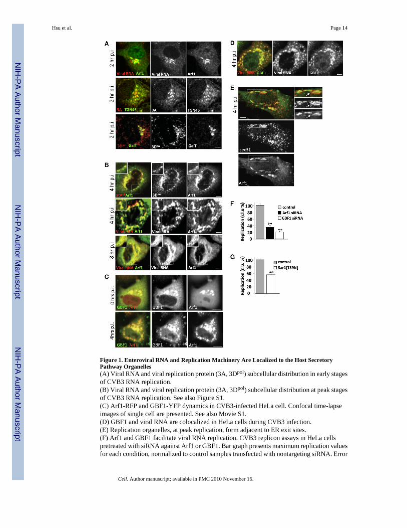
Figure 1. Enteroviral RNA and Replication Machinery Are Localized to the Host SecretoryPathway Organelles(A) Viral RNA and viral replication protein (3A, 3Dpol) subcellular distribution in early stagesof CVB3 RNA replication.(B) Viral RNA and viral replication protein (3A, 3Dpol) subcellular distribution at peak stagesof CVB3 RNA replication. See also Figure S1.(C) Arf1-RFP and GBF1-YFP dynamics in CVB3-infected HeLa cell. Confocal time-lapseimages of single cell are presented. See also Movie S1.(D) GBF1 and viral RNA are colocalized in HeLa cells during CVB3 infection.(E) Replication organelles, at peak replication, form adjacent to ER exit sites.(F) Arf1 and GBF1 facilitate viral RNA replication. CVB3 replicon assays in HeLa cellspretreated with siRNA against Arf1 or GBF1. Bar graph presents maximum replication valuesfor each condition, normalized to control samples transfected with nontargeting siRNA. Error
Hsu et al. Page 14
Cell. Author manuscript; available in PMC 2010 November 16.
NIH
-PA Author Manuscript
NIH
-PA Author Manuscript
NIH
-PA Author Manuscript
bars are SEM from eight replicates for each condition (**p < 0.01). See also Figures S2A–S2Cand Table S1.(G) Functional ER exit sites facilitate viral RNA replication. PV replicon assays in HeLa cellstransiently expressing Sar1[T39N] plasmid. Bar graph presents maximum replication values,normalized to control samples transfected with GFP. Error bars are SEM from eight replicatesfor each condition (**p < 0.01). See also Table S1. r.l.u% = relative light unit %. Allfluorescence images were confocal images of optical slice thickness ~1 mm. Scale bar, 10μm.
Hsu et al. Page 15
Cell. Author manuscript; available in PMC 2010 November 16.
NIH
-PA Author Manuscript
NIH
-PA Author Manuscript
NIH
-PA Author Manuscript

Figure 2. Reorganization of Secretory Pathway Organelles after Enteroviral InfectionHeLa cells (A) coexpressing Arf1-RFP/εCOP-YFP; expressing Arf1-GFP (B, C, E–I); orcoexpressing Arf1-RFP/GalT-YFP (see Movie S2) (D) were infected with CVB3 for 4 hr. Cellsin (B), (C), (E)–(I) were fixed and coimmunostained with anti-GFP and (B) anti-clathrin heavychain; (C) anti-γ-adaptin; (E) anti-GM130; (F) anti-TGN46; (G) anti-GGA1; (H) anti-Rab1b;(I) anti-ERGIC53 antibodies. Arrows in (A), (B), (C), and (D) indicate Arf1-labeledmembranes that do not label with εCOP-YFP, clathrin, γ-adaptin, and GalT-YFP, respectively.See also Figures S3A–S3H and Figure S4.All fluorescence images were confocal images of optical slice thickness ~1 μm. Scale bar, 10μm.
Hsu et al. Page 16
Cell. Author manuscript; available in PMC 2010 November 16.
NIH
-PA Author Manuscript
NIH
-PA Author Manuscript
NIH
-PA Author Manuscript

Figure 3. PI4KIIIβ Is Localized to Enteroviral RNA Replication Organelles(A) PI4KIIIβ and Arf1 are colocalized throughout CVB3 infection. HeLa cells expressingArf1-GFP infected with CVB3 were immunostained with anti-PI4KIIIβ and anti-GFPantibodies.(B) GBF1/Arf1 inactivation leads to PI4KIIIβ dispersal. HeLa cells were infected with CVB3for 4 hr and treated with 10 μg/ml of BFA for 30 min. 75% ± 5% (n = 10 cells) of PI4KIIIβ-associated fluorescence was dispersed upon BFA treatment. (C) PI4KIIIβ is in physicalcomplex with viral replication enzymes. HeLa cell lysates from cells infected with CVB3 ormock (−) for 4 hr were immunoprecipitated with anti-PI4KIIIβ antibodies. Input samplesverifying the presence of TGN46 in the lysates prior to immunoprecipitation are presented inthe bottom panel.All fluorescence images were confocal images of optical slice thickness ~1 mm. Scale bar, 10μm.
Hsu et al. Page 17
Cell. Author manuscript; available in PMC 2010 November 16.
NIH
-PA Author Manuscript
NIH
-PA Author Manuscript
NIH
-PA Author Manuscript
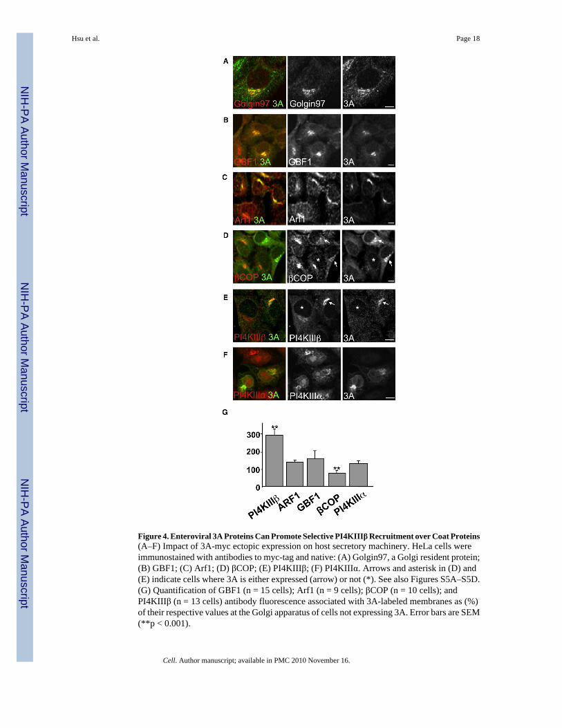
Figure 4. Enteroviral 3A Proteins Can Promote Selective PI4KIIIβ Recruitment over Coat Proteins(A–F) Impact of 3A-myc ectopic expression on host secretory machinery. HeLa cells wereimmunostained with antibodies to myc-tag and native: (A) Golgin97, a Golgi resident protein;(B) GBF1; (C) Arf1; (D) βCOP; (E) PI4KIIIβ; (F) PI4KIIIα. Arrows and asterisk in (D) and(E) indicate cells where 3A is either expressed (arrow) or not (*). See also Figures S5A–S5D.(G) Quantification of GBF1 (n = 15 cells); Arf1 (n = 9 cells); βCOP (n = 10 cells); andPI4KIIIβ (n = 13 cells) antibody fluorescence associated with 3A-labeled membranes as (%)of their respective values at the Golgi apparatus of cells not expressing 3A. Error bars are SEM(**p < 0.001).
Hsu et al. Page 18
Cell. Author manuscript; available in PMC 2010 November 16.
NIH
-PA Author Manuscript
NIH
-PA Author Manuscript
NIH
-PA Author Manuscript
All fluorescence images were confocal images of optical slice thickness ~1 μm. Scale bar, 10μm.
Hsu et al. Page 19
Cell. Author manuscript; available in PMC 2010 November 16.
NIH
-PA Author Manuscript
NIH
-PA Author Manuscript
NIH
-PA Author Manuscript

Figure 5. PI4KIIIβ Activity Is Required for Enteroviral RNA Replication(A and B) PIK93 block of PI4KIIIβ activity inhibits CVB3 and PV RNA replication. CVB3and PV replicon assays in cells treated with 500 nM and 1 μM PIK93 are shown. Bar graphspresent maximum replication values for CVB3 (A) and PV (B) with PIK93 treatment,normalized to control (DMSO) treatment. See also Figures S6A–S6C.(C) Kinetics of inhibition by PIK93 presented for the CVB3 replicon.(D and E) Reduction of PI4KIIIβ levels with siRNA inhibits CVB3 and PV RNA replication.CVB3 and PV replicon assays in HeLa cells; bar graphs present maximum replication valuesfor each condition, normalized to control (nontargeting) siRNA. See also Figure S2A.(F) Kinetics of inhibition by PI4KIIIβ siRNA presented for PV replicon.(G and H) Expression of ectopic kinase-dead PI4KIIIβ (PI4KIIIβ-KD) inhibits CVB3 and PVreplication. CVB3 and PV replicon assays in HeLa cells; bar graphs present maximumreplication values for each condition, normalized to control (GFP) plasmid ectopic expression.(I) Kinetics of inhibition by PI4KIIIβ-KD presented for the CVB3 replicon. Error bars in eachCVB3 assay are SEM of six CVB3 samples and each PV assay is SEM for eight PV samples.(**p < 0.01).(J and K) PI4KIIIβ activity regulates viral RNA synthesis. Cell-free PV RNA translation (J)and synthesis (K) assays performed in the presence of PIK93.
Hsu et al. Page 20
Cell. Author manuscript; available in PMC 2010 November 16.
NIH
-PA Author Manuscript
NIH
-PA Author Manuscript
NIH
-PA Author Manuscript
Figure 6. PI4P Lipid Microenvironment within Replication Organelles Regulates both Enteroviraland Flaviviral RNA Replication(A) Cellular PI4P lipid levels rise in CVB3 infection. Total cellular PI4P lipids were quantifiedover time. Error bars are SEM from duplicate samples (**p < 0.01).(B) Reduction in PI4P lipid levels inhibits enteroviral RNA replication. PV replicon assays inHeLa cells ectopically expressing Sac1 are shown. Inset: western blot showing increase in Sac1levels after 16 hr of ectopic expression. Bar graph presents maximum replication values,normalized to control (GFP) plasmid ectopic expression. Error bars are SEM from eightreplicates cells (**p < 0.01).(C) PI4P lipids localize to enteroviral replication organelles. Time-lapse confocal images ofsingle HeLa cell infected with CVB3, coexpressing FAPP1PH-GFP/Arf1-RFP (see also MovieS3).
Hsu et al. Page 21
Cell. Author manuscript; available in PMC 2010 November 16.
NIH
-PA Author Manuscript
NIH
-PA Author Manuscript
NIH
-PA Author Manuscript
(D) PI4KIIIβ activity is responsible for PI4P lipids at enteroviral replication organelles. Time-lapse confocal image of single HeLa cell coexpressing FAPP1-PHGFP/ARF1-RFP pre- andpost-PIK93 treatment at 4 hr post-infection with CVB3.(E) Quantification of FAPP1PH-GFP to Arf1-RFP fluorescence from (D) expressed as a ratio.Error bars are SEM from ten cells for each condition.(F and G) PV RNA polymerase specifically and preferentially binds PI4P lipids. Purifiedrecombinant 3Dpol enzyme was incubated with membrane strips that were previously spottedwith different lipids. Antibodies detected 3Dpol binding. Two representative blots are shown.Binding was quantified and plotted in bar graph as a ratio over background non-lipid spottedmembrane from three different experiments for each lipid type.(H) Cellular PI4P lipid levels rise in HCV replicating cells. Quantification of PI4P with anti-PI4P primary antibodies of 3-5B(HCV) cells, normalized as % of PI4P lipid quantificationwithin Huh7 cells. Error bars are SEM from ten cells for each cell type.(I) PI4P lipids localize to HCV replication membranes. PI4P lipid and NS5A proteindistribution in Huh7 and 3-5B(HCV) cells were determined by immunostaining with anti-PI4Pand anti-NS5A antibodies.(J and K) PI4KIIIβ is responsible for a significant fraction of PI4P lipids at HCV replicationmembranes. 3-5B(HCV) cells were treated with nontargeting, PI4KIIIβ, or PI4KIIIα siRNA.Cells were immunostained and quantified for PI4P lipids. Representative images of groups ofsiRNA-treated cells are shown (J). Quantification was done on 20 cells for each siRNAtreatment condition.(L) Reduction of PI4P lipids inhibits HCV replication. HCV replicon assays conducted withJ6/JFH (p7-Rluc2A) replicons in Huh7 cells ectopically expressing Sac1, PI4KIIIβ-KD, orboth plasmids are shown. Bar graph presents maximum replication value normalized to control(GFP) plasmid ectopic expression. Error bars are SEM from eight replicates of cells for eachtreatment condition (**p < 0.01).All fluorescence images were confocal images of optical slice thickness ~1 mm. Scale bar, 10μm.
Hsu et al. Page 22
Cell. Author manuscript; available in PMC 2010 November 16.
NIH
-PA Author Manuscript
NIH
-PA Author Manuscript
NIH
-PA Author Manuscript
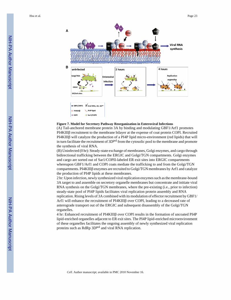
Figure 7. Model for Secretory Pathway Reorganization in Enteroviral Infections(A) Tail-anchored membrane protein 3A by binding and modulating GBF1/Arf1 promotesPI4KIIIβ recruitment to the membrane bilayer at the expense of coat protein COPI. RecruitedPI4KIIIβ will catalyze the production of a PI4P lipid micro-environment (red lipids) that willin turn facilitate the recruitment of 3Dpol from the cytosolic pool to the membrane and promotethe synthesis of viral RNA.(B) Uninfected (0 hr): Steady-state exchange of membranes, Golgi enzymes, and cargo throughbidirectional trafficking between the ERGIC and Golgi/TGN compartments. Golgi enzymesand cargo are sorted out of Sar1/COPII-labeled ER exit sites into ERGIC compartmentswhereupon GBF1/Arf1 and COPI coats mediate the trafficking to and from the Golgi/TGNcompartments. PI4KIIIβ enzymes are recruited to Golgi/TGN membranes by Arf1 and catalyzethe production of PI4P lipids at these membranes.2 hr: Upon infection, newly synthesized viral replication enzymes such as the membrane-bound3A target to and assemble on secretory organelle membranes but concentrate and initiate viralRNA synthesis on the Golgi/TGN membranes, where the pre-existing (i.e., prior to infection)steady-state pool of PI4P lipids facilitates viral replication protein assembly and RNAreplication. Rising levels of 3A combined with its modulation of effector recruitment by GBF1/Arf1 will enhance the recruitment of PI4KIIIβ over COPI, leading to a decreased rate ofanterograde transport out of the ERGIC and subsequent disassembly of the Golgi/TGNorganelles.4 hr: Enhanced recruitment of PI4KIIIβ over COPI results in the formation of uncoated PI4Plipid-enriched organelles adjacent to ER exit sites. The PI4P lipid-enriched microenvironmentof these organelles facilitates the ongoing assembly of newly synthesized viral replicationproteins such as RdRp 3Dpol and viral RNA replication.
Hsu et al. Page 23
Cell. Author manuscript; available in PMC 2010 November 16.
NIH
-PA Author Manuscript
NIH
-PA Author Manuscript
NIH
-PA Author Manuscript