Highly Dynamic Host Actin Reorganization around Developing Plasmodium Inside Hepatocytes
9
Highly Dynamic Host Actin Reorganization around Developing Plasmodium Inside Hepatocytes Carina S. S. Gomes-Santos 1,2 , Maurice A. Itoe 1 , Cristina Afonso 1 , Ricardo Henriques 3 , Rui Gardner 4 , Nuno Sepu ´ lveda 4,5 , Pedro D. Simo ˜ es 6 , Helena Raquel 6 , Anto ´ nio Paulo Almeida 7 , Luis F. Moita 6 , Friedrich Frischknecht 8 *, Maria M. Mota 1 * 1 Malaria Unit, Instituto de Medicina Molecular, Faculdade de Medicina Universidade de Lisboa, Lisboa, Portugal, 2 PhD Programme in Experimental Biology and Biomedicine, Center for Neuroscience and Cell Biology, University of Coimbra, Coimbra, Portugal, 3 Cell Biology Unit, Instituto de Medicina Molecular, Universidade de Lisboa, Lisboa, Portugal, 4 Instituto Gulbenkian de Cie ˆncia, Oeiras, Portugal, 5 Center of Statistics and Applications, University of Lisbon, Lisboa, Portugal, 6 Cell Biology of the Immune System Unit, Instituto de Medicina Molecular, Universidade de Lisboa, Lisboa, Portugal, 7 Unidade de Entomologia Me ´dica/UPMM, Instituto de Higiene e Medicina Tropical, Universidade Nova de Lisboa, Lisboa, Portugal, 8 Parasitology, Department of Infectious Diseases, University of Heidelberg Medical School, University of Heidelberg, Heidelberg, Germany Abstract Plasmodium sporozoites are transmitted by Anopheles mosquitoes and infect hepatocytes, where a single sporozoite replicates into thousands of merozoites inside a parasitophorous vacuole. The nature of the Plasmodium-host cell interface, as well as the interactions occurring between these two organisms, remains largely unknown. Here we show that highly dynamic hepatocyte actin reorganization events occur around developing Plasmodium berghei parasites inside human hepatoma cells. Actin reorganization is most prominent between 10 to 16 hours post infection and depends on the actin severing and capping protein, gelsolin. Live cell imaging studies also suggest that the hepatocyte cytoskeleton may contribute to parasite elimination during Plasmodium development in the liver. Citation: Gomes-Santos CSS, Itoe MA, Afonso C, Henriques R, Gardner R, et al. (2012) Highly Dynamic Host Actin Reorganization around Developing Plasmodium Inside Hepatocytes. PLoS ONE 7(1): e29408. doi:10.1371/journal.pone.0029408 Editor: Stefan Kappe, Seattle Biomedical Research Institute - University of Washington, United States of America Received September 6, 2011; Accepted November 28, 2011; Published January 6, 2012 Copyright: ß 2012 Gomes-Santos et al. This is an open-access article distributed under the terms of the Creative Commons Attribution License, which permits unrestricted use, distribution, and reproduction in any medium, provided the original author and source are credited. Funding: The work was supported by Fundac ¸a ˜o para a Cie ˆncia e Tecnologia (FCT) of the Portuguese Ministry of Science (PTDC/SAU-GMG/100313/2008), the Federal German Ministry of education and Science (Biofuture) C.S.S.G-S. was supported by an FCT fellowship (SFRH/BD/15888/2005). M.A.I. was funded by the EU FP 7 Marie Curie Initial Training Network ‘‘Intervention Strategies against Malaria (InterMalTraining)’’, contract no. 215281. L.F.M. is a Young Investigator from the Human Frontier Science Program and receives support from Fundac ¸a ˜o Luso-Americana para o Desenvolvimento and Fundac ¸a ˜o para a Cie ˆncia e a Tecnologia (PTDC/SAU-MII/69280/2006 and PTDC/SAU-MII/78333/2006). F.F. thanks the Chica and Heinz Schaller Foundation for support. M.M.M. and F.F. are members of the EU FP7 Network of Excellence EVIMalaR. The funders had no role in study design, data collection and analysis, decision to publish, or preparation of the manuscript. Competing Interests: The authors have declared that no competing interests exist. * E-mail: [email protected] (FF); [email protected] (MMM) Introduction Diverse pathogens have developed numerous strategies to successfully survive and replicate inside host cells. They often subvert signalling pathways and cytoskeletal components of the host cell to avoid the host’s immune system and for direct access to host metabolites [1]. The bacteria Salmonella enterica, for example, induces host cell actin reorganization and ruffle formation to facilitate its internalization into non-phagocytic cells [2], while the vaccinia virus moves along microtubules and polymerizes actin to form comet tails that allow viral dissemination into non-infected cells [reviewed in 3]. Besides bacteria and viruses, protozoan parasites are also capable of manipulating the host cell cytoskeleton. The apicomplexan parasite Cryptosporidium parvum, responsible for diarrheal illness, induces the reorganization of the host’s actin network into a plaque-like structure that separates the parasite from the cell cytoplasm, thereby creating an intracellular but extracytoplasmic niche, where it replicates [4]. During Theileria infection, host microtubules associate with the parasite, which divides in synchrony with the host cell [5,6]. Invasion by Toxoplasma gondii induces the formation of a host F-actin ring at the junction site [7] and, at a later stage of infection, T. gondii recruits microtubules, proposed to form conduits along which host organelles are transported to the parasitophorous vacuole [8]. The malaria parasites (Plasmodium spp.) of mammals first replicate asexually in hepatocytes and later in red blood cells (RBCs). Several studies show that, within RBCs, Plasmodium exports proteins to the host cell cytosol that manipulate the host cell cytoskeleton, important for parasite egress and progression of infection [9,10,11]. However, during the liver stage of infection, few reports exist on the interaction of the host cell cytoskeleton with Plasmodium. While no significant reorganization of host microtubules or actin was reported in fixed cells at 24 hours of Plasmodium spp. development [12], an F-actin ring in the cell–parasite junction was observed during invasion of hepatocytes by sporozoites [7]. Here, we investigate the hepatocyte actin and microtubule organization during Plasmodium berghei development, using live cell imaging. Results and Discussion Reorganization of hepatocyte actin, but not tubulin, occurs around developing P. berghei To investigate a potential reorganization of the hepatocyte cytoskeleton during Plasmodium infection, we established Huh7 cell PLoS ONE | www.plosone.org 1 January 2012 | Volume 7 | Issue 1 | e29408
Transcript of Highly Dynamic Host Actin Reorganization around Developing Plasmodium Inside Hepatocytes
Highly Dynamic Host Actin Reorganization aroundDeveloping Plasmodium Inside HepatocytesCarina S. S. Gomes-Santos1,2, Maurice A. Itoe1, Cristina Afonso1, Ricardo Henriques3, Rui Gardner4, Nuno
Sepulveda4,5, Pedro D. Simoes6, Helena Raquel6, Antonio Paulo Almeida7, Luis F. Moita6, Friedrich
Frischknecht8*, Maria M. Mota1*
1 Malaria Unit, Instituto de Medicina Molecular, Faculdade de Medicina Universidade de Lisboa, Lisboa, Portugal, 2 PhD Programme in Experimental Biology and
Biomedicine, Center for Neuroscience and Cell Biology, University of Coimbra, Coimbra, Portugal, 3 Cell Biology Unit, Instituto de Medicina Molecular, Universidade de
Lisboa, Lisboa, Portugal, 4 Instituto Gulbenkian de Ciencia, Oeiras, Portugal, 5 Center of Statistics and Applications, University of Lisbon, Lisboa, Portugal, 6 Cell Biology of
the Immune System Unit, Instituto de Medicina Molecular, Universidade de Lisboa, Lisboa, Portugal, 7 Unidade de Entomologia Medica/UPMM, Instituto de Higiene e
Medicina Tropical, Universidade Nova de Lisboa, Lisboa, Portugal, 8 Parasitology, Department of Infectious Diseases, University of Heidelberg Medical School, University of
Heidelberg, Heidelberg, Germany
Abstract
Plasmodium sporozoites are transmitted by Anopheles mosquitoes and infect hepatocytes, where a single sporozoitereplicates into thousands of merozoites inside a parasitophorous vacuole. The nature of the Plasmodium-host cell interface,as well as the interactions occurring between these two organisms, remains largely unknown. Here we show that highlydynamic hepatocyte actin reorganization events occur around developing Plasmodium berghei parasites inside humanhepatoma cells. Actin reorganization is most prominent between 10 to 16 hours post infection and depends on the actinsevering and capping protein, gelsolin. Live cell imaging studies also suggest that the hepatocyte cytoskeleton maycontribute to parasite elimination during Plasmodium development in the liver.
Citation: Gomes-Santos CSS, Itoe MA, Afonso C, Henriques R, Gardner R, et al. (2012) Highly Dynamic Host Actin Reorganization around Developing PlasmodiumInside Hepatocytes. PLoS ONE 7(1): e29408. doi:10.1371/journal.pone.0029408
Editor: Stefan Kappe, Seattle Biomedical Research Institute - University of Washington, United States of America
Received September 6, 2011; Accepted November 28, 2011; Published January 6, 2012
Copyright: � 2012 Gomes-Santos et al. This is an open-access article distributed under the terms of the Creative Commons Attribution License, which permitsunrestricted use, distribution, and reproduction in any medium, provided the original author and source are credited.
Funding: The work was supported by Fundacao para a Ciencia e Tecnologia (FCT) of the Portuguese Ministry of Science (PTDC/SAU-GMG/100313/2008), theFederal German Ministry of education and Science (Biofuture) C.S.S.G-S. was supported by an FCT fellowship (SFRH/BD/15888/2005). M.A.I. was funded by the EUFP 7 Marie Curie Initial Training Network ‘‘Intervention Strategies against Malaria (InterMalTraining)’’, contract no. 215281. L.F.M. is a Young Investigator from theHuman Frontier Science Program and receives support from Fundacao Luso-Americana para o Desenvolvimento and Fundacao para a Ciencia e a Tecnologia(PTDC/SAU-MII/69280/2006 and PTDC/SAU-MII/78333/2006). F.F. thanks the Chica and Heinz Schaller Foundation for support. M.M.M. and F.F. are members of theEU FP7 Network of Excellence EVIMalaR. The funders had no role in study design, data collection and analysis, decision to publish, or preparation of themanuscript.
Competing Interests: The authors have declared that no competing interests exist.
* E-mail: [email protected] (FF); [email protected] (MMM)
Introduction
Diverse pathogens have developed numerous strategies to
successfully survive and replicate inside host cells. They often
subvert signalling pathways and cytoskeletal components of the
host cell to avoid the host’s immune system and for direct access to
host metabolites [1]. The bacteria Salmonella enterica, for example,
induces host cell actin reorganization and ruffle formation to
facilitate its internalization into non-phagocytic cells [2], while the
vaccinia virus moves along microtubules and polymerizes actin to
form comet tails that allow viral dissemination into non-infected
cells [reviewed in 3]. Besides bacteria and viruses, protozoan
parasites are also capable of manipulating the host cell
cytoskeleton. The apicomplexan parasite Cryptosporidium parvum,
responsible for diarrheal illness, induces the reorganization of the
host’s actin network into a plaque-like structure that separates the
parasite from the cell cytoplasm, thereby creating an intracellular
but extracytoplasmic niche, where it replicates [4]. During Theileria
infection, host microtubules associate with the parasite, which
divides in synchrony with the host cell [5,6]. Invasion by
Toxoplasma gondii induces the formation of a host F-actin ring at
the junction site [7] and, at a later stage of infection, T. gondii
recruits microtubules, proposed to form conduits along which host
organelles are transported to the parasitophorous vacuole [8].
The malaria parasites (Plasmodium spp.) of mammals first replicate
asexually in hepatocytes and later in red blood cells (RBCs). Several
studies show that, within RBCs, Plasmodium exports proteins to the
host cell cytosol that manipulate the host cell cytoskeleton, important
for parasite egress and progression of infection [9,10,11]. However,
during the liver stage of infection, few reports exist on the interaction
of the host cell cytoskeleton with Plasmodium. While no significant
reorganization of host microtubules or actin was reported in fixed
cells at 24 hours of Plasmodium spp. development [12], an F-actin ring
in the cell–parasite junction was observed during invasion of
hepatocytes by sporozoites [7]. Here, we investigate the hepatocyte
actin and microtubule organization during Plasmodium berghei
development, using live cell imaging.
Results and Discussion
Reorganization of hepatocyte actin, but not tubulin,occurs around developing P. berghei
To investigate a potential reorganization of the hepatocyte
cytoskeleton during Plasmodium infection, we established Huh7 cell
PLoS ONE | www.plosone.org 1 January 2012 | Volume 7 | Issue 1 | e29408
lines stably expressing mCherry::b-actin or mCherry::a-tubulin
fusion proteins. Anti-a-tubulin antibody or phalloidin labelling
showed that all microtubules stained with the antibody were
positive for mCherry::a-tubulin and all the filamentous actin (F-
actin) structures stained with phalloidin, were also positive for
mCherry::b-actin, showing integration of exogenous proteins into
the microtubules or the F-actin of the living cells, respectively (Fig.
S1). Transformed cell lines were indistinguishable from the parent
lines, with unperturbed key cellular events involving cytoskeletal
dynamics in both mCherry lines (data not shown). Furthermore,
infection of these cells with GFP-Pb proceeded at the same rate as
in control Huh7 cells (Fig. S2).
We next aimed to identify the possible interactions between
these components of the host cell cytoskeleton and the developing
Plasmodium parasite. Cells from both cell lines were infected with
GFP-Plasmodium berghei (GFP-Pb) sporozoites and observed by wide
field fluorescence microscopy at different times after infection.
Time lapse experiments, with 20 seconds acquisition intervals,
were performed between 3 and 34 hours p.i. to follow the host
cytoskeleton dynamics around the parasite during its development.
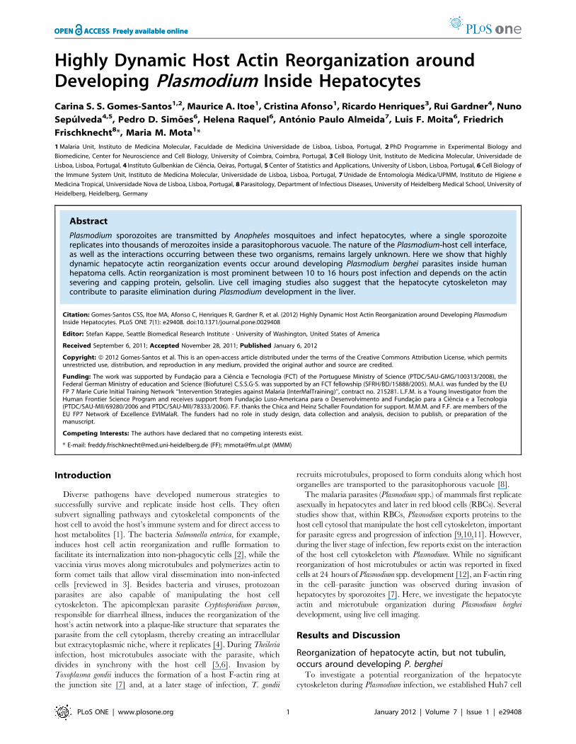
No significant host microtubule reorganization was observed
around 238 GFP-Pb parasites (Fig. 1A; Movie S1). However, clear
host cell actin reorganization events, characterized by changes of
mCherry::b-actin fluorescence around the parasites, were ob-
served around 77 out of 562 developing GFP-Pb (1462%)
analysed between 3 and 34 hours p.i. (Fig. 1A; Movie S2). Host
actin reorganization events were highly dynamic, comprising
cycles of polymerization and depolymerization around the parasite
(Movie S2). Data analysis showed that, although present
throughout infection, this phenomenon occurred preferentially
between 10 to 16 hours p.i. (2363%, p,0.01) (Fig. 1B). Although
not much is known about the biological processes occurring during
intra-hepatic Plasmodium development, the interval between 10 and
16 hours p.i. may coincide with an important step in the
preparation for the extensive nuclear replication that starts soon
after that period [13]. We next determined whether actin
polymerization around developing GFP-Pb also occurs during
liver infection in vivo, by staining liver slices of BALB/c mice
24 hours p.i., with the F-actin binding toxin fluorescent conjugat-
ed, phalloidin. Clear actin rings were observed around approx-
imately 4% (3 out of 76) of the exoerythrocytic forms (EEFs)
analysed by fluorescence confocal microscopy (Fig. 1C). Thus, our
observations show that hepatocyte actin reorganization events also
occur during EEF development in the liver of infected mice. The
lower percentage of actin rings around mouse liver EEFs,
compared to the live cell imaging experiments formerly presented,
is also observed in vitro around fixed EEFs (data not shown). This is
probably due to the fact that the observed actin polymerization
events are extremely dynamic, as shown in our live cell imaging
experiments, and as such much more difficult to capture in fixed
conditions.
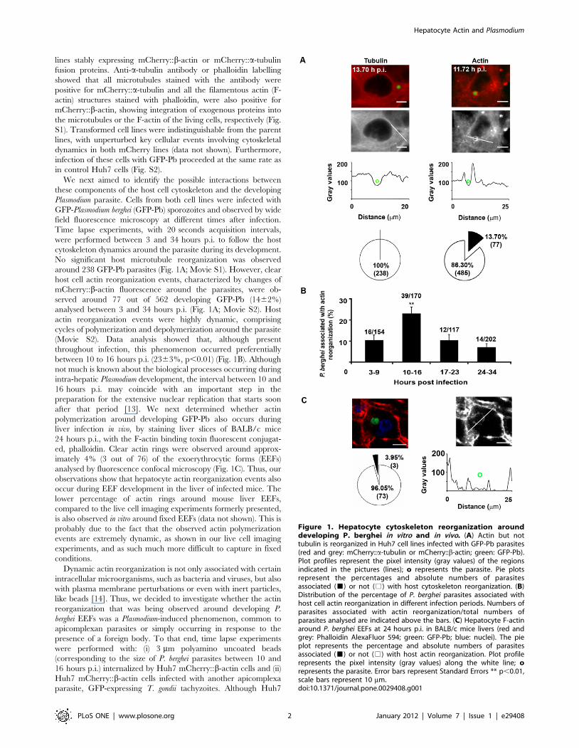
Dynamic actin reorganization is not only associated with certain
intracellular microorganisms, such as bacteria and viruses, but also
with plasma membrane perturbations or even with inert particles,
like beads [14]. Thus, we decided to investigate whether the actin
reorganization that was being observed around developing P.
berghei EEFs was a Plasmodium-induced phenomenon, common to
apicomplexan parasites or simply occurring in response to the
presence of a foreign body. To that end, time lapse experiments
were performed with: (i) 3 mm polyamino uncoated beads
(corresponding to the size of P. berghei parasites between 10 and
16 hours p.i.) internalized by Huh7 mCherry::b-actin cells and (ii)
Huh7 mCherry::b-actin cells infected with another apicomplexa
parasite, GFP-expressing T. gondii tachyzoites. Although Huh7
Figure 1. Hepatocyte cytoskeleton reorganization arounddeveloping P. berghei in vitro and in vivo. (A) Actin but nottubulin is reorganized in Huh7 cell lines infected with GFP-Pb parasites(red and grey: mCherry::a-tubulin or mCherry::b-actin; green: GFP-Pb).Plot profiles represent the pixel intensity (gray values) of the regionsindicated in the pictures (lines); o represents the parasite. Pie plotsrepresent the percentages and absolute numbers of parasitesassociated (&) or not (%) with host cytoskeleton reorganization. (B)Distribution of the percentage of P. berghei parasites associated withhost cell actin reorganization in different infection periods. Numbers ofparasites associated with actin reorganization/total numbers ofparasites analysed are indicated above the bars. (C) Hepatocyte F-actinaround P. berghei EEFs at 24 hours p.i. in BALB/c mice livers (red andgrey: Phalloidin AlexaFluor 594; green: GFP-Pb; blue: nuclei). The pieplot represents the percentage and absolute numbers of parasitesassociated (&) or not (%) with host actin reorganization. Plot profilerepresents the pixel intensity (gray values) along the white line; orepresents the parasite. Error bars represent Standard Errors ** p,0.01,scale bars represent 10 mm.doi:10.1371/journal.pone.0029408.g001
Hepatocyte Actin and Plasmodium
PLoS ONE | www.plosone.org 2 January 2012 | Volume 7 | Issue 1 | e29408
cells are not professional phagocytes, they clearly internalize beads
following a 1 hour starvation period. These experiments were
performed between 10 and 16 hours post-bead internalization or
T. gondii infection, corresponding to the interval of P. berghei
infection where there is the highest percentage of Plasmodium
parasites associated with host actin reorganization, employing
experimental conditions that mimicked infection by Plasmodium
(see Material and Methods).
Comparison of actin reorganization events, during P. berghei
infection (22.963%), T. gondii infection (3.3%, s.e. = 3%,
CI95 = (0.1%,17%)) or after internalization of beads (7.6%,
s.e. = 3%, CI95 = (3.5%,14.5%)), showed that the events observed
around T. gondii or beads were significantly less frequent than those
occurring around Plasmodium (Fig. 2). The few events occurring
around beads or T. gondii were also less intense and dynamic than
those observed around P. berghei in the same period of time (Movie
S3). Previous work by Yam and colleagues show that similar actin
dynamics can be observed around E-cadherin covered beads, E.
coli and Listeria-containing phagosomes in MDCK cells [14].
However, our work shows that, in hepatocytes, the actin dynamics
around Plasmodium is significantly more frequent and intense, when
compared to the one observed around uncoated beads or the
related parasite, T. gondii. This suggests that, although the
phenomena that we observe may be part of the normal actin
dynamics of the cells, specific features of the Plasmodium, or of its
parasitophorous vacuole membrane, also play an important role in
the process.
Dynamics of hepatocyte actin reorganization eventsassociated with developing P. berghei
We termed the actin reorganization events around Plasmodium
actin clouds. The dynamics of actin clouds were characterized by
the accumulation of actin in close vicinity of the PV, which
appeared either asymmetrical, moving around the vacuole
(Fig. 3A; Movie S4), or symmetrical, surrounding the entire PV
(Fig. 3A; Movie S2). Actin clouds could also rearrange into polar
structures reminiscent of actin tails (Fig. 3B; Movie S5). Actin
reorganization could last for the whole duration of the movie, e.g.
2 hours, while others lasted for just a few minutes or even seconds.
In all cases, actin clouds were very dynamic, as seen in Movies S4
and S5.
The parasites associated with actin clouds usually showed a non
progressive movement (Movie S4). However, some parasites
associated with tail-like actin clouds were clearly prone to
translocation inside the cell. The longest distance observed for
an intracellular parasite translocation event was 60 mm, which
occurred between two positions separated by 19 mm within the cell
(Fig. 3C). No specific directional pattern was observed from 12
moving parasites.
Host cell actin reorganization observed around P. berghei
resembles the type of actin dynamics associated with other
pathogenic systems, such as Listeria-containing phagosomes [14],
or even with normal cellular functions, such as the movement of
endosomes or other intracellular vesicles inside the cell cytosol
[15,16]. Similarly, Plasmodium is also surrounded by the para-
sitophorous vacuole membrane, suggesting that actin dynamics
observed around this apicomplexan parasite might originate at this
membrane. Indeed, it has been demonstrated that membranes can
be associated with actin polymerization events, depending on their
lipid content [17]. Considering this, it is tempting to hypothesize
that the actin dynamics observed around Plasmodium might be
related with active vacuole membrane remodelling as part of the
vacuole maturation process during parasite development.
Gelsolin is involved in host actin reorganizationassociated with developing P. berghei
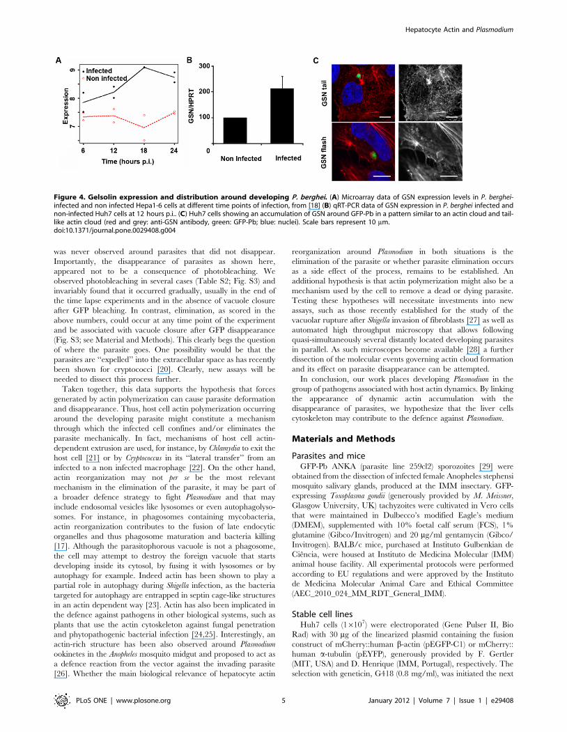
We next sought to identify actin related proteins involved in the
dynamic events observed around the developing Plasmodium. A
recent microarray screen of Plasmodium-infected versus non-
infected cells [18] showed that gelsolin (GSN) transcripts are
significantly up-regulated throughout infection, during all time
points of infection analysed in that study (Fig. 4A). GSN severs
actin filaments and remains attached to the barbed end of the
short filament, preventing elongation. When GSN uncaps these
filaments, many actin polymerization points become available in
the cell cytosol to generate new actin filaments [19]. We confirmed
the microarray data by quantitative RT-PCR (qRT-PCR),
comparing infected Huh7 cells 12 hours p.i. and non-infected
cells (Fig. 4B). When infected Huh7 cells were stained with an anti-
GSN antibody, structures resembling actin clouds were observed
(Fig. 4C).
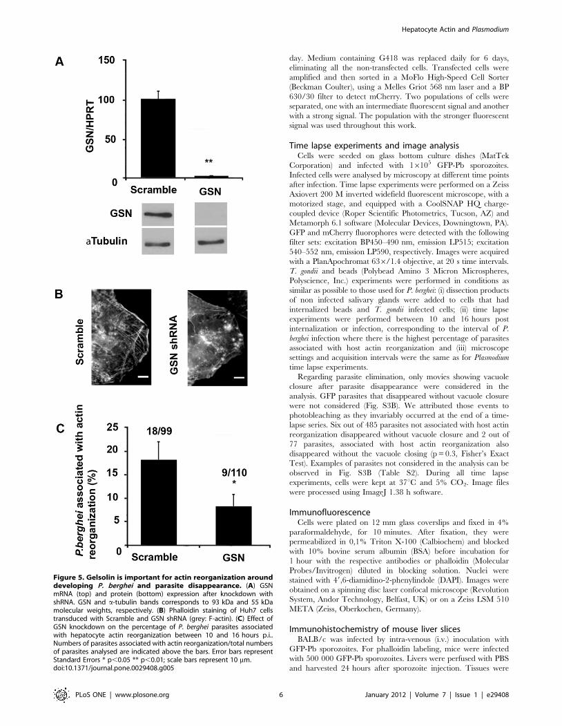
To determine the contribution of GSN to actin reorganization
events, we performed GSN knockdown experiments in the Huh7
mCherry::b-actin cell line by lentiviral-delivered shRNA (Fig. 5A),
followed by infection with GFP-Pb sporozoites. Time lapse
experiments of Plasmodium infection (10 to 16 hours p.i.) showed
that when GSN expression is efficiently down-modulated
(97.560.5%, Fig. 5A), actin structures are perturbed (Fig. 5B)
and the percentage of parasites associated with actin clouds is
significantly lower than that observed in the control situation
(Fig. 5C). Attempts to deplete other actin related proteins, such as
Arp2 and Arp3 from the Arp2/3 complex as well as N-WASP,
usually involved in this type of actin phenomenon, were made but
without success due to significant cell death after knockdown of
these proteins. The results clearly show that, although other
proteins are also likely to be involved in P. berghei-associated
hepatocyte actin reorganization, GSN is an important player in
this process. Most probably contributing to the actin turnover and
consequently the dynamics of the actin clouds.
Biological relevance of hepatocyte actin reorganizationaround developing Plasmodium
Many pathogens hijack the host cytoskeleton for their own
benefit, either to spread from cell to cell or to capture important
nutrients [3]. During live cell imaging experiments we noticed the
disappearance of some parasites to coincide with extremely
Figure 2. Hepatocyte cytoskeleton reorganization depends onthe presence of P. berghei. Percentage of parasites (P. berghei and T.gondii) and beads associated with hepatocyte actin reorganizationbetween 10 and 16 hours p.i. or bead internalization. Numbers ofparasites or beads associated with actin reorganization/total numbersof parasites or beads analysed are indicated above the bars. Error barsrepresent Standard Errors * p,0.05 *** p#0.001.doi:10.1371/journal.pone.0029408.g002
Hepatocyte Actin and Plasmodium
PLoS ONE | www.plosone.org 3 January 2012 | Volume 7 | Issue 1 | e29408

dynamic actin events. Indeed, 5.2% of the parasites recorded
during actin reorganization events disappear (Fig. 6; Movie S6;
Table S1). In the experiment shown in movie S6, a progressively
stronger actin cloud around the vacuole was observed, which first
deformed and finally eliminated the parasite leading to an
apparent vacuole closure (Fig. 6). In contrast, only 0.8% of the
parasites not associated with actin reorganization show the same
phenotype (Table S1). This significant difference (p = 0.015, odds
ratio of 6.5, Fisher’s Exact Test) implies that hepatocyte actin
dynamics is positively associated with parasite elimination
throughout infection and that it is 6.5 times more likely that a
parasite disappears associated with an actin event than in the
absence of that event. The observation that not all actin
reorganization events were associated with parasite elimination,
might be due to the fact that some parasites are more fit than
others and therefore offer more resistance to intense actin
polymerization and consequent mechanical elimination. However,
strong accumulation of actin as the one shown in Fig. 6/movie S6
Figure 3. Dynamics of hepatocyte actin clouds around developing P. berghei. Asymmetrical and symmetrical actin clouds (A) as well as tail-like actin clouds (B) are present around GFP-Pb (red and grey: mCherry::b-actin; green: GFP-Pb), surface plots represent the intensity of the pixels ofthe mCherry::b-actin channel in the selected region (square). (C) Translocation of a GFP-Pb, associated with a tail-like actin cloud. The tracked path isindicated in blue (red and grey: mCherry::b-actin; green: GFP-Pb). Scale bars represent 10 mm. Some frames present the same time points as theacquisition interval between individual images was 20 s ( = 0.0056 h) and thus too small to annotate in consecutive frames, when using a time scaleof hours.doi:10.1371/journal.pone.0029408.g003
Hepatocyte Actin and Plasmodium
PLoS ONE | www.plosone.org 4 January 2012 | Volume 7 | Issue 1 | e29408
was never observed around parasites that did not disappear.
Importantly, the disappearance of parasites as shown here,
appeared not to be a consequence of photobleaching. We
observed photobleaching in several cases (Table S2; Fig. S3) and
invariably found that it occurred gradually, usually in the end of
the time lapse experiments and in the absence of vacuole closure
after GFP bleaching. In contrast, elimination, as scored in the
above numbers, could occur at any time point of the experiment
and be associated with vacuole closure after GFP disappearance
(Fig. S3; see Material and Methods). This clearly begs the question
of where the parasite goes. One possibility would be that the
parasites are ‘‘expelled’’ into the extracellular space as has recently
been shown for cryptococci [20]. Clearly, new assays will be
needed to dissect this process further.
Taken together, this data supports the hypothesis that forces
generated by actin polymerization can cause parasite deformation
and disappearance. Thus, host cell actin polymerization occurring
around the developing parasite might constitute a mechanism
through which the infected cell confines and/or eliminates the
parasite mechanically. In fact, mechanisms of host cell actin-
dependent extrusion are used, for instance, by Chlamydia to exit the
host cell [21] or by Cryptococcus in its ‘‘lateral transfer’’ from an
infected to a non infected macrophage [22]. On the other hand,
actin reorganization may not per se be the most relevant
mechanism in the elimination of the parasite, it may be part of
a broader defence strategy to fight Plasmodium and that may
include endosomal vesicles like lysosomes or even autophagolyso-
somes. For instance, in phagosomes containing mycobacteria,
actin reorganization contributes to the fusion of late endocytic
organelles and thus phagosome maturation and bacteria killing
[17]. Although the parasitophorous vacuole is not a phagosome,
the cell may attempt to destroy the foreign vacuole that starts
developing inside its cytosol, by fusing it with lysosomes or by
autophagy for example. Indeed actin has been shown to play a
partial role in autophagy during Shigella infection, as the bacteria
targeted for autophagy are entrapped in septin cage-like structures
in an actin dependent way [23]. Actin has also been implicated in
the defence against pathogens in other biological systems, such as
plants that use the actin cytoskeleton against fungal penetration
and phytopathogenic bacterial infection [24,25]. Interestingly, an
actin-rich structure has been also observed around Plasmodium
ookinetes in the Anopheles mosquito midgut and proposed to act as
a defence reaction from the vector against the invading parasite
[26]. Whether the main biological relevance of hepatocyte actin
reorganization around Plasmodium in both situations is the
elimination of the parasite or whether parasite elimination occurs
as a side effect of the process, remains to be established. An
additional hypothesis is that actin polymerization might also be a
mechanism used by the cell to remove a dead or dying parasite.
Testing these hypotheses will necessitate investments into new
assays, such as those recently established for the study of the
vacuolar rupture after Shigella invasion of fibroblasts [27] as well as
automated high throughput microscopy that allows following
quasi-simultaneously several distantly located developing parasites
in parallel. As such microscopes become available [28] a further
dissection of the molecular events governing actin cloud formation
and its effect on parasite disappearance can be attempted.
In conclusion, our work places developing Plasmodium in the
group of pathogens associated with host actin dynamics. By linking
the appearance of dynamic actin accumulation with the
disappearance of parasites, we hypothesize that the liver cells
cytoskeleton may contribute to the defence against Plasmodium.
Materials and Methods
Parasites and miceGFP-Pb ANKA (parasite line 259cl2) sporozoites [29] were
obtained from the dissection of infected female Anopheles stephensi
mosquito salivary glands, produced at the IMM insectary. GFP-
expressing Toxoplasma gondii (generously provided by M. Meissner,
Glasgow University, UK) tachyzoites were cultivated in Vero cells
that were maintained in Dulbecco’s modified Eagle’s medium
(DMEM), supplemented with 10% foetal calf serum (FCS), 1%
glutamine (Gibco/Invitrogen) and 20 mg/ml gentamycin (Gibco/
Invitrogen). BALB/c mice, purchased at Instituto Gulbenkian de
Ciencia, were housed at Instituto de Medicina Molecular (IMM)
animal house facility. All experimental protocols were performed
according to EU regulations and were approved by the Instituto
de Medicina Molecular Animal Care and Ethical Committee
(AEC_2010_024_MM_RDT_General_IMM).
Stable cell linesHuh7 cells (16107) were electroporated (Gene Pulser II, Bio
Rad) with 30 mg of the linearized plasmid containing the fusion
construct of mCherry::human b-actin (pEGFP-C1) or mCherry::
human a-tubulin (pEYFP), generously provided by F. Gertler
(MIT, USA) and D. Henrique (IMM, Portugal), respectively. The
selection with geneticin, G418 (0.8 mg/ml), was initiated the next
Figure 4. Gelsolin expression and distribution around developing P. berghei. (A) Microarray data of GSN expression levels in P. berghei-infected and non infected Hepa1-6 cells at different time points of infection, from [18] (B) qRT-PCR data of GSN expression in P. berghei infected andnon-infected Huh7 cells at 12 hours p.i.. (C) Huh7 cells showing an accumulation of GSN around GFP-Pb in a pattern similar to an actin cloud and tail-like actin cloud (red and grey: anti-GSN antibody, green: GFP-Pb; blue: nuclei). Scale bars represent 10 mm.doi:10.1371/journal.pone.0029408.g004
Hepatocyte Actin and Plasmodium
PLoS ONE | www.plosone.org 5 January 2012 | Volume 7 | Issue 1 | e29408
day. Medium containing G418 was replaced daily for 6 days,
eliminating all the non-transfected cells. Transfected cells were
amplified and then sorted in a MoFlo High-Speed Cell Sorter
(Beckman Coulter), using a Melles Griot 568 nm laser and a BP
630/30 filter to detect mCherry. Two populations of cells were
separated, one with an intermediate fluorescent signal and another
with a strong signal. The population with the stronger fluorescent
signal was used throughout this work.
Time lapse experiments and image analysisCells were seeded on glass bottom culture dishes (MatTek
Corporation) and infected with 16105 GFP-Pb sporozoites.
Infected cells were analysed by microscopy at different time points
after infection. Time lapse experiments were performed on a Zeiss
Axiovert 200 M inverted widefield fluorescent microscope, with a
motorized stage, and equipped with a CoolSNAP HQ charge-
coupled device (Roper Scientific Photometrics, Tucson, AZ) and
Metamorph 6.1 software (Molecular Devices, Downingtown, PA).
GFP and mCherry fluorophores were detected with the following
filter sets: excitation BP450–490 nm, emission LP515; excitation
540–552 nm, emission LP590, respectively. Images were acquired
with a PlanApochromat 636/1.4 objective, at 20 s time intervals.
T. gondii and beads (Polybead Amino 3 Micron Microspheres,
Polyscience, Inc.) experiments were performed in conditions as
similar as possible to those used for P. berghei: (i) dissection products
of non infected salivary glands were added to cells that had
internalized beads and T. gondii infected cells; (ii) time lapse
experiments were performed between 10 and 16 hours post
internalization or infection, corresponding to the interval of P.
berghei infection where there is the highest percentage of parasites
associated with host actin reorganization and (iii) microscope
settings and acquisition intervals were the same as for Plasmodium
time lapse experiments.
Regarding parasite elimination, only movies showing vacuole
closure after parasite disappearance were considered in the
analysis. GFP parasites that disappeared without vacuole closure
were not considered (Fig. S3B). We attributed those events to
photobleaching as they invariably occurred at the end of a time-
lapse series. Six out of 485 parasites not associated with host actin
reorganization disappeared without vacuole closure and 2 out of
77 parasites, associated with host actin reorganization also
disappeared without the vacuole closing (p = 0.3, Fisher’s Exact
Test). Examples of parasites not considered in the analysis can be
observed in Fig. S3B (Table S2). During all time lapse
experiments, cells were kept at 37uC and 5% CO2. Image files
were processed using ImageJ 1.38 h software.
ImmunofluorescenceCells were plated on 12 mm glass coverslips and fixed in 4%
paraformaldehyde, for 10 minutes. After fixation, they were
permeabilized in 0,1% Triton X-100 (Calbiochem) and blocked
with 10% bovine serum albumin (BSA) before incubation for
1 hour with the respective antibodies or phalloidin (Molecular
Probes/Invitrogen) diluted in blocking solution. Nuclei were
stained with 49,6-diamidino-2-phenylindole (DAPI). Images were
obtained on a spinning disc laser confocal microscope (Revolution
System, Andor Technology, Belfast, UK) or on a Zeiss LSM 510
META (Zeiss, Oberkochen, Germany).
Immunohistochemistry of mouse liver slicesBALB/c was infected by intra-venous (i.v.) inoculation with
GFP-Pb sporozoites. For phalloidin labeling, mice were infected
with 500 000 GFP-Pb sporozoites. Livers were perfused with PBS
and harvested 24 hours after sporozoite injection. Tissues were
Figure 5. Gelsolin is important for actin reorganization arounddeveloping P. berghei and parasite disappearance. (A) GSNmRNA (top) and protein (bottom) expression after knockdown withshRNA. GSN and a-tubulin bands corresponds to 93 kDa and 55 kDamolecular weights, respectively. (B) Phalloidin staining of Huh7 cellstransduced with Scramble and GSN shRNA (grey: F-actin). (C) Effect ofGSN knockdown on the percentage of P. berghei parasites associatedwith hepatocyte actin reorganization between 10 and 16 hours p.i..Numbers of parasites associated with actin reorganization/total numbersof parasites analysed are indicated above the bars. Error bars representStandard Errors * p,0.05 ** p,0.01; scale bars represent 10 mm.doi:10.1371/journal.pone.0029408.g005
Hepatocyte Actin and Plasmodium
PLoS ONE | www.plosone.org 6 January 2012 | Volume 7 | Issue 1 | e29408

then fixed in 4% PFA for 24 hours at 4uC, washed with PBS for
1 hour and then sliced into 50 mm sections using a vibratome
(VT1000S, Leica). Sections were again fixed for 5 minutes with
4% PFA, permeabilized for 1 hour with 0.3% Triton X-100 and
blocked for 2 hours, with 1% BSA. Sections were then incubated
for 24 hours at 4uC in blocking solution containing rabbit anti-
GFP FITC (Molecular Probes/Invitrogen) and AlexaFluor 594
phalloidin (Molecular Probes/Invitrogen). Nuclei were stained for
30 minutes with DAPI. Images were acquired in a Zeiss LSM 510
META (Zeiss, Oberkochen, Germany).
P. berghei-infected hepatoma cell microarray analysisThe GeneChipH Mouse Expression 430 2.0 array con-
tains45000 probesets, covering 39000 transcripts and variants
from over 34000 well characterized mouse genes. Data was
obtained from previous publication [18] and all raw data is
MIAME compliant and accessible through Array Express or
GEO, accession number: E-MEXP-667.
Lentiviral shRNA knockdown of GelsolinPlasmids encoding lentiviruses expressing shRNAs were ob-
tained from the library of the RNAi Consortium (TRC) [30].
Viruses were produced as previously described [30]. Five different
hairpins were initially used and their GSN knockdown efficiency
was compared by qRT-PCR against a scramble hairpin. All shRNA
sequences efficiently knockdown GSN (.80%) and the following
shRNA sequence,CCGGCGACAGCTACATCATTCTGTACT-
CGAGTACAGAATGATGTAGCTGTCGTTTTT, was chosen
to use in the live imaging experiments. For lentivirus infection,
56103 Huh7 mCherry::b-actin cells were seeded on a 96-well plate.
In the following day, 10 ml of virus were added to each well, in the
presence of medium containing 8 mg/ml of polybrene (Sigma) and
the plate was centrifuged at 974 g for 90 minutes, at 37uC. The
medium was then removed and supplemented RPMI was added.
Selection of non-transduced cells started 48 hours later, with 4 mg/
ml of puromycin (Calbiochem). Cells were infected and used in time
lapse experiments after 48 hours of selection, GSN expression was
quantified by qRT-PCR and Western Blot as described before [31].
Antibodies used in the Western blot include anti-GSN (BD
Transduction Laboratories), anti-a tubulin (Sigma) and HRP
conjugated anti-mouse (Amersham).
Statistical AnalysisSeveral data are presented in the form of percentages. For low
percentages (,8%), we computed exact confidence intervals for
the respective probability. For the remaining percentages, we
calculated traditional asymptotic confidence intervals based on the
standard error (s.e.) associated with the estimates. Pearson
independence test for two-way contingency tables was used to (i)
assess culture-plate effects in time lapse experiments performed on
different days and (ii) compare the frequency of parasites or beads
associated with host actin reorganization. In (i), the null hypothesis
was that all different plates referring to the same infection period
show a similar relative frequency of parasites or beads associated
with host actin reorganization. Since this hypothesis could be
accepted at the 5% significance level, data from different plates
imaged at different days were pooled together for further analysis.
In (ii), the null hypothesis is that there is no difference between the
groups. Fisher’s exact test for 262 tables was used to assess
whether the disappearance of parasites was or not associated with
host actin reorganization. The application of this exact test is
justified by the presence of an unbalanced 262 table, which would
lead to unreliable p-values for the traditional Pearson’s indepen-
dence test. Since we could reject the independence hypothesis
between parasite disappearance and host actin reorganization, we
extended further the analysis by calculating the odds ratio and the
respective 95% confidence interval. Student’s T test was used to
analyze the data from the remaining experiments because the data
was following a Gaussian distribution. In all above-mentioned
tests, the null hypothesis was accepted when the p-value.0.05 or
Figure 6. Actin reorganization is associated with P. berghei disappearance. Frames of movie S6 where it is possible to observe parasitedeformation and disappearance coincident with hepatocyte actin reorganization (red: mCherry::b-actin; green: GFP-Pb; grey pictures represent singlechannels), scale bars represent 10 mm.doi:10.1371/journal.pone.0029408.g006
Hepatocyte Actin and Plasmodium
PLoS ONE | www.plosone.org 7 January 2012 | Volume 7 | Issue 1 | e29408
rejected, otherwise. In the same vein, all confidence intervals were
computed at a 95% confidence level. All statistical data analysis
was carried out using SPSS 11.0. for windows and R software
(http://www.r-project.org).
Supporting Information
Figure S1 Huh7 cells stably expressing mCherry::hu-man b-actin or mCherry::human a-tubulin. Immunofluo-
rescence of Huh7 mCherry::human b-actin and Huh7 mCher-
ry::human a-tubulin stained with phalloidin or an antibody anti-a-
tubulin respectively (red: mCherry::b-actin or mCherry::a-tubulin;
green: phalloidin Alexa Fluor 488 or anti-a-tubulin antibody; blue:
nuclei), scale bars represent 10 mm.
(TIF)
Figure S2 Comparison of P.berghei infection in Huh7cells vs Huh7 mCherry:: human b-actin or Huh7mCherry::human a-tubulin cell lines, 48 hours afterinfection. Cells were infected with 36104 GFP-Pb sporozoites
and infection was measured by flow cytometry.
(TIF)
Figure S3 P. berghei elimination versus photobleachingduring time lapse experiments. (A) GFP-Pb elimination in
the absence of host actin reorganization. Note that the left parasite
disappears completely, while the right parasite remains. (B) GFP-
Pb photobleaching. Both parasites gradually lose their fluorescence
during the time lapse experiment. The place where parasites were
visible remains in the mCherry::b-actin channel. Arrows indicate
the position of where the parasite is after bleaching. (red:
mCherry::b-actin; green: GFP-Pb; grey pictures represent single
channels); Scale bars represent 10 mm.
(TIF)
Movie S1 Time lapse experiment showing no tubulin reorgani-
zation around GFP-Pb, representative of the 238 parasites
analysed with this cell line (red: mCherry::a-tubulin; green:
GFP-Pb; grey pictures represent single channels), scale bar
represents 10 mm.
(MOV)
Movie S2 Time lapse experiment of Huh7 mCherry::b-actin
infected with GFP-Pb, showing a transient and symmetrical
reorganization of host actin around the parasite (red and grey:
mCherry::b-actin; green: GFP-Pb), scale bar represents 10 mm.
(MOV)
Movie S3 Comparative actin reorganization events in Huh7
mCherry::b-actin infected with P. berghei (left), T. gondii (middle)
and 3 mm beads (right), respectively. Only mCherry::b- actin
channels are shown. Squares in the initial frames represent the
regions where the parasites or the beads are located. The
permanent fluorescent ring around the bead corresponds to
autofluorescence, as it is also observed in beads imaged in the
absence of cells, when the same microscope settings are employed,
scale bars represent 10 mm.
(MOV)
Movie S4 Asymmetrical actin clouds around GFP-Pb (red and
grey: mCherry::b-actin; green: GFP-Pb), scale bar represents
10 mm.
(MOV)
Movie S5 Tail-like actin cloud associated with GFP-Pb (red and
grey: mCherry::b-actin; green: GFP-Pb), scale bar represents
10 mm.
(MOV)
Movie S6 Host actin reorganization event coinciding with P.
berghei deformation and disappearance (red: mCherry::b-actin;
green and grey: GFP-Pb), scale bar represents 10 mm.
(MOV)
Table S1 GFP-Pb elimination in the presence and absence of
actin reorganization.
(DOCX)
Table S2 GFP-Pb photobleaching in the presence and absence
of host actin reorganization.
(DOCX)
Acknowledgments
We thank F. Gertler (MIT, USA) and D. Henrique (IMM, Portugal) for
providing mCherry::human b-actin and mCherry::human a-tubulin
plasmids, respectively, M. Meissner for GFP expressing T. gondii, J. Rino
for microscopy assistance, C. Carret for statistical help and interpretation,
A. Grosso for R software assistance, E. Gomes and A. Jacinto for scientific
discussion, F. Baptista and A. Parreira for technical assistance and M.
Prudencio, G. Mair and C. Carret for manuscript revision.
Author Contributions
Conceived and designed the experiments: CSSG-S CA FF MMM.
Performed the experiments: CSSG-S MAI. Analyzed the data: CSSG-S
MAI. Contributed reagents/materials/analysis tools: RH RG NS PS HR
APA LFM. Wrote the paper: CSSG-S FF MMM.
References
1. Schaible U, Haas A (2009) Intracellular Niches of Microbes: A Pathogens Guide
Through the Host Cell.
2. Patel JC, Galan JE (2005) Manipulation of the host actin cytoskeleton by
Salmonella–all in the name of entry. Curr Opin Microbiol 8: 10–15.
3. Munter S, Way M, Frischknecht F (2006) Signaling during pathogen infection.
Sci STKE 2006: re5.
4. Elliott DA, Clark DP (2000) Cryptosporidium parvum induces host cell actin
accumulation at the host-parasite interface. Infect Immun 68: 2315–2322.
5. Frenal K, Soldati-Favre D (2009) Role of the parasite and host cytoskeleton in
apicomplexa parasitism. Cell Host Microbe 5: 602–611.
6. von Schubert C, Xue G, Schmuckli-Maurer J, Woods KL, Nigg EA, et al. (2010)
The transforming parasite Theileria co-opts host cell mitotic and central spindles
to persist in continuously dividing cells. PLoS Biol 8.
7. Gonzalez V, Combe A, David V, Malmquist NA, Delorme V, et al. (2009) Host
cell entry by apicomplexa parasites requires actin polymerization in the host cell.
Cell Host Microbe 5: 259–272.
8. Coppens I, Dunn JD, Romano JD, Pypaert M, Zhang H, et al. (2006)
Toxoplasma gondii sequesters lysosomes from mammalian hosts in the vacuolar
space. Cell 125: 261–274.
9. Oh SS, Voigt S, Fisher D, Yi SJ, LeRoy PJ, et al. (2000) Plasmodium falciparum
erythrocyte membrane protein 1 is anchored to the actin-spectrin junction and
knob-associated histidine-rich protein in the erythrocyte skeleton. Mol Biochem
Parasitol 108: 237–247.
10. Pei X, Guo X, Coppel R, Mohandas N, An X (2007) Plasmodium falciparum
erythrocyte membrane protein 3 (PfEMP3) destabilizes erythrocyte membrane
skeleton. J Biol Chem 282: 26754–26758.
11. Millholland MG, Chandramohanadas R, Pizarro A, Wehr A, Shi H, et al. (2011)
The malaria parasite progressively dismantles the host erythrocyte cytoskeleton
for efficient egress. Mol Cell Proteomics.
12. Bano N, Romano JD, Jayabalasingham B, Coppens I (2007) Cellular
interactions of Plasmodium liver stage with its host mammalian cell.
Int J Parasitol 37: 1329–1341.
13. Sturm A, Graewe S, Franke-Fayard B, Retzlaff S, Bolte S, et al. (2009) Alteration
of the parasite plasma membrane and the parasitophorous vacuole membrane
during exo-erythrocytic development of malaria parasites. Protist 160: 51–63.
14. Yam PT, Theriot JA (2004) Repeated cycles of rapid actin assembly and
disassembly on epithelial cell phagosomes. Mol Biol Cell 15: 5647–5658.
15. Merrifield CJ, Moss SE, Ballestrem C, Imhof BA, Giese G, et al. (1999)
Endocytic vesicles move at the tips of actin tails in cultured mast cells. Nat Cell
Biol 1: 72–74.
16. Taunton J (2001) Actin filament nucleation by endosomes, lysosomes and
secretory vesicles. Curr Opin Cell Biol 13: 85–91.
Hepatocyte Actin and Plasmodium
PLoS ONE | www.plosone.org 8 January 2012 | Volume 7 | Issue 1 | e29408

17. Anes E, Kuhnel MP, Bos E, Moniz-Pereira J, Habermann A, et al. (2003)
Selected lipids activate phagosome actin assembly and maturation resulting in
killing of pathogenic mycobacteria. Nat Cell Biol 5: 793–802.
18. Albuquerque SS, Carret C, Grosso AR, Tarun AS, Peng X, et al. (2009) Host
cell transcriptional profiling during malaria liver stage infection reveals a
coordinated and sequential set of biological events. BMC Genomics 10: 270.
19. Sun HQ, Yamamoto M, Mejillano M, Yin HL (1999) Gelsolin, a multifunctional
actin regulatory protein. J Biol Chem 274: 33179–33182.
20. Johnston SA, May RC (2010) The human fungal pathogen Cryptococcus
neoformans escapes macrophages by a phagosome emptying mechanism that is
inhibited by Arp2/3 complex-mediated actin polymerisation. PLoS Pathog 6.
21. Hybiske K, Stephens RS (2007) Mechanisms of host cell exit by the intracellular
bacterium Chlamydia. Proc Natl Acad Sci U S A 104: 11430–11435.
22. Ma H, Croudace JE, Lammas DA, May RC (2007) Direct cell-to-cell spread of a
pathogenic yeast. BMC Immunol 8: 15.
23. Mostowy S, Bonazzi M, Hamon MA, Tham TN, Mallet A, et al. (2010)
Entrapment of intracytosolic bacteria by septin cage-like structures. Cell Host
Microbe 8: 433–444.
24. Kobayashi I, Hakuno H (2003) Actin-related defense mechanism to reject
penetration attempt by a non-pathogen is maintained in tobacco BY-2 cells.
Planta 217: 340–345.
25. Tian M, Chaudhry F, Ruzicka DR, Meagher RB, Staiger CJ, et al. (2009)
Arabidopsis actin-depolymerizing factor AtADF4 mediates defense signaltransduction triggered by the Pseudomonas syringae effector AvrPphB. Plant
Physiol 150: 815–824.
26. Mendes AM, Schlegelmilch T, Cohuet A, Awono-Ambene P, De Iorio M, et al.(2008) Conserved mosquito/parasite interactions affect development of Plasmo-
dium falciparum in Africa. PLoS Pathog 4: e1000069.27. Ray K, Bobard A, Danckaert A, Paz-Haftel I, Clair C, et al. (2010) Tracking the
dynamic interplay between bacterial and host factors during pathogen-induced
vacuole rupture in real time. Cell Microbiol 12: 545–556.28. Frischknecht F, Renaud O, Shorte SL (2006) Imaging today’s infectious
animalcules. Curr Opin Microbiol 9: 297–306.29. Franke-Fayard B, Trueman H, Ramesar J, Mendoza J, van der Keur M, et al.
(2004) A Plasmodium berghei reference line that constitutively expresses GFP ata high level throughout the complete life cycle. Mol Biochem Parasitol 137:
23–33.
30. Moffat J, Grueneberg DA, Yang X, Kim SY, Kloepfer AM, et al. (2006) Alentiviral RNAi library for human and mouse genes applied to an arrayed viral
high-content screen. Cell 124: 1283–1298.31. Prudencio M, Rodrigues CD, Hannus M, Martin C, Real E, et al. (2008)
Kinome-wide RNAi screen implicates at least 5 host hepatocyte kinases in
Plasmodium sporozoite infection. PLoS Pathog 4: e1000201.
Hepatocyte Actin and Plasmodium
PLoS ONE | www.plosone.org 9 January 2012 | Volume 7 | Issue 1 | e29408