TPCs: Endolysosomal channels for Ca2+ mobilization from acidic organelles triggered by NAADP
17
TPCs: Endolysosomal channels for Ca 2+ mobilization from acidic organelles triggered by NAADP Michael X. Zhu a,* , Jianjie Ma b , John Parrington c , Antony Galione c , and A. Mark Evans d a Department of Neuroscience and Center for Molecular Neurobiology, The Ohio State University, Columbus, OH, USA b Department of Physiology and Biophysics, UMDNJ-Robert Wood Johnson Medical School, Piscataway, NJ, USA c Department of Pharmacology, University of Oxford, Mansfield Road, Oxford, UK d Centre for Integrative Physiology, College of Medicine and Veterinary Medicine, University of Edinburgh, Edinburgh, Scotland, UK Abstract Two-pore channels (TPCs or TPCNs) are novel members of the large superfamily of voltage-gated cation channels with slightly higher sequence homology to the pore-forming subunits of voltage- gated Ca 2+ and Na + channels than most other members. Recent studies demonstrate that TPCs locate to endosomes and lysosomes and form Ca 2+ release channels that respond to activation by the Ca 2+ mobilizing messenger, nicotinic acid adenine dinucleotide phosphate (NAADP). With multiple endolysosomal targeted NAADP receptors now identified, important new insights into the regulation of endolysosomal function in health and disease will therefore be unveiled. Keywords Acidic organelle; Calcium signaling; Calcium channel; IP3 receptor; Ryanodine receptor; Bafilomycin 1. Two-pore channels represent an evolutionary link between the single and four pore-domain architectures of voltage-gated cation channels Ion channels play pivotal roles in signal transduction. Although many of the initial discoveries concerning ion channel functions were made in excitable cells, revealing the importance of these protein complexes in such essential activities as electric signal conduction and synaptic transmission in nerves, contraction of muscles, and hormone secretion of the endocrine system, it is important to know that ion channels are indispensable for all cells, including non-excitable cell types such as hepatocytes, adipocytes, keratinocytes, blood cells, endothelial and epithelial cells. Essentially, these channels are passages for ions to cross the lipid barriers of the plasma membrane and the membranes of intracellular organelles. These are tightly regulated processes and the direction of ionic movement is governed by the electrochemical gradients of the ion across a given membrane. For the majority of cells, the ionic movement only concerns Na + , K + , Ca 2+ , and Cl − because these are the major ions in the intracellular milieu and extracellular fluid. While Na + , K + and Cl − play critical roles in regulating membrane potential and, to some *Corresponding author. Fax: +1 614 292 5379. [email protected] (M.X. Zhu). NIH Public Access Author Manuscript FEBS Lett. Author manuscript; available in PMC 2010 May 17. Published in final edited form as: FEBS Lett. 2010 May 17; 584(10): 1966–1974. doi:10.1016/j.febslet.2010.02.028. NIH-PA Author Manuscript NIH-PA Author Manuscript NIH-PA Author Manuscript
Transcript of TPCs: Endolysosomal channels for Ca2+ mobilization from acidic organelles triggered by NAADP

TPCs: Endolysosomal channels for Ca2+ mobilization from acidicorganelles triggered by NAADP
Michael X. Zhua,*, Jianjie Mab, John Parringtonc, Antony Galionec, and A. Mark Evansda Department of Neuroscience and Center for Molecular Neurobiology, The Ohio State University,Columbus, OH, USAb Department of Physiology and Biophysics, UMDNJ-Robert Wood Johnson Medical School,Piscataway, NJ, USAc Department of Pharmacology, University of Oxford, Mansfield Road, Oxford, UKd Centre for Integrative Physiology, College of Medicine and Veterinary Medicine, University ofEdinburgh, Edinburgh, Scotland, UK
AbstractTwo-pore channels (TPCs or TPCNs) are novel members of the large superfamily of voltage-gatedcation channels with slightly higher sequence homology to the pore-forming subunits of voltage-gated Ca2+ and Na+ channels than most other members. Recent studies demonstrate that TPCs locateto endosomes and lysosomes and form Ca2+ release channels that respond to activation by theCa2+ mobilizing messenger, nicotinic acid adenine dinucleotide phosphate (NAADP). With multipleendolysosomal targeted NAADP receptors now identified, important new insights into the regulationof endolysosomal function in health and disease will therefore be unveiled.
KeywordsAcidic organelle; Calcium signaling; Calcium channel; IP3 receptor; Ryanodine receptor;Bafilomycin
1. Two-pore channels represent an evolutionary link between the single andfour pore-domain architectures of voltage-gated cation channels
Ion channels play pivotal roles in signal transduction. Although many of the initial discoveriesconcerning ion channel functions were made in excitable cells, revealing the importance ofthese protein complexes in such essential activities as electric signal conduction and synaptictransmission in nerves, contraction of muscles, and hormone secretion of the endocrine system,it is important to know that ion channels are indispensable for all cells, including non-excitablecell types such as hepatocytes, adipocytes, keratinocytes, blood cells, endothelial and epithelialcells. Essentially, these channels are passages for ions to cross the lipid barriers of the plasmamembrane and the membranes of intracellular organelles. These are tightly regulated processesand the direction of ionic movement is governed by the electrochemical gradients of the ionacross a given membrane. For the majority of cells, the ionic movement only concerns Na+,K+, Ca2+, and Cl− because these are the major ions in the intracellular milieu and extracellularfluid. While Na+, K+ and Cl− play critical roles in regulating membrane potential and, to some
*Corresponding author. Fax: +1 614 292 5379. [email protected] (M.X. Zhu).
NIH Public AccessAuthor ManuscriptFEBS Lett. Author manuscript; available in PMC 2010 May 17.
Published in final edited form as:FEBS Lett. 2010 May 17; 584(10): 1966–1974. doi:10.1016/j.febslet.2010.02.028.
NIH
-PA Author Manuscript
NIH
-PA Author Manuscript
NIH
-PA Author Manuscript

extent, substance transport, Ca2+ is pivotal for cell signaling (see later). Depending on theirion selectivity, the ion channels have been designated as Na+, K+, Ca2+, Cl− channels or non-selective cation channels. Based on the mode of activation, they are also called voltage-gated,ligand-gated, Ca2+- activated, second messenger-operated, etc.
Building on the early physiological and pharmacological studies, in the past three decadesmolecular cloning and sequence analyses have revealed a large number of ion channel proteins,which are not always evolutionarily related. Among them, the voltage-gated K+, Ca2+ andNa+ channels are evolutionarily linked [1]. The ion-conducting pore of the K+ channels isformed by four pore-forming subunits, which are either identical or distinct but share sequencehomology. Each subunit contains six transmembrane (TM) α helical segments that can bedivided into two parts. The first four (S1–S4) are involved in channel regulation includingvoltage- sensing but not ion permeation. The last two (S5 and S6), together with a hydrophobicsegment in between them called the pore-loop (P-loop), form the ion conducting pore, whichshare sequence similarity with the simplest form of this ion channel super-family, the inwardlyrectifying K+ (IRK) channels, which contain just two TM segments and the P-loop in eachsubunit. The IRK channels are also tetrameric with respect to pore formation. In the case oftwo-pore K+ channels (K2P), two of the 2-TM units are linked as one polypeptide and thechannels then require two subunits to form the pore [1].
It is quite clear that similar multiplication events have occurred for the 6-TM unit channels asthe pore-forming subunits of voltage-gated Ca2+ and Na+ channels contain four homologousrepeats of the 6-TM unit. It is believed that these channels arose from two rounds of duplicationsof ancestral single segment 6-TM channels [2]. However, for a long time the intermediate 6-TM two-pore segment channels were not known. Recently, multiple two-pore segmentchannels (TPCs, TPCNs for gene names) in vertebrate species and at least one in higher landplants have been identified [3–5]. Not only do they contain two clearly defined 6-TMhomologous repeats (Fig. 1A), but also more importantly, they share sequence similarity withthe voltage-gated Ca2+ and Na+ channels. Therefore, these channels most likely representevolutionary intermediates of the pore-forming subunits of voltage-gated Ca2+ and Na+
channels.
The first two-pore channel sequence (rat TPC1) was reported in 2000 by Ishibashi et al. [3]. Itwas so named because the predicted full-length amino acid sequence contains two separatewell-defined 6-TM homologous repeats with homology to voltage-gated channels. Strictlyspeaking, this name is somewhat misleading because it could imply that the channel had twoion-conducting pores. However, the same caveat would apply to the two-pore K+ channels.Therefore, in the GenBank database, some of the two-pore K+ channels are designated two-pore domain K+ channels and TPCNs are called two-pore segment channels. This is not uniformthroughout the database, however, and some of the TPCNs are also listed as two-pore calciumchannels. As yet, there has not been a concerted effort on unifying the nomenclature for theTPCs or TPCNs, which may be worth doing at some point because of the increasing numberof homologous sequences identified from diverse species.
2. Multiple TPCN genes in animals and plantsEver since the report of the first TPC sequence, the function(s) of these channels had remainedmysterious for many years despite the identification of many homologous sequences, includinga non-allelic homologue, TPC2, first from human (GenBank Acc # AY029200, first depositedin April, 2001) and then from other vertebrate species. The rapid progress in genome projectshas made available new TPC sequences from a large number of species at an astonishing rate,resulting mainly from computer-based prediction of mRNA sequences from genomic dataassemblies, and to a lesser extent, from expressed sequence tags (ESTs), which consist of
Zhu et al. Page 2
FEBS Lett. Author manuscript; available in PMC 2010 May 17.
NIH
-PA Author Manuscript
NIH
-PA Author Manuscript
NIH
-PA Author Manuscript

results obtained from random sequencing of cDNA libraries. Few of these sequences have beenconfirmed by full-length cDNA isolation and sequencing. Therefore, they are prone to errorsmade during sequencing, intron/exon prediction, and data annotation. Not surprisingly, someof the database sequences originally annotated as being TPCN1 and TPCN2, turned out to beyet another non-allelic TPCN gene, i.e. TPCN3, only at a much later time [5]. One reason forthis could be that TPCN3 is not present in the “model” mammalian species, human, mouse,rat, and chimp, of which the genome sequences are complete. Another could be due to certainintronic regions being incorrectly included in the predicted TPCN1 and TPCN2 cDNAsequences of many vertebrate species so that TPCN3 did not stand out as being very different.These, hopefully, will all be corrected soon so as to allow for the phylogenetic relationshipsof TPCs in animals to be clarified.
Indeed, the TPCN genes have been separated early on in evolution. Three TPCN genes can befound in Trichoplax (Trichoplax adhaerens), a basal group of metazoa, or multicellular animals(Fig. 2). At least six TPCN genes are present in the choanoflagellate, Monosiga brevicollisMX1, one of the closest unicellular relatives of animals. As shown in Fig. 1B, pair-wisecomparisons of the three mammalian TPC proteins indicate very low homology between anypair. The most conserved regions are at the second half of the two 6-TM units, which areinvolved in forming the ion conducting pores and which only share less than 40% amino acididentity. In the non-TM regions, the amino acid identity can be as low as <10%, indicative ofbasically no homology. The most important fact, however, is that all TPCs share the samemembrane topology, i.e. 12 TM segments including two well-defined 6-TM pore-formingdomains. According to this definition, there can still be additional TPCs (TPC5?) in loweranimal species, for example, lancelets, sea urchins, sea squirts, and choanoflagellates (Fig. 2),although whether or not they contain just two 6-TM repeats still awaits confirmation by theexamination of experimentally isolated full-length cDNA sequences. On the other hand,orthologs for these genes are not found in vertebrates, especially those mammals for whichcomplete genomic sequences are available. This suggests an interesting possibility that TPCsmight have been more widespread during the early phases of animal evolution and that theyselectively became extinct in higher animal species due to, perhaps, redundancy conferred bymore recently evolved genes or the elimination of certain cellular function. For instance, ifTPCs are intermediate channels of the four-pore segment voltage-gated Ca2+ and Na+ channels,some of the ancient TPCs may have mediated voltage-dependent Ca2+/Na+ influx functionsand then later been deleted because of the appearance of the CACNA genes that could providefor channels better fit for the purpose.
In this regard, it may be understandable why not every one of the TPCN1–3 genes is alwayspresent across the animal phyla. Most significantly, TPCN3 is absent in mice, rats and primates,while being present in most other mammals [5]. While it is obvious from inspection of thecomplete genomic sequences that TPCN3 is absent in these species, the deletion of this genefrom rodents and primates may represent independent evolutionary events. Whereas mice andrats lack the entire TPCN3 gene, other rodents, e.g. squirrels and guinea pigs, appear to stillpossess this gene as indicated by the presence of TPC3 coding sequences in the incompletegenomic database [6]. On the other hand, a search of the completed genomic database forhumans and chimps revealed sequences coding for just the N-terminal third of TPC3, which,however, can only give rise to a pseudogene because of the presence of stop codons. Forhumans, the first half of this remaining sequence is also present at a high frequency in the ESTdatabase, suggesting that it is transcribed despite the fact it cannot encode a functional protein.Interestingly, even though the later two-thirds of the TPCN3 gene is completely missing inhumans and chimps, the corresponding sequences are present in rhesus monkeys (Macacamulatta). However, because intron acceptors and donors are missing in many areas and multiplestop codons are present in the presumed coding regions based on the alignment with othermammalian TPC3 proteins, the monkey TPCN3 still represents a pseudogene. Therefore, it
Zhu et al. Page 3
FEBS Lett. Author manuscript; available in PMC 2010 May 17.
NIH
-PA Author Manuscript
NIH
-PA Author Manuscript
NIH
-PA Author Manuscript

would appear that the loss of TPCN3 gene in high primates was a relatively recent event inevolution.
In other animals, TPCN1 and/or TPCN2 may be lost as well. At the present time, informationon the presence of TPCN genes across the animal phyla is rather scarce because for most speciesthe genomic data are incomplete. However, complete genomic sequences are available forcertain model species. Thus, it is clear that Caenorhabditis elegans and Drosophilamelanogaster, and perhaps all flies and mosquitoes, do not possess any TPCNs. By contrast,other insects, such as beetles, lice, bees/wasps, silkworms, and aphids do have TPCN1. So whatis it in the biology of bees and silkworms that requires a TPC whereas in flies and mosquitoes,this is not important at all? Answers to questions like this should provide for advances in ourunderstanding of the detailed biology of different animal species, as well as the unique roleeach TPC plays. Further interest in this area may be boosted by the facts that the completegenomic sequences of two sea squirt species, Ciona savignyi and Ciona intestinalis, containonly TPCN2 and TPCN3, but no TPCN1, and the TPCN2 sequences have no introns, possiblyindicative of a retroviral mediated process that gave rise to the TPCN2 gene later in this genus.In addition, the sea squirts have possibly another TPCN gene (TPCN5), which, as describedabove, is unlikely to be present in vertebrates. Conversely, sequences for TPCN1 and TPCN3(or TPCN4, see Fig. 2) but not TPCN2 are found in choanoflagellates; so why is there noTPCN2 or has it perhaps become so divergent from that of other species that it is no longerrecognizable as a TPC2 ortholog? After all, these unicellular “animals” have a few moreTPCN genes than the multicellular animals. It may not be a total surprise, therefore, if someof them possess similar functions to those of the voltage-gated Ca2+ and/or Na+ channels inhigher animals. In a few more advanced animal species, e.g. lancelets, there seem to be at leasttwo related TPCN3 genes (TPCN3 and TPCN4, see Fig. 2). If this is a result of gene duplication,it must have happened quite early in evolution because ticks have a homologue that moreclosely resembles TPCN4 rather than TPCN3. The genomes of flatworms, including theparasite Schistosoma japonicum, contain a TPCN2-like sequence, suggesting that TPC2 maycarry the major function of TPCs in this genus. Furthermore, while most land plants have asingle TPCN gene (e.g. AtTPC1), some, e.g. the moss, Physcomitrella patens, have up to nineTPCN genes, with two being more closely related to AtTPC1 and the other seven of nine beingmore distant [7]. Importantly, despite what is implied by the name, the plant TPC1 is not anycloser to animal TPCN1 than to any other TPCNs. Therefore, the multiple animal TPCN genesmust have appeared after the divergence of animals and plants. Interestingly, none of thecomplete green algae genomes (Chlamydomonas reinhardtii, Ostreococcus lucimarinus, andOstreococcus tauri) contain any TPCN-like sequence. These observations therefore suggestthat although these ancient genes represent an important ion channel family, they are notessential for the survival of all animal species.
3. Two-pore channels localize to acidic organelles in animal cellsSince there are no homologous sequences to the pore-forming subunits of voltage-gatedCa2+ and Na+ channels in land plants, AtTPC1 had been considered a candidate gene forvoltage-dependent Ca2+ channels in plants [4,7]. However, more recent studies demonstratedthat AtTPC1 localizes to the plant vacuolar membrane and forms the well-characterized slow-vacuolar channel that mediates vacuolar Ca2+ release in response to a rise in cytoplasmicCa2+ [8]. This function is crucial for seed germination and stomatal guard-cell closure inresponse to external Ca2+ [8].
Early studies of the function of animal TPCs met with the difficulty of detecting the channelfunction using electrophysiological techniques after heterologous expression in varioussystems [3]. A turning point was the discovery that these channels are not expressed on theplasma membrane, but rather they localize to intracellular acidic organelles. Several years ago,
Zhu et al. Page 4
FEBS Lett. Author manuscript; available in PMC 2010 May 17.
NIH
-PA Author Manuscript
NIH
-PA Author Manuscript
NIH
-PA Author Manuscript

we observed that when expressed stably in HEK293 cells, the HA-tagged human TPC2appeared as puncta in the intracellular compartments. Using fluorescent probes formitochondria and lysosomes, we found that the HA-TPC2 signal coincided with that of thelysosome probe, and additional immunocytochemical studies demonstrated that both theendogenous and heterologously expressed TPC2 colocalized nearly perfectly with markers oflysosomal membrane proteins [5]; similar results were obtained for the expressed mouse TPC2containing HA or fluorescent protein tags placed at either the amino- or carboxyl-terminus.That TPCs form dimeric channels was confirmed by coimmunoprecipitation experimentswhich demonstrated that differentially tagged TPC2 proteins exist in a complex [9].
Expressed human TPC1 displayed much poorer colocalization with lysosomal markers thandid TPC2 and partial overlap with markers of early and late endosomes [5]. It may be importantto note, however, that to date no particular organelle markers have been found to perfectlycolocalize with the expressed TPC1. Thus, it would be of interest in future studies to morespecifically define the subpopulation(s) of endosomes where TPC1 resides. Because TPC1 andTPC2 colocalize poorly in immunocytochemical experiments and do not coimmunoprecipitate(MXZ, unpublished result, also see [9]), the possibility that they may coassemble intoheteromeric channels is rather low. However, a slight possibility may exist that small fractionsof TPC1 and TPC2 can merge as a result of endosome-lysosome fusion, a commonphysiological event for most cells [10]. Equally mysterious is the subcellular localization ofTPC3. While the ectopically expressed chicken TPC3 showed a preferential colocalizationwith markers of recycling endosomes, rabbit TPC3 seems to have a slightly broaderdistribution, including fractional targeting to both late endosomes and lysosomes (MXZ,unpublished results). This could be due to the lack of a control mechanism for TPC3 targetingin HEK293 cells since the cells do not endogenously express this isoform. Nevertheless, themajority of the TPC3 protein was found to be associated with subpopulations of endosomesthat are typically not associated with TPC1. It is thus likely that the three mammalian TPCslocalize mainly to different populations of endosomes and lysosomes (Fig. 3), which becauseof their low luminal pH, are referred to as acidic organelles. In this respect, the intracellularlocalization of the mammalian TPCs is consistent with the expression of AtTPC1 in plantvacuolar membranes, suggesting a general role for TPCs in regulating ion fluxes across themembrane of intracellular, acidic organelles.
A couple of recent reports have indeed corroborated our findings that all TPCs are associatedwith acidic organelles in human cells [9,11]. There are, however, some notable differences inthe detailed distribution patterns which may have resulted from the fact that transient, ratherthan stable, expression was used in these studies [9,11]. For example, in one study a substantialamount of TPC2 was also found in the endoplasmic reticulum (ER) of HEK293 cells [9]. Giventhat membrane proteins are made in the ER, it is perhaps not so surprising that a substantialamount TPC2 was present in the ER when transiently overexpressed. In another study TPC1was found to be equally distributed in endosomes and lysosomes of SKBR3 human breastcancer cells [11], which could be attributed to the use of transient transfection, the use of adifferent host cell line and/or errors of designation due to the limited number of organellemarkers tested. Nonetheless, these studies all agree that mature and functional TPC1 and TPC2both localize to acidic organelles but not to the ER, the Golgi apparatus, mitochondria or plasmamembrane [5]. More recent studies with sea urchin TPCs also support this idea [12,13].However, the most important and groundbreaking discovery about the TPCs is that thesechannels mediate Ca2+ release from endolysosomes in response to a well-characterized Ca2+
mobilizing second messenger, namely nicotinic acid adenine dinucleotide phosphate(NAADP) [5].
Zhu et al. Page 5
FEBS Lett. Author manuscript; available in PMC 2010 May 17.
NIH
-PA Author Manuscript
NIH
-PA Author Manuscript
NIH
-PA Author Manuscript

4. NAADP mobilizes Ca2+ from endolysosomes via two-pore channelsNAADP was first reported to be a potent Ca2+ mobilizing molecule in 1995 [14]. The initialcharacterizations in sea urchin eggs revealed that NAADP was extremely potent, acting at <1nM concentrations, and identified that it mobilized different Ca2+ storage compartment(s)distinct from the sarco/endoplasmic reticulum (S/ER) mobilized by the other well-knownCa2+ mobilizing messengers: inositol 1,4,5-trisphosphate (IP3), and cyclic ADP ribose(cADPR) [15]. The latter two mobilize Ca2+ from the S/ER through activation of IP3 receptors(IP3Rs) and ryanodine receptors (RyRs), respectively. However, both the nature of the storetargeted by NAADP and the molecular identity of the NAADP receptor have remained debated,despite the fact that its tendency to target internal Ca2+ stores distinct from the S/ER has beendemonstrated in multiple mammalian cell types and linked to a diverse array of physiologicalfunctions including digestive enzyme and insulin secretion [16,17], cardiac [18] and smoothmuscle contractions [19,20], neurotransmitter release and neurite outgrowth [21,22], andactivation of platelets [23] and T-lymphocytes [24]. A major leap in our understanding wasprovided in 2002 by the demonstration that the reserve granules, lysosome-related organelles,represented the intracellular storage pool mobilized by NAADP in sea urchin eggs [25]. Notonly did this study identify acidic stores as a novel Ca2+ storage pool for Ca2+ signaling, butit also offered powerful new experimental tools to address the involvement of the acidic storesin the process of Ca2+ signaling. Thus, it was demonstrated that the vacuolar-H+-ATPaseinhibitors, bafilomycin A1 and concanamycin A, and a substrate of the lysosome-specificexopeptidase cathepsin C, glycylphenylalanine 2-naphthylamide (GPN), can be employed todeplete Ca2+ from acidic organelles without affecting the S/ER Ca2+ stores, much like the useof thapsigargin in depleting Ca2+ from S/ER. Thereafter, acidic stores have been shown to beinvolved in NAADP-evoked Ca2+ release in many mammalian cell types [18,20,22,26], andsome studies have also identified a role in this process not only of lysosomes, but of endosomesas well [27]. On the other hand, evidence exists that thapsigargin-sensitive stores and RyRsare involved in NAADP-elicited Ca2+ responses in some cell types [28,29].
The localization of the NAADP targeted store(s) to acidic organelles suggested the presenceof NAADP-sensitive Ca2+ release channels on endolysosomal membranes. Since TPCs arehomologous to voltage-gated Ca2+ channels and they localize to acidic organelles, we testedthe hypothesis that they form NAADP receptors. This led to an extensive group effort involvingfive collaborating laboratories on molecular characterizations of the putative NAADPreceptors. The main results have been presented in detail in our recent publication [5] and theimplications of these findings are further discussed in a number of review articles [6,30–32].Briefly, several lines of evidence were obtained to support the involvement of TPCs inNAADP-mediated signaling. First, membranes prepared from TPC2-overexpressing cellsdisplayed an increased specific binding to [32P]NAADP as compared to those from wild-typeHEK293 cells. The TPC2-expressing membranes bind to NAADP with a high affinity of ~5nM and a low affinity of ~10 μM, similar to the values obtained for membranes prepared frommouse liver, which has a high level of TPC2 expression and is enriched in lysosomes. Second,the NAADP-evoked intracellular Ca2+ concentration ([Ca2+]i) increase in HEK293 cells wasgreatly enhanced by the overexpression of TPC2 and this effect was attenuated by shRNAknockdown of TPC2 expression. Because NAADP is membrane impermeable and there is nota defined extracellular ligand that only triggers NAADP production without affecting otherCa2+ signaling messengers, e.g. IP3, we used two methods to raise the NAADP concentrationinside the cell. One method employed intracellular dialysis of a known concentration ofNAADP into Fura2-loaded cells through a patch pipette using the whole-cell configuration ofthe patch clamp technique and the evoked [Ca2+]i transients were monitored by fluorescenceratio imaging [19,20]. Another one used flash photolysis to generate free NAADP from caged-NAADP that was dialyzed into the cell together with the fluorescence Ca2+ indicator Fluo3and the fluorescence changes monitored by photometry. In both cases, wild type cells showed
Zhu et al. Page 6
FEBS Lett. Author manuscript; available in PMC 2010 May 17.
NIH
-PA Author Manuscript
NIH
-PA Author Manuscript
NIH
-PA Author Manuscript
only small and localized Ca2+ transients while cells stably overexpressing human TPC2displayed a robust Ca2+ transient in response to NAADP. The responses were abolished bytreating the cells with bafilomycin A1, revealing the involvement of acidic stores [5]. Thedialysis experiments demonstrated that the response in the TPC2-overexpressing cells waselicited by as low as 10 nM NAADP, consistent with the high binding affinity obtained in thebinding experiments and the published ligand sensitivity in native cells [16,20]. The photolysisexperiments established that there was no or little delay between the introduction of NAADPand the initial phase of the [Ca2+]i rise, supporting a direct interaction of the second messengerwith the channel. Furthermore, in cultured human hepatocytes, HepG2 cells, bath appliedNAADP was given access to the cytoplasm through damaged plasma membrane regionscreated by UV-laser stimulation at a high intensity and a high frequency. Under these conditionsthe NAADP-induced [Ca2+]i rise was readily detectable in the wild type HepG2 cells.Consistent with our studies on recombinant TPC2 in the HEK293 cell expression system, theseresponses were greatly diminished after treatment with bafilomycin A1 or after shRNAknockdown of TPC2 in these native cells. Further support for our proposal that TPC2 representsan NAADP-gated release channel was obtained from pancreatic β cells isolated from eitherthe wild type or Tpcn2 knockout mice. In the wild type β cells, pipette dialysis of 100 nMNAADP elicited a flickering cation conductance of variable amplitude in a manner dependenton changes in [Ca2+]i. Most likely, this reflects discrete Ca2+ release from the vesicularendolysosomal stores of variable sizes (see later) and the consequent activation of a Ca2+-activated non-selective cation channel of unknown molecular identity; although in alllikelihood this could be either TRPM4 or TRPM5 [33]. Importantly, the NAADP-dependentactivation of the cation conductance was not detected in β cells from Tpcn2 knockout mice,suggesting a critical role of TPC2 in the NAADP response in pancreatic β cells.
5. Coupling to S/ER and the general triggering role of TPC-mediated Ca2+
mobilizationRecent reports by others generally support our conclusion that TPCs mediate Ca2+ release fromacidic organelles in an NAADP-dependent manner [9,11,13]. The first of these studies utilizedHEK293 cells that transiently expressed mouse TPC2, and found that intracellular dialysis ofNAADP, but not NADP, elicited a rise in [Ca2+]i with a bell-shaped dose response curve thatpeaked at 30 nM NAADP. No such responses were detected in cells transfected with controlplasmid or cells in which TPC1 had been transiently expressed [9]. In marked contrast, asubsequent study of SKBR3 cells in which TPC1 was transiently overexpressed, microinjectionof bolus volume of intracellular solution with as low as 10 nM NAADP was shown to evokea robust [Ca2+]i rise that was only attainable in mock transfected cells when 10 μM NAADPwas used. The response to 10 μM NAADP in control cells was strongly attenuated with theknockdown of TPC1 expression by RNA interference and by the transfection of a TPC1 mutant,suggesting that TPC1 is important for the NAADP-evoked Ca2+ response in these cells [11].
However, some differences in the nature of the Ca2+ transients observed via TPC1 and TPC2,respectively, in our study and in those of others are evident and may be of some significance.Firstly, the Ca2+ transients observed by us in HEK293 cells that stably overexpressed TPC2were clearly biphasic, with a slow pacemaker phase followed by a large Ca2+ transient. Thelarge transient, but not the initial pacemaker phase, was selectively blocked by pretreatmentof cells with thapsigargin, a SERCA inhibitor that depletes S/ER Ca2+ store, and by intracellulardialysis of heparin, an IP3R inhibitor. Thus, the secondary transient must have arisen from ERCa2+ release through activation of IP3Rs [5]. By contrast, pretreatment with bafilomycin A1,which depletes acidic Ca2+ stores by disrupting the pH gradient, eliminated the entire NAADP-evoked Ca2+ transient, including the initial pacemaker phase. These findings suggested notonly that the initial phase is dependent on Ca2+ release from acidic stores, but also that thelarge, secondary transient was a consequence of prior Ca2+ release from acidic stores. This
Zhu et al. Page 7
FEBS Lett. Author manuscript; available in PMC 2010 May 17.
NIH
-PA Author Manuscript
NIH
-PA Author Manuscript
NIH
-PA Author Manuscript
supports the idea that NAADP-induced Ca2+ signals from acidic stores generate a globalCa2+ signal by triggering further S/ER Ca2+ release through the mechanism of Ca2+-inducedCa2+ release (CICR) via IP3Rs and/or RyRs [34], as described for a variety of native cell types[16,18–20,24] including pancreatic acinar cells [35]. In marked contrast to our findings,however, the second study that utilized transient overexpression of mouse TPC2 in HEK293cells, reported that the response to NAADP was only partially blocked by bafilomycin A1 andremained completely unaffected by thapsigargin pretreatment. The reason for thesediscrepancies is unclear but could be related to the small amount of BAPTA (0.05 mM)included in the pipette solution, which perhaps would limit Ca2+ diffusion and thereby reducethe coupling efficiency to the IP3Rs [9].
Secondly, in wild type HEK293 cells and a stable cell line that overexpressed human TPC1,we only observed localized Ca2+ transients in response to NAADP. The TPC1 cells showedincreased sensitivity to NAADP and more frequent Ca2+ signals as compared to the wild types,but the signals did not spread to the entire cell, unlike in the case of TPC2-overexpressing cells.This lack of coupling to the ER probably reflects the more restricted distribution of TPC1-expressing endosomes and the relatively smaller sizes of these vesicles as compared to theTPC2-expressing lysosomes. In marked contrast, others have reported on studies in whichTPC1 was transiently overexpressed in SKBR3 cells where microinjection of a bolus volumeof intracellular solution containing 10 nM NAADP evoked large Ca2+ transients whichoccurred without delay and were inhibited by ryanodine [11]. One possible explanation for thisdiscrepancy could be that transient overexpression may provide sufficient TPC1 expression inlysosomes or in a greater number of endosomes, thereby allowing for coupling to the ER anda global Ca2+ response to NAADP. Alternatively, differences in outcome could be due to thefact that coupling was mostly mediated by RyRs, instead of the IP3Rs which represent thepredominant ER Ca2+ release channel in HEK293 cells [36].
Nonetheless, the general idea that NAADP-evoked Ca2+ release from acidic organelles canbecome global is very intriguing because by the vesicular nature of endolysosomes and thelack of CICR via NAADP receptors [15,37] one may expect to generate only local and“quantal” Ca2+ release events. It seems likely, therefore, that multiple endolysosome vesicleshave to release Ca2+ in a concerted manner at a site(s) that may have to be very close to anIP3R or RyR in order to raise the local [Ca2+] high enough to breach the activation thresholdof IP3Rs and/or RyRs. This will of course depend on the number and sizes of the endolysosomesaffected, as well as the timing and subcellular locations of the release events. In addition, theCa2+ sensitivity of the specific IP3R and/ RyR isoform may also be important. Therefore, notall NAADP-evoked, local Ca2+ signals may convert into a global one. However, once anIP3R or RyR, or a cluster of IP3Rs or RyRs, is activated by the Ca2+ released from acidic stores,it will likely produce regenerative Ca2+ waves throughout the entire cell due to the propertyof CICR conferred by these receptors. Thus, NAADP-induced global Ca2+ signals are likelyinitiated in an all-or-none manner as has been shown in pulmonary arterial smooth muscle[19].
Therefore, the sizes and the number of acidic stores affected by NAADP will matter in termsof coupling efficiency between the localized Ca2+ signals generated by endolysosomal Ca2+
release and the consequent regenerative global Ca2+ waves are dependent on further S/ERCa2+ release mediated via IP3Rs and/or RyRs. Thus, it is likely that the expression levels andspatial distribution of the TPCs will greatly affect the overall sensitivity of acidic stores toNAADP and the net local Ca2+ levels achieved by spatial and temporal summation of individual“quanta” of Ca2+ released from the acidic stores (Fig. 4; for more extensive discussion on“quantal” Ca2+ release from acidic stores, see [6,32]). On the other hand, it is likely that localCa2+ signals from acidic stores will have important physiological significance in their own
Zhu et al. Page 8
FEBS Lett. Author manuscript; available in PMC 2010 May 17.
NIH
-PA Author Manuscript
NIH
-PA Author Manuscript
NIH
-PA Author Manuscript

right [38], not least through the regulation of the luminal pH of both endosomes and lysosomes(see below).
6. Physiological roles and beyondTo date, very little is known about the physiological functions of TPCs. Although TPCs arenot the only molecular candidates that have been proposed to function as NAADP receptors[29,39], it can be anticipated that many of the physiological functions previously ascribed toNAADP-evoked Ca2+ signaling are actually mediated by these channels. These include butare not limited to fertilization of marine invertebrates, such as sea urchins and starfish [40,41]. Indeed, all three TPCs have been isolated from sea urchins [12,13] and functional andbiochemical studies show that these channels possess many similar properties to nativeNAADP receptors from sea urchin egg samples [12]. In mammalian cells, NAADP has beenshown to play key functions in pancreatic acinar and β cells [16,17,26], cardiac and smoothmuscle cells [18–20], T lymphocytes [24], platelets [23], and neurons [21,22]. With respect topancreatic β cells, our study of Tpcn2 knockout mice has revealed a role for TPC2 in glucose-evoked Ca2+ response and insulin secretion in pancreatic β cells and further details of the roleof TPCs in glucose-dependent insulin secretion are forthcoming [42]. Likewise, revisiting allsystems so far studied with new probes against TPC1 and TPC2 will reveal to what extenteither TPC isoform contributes to a given function and provide further mechanistic details ofthe regulation of these cell types. New avenues may also be opened via genetic analyses, suchas those that have shown that the polymorphic variants of the TPCN2 gene at two locations areassociated with hair color in Northern Europeans [43], which would be consistent with TPC2function in melanosomes, lysosomal-related organelles of the melanocytes, which produce andrelease melanin to keratinocytes for pigmentation.
A further consideration should also be given to the luminal side of the acidic organelles becausethe NAADP-evoked Ca2+ release not only depletes luminal Ca2+ but causes intraluminalalkalinization as well [44]. The TPC-mediated Ca2+ release would then critically modulate theactivity of pH-sensitive hydrolytic lysosomal enzymes, such as glucocerebrosidase and acidsphingomyelinase, which exhibit a marked loss of function at pH >5 [45,46] and thereby leadto accumulation of macromolecules such as glucocerebroside and sphigomylin. Deficienciesin these enzymes are known to cause lysosomal storage disorders such as Gaucher’s diseaseand Niemann–Pick disease (types A and B), respectively. In this respect, the recent findingthat a reduction in luminal Ca2+ of acidic compartments is observed in cells expressing themutant Niemann–Pick type C1 protein (NPC1) and that this is a key step leading to abnormallipid storage in lysosomes is particularly worth noting [47]. On the other hand, anotherlysosomal storage disease, mucolipidosis IV, which is caused by the loss of function ofTRPML1, appears to have an enhanced luminal Ca2+ content (AG, unpublished observation).Therefore, a properly balanced Ca2+ homeostasis at the luminal side of acidic stores may bequite important for lysosomal enzyme functions. Given the importance of TPC2 in regulatinglysosomal Ca2+ content, it is also interesting to note that the chromosomal region containingTPCN2 has been linked to autosomal recessive non-syndromic hearing impairment in at leastfive families [48,49] and sequence variants in TPCN2 exons and introns have been documentedfor one of them [48]. Since progressive hearing loss is common among several lysosomalstorage diseases [50], it is entirely possible that altered TPC2 function contributes to thisdisease.
Autophagy could be another function affected by TPC2, with important implications in cancer.This cellular function serves not only to regulate programmed cell death, but also the recyclingof organelles, such as mitochondria, through a process of degradation involving lysosomalhydrolases [51]. It is notable, therefore, that disrupting the pH gradient of acidic stores withbafilomycin A1, NH3, chloroquine diphosphate, or nigericin leads to block of autophagosome-
Zhu et al. Page 9
FEBS Lett. Author manuscript; available in PMC 2010 May 17.
NIH
-PA Author Manuscript
NIH
-PA Author Manuscript
NIH
-PA Author Manuscript

lysosome fusion in a similar manner [52]. Since the immediate consequence of intraluminalalkalinization is also luminal Ca2+ loss and Ca2+ plays a pivotal role in vesicle trafficking andfusion [10], it can be extrapolated that luminal Ca2+ loss is the more direct cause of theinhibitory effect of the above agents on the fusion among endosomes, autophagosomes,amphisomes, and lysosomes. Although direct evidence is still lacking at the moment, thefinding that TPCN2 is frequently overexpressed in primary tumors of human oral cancers evenin the absence of gene amplification implicates this channel as a contributing factor in thegrowth or metastatic potential of cancer cells [53].
Furthermore, TPCs may regulate endocytosis, a process that involves vesicle trafficking andfusion (Fig. 3). Active non-synchronous movements of TPC1 and TPC2-expressing vesicleshave been detected in live-cell imaging experiments using GFP-tagged proteins [5]. TheNAADP-evoked Ca2+ release through TPC1 and/ or TPC2 could contribute to Ca2+ signalingat all stages of multiple membrane fusion events that are dependent on Ca2+ for the effectiveformation of the SNARE complexes [10]. Thus, transport of proteins between lysosomes, Golgiapparatus, and plasma membrane via endosomes may involve spatially restricted Ca2+ releasefrom acidic stores via TPCs. This perhaps is another tightly regulated process that requires afinite balance of TPC expression and function, as a recent study has revealed that bothoverexpression of TPC1 or TPC2 and inhibition of the NAADP receptor function can disruptsubstance transport through endocytic pathways [12].
Finally, the biophysical properties of TPC1 and TPC2 remain to be elucidated. Recently, it hasbeen demonstrated that reconstitution of an immunopurified human TPC2 complex in planarlipid bilayers gives rise to an NAADP-sensitive conductance that can be carried by either K+
or Ca2+, with single channel conductance values that differ from that of IP3Rs and RyRs. Moreimportantly, the high sensitivity (nM) of the channel to the cis application of NAADP(cytoplasmic side) is only attained by maintaining a high [Ca2+] (200 μM) at the trans or theluminal side [54]. This gives the first direct demonstration that mammalian TPCs formfunctional Ca2+-permeable channels that respond to NAADP with a high affinity at thenanomolar level. Thus, more detailed biophysical characterizations on TPC1, TPC2, and TPC3channels should be forthcoming and regulatory details of these channels will be revealed soon.
AcknowledgmentsThe authors thank US National Institutes of Health (to M.X.Z., A.M.E., and J.M.), Wellcome Trust (to A.M.E., J.P.,and A.G.), American Heart Association (to M.X.Z.) and British Heart Foundation (to A.M.E.) for supporting theirresearch.
References1. Yu FH, Yarov-Yarovoy V, Gutman GA, Catterall WA. Overview of molecular relationships in the
voltage-gated ion channel superfamily. Pharmacol Rev 2005;57:387–395. [PubMed: 16382097]2. Anderson PA, Greenberg RM. Phylogeny of ion channels: clues to structure and function. Comp
Biochem Physiol B Biochem Mol Biol 2001;129:17–28. [PubMed: 11337248]3. Ishibashi K, Suzuki M, Imai M. Molecular cloning of a novel form (two-repeat) protein related to
voltage-gated sodium and calcium channels. Biochem Biophys Res Commun 2000;270:370–376.[PubMed: 10753632]
4. Furuichi T, Cunningham KW, Muto S. A putative two pore channel AtTPC1 mediates Ca2+ flux inArabidopsis leaf cells. Plant Cell Physiol 2001;42:900–905. [PubMed: 11577183]
5. Calcraft PJ, Ruas M, Pan Z, Cheng X, Arredouani A, Hao X, Tang J, Rietdorf K, Teboul L, ChuangKT, Lin P, Xiao R, Wang C, Zhu Y, Lin Y, Wyatt CN, Parrington J, Ma J, Evans AM, Galione A, ZhuMX. NAADP mobilizes calcium from acidic organelles through two-pore channels. Nature2009;459:596–600. [PubMed: 19387438]
Zhu et al. Page 10
FEBS Lett. Author manuscript; available in PMC 2010 May 17.
NIH
-PA Author Manuscript
NIH
-PA Author Manuscript
NIH
-PA Author Manuscript

6. Zhu M, Ma J, Parrington J, Calcraft PJ, Galione A, Evans AM. Calcium signaling via two-porechannels: local or global, that is the question. Am J Physiol Cell Physiol 2009 Dec 16;2 2010. Epubahead of print.
7. Wheeler GL, Brownlee C. Ca2+ signalling in plants and green algae–changing channels. Trends PlantSci 2008;13 (9):506–514. [PubMed: 18703378]
8. Peiter E, Maathuis FJ, Mills LN, Knight H, Pelloux J, Hetherington AM, Sanders D. The vacuolarCa2+-activated channel TPC1 regulates germination and stomatal movement. Nature 2005;434:404–408. [PubMed: 15772667]
9. Zong X, Schieder M, Cuny H, Fenske S, Gruner C, Rotzer K, Griesbeck O, Harz H, Biel M, Wahl-Schott C. The two-pore channel TPCN2 mediates NAADP-dependent Ca2+-release from lysosomalstores. Pflugers Arch 2009;458:891–899. [PubMed: 19557428]
10. Luzio JP, Rous BA, Bright NA, Pryor PR, Mullock BM, Piper RC. Lysosome–endosome fusion andlysosome biogenesis. J Cell Sci 2000;113:1515–1524. [PubMed: 10751143]
11. Brailoiu E, Churamani D, Cai X, Schrlau MG, Brailoiu GC, Gao X, Hooper R, Boulware MJ, DunNJ, Marchant JS, Patel S. Essential requirement for two-pore channel 1 in NAADP-mediated calciumsignaling. J Cell Biol 2009;186:201–209. [PubMed: 19620632]
12. Ruas M, Rietdorf K, Arredouani A, Davis LC, Lloyd-Evans E, Koegel H, Funnell TM, Morgan AJ,Ward JA, Watanabe K, Cheng X, Churchill GC, Zhu MX, Platt FM, Wessel GM, Parrington J,Galione A. (accepted for publication). Purified TPC isoforms form NAADP receptors with distinctroles for Ca2+ signalling and endo-lysosomal trafficking. Curr Biol.
13. Brailoiu E, Hooper R, Cai X, Brailoiu GC, Keebler MV, Dun NJ, Marchant JS, Patel S. An ancestraldeuterostome family of two-pore channels mediate nicotinic acid adenine dinucleotide phosphate-dependent calcium release from acidic organelles. J Biol Chem 2010;285:2897–2901. [PubMed:19940116]
14. Lee HC, Aarhus R. A derivative of NADP mobilizes calcium stores insensitive to inositoltrisphosphate and cyclic ADP-ribose. J Biol Chem 1995;270:2152–2157. [PubMed: 7836444]
15. Genazzani AA, Galione A. Nicotinic acid-adenine dinucleotide phosphate mobilizes Ca2+ from athapsigargin-insensitive pool. Biochem J 1996;315:721–725. [PubMed: 8645149]
16. Cancela JM, Churchill GC, Galione A. Coordination of agonist-induced Ca2+-signalling patterns byNAADP in pancreatic acinar cells. Nature 1999;398:74–76. [PubMed: 10078532]
17. Masgrau R, Churchill GC, Morgan AJ, Ashcroft SJ, Galione A. NAADP, a new second messengerfor glucose-induced Ca2+ responses in clonal pancreatic beta cells. Curr Biol 2003;13:247–251.[PubMed: 12573222]
18. Macgregor A, Yamasaki M, Rakovic S, Sanders L, Parkesh R, Churchill GC, Galione A, Terrar DA.NAADP controls cross-talk between distinct Ca2+ stores in the heart. J Biol Chem 2007;282:15302–15311. [PubMed: 17387177]
19. Boittin FX, Galione A, Evans AM. Nicotinic acid adenine dinucleotide phosphate mediates Ca2+
signals and contraction in arterial smooth muscle via a two-pool mechanism. Circ Res 2002;91:1168–1175. [PubMed: 12480818]
20. Kinnear NP, Boittin FX, Thomas JM, Galione A, Evans AM. Lysosome-sarcoplasmic reticulumjunctions. A trigger zone for calcium signaling by nicotinic acid adenine dinucleotide phosphate andendothelin-1. J Biol Chem 2004;279:54319–54326. [PubMed: 15331591]
21. Brailoiu E, Patel S, Dun NJ. Modulation of spontaneous transmitter release from the frogneuromuscular junction by interacting intracellular Ca2+ stores: critical role for nicotinic acid adeninedinucleotide phosphate (NAADP). Biochem J 2003;373:313–318. [PubMed: 12749764]
22. Brailoiu E, Churamani D, Pandey V, Brailoiu GC, Tuluc F, Patel S, Dun NJ. Messenger-specific rolefor nicotinic acid adenine dinucleotide phosphate in neuronal differentiation. J Biol Chem2006;281:15923–15928. [PubMed: 16595650]
23. Lopez JJ, Redondo PC, Salido GM, Pariente JA, Rosado JA. Two distinct Ca2+ compartments showdifferential sensitivity to thrombin, ADP and vasopressin in human platelets. Cell Signal2006;18:373–381. [PubMed: 16095882]
24. Berg I, Potter BV, Mayr GW, Guse AH. Nicotinic acid adenine dinucleotide phosphate (NAADP+)is an essential regulator of T-lymphocyte Ca2+-signaling. J Cell Biol 2000;150:581–588. [PubMed:10931869]
Zhu et al. Page 11
FEBS Lett. Author manuscript; available in PMC 2010 May 17.
NIH
-PA Author Manuscript
NIH
-PA Author Manuscript
NIH
-PA Author Manuscript

25. Churchill GC, Okada Y, Thomas JM, Genazzani AA, Patel S, Galione A. NAADP mobilizes Ca2+
from reserve granules, a lysosome-related organelle, in sea urchin eggs. Cell 2002;111:703–708.[PubMed: 12464181]
26. Yamasaki M, Masgrau R, Morgan AJ, Churchill GC, Patel S, Ashcroft SJ, Galione A. Organelleselection determines agonist-specific Ca2+ signals in pancreatic acinar and beta cells. J Biol Chem2004;279:7234–7240. [PubMed: 14660554]
27. Menteyne A, Burdakov A, Charpentier G, Petersen OH, Cancela JM. Generation of specific Ca2+
signals from Ca2+ stores and endocytosis by differential coupling to messengers. Curr Biol2006;16:1931–1937. [PubMed: 17027490]
28. Gerasimenko JV, Maruyama Y, Yano K, Dolman NJ, Tepikin AV, Petersen OH, Gerasimenko OV.NAADP mobilizes Ca2+ from a thapsigargin-sensitive store in the nuclear envelope by activatingryanodine receptors. J Cell Biol 2003;163:271–282. [PubMed: 14568993]
29. Dammermann W, Guse AH. Functional ryanodine receptor expression is required for NAADP-mediated local Ca2+ signaling in T-lymphocytes. J Biol Chem 2005;280:21394–21399. [PubMed:15774471]
30. Galione A, Evans AM, Ma J, Parrington J, Arredouani A, Cheng X, Zhu MX. The acid test: thediscovery of two-pore channels (TPCs) as NAADP-gated endolysosomal Ca2+ release channels.Pflugers Arch 2009;458:869–876. [PubMed: 19475418]
31. Guse AH. Second messenger signaling: multiple receptors for NAADP. Curr Biol 2009;19:R521–R523. [PubMed: 19602416]
32. Zhu MX, Evans AM, Ma J, Parrington J, Galione A. Two-pore channels for integrative Ca2+ signaling.Commun Integr Biol 2010;3:1–6.
33. Vennekens R, Nilius B. Insights into TRPM4 function, regulation and physiological role. Handb ExpPharmacol 2007;179:269–285. [PubMed: 17217063]
34. Bootman MD, Lipp P. Ringing changes to the ‘bell-shaped curve’. Curr Biol 1999;9:876–878.35. Cancela JM, Van Coppenolle F, Galione A, Tepikin A, Petersen OH. Transformation of local Ca2+
spikes to global Ca2+ transients: the combinatorial roles of multiple Ca2+ releasing messengers.EMBO J 2002;21:909–919. [PubMed: 11867519]
36. Aoyama M, Yamada A, Wang J, Ohya S, Furuzono S, Goto T, Hotta S, Ito Y, Matsubara T, ShimokataK, Chen SR, Imaizumi Y, Nakayama S. Requirement of ryanodine receptors for pacemaker Ca2+
activity in ICC and HEK293 cells. J Cell Sci 2004;117:2813–2825. [PubMed: 15169838]37. Chini EN, Dousa TP. Nicotinate-adenine dinucleotide phosphate-induced Ca2+-release does not
behave as a Ca2+-induced Ca2+-release system. Biochem J 1996;316:709–711. [PubMed: 8670142]38. Macrez N, Mironneau J. Local Ca2+ signals in cellular signalling. Curr Mol Med 2004;4:263–275.
[PubMed: 15101684]39. Zhang F, Li PL. Reconstitution and characterization of a nicotinic acid adenine dinucleotide phosphate
(NAADP)-sensitive Ca2+ release channel from liver lysosomes of rats. J Biol Chem2007;282:25259–25269. [PubMed: 17613490]
40. Churchill GC, O’Neill JS, Masgrau R, Patel S, Thomas JM, Genazzani AA, Galione A. Sperm delivera new second messenger. Curr Biol 2003;13:125–128. [PubMed: 12546785]
41. Moccia F, Lim D, Kyozuka K, Santella L. NAADP triggers the fertilization potential in starfishoocytes. Cell Calcium 2004;36:515–524. [PubMed: 15488601]
42. Arredouani A, Parkesh R, Pillinger T, Coltart G, Clough F, Shimomura K, Aschcroft F, Churchill G,Galione A. Essential role of NAADP-evoked calcium release in glucose-mediated depolarization,[Ca2+]i spiking and insulin secretion in mouse pancreatic beta cell. Diabetologia 2009;52 (Suppl1):S176.
43. Sulem P, Gudbjartsson DF, Stacey SN, Helgason A, Rafnar T, Jakobsdottir M, Steinberg S,Gudjonsson SA, Palsson A, Thorleifsson G, Palsson S, Sigurgeirsson B, Thorisdottir K, RagnarssonR, Benediktsdottir KR, Aben KK, Vermeulen SH, Goldstein AM, Tucker MA, Kiemeney LA,Olafsson JH, Gulcher J, Kong A, Thorsteinsdottir U, Stefansson K. Two newly identified geneticdeterminants of pigmentation in Europeans. Nat Genet 2008;40:835–837. [PubMed: 18488028]
44. Morgan AJ, Galione A. NAADP induces pH changes in the lumen of acidic Ca2+ stores. Biochem J2007;402:301–310. [PubMed: 17117921]
Zhu et al. Page 12
FEBS Lett. Author manuscript; available in PMC 2010 May 17.
NIH
-PA Author Manuscript
NIH
-PA Author Manuscript
NIH
-PA Author Manuscript

45. van Weely S, van den Berg M, Barranger JA, Sa Miranda MC, Tager JM, Aerts JM. Role of pH indetermining the cell-type-specific residual activity of glucocerebrosidase in type 1 Gaucher disease.J Clin Invest 1993;91:1167–1175. [PubMed: 8450045]
46. Schneider PB, Kennedy EP. Sphingomyelinase in normal human spleens and in spleens from subjectswith Niemann–Pick disease. J Lipid Res 1967;8:202–209. [PubMed: 4962590]
47. Lloyd-Evans E, Morgan AJ, He X, Smith DA, Elliot-Smith E, Sillence DJ, Churchill GC, SchuchmanEH, Galione A, Platt FM. Niemann–Pick disease type C1 is a sphingosine storage disease that causesderegulation of lysosomal calcium. Nat Med 2008;14:1247–1255. [PubMed: 18953351]
48. Kalay E, Caylan R, Kiroglu AF, Yasar T, Collin RW, Heister JG, Oostrik J, Cremers CW, BrunnerHG, Karaguzel A, Kremer H. A novel locus for autosomal recessive nonsyndromic hearingimpairment, DFNB63, maps to chromosome 11q13.2–q13.4. J Mol Med 2007;85:397–404.[PubMed: 17211611]
49. Khan SY, Riazuddin S, Tariq M, Anwar S, Shabbir MI, Riazuddin SA, Khan SN, Husnain T, AhmedZM, Friedman TB, Riazuddin S. Autosomal recessive nonsyndromic deafness locus DFNB63 atchromosome 11q13.2–q13.3. Hum Genet 2007;120:789–793. [PubMed: 17066295]
50. Kamphoven JH, de Ruiter MM, Winkel LP, Van den Hout HM, Bijman J, De Zeeuw CI, Hoeve HL,Van Zanten BA, Van der Ploeg AT, Reuser AJ. Hearing loss in infantile Pompe’s disease anddetermination of underlying pathology in the knockout mouse. Neurobiol Dis 2004;16:14–20.[PubMed: 15207257]
51. Gozuacik D, Kimchi A. Autophagy as a cell death and tumor suppressor mechanism. Oncogene2004;23:2891–2906. [PubMed: 15077152]
52. Klionsky DJ, Elazar Z, Seglen PO, Rubinsztein DC. Does bafilomycin A1 block the fusion ofautophagosomes with lysosomes? Autophagy 2008;4:849–950. [PubMed: 18758232]
53. Huang X, Godfrey TE, Gooding WE, McCarty KS Jr, Gollin SM. Comprehensive genome andtranscriptome analysis of the 11q13 amplicon in human oral cancer and synteny to the 7F5 ampliconin murine oral carcinoma. Genes Chromosomes Cancer 2006;45:1058–1069. [PubMed: 16906560]
54. Pitt, SJ.; Funnell, T.; Zhu, MX.; Sitsapesan, M.; Venturi, E.; Parrington, J.; Ruas, M.; Galione, A.;Sitsapesan, R. Luminal Ca2+ is a major sensitiser of two-pore channels to NAADP. 54th Annualmeeting of Biophysical Society; 2010. Abstract 161.15
Zhu et al. Page 13
FEBS Lett. Author manuscript; available in PMC 2010 May 17.
NIH
-PA Author Manuscript
NIH
-PA Author Manuscript
NIH
-PA Author Manuscript
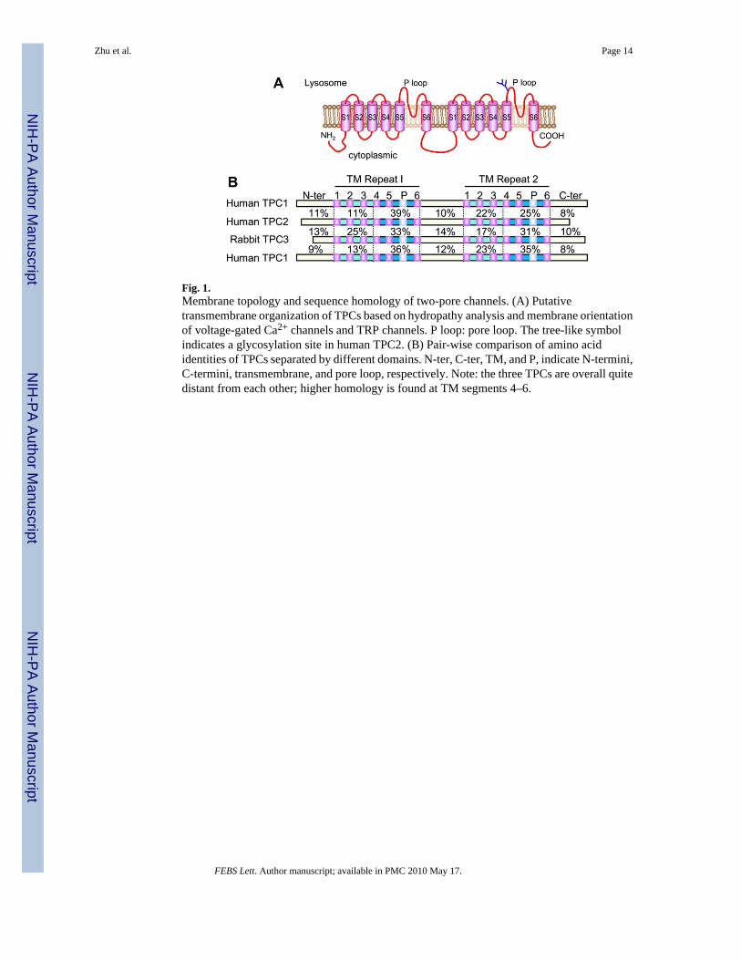
Fig. 1.Membrane topology and sequence homology of two-pore channels. (A) Putativetransmembrane organization of TPCs based on hydropathy analysis and membrane orientationof voltage-gated Ca2+ channels and TRP channels. P loop: pore loop. The tree-like symbolindicates a glycosylation site in human TPC2. (B) Pair-wise comparison of amino acididentities of TPCs separated by different domains. N-ter, C-ter, TM, and P, indicate N-termini,C-termini, transmembrane, and pore loop, respectively. Note: the three TPCs are overall quitedistant from each other; higher homology is found at TM segments 4–6.
Zhu et al. Page 14
FEBS Lett. Author manuscript; available in PMC 2010 May 17.
NIH
-PA Author Manuscript
NIH
-PA Author Manuscript
NIH
-PA Author Manuscript

Fig. 2.Distribution and phylogenetic relationship of TPC proteins in animals and plants. The unrootedphylogenetic tree was made for known TPC sequences from representative members of animalkingdom and two land plants using ClusterW (http://clustalw.genome.jp/) and plotted usingneighbour-joining algorithm. The N- and C- termini were removed and for some sequencesthe loop between the two transmembrane domains were also partially removed beforeperforming the alignment. GenBank accession numbers for the corresponding nucleotidesequences are shown in parentheses.
Zhu et al. Page 15
FEBS Lett. Author manuscript; available in PMC 2010 May 17.
NIH
-PA Author Manuscript
NIH
-PA Author Manuscript
NIH
-PA Author Manuscript

Fig. 3.Subcellular distribution and potential function of mammalian TPCs. Diagram depicts thelocations of human TPC1 and chicken TPC3 in different endosome populations and of humanTPC2 in lysosomes when stably expressed in HEK293 cells. In addition to mediating NAADP-evoked Ca2+ release from endolysosomes, other potential functions for TPCs expressed in theacidic organelles are indicated. SOC and ROC: store- and receptor-operated channels, TGN:trans-Golgi network (adapted from [10] with modifications).
Zhu et al. Page 16
FEBS Lett. Author manuscript; available in PMC 2010 May 17.
NIH
-PA Author Manuscript
NIH
-PA Author Manuscript
NIH
-PA Author Manuscript

Fig. 4.The triggering role of TPC-mediated Ca2+ mobilization. The flow chart shows pathways forNAADP, cADPR and IP3 production and their effects on intracellular Ca2+. The drawings atthe bottom depict “quantal” Ca2+ signals generated by TPCs (left, lighter color and higherposition for higher Ca2+ concentrations) and global Ca2+ signals generated by activatingIP3Rs and/or RyRs (right), as well as their contributions to the two phases of Ca2+ transientsevoked by NAADP (blue arrows). Red dashed lines indicate alternative pathways. The greenpathway on the left shows an example of physiological functions directly regulated by the localCa2+ signals. ARC, ADP-ribose cyclase, including CD38, PLC, phospholipase C, Ψ,membrane potential. ICa, Ca2+ currents through plasma membrane.
Zhu et al. Page 17
FEBS Lett. Author manuscript; available in PMC 2010 May 17.
NIH
-PA Author Manuscript
NIH
-PA Author Manuscript
NIH
-PA Author Manuscript