Two DNA nanomachines map pH changes along intersecting endocytic pathways inside the same cell

Molecular Biology of the CellVol. 20, 3125–3141, July 1, 2009
Revisiting the Role of Cystic Fibrosis TransmembraneConductance Regulator and Counterion Permeability in thepH Regulation of Endocytic OrganellesHerve Barriere,* Miklos Bagdany,* Florian Bossard,* Tsukasa Okiyoneda,*Gabriella Wojewodka,† Dieter Gruenert,‡ Danuta Radzioch,† andGergely L. Lukacs*
*Department of Physiology, McGill University, Montreal, Quebec, H3G 1Y6, Canada; †Research Institute ofthe McGill University Health Center, Montreal, Quebec, H3G 1A4 Canada; and ‡Research Institute, CaliforniaPacific Medical Center, San Francisco, CA 94107
Submitted January 21, 2009; Revised April 10, 2009; Accepted April 27, 2009Monitoring Editor: Sandra L. Schmid
Organellar acidification by the electrogenic vacuolar proton-ATPase is coupled to anion uptake and cation efflux topreserve electroneutrality. The defective organellar pH regulation, caused by impaired counterion conductance of themutant cystic fibrosis transmembrane conductance regulator (CFTR), remains highly controversial in epithelia andmacrophages. Restricting the pH-sensitive probe to CFTR-containing vesicles, the counterion and proton permeability,and the luminal pH of endosomes were measured in various cells, including genetically matched CF and non-CF humanrespiratory epithelia, as well as cftr�/� and cftr�/� mouse alveolar macrophages. Passive proton and relative counterionpermeabilities, determinants of endosomal, lysosomal, and phagosomal pH-regulation, were probed with FITC-conju-gated transferrin, dextran, and Pseudomonas aeruginosa, respectively. Although CFTR function could be documented inrecycling endosomes and immature phagosomes, neither channel activation nor inhibition influenced the pH in any ofthese organelles. CFTR heterologous overexpression also failed to alter endocytic organellar pH. We propose that therelatively large CFTR-independent counterion and small passive proton permeability ensure efficient shunting of theproton-ATPase–generated membrane potential. These results have implications in the regulation of organelle acidifica-tion in general and demonstrate that perturbations of the endolysosomal organelles pH homeostasis cannot be linked tothe etiology of the CF lung disease.
INTRODUCTION
Dynamic networks of secretory and endocytic organelles areresponsible for the biosynthesis, sorting, and delivery ofprotein and lipid cargo, as well as the cellular uptake ofnutrients and a variety of pathogens (Mukherjee et al., 1997;Wu et al., 2001). The physiological functions of the secretoryand endocytic pathway require progressively decreasing lu-minal pH from the endoplasmic reticulum (ER) to secretoryvesicles and from endocytic vesicles to lysosomes, respec-tively (Mellman et al., 1986; Mukherjee et al., 1997; Weisz,2003a). Acidification of the Golgi complex, for example, isnecessary to ensure the pH optimum for sialylation, sulfa-tion, fucosylation of polypeptides and ligand dissociationfrom the KDEL and mannose-6 phosphate receptor family(Kim et al., 1998; Weisz, 2003a; Olson et al., 2008). Endolyso-somal acidification is essential for ligand-receptor sorting,activation of hydrolytic enzymes, as well as homo- andheterotypic membrane fusion and fission to maintain endo-cytosis and recycling (Mellman et al., 1986; Mukherjee et al.,1997). Phagolysosomal acidification is a prerequisite for the
efficient destruction of ingested bacteria and presentation ofantigens as part of the innate and acquired immune re-sponse (Steinberg et al., 2007a).
Regulation of organellar acidification is dictated by theinterplay of multiple ion transport systems, second messen-gers and cytoplasmic factors. One of the most importantplayers is the V-type adenosine triphosphatase (v-ATPase)proton pump, a multisubunit enzyme complex assemblingfrom the V0 and V1 subunit (Jefferies et al., 2008; Marshanskyand Futai, 2008). The v-ATPase utilizes the energy of ATPhydrolysis to build up the organellar electrochemical protongradient. Because the v-ATPase is an electrogenic pump,macroscopic proton translocation is accompanied by an-ion uptake or cation efflux to preserve electroneutrality(Marshansky and Futai, 2008). Severe reduction in the coun-terion permeability leads to the acidification defect by thedevelopment of large, positive inside membrane potential,imposing energetic barrier on proton uptake. Based on theseconsiderations, counterion conductance was proposed toplay a determinant role in organellar acidification (Mellmanet al., 1986; Jentsch, 2007) and was supported by the acidifi-cation defect of cells deficient of members of the intracellularClC chloride channel family (Hara-Chikuma et al., 2005a;Hara-Chikuma et al., 2005b; Scheel et al., 2005; Jentsch, 2007;Graves et al., 2008). Aside the counterion conductance, ac-cumulating evidence indicate that both the passive protonleak (Chandy et al., 2001; Wu et al., 2001) and direct regula-
This article was published online ahead of print in MBC in Press(http://www.molbiolcell.org/cgi/doi/10.1091/mbc.E09–01–0061)on May 6, 2009.
Address correspondence to: Gergely L. Lukacs ([email protected]).
© 2009 by The American Society for Cell Biology 3125

tion of the v-ATPase (Marshansky and Futai, 2008) contrib-ute to the development of organelle-specific pH.
Cystic fibrosis (CF), one of the most prevalent geneticdiseases of the Caucasian population, is caused by the func-tional defect of the cystic fibrosis transmembrane conduc-tance regulator (CFTR). CFTR, a member of the ABC trans-porter family, is a protein kinase A (PKA)-regulated chloridechannel and regulator of numerous epithelial transporters(Riordan et al., 1989; Riordan, 2008). The multifaceted clini-cal manifestations of CF are, in part, attributed to the im-paired transepithelial salt and water movement, culminatingin recurrent bacterial infection, colonization, and hyperin-flammation of the lung, representing the major cause ofmorbidity in CF (Boucher, 2007). The mechanism of the CFlung disease, though not completely elucidated, can be at-tributed to the combination of defective airway surface liq-uid hydration and mucocilliary clearance, excessive mucussecretion, and Na� resorption (Boucher, 2007).
Based on the notion that counterion permeability is one ofthe determinants of organellar acidification (Forgac, 1998),the CFTR chloride channel was endowed with the capacityto regulate the acidification of the trans-Golgi, trans-Golginetwork (TGN), endosome, phagosome, and lysosome inselected epithelia and alveolar macrophages (Barasch et al.,1991; Poschet et al., 2001, 2002; Di et al., 2006; Teichgraber etal., 2008). This paradigm offered a possible explanation forthe metabolic and cellular pathology of CF respiratory epi-thelia as well as alveolar macrophages. Defective acidifica-tion of the TGN was proposed to result in undersialylationof glycoconjugates and to promote adhesion and coloniza-tion of Pseudomonas aeruginosa and Staphylococcus aureus torespiratory epithelia (Barasch et al., 1991; Poschet et al., 2001;Campodonico et al., 2008). Likewise, impaired acidificationof the endolysosomes was invoked in membrane traffickingdefect (Barasch et al., 1991), the hyperinflammatory state ofrespiratory epithelia (Teichgraber et al., 2008) and compro-mised destruction of bacteria in alveolar macrophages (Di etal., 2006). Intriguingly, hyperacidification of the TGN andendosomes was also reported in CF respiratory epithelia andexplained by consequence of the hyperactive epithelial so-dium channel (ENaC; Poschet et al., 2001, 2006). Finally,using CFTR overexpression systems and loss-of-functionstudies, other investigators reported that neither the secre-tory (Golgi and TGN) nor the endocytic organelles (endo-somes, lysosomes, and phagosomes) display a CFTR-depen-dent acidification defect (Lukacs et al., 1992; Biwersi andVerkman, 1994; Dunn et al., 1994; Root et al., 1994; Seksek etal., 1996; Machen et al., 2001; Haggie and Verkman, 2007,2009b; Lamothe and Valvano, 2008). Given these discrepan-cies, improved methodologies and better understanding ofthe biophysical basis of organellar acidification are requiredto rationalize the possible contribution of CFTR activity tothe pH regulation of intracellular organelles. These consid-erations led us to determine the intrinsic, passive protonpermeability and CFTR-independent and -dependent rela-tive counterion permeability of endosomes, lysosomes, andphagosomes in several cellular models, including geneti-cally matched CF and non-CF respiratory epithelia and al-veolar macrophages.
Using single-cell fluorescence ratio image analysis (FRIA),we show that the CFTR-independent and overall counterionpermeability was remarkably higher than the passive protonpermeability of endosomes, and therefore, CFTR activationcannot interfere with the endosomal pH regulation in CFrespiratory epithelia and alveolar macrophages, as well asother heterologous expression systems. A similar phenom-enon was discovered in lysosomes and phagolysosomes of
respiratory epithelia and primary or immortalized mousemacrophages, respectively, with the caveat that recyclingcompromised the channel accumulation in both lysosomesand phagosomes as well. Our results have implications forthe pH regulation of endocytic organelles and documentthat other factor than the acidification defect of endocyticorganelles accounts for the hyperinflammatory CF lung phe-notype.
MATERIALS AND METHODS
DNA Constructs and ChemicalsThe extracellular triple hemagglutinin (3HA) tag, consisting of amino acidresidues of SLEYPYDVPDY-ASYPYDVPDYAYPYDVPD, was inserted intothe fourth extracellular loop after residue 897 in the G551D CFTR, as describedfor the wild-type (wt) and �F508 CFTR (Sharma et al., 2004; Pedemonte et al.,2005). The G551D CFTR cDNA was kindly provided by Dr. J. Rommens (Hos-pital for Sick Children, Toronto). All chemicals of the highest available puritywere obtained from Sigma-Aldrich (Oakville, ON, Canada) except when indi-cated. Bafilomycin A1 (Baf) was from LC Laboratories (Woburn, MA).
CellsHeLa, Madin-Darby canine kidney (MDCK) and RAW264.7 cells were grownin Dulbecco’s modified Eagle’s medium (DMEM) containing 10% fetal bovineserum (FBS) in a thermostated cell culture incubator in 5% CO2 at 37°C. Babyhamster kidney (BHK) cells were grown in DMEM/F12 containing 5% FBS.IB3 cells (kindly provided by Dr. P. Zeitlin, Johns Hopkins University) weregrown in LHC-8 basal medium (Invitrogen, Carlsbad, CA) containing 5% FBS.The IB3 cells have �F508/W1282X genotype (Zeitlin et al., 1991). The humanbronchial epithelial cell line derived from a CF patient with a �F508/�F508genotype (CFBE41o-, designated as CFBE) have been characterized (Cozens etal., 1994) and were cultured in MEM with GlutaMAX (Invitrogen, Carlsbad,CA) supplemented with 10% FBS serum at 37°C in a 5% CO2-humidifiedincubator as described (Gruenert et al., 1995). To allow differentiation of theMDCK and CFBE, epithelial cells were cultured at confluence for 3–5 d.Primary macrophages were obtained by intraperitoneal and bronchoalveolarlavages as previously described (Guilbault et al., 2008). Macrophages wereisolated by centrifugation and resuspended in RPMI 1640 with 10% FBS andseeded on polylysine (Sigma)-coated glass coverslips. Coverslips were coatedaccording to manufacturer’s instructions. Experiments were performed 24–48h after plating the cells.
All cell types used were either transiently or stably transfected with wt orG551D CFTR-3HA harboring triple hemagglutinin tags in the fourth extra-cellular loop (Sharma et al., 2004). BHK cell expressing the wt and G551DCFTR-3HA was generated as previously described and clones were selectedin the presence of 500 �M methotrexate (Sharma et al., 2001).
CFTR variants were stably expressed in the CFBE and HeLa cells usinglentiviral vectors by Dr. J. Wakefield (Tranzyme, Birmingham, AL) and se-lected in the presence of 5 �g/mP puromycin as described (Bebok et al., 2005).IB3 bronchial epithelia, expressing endogenous �F508 and W1282X CFTR atundetectable levels, were stably transfected with the pCEP4 expression plas-mid encoding the wt or G551D CFTR-3HA. A mixture of clones were selectedin hygromycin B. Transfected cells were maintained in LHC-8 containing 5%FBS and 100 �g � ml�1 hygromycin B. RAW cells were transiently transfectedwith CFTR encoding pNut-CFTR using FuGENE6 as DNA complexing agent(Roche, Basel, Switzerland) according to the manufacturer’s recommendationand analyzed after 48 h. MDCK cells were infected with retrovirus encodingthe CFTR-3HA variants as described previously (Benharouga et al., 2003).
AnimalsInbred C57BL/6-Cftr�/� heterozygous mice were maintained and bred in theAnimal Facility of the McGill University Health Center Research Institute. Allpups were genotyped between 12 and 14 d of age. The animals were kept incages with sterile corn bedding (Anderson, Bestmonro, LA) and maintainedin ventilated racks (Lab Products, Seaford, DE). Age-matched C57BL/6-Cftr�/� mice and C57BL/6-Cftr�/� mice were maintained in murine patho-gen–, Helicobacter-, and parasite-free conditions. They were housed (1–4 ani-mals/cage), bred, and maintained in a facility under specific pathogen-freeconditions. Mice were fed with either the NIH-31–modified, irradiated mousediet for wt mice (Harlan Teklad, Indianapolis, IN) or a liquid diet starting at14 d of age for knockout mice (Peptamen liquid diet; Nestle Canada, Bramp-ton, ON, Canada). The liquid diet was freshly prepared each morning andprovided in 50-ml centrifuge tubes (Fisher Scientific, Nepean, ON, Canada).Mice used for experiments were between 19 and 22 wk of age. Experimentalprocedures with mice were conducted in accordance with the CanadianCouncil on Animal Care guidelines and with the approval of the FacilityAnimal Care Committee of the Montreal General Hospital Research Institute(Montreal, PQ, Canada).
H. Barriere et al.
Molecular Biology of the Cell3126

Labeling of Endocytic Organelles with pH-sensitive DyesThe luminal pH of early endosomes, recycling endosomes, lysosomes, andphagosomes was routinely determined after the selective labeling of therespective compartment with fluorescein isothiocyanate transferrin (FITC)-conjugated cargo (e.g., CFTR, transferrin, dextran, and P. aeruginosa PAO1) byFRIA as described for CFTR and other cargo molecules (Sharma et al., 2004;Barriere et al., 2007; Kumar et al., 2007; Barriere and Lukacs, 2008; Duarri et al.,2008; Varghese et al., 2008; Glozman et al., 2009).
To label internalized CFTR, cells were incubated with anti-HA (1:500 dilu-tion equivalent to 10 �g/ml, MMS101R, Covance Laboratories, Madison, WI)primary Ab and FITC-conjugated goat anti-mouse secondary Fab (1:500 di-lution, Jackson ImmunoResearch Laboratories, West Grove, PA) by incubat-ing primary for 1 h at 37°C. Cells were then washed (140 mM NaCl, 5 mMKCl, 20 mM HEPES, 10 mM glucose, 0.1 mM CaCl2, and 1 mM MgCl2, pH 7.3)and chased for indicated time at 37°C. Fluid-phase Ab uptake was notdetectable in mock-transfected cells (data not shown). When indicated, cellsurface–resident CFTR was labeled on ice by successive incubation with theprimary anti-HA Ab and secondary FITC-Fab.
To confirm that the primary and secondary Ab remains bound to CFTRduring FRIA experiments, the pH resistance of Ab binding was measured byimmunoperoxidase assay. After anti-HA– and HRP-conjugated secondary Abbinding to CFTR-expressing HeLa cells, the extracellular medium pH wasadjusted to pH 7.2, 5.0, and 2.5 for 5 min (van Kerkhof et al., 2001; 0.15 MNaCl, 50 mM glycine, 0.1% BSA, 20 mM MES, for pH 2.5 and 5.0, 10 mMHEPES for pH 7.2). The amount of bounded HRP-conjugated Ab was mea-sured by Amplex-Red as substrate, using a POLARstar OPTIMA fluorescenceplate-reader (BMG Labtech, Offenburg, Germany; Barriere et al., 2006). The Abbinding was virtually unaltered at pH 5.0, but reduced by 50% at pH 2.5(Supplemental Figure S1A).
Recycling endosomes were labeled with FITC-transferrin (Tf; 15 �g/ml,45-min loading after 45 min serum-depletion at 37°C) and chased for 0–3 min.Lysosomal pH was measured with similar results on cells labeled by over-night fluid-phase uptake of FITC-dextran alone or in combination with Ore-gon Green 488-dextran (50 �g/ml, MW 10 kDa, Molecular Probes, Eugene,OR) and chased for �3 h.
Phagosomal pH was monitored following the uptake of FITC- or FITC/TRITC-conjugated P. aeruginosa (PAO1 strain, kindly provided by Dr. M.Parsek, University of Washington, Seattle). From an overnight culture �5 �108 bacteria were opsonized in 20% FBS-PBS for 30 min at 37°C with agitation.Cells were washed with PBS and labeled in the presence of 1 mg/ml FITC,TRITC, or a combination of both in PBS at pH 8.0 for 30 min at roomtemperature under rotation. Excess of fluorescent dyes was removed byrepeated centrifugation in ice-cold PBS. Bacteria were snap-frozen and storedat �80°C.
Organellar pH MeasurementFRIA of endocytic organelles was performed on an Axiovert 100 invertedfluorescence microscope (Carl Zeiss MicroImaging, Toronto, ON, Canada) atroom temperature equipped with a Hamamatsu ORCA-ER 1394 (Hamamatsu,Japan) cooled CCD camera and a Planachromat (63� NA 1.4) objectiveessentially as described previously (Sharma et al., 2004; Barriere et al., 2007;Barriere and Lukacs, 2008; Glozman et al., 2009). Image acquisition and FRIAwere performed with MetaFluor software (Molecular Devices, Downingtown,PA). Images were acquired at 490 � 5 and 440 � 10-nm excitation wave-lengths, using a 535 � 25-nm emission filter. To determine the initial rates ofacidification of endosomes (see Figure 4E), CFTR was labeled with anti-HAand FITC-conjugated goat anti-mouse Fab sequentially for 1 h on ice andchased at 37°C for the indicated times. The cell surface remaining Abs wereacid-stripped before image acquisition (van Kerkhof et al., 2001).
In situ calibration curves, describing the relationship between the fluores-cence ratio values and endosome, lysosome, or phagosome pH, served tocalculate the luminal pH of individual vesicles after fluorescence backgroundsubtraction at both excitation wavelengths (e.g., Supplemental Figure S1,B–D). In situ calibration was performed by clamping the vesicular pH be-tween 4.5 and 7.4 in K�-rich medium (135 mM KCl, 10 mM NaCl, 20 mMHEPES, or 20 mM MES, 1 mM MgCl2, and 0.1 mM CaCl2) with 10 �Mnigericin, 10 �M monensin, 0.4 �M Baf, and 20 �M carbonyl cyanide 3-chlo-rophenylhydrazone (CCCP; Sigma-Aldrich) and recording the fluorescenceratios. Calibration curves were obtained for each cargo molecules and re-peated at regular intervals. As an internal control, one-point calibration wasperformed on each coverslip by clamping the organellar pH to 6.5 with 10 �Mmonensin and nigericin, 0.4 �M Baf, and 20 �M CCCP. In each experiment thepH of 200–800 endosomes/lysosomes and 100–400 phagosomes was deter-mined. Mono- or multipeak Gaussian distributions of vesicular pH valueswere obtained with Origin 7.0 software (OriginLab, Northampton, MA), andthe results of individual experiments were illustrated. The mean pH of eachvesicle population was calculated as the arithmetic mean of the data in eachindividual experiment using 200–800 vesicles from 15 to 60 cells. At leastthree independent experiments were performed for each condition.
Determination of the Relative Counterion Permeabilityand Buffer Capacity of OrganellesRapid dissipation of the organellar pH gradient by the proton pump inhibitor(Baf) and the protonophore (CCCP) was used to determine the relativecounterion permeability of the organelles. Cells were labeled as describedabove and the organellar pH was continuously monitored by FRIA for theindicated time. pH dissipation was measured in the presence of 0.4 �M Bafand 20 �M CCCP. To measure the passive proton leak, only Baf was added.Both Baf and CCCP were used at saturating concentrations (Lukacs et al.,1990, 1991; Hackam et al., 1997; Steinberg et al., 2007b). When indicated, CFTRwas activated with the PKA agonist cocktail (20 �M forskolin, 0.5 mM8-(4-chlorophenyl-thio)adenosine 3�,5�-cyclic monophosphate sodium saltCPT-cAMP, and 0.2 mM isobutylmethylxanthine IBMX) for 3 min beforeBaf�CCCP addition. FRIA was performed as described above. The pH dis-sipation rate was calculated from the initial slope of the fluorescence ratiochange. The buffer capacity of endocytic organelles was calculated from theextent of rapid alkalinization after the addition of 0.5–2 mM NH4Cl in thepresence or absence of Baf. The calculation was based on the formula fromRoos and Boron (Roos and Boron, 1981; Sonawane and Verkman, 2003).
The Passive Proton Permeability DeterminationThe passive proton permeability of recycling endosomes, lysosomes, andphagosomes was calculated according to the following equation:
PH��dpHo
dt. V
S. �v
([H�]o�[H�]c)
where PH� is the organellar passive proton permeability in cm � s�1, dpHo/dtis the organellar proton flux in pH � s�1, V is the organellar volume in cm3, Sis the organellar surface in cm2, �v is the organellar buffer capacity inM � pH�1, and (H�o � H�c), is the transmembrane proton gradient be-tween the organelle lumen and the cytosol (Chandy et al., 2001; Grabe andOster, 2001). The passive proton flux was measured by monitoring the or-ganellar alkalinization rate immediately after 400 nM Baf addition as shownin Figure 2D. The buffer capacity was measured by monitoring the rapidalkalinization of organelles after NH4Cl addition in the presence of Baf asdetailed in Materials and Methods and in Chandy et al. (2001). The surface andvolume of endosomes and lysosomes were obtained from published data(Griffiths et al., 1989). The phagosome volume and surface calculation wasbased on the assumption that average phagosome diameter is 3 �m and thedistance between the bacterial wall and the inner leaflet of the phagosomalmembrane is 100 nm, based on EM observations (Hart et al., 1987; Chastellier,2008). In light of the high counterion conductance of the endocytic organellesand the modest membrane potential of phagolysosomes (�25 mV includingthe Donnan potential effect; Steinberg et al., 2007b), we assumed that themembrane potential has modest contribution to the electrochemical protondriving force in endocytic organelles. Therefore, the passive proton per-meability was calculated by taking into consideration only the chemicalproton driving force, and thus it likely represents an overestimate. Thecytoplasmic pH was assumed to be constant (pH 7.3) during the protonefflux measurements (Mukherjee et al., 1997).
Immunofluorescence MicroscopyCFTR subcellular distribution was determined after internalization of an-ti-HA Ab for 1 h in DMEM and chased for 0.5 h. CFTR was visualized byFITC-conjugated goat anti-mouse Fab or TRITC-conjugated goat anti-mouseafter cell fixation. During the last 45 min, cells were labeled with FITC-Tf orTRITC-Tf (15 �g/ml) after 45-min serum depletion to visualize recyclingendosomes. Early endosome marker 1 (EEA1) and Rab5 were detected byindirect immunostaining using rabbit polyclonal anti-EEA-1 and anti-Rab5antibodies from Abcam (Cambridge, United Kingdom) and Santa Cruz Bio-technology, (Santa Cruz, CA), respectively. Lysosomes were labeled with FITC-dextran (50 �g/ml, MW 10 kDa) as described for the pH determination. Insome experiments, the lysosome was stained with mouse monoclonal anti-Lamp-2 Ab (H4B4 Ab was developed by Dr. J. Thomas August and Dr. JamesE. K. Hildreth and was obtained from the Developmental Studies HybridomaBank maintained by The University of Iowa, Department of Biological Sci-ences, Iowa City, IA). Single optical sections were collected by Zeiss LSM510laser confocal fluorescence microscope, equipped with a Plan-Apochromat63X/1.4 (Carl Zeiss Microimaging, Thornwood, NY) as described (Lechard-eur et al., 2004). Images were processed with Adobe Photoshop (AdobeSystems, San Jose, CA) software. CFTR phagosomal colocalization was mea-sured by internalizing anti-HA Ab complexed with TRITC-conjugated goatanti-mouse IgG for 1 h in DMEM and chased for 0.5 h, and then FITC-PAO1bacteria were phagocytosed for the indicated time. Colocalization was per-formed using the “Colocalization” feature in the Volocity 4.1.0 (Improvisionsoftware; Molecular Devices, Sunnyvale, CA) software as described in theSupplemental Materials.
Determinants of Endocytic Compartments pH
Vol. 20, July 1, 2009 3127

Western BlottingCFTR expression level of cell lines was determined by immunoblotting usinganti-HA (MMS101R, Covance) for epithelial cells or anti-CFTR antibodies(M3A7 and L12B4, Chemokine, East Orange, NJ) for macrophages. Cell ly-sates were prepared using RIPA buffer containing 10 �g/ml leupeptin, pep-statin, 100 �M phenylmethylsulfonyl fluoride, 10 �M MG132, and 10 mMN-ethylmaleimide, and immunoblotting of CFTR was performed as describedpreviously using enhanced chemiluminescence (Sharma et al., 2004).
Iodide Efflux AssayThe plasma membrane cAMP-dependent halide conductance of transfectedand parental cells was determined with the iodide efflux as described (Sharmaet al., 2001). The parental cells have no or negligible amount of cAMP-activated halide conductance. The CFTR inhibitor MalH2 (kindly provided byDr. A. Verkman, University of California, San Francisco) was added simul-taneously with the PKA agonist cocktail containing 20 �M forskolin, 0.5 mMCPT-cAMP, and 0.2 mM IBMX. The pH sensitivity of the iodide efflux wasdetermined after incubating the cells at pH 5 during the last 10 min of theiodide loading and the efflux measurement in 20 mM MES-supplementedloading and efflux buffer, respectively. The iodide-selective electrode wascalibrated in MES containing buffer at pH 5.
Statistical AnalysisExperiments were repeated at least three times or as indicated. Data aremeans � SEM. Significance was assessed by calculating two-tailed p values at95% confidence level with unpaired t test, using Prism software (GraphPadSoftware, San Diego, CA).
RESULTS
Considering the low plasma membrane density and slowmetabolic turnover rate of CFTR in various expression sys-tems (Heda et al., 2001; Sharma et al., 2004), it is conceivablethat only a small population of secretory and newly formedendocytic vesicles contain the channel. Previous studies,including our own, used pH probes (e.g., cellubrevin-phlu-orin, synaptobrevin-GFP-phluorin, FITC-transferrin, andFITC-dextran) that labeled organelles in CFTR-independentmanner (Barasch et al., 1991; Lukacs et al., 1992; Biwersi and
Verkman, 1994; Dunn et al., 1994; Seksek et al., 1996; Botelhoet al., 2000; Chandy et al., 2001; Poschet et al., 2002; Machenet al., 2003; Haggie and Verkman, 2007, 2009). Here weintroduced two novel approaches to monitor the CFTR-dependent endosomal pH regulation with high specificity.First, we implemented a method that restricted the pH-sensitive probe to CFTR-containing vesicles (Figure 1A;Sharma et al., 2004; Glozman et al., 2009). Second, to controlCFTR-independent endosomal pH changes provoked by theactivation of cAMP-dependent PKA, a direct regulator of thev-ATPase and Na�/H� exchanger activity (Marshanskyand Futai, 2008), we utilized the G551D CFTR-3HA, a classIV CF mutation with severely impaired channel activity(Becq et al., 1994, 2007). In the following experiments, firstwe validated the use of G551D CFTR-3HA and the pHmeasurement methodology as well as the counterion andpassive proton permeability determination in nonpolarizedBHK cells. Subsequent studies were performed on geneti-cally matched CF and non-CF human respiratory epithelia(IB3 and CFBE) as well as cftr�/� and cftr�/� primary mousemacrophages and other heterologous CFTR overexpressionsystems. These studies not only allowed us to assess theeffect of CFTR deficiency in genetically matched respiratoryepithelia and alveolar macrophages, but also to determinewhether the endogenous counterion permeability can limitthe acidification in CFTR-independent cellular models.
Comparison of the Biosynthetic and Endocytic MembraneTrafficking of wt and G551D CFTR-3HATo utilize the G551D CFTR-3HA for monitoring the vesicu-lar pH without conferring PKA-dependent chloride perme-ability to endocytic organelles, first we assessed the mem-brane trafficking pathway of the G551D CFTR in relation toits wt counterpart. BHK cells were stably transfected withG551D and wt CFTR-3HA. The steady-state expression level
Figure 1. Selective labeling of CFTR-3HA–containingendosomes with pH-sensitive probes. (A) Schematiccomparison of the subcellular distribution of CFTR andvarious endosomal pH probes. It is predicted that CFTRhas partially overlapping localization with the fluid-phase marker dextran-FITC (left panel) and cellubrevinor synaptobrevin coupled with GFP-Phluorin (middlepanel). Completely overlapping distribution of the pH-probe and endosomal CFTR is anticipated by labelingthe exofacial 3HA-tag of CFTR with anti-HA Ab andFITC-Fab. (B) Immunoblot analysis of wt and G551DCFTR-3HA expression in stably transfected BHK cells.Equal amounts of cell lysates were immunoblotted withanti-HA Ab. Core- and complex-glycosylated CFTR arelabeled, indicated by empty and filled arrowhead, re-spectively. (C) Plasma membrane halide conductancewas measured by the iodide efflux assay in BHK mono-layers expressing wt or G551D CFTR. PKA was stimu-lated with 20 �M forskolin, 0.2 mM IBMX, and 0.5 mMCPT-cAMP cocktail (�FK) at the indicated time at 22°C.Data are means of triplicate determinations from a sin-gle representative experiment. (D) Subcellular localiza-tion of internalized CFTR in BHK cells. Internalized wtand G551D CFTR-3HA was labeled with anti-HA Aband visualized by TRITC-conjugated secondary Fab. Ly-sosomes and recycling endosomes were labeled with 50�g/ml FITC-dextran (loaded overnight and chased for3 h) and 15 �g/ml FITC-Tf (loaded for 45 min in serum-free medium), respectively. Single optical sections wereobtained by laser confocal fluorescence microscopy. Bar,10 �m.
H. Barriere et al.
Molecular Biology of the Cell3128

of the core- and complex-glycosylated G551D and wt CFTRwas comparable, measured by immunoblotting (Figure 1B).Likewise, the cell surface expression of the wt and G551DCFTR-3HA was similar, determined by anti-HA Ab-bindingassay (Supplemental Figure S2A). PKA activation by fors-kolin, CPT-cAMP, and IBMX failed to activate G551D CFTRcontrary to the wt channel, monitored by the iodide effluxassay (Figure 1C). The biosynthetic processing efficiency,metabolic stability, and internalization rates of the complex-glycosylated wt and G551D CFTR, measured by pulse-chaseexperiments, and anti-HA Ab uptake assay, respectively,were similar (Supplemental Figure S2, B and C, and data notshown).
To determine the postendocytic fate of the G551D CFTR,internalized channels were colocalized with the Tf receptor(Tf-R), a marker of recycling endosomes (Mukherjee et al.,1997). CFTR was labeled by in vivo anti-HA Ab capture at37°C for 1 h. Tf-Rs were visualized by FITC-Tf. Quantitativecolocalization of CFTR, on micrographs obtained by fluores-cence laser confocal microscopy (FLCM), revealed that 85 �2 and 80 � 4% (mean � SEM, n � 4 experiments) ofinternalized Tf was colocalized with G551D and wt CFTR,respectively. Conversely, 52 � 4% of wt and 62 � 3% ofG551D CFTR were confined to Tf-positive endosomes (Sup-plemental Figure S3A). Confinement of internalized G551Dand wt CFTR to early endosomes was confirmed with theircolocalization with rab5 and EEA1 (Supplemental FigureS3B and data not shown) and exclusion from FITC-dextran–loaded lysosomes (Figure 1D), an observation confirmed onother cells (see below). These results, jointly, indicate thatthe wt and G551D CFTR have overlapping postendocyticmembrane trafficking that was further validated by vesicularpH measurements (see below).
Monitoring the Postendocytic Fate of G551D CFTR byVesicular pH DeterminationBased on the characteristic pH of the endolysosomal com-partment (Mukherjee et al., 1997), the postendocytic sortingof G551D and wt CFTR could be inferred from the luminalpH of internalized CFTR-containing vesicles, as has beenshown for a variety of cargo molecules (Barriere et al., 2007;Kumar et al., 2007; Duarri et al., 2008; Varghese et al., 2008).Cell-surface CFTR-3HA was labeled with primary anti-HAand FITC-conjugated secondary Fab on ice. After the re-moval of the excess extracellular Ab, internalization wasinitiated by raising the temperature to 37°C (Figure 2A). Theluminal pH of individual endocytic vesicles was measuredby FRIA, using 450- and 490-nm excitation wavelengthswith an in situ calibration technique, and was plotted astheir frequency distribution (Figure 2, A–C, Barriere et al.,2007; Barriere and Lukacs, 2008; Glozman et al., 2009). Al-though the cell-surface–bound probe (0-h chase) was ex-posed to the extracellular medium pH (�7.4), more than90% of vesicles acidified to pH �6.3–6.5 after a 0.5–1-h chaseat 37°C (Figure 2A). Comparable results were obtained bycontinuous labeling of CFTR at 37°C for 1 h to enhance thesignal-to-noise ratio (Figure 2B). Importantly, G551D, simi-lar to the wt CFTR, was targeted to mildly acidic recyclingendosomes after 1-h chase (pH � 6.52, Figure 2C). Consid-ering that the recycling endosomes mean pH is 6.4–6.5,measured in FITC-Tf–loaded BHK cells (Sharma et al., 2004),these results indicate that G551D like its wt counterpartrecycles back to the cell surface and largely avoids lysosomaldelivery. This conclusion is also in line with the limited(�8%) colocalization of the wt and G551D CFTR withLamp2- and dextran-loaded lysosomes, determined by theVolocity program (see Figures 1D and 3C and Supplemental
Figure S3D). CFTR was also confined to recycling endo-somes after 4-h chase (data not shown). No significant dis-sociation of Ab from CFTR was observed at pH 5 (seeMaterials and Methods). Furthermore, the metabolic stabilityof the Ab-bound CFTR complex remained unaltered (Sharmaet al., 2004), consistent with the notion that Ab binding didnot provoke premature lysosomal degradation of the chan-nel. Thus the G551D CFTR could serve as a negative controlfor evaluating the contribution of wt CFTR activation to theendosomal pH regulation.
The Effect of CFTR Activation on the CounterionConductance and the Steady-State pH of Endosomes InSituTo assess the relative magnitude of CFTR-dependent coun-terion permeability and the passive proton leak of individ-ual endosomes, determinants of the endosomal pH, wemodified a technique developed to measure these parame-ters in cell suspension of mouse peritoneal macrophages andChinese hamster ovary (CHO) cells (Lukacs et al., 1990,1992). The assay is based on the assumption that the dissi-pation rate of vesicular pH gradient by high concentration ofprotonophore (20 �M FCCP or CCCP) is rate-limited by theendosomal counterion conductance in the presence of theH�-ATPase inhibitor, Baf (Lukacs et al., 1990, 1992). Underthese conditions, activation of CFTR should enhance theinitial rate of Baf�CCCP–induced H�-efflux by the provi-sion of additional Cl� conductance and dissipation of theinside negative endosomal membrane potential generatedby the H� efflux (Figure 2D, right panel). This was a reason-able assumption, considering that the luminal Cl� concen-tration of newly formed endosomes was rapidly reducedfrom 150 to 10–50 mM (Hara-Chikuma et al., 2005b).
The pH of individual, CFTR-3HA–containing endosomeslabeled by primary anti-HA– and FITC-conjugated second-ary Fab, was determined by FRIA as a function of time. Afterthe inhibition of the vacuolar H�-ATPase activity by Baf,slow endosomal alkalinization, caused by the passive H�
efflux along the proton electrochemical gradient, was de-tected (Figure 2D). The observation that saturating concen-tration of protonophore CCCP (20 �M) accelerated the Baf-induced pH dissipation rate by nearly 10-fold (Figure 2D)suggests that early endosomes have a relatively small pas-sive proton and large counterion conductance.
Incomplete inhibition of the H�-ATPase cannot accountfor the slow pH dissipation rate, because the Baf dose–response curve indicated that the v-ATPase was fully inhib-ited at 0.5 �M Baf concentration in vivo (data not shown;Lukacs et al., 1990). These observations also imply that arelatively slow H�-ATPase activity can maintain the endo-somal pH gradient at steady state. Similar results were ob-served in parental BHK cells, ruling out the possibility thatCFTR expression is responsible for the high constitutivecounterion conductance and limited proton leak of endo-somes (see Supplemental Figure S3E).
Activation of wt CFTR by PKA stimulation increased theCCCP�Baf–induced endosomal pH dissipation rate bythreefold (Figure 2, E and F). In sharp contrast, the pHdissipation remained unaltered in G551D CFTR-expressingcells (Figures 2, E and F, and 4B). These results confirmedthat wt, but not the G551D CFTR, is susceptible to PKAactivation in endosomes (Becq et al., 1994).
To determine the consequence of CFTR activation on thesteady-state endosomal pH, the luminal pH values wereplotted before and after 3-min stimulation by PKA agonists.The endosomal pH of PKA-stimulated cells remained unal-
Determinants of Endocytic Compartments pH
Vol. 20, July 1, 2009 3129

tered, regardless whether wt or G551D CFTR was expressed(Figure 2, F, right panel, and G).
A small fraction of internalized CFTR was confined tovesicles with pH � 6 and pH � 6.6 after 1-h chase, likelyrepresenting channels in late endosomes en route to lyso-somes and in endocytic carrier vesicles, respectively (Figure2, A and B; Sharma et al., 2004). Analysis of the luminal pHof these vesicles revealed that PKA-dependent CFTR activa-tion was unable to influence the steady-state pH of lateendosomes/lysosomes and endocytic carrier vesicles (Fig-ure 2G).
Endosomal pH Regulation Is Not Influenced by CFTRAblation or Overexpression in CF Epithelia andHeterologous Cell Models, RespectivelyThe inability of CFTR functional overexpression to hy-peracidify BHK endosomes suggested that the relativelyhigh endogenous counterion permeability in the presenceof a small passive proton leak cannot limit the protonaccumulation by the v-ATPase in cells that have no en-dogenous CFTR. This inference was tested by overex-
pressing wt CFTR in HeLa cells and polarized MDCKepithelia.
The loss of endogenous CFTR on the endosomal pH ho-meostasis was examined using genetically matched CF andnon-CF respiratory epithelia. To this end, wt and G551DCFTR-3HA were stably expressed in IB3 and CFBE cells,widely used models of CF respiratory epithelia lacking func-tional CFTR (Gruenert et al., 1995; Bruscia et al., 2002). IB3and CFBE cells were derived from the bronchial epithelia ofCF patients with �F508/W1282X and �F508/�F508 CFTRgenotypes, respectively (Zeitlin et al., 1991; Cozens et al.,1994) and have no detectable CFTR expression by immuno-blotting (Figure 3A). Although IB3 cells are nonpolarized,both CFBE and MDCK cells were differentiated into polar-ized monolayers to ensure selective labeling of apical endo-somes (see Materials and Methods).
Heterologous expression of wt and G551D CFTR-3HAwas verified by immunoblotting (Figure 3A) and cell surfaceanti-HA Ab-binding assay (Supplemental Figure 2A). Iodideefflux assay revealed that wt but not G551D CFTR expres-sion conferred PKA-stimulated plasma membrane halide
Figure 2. Monitoring wt and G551D CFTR-3HA endo-cytic sorting and pH regulation by FRIA in BHK cells.(A) Determining the sorting pathway of internalizedCFTR by vesicular pH (pHv) measurement. Anti-HAantibody and FITC-conjugated secondary Fab wasbound to CFTR-expressing BHK cells for 1 h at 0°C.Then the temperature was raised to 37°C for 0–1 h, andthe pHv was measured by FRIA. Data are expressed asfrequency of pHv and means (� SEM) pHv of the majorendosomal population. The number of vesicles ana-lyzed in a single experiment is indicated. (B) Wt CFTRwas labeled with anti-HA primary Ab and FITC-conju-gated secondary Fab for 1 h at 37°C and was chased for30 min before FRIA. (C) G551D CFTR was labeled asdescribed in B. (D) Determination of the passive protonand relative counterion permeability of wt CFTR-con-taining endosomes. Right panel, to unravel the CFTRdependent counterion permeability, protonophere(CCCP) was used to rapidly dissipate the pH gradientin the presence of Baf. H� egress generated a negativeinside membrane potential that was dissipated by Cl�efflux via CFTR and uptake of cations. Left panel, theendosomal pH was monitored as a function of time asdescribed in B. For each experiment, 30–50 vesicleswere tracked simultaneously. The vacuolar H�-ATPasewas inhibited with 0.4 �M Baf to unmask the passiveproton permeability. CCCP (20 �M), a protonophore,induced a rapid dissipation of the endosomal pH gra-dient, indicating the presence of constitutively activecounterion conductance in endosomes. Addition ofNH4Cl (20 mM) dissipated the pH gradient. Wt CFTRwas labeled as in B. (E) CFTR activation enhances thecounterion permeability of wt but not G551D CFTR-expressing endosomes in BHK cells. Endosomes werelabeled as described in B and the pH dissipation ratewas measured after addition of 0.4 �M Baf and 20 �MCCCP by FRIA. CFTR was activated with the PKAagonist cocktail (20 �M forskolin, 0.2 mM IBMX, and 0.5mM CPT-cAMP cocktail FK) for 2–3 min before thepH dissipation was induced. Traces obtained in thepresence of activated PKA are labeled by �FK. (F)Quantification of pH dissipation rate (left panel) andvesicular pH (right panel) after PKA activation. The pHdissipation rate was measured in experiments de-scribed in E. (G) No significant variation in late andearly endosomal pH of wt CFTR-containing vesicles
was detectable. The PKA sensitivity of a selected subpopulation of CFTR-containing vesicles with distinct initial pH was determined as inB. Means � SEM; n � 3–5.
H. Barriere et al.
Molecular Biology of the Cell3130

conductance in HeLa, IB3, and CFBE cells (Figure 3B). Noneof the parental cells had detectable endogenous CFTR- andPKA-activated halide conductance (Figure 3, A and B, anddata not shown).
Internalized wt and G551D CFTR were primarily targetedto early endosomes and excluded from lysosomes in IB3,CFBE, and HeLa cells, visualized by colocalization withFITC-Tf, EEA1, or rab5 and exclusion from dextran- orLamp2-containing lysosome (Figure 3C, Supplemental Fig-ure S3, C and D, and data not shown). These results indicatethat the recycling propensity of endocytosed G551D CFTR isindependent of the cellular expression system, as observedpreviously for wt CFTR (Sharma et al., 2004; Gentzsch et al.,2007; Varga et al., 2008).
The PKA-induced acceleration of the pH dissipation ratein the presence of CCCP�Baf showed that wt CFTR canfunction as a chloride conductive efflux pathway to facilitateH� egress from early endosomes of CFBE, IB3, HeLa, andMDCK cells (Figure 4, A and B). This conclusion was con-firmed by the inability of PKA to activate H� efflux fromendosomes of G551D CFTR-expressing cells (Figure 4B).Importantly, despite full activation of wt CFTR by the ago-nist cocktail in 1.5–2 min at room temperature (see Supple-mental Figure S4B), no significant change in the steady-statepH of wt CFTR-expressing endosomes, including the CFBEand IB3 respiratory epithelia, was observed relative to thatin G551D CFTR-expressing cells (Figure 4C). Likewise, wewere unable to detect significant changes in the initial acid-ification rate of early endosomes, after the synchronizedinternalization of Ab-labeled CFTR from the cell surface(Figure 4, E and F). These data strongly suggest that theCFTR-independent counterion permeability is sufficient to
ensure unrestricted proton accumulation during the acidifi-cation and at steady state by the v-ATPase. Finally, the HeLaand MDCK data suggest that the endosomal acidification isnot limited by their endogenous counterion permeability,because provision of exogenous CFTR chloride conductanceat an inwardly directed electrochemical chloride gradient(Sonawane and Verkman, 2003) could not hyperacidify en-dosomes.
CFTR Effect on the pH Homeostasis of RecyclingEndosomesAlthough the anti-HA and FITC-Fab labeling of CFTR en-abled us to restrict the pH measurements to CFTR-contain-ing vesicles, we wanted to establish whether the Ab bindinginterferes with the channel function. To assess the potentialeffect of Ab labeling on CFTR transport activity, CFTR-3HA–expressing BHK cells were incubated with saturating con-centration of anti-HA Ab (37°C for 30 min). The mean PKA-dependent whole cell current density was reduced by 42%(from 35.1 � 8.0 to 20.5 � 6.7 pA/pF) in the presence ofanti-HA Ab measured by the patch-clamp technique (Sup-plemental Figure S4A). The activation kinetics of CFTR byPKA agonists remained unaltered (Supplemental FigureS4B). Although these observations suggested that theCFTR-Ab complex retains significant activity, we sought analternative assay to evaluate the CFTR-dependent endosomalpH regulation in the presence of fully functional channels.
On the basis of the overlapping subcellular distribution ofFITC-Tf with wt and G551D CFTR (Figure 1D and Supplemen-tal Figure S3A), we followed the endosomal pH after labelingthe recycling endosomes with FITC-Tf. The Baf�CCCP–in-duced pH dissipation rates of recycling endosomes were
Figure 3. Wt and G551D CFTR expression, func-tion, and postendocytic localization in CF respira-tory epithelia and HeLa cells. (A) CFTR expressionwas probed by immunoblot analysis in mock, wt,and G551D CFTR-3HA expressing CFBE and IB3respiratory epithelia, as well as in HeLa cells. Equalamounts of cell lysates were immunoblotted withanti-HA Ab. (B) Wt and G551D CFTR activity wasmeasured by the iodide efflux assay as described inFigure 1C. Data are means of triplicate determina-tions from a representative experiment. (C) Localiza-tion of internalized wt and G551D CFTR in CFBEand IB3 cells. Internalized CFTR was labeled as de-scribed in Figure 1D. Endosomes were visualized by15 �g/ml FITC-Tf loading or by indirect immuno-staining of EEA1 and rab5. Lysosomes were identi-fied by 50 �g/ml FITC-dextran loading as describedin Materials and Methods or by indirect Lamp2 immu-nostaining. Single optical sections were obtained bylaser confocal fluorescence microscopy. Bar, 10 �m.
Determinants of Endocytic Compartments pH
Vol. 20, July 1, 2009 3131

determined in IB3 respiratory epithelia, as well as in BHKand HeLa cells. The endosomal counterion conductance wasstimulated by about twofold with PKA agonists in wt, butnot in G551D CFTR or parental cells (Supplemental FigureS3E). This suggests that the anti-HA Ab binding did notsignificantly limit the CFTR-dependent counterion flux inearly or recycling endosomes. The steady-state endosomalpH was not, or only marginally affected by the wt CFTRactivation relative to that of the G551D CFTR (Figure 4D),supporting our conclusion that neither ablation nor overex-pression of CFTR influences the endosomal pH regulation,presumably due to the relatively high CFTR-independentcounterion conductance.
Internalized CFTR Traverses the Immature PhagosomeAlthough there is no direct evidence available for the func-tional expression of CFTR in phagosomes, recent observationssuggested that the phagolysosomal acidification of CFTR-defi-cient alveolar macrophages was severely compromised (Di etal., 2006). These results could not be confirmed by Haggie andcoworkers (Haggie and Verkman, 2007). Considering that thephagosomal membrane undergoes substantial compositionalchange during maturation (Steinberg and Grinstein, 2007), it
was plausible to assume that CFTR may traverse the limitingmembrane of phagosomes and facilitate acidification by pro-vision of chloride as a counterion. To assess CFTR localizationand impact on the phagosomal proton and counterion perme-ability, first we used transiently transfected RAW264.7 mouseperitoneal macrophages. We were unable to detect endoge-nous CFTR by immunoblotting and iodide efflux assay inRAW cells (Figure 5, A and B, and data not shown). Transientexpression of CFTR was verified by immunoblotting and func-tional assay (Figure 5, A and B). Immunostaining showed thatthe internalized channel was targeted to Tf-labeled recyclingendosomes (�90% colocalization) and largely excluded fromdextran-labeled lysosomes (�10% colocalization) after the la-beling of endocytosed CFTR by anti-HA Ab capture in vivo(Figure 5C).
To assess whether the activation of the phagocytic signal-ing cascade influences the subcellular targeting of CFTR, thepostendocytic fate of the channel was determined in cellsingesting fluorophore-labeled P. aeruginosa (PAO1). Controlexperiments showed that after the engulfment of the FITC-labeled P. aeruginosa, newly formed phagosomes maturedinto phagolysosomes in �20 min, as shown by their fusionwith TRITC-dextran loaded lysosomes (Figure 5C, bottom
Figure 4. CFTR complementation of CF respiratoryepithelia and CFTR overexpression in HeLa and MDCKcells has no effect on the endosomal pH. (A and B)Measurement of endosomal pH dissipation rates in wtand G551D CFTR-3HA complemented CFBE and IB3 CFrespiratory epithelia and overexpressing HeLa andMDCK cells. CFTR labeling was performed as describedin Figure 2B. The endosomal pH dissipation rates weredetermined as described in Figure 2, E and F. (C) Ve-sicular pH of CFTR-containing vesicle was measuredafter 3 min of PKA stimulation as in Figure 2E in theindicated cell lines. Data were analyzed by two-tailedunpaired t tests and indicated as follows: * p � 0.0347for CFTR G551D control versus forskolin in HeLa. (D)Vesicular pH of FITC-Tf containing vesicle before andafter 3 min of PKA stimulation with the agonist cocktail.Means � SEM; n � 3–5. FITC-Tf loading was describedin Materials and Methods. (E) Initial acidification rates ofendosomes were measured in wt and G551D CFTR-3HA–expressing HeLa and IB3 cells. CFTR labeling wasperformed on ice as described in Figure 2A and moni-tored by FRIA after increasing the temperature to 37°Cfor the indicated time in the presence or absence of PKAactivators. Means � SEM from three independent ex-periments. The pH of �10,000 and 8000 vesicles wasmeasured in studies depicted on the top and bottompanels, respectively. (F) Initial acidification rates weremeasured after 4 min CFTR internalization as in E in theindicated cell lines. Means � SEM; n � 3.
H. Barriere et al.
Molecular Biology of the Cell3132

panels). Colocalization of bacteria with labeled Tf-R con-firmed that the assay can reproduce the transient recruit-ment of Tf-receptors into immature phagosomes (Figure 5C,bottom panels) as reported earlier (Botelho et al., 2000).
CFTR recruitment to phagosomes was followed after label-ing the endocytic CFTR-3HA pool with anti-HA Ab for 1–2 h at37°C. P. aeruginosa was then bound to the plasma membrane at4°C, and phagocytosis was initiated by raising the temperatureto 37°C. Limited colocalization of CFTR with newly formedphagosomes was observed after 5 min of incubation by FLCM(Figure 5D). In sharp contrast, CFTR was largely undetectablein mature phagosomes 15–30 min after P. aeruginosa ingestion(Figure 5D). Similar results were obtained upon labeling CFTRon ice for 1 h before phagocytosis and initiating both internal-ization and phagocytosis simultaneously (data not shown).These results suggest that CFTR and ingested bacteria haveonly transiently overlapping postendocytic trafficking path-ways and CFTR is virtually eliminated from mature phago-somes in RAW macrophages. Another possibility is that theanti-HA Ab complex is susceptible to rapid degradationin the proteolytically active phagosomes. This is unlikely to bethe case because the labeled CFTR-Ab complex remained de-tectable after 90-min chase with comparable staining intensity,and inhibition of phagolysosomal proteases did not preventthe removal of Ab-labeled CFTR from phagosomes (see Figure6F and data not shown). To further support the observationthat CFTR is only transiently confined to immature phago-
somes, we compared the luminal pH of CFTR-containing ves-icles in phagocytosing RAW cells.
CFTR-independent Endosomal and PhagosomalAcidification in RAW MacrophagesThe postendocytic trafficking of transiently expressed CFTR-3HA was determined by FRIA in RAW macrophages. The pHdistribution profile of internalized wt CFTR-3HA indicatedthat the channels were primarily targeted to recycling endo-somes in accord with immunolocalization data (Figure 6A).The PKA-dependent phosphorylation augmented the Baf�CCCP–induced endosomal pH dissipation by threefold, con-firming that the channel is functional in endosomes (Figure6, B and C). Remarkably, preincubating the cells with thewater-soluble MalH2, a specific inhibitor of CFTR (Muan-prasat et al., 2004; Sonawane et al., 2008), fully suppressedthe PKA-dependent pH dissipation of recycling endosomes(Figure 6, B and C).
Next, the PKA sensitivity of the endogenous counterionpermeability of Tf-labeled recycling endosomes was mea-sured. The Baf�CCCP–induced pH dissipation assay re-vealed that recycling endosomes have PKA-stimulated andMalH2-sensitive endogenous counterion conductance (Fig-ure 5B). This observation suggests that RAW macrophagesexpress a low level of endogenous CFTR that is below thedetection limit of immunoblotting (Figure 6B). Neither PKA-activation nor MalH2 inhibition caused any significant
Figure 5. Expression, activity, and membrane traffick-ing of CFTR-3HA in RAW macrophages. (A) CFTRplasma membrane channel activity was measured bythe iodide efflux assay in transiently transfected RAWcells as described in Figure 1C. Data are means of trip-licate determinations. (B) CFTR expression in tran-siently transfected RAW macrophages was visualizedby immunoblotting using M3A7 and L12B4 anti-CFTRAb. Equal amounts of cell lysates were immunoblotted.(C) Subcellular localization of internalized wt CFTR-3HA in transiently transfected RAW cells and the mat-uration of phagosomes. Top panels, internalized CFTRwas colocalized with recycling endosomes and ex-cluded from lysosomes. FITC-Tf and FITC-dextran la-beling of recycling endosomes and lysosomes, respec-tively, was performed as described in Figure 1D. Singleoptical sections were obtained by fluorescence laser con-focal microscopy (FLCM). Lower panels, phagosomalmaturation was monitored by the colocalization of syn-chronously ingested, FITC-conjugated P. aeruginosa(PAO1) with TRITC-Tf– and TRITC-dextran–labeled ly-sosomes as a function of incubation time at 37°C. Thelabeling of recycling endosomes and lysosomes are de-scribed in Materials and Methods. Single optical sectionswere obtained by FLCM. Bar, 10 �m. (D) Colocalizationof FITC-conjugated P. aeruginosa with Ab-labeled CFTRin RAW macrophages. Transiently expressed CFTR-3HA was labeled in vivo by Ab capture for 90 minwithout chase with anti-HA Ab and TRITC-conjugatedsecondary Fab at 37°C. Synchronously initiated phago-cytosis of FITC-conjugated P. aeruginosa was performedfor the indicated time before fixing the cells with para-formaldehyde. Single optical sections were obtained byFLCM. Bar, 10 �m.
Determinants of Endocytic Compartments pH
Vol. 20, July 1, 2009 3133

change in the early endosomal pH of parental and CFTR-expressing RAW cells (Figure 6D).
It has been accepted that the rapid acidification of newlyformed phagosomes is mediated by the vacuolar protonATPase in macrophages (Lukacs et al., 1990, 1991; Hackam etal., 1997; Yates et al., 2005) and precedes the phagolysosomalfusion (Geisow et al., 1981). CFTR-dependent chloride up-take may promote proton accumulation by shunting themembrane potential of immature phagosomes. To addressthis scenario, macrophages were allowed to engulf FITC-labeled P. aeruginosa synchronously, in parental and CFTRoverexpressing RAW cells, and the phagosomal pH wasmonitored by FRIA. CFTR expressors were identified byanti-HA Ab and TRITC-Fab staining. Neither overexpres-sion nor inhibition of CFTR by MalH2 influenced the rapid,early phase of the Baf-sensitive acidification of newlyformed phagosomes (Figure 6E). After 30 min of bacterialengulfment, the phagosomal H� concentration reached pH�5 regardless of CFTR activity (Figure 6E).
The possible influence of phagocytosis on the postendo-cytic CFTR sorting next was assessed using FRIA. Both thecell surface binding of P. aeruginosa and CFTR labeling byanti-HA and FITC-Fab was performed on ice. Internalizationand phagocytosis was initiated by shifting the temperatureto 37°C. After transient confinement of wt CFTR to vesicleswith luminal pH �6.0, likely representing sorting endo-somes and immature phagosomes, the channel was trans-ferred into recycling endosomes, characterized by more al-kaline luminal pH (�6.3–6.5; Figure 6F). The CFTR transferto recycling endosomes coincided with the progressive acid-ification of phagosomes and the formation of the maturephagolysosomes with luminal pH �5.0, determined in sep-arate samples (Figure 6E). These observations underscorethe distinct trafficking route of CFTR and bacteria in RAWcells. The phagosomal acidification was blocked by Baf andreversed by NH4Cl (Figure 6, E and G). CFTR activation hadno measurable effect on the steady-state pH of CFTR-con-taining endosomes and early or mature phagosomes (Figure6, G and H). On the basis of the immunochemical localiza-tion and pH measurements, however, we could not rule outthat a small fraction of CFTR reached the lysosomes. Next,we assessed CFTR function in lysosomal pH regulation.
Acidification of Endolysosomes and Phagosomes IsCFTR-independent in Primary Alveolar and PeritonealMacrophagesCFTR was proposed to be indispensable for the normal acidi-fication and bactericidal effect of phagolysosomal compart-ment in alveolar macrophages (Di et al., 2006). Functional ex-pression of CFTR was measured by the pH dissipation assay inprimary alveolar and peritoneal macrophages harvested fromcftr�/� and cftr�/� mice. The pH dissipation rate of FITC-Tf–labeled recycling endosomes revealed that only cftr�/�, but notcftr�/� macrophages possessed PKA-activated and MalH2-
Figure 6. The phagosomal acidification is CFTR-independent inRAW macrophages. (A) The pH distribution profile of wt CFTR-containing endosomes in transiently transfect RAW macrophages.FRIA of internalized CFTR-3HA was performed as described inFigure 2B. (B) The pH dissipation rate of FITC-Tf– or FITC-CFTR–containing endosomes was measured in mock or wt CFTR-3HA–transfected RAW cells as indicated. The pH dissipation was initiatedby Baf�CCCP addition as described in Figure 2E and also shown inC. MalH2, 20 �M, was added to inhibit CFTR during labeling andthe measurement. (C) The CFTR-dependent PKA-stimulated endo-somal pH dissipation was inhibited by MalH2. The pH dissipationof FITC-Tf–loaded endosomes was measured in the absence orpresence of PKA stimulation (�FK) as in B. MalH2 (20 �M) wasused during the labeling and the measurement to inhibit CFTRactivity. (D) Vesicular pH of FITC-Tf– or FITC-CFTR–containingvesicle in the absence or presence of PKA activation (�FK) for 3min. Vesicular pH measurements were performed as in C. Whenindicated, 20 �M MalH2 was included. (E) Phagosomal acidificationkinetics of RAW macrophages. Phagosomal pH was measured aftersynchronous ingestion of FITC-conjugated P. aeruginosa (PAO1) byFRIA in control and transiently transfected RAW cells with CFTR-3HA (�CFTR). CFTR-expressing cells were identified by labelingthe channel with anti-HA Ab and TRITC-conjugated Fab capturebefore phagocytosis. MalH2 was added to the medium as in B.Means � SEM; n � 4. (F) Effect of phagocytosis on the pH of CFTR-containing vesicles in RAW cells. The postendocytic trafficking of syn-chronously internalized CFTR was measured by FRIA as in Figure 2A.CFTR was labeled with anti-HA Ab and FITC-conjugated Fab on ice.
TRITC-conjugated P. aeruginosa was adsorbed to the cell surface at4°C. pH measurements were performed on double-stained cellsafter raising the temperature to 37°C for the indicated time.Means � SEM; n � 4. (G) Vesicular pH and pH dissipation rates ofCFTR-containing vesicles after 5- or 30-min chase in RAW cellstransiently expressing wt CFTR-3HA. CFTR was labeled as de-scribed in F. (H) The steady-state pH of CFTR-containing endo-somes and PAO1 ingested phagosomes are insensitive to PKAstimulation. The vesicular pH was determined before and after 3min of stimulation with the PKA agonist cocktail (�FK) by FRIA.Means � SEM; n � 4.
H. Barriere et al.
Molecular Biology of the Cell3134
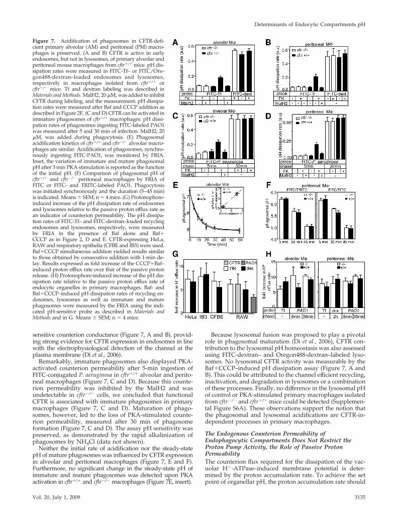
sensitive counterion conductance (Figure 7, A and B), provid-ing strong evidence for CFTR expression in endosomes in linewith the electrophysiological detection of the channel at theplasma membrane (Di et al., 2006).
Remarkably, immature phagosomes also displayed PKA-activated counterion permeability after 5-min ingestion ofFITC-conjugated P. aeruginosa in cftr�/� alveolar and perito-neal macrophages (Figure 7, C and D). Because this counte-rion permeability was inhibited by the MalH2 and wasundetectable in cftr�/� cells, we concluded that functionalCFTR is associated with immature phagosomes in primarymacrophages (Figure 7, C and D). Maturation of phago-somes, however, led to the loss of PKA-stimulated counte-rion permeability, measured after 30 min of phagosomeformation (Figure 7, C and D). The assay pH sensitivity waspreserved, as demonstrated by the rapid alkalinization ofphagosomes by NH4Cl (data not shown).
Neither the initial rate of acidification nor the steady-statepH of mature phagosomes was influenced by CFTR expressionin alveolar and peritoneal macrophages (Figure 7, E and F).Furthermore, no significant change in the steady-state pH ofimmature and mature phagosomes was detected upon PKAactivation in cftr�/� and cftr�/� macrophages (Figure 7E, insert).
Because lysosomal fusion was proposed to play a pivotalrole in phagosomal maturation (Di et al., 2006), CFTR con-tribution to the lysosomal pH homeostasis was also assessedusing FITC-dextran– and Oregon488-dextran–labeled lyso-somes. No lysosomal CFTR activity was measurable by theBaf�CCCP–induced pH dissipation assay (Figure 7, A andB). This could be attributed to the channel efficient recycling,inactivation, and degradation in lysosomes or a combinationof these processes. Finally, no difference in the lysosomal pHof control or PKA-stimulated primary macrophages isolatedfrom cftr�/� and cftr�/� mice could be detected (Supplemen-tal Figure S6A). These observations support the notion thatthe phagosomal and lysosomal acidifications are CFTR-in-dependent processes in primary macrophages.
The Endogenous Counterion Permeability ofEndophagocytic Compartments Does Not Restrict theProton Pump Activity, the Role of Passive ProtonPermeabilityThe counterion flux required for the dissipation of the vac-uolar H�-ATPase–induced membrane potential is deter-mined by the proton accumulation rate. To achieve the setpoint of organellar pH, the proton accumulation rate should
Figure 7. Acidification of phagosomes in CFTR-defi-cient primary alveolar (AM) and peritoneal (PM) macro-phages is preserved. (A and B) CFTR is active in earlyendosomes, but not in lysosomes, of primary alveolar andperitoneal mouse macrophages from cftr�/� mice. pH dis-sipation rates were measured in FITC-Tf– or FITC/Ore-gon488-dextran–loaded endosomes and lysosomes,respectively in macrophages isolated from cftr�/� orcftr�/� mice. Tf and dextran labeling was described inMaterials and Methods. MalH2, 20 �M, was added to inhibitCFTR during labeling, and the measurement. pH dissipa-tion rates were measured after Baf and CCCP addition asdescribed in Figure 2E. (C and D) CFTR can be activated inimmature phagosomes of cftr�/� macrophages. pH dissi-pation rates of phagosomes ingesting FITC-labeled PAO1was measured after 5 and 30 min of infection. MalH2, 20�M, was added during phagocytosis. (E) Phagosomalacidification kinetics of cftr�/� and cftr�/� alveolar macro-phages are similar. Acidification of phagosomes, synchro-nously ingesting FITC-PAO1, was monitored by FRIA.Inset, the variation of immature and mature phagosomalpH after 3 min PKA stimulation is reported as the functionof the initial pH. (F) Comparison of phagosomal pH ofcftr�/� and cftr�/� peritoneal macrophages by FRIA ofFITC or FITC- and TRITC-labeled PAO1. Phagocytosiswas initiated synchronously and the duration (5–45 min)is indicated. Means � SEM; n � 4 mice. (G) Protonophore-induced increase of the pH dissipation rate of endosomesand lysosomes relative to the passive proton efflux rate asan indicator of counterion permeability. The pH dissipa-tion rates of FITC-Tf– and FITC-dextran–loaded recyclingendosomes and lysosomes, respectively, were measuredby FRIA in the presence of Baf alone and Baf�CCCP as in Figure 2, D and E. CFTR-expressing HeLa,RAW and respiratory epithelia (CFBE and IB3) were used.Baf�CCCP simultaneous addition yielded results similarto those obtained by consecutive addition with 1-min de-lay. Results expressed as fold increase of the CCCP�Baf–induced proton efflux rate over that of the passive protonrelease. (H) Protonophore-induced increase of the pH dis-sipation rate relative to the passive proton efflux rate ofendocytic organelles in primary macrophages. Baf- andBaf�CCCP–induced pH dissipation rates of recycling en-dosomes, lysosomes as well as immature and maturephagosomes were measured by the FRIA using the indi-cated pH-sensitive probe as described in Materials andMethods and in G. Means � SEM; n � 4 mice.
Determinants of Endocytic Compartments pH
Vol. 20, July 1, 2009 3135

be adjusted according to the buffer capacity and the protonleak (Wu et al., 2001). Considering that CFTR overexpres-sion, activation, and ablation failed to change the organellaracidification in endolysosomes and phagosomes, we hy-pothesized that the endogenous, CFTR-independent coun-terion permeability is sufficiently high to dissipate the mem-brane potential (Steinberg et al., 2007b) and thereby cannotimpede the proton translocation rate. This assumption im-plies that the passive proton permeability of endocytic or-ganelles is small in comparison with their counterion per-meability. To support this prediction, we assessed thecounterion and passive proton permeabilities of endocyticorganelles in respiratory epithelia, macrophages, and otherheterologous expression systems, assuming that the passivepH dissipation rate is proportional with the proton perme-abilities.
The Baf-induced proton efflux reflects the passive protonleak that is compensated by the v-ATPase activity at thesteady-state pH. Comparison of the Baf- and Baf�CCCP–induced pH dissipation rates revealed that the CFTR-inde-pendent counterion permeability was �5–10 times higherthan the passive proton permeability both in recycling en-dosomes and lysosomes of IB3 and CFBE epithelia (Figure7G). Similar data were obtained in primary macrophages(Figure 7H). These results strongly suggest that the counte-rion permeability is sufficiently high to support the acidifi-cation of organelles in CFTR-deficient respiratory epitheliaand primary macrophages.
Decreasing passive proton permeability along the secre-tory pathway was proposed as a critical determinant of theprogressively increasing luminal acidification (Wu et al.,2001). An analogous role of the passive proton permeabilitymay prevail along the endocytic pathway. To determine thepassive proton permeability of endocytic organelles, first thebuffer capacity of endosomes, lysosomes, and phagosomes
was measured, using the ammonium chloride pulse tech-nique (Roos and Boron, 1981; Figure 8, A and B). The passiveproton permeability was calculated based on the Baf-in-duced proton efflux rate and the assumption that the pre-dominant driving force is the transmembrane pH gradient ata constant cytoplasmic pH 7.3 as described in Materials andMethods. The passive proton permeability of lysosomes wasat least twofold lower than in recycling endosomes in BHK,HeLa, IB3, and CFBE cells (Figure 8C), supporting the notionthat down-regulation of the passive proton leak may con-tribute to the progressive acidification along the endocyticpathway. For comparison, the passive proton permeabilityof the ER and Golgi compartment was also plotted, as de-termined in previous publications (Llopis et al., 1998;Chandy et al., 2001; Wu et al., 2001; Weisz, 2003b).
DISCUSSION
The paucity of our understanding of CFTR function in or-ganellar pH homeostasis is illustrated by the fact that CFTRis invoked in facilitating (Barasch et al., 1991; Di et al., 2006;Teichgraber et al., 2008), inhibiting (Poschet et al., 2002), orhaving no effect on the acidification of exocytic and endo-cytic organelles (Lukacs et al., 1992; Dunn et al., 1994; Root etal., 1994; Seksek et al., 1996; Gibson et al., 2000; Machen et al.,2001; Haggie and Verkman, 2007, 2009b; Lamothe andValvano, 2008). Comparison of these studies is hampered bydifferent cellular models and pH detection methodologiesused as recently reviewed by Haggie and Verkman (2009a).Because organellar acidification has fundamental influenceon various cellular functions with proposed relevance to thehyperinflammatory CF lung infection (Konstan et al., 1994;Pier et al., 1996; Rowe et al., 2005; Sagel et al., 2007), ourprimary goal was to assess the role of counterion conduc-tance in general, and CFTR in particular, in the pH regula-
Figure 8. Determination of the passive proton perme-ability of endocytic organelles. (A) Measurement of thebuffer capacity of endocytic organelles. The lysosomalcompartment of BHK cells were loaded with dextran asdescribed in Materials and Methods, and the organellarpH was monitored by FRIA. The extent of lysosomealkalinization was measured after the addition of smallamounts of NH4Cl (e.g., 0.1–2 mM) in the presence ofBaf to prevent the compensatory activation of the v-ATPase. The number indicates the final concentration ofNH4Cl in mM. The buffer capacity was calculated fromthe alkalinization, induced by the 0.5–1 mM NH4Cl asdescribed in Roos and Boron (1981) and Chandy et al.(2001). (B) The buffer capacity of recycling endosomes,lysosomes, and phagosomes was measured on FITC-Tf–and FITC-dextran–loaded organelles as described in A.The early and mature phagosomal buffer capacitieswere measured after 5 and 30 min phagocytosis of FITC-conjugated P. aeruginosa. (C) The passive proton perme-ability of recycling endosomes, lysosomes, and phago-somes. The passive proton efflux rate from the indicatedorganelles was measured in the presence of 400 nM Bafby FRIA after their selective labeling as described inMaterials and Methods. The passive proton permeabilitywas calculated as described in Materials and Methods. Forcomparison, the passive proton permeability of theGolgi compartment and the ER was indicated obtainedfrom previous publications (Llopis et al., 1998; Chandyet al., 2001; Wu et al., 2001; Weisz, 2003b).
H. Barriere et al.
Molecular Biology of the Cell3136

tion of endocytic compartments in well-defined cellularmodels.
The Endogenous Counterion Conductance Is NotRate-limiting for Endosomal Acidification of CFRespiratory EpitheliaDuring the past decade, three major determinants of or-ganellar acidification have been established: the counterionconductance, the intrinsic proton leak, and the v-ATPaseactivity (Weisz, 2003b). The major counterion conductanceof endolysosomes consists of members of two branches ofthe ClC gene family; ClC-3, -4, and -5, and ClC-6 and -7(Marshansky et al., 2002; Jentsch et al., 2005; Picollo andPusch, 2005; Jentsch, 2007). Subcellular localization of theClC-3, -4, and -5 in concert with functional data suggest thatthese ClC transporters are permissive for the acidification ofearly endosomes and synaptic vesicles by providing asignificant electrical shunt pathway (Stobrawa et al., 2001;Suzuki et al., 2006; Maritzen et al., 2008). Ablation of ClC-4and -5 impairs the acidification of endosomes (Gunther et al.,1998; Hara-Chikuma et al., 2005a), whereas ClC-3 depletioncauses defective acidification of synaptic vesicles (Stobrawaet al., 2001; Maritzen et al., 2008) as well as early endosomes(Li et al., 2002; Hara-Chikuma et al., 2005b). Although addi-tional Cl� transport pathway may be involved (Schenck etal., 2009). ClC-4, -5, and -7 are now known to operate asvoltage-dependent Cl�/H� antiporters (Picollo and Pusch,2005; Scheel et al., 2005; Graves et al., 2008). If two Cl� ionsare exchanged for one H�, as determined for the ClC-7, asingle transport cycle of the ClC-3, -4, and -5 ensures chargeneutralization of two protons uptake at the cost of oneproton efflux. Impaired functional expression of ClC-3, -4, or-5, therefore, impedes the v-ATPase–mediated proton accu-mulation by increasing the inside positive membrane poten-tial (Marshansky et al., 2002). Although these data providesupportive evidence for the permissive role of ClC-3, -4, and-5 in endosomal acidification, they are short on demonstrat-ing that the overall counterion permeability limits the pro-ton pump activity in vivo, a prerequisite for CFTR to directlyinfluence the endosomal pH regulation.
To compare the CFTR-independent and CFTR-dependentcounterion and proton permeabilities, endosomes contain-ing functional (wt) or nonfunctional (G551D) CFTR werelabeled with the pH-sensitive fluorophore, restricting theprobe to CFTR-expressing vesicles. Using saturating concen-tration of Baf in the absence or in combination with a prot-onophore, we determined the relative magnitude of passiveproton leak and the maximum rate of pH dissipation, anindicator of the lower limit of endosomal counterion perme-ability. The CFTR-independent counterion permeability wasseveral-fold larger than the passive proton permeability inapical endosomes of polarized bronchial epithelia (CFBE)derived from CF patient. Importantly, although CFTRcomplementation of CFBE and IB3 conferred a threefoldincrease in counterion permeability (Figure 4B and Supple-mental Figure S3E), it failed to alter the passive protonpermeability and the initial acidification rate, important de-terminants of organellar pH (Figure 4, E and F, and data notshown; Chandy et al., 2001; Machen et al., 2003). Theseresults are complemented with studies on cells (HeLa, BHK,and MDCK) where organellar acidification is independent ofCFTR. The channel overexpression failed to hyperacidify theendolysosomal compartments (Figure 4, C and D, and Sup-plemental Figure S6C), implying that the v-ATPase activityis not limited by endogenous counterion permeability. Sim-ilar results were obtained in IB3 respiratory epithelia andHeLa cells using FITC-Tf labeling of recycling endosomes
and jointly support the notion that the endosomal pH cannotbe regulated by modest alteration of the relatively largecounterion conductance under physiological conditions.
Identification of two ubiquitously expressing endolysoso-mal cation efflux pathways supports the inference that theCFTR-independent counterion permeability is significantand cannot restrict the relatively slow endosomal protonaccumulation. The TRPV2, a member of the transient recep-tor potential channel family, was confined to early endo-somes (Saito et al., 2007). Its limited selectivity toward Ca2�
over K�, activation by luminal acidification and low chlo-ride concentration makes it a plausible candidate to contrib-ute to cation efflux accompanying proton accumulation sim-ilar to the TRPML1 (mucolipin1) channel, localized to lateendosomes and lysosomes and invoked in iron release(Dong et al., 2008).
CFTR-independent Lysosomal AcidificationThe lysosomal acidification defect (pH �5.9) was implicatedin ceramide accumulation, which in turn caused excessivesecretion of the proinflammatory cytokine interleukin-1(IL-1) and the keratinocyte-derived chemokine (the mousehomologue of IL-8), cell death, and susceptibility to P.aeruginosa infection of respiratory epithelia (Bruscia et al.,2008; Teichgraber et al., 2008). Importantly, alkalinization oflysosomes to pH �5.9 was accounted for the phagosomalacidification defect and intracellular proliferation of P.aeruginosa in cftr�/� alveolar macrophages (Di et al., 2006).We were unable to reproduce any alkalinization of lyso-somes in CF respiratory epithelia (CFBE or IB3) comparedwith their CFTR-complemented counterparts (Figure 4C,and Supplemental Figure S6C). Likewise, we could not re-solve significant change in the lysosomal pH of alveolar orperitoneal macrophages derived from cftr�/� and cftr�/�
mouse or after the channel activation by PKA agonist. Theseresults confirm and expand recent observations indicatingthat the phagosomal/lysosomal acidification of �F508-CFTRalveolar and CFTR-deficient bone marrow mouse macro-phages, as well as primary respiratory epithelia are pre-served (Haggie and Verkman, 2007, 2009b; Lamothe andValvano, 2008).
Albeit CFTR could be activated in endosomes, no channelfunction was detected in lysosomes of CFTR complementedCFBE and IB3 epithelia by the pH dissipation assay (Sup-plemental Figure S6B) and no difference in the lysosomal pHcould be established (Supplemental Figure S6C). Similardata were obtained in RAW, BHK, and HeLa cells (Figure 4,C and D, and data not shown). These results cannot beattributed to a significant increase of the electrochemicalchloride gradient across the lysosomal membrane, becausethe luminal chloride concentration of late endosomes/lyso-somes was found to be 40–50 mM (Hara-Chikuma et al.,2005b).
Cell type differences and pH measurement methodologymay explain the discordant data on lysosomal pH. Teichgra-ber et al. (2008) monitored the lysosomal pH of freshlyisolated respiratory epithelia with uncharacterized cellularcomposition using lysosensor-green D189 that labeled acidicorganelles nonselectively. The calibration was performed onpermeabilized cells, based on the correlation between theluminal pH and the fluorescence intensity of the dye. Cellpermeabilization in extracellular medium (containing 132mM NaCl and 1 mM CaCl2) likely induced significantchanges in organelle composition and adsorption of lysosen-sor-green to cytosolic constituents. Because the fluorescenceintensity of lysosensor-green was measured in areas of 1.5 �1.5 mm and not in individual vesicles (Teichgraber et al.,
Determinants of Endocytic Compartments pH
Vol. 20, July 1, 2009 3137

2008), nonspecific changes during the calibration could notbe ruled out. Our measurements were performed on well-characterized bronchial epithelia (CFBE and IB3) that havebeen used extensively (Zeitlin et al., 1991; Cozens et al., 1994;Gruenert et al., 1995; Moyer et al., 1999; Bebok et al., 2005;Swiatecka-Urban et al., 2005; Varga et al., 2008). We relied onratiometric pH determination of FITC/Oregon488-dextran–labeled individual lysosomes that allowed concentration-independent, in situ calibration, a standard assay to monitorendolysosomal pH that could also resolve small pH changesin subpopulations of organelles (Haggie and Verkman,2009b; see Supplemental Figure S1, B–D). Experiments werealso carried out on FITC/TMR-dextran labeled lysosomes,to validate the assay pH sensitivity, with identical results(data not shown).
Several considerations support our conclusion that lyso-somal acidification is independent of CFTR in respiratoryepithelia and macrophages. 1) Internalized CFTR constitu-tively recycles back to the cell surface and only �10% ofcomplex-glycosylated channels are targeted to lysosomes in1–2 h. Highly efficient recycling preserves the long metabolichalf-life of CFTR in post-Golgi compartments and was ob-served in a wide variety of heterologous expression systems,including respiratory epithelia (Varga et al., 2008). 2) CFmutations provoked ubiquitination and rapid down-regula-tion of CFTR from the cell surface by lysosomal proteolysis(Sharma et al., 2004). We assume that a similar mechanism isresponsible for the nonnative wt CFTR turnover (Sharma etal., 2004). Indeed, CFTR immunoreactive degradation inter-mediates were detectable in purified lysosomes upon inhi-bition of lysosomal protease (Benharouga et al., 2001). There-fore, CFTR channel density in the lysosomal membrane islimited and immunolocalization studies could visualize par-tially degraded, nonfunctional channels (Di et al., 2006). 3)Even if low copy number of CFTR is inserted into thelysosomal membrane, the acidic pH inhibits by �50% thechannel activity. This inference is based on the inhibitoryeffect of pH 5 on the PKA-activated plasma membrane ha-lide conductance in BHK cells (Supplemental Figure S5). 4)Finally, and perhaps most importantly, we showed that theCFTR-independent endogenous counterion permeability oflysosomes was 5–10-fold higher than the passive protonpermeability in CFBE and IB3 respiratory epithelia and al-veolar macrophages (Figure 7, G and H). Remarkably, thelysosomal passive proton permeability appears to be �1�m/s, the lowest among acidic organelles of the endocyticand secretory pathway (Figure 8C), maximizing the acidifi-cation efficiency, whereas minimizing the counterion fluxrequirement. These observations imply that the transmem-brane potential generated by lysosomal proton accumula-tion is largely dissipated by CFTR-independent counterionmovements, mediated by the ubiquitously expressed ClC6and ClC7 as Cl/H exchangers and a subset of cation-protonexchangers and cation channels (Marshansky et al., 2002;Jentsch, 2007; Orlowski and Grinstein, 2007; Dong et al.,2008).
CFTR Expression and Activation Does Not InfluencePhagosomal AcidificationIt was proposed that CFTR is essential for the bactericidalactivity of alveolar macrophages by ensuring phagolyso-somes acidification (Di et al., 2006). Ingested bacteria inCFTR-deficient alveolar macrophages remained in mildlyacidic phagosomes because of the lysosomal acidificationdefect (Di et al., 2006). Importantly, no phagosomal acidifi-cation defect was reported in alveolar macrophages from�F508 cftr homozygous mice or by inhibiting the wt channel
with the channel blocker, CFTRinh-172 (Haggie and Verkman,2007). Likewise, the phagosomal acidification of bone mar-row macrophages obtained from cftr�/� mouse was unaf-fected (Lamothe and Valvano, 2008). To address whetherexpression of �F508-CFTR and cell-specific differences couldexplain the contrasting results, our studies were performedon CFTR-deficient alveolar and peritoneal macrophages. Nodifference in the phagosomal acidification rate and extentwas documented in CFTR-deficient and control macro-phages (Figure 7, E and F). Importantly, similar results wereobtained upon inhibition of the endogenous CFTR activityby MalH2 in RAW macrophages. Analysis of CFTR mem-brane trafficking, as well as the relative proton and counte-rion permeabilities, provided compelling rationale for theinability of CFTR to regulate phagosomal acidification inthese models.
CFTR was only transiently confined to newly formedphagosomes and eliminated from mature phagosomes,demonstrated by CFTR immunolocalization (Figure 5D), aswell as by vesicular pH determination of FITC-labeled CFTRin RAW macrophages (Figure 6, D and F). Exogenous over-expression of CFTR-3HA cannot account for this phenom-enon, because endogenous CFTR was identified in theimmature phagosome of RAW, alveolar, and peritonealmacrophages by the PKA- and MalH2-sensitive pH dissipa-tion (Figures 6B and 7, A and B). The rapid loss of CFTRduring phagosomal maturation parallels the Tf-R retrievalfor recycling (Figure 6, C and D), demonstrated to be accom-plished by COPI-coated vesicle budding (Botelho et al.,2000).
Finally, we showed that the CFTR-independent counte-rion permeability of both the immature and mature phago-some is several-fold higher than its endogenous proton per-meability. The relatively high counterion permeability ofCFTR-deficient and wt primary macrophages (as well asRAW cells) is comparable to that observed in thioglycolateelicited peritoneal and RAW macrophages (Lukacs et al.,1990). The modest phagosomal membrane potential (12–27mV) of RAW macrophages is in accord with efficient elec-trical shunting (Steinberg et al., 2007a). We envision thatsimilarly low phagosomal membrane potential develops inprimary CF alveolar macrophages in the presence of theendogenous counterion permeability. Therefore, neitherCFTR complementation nor overexpression influences thephagosomal pH homeostasis during the fast acidificationphase or at steady state (Haggie and Verkman, 2007; Figure6, D and F).
In summary, our results, obtained on genetically matchedrespiratory epithelial cell lines and primary alveolar macro-phages, indicate that CFTR has no role in the acidification ofendosomes, lysosomes, and phagosomes because of the rel-atively large counterion and limited proton permeability ofthese organelles. Therefore, other factors than the acidifica-tion defect must be invoked in defining the connection be-tween CF mutations and the severe lung pathology. At amore general level, our results showed that the counterionpermeability of endocytic organelles exceeds several fold therequirement for shunting the v-ATPase generated mem-brane potential in all cell types examined. Thus, limitedattenuation or elevation of the counterion permeability can-not play a decisive role in the organellar acidification, con-sistent with the emerging paradigm suggesting that subunitcomposition of the v-ATPase and the passive proton leak arethe most critical factors in adjusting the luminal pH of acidicorganelles (Hurtado-Lorenzo et al., 2006). The smallest pas-sive proton permeability of lysosomes among acidic or-ganelles is consistent with this hypothesis (Figure 8C).
H. Barriere et al.
Molecular Biology of the Cell3138

ACKNOWLEDGMENTS
The authors thank Dr. Johanna Rommens for the G511D CFTR cDNA, Dr.Matt Parsek (University of Seattle) for the P. aeruginosa PAO1 strain, Dr. AlanVerkman (University of California San Francisco) for the MalH2 inhibitor andDr. Pamela Zeitlin (Johns Hopkins University) for the IB3 cells. We thankHadi Salah for generating the CFTR-transfected IB3 cells. This work wasfunded by the Canadian Institute of Health Research (CIHR), by the NationalInstitutes of Health National Institute of Diabetes, and Digestive, and KidneyDiseases, by the Canada Foundation for Innovation to G.L., and by the CIHRand the Canadian Cystic Fibrosis Foundation to D.R. G.L. also holds a CanadaResearch Chair in the Molecular Cell Biology of Cystic Fibrosis.
REFERENCES
Barasch, J., Kiss, B., Prince, A., Saiman, L., Gruenert, D., and al-Awqati, Q.(1991). Defective acidification of intracellular organelles in cystic fibrosis.Nature 352, 70–73.
Barriere, H., and Lukacs, G. L. (2008). Analysis of endocytic trafficking bysingle-cell fluorescence ratio imaging. Curr. Protoc. Cell Biol. Chapter 15, Unit15 13.
Barriere, H., Nemes, C., Du, K., and Lukacs, G. L. (2007). Plasticity of polyu-biquitin recognition as lysosomal targeting signals by the endosomal sortingmachinery. Mol. Biol. Cell 18, 3952–3965.
Barriere, H., Nemes, C., Lechardeur, D., Khan-Mohammad, M., Fruh, K., andLukacs, G. L. (2006). Molecular basis of oligoubiquitin-dependent internaliza-tion of membrane proteins in mammalian cells. Traffic 7, 282–297.
Bebok, Z., Collawn, J. F., Wakefield, J., Parker, W., Li, Y., Varga, K., Sorscher,E. J., and Clancy, J. P. (2005). Failure of cAMP agonists to activate rescueddeltaF508 CFTR in CFBE41o- airway epithelial monolayers. J. Physiol. 569,601–615.
Becq, F., Jensen, T. J., Chang, X. B., Savoia, A., Rommens, J. M., Tsui, L. C.,Buchwald, M., Riordan, J. R., and Hanrahan, J. W. (1994). Phosphatase inhib-itors activate normal and defective CFTR chloride channels. Proc. Natl. Acad.Sci. USA 91, 9160–9164.
Benharouga, M., Haardt, M., Kartner, N., and Lukacs, G. L. (2001). COOH-terminal truncations promote proteasome-dependent degradation of maturecystic fibrosis transmembrane conductance regulator from post-Golgi com-partments. J. Cell Biol. 153, 957–970.
Benharouga, M., Sharma, M., So, J., Haardt, M., Drzymala, L., Popov, M.,Schwapach, B., Grinstein, S., Du, K., and Lukacs, G. L. (2003). The role of theC terminus and Na�/H� exchanger regulatory factor in the functionalexpression of cystic fibrosis transmembrane conductance regulator in nonpo-larized cells and epithelia. J. Biol. Chem. 278, 22079–22089.
Biwersi, J., and Verkman, A. S. (1994). Functional CFTR in endosomal com-partment of CFTR-expressing fibroblasts and T84 cells. Am J. Physiol. 266,C149–C156.
Bompadre, S. G., Sohma, Y., Li, M., and Hwang, T. C. (2007). G551D andG1349D, two CF-associated mutations in the signature sequences of CFTR,exhibit distinct gating defects. J. Gen. Physiol. 129, 285–298.
Botelho, R. J., Hackam, D. J., Schreiber, A. D., and Grinstein, S. (2000). Role ofCOPI in phagosome maturation. J. Biol. Chem. 275, 15717–15727.
Boucher, R. C. (2007). Cystic fibrosis: a disease of vulnerability to airwaysurface dehydration. Trends Mol. Med. 13, 231–240.
Bruscia, E., Sangiuolo, F., Sinibaldi, P., Goncz, K. K., Novelli, G., and Gruenert,D. C. (2002). Isolation of CF cell lines corrected at DeltaF508-CFTR locus bySFHR-mediated targeting. Gene Ther. 9, 683–685.
Bruscia, E. M., Zhang, P. X., Ferreira, E., Caputo, C., Emerson, J. W., Tuck, D.,Krause, D. S., and Egan, M. E. (2009). Macrophages directly contribute to theexaggerated inflammatory response in CFTR-/- mice. Am J. Respir. Cell. Mol.Biol. 40, 295–304.
Campodonico, V. L., Gadjeva, M., Paradis-Bleau, C., Uluer, A., and Pier, G. B.(2008). Airway epithelial control of Pseudomonas aeruginosa infection in cysticfibrosis. Trends Mol. Med. 14, 120–133.
Chandy, G., Grabe, M., Moore, H. P., and Machen, T. E. (2001). Proton leakand CFTR in regulation of Golgi pH in respiratory epithelial cells. Am J.Physiol. Cell Physiol. 281, C908–C921.
Chastellier, C. D. (2008). EM analysis of phagosomes. Methods Mol. Biol. 445,261–285.
Cozens, A. L., Yezzi, M. J., Kunzelmann, K., Ohrui, T., Chin, L., Eng, K.,Finkbeiner, W. E., Widdicombe, J. H., and Gruenert, D. C. (1994). CFTRexpression and chloride secretion in polarized immortal human bronchialepithelial cells. Am J. Respir. Cell. Mol. Biol. 10, 38–47.
Di, A., et al. (2006). CFTR regulates phagosome acidification in macrophagesand alters bactericidal activity. Nat. Cell Biol. 8, 933–944.
Dong, X. P., Cheng, X., Mills, E., Delling, M., Wang, F., Kurz, T., and Xu, H.(2008). The type IV mucolipidosis-associated protein TRPML1 is an endoly-sosomal iron release channel. Nature 455, 992–996.
Duarri, A., et al. (2008). Molecular pathogenesis of megalencephalic leukoen-cephalopathy with subcortical cysts: mutations in MLC1 cause folding de-fects. Hum Mol. Genet 17, 3728–3739.
Dunn, K. W., Park, J., Semrad, C. E., Gelman, D. L., Shevell, T., and McGraw,T. E. (1994). Regulation of endocytic trafficking and acidification are indepen-dent of the cystic fibrosis transmembrane regulator. J. Biol. Chem. 269, 5336–5345.
Forgac, M. (1998). Structure, function and regulation of the vacuolar (H�)-ATPases. FEBS Lett. 440, 258–263.
Geisow, M. J., D’Arcy Hart, P., and Young, M. R. (1981). Temporal changes oflysosome and phagosome pH during phagolysosome formation in macro-phages: studies by fluorescence spectroscopy. J. Cell Biol. 89, 645–652.
Gentzsch, M., Choudhury, A., Chang, X. B., Pagano, R. E., and Riordan, J. R.(2007). Misassembled mutant DeltaF508 CFTR in the distal secretory pathwayalters cellular lipid trafficking. J. Cell Sci. 120, 447–455.
Gibson, G. A., Hill, W. G., and Weisz, O. A. (2000). Evidence against theacidification hypothesis in cystic fibrosis. Am J. Physiol. Cell Physiol. 279,C1088–C1099.
Glozman, R., Okiyoneda, T., Mulvihill, C. M., Rini, J. M., Barriere, H., andLukacs, G. L. (2009). N-glycans are direct determinants of CFTR folding andstability in secretory and endocytic membrane traffic. J. Cell Biol. 184, 847–862.
Grabe, M., and Oster, G. (2001). Regulation of organelle acidity. J. GenPhysiol. 117, 329–344.
Graves, A. R., Curran, P. K., Smith, C. L., and Mindell, J. A. (2008). TheCl�/H� antiporter ClC-7 is the primary chloride permeation pathway inlysosomes. Nature 453, 788–792.
Griffiths, G., Back, R., and Marsh, M. (1989). A quantitative analysis of theendocytic pathway in baby hamster kidney cells. J. Cell Biol. 109, 2703–2720.
Gruenert, D. C., Finkbeiner, W. E., and Widdicombe, J. H. (1995). Culture andtransformation of human airway epithelial cells. Am J. Physiol. 268, L347–L360.
Guilbault, C., et al. (2008). Fenretinide corrects newly found ceramide defi-ciency in cystic fibrosis. Am J. Respir Cell. Mol. Biol. 38, 47–56.
Gunther, W., Luchow, A., Cluzeaud, F., Vandewalle, A., and Jentsch, T. J.(1998). ClC-5, the chloride channel mutated in Dent’s disease, colocalizes withthe proton pump in endocytotically active kidney cells. Proc. Natl. Acad. Sci.USA 95, 8075–8080.
Hackam, D. J., Rotstein, O. D., Zhang, W. J., Demaurex, N., Woodside, M.,Tsai, O., and Grinstein, S. (1997). Regulation of phagosomal acidification.Differential targeting of Na�/H� exchangers, Na�/K�-ATPases, and vac-uolar-type H�-ATPases. J. Biol. Chem. 272, 29810–29820.
Haggie, P. M., and Verkman, A. S. (2007). Cystic fibrosis transmembraneconductance regulator-independent phagosomal acidification in macro-phages. J. Biol. Chem. 282, 31422–31428.
Haggie, P. M., and Verkman, A. S. (2009a). Defective Organellar Acidificationas a Cause of Cystic Fibrosis Lung Disease—Re-Examination of a RecurringHypothesis. Am J. Physiol. Lung Cell Mol. Physiol. 296, L859–L867.
Haggie, P. M., and Verkman, A. S. (2009b). Unimpaired lysosomal acidifica-tion in respiratory epithelial cells in cystic fibrosis. J. Biol. Chem. 284, 7681–7686.
Hara-Chikuma, M., Wang, Y., Guggino, S. E., Guggino, W. B., and Verkman,A. S. (2005a). Impaired acidification in early endosomes of ClC-5 deficientproximal tubule. Biochem. Biophys. Res. Commun. 329, 941–946.
Hara-Chikuma, M., Yang, B., Sonawane, N. D., Sasaki, S., Uchida, S., andVerkman, A. S. (2005b). ClC-3 chloride channels facilitate endosomal acidifi-cation and chloride accumulation. J. Biol. Chem. 280, 1241–1247.
Hart, P. D., Young, M. R., Gordon, A. H., and Sullivan, K. H. (1987). Inhibitionof phagosome-lysosome fusion in macrophages by certain mycobacteria canbe explained by inhibition of lysosomal movements observed after phagocy-tosis. J. Exp. Med. 166, 933–946.
Heda, G., Tanwani, M., and Marino, C. (2001). The �F508 mutation shortensthe biochemical half-life of plasma membrane CFTR in polarized epithelialcells. Am J. Physiol. Cell Physiol. 49, C166–C174.
Hurtado-Lorenzo, A., et al. (2006). V-ATPase interacts with ARNO and Arf6 inearly endosomes and regulates the protein degradative pathway. Nat. CellBiol. 8, 124–136.
Determinants of Endocytic Compartments pH
Vol. 20, July 1, 2009 3139

Jefferies, K. C., Cipriano, D. J., and Forgac, M. (2008). Function, structureand regulation of the vacuolar (H�)-ATPases. Arch. Biochem. Biophys.476, 33– 42.
Jentsch, T. J. (2007). Chloride and the endosomal-lysosomal pathway: emerg-ing roles of CLC chloride transporters. J. Physiol. 578, 633–640.
Jentsch, T. J., Poet, M., Fuhrmann, J. C., and Zdebik, A. A. (2005). Physiolog-ical functions of CLC Cl� channels gleaned from human genetic disease andmouse models. Annu. Rev. Physiol. 67, 779–807.
Kim, J. H., Johannes, L., Goud, B., Antony, C., Lingwood, C. A., Daneman, R.,and Grinstein, S. (1998). Noninvasive measurement of the pH of the endo-plasmic reticulum at rest and during calcium release. Proc. Natl. Acad. Sci.USA 95, 2997–3002.
Konstan, M. W., Hilliard, K. A., Norvell, T. M., and Berger, M. (1994).Bronchoalveolar lavage findings in cystic fibrosis patients with stable, clini-cally mild lung disease suggest ongoing infection and inflammation. Am J.Respir. Crit. Care Med. 150, 448–454.
Kumar, K. G., et al. (2007). Site-specific ubiquitination exposes a linear motifto promote interferon-alpha receptor endocytosis. J. Cell Biol. 179, 935–950.
Lamothe, J., and Valvano, M. A. (2008). Burkholderia cenocepacia-induced delayof acidification and phagolysosomal fusion in cystic fibrosis transmembraneconductance regulator (CFTR)-defective macrophages. Microbiology 154,3825–3834.
Lechardeur, D., Xu, M., and Lukacs, G. L. (2004). Contrasting nuclear dynam-ics of the caspase-activated DNase (CAD) in dividing and apoptotic cells.J. Cell Biol. 167, 851–862.
Li, X., Wang, T., Zhao, Z., and Weinman, S. A. (2002). The ClC-3 chloridechannel promotes acidification of lysosomes in CHO-K1 and Huh-7 cells.Am J. Physiol. Cell Physiol. 282, C1483–C1491.
Llopis, J., McCaffery, J. M., Miyawaki, A., Farquhar, M. G., and Tsien, R. Y.(1998). Measurement of cytosolic, mitochondrial, and Golgi pH in singleliving cells with green fluorescent proteins. Proc. Natl. Acad. Sci. USA 95,6803–6808.
Lukacs, G. L., Chang, X. B., Kartner, N., Rotstein, O. D., Riordan, J. R., andGrinstein, S. (1992). The cystic fibrosis transmembrane regulator is presentand functional in endosomes. Role as a determinant of endosomal pH. J. Biol.Chem. 267, 14568–14572.
Lukacs, G. L., Rotstein, O. D., and Grinstein, S. (1990). Phagosomal acidifica-tion is mediated by a vacuolar-type H(�)-ATPase in murine macrophages.J. Biol. Chem. 265, 21099–21107.
Lukacs, G. L., Rotstein, O. D., and Grinstein, S. (1991). Determinants of thephagosomal pH in macrophages. In situ assessment of vacuolar H(�)-ATPaseactivity, counterion conductance, and H� “leak.” J. Biol. Chem. 266, 24540–24548.
Machen, T. E., Chandy, G., Wu, M., Grabe, M., and Moore, H. P. (2001). Cysticfibrosis transmembrane conductance regulator and H� permeability in reg-ulation of Golgi pH. J. Pancreas 2, 229–236.
Machen, T. E., Leigh, M. J., Taylor, C., Kimura, T., Asano, S., and Moore, H. P.(2003). pH of TGN and recycling endosomes of H�/K�-ATPase-transfectedHEK-293 cells: implications for pH regulation in the secretory pathway. Am J.Physiol. Cell Physiol. 285, C205–C214.
Maritzen, T., Keating, D. J., Neagoe, I., Zdebik, A. A., and Jentsch, T. J. (2008).Role of the vesicular chloride transporter ClC-3 in neuroendocrine tissue.J. Neurosci. 28, 10587–10598.
Marshansky, V., Ausiello, D. A., and Brown, D. (2002). Physiological impor-tance of endosomal acidification: potential role in proximal tubulopathies.Curr. Opin. Nephrol. Hypertens. 11, 527–537.
Marshansky, V., and Futai, M. (2008). The V-type H�-ATPase in vesiculartrafficking: targeting, regulation and function. Curr. Opin. Cell Biol. 20, 415–426.
Mellman, I., Fuchs, R., and Helenius, A. (1986). Acidification of the endocyticand exocytic pathways. Annu. Rev. Biochem. 55, 663–700.
Moyer, B. D., et al. (1999). A PDZ-interacting domain in CFTR is an apicalmembrane polarization signal. J. Clin. Invest. 104, 1353–1361.
Muanprasat, C., Sonawane, N. D., Salinas, D., Taddei, A., Galietta, L. J., andVerkman, A. S. (2004). Discovery of glycine hydrazide pore-occluding CFTRinhibitors: mechanism, structure-activity analysis, and in vivo efficacy. J. GenPhysiol. 124, 125–137.
Mukherjee, S., Ghosh, R. N., and Maxfield, F. R. (1997). Endocytosis. Physiol.Rev. 77, 759–803.
Olson, L. J., Hindsgaul, O., Dahms, N. M., and Kim, J. J. (2008). Structuralinsights into the mechanism of pH-dependent ligand binding and release by
the cation-dependent mannose 6-phosphate receptor. J. Biol. Chem. 283,10124–10134.
Orlowski, J., and Grinstein, S. (2007). Emerging roles of alkali cation/protonexchangers in organellar homeostasis. Curr. Opin. Cell Biol. 19, 483–492.
Pedemonte, N., Lukacs, G. L., Du, K., Caci, E., Zegarra-Moran, O., Galietta,L. J., and Verkman, A. S. (2005). Small-molecule correctors of defectiveDeltaF508-CFTR cellular processing identified by high-throughput screening.J. Clin. Invest. 115, 2564–2571.
Picollo, A., and Pusch, M. (2005). Chloride/proton antiporter activity ofmammalian CLC proteins ClC-4 and ClC-5. Nature 436, 420–423.
Pier, G. B., Grout, M., Zaidi, T. S., and Goldberg, J. B. (1996). How mutantCFTR may contribute to Pseudomonas aeruginosa infection in cystic fibrosis.Am J. Respir. Crit. Care Med. 154, S175–S182.
Poschet, J. F., Boucher, J. C., Tatterson, L., Skidmore, J., Van Dyke, R. W., andDeretic, V. (2001). Molecular basis for defective glycosylation and Pseudomo-nas pathogenesis in cystic fibrosis lung. Proc. Natl. Acad. Sci. USA 98, 13972–13977.
Poschet, J. F., Fazio, J. A., Timmins, G. S., Ornatowski, W., Perkett, E.,Delgado, M., and Deretic, V. (2006). Endosomal hyperacidification in cysticfibrosis is due to defective nitric oxide-cylic GMP signalling cascade. EMBORep. 7, 553–559.
Poschet, J. F., Skidmore, J., Boucher, J. C., Firoved, A. M., Van Dyke, R. W.,and Deretic, V. (2002). Hyperacidification of cellubrevin endocytic compart-ments and defective endosomal recycling in cystic fibrosis respiratory epithe-lial cells. J. Biol. Chem. 277, 13959–13965.
Riordan, J. R. (2008). CFTR function and prospects for therapy. Annu. Rev.Biochem. 77, 701–726.
Riordan, J. R., et al. (1989). Identification of the cystic fibrosis gene: cloningand characterization of complementary DNA. Science 245, 1066–1073.
Roos, A., and Boron, W. F. (1981). Intracellular pH. Physiol. Rev. 61, 296–434.
Root, K. V., Engelhardt, J. F., Post, M., Wilson, J. W., and Van Dyke, R. W.(1994). CFTR does not alter acidification of L cell endosomes. Biochem.Biophys. Res. Commun. 205, 396–401.
Rowe, S. M., Miller, S., and Sorscher, E. J. (2005). Cystic fibrosis. N. Engl.J. Med. 352, 1992–2001.
Sagel, S. D., Chmiel, J. F., and Konstan, M. W. (2007). Sputum biomarkers ofinflammation in cystic fibrosis lung disease. Proc. Am Thorac. Soc. 4, 406–417.
Saito, M., Hanson, P. I., and Schlesinger, P. (2007). Luminal chloride-depen-dent activation of endosome calcium channels: patch clamp study of enlargedendosomes. J. Biol. Chem. 282, 27327–27333.
Scheel, O., Zdebik, A. A., Lourdel, S., and Jentsch, T. J. (2005). Voltage-dependent electrogenic chloride/proton exchange by endosomal CLC pro-teins. Nature 436, 424–427.
Schenck, S., Wojcik, S. M., Brose, N., and Takamori, S. (2009). A chlorideconductance in VGLUT1 underlies maximal glutamate loading into synapticvesicles. Nat. Neurosci. 12, 156–162.
Seksek, O., Biwersi, J., and Verkman, A. S. (1996). Evidence against defectivetrans-Golgi acidification in cystic fibrosis. J. Biol. Chem. 271, 15542–15548.
Sharma, M., Benharouga, M., Hu, W., and Lukacs, G. L. (2001). Conforma-tional and temperature-sensitive stability defects of the delta F508 cysticfibrosis transmembrane conductance regulator in post-endoplasmic reticulumcompartments. J. Biol. Chem. 276, 8942–8950.
Sharma, M., et al. (2004). Misfolding diverts CFTR from recycling to degra-dation: quality control at early endosomes. J. Cell Biol. 164, 923–933.
Sonawane, N. D., and Verkman, A. S. (2003). Determinants of Cl� inrecycling and late endosomes and Golgi complex measured using fluorescentligands. J. Cell Biol. 160, 1129–1138.
Sonawane, N. D., Zhao, D., Zegarra-Moran, O., Galietta, L. J., and Verkman,A. S. (2008). Nanomolar CFTR inhibition by pore-occluding divalent polyeth-ylene glycol-malonic acid hydrazides. Chem. Biol 15, 718–728.
Steinberg, B. E., and Grinstein, S. (2007). Assessment of phagosome formationand maturation by fluorescence microscopy. Methods Mol. Biol. 412, 289–300.
Steinberg, B. E., Huynh, K. K., and Grinstein, S. (2007a). Phagosomal acidifi-cation: measurement, manipulation and functional consequences. Biochem.Soc. Trans. 35, 1083–1087.
Steinberg, B. E., Touret, N., Vargas-Caballero, M., and Grinstein, S. (2007b). Insitu measurement of the electrical potential across the phagosomal membraneusing FRET and its contribution to the proton-motive force. Proc. Natl. Acad.Sci. USA 104, 9523–9528.
H. Barriere et al.
Molecular Biology of the Cell3140

Stobrawa, S. M., et al. (2001). Disruption of ClC-3, a chloride channel ex-pressed on synaptic vesicles, leads to a loss of the hippocampus. Neuron 29,185–196.
Suzuki, T., Rai, T., Hayama, A., Sohara, E., Suda, S., Itoh, T., Sasaki, S., andUchida, S. (2006). Intracellular localization of ClC chloride channels and theirability to form hetero-oligomers. J. Cell. Physiol. 206, 792–798.
Swiatecka-Urban, A., et al. (2005). The short apical membrane half-life ofrescued �F508-cystic fibrosis transmembrane conductance regulator (CFTR)results from accelerated endocytosis of �F508-CFTR in polarized humanairway epithelial cells. J. Biol. Chem. 280, 36762–36772.
Teichgraber, V., et al. (2008). Ceramide accumulation mediates inflammation,cell death and infection susceptibility in cystic fibrosis. Nat. Med. 14, 382–391.
van Kerkhof, P., Sachse, M., Klumperman, J., and Strous, G. J. (2001). Growthhormone receptor ubiquitination coincides with recruitment to clathrin-coated membrane domains. J. Biol. Chem. 276, 3778–3784.
Varga, K., Goldstein, R. F., Jurkuvenaite, A., Chen, L., Matalon, S., Sorscher,E. J., Bebok, Z., and Collawn, J. F. (2008). Enhanced cell-surface stability of
rescued �F508 cystic fibrosis transmembrane conductance regulator (CFTR)by pharmacological chaperones. Biochem. J. 410, 555–564.
Varghese, B., et al. (2008). Polyubiquitination of prolactin receptor stimulatesits internalization, postinternalization sorting, and degradation via the lyso-somal pathway. Mol. Cell. Biol. 28, 5275–5287.
Weisz, O. A. (2003a). Acidification and protein traffic. Int. Rev. Cytol. 226,259–319.
Weisz, O. A. (2003b). Organelle acidification and disease. Traffic 4, 57–64.
Wu, M. M., Grabe, M., Adams, S., Tsien, R. Y., Moore, H. P., and Machen, T. E.(2001). Mechanisms of pH regulation in the regulated secretory pathway.J. Biol. Chem. 276, 33027–33035.
Yates, R. M., Hermetter, A., and Russell, D. G. (2005). The kinetics of phago-some maturation as a function of phagosome/lysosome fusion and acquisi-tion of hydrolytic activity. Traffic 6, 413–420.
Zeitlin, P. L., Lu, L., Rhim, J., Cutting, G., Stetten, G., Kieffer, K. A., Craig, R.,and Guggino, W. B. (1991). A cystic fibrosis bronchial epithelial cell line:immortalization by adeno-12-SV40 infection. Am J. Respir. Cell. Mol. Biol. 4,313–319.
Determinants of Endocytic Compartments pH
Vol. 20, July 1, 2009 3141
Copyright © 2022 FDOKUMEN