Regulation of Photoactivation in Vertebrate Short Wavelength Visual Pigments: Protonation of the...
11
Regulation of Photoactivation in Vertebrate Short Wavelength Visual Pigments: Protonation of the Retinylidene Schiff Base and a Counterion Switch Lavoisier S. Ramos, ‡,§ Min-Hsuan Chen, §,| Barry E. Knox,* ,| and Robert R. Birge* ,‡ Departments of Chemistry and of Molecular and Cell Biology, UniVersity of Connecticut, Storrs, Connecticut 06269, and Departments of Biochemistry and Molecular Biology and of Ophthalmology, SUNY Upstate Medical UniVersity, Syracuse, New York 13210 ReceiVed January 23, 2007; ReVised Manuscript ReceiVed March 12, 2007 ABSTRACT: Xenopus violet cone opsin (VCOP) and its counterion variant (VCOP-D108A) are expressed in mammalian COS1 cells and regenerated with 11-cis-retinal. The phototransduction process in VCOP- D108A is investigated via cryogenic electronic spectroscopy, homology modeling, molecular dynamics, and molecular orbital theory. The VCOP-D108A variant is a UV-like pigment that displays less efficient photoactivation than the mouse short wavelength sensitive visual pigment (MUV) and photobleaching properties that are significantly different. Theoretical calculations trace the difference to the protonation state of the nearby glutamic acid residue E176, which is the homology equivalent of E181 in rhodopsin. We find that E176 is negatively charged in MUV but neutral (protonated) in VCOP-D108A. In the dark state, VCOP-D108A has an unprotonated Schiff base (SB) chromophore (λ max ) 357 nm). Photolysis of VCOP-D108A at 70 K generates a bathochromic photostationary state (λ max ) 380 nm). We identify two lumi intermediates, wherein the transitions from batho to the lumi intermediates are temperature- and pH-dependent. The batho intermediate decays to a more red-shifted intermediate called lumi I. The SB becomes protonated during the lumi I to lumi II transition. Decay of lumi II forms meta I, followed by the formation of meta II. We conclude that even in the absence of a primary counterion in VCOP-D108A, the SB becomes protonated during the photoactivation cascade. We examine the relevance of this observation to the counterion switch mechanism of visual pigment activation. The visual pigments of the vertebrate retina are classified into five different families: M/LWS (mid and long wave- length sensitive), SWS1 (short wavelength sensitive 1), SWS2 (short wavelength sensitive 2), RH1 (rhodopsins), and RH2 (rodlike pigments) (1-4). Each family of opsins mediates sensitivity in a specific region of the spectrum. All vertebrate opsins are heptahelical membrane proteins found in the outer segment of the photoreceptor cells. The vertebrate opsins have a lysine residue in the seventh transmembrane helix to which an 11-cis-retinal chromophore is covalently attached (K296 in rhodopsin and K291 in VCOP) via a Schiff base (SB) 1 linkage. In all vertebrate opsins, except for some SWS1 pigments, the SB is proto- nated. A negatively charged residue, also called the primary counterion, in transmembrane helix 3 (E113 in rhodopsin, E108 in MUV, and D108 in VCOP) stabilizes the proto- nated SB (PSB). One photoactivation model for rhodopsin, called the counterion switch model, proposes that another residue in extracellular loop 2 (E181 in rhodopsin and E176 in both VCOP and MUV) serves as a counterion and is involved in the activation of rhodopsin (5, 6). The counterion switch occurs during a conformational change, which moves E181 closer to, and E113 farther from, the SB of rhodopsin. Our previous studies show that the UV pigments also include a counterion switch (6-8). Analogous to rhodopsin, the counterion switch from the primary counte- rion, E108, to E176 in MUV occurs during the lumi to meta I transition. The SWS1 pigments have been the focus of our research because little is known about the chromophore-opsin interactions and unique photoactivation properties in these cone pigments. The SWS1 family of vertebrate visual pigments has an absorption maximum between 350 and 450 nm and is divided into two groups: those that absorb in the 400-440 nm range (blue/violet) and those that absorb in the 350-390 nm range (UV). From our previous studies, we have shown that some of the SWS1 pigments have an unprotonated SB, and this explains the wide energy gap between the violet/blue cones and the UV cones (7, 8). Cryogenic UV-vis spectroscopy reveals the photoactivation sequences of the two SWS1 pigments, the Xenopus violet cone pigment (VCOP; λ max ) 427 nm) and the mouse UV cone pigment (MUV; λ max ) 360 nm). VCOP has a PSB * To whom correspondence should be addressed. R.R.B.: telephone, (860) 486-6720; fax, (860) 486-2981; e-mail, [email protected]. B.E.K.: telephone, (315) 464-8719; fax, (315) 464-8750; e-mail, [email protected]. ‡ University of Connecticut. § These authors contributed equally to this work. | SUNY Upstate Medical University. 1 Abbreviations: VCOP, Xenopus laeVis violet cone opsin; MUV, mouse UV cone opsin; SB, Schiff base; PSB, protonated Schiff base; HEPES, N-(2-hydroxyethyl)piperazine-N′-2-ethanesulfonic acid; PSS, photostationary state; batho, lumi I, lumi II, meta I, and meta II, discrete thermal intermediates in the photobleaching pathway of visual pigments; VMD, visual molecular dynamics; MNDO-PSDCI, modified neglect of differential overlap with partial single- and double-configuration interaction. 10.1021/bi700138g CCC: $37.00 © xxxx American Chemical Society PAGE EST: 11 Published on Web 04/18/2007
-
Upload
learn-uconn -
Category
Documents
-
view
0 -
download
0
Transcript of Regulation of Photoactivation in Vertebrate Short Wavelength Visual Pigments: Protonation of the...

Regulation of Photoactivation in Vertebrate Short Wavelength Visual Pigments:Protonation of the Retinylidene Schiff Base and a Counterion Switch
Lavoisier S. Ramos,‡,§ Min-Hsuan Chen,§,| Barry E. Knox,*,| and Robert R. Birge*,‡
Departments of Chemistry and of Molecular and Cell Biology, UniVersity of Connecticut, Storrs, Connecticut 06269, andDepartments of Biochemistry and Molecular Biology and of Ophthalmology, SUNY Upstate Medical UniVersity,
Syracuse, New York 13210
ReceiVed January 23, 2007; ReVised Manuscript ReceiVed March 12, 2007
ABSTRACT: Xenopusviolet cone opsin (VCOP) and its counterion variant (VCOP-D108A) are expressedin mammalian COS1 cells and regenerated with 11-cis-retinal. The phototransduction process in VCOP-D108A is investigated via cryogenic electronic spectroscopy, homology modeling, molecular dynamics,and molecular orbital theory. The VCOP-D108A variant is a UV-like pigment that displays less efficientphotoactivation than the mouse short wavelength sensitive visual pigment (MUV) and photobleachingproperties that are significantly different. Theoretical calculations trace the difference to the protonationstate of the nearby glutamic acid residue E176, which is the homology equivalent of E181 in rhodopsin.We find that E176 is negatively charged in MUV but neutral (protonated) in VCOP-D108A. In the darkstate, VCOP-D108A has an unprotonated Schiff base (SB) chromophore (λmax ) 357 nm). Photolysis ofVCOP-D108A at 70 K generates abathochromicphotostationary state (λmax ) 380 nm). We identify twolumi intermediates, wherein the transitions frombatho to the lumi intermediates are temperature- andpH-dependent. Thebatho intermediate decays to a more red-shifted intermediate calledlumi I. The SBbecomes protonated during thelumi I to lumi II transition. Decay oflumi II forms metaI, followed by theformation ofmetaII. We conclude that even in the absence of a primary counterion in VCOP-D108A,the SB becomes protonated during the photoactivation cascade. We examine the relevance of thisobservation to the counterion switch mechanism of visual pigment activation.
The visual pigments of the vertebrate retina are classifiedinto five different families: M/LWS (mid and long wave-length sensitive), SWS1 (short wavelength sensitive 1),SWS2 (short wavelength sensitive 2), RH1 (rhodopsins),and RH2 (rodlike pigments) (1-4). Each family of opsinsmediates sensitivity in a specific region of the spectrum.All vertebrate opsins are heptahelical membrane proteinsfound in the outer segment of the photoreceptor cells. Thevertebrate opsins have a lysine residue in the seventhtransmembrane helix to which an 11-cis-retinal chromophoreis covalently attached (K296 in rhodopsin and K291 inVCOP) via a Schiff base (SB)1 linkage. In all vertebrateopsins, except for some SWS1 pigments, the SB is proto-nated. A negatively charged residue, also called the primarycounterion, in transmembrane helix 3 (E113 in rhodopsin,
E108 in MUV, and D108 in VCOP) stabilizes the proto-nated SB (PSB). One photoactivation model for rhodopsin,called the counterion switch model, proposes that anotherresidue in extracellular loop 2 (E181 in rhodopsin and E176in both VCOP and MUV) serves as a counterion and isinvolved in the activation of rhodopsin (5, 6). The counterionswitch occurs during a conformational change, whichmoves E181 closer to, and E113 farther from, the SB ofrhodopsin. Our previous studies show that the UV pigmentsalso include a counterion switch (6-8). Analogous torhodopsin, the counterion switch from the primary counte-rion, E108, to E176 in MUV occurs during thelumi to metaI transition.
The SWS1 pigments have been the focus of our researchbecause little is known about the chromophore-opsininteractions and unique photoactivation properties in thesecone pigments. The SWS1 family of vertebrate visualpigments has an absorption maximum between 350 and 450nm and is divided into two groups: those that absorb in the400-440 nm range (blue/violet) and those that absorb inthe 350-390 nm range (UV). From our previous studies,we have shown that some of the SWS1 pigments have anunprotonated SB, and this explains the wide energy gapbetween the violet/blue cones and the UV cones (7, 8).Cryogenic UV-vis spectroscopy reveals the photoactivationsequences of the two SWS1 pigments, theXenopusvioletcone pigment (VCOP;λmax ) 427 nm) and the mouse UVcone pigment (MUV;λmax ) 360 nm). VCOP has a PSB
* To whom correspondence should be addressed. R.R.B.: telephone,(860) 486-6720; fax, (860) 486-2981; e-mail, [email protected].: telephone, (315) 464-8719; fax, (315) 464-8750; e-mail,[email protected].
‡ University of Connecticut.§ These authors contributed equally to this work.| SUNY Upstate Medical University.1 Abbreviations: VCOP,Xenopus laeVis violet cone opsin; MUV,
mouse UV cone opsin; SB, Schiff base; PSB, protonated Schiff base;HEPES,N-(2-hydroxyethyl)piperazine-N′-2-ethanesulfonic acid; PSS,photostationary state;batho, lumi I, lumi II, metaI, andmetaII, discretethermal intermediates in the photobleaching pathway of visual pigments;VMD, visual molecular dynamics; MNDO-PSDCI, modified neglectof differential overlap with partial single- and double-configurationinteraction.
10.1021/bi700138g CCC: $37.00 © xxxx American Chemical SocietyPAGE EST: 11Published on Web 04/18/2007

chromophore in the dark, and its photoactivation pathwayis remarkably similar to that observed in rhodopsin.
Fourier transform infrared spectroscopy (FTIR) studiesshow that MUV has an unprotonated SB, which explainsthe significant hypsochromic shift in its absorption maximumin the dark state compared to that of VCOP (8). Ourhomology model of MUV predicts that the counterion (E108)is sufficiently close to extract the proton from the SBchromophore which provides insight into why MUV has anunprotonated SB chromophore in the dark state. Among theSWS1 pigments, positions 81, 85, and 88 (86, 90, and 93,respectively, in rho numbering) were identified as keyresidues that modulate the environment of the SB to favor acharged (protonated) or a neutral chromophore (9-13).Besides the protonation state of the SB, the relative positionof the counterion with respect to the SB and the arrangementof the residues in the chromophore binding pocket contributeto the overall mechanism of spectral tuning in SWS1pigments.
To understand the electrostatic interactions between thechromophore and the surrounding key residues, variants ofthe pigment were characterized by using UV-vis spectros-copy. Site-directed mutagenesis has shown that the D108residue serves as a primary counterion in VCOP and theremoval of this charged residue results in a dramatic changein the pKa of the SB (14). In previous studies, we havereplaced the aspartic acid residue at position 108 withsimilarly sized and neutral residues, such as glutamine andasparagine. D108Q did not bind retinal, while D108N (λmax
) 365 nm) exhibits a significant decrease in the level ofbinding with 11-cis-retinal. Replacing the aspartic acid atposition 108 with a neutral residue, such as alanine, gave ablue-shifted absorption maximum. Like that of MUV, theabsorption maximum of VCOP-D108A (λmax ) 357 nm,25 °C) suggests that it has an unprotonated SB. The VCOP-D108A pigment exhibits a significantly long-livedmetaIIintermediate but is incapable of activating bovine rodtransducin in the dark (14).
In this study, the photobleaching pathway of VCOP-D108A is characterized and the possible role of E176 isinvestigated. In this report, we show that the earlierintermediates of the photoactivation sequence of VCOP-D108A are significantly affected by the removal of a chargedresidue at position 108. However, neutralizing the primarycounterion in VCOP does not hinder the formation of anactivated opsin,metaII. We conclude that the protonationstate of E176 plays an important role in the formation kineticsand properties of the early intermediates, and our resultsprovide new insights into the nature and relevance of thecounterion switch mechanism.
MATERIALS AND METHODS
Site-Directed Mutagenesis and Visual Pigment Expressionand Purification.The epitope-tagged VCOP plasmids usedfor site-directed mutagenesis and protein expression havebeen described previously (14). The mutants were expressedin COS1 cells by transient transfection, purified by immu-noaffinity chromatography, and analyzed as described in ref14. Pigments were eluted in buffer Y1 [50 mM HEPES, 140mM NaCl, and 3 mM MgCl2 (pH 6.6)] with 0.1%N-dodecylâ-D-maltoside and 20% glycerol.
Cryogenic Electronic Spectroscopy.Samples were pre-pared in 67% glycerol, 0.05%N-dodecylâ-D-maltoside, and1× buffer Y [50 mM HEPES, 140 mM NaCl, and 3 mMMgCl2 (pH 6.8)]. All experiments for studies at 70-230 Kwere performed in a helium-refrigerated cryostat (APD, Inc.)coupled to a Cary 50 UV-vis spectrophotometer. For studiesat 230-280 K, samples were placed in a temperature-controlled sample holder inside the Cary 5000 UV-visspectrometer. To generate the photostationary states, sampleswere illuminated with a Photomax system equipped with a200 W arc lamp and a monochromator (Oriel, Stratford, CT).The samples were illuminated until no further spectralchanges could be observed.
To determine the chromophore conformation, retinaloximes were extracted and analyzed using HPLC andmethods reported previously (15).
Homology Models and Computational Methods.Proteinmodeling was performed using molecular dynamics methodsmodified from those of Schulten and colleagues (16).Homology models of VCOP were constructed using thecrystal structure of rhodopsin (PDB entry 1U19) (17).Homology models of rhodopsin, VCOP, and VCOP-D108Awere constructed using Modeler (18). Thirty-six internalhydration water molecules were introduced using the rhodop-sin crystal structure and those predicted by DOWSER (19).The complete protein homology model with internal hydra-tion water molecules was minimized in NAMD (20) for 4000steps. The membrane was included in the model using1-palmitoyl-2-oleoylphosphatidylcholine (POPC) moleculesto simulate the unsaturated phospholipids of the outer
FIGURE 1: Photochemistry of VCOP-D108A (pH 6.8) at 70 K. Theformation of batho was observed during the illumination of thepigment with 310 nm light. (A) Spectra of the pigment aftersuccessive illumination at 70 K. (B) Difference spectra of il-luminated minus dark VCOP-D108A.
B Ramos et al. Biochemistry

segment (21). A lipid box was built using VMD (90 Å×90 Å) and the protein embedded by using VMD. Lipidmolecules overlapping with the protein were removed, and
the final model contained 169 POPC molecules. A shortmolecular dynamics (120 ps) simulation was performed onthe protein-membrane model to relax the protein-lipid
FIGURE 2: Thermal decay of the photostationary state (PSS368) formed at 70 K. The formation oflumi I and lumi II intermediates isobserved at 190 and 200 K, respectively. (A) After illumination at 70 K, the sample was warmed in increments of 10 K, equilibrated for1 h, and cooled to 70 K before the spectrum was recorded. (B) Difference spectra of the warmed sample minus the PSS368 spectrum, fromthe data set in panel A. (C) After illumination at 180 K, the sample was warmed in increments of 10 K and equilibrated for several hours(as indicated). (D) Difference spectra of the warmed sample minus the PSS368 spectrum, from the data in panel C. The insets in panels Aand C show plots of the absorbance changes monitored at 280, 390, and 465 nm during the temperature ramping.
FIGURE 3: Formation of thelumi I, lumi II, andmetaI intermediates. (A) VCOP-D108A (pH 6.8) was illuminated with 310 nm light at 233K for several hours until no spectral change was observed. The inset in panel A shows a plot of absorbance changes monitored at 390 and465 nm during the illumination, corresponding to the formation oflumi I and lumi II, respectively. (B) Difference spectra of the illuminatedsample minus the dark state at 233 K. (C) Formation ofmetaI was observed when the sample was further equilibrated for several hoursin the dark at 233 K after illumination. The inset in panel C shows an expanded scale showing the shift of the chromophore band to 440nm. (D) Difference spectra of the illuminated sample minus the dark VCOP-D108A pigment.
Biochemistry Photoactivation of Vertebrate SWS1 Visual PigmentC

interface. The system was then solvated with water molecules(TIP3) by using VMD. The fully solvated system haddimensions of 90 Å× 90 Å × 100 Å and contained 13 542water molecules. After solvation, 38 sodium and chlorideions were added to neutralize the system, and these weremanually redistributed on the basis of the surface potentialmap generated by GRASP (22). All molecular dynamicsimulations were carried out with full PME calculations forelectrostatic interactions. To ensure that the system wasrelaxed systematically, simulations were carried out using aLangevin bath as follows: 25 ps with the protein fixed, 25ps with the entire protein harmonically constrained, 25 pswith the peptide backbone harmonically constrained, 25 pswith R-carbons harmonically constrained, and finally 25 pswith only the retinal constrained. NPT ensemble (constantpressure and temperature) simulation was then performedfor 2 ns. The same procedure was followed for the simulationof earlier intermediates of the visual pigments with an all-trans-retinal chromophore.
Following molecular dynamics, the chromophore wasoptimized by using the PM3 Hamiltonian within MOZYME(23-25) as implemented within Chem3D and MopacUltra(CambridgeSoft, Cambridge, MA). During the optimizationof the chromophore, only the chromophore and the side chainof Lys 291 were allowed to move while the rest of the proteinwas held fixed. Optimization of the chromophore eliminatesminor flaws in the chromophore geometry that lead to
unrealistic transition energy calculations. The photophysicalproperties of the chromophore were calculated by usingmodified neglect of differential overlap with partial single-and double-configuration interaction (MNDO-PSDCI) mo-lecular orbital theory (26) and parametrizations as describedpreviously (27, 28). The PM3 Hamiltonian was selected forall the calculations because it routinely yields the mostrealistic transition energies and oscillator strength. All thecalculations reported here assumed a 6-s-cis conformationof theâ-ionone ring of the chromophore, and test calculationsverified that this conformation has an energy lower than thatof the 6-s-trans conformation.
RESULTS
Formation of Early Intermediates.Replacing the residueof VCOP at position 108 with a neutral residue results in ahypsochromic shift in the absorption maximum from 425 to357 nm. The absorption maximum red shifts to 363 nm whenVCOP-D108A is cooled to 70 K. The pigment was il-luminated with 310 nm light, forming a bathochromicphotostationary state as shown in Figure 1. The photolysisof the pigment at 70 K resulted in a 5 nm redshift in thechromophore peak from 363 to 368 nm. The illuminationwas carried out for several hours to ensure that the photo-stationary state (PSS368) was achieved with maximal forma-tion of a batho intermediate. Retinal extraction of PSS368in VCOP-D108A indicated a mixture of retinal isomers: 75%all-trans, 12% 11-cis, and 13% 9-cis isomers.
The temperature of thebatho photostationary state wasgradually increased in increments of 5 K and displayed nospectral shift from 70 to 180 K (Figure 2A,B). To avoidartifacts associated with temperature-dependent baselineshifts, the sample was cooled back to 70 K prior to collectionof the spectra. Further warming of the pigment subsequentlyshifted the absorption maximum toward the red and formedthe next thermally stable intermediate at 190 K. Thedifference spectra show a positive band at 390 nm togetherwith a small increase in the absorbance of the opsin peak at280 nm. The temperature of the sample was increased to200 K, and this generated two bands located in the 390 and450 nm region. To look closely at the formation of the 390nm peak and to probe the nature of the absorbance in the450 nm region, we performed another set of experiments.As shown in panels C and D of Figure 2, the illuminatedsample was equilibrated at 190, 200, and 210 K for anextended period of time to achieve more complete conversionto the appropriate, thermally stable intermediate. Similar towhat was observed in the first set of data shown in Figure2A, the 390 nm band formed when the temperature of thesample was at 190 K. The difference spectra in Figure 2Dshow a negative change in absorbance at 350 nm, which isdue to the decay of thebatho VCOP-D108A. The twopositive bands located at 390 and 470 nm correspond to theformation of two intermediates, which we label, in order offormation, lumi I and lumi II, respectively.
Formation of Later Intermediates.The later intermediatesof the photoactivation sequence of VCOP-D108A werestudied at higher temperatures (>230 K). Illumination of thepigment at 233 K (Figure 3) resulted in the formation of amixture of the two intermediates (λmax ) 394 nm, andλmax
) 460 nm). The inset of Figure 3A shows that the increase
FIGURE 4: Formation and decay of later intermediates of VCOP-D108A. (A) After illumination with 310 nm light at 233 K, thesample was warmed in increments of 5 K. The inset in panel Ashows a plot of absorbance changes monitored at 360 and 450 nm.The meta I starts to decay to themeta II intermediate at 253 K.(B) Difference spectra of the warmed sample minus the illuminatedsample at 233 K.
D Ramos et al. Biochemistry
in the absorbance at 390 nm ended after illumination for 40min, while an increase in the absorbance at 465 nm bandwas observed after illumination for 30 min. The sample waskept in the dark after illumination for a few hours, and thisresulted in a spectral shift centered at 450 nm as shown inthe difference spectra in Figure 3D. This 450 nm band isassociated with the formation of themetaI intermediate. ThemetaI formation in VCOP-D108A is observed at a slightlylower temperature compared to that of VCOP. With a gradualincrease in temperature, the absorption band at 450 nmslowly decreased while the band at 360 nm gained intensity,associated with the decay ofmeta I and the formation ofmetaII (Figure 4).
To investigate the effect of the pH environment on thephotoactivation sequence of VCOP-D108A, the pigment wassuspended in buffers at pH 5.9, 6.8, and 8.0 as shown inFigure 5. The absorption maximum of VCOP-D108A in thedark was unaffected by the pH change. The same photoac-tivation intermediates were formed under all three pHconditions. However, when the pH was lowered to 5.9, theformation of the intermediates at 233 K was faster comparedto that at pH 6.8 and 8.0.Lumi I formation was alreadyobserved after illumination for 3 min. After 45 min, therewas little lumi I remaining and most of the pigment waslumiII. For samples at pH 6.8 and 8.0, the formation oflumi IIwas observed only after illumination for 14 min.
DeriVation of the Spectra of the Intermediates.Thecalculated electronic spectra of the pure intermediates inFigure 6 were generated from the spectra of temperature-trapped intermediates via spectral deconvolution. The darkstate andbatho intermediates of VCOP-D108A are compa-rable to those of MUV. In contrast to other visual pigments,the batho intermediate of VCOP-D108A is followed by ared-shifted intermediate,lumi I. The absorption maxima oflumi II and meta I are red-shifted compared to that of the
PSB in solution, indicating that the SB at these intermediatesis protonated. Analogous to what is observed inmetaII ofVCOP and MUV, the absorption maximum ofmeta II inVCOP-D108A is∼370 nm, characteristic of an unprotonatedSB.
DISCUSSION
Previous studies on the rhodopsin counterion variant,E113Q, showed a pH dependence of the absorption maxi-mum of the pigment (29, 30). The protonation state of thechromophore SB in E113Q-rhodopsin is dependent on thepH in the range of 4-9, whereas VCOP and VCOP-D108Aare pH-independent. VCOP-D108A maintains an absorptionmaximum of 357 nm in this pH range (pH 4-9), whichimplies that the pKa values of the retinal Schiff base linkagesare outside of the investigated pH range (14).
PhotoactiVation Sequence of VCOP-D108A.The VCOP-D108A counterion variant is a violet sensitive pigmentconverted into a UV-like pigment, which has an unprotonatedSB in the dark. In MUV, the SB is unprotonated but thecounterion (E108) in transmembrane 3 is still present. Thecounterion in VCOP is D108, which has a carbon linkageone atom shorter than the counterion in MUV (E108).Previously, we replaced the counterion in VCOP with aglutamic acid residue (D108E), anticipating that we wouldconvert VCOP into a UV-like pigment (14). However, theD108E mutation produced an only 12 nm blue shift in theλmax. The D108A and D108G mutations in VCOP resultedin a dramatic change in theλmax (14). The results led us toinvestigate the photoactivation properties of the VCOP-D108A pigment.
Will VCOP-D108A photoactivate without a chargedresidue at position 108 and with an unprotonated SB? Toaddress this question, we have carried out a detailed
FIGURE 5: Photobleaching difference spectra of VCOP-D108A at (A) pH 5.9, (B) 6.8, and (C) 8.0. The spectra were generated by illuminatingthe sample with 310 nm light at 233 K for several hours as indicated (in minutes). (D) The absorbance changes at 390 nm at different pHsare plotted vs the illumination time. The inset in panel D shows a plot of the absorbance changes at 465 nm.
Biochemistry Photoactivation of Vertebrate SWS1 Visual PigmentE

spectroscopic study of VCOP-D108A. The low-temperaturespectroscopy experiment reveals a trappablebathointermedi-ate with aλmax of 380 nm. The darkf batho spectra ofVCOP-D108A are analogous to thebathoformation in MUVas shown in Figure 6. The intermediates afterbatho aregreatly affected by the D108A substitution. Unlike the casein MUV or MUV-E108Q, the VCOP-D108Abatho isconverted tolumi I, accompanied by a small red shift in thechromophore band (λmax ) 394 nm). A gradual increase inthe temperature from 180 to 200 K convertslumi I to lumiII and dramatically red shifts the absorption maximum to460 nm. Twolumi intermediates, which are both red-shiftedcompared to thebathointermediate, have not been observedpreviously in other vertebrate opsins. In most cases, a blue-shifted intermediate (BLI in MUV and BSI in rhodopsin) isobserved afterbathoand beforelumi (7, 31, 32). Thebathof BSI transition in rhodopsin has been interpreted as therelaxation of the chromophore, while the BSIf lumitransition in rhodopsin involves protein conformationalchanges (16, 31, 33, 34). In the case of VCOP-D108A,lumi
I (λmax ) 394 nm) has a red-shifted absorption maximumcompared to that ofbatho, which can be trapped at<200K. Formation of lumi I via thermal decay of thebathointermediate could be interpreted as a structural rearrange-ment of the protein. Atlumi I (λmax ) 394 nm), the SB isstill unprotonated, but opsin movement could be detected inthe aromatic region (∼280 nm) of the absorbance spectrumof the batho f lumi transition (Figure 2A). Evidently,changes in the interactions between the SB and nearbyresidues or water molecule could lead to thelumi I red shift.The∼65 nm spectral shift in the absorbance spectra towardthe red during the formation oflumi II in VCOP-D108Ainfers that the SB becomes protonated during thelumi I flumi II transition.Meta I decays tometaII at >230 K, withan apparently normal path, unlike the formation of anaberrantmetaI in the MUV-E108Q counterion variant (4).The results for VCOP-D108A support the notion that proper
FIGURE 6: Electronic spectra of the photobleaching intermediatesof VCOP, MUV, and VCOP-D108A generated via SVD spectraldecomposition of the temperature-trapped photostationary statespectra. The absorption maxima are accurate to(2 nm. Note thatVCOPbathodecays tolumi. MUV bathodecays to a blue-shiftedintermediate called BLI, whereas VCOP-D108Abathodecays toa red-shifted intermediate calledlumi I.
Table 1: Distances between the Schiff Base Nitrogen of theChromophore and the Hydrogen Atoms of Nearby Water Moleculesand Residues in VCOP-D108Aa
protein and atoms distance after 2 ns MD (Å)
Dark State (11-cis-retinal)b
retinal and H2ONZ(Ret)-H1 2.26NZ(Ret)-H2 3.49
retinal and Ser-85NZ(Ret)-HO 4.67
retinal and Ser-181NZ(Ret)-HO 2.65
Lumi I (all-trans-retinal)b
retinal and H2ONZ(Ret)-H1 2.81NZ(Ret)-H2 3.56
retinal and Ser-85NZ(Ret)-HO 5.21
retinal and Ser-181NZ(Ret)-HO 7.70
a The distances between the Schiff base nitrogen of the retinal[NZ(ret)] and the hydrogen atoms of Ser-85, Ser-181, and nearby watermolecule were measured. The water molecule closest to the SB wasselected.b The retinal chromophore in the models has an unprotonatedSB.
FIGURE 7: Comparison of the photoactivation sequences ofrhodopsin, MUV, VCOP, and VCOP-D108A based on low-temperature experiments. The data for the photobleaching inter-mediates of rhodopsin, VCOP, and MUV are from previous studies(7, 31, 43).
F Ramos et al. Biochemistry

formation of meta I required a lumi intermediate with aprotonated Schiff base.
The SB in VCOP-D108A first becomes protonated inlumiII. Where does the proton come from? The first possibleproton donor is the highly conserved amino acid in extra-cellular loop II, E176. This hypothesis requires that E176be protonated in the dark state or be protonated prior tolumiII formation. In rhodopsin, the protonation state of thisresidue is still unresolved (5, 35). The second possible protonsource is solvent. Molecular models indicate that there couldbe one to two water molecules accessible to the SB. Table1 shows the distances among the nearest water molecule tothe Schiff base, selected polar residues, and the SB nitrogen.Lowering the pH from 6.8 to 5.9 increased the kinetics oflumi II formation, suggesting that the protonation of the SBis faster. This could indicate that E176 or the SB becomesmore accessible to solvent during the transition fromlumi Ito lumi II. The photobleaching sequence of VCOP-D108Ais compared to those of rhodopsin, VCOP, and MUV inFigure 7.
Binding Site Models.To characterize the protonation eventof the chromophore in VCOP-D108A, we have generatedmodels of the dark state of the pigments. The homologymodels of VCOP, MUV, and VCOP-D108A were followedby successive energy minimizations. The molecular dynamicsimulations performed were identical for all pigments.Selected residues in the binding sites of both VCOP andVCOP-D108A are shown in Figure 8. Both VCOP andVCOP-D108A models predict that the chromophore has
direct access to a few water molecules. We have alsogenerated possible models of the earlier intermediates ofVCOP-D108A, wherein the retinal chromophore is in theall-transconformation with the protonated and unprotonatedSB (not shown). Table 1 shows the distances from the SBnitrogen of the chromophore to nearby hydrogen atoms ofwater molecules and selected polar residues. In all the modelsthat have been generated, the SB nitrogen of the chromophoreremains in the proximity (within 3 Å) of a hydrogen atom,either from a nearby water molecule or from a polar residuein the binding site. This observation provides an explanationof how the SB becomes protonated in VCOP-D108A.
The photophysical properties of the bound chromophorein WT VCOP and VCOP-D108A were calculated by usingMNDO-PSDCI molecular orbital theory. The results arepresented in Table 2, which lists the calculated absorptionmaximum as a function of the number of binding siteresidues included in the calculation. Analysis of these dataindicates that deprotonation of the Schiff base is the primarysource of the blue shift in VCOP-D108A. Indeed, thecalculations on VCOP-D108A yield absorption maxima thatare very close to those predicted by MNDO-PSDCI calcula-tions on MUV (7), but there is one important differenceobserved. In all cases, the lowest-lying singlet state in MUVis calculated to be the strongly allowed1Bu* +-like state, asin VCOP (7). In contrast, VCOP-D108A is calculated to havea lowest-lying1Ag* --like state (see Table 2). We believethis difference in excited-state level ordering is responsiblefor the observed decrease in the relative quantum efficiencyof the primary event in VCOP-D108A relative to MUV(Figure 9). Our analysis was based on the spectroscopicobservation ofbathoformation and used the same methodsand procedures as outlined in Kim et al. (36). Ratios basedon the initial formation (0.872) versus photostationary state(0.869) are nearly identical, and we conclude that VCOP-D108A has a primary event quantum efficiency lower thanthat of MUV by 0.87( 0.05. The quantum mechanical originof this difference is believed to be associated with adifference in the potential energy surfaces for photoisomer-ization. When the1Bu*+-like state is the lowest excited singletstate, as it is in rhodopsin and MUV, a barrierless excited-state potential surface is generated due to repulsion between
Table 2: Spectroscopic Properties of the Chromophore in Wild-Type VCOP and VCOP-D108A as a Function of Binding Site Environmenta
VCOP (expλmax ) 425 nm) VCOP-D108A (expλmax ) 357 nm)
propertyresidues included
in calculationvalue(nm) property
residues includedin calculation
value(nm)
∆E (S1) LYR 595 ∆E (S2) LYRb 371f ) 0.38 f ) 1.30
∆E (S1) LYR, S85, D108 417 ∆E (S2) LYR,b S85, A108 371f ) 0.54 f ) 1.32
∆E (S1) LYR, S85, D108, E176, S181 430 ∆E (S2) LYR,b S85, A108, E176, S181 374f ) 0.48 f ) 1.34
∆E (S1) LYR, S85, D108, E176, S181,H2O within 4 Å
413 ∆E (S2) LYR,b S85, A108, E176, S181,water within 4 Å
371
f ) 0.57 f ) 1.35∆E (S1) LYR, S85, D108, E176, S181, W186,
C206, F207, Y260, S290425 ∆E (S2) LYR,b S85, A108, E176, S181, W186,
C206, F207, Y260, S290367
f ) 0.57 f ) 1.22a The MNDO-PSDCI calculations included full single- and double-configuration interaction (CI) from the chromophore (LYR)π-system. All the
data are given for the low-lying strongly allowed1Bu*-like excited singlet state, which is responsible for theλmax absorption band. The oscillatorstrength (f) is also given with the calculatedλmax. In some calculations for VCOP-D108A, the lowest singlet state was a doubly excited1Ag*-likestate with a relatively low oscillator strength.b The retinal chromophore (LYR) of the VCOP-D108A model has an unprotonated Schiff base.
Table 3: Comparison of Calculated Energies of MUV andVCOP-D108A Models
heat of formation (kcal/mol)a
models protonated E176 charged E176
MUV -12484.7 -12597.2VCOP-D108A -13728.2 -13702.4
a Both models contain an 11-cis-retinal chromophore bound to theopsin via an unprotonated SB linkage. Each model underwent 2 nsof dynamics with initial conditions altered on the basis of theprotonation state of Glu-176 (see Materials and Methods). The finalheat of formation was calculated by using MOZYME and a PM3Hamiltonian.
Biochemistry Photoactivation of Vertebrate SWS1 Visual PigmentG

this state and the higher-energy1Ag* --like state during 11-cis to 11-trans isomerization (37). This level orderinggenerates not only a barrierless photoisomerization pathwaybut also an interconversion pathway that is vibrationally
coherent (38). When the lowest two excited singlet statesare reversed, a small barrier is created that decreases thequantum efficiency. Two-photon studies are underway toexamine level ordering in MUV and VCOP-D108A spec-troscopically.
Of greater interest to this investigation is the observationof the significant difference in the photobleaching sequencesof VCOP-D108A versus MUV. Both are UV pigments withunprotonated Schiff base chromophores, but as demonstratedin Figure 7, the nature of the photobleaching sequence issignificantly different. VCOP-D108A has twolumi inter-mediates, ametaI state which is blue-shifted relative to thelumi II intermediate, and ametaII intermediate that is blue-shifted relative to all of themetaII intermediates observedin the other pigments (Figure 7). In contrast, MUV has ablue-shifted intermediate that is formed from thebathointermediate (BLI), which is roughly comparable to the blue-shifted intermediate (BSI) observed in rhodopsin. We believethe significant differences in the photobleaching sequencesof VCOP-D108A versus MUV can be traced to the proto-nation state of Glu-176, a residue that lies above the centerof the chromophore (Figure 8). MOZYME calculationsindicate that Glu-176 is negatively charged in MUV butneutral (protonated) in VCOP-D108A (Table 3). This dif-ference in the protonation state of a binding site residuegenerates significant differences in the electrostatic fields inthe plane of the chromophores in these two pigments (Figure10). Nevertheless, both of these pigments display oneimportant similarity. The chromophore becomes protonatedduring the photobleaching sequence. We have proposed thatthis characteristic of UV pigments reflects the mechanisticimportance of the counterion switch mechanism (5) to theactivation process of both rhodopsin and cone pigments (6,7), but in the case of the UV pigments, which uniformly
FIGURE 8: Selected VCOP and VCOP-D108A binding site residuesfollowing a 2 nsmolecular dynamics simulation. TheR-carbonsare colored black, and the hydrogen atoms are not shown exceptfor those on the water molecules (WAT). VCOP has a protonatedSB retinal chromophore, whereas VCOP-D108A has an unproto-nated SB chromophore.
FIGURE 9: Quantum yield analysis of the primary events in MUVand VCOP-D108A based on the formation of thebathointermediateas a function of time. The kinetic and photostationary state analysespredict a primary event quantum efficiency in VCOP-D108Adivided by that in MUV of 0.87( 0.05.
FIGURE 10: Electrostatic comparison of the binding sites of VCOP-D108A and MUV after both proteins underwent two 2 ns dynamics.The contours show the ground state electrostatic fields in the planeof the chromophore where the red contours indicate excess positivecharge and the blue contours indicate excess negative charge.Charges on the atoms of the chromophore and Glu-176 wereexcluded.
H Ramos et al. Biochemistry
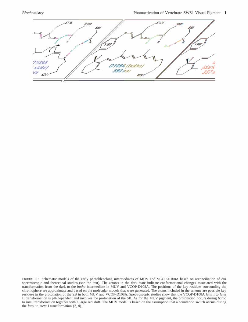
FIGURE 11: Schematic models of the early photobleaching intermediates of MUV and VCOP-D108A based on reconciliation of ourspectroscopic and theoretical studies (see the text). The arrows in the dark state indicate conformational changes associated with thetransformation from the dark to thebatho intermediate in MUV and VCOP-D108A. The positions of the key residues surrounding thechromophore are approximate and based on the molecular models that were generated. The atoms included in the scheme are possible keyresidues in the protonation of the SB in both MUV and VCOP-D108A. Spectroscopic studies show that the VCOP-D108Alumi I to lumiII transformation is pH-dependent and involves the protonation of the SB. As for the MUV pigment, the protonation occurs duringbathoto lumi transformation together with a large red shift. The MUV model is based on the assumption that a counterion switch occurs duringthe lumi to metaI transformation (7, 8).
Biochemistry Photoactivation of Vertebrate SWS1 Visual PigmentI

exhibit a deprotonated Schiff base chromophore in the darkstate, the counterion switch process is complicated by thefact that the chromophore must become protonated to interactwith the secondary counterion. The observation that VCOP-D108A and MUV have different protonation states for Glu-176 in the dark provides an interesting challenge to providea realistic molecular model of the photobleaching processesin these two proteins. Our proposed schemes are shown inFigure 11. Although these schemes are consistent with theobserved absorption maxima, other possibilities exist, andfurther work will be required to accurately assign the natureof these intermediates.
What remains is to examine the relevance of theseobservations to the photobleaching processes of visualpigments in general. Binding site-induced shifts of thechromophores in the intermediates of rhodopsin, MUV,VCOP, and VCOP-D108A are compared in Figure 12. Theshifts are calculated using a very simple model based on theassumption that an isolated protonated Schiff base has anabsorption maximum at 600 nm. The trends are the mostimportant as they provide a simple but useful perspectiveon the chromophore-protein interactions during the photo-bleaching process. As can be observed by reference to Figure12, the primary events in all four proteins involve a nearlyidentical bathochromic shift of∼0.2 eV. This represents acurious observation because it is commonly assumed that asignificant portion of both the bathochromic shift and energystorage of the primary event is associated with chargeseparation of the positively charged chromophore from theprimary counterion (39-42), but the two UV pigments withunprotonated Schiff base chromophores exhibit shifts virtu-ally identical to those observed in the two pigments withprotonated 11-cis chromophores. This result suggests thatconformational distortion is the primary source of thebathochromic shift of the primary events in these visualpigments. The question of whether the same observation alsoapplies to energy storage remains. To help answer thisquestion, photocalorimetric studies of the primary event inMUV are currently in progress (42). Of equal importance isthe observation that E176 is protonated in VCOP-D108Abut deprotonated (negatively charged) in MUV (Table 3).
Both pigments are able to undergo successful photoactivation,but the process in VCOP-D108A is less efficient and involvesa convoluted photobleaching sequence significantly differentfrom that observed in MUV. Our studies support theimportance of having a negatively charged glutamic acidresidue at position 176 in the UV pigments. The questionthat remains to be answered, however, is whether thehomology equivalent residue in rhodopsin (E181) is alsonegatively charged. The state of protonation of this residuein the dark state of rhodopsin is a current subject of debate(5, 35). These studies suggest, by inference, that E181 isdeprotonated in rhodopsin.
COMMENTS AND CONCLUSIONS
The single-amino acid D108A substitution converts theviolet cone pigment ofXenopus laeVis (VCOP) into afunctional UV pigment which photobleaches. When wecompare the photophysical properties of this protein withthose of the native mouse UV pigment (MUV), we find thatVCOP-D108A has a less efficient primary event andundergoes a convoluted photobleaching sequence. We tracethe difference to the protonation state of the glutamic acidresidue that is located near the center of the chromophorepolyene chain, E176. Nevertheless, both proteins generate aprotonated chromophore during the photobleaching sequence,providing further evidence that a counterion switch (orcounterion creation) is requisite for UV pigment activation.Although we do not yet fully understand the molecular detailsof this process, our schematics in Figure 11 provide apreliminary model. The fact that all rhodopsin and conepigments investigated to date generate photobleacing inter-mediates which involve the interaction of a protonated Schiffbase chromophore with a negatively charged E176 (or E181)residue suggests that visual pigments must undergo signifi-cant helical displacements during photoactivation, but con-trary to intuition, studies of the UV pigments suggest thatthis process is not driven by an electrostatic interactionbetween the PSB(+) chromophore and the secondary coun-terion [E176(-) or E181(-)]. Rather, this interaction iscreated by large amplitude motion of theR-helices thattranslocate the Schiff base region of the chromophore closerto the secondary counterion. In the case of both rhodopsinand VCOP, the large amplitude motion creates a counterionswitch. In the case of the UV pigments, the large amplitudemotion generates a binding site environment that inducesprotonation of the chromophore. It is logical to conclude thatthe large amplitude motion in all of these proteins is similar.However, before we accept the counterion switch (orcreation) model as being a universal feature of all visualpigments, we note that most red cones have no glutamic acidresidue in this position. Rather, most red cones have ahistidine residue at the homologous position 181 (6). Studieson the red cone pigments are needed and should provide uswith new insights into the nature of the large amplitudemotions that are important in the activation mechanism ofthe visual pigments.
REFERENCES
1. Ebrey, T., and Koutalos, Y. (2001) Vertebrate photoreceptors,Prog. Retinal Eye Res. 20, 49-94.
2. Yokoyama, S., and Shi, Y. (2000) Genetics and evolution ofultraviolet vision in vertebrates,FEBS Lett. 486, 167-172.
FIGURE 12: Protein-induced blue shifts associated with incorpora-tion of the Schiff base or protonated Schiff base chromophores intothe binding sites of the dark states and early intermediates ofrhodopsin (Rho), the violet cone opsin ofXenopus(VCOP), mouseUV (MUV), and VCOP-D108A. The shift is calculated by assumingthat an isolated all-trans protonated Schiff base chromophore hasan absorption maximum of 600 nm.
J Ramos et al. Biochemistry
3. Yokoyama, S., Isenberg, K. E., and Wright, A. F. (1989) Adaptiveevolution of G-protein coupled receptor genes,Mol. Biol. EVol.6, 342-353.
4. Yokoyama, R., and Yokoyama, S. (1990) Convergent evolutionof the red- and green-like visual pigment genes in fish,Astyanaxfasciatus, and human,Proc. Natl. Acad. Sci. U.S.A. 87, 9315-9318.
5. Yan, E. C. Y., Kasmi, M. A., Ganim, Z., Hou, J. M., Pan, D.,Chang, B. S. W., Sakmar, T. P., and Mathies, R. A. (2003) Retinalcounterion switch in the photoactivation of the G protein-coupledreceptor rhodopsin,Proc. Natl. Acad. Sci. U.S.A. 100, 9262-9267.
6. Birge, R. R., and Knox, B. E. (2003) Perspectives on the counterionswitch-induced photoactivation of the G protein-coupled receptorrhodopsin,Proc. Natl. Acad. Sci. U.S.A. 100, 9105-9107.
7. Kusnetzow, A., Dukkipati, A., Babu, K. R., Ramos, L., Knox, B.E., and Birge, R. R. (2004) Vertebrate ultraviolet visual pig-ments: Protonation of the retinylidene Schiff base and a counterionswitch during photoactivation,Proc. Natl. Acad. Sci. U.S.A. 101,941-946.
8. Dukkipati, A., Kusnetzow, A., Babu, K. R., Ramos, L., Singh,D., Knox, B. E., and Birge, R. R. (2002) Phototransduction byvertebrate ultraviolet visual pigments: Protonation of the reti-nylidene Schiff base following photobleaching,Biochemistry 41,9842-9851.
9. Cowing, J. A., Poopalasundaram, S., Wilkie, S. E., Robinson, P.R., Bowmaker, J., and Hunt, D. (2002) The molecular mechanismfor the spectral shifts between vertebrate ultraviolet- and violet-sensitive cone visual pigments,Biochem. J. 367, 129-135.
10. Fasick, J., Applebury, M., and Oprian, D. (2002) Spectral tuningin the mammalian shortwavelength sensitive cone pigments,Biochemistry 41, 6860-6865.
11. Hunt, D., Cowing, J. A., Wilkie, S. E., Parry, J. W., Poopala-sundaram, S., and Bowmaker, J. (2004) Divergent mechanismsfor the tuning of shortwave sensitive visual pigments in verte-brates,Photochem. Photobiol. 3, 713-720.
12. Yokoyama, S., Radlwimmer, F., and Blow, N. (2000) Ultravioletpigments in birds evolved from violet pigments by a single aminoacid change,Proc. Natl. Acad. Sci. U.S.A. 97, 7366-7371.
13. Wilkie, S. E., Robinson, P. R., Cronin, T. W., Poopalasundaram,S., Bowmaker, J. K., and Hunt, D. M. (2000) Spectral tuning ofavian violet- and ultraviolet-sensitive visual pigments,Biochem-istry 39, 7895-7901.
14. Babu, K. R., Dukkipati, A., Birge, R. R., and Knox, B. E. (2001)Regulation of phototransduction in short wavelength cone visualpigments via the retinylidene Schiff base counterion,Biochemistry40, 13760-13766.
15. Vought, B. W., Dukkipati, A., Max, M., Knox, B. E., and Birge,R. R. (1999) Photochemistry of the primary event in short-wavelength visual opsins at low temperature,Biochemistry 38,11287-11297.
16. Saam, J., Tajkhorshid, E., Hayashi, S., and Schulten, K. (2002)Molecular dynamics investigation of primary photoinduced eventsin the activation of rhodopsin,Biophys. J. 83, 3097-3112.
17. Okada, T., Sugihara, M., Bondar, A. N., Elstner, M., Entel, P.,and Buss, V. (2004) The retinal conformation and its environmentin rhodopsin in light of a new 2.2 Å crystal structure,J. Mol.Biol. 342, 571-583.
18. Marti-Renom, M. A., Stuart, A. C., Fiser, A., Sanchez, R., Melo,F., and Sali, A. (2000) Comparative protein structure modelingof genes and genomes,Annu. ReV. Biophys. Biomol. Struct. 29,291-325.
19. Zhang, L., and Hermans, J. (1996) Hydrophilicity of cavities inproteins,Proteins 24, 433-438.
20. Kale, L., Skeel, R., Bhandarkar, M., Brunner, R., Gursoy, A.,Krawetz, N., Phillips, J., Shinozaki, A., Varadarajan, K., andSchulten, K. (1999) NAMD2: Greater scalability for parallelmolecular dynamics,J. Comput. Phys. 151, 283-312.
21. Fong, S. L., Tsin, A. T., Bridges, C. D., and Liou, G. I. (1982)Detergents for extraction of visual pigments: Types, solubilization,and stability,Methods Enzymol. 81, 133-140.
22. Nicholls, A., Sharp, K. A., and Honig, B. (1991) Protein foldingand association: Insights from the interfacial and thermodynamicproperties of hydrocarbons,Proteins 11, 281-296.
23. Stewart, J. J. P. (1999)MOZYME, Fujitsu Limited, Tokyo,Japan.
24. Stewart, J. J. P. (1996) Application of localized molecular orbitalsto the solution of semiempirical self-consistet field equations,Int.J. Quant. Chem. 58, 133-146.
25. Stewart, J. J. P. (1997) Calculation of the geometry of a smallprotein using semiempirical methods,THEOCHEM, 195-205.
26. Martin, C. H., and Birge, R. R. (1998) Reparameterizing MNDOfor excited-state calculations using ab initio effective Hamiltoniantheory: Application to the 2,4-pentadien-1-iminium cation,J.Phys. Chem. A 102, 852-860.
27. Kusnetzow, A., Singh, D. L., Martin, C. H., Barani, I., and Birge,R. R. (1999) Nature of the chromophore binding site of bacteri-orhodopsin. The potential role of Arg-82 as a principal counterion,Biophys. J. 76, 2370-2389.
28. Ren, L., Martin, C. H., Wise, K. J., Gillespie, N. B., Luecke, H.,Lanyi, J. K., Spudich, J. L., and Birge, R. R. (2001) Molecularmechanism of spectral tuning in sensory rhodopsin II,Biochem-istry 40, 13906-13914.
29. Sakmar, T. P., Franke, R. R., and Khorana, H. G. (1989) Glutamicacid 113 serves as the retinylidene Schiff base counterion in bovinerhodopsin,Proc. Natl. Acad. Sci. U.S.A. 86, 8309-8313.
30. Zhukovsky, E. A., and Oprian, D. D. (1989) Effect of carboxylicacid side chains on the absorption maximum of visual pigments,Science 246, 928-931.
31. Kliger, D. S., and Lewis, J. W. (1995) Spectral and kineticcharacterization of visual pigment photointermediates,Isr. J.Chem. 35, 289-307.
32. Jager, S., Lewis, J. W., Zvyaga, T. A., Szundi, I., Sakmar, T. P.,and Kliger, D. S. (1997) Time-resolved spectroscopy of the earlyphotolysis intermediates of rhodopsin Schiff base counterionmutants,Biochemistry 36, 1999-2009.
33. Jager, S., Lewis, J. W., Zvyaga, T. A., Szundi, I., Sakmar, T. P.,and Kliger, D. S. (1997) Chromophore structural changesin rhodopsin from nanoseconds to microseconds followingpigment photolysis,Proc. Natl. Acad. Sci. U.S.A. 94, 8557-8562.
34. Pan, D., Ganim, Z., Kim, J. E., Verhoeven, M. A., Lugtenburg,J., and Mathies, R. A. (2002) Time-resolved resonance Ramananalysis of chromophore structural changes in the formation anddecay of rhodopsin’s BSI intermediate,J. Am. Chem. Soc. 124,4857-4864.
35. Ludeke, S., Beck, M., Yan, E. C. Y., Sakmar, T. P., Siebert, F.,and Vogel, R. (2005) The role of Glu 181 in the photoactivationof rhodopsin,J. Mol. Biol. 353, 345-356.
36. Kim, J. E., Tauber, M. J., and Mathies, R. A. (2001) Wavelength-dependent cis-trans isomerization in vision,Biochemistry 40,13774-13778.
37. Tallent, J. R., Hyde, E. Q., Findsen, L. A., Fox, G. C., and Birge,R. R. (1992) Molecular dynamics of the primary photochemicalevent in rhodopsin,J. Am. Chem. Soc. 114, 1581-1592.
38. Wang, Q., Schoenlein, R. W., Peteanu, L. A., Mathies, R. A., andShank, C. V. (1994) Vibrationally coherent photochemistry in thefemtosecond primary event of vision,Science 266, 422-424.
39. Cooper, A. (1979) Energy uptake in the first step of visualexcitation,Nature 282, 531-533.
40. Schick, G. A., Cooper, T. M., Holloway, R. A., Murray, L. P.,and Birge, R. R. (1987) Energy storage in the primary photo-chemical events of rhodopsin and isorhodopsin,Biochemistry 26,2556-2562.
41. Honig, B., Ebrey, T., Callender, R. H., Dinur, U., and Ottolenghi,M. (1979) Photoisomerization, energy storage, and charge separa-tion: A model for light energy transduction in visual pigmentsand bacteriorhodopsin,Proc. Natl. Acad. Sci. U.S.A. 76, 2503-2507.
42. Birge, R. R., and Vought, B. W. (2000) Energetics of rhodopsinphotobleaching: Photocalorimetric studies of energy storage inthe early and later intermediates, inMethods in Enzymology(Palczewski, K., Ed.) pp 143-163, Academic Press, New York.
43. Kusnetzow, A., Dukkipati, A., Babu, K. R., Singh, D., Vought,B. W., Knox, B. E., and Birge, R. R. (2001) The PhotobleachingSequence of a Short-Wavelength Visual Pigment,Biochemistry40, 7832-7844.
BI700138G
Biochemistry PAGE EST: 11 Photoactivation of Vertebrate SWS1 Visual PigmentK